Embed Size (px)
Citation preview
Journ
alof
Cell
Scie
nce
Energy metabolism and energy-sensing pathways inmammalian embryonic and adult stem cell fate
Victoria A. Rafalski1,2, Elena Mancini1 and Anne Brunet1,2,3,*1Department of Genetics, Stanford University, Stanford, CA 94305, USA2Neurosciences Program, Stanford University, Stanford, CA 94305, USA3Glenn Laboratories for the Biology of Aging, Stanford, CA 94305, USA
*Author for correspondence ([email protected])
Journal of Cell Science 125, 5597–5608� 2012. Published by The Company of Biologists Ltddoi: 10.1242/jcs.114827
SummaryMetabolism is influenced by age, food intake, and conditions such as diabetes and obesity. How do physiological or pathologicalmetabolic changes influence stem cells, which are crucial for tissue homeostasis? This Commentary reviews recent evidence that stemcells have different metabolic demands than differentiated cells, and that the molecular mechanisms that control stem cell self-renewal
and differentiation are functionally connected to the metabolic state of the cell and the surrounding stem cell niche. Furthermore, wepresent how energy-sensing signaling molecules and metabolism regulators are implicated in the regulation of stem cell self-renewal anddifferentiation. Finally, we discuss the emerging literature on the metabolism of induced pluripotent stem cells and how manipulating
metabolic pathways might aid cellular reprogramming. Determining how energy metabolism regulates stem cell fate should shed lighton the decline in tissue regeneration that occurs during aging and facilitate the development of therapies for degenerative or metabolicdiseases.
Key words: Metabolism, Stem cells, ESCs, Reprogramming, iPSCs, HSCs, NSCs, AMPK, FOXO, mTOR, SIRT1, Insulin, Hypoxia, Aging
IntroductionStem cells serve as the origin for all tissues during embryonic andpostnatal development, and contribute to tissue homeostasis and
repair throughout adult life. Stem cells hold great promise forreplacement therapies for degenerative diseases and age-related
disorders. Embryonic, postnatal and adult stem cells share two
crucial characteristics: the ability to produce at least one daughterstem cell upon division (self-renewal) and the ability to generate
differentiated cells (potency). Stem cell potency varies dependingon the type of stem cell. For example, embryonic stem cells
(ESCs) are pluripotent and can generate all three germ layers
(endoderm, ectoderm and mesoderm) (Thomson et al., 1998).Stem cells that are present in adult tissues can be either
multipotent or unipotent (i.e. giving rise to multiple
differentiated cell types, or only one cell type, respectively)(Nakada et al., 2011). Interestingly, induced pluripotent stem
cells (iPSCs) can be generated from either embryonic or adultdifferentiated cells upon expression of specific combinations of
transcription factors (Takahashi and Yamanaka, 2006) (Box 1).
As iPSCs can be generated from a specific patient, the use ofthese cells avoids potential medical or ethical issues when
considering their application in regenerative medicine.
Emerging evidence suggests that pluripotent stem cells and
certain adult stem cells are metabolically distinct from theirdifferentiated counterparts and that these metabolic properties are
important for stem cell identity. Furthermore, molecular
regulators of energy metabolism have essential roles in stemcell fate, in particular, the decision to self-renew or differentiate.
Finally, stem cells respond to fluctuations in organismal energy
states in vivo. This Commentary will discuss the connectionsbetween stem cells and energy metabolism, focusing on human
and mouse stem cells. The influence of metabolism on stem cells
in other species has been described elsewhere (Jasper and Jones,2010). The main stem cell types that will be discussed are ESCsand iPSCs as examples of pluripotent stem cells, and neural stem
cells (NSCs) and hematopoietic stem cells (HSCs) as examples ofadult tissue-specific stem cells.
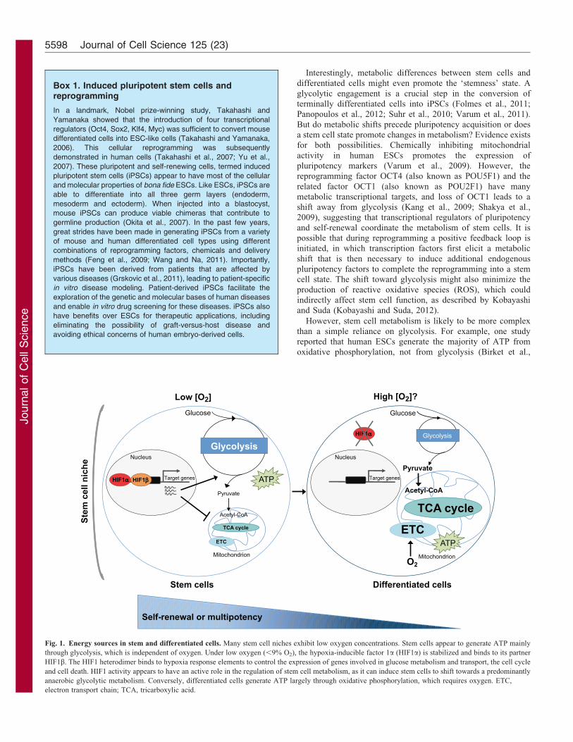
Metabolic properties of stem cellsStem cells appear to depend mostly on glycolysis forproduction of ATP
In contrast to differentiated cells, many stem cells appear torely to a greater extent on glycolysis than on oxidativephosphorylation to generate adenosine-59-triphosphate (ATP).
Bioenergetics studies have revealed that human ESCs (Zhanget al., 2011b; Zhou et al., 2012) depend, in a large part,
on glycolysis for ATP production (Fig. 1). Consistently,mitochondria are less complex and fewer in number in humanESCs than in their differentiated progeny (Cho et al., 2006;
Facucho-Oliveira et al., 2007; St John et al., 2005; Varum et al.,2011; Zhang et al., 2011b). Furthermore, studies analyzingmitochondrial respiration, glycolytic flux or proteomic profiles of
purified adult HSCs have shown that these adult stem cells relyprimarily on glycolysis to generate ATP (Miharada et al., 2011;Simsek et al., 2010; Unwin et al., 2006). The dependency of stem
cells on glycolysis for ATP generation is reminiscent of that ofcancer cells (Hsu and Sabatini, 2008; Warburg, 1956). Unlike
oxidative phosphorylation, glycolysis can proceed anaerobically,raising the possibility that the dependency of a stem cell onglycolysis is an adaptation to the low oxygen levels that are
present in vivo during development and in an adult stem cellmicroenvironment or ‘niche’ (see below) (Fig. 1).
ARTICLE SERIES: Stem Cells Commentary 5597
Journ
alof
Cell
Scie
nce
Interestingly, metabolic differences between stem cells and
differentiated cells might even promote the ‘stemness’ state. A
glycolytic engagement is a crucial step in the conversion of
terminally differentiated cells into iPSCs (Folmes et al., 2011;
Panopoulos et al., 2012; Suhr et al., 2010; Varum et al., 2011).
But do metabolic shifts precede pluripotency acquisition or does
a stem cell state promote changes in metabolism? Evidence exists
for both possibilities. Chemically inhibiting mitochondrial
activity in human ESCs promotes the expression of
pluripotency markers (Varum et al., 2009). However, the
reprogramming factor OCT4 (also known as POU5F1) and the
related factor OCT1 (also known as POU2F1) have many
metabolic transcriptional targets, and loss of OCT1 leads to a
shift away from glycolysis (Kang et al., 2009; Shakya et al.,
2009), suggesting that transcriptional regulators of pluripotency
and self-renewal coordinate the metabolism of stem cells. It is
possible that during reprogramming a positive feedback loop is
initiated, in which transcription factors first elicit a metabolic
shift that is then necessary to induce additional endogenous
pluripotency factors to complete the reprogramming into a stem
cell state. The shift toward glycolysis might also minimize the
production of reactive oxidative species (ROS), which could
indirectly affect stem cell function, as described by Kobayashi
and Suda (Kobayashi and Suda, 2012).
However, stem cell metabolism is likely to be more complex
than a simple reliance on glycolysis. For example, one study
reported that human ESCs generate the majority of ATP from
oxidative phosphorylation, not from glycolysis (Birket et al.,
Box 1. Induced pluripotent stem cells andreprogramming
In a landmark, Nobel prize-winning study, Takahashi and
Yamanaka showed that the introduction of four transcriptional
regulators (Oct4, Sox2, Klf4, Myc) was sufficient to convert mouse
differentiated cells into ESC-like cells (Takahashi and Yamanaka,
2006). This cellular reprogramming was subsequently
demonstrated in human cells (Takahashi et al., 2007; Yu et al.,
2007). These pluripotent and self-renewing cells, termed induced
pluripotent stem cells (iPSCs) appear to have most of the cellular
and molecular properties of bona fide ESCs. Like ESCs, iPSCs are
able to differentiate into all three germ layers (endoderm,
mesoderm and ectoderm). When injected into a blastocyst,
mouse iPSCs can produce viable chimeras that contribute to
germline production (Okita et al., 2007). In the past few years,
great strides have been made in generating iPSCs from a variety
of mouse and human differentiated cell types using different
combinations of reprogramming factors, chemicals and delivery
methods (Feng et al., 2009; Wang and Na, 2011). Importantly,
iPSCs have been derived from patients that are affected by
various diseases (Grskovic et al., 2011), leading to patient-specific
in vitro disease modeling. Patient-derived iPSCs facilitate the
exploration of the genetic and molecular bases of human diseases
and enable in vitro drug screening for these diseases. iPSCs also
have benefits over ESCs for therapeutic applications, including
eliminating the possibility of graft-versus-host disease and
avoiding ethical concerns of human embryo-derived cells.
HIF1αα
Differentiated cells Stem cells
Acetyl-CoA
Pyruvate
Glucose
O2
Pyruvate
Acetyl-CoA
Self-renewal or multipotency
Stem
cel
l nic
he
Lactate
t
Nucleus
Low [O2]
TCA cycle
Glycolysis
ETC
TCA cycle ETC
High [O2]?
Mitochondrion
Glycolysis
HIF1ββHIF1αα Target genes
ATP
ATP
Glucose
Lactate
t
Nucleus
Target genes
Mitochondrion
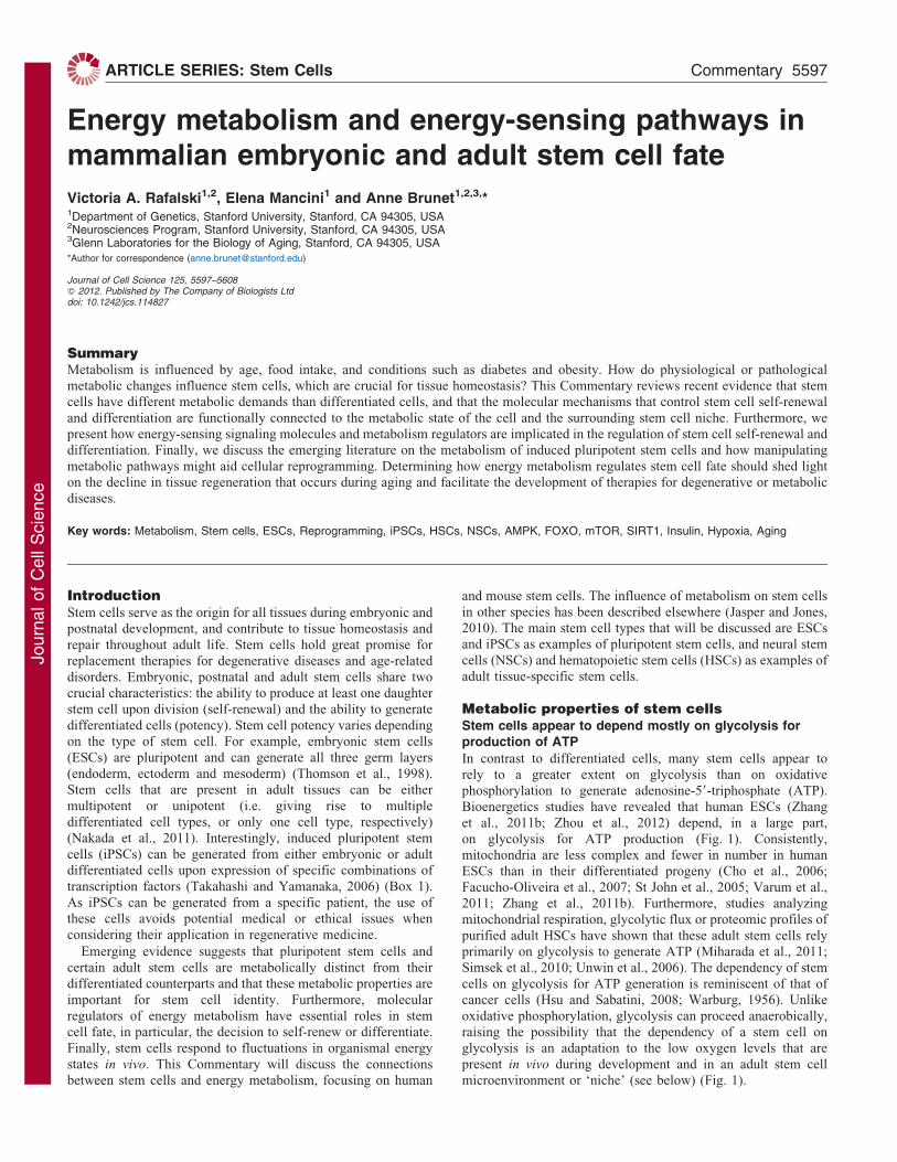
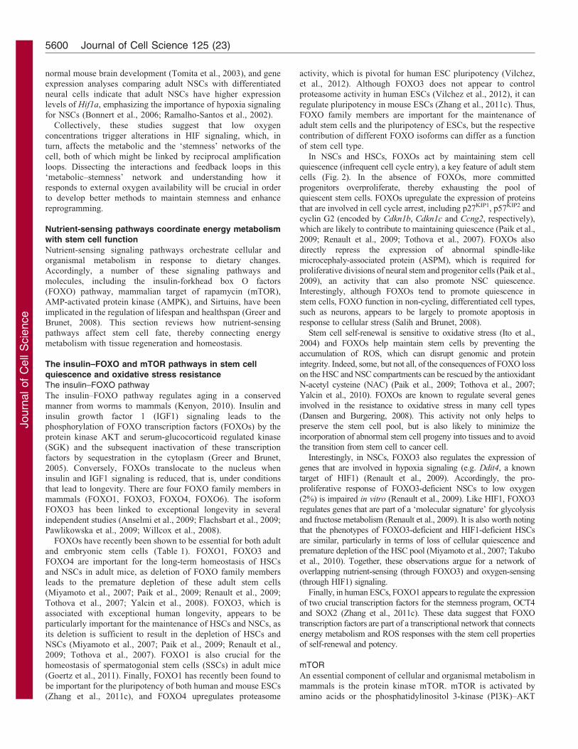
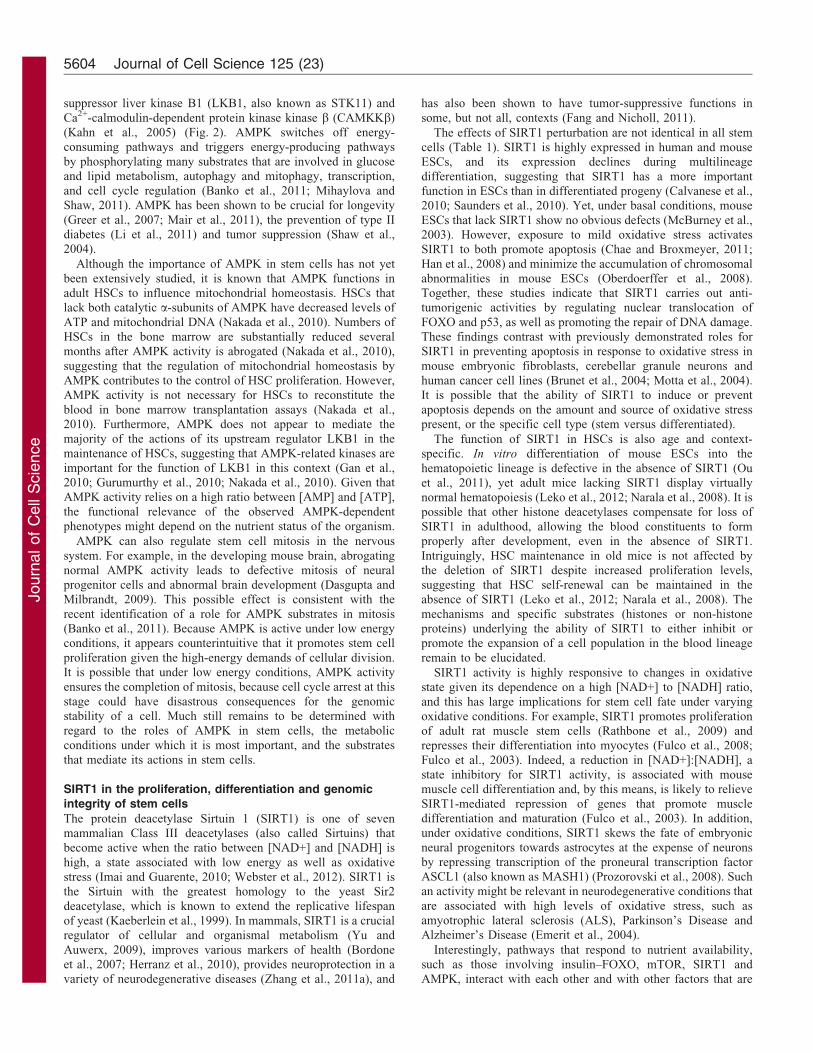
Fig. 1. Energy sources in stem and differentiated cells. Many stem cell niches exhibit low oxygen concentrations. Stem cells appear to generate ATP mainly
through glycolysis, which is independent of oxygen. Under low oxygen (,9% O2), the hypoxia-inducible factor 1a (HIF1a) is stabilized and binds to its partner
HIF1b. The HIF1 heterodimer binds to hypoxia response elements to control the expression of genes involved in glucose metabolism and transport, the cell cycle
and cell death. HIF1 activity appears to have an active role in the regulation of stem cell metabolism, as it can induce stem cells to shift towards a predominantly
anaerobic glycolytic metabolism. Conversely, differentiated cells generate ATP largely through oxidative phosphorylation, which requires oxygen. ETC,
electron transport chain; TCA, tricarboxylic acid.
Journal of Cell Science 125 (23)5598
Journ
alof
Cell
Scie
nce
2011). Such a difference to other studies is not completely
understood yet, but could arise from the sensitivity of the
detection technology used, or the fact that there might be slightly
different stages of pluripotency – and thus metabolism – among
ESCs (Zhou et al., 2012). Mouse ESCs also depend on the amino
acid threonine as a crucial source of energy, whereby the citric
acid cycle metabolite acetyl-coenzyme A is generated through
the action of threonine dehydrogenase (Shyh-Chang et al., 2013;
Wang et al., 2011; Wang et al., 2009). Interestingly, lipid
metabolism is also emerging as a key regulator of stem cell
maintenance and differentiation (Ito et al., 2012; Knobloch et al.,
2013). A more in-depth analysis of the metabolism of stem cells
is needed to clarify the circumstances under which the different
components of respiration are utilized (Box 2).
It is worth noting that metabolic information obtained from
cultured stem cells might not reflect the metabolism of stem cells in
their natural niche for two main reasons (Joseph and Morrison, 2005).
First, culture systems can differ quite dramatically from stem cell
niches in terms of nutrient availability, cell–cell and cell–extracellular
matrix contacts, and oxygen availability (Mohyeldin et al., 2010).These differences might be particularly significant for cells that are
normally relatively quiescent, but are triggered to divide rapidly oncethey are exposed to growth factors in vitro, such as adult NSCs(Morshead et al., 1994). Second, stem cell cultures are often a
heterogeneous mixture of stem cells, progenitors and differentiatingcells. Future metabolic studies performed on small numbers of cellsimmediately after their isolation from tissue will aid ourunderstanding of the metabolic profile of stem cells in vivo (Box 2).
Regardless of technical limitations, it is becoming increasinglyclear that stem cells harbor different energy metabolism
requirements compared with differentiating progeny. Theunique metabolic properties of stem cells could be harnessed tofacilitate the development of stem-cell-targeted therapies, inwhich stem cells are selectively directed to self-renew or
differentiate by manipulating their metabolic state.
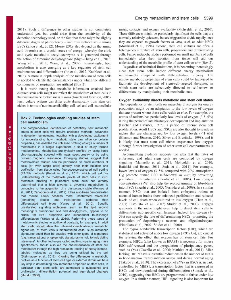
Oxygen availability directs metabolic and stem cell states
The dependency of stem cells on anaerobic glycolysis for energyproduction might be an adaptation to the low levels of oxygen
that are present where these cells reside in vivo. For example, theuterus of rodents has particularly low levels of oxygen (3.5–5%)during the period of late blastocyst development and implantation
(Fischer and Bavister, 1993), a period of intense stem cellproliferation. Adult HSCs and NSCs are also thought to reside inniches that are characterized by low oxygen levels (,1–6%)(Eliasson and Jonsson, 2010; Silver and Erecinska, 1998), and it
is likely that most stem cell niches experience low oxygenalthough further investigation of other stem cell compartments isneeded.
Accumulating evidence supports the idea that the fates ofembryonic and adult stem cells are controlled by oxygensignaling (Mannello et al., 2011; Mohyeldin et al., 2010;
Rafalski and Brunet, 2011; Suda et al., 2011). For example,lower levels of oxygen (3–5% compared with 20% atmosphericO2) promote human ESC self-renewal in vitro by preventing
premature differentiation (Ezashi et al., 2005). Low oxygenconcentrations (5%) also help the reprogramming of fibroblastsinto iPSCs (Ezashi et al., 2005; Yoshida et al., 2009). In a similar
manner, NSCs that are isolated from embryonic rodent orneonatal human brains show enhanced proliferation and reducedlevels of cell death when cultured in low oxygen (Chen et al.,2007; Pistollato et al., 2007; Studer et al., 2000). Oxygen
gradients in the niche might even help to direct stem cells todifferentiate into specific cell lineages. Indeed, low oxygen (3–5%) can specify the fate of differentiating NSCs, promoting the
production of dopaminergic neurons and oligodendrocytes(Pistollato et al., 2007; Studer et al., 2000).
The hypoxia-inducible transcription factors (HIF), which are
stabilized and activated under low oxygen (,9% O2), are crucialfor relaying the effect that oxygen has on stem cell fate. Forexample, HIF2a (also known as EPAS1) is necessary for mouse
ESC self-renewal and the upregulation of pluripotency genes,such as Oct4 (Covello et al., 2006; Mathieu et al., 2011). Micelacking HIF1a have substantial reductions in the number of HSCs
in bone marrow transplantation assays and during normal aging(Takubo et al., 2010). The expression of Hif1a in HSCs is, in part,controlled by MEIS1, a transcription factor that is expressed in
HSCs and downregulated during differentiation (Simsek et al.,2010), suggesting that HSCs are programmed to thrive under lowoxygen. In a similar manner, HIF1 signaling is also important for
Box 2. Technologies enabling studies of stemcell metabolism
The comprehensive identification of potentially new metabolic
states in stem cells will require unbiased methods. Advances
in detection technologies, together with a developing excitement
for understanding how metabolic state can influence cellular
properties, has enabled the unbiased profiling of large numbers of
metabolites in a single experiment, a field of study termed
metabolomics. Metabolites are typically profiled by using liquid
chromatography coupled with mass spectrometry or by using
nuclear magnetic resonance. Emerging studies suggest that
metabolomics studies can be performed on small numbers of
cells (or even single cells) directly after their isolation and
purification from tissue using fluorescence-activated cell sorting
(FACS) methods (Rubakhin et al., 2011), which will aid our
understanding of the metabolite profile of stem cells in vivo.
Metabolic profiling of pluripotent stem cells has already
determined that a bias towards a glycolytic metabolism is
conducive to the acquisition of a pluripotency state (Folmes et
al., 2011; Panopoulos et al., 2012). It has also been demonstrated
that mouse ESCs possess more unsaturated molecules
(containing double- and triple-bonded carbons) than
differentiated cell types (Yanes et al., 2010). Specific
unsaturated signaling molecules, such as the lipid second
messengers arachidonic acid and diacylglyercol, appear to be
crucial for ESC properties and subsequent multilineage
differentiation (Yanes et al., 2010). Performing these types of
metabolomic studies in additional contexts, for example, in adult
stem cells, should allow the unbiased identification of ‘metabolic
signatures’ of stem versus differentiated cells. Such metabolic
signatures could then be coupled with other types of signatures
(e.g. transcriptional or epigenetic signatures) to help to truly define
‘stemness’. Another technique called multi-isotope imaging mass
spectrometry should also aid the characterization of stem cell
metabolism through the high-resolution tracking of heavy isotope-
labeled molecules as they are being utilized by the cell
(Steinhauser et al., 2012). Knowing the differences in metabolic
profiles as a function of stem cell type or external stimuli will be a
key step in determining how metabolic properties of stem cells, in
particular adult stem cells, are connected to quiescence and
proliferation, differentiation potential and age-related changes
(Rando, 2006).
Energy metabolism and stem cells 5599
Journ
alof
Cell
Scie
nce
normal mouse brain development (Tomita et al., 2003), and geneexpression analyses comparing adult NSCs with differentiatedneural cells indicate that adult NSCs have higher expressionlevels of Hif1a, emphasizing the importance of hypoxia signaling
for NSCs (Bonnert et al., 2006; Ramalho-Santos et al., 2002).
Collectively, these studies suggest that low oxygen
concentrations trigger alterations in HIF signaling, which, inturn, affects the metabolic and the ‘stemness’ networks of thecell, both of which might be linked by reciprocal amplification
loops. Dissecting the interactions and feedback loops in this‘metabolic–stemness’ network and understanding how itresponds to external oxygen availability will be crucial in order
to develop better methods to maintain stemness and enhancereprogramming.
Nutrient-sensing pathways coordinate energy metabolismwith stem cell function
Nutrient-sensing signaling pathways orchestrate cellular andorganismal metabolism in response to dietary changes.Accordingly, a number of these signaling pathways and
molecules, including the insulin-forkhead box O factors(FOXO) pathway, mammalian target of rapamycin (mTOR),AMP-activated protein kinase (AMPK), and Sirtuins, have been
implicated in the regulation of lifespan and healthspan (Greer andBrunet, 2008). This section reviews how nutrient-sensingpathways affect stem cell fate, thereby connecting energymetabolism with tissue regeneration and homeostasis.
The insulin–FOXO and mTOR pathways in stem cellquiescence and oxidative stress resistanceThe insulin–FOXO pathway
The insulin–FOXO pathway regulates aging in a conservedmanner from worms to mammals (Kenyon, 2010). Insulin and
insulin growth factor 1 (IGF1) signaling leads to thephosphorylation of FOXO transcription factors (FOXOs) by theprotein kinase AKT and serum-glucocorticoid regulated kinase(SGK) and the subsequent inactivation of these transcription
factors by sequestration in the cytoplasm (Greer and Brunet,2005). Conversely, FOXOs translocate to the nucleus wheninsulin and IGF1 signaling is reduced, that is, under conditions
that lead to longevity. There are four FOXO family members inmammals (FOXO1, FOXO3, FOXO4, FOXO6). The isoformFOXO3 has been linked to exceptional longevity in several
independent studies (Anselmi et al., 2009; Flachsbart et al., 2009;Pawlikowska et al., 2009; Willcox et al., 2008).
FOXOs have recently been shown to be essential for both adultand embryonic stem cells (Table 1). FOXO1, FOXO3 andFOXO4 are important for the long-term homeostasis of HSCsand NSCs in adult mice, as deletion of FOXO family members
leads to the premature depletion of these adult stem cells(Miyamoto et al., 2007; Paik et al., 2009; Renault et al., 2009;Tothova et al., 2007; Yalcin et al., 2008). FOXO3, which is
associated with exceptional human longevity, appears to beparticularly important for the maintenance of HSCs and NSCs, asits deletion is sufficient to result in the depletion of HSCs and
NSCs (Miyamoto et al., 2007; Paik et al., 2009; Renault et al.,2009; Tothova et al., 2007). FOXO1 is also crucial for thehomeostasis of spermatogonial stem cells (SSCs) in adult mice
(Goertz et al., 2011). Finally, FOXO1 has recently been found tobe important for the pluripotency of both human and mouse ESCs(Zhang et al., 2011c), and FOXO4 upregulates proteasome
activity, which is pivotal for human ESC pluripotency (Vilchez,et al., 2012). Although FOXO3 does not appear to controlproteasome activity in human ESCs (Vilchez et al., 2012), it can
regulate pluripotency in mouse ESCs (Zhang et al., 2011c). Thus,FOXO family members are important for the maintenance ofadult stem cells and the pluripotency of ESCs, but the respective
contribution of different FOXO isoforms can differ as a functionof stem cell type.
In NSCs and HSCs, FOXOs act by maintaining stem cellquiescence (infrequent cell cycle entry), a key feature of adult stemcells (Fig. 2). In the absence of FOXOs, more committed
progenitors overproliferate, thereby exhausting the pool ofquiescent stem cells. FOXOs upregulate the expression of proteinsthat are involved in cell cycle arrest, including p27KIP1, p57KIP2 and
cyclin G2 (encoded by Cdkn1b, Cdkn1c and Ccng2, respectively),which are likely to contribute to maintaining quiescence (Paik et al.,2009; Renault et al., 2009; Tothova et al., 2007). FOXOs also
directly repress the expression of abnormal spindle-likemicrocephaly-associated protein (ASPM), which is required forproliferative divisions of neural stem and progenitor cells (Paik et al.,
2009), an activity that can also promote NSC quiescence.Interestingly, although FOXOs tend to promote quiescence instem cells, FOXO function in non-cycling, differentiated cell types,such as neurons, appears to be largely to promote apoptosis in
response to cellular stress (Salih and Brunet, 2008).
Stem cell self-renewal is sensitive to oxidative stress (Ito et al.,2004) and FOXOs help maintain stem cells by preventing theaccumulation of ROS, which can disrupt genomic and protein
integrity. Indeed, some, but not all, of the consequences of FOXO losson the HSC and NSC compartments can be rescued by the antioxidantN-acetyl cysteine (NAC) (Paik et al., 2009; Tothova et al., 2007;
Yalcin et al., 2010). FOXOs are known to regulate several genesinvolved in the resistance to oxidative stress in many cell types(Dansen and Burgering, 2008). This activity not only helps topreserve the stem cell pool, but is also likely to minimize the
incorporation of abnormal stem cell progeny into tissues and to avoidthe transition from stem cell to cancer cell.
Interestingly, in NSCs, FOXO3 also regulates the expression ofgenes that are involved in hypoxia signaling (e.g. Ddit4, a known
target of HIF1) (Renault et al., 2009). Accordingly, the pro-proliferative response of FOXO3-deficient NSCs to low oxygen(2%) is impaired in vitro (Renault et al., 2009). Like HIF1, FOXO3
regulates genes that are part of a ‘molecular signature’ for glycolysisand fructose metabolism (Renault et al., 2009). It is also worth notingthat the phenotypes of FOXO3-deficient and HIF1-deficient HSCs
are similar, particularly in terms of loss of cellular quiescence andpremature depletion of the HSC pool (Miyamoto et al., 2007; Takuboet al., 2010). Together, these observations argue for a network ofoverlapping nutrient-sensing (through FOXO3) and oxygen-sensing
(through HIF1) signaling.
Finally, in human ESCs, FOXO1 appears to regulate the expressionof two crucial transcription factors for the stemness program, OCT4and SOX2 (Zhang et al., 2011c). These data suggest that FOXO
transcription factors are part of a transcriptional network that connectsenergy metabolism and ROS responses with the stem cell propertiesof self-renewal and potency.
mTOR
An essential component of cellular and organismal metabolism inmammals is the protein kinase mTOR. mTOR is activated byamino acids or the phosphatidylinositol 3-kinase (PI3K)–AKT
Journal of Cell Science 125 (23)5600
Journ
alof
Cell
Scie
nce
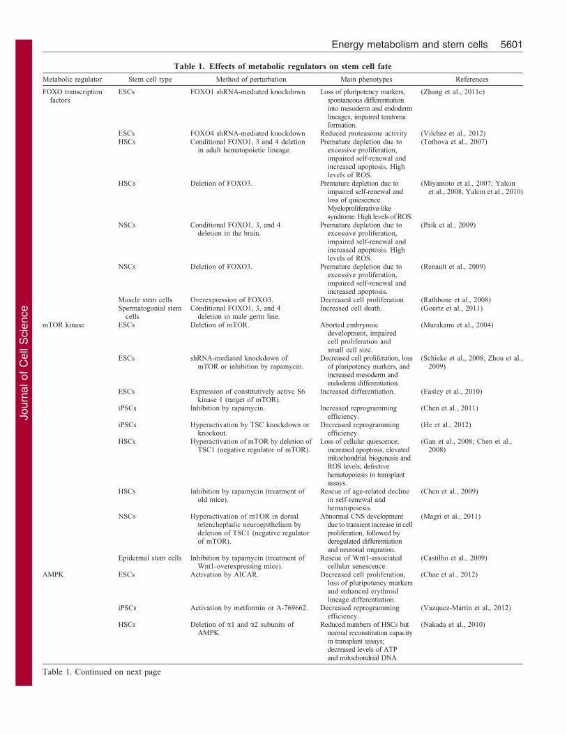
Table 1. Effects of metabolic regulators on stem cell fate
Metabolic regulator Stem cell type Method of perturbation Main phenotypes References
FOXO transcriptionfactors
ESCs FOXO1 shRNA-mediated knockdown. Loss of pluripotency markers,spontaneous differentiationinto mesoderm and endodermlineages, impaired teratomaformation.
(Zhang et al., 2011c)
ESCs FOXO4 shRNA-mediated knockdown Reduced proteasome activity (Vilchez et al., 2012)HSCs Conditional FOXO1, 3 and 4 deletion
in adult hematopoietic lineage.Premature depletion due to
excessive proliferation,impaired self-renewal andincreased apoptosis. Highlevels of ROS.
(Tothova et al., 2007)
HSCs Deletion of FOXO3. Premature depletion due toimpaired self-renewal andloss of quiescence.Myeloproliferative-likesyndrome. High levels of ROS.
(Miyamoto et al., 2007; Yalcinet al., 2008, Yalcin et al., 2010)
NSCs Conditional FOXO1, 3, and 4deletion in the brain.
Premature depletion due toexcessive proliferation,impaired self-renewal andincreased apoptosis. Highlevels of ROS.
(Paik et al., 2009)
NSCs Deletion of FOXO3. Premature depletion due toexcessive proliferation,impaired self-renewal andincreased apoptosis.
(Renault et al., 2009)
Muscle stem cells Overexpression of FOXO3. Decreased cell proliferation. (Rathbone et al., 2008)Spermatogonial stem
cellsConditional FOXO1, 3, and 4
deletion in male germ line.Increased cell death. (Goertz et al., 2011)
mTOR kinase ESCs Deletion of mTOR. Aborted embryonicdevelopment, impairedcell proliferation andsmall cell size.
(Murakami et al., 2004)
ESCs shRNA-mediated knockdown ofmTOR or inhibition by rapamycin.
Decreased cell proliferation, lossof pluripotency markers, andincreased mesoderm andendoderm differentiation.
(Schieke et al., 2008; Zhou et al.,2009)
ESCs Expression of constitutively active S6kinase 1 (target of mTOR).
Increased differentiation. (Easley et al., 2010)
iPSCs Inhibition by rapamycin. Increased reprogrammingefficiency.
(Chen et al., 2011)
iPSCs Hyperactivation by TSC knockdown orknockout.
Decreased reprogrammingefficiency.
(He et al., 2012)
HSCs Hyperactivation of mTOR by deletion ofTSC1 (negative regulator of mTOR).
Loss of cellular quiescence,increased apoptosis, elevatedmitochondrial biogenesis andROS levels; defectivehematopoiesis in transplantassays.
(Gan et al., 2008; Chen et al.,2008)
HSCs Inhibition by rapamycin (treatment ofold mice).
Rescue of age-related declinein self-renewal andhematopoiesis.
(Chen et al., 2009)
NSCs Hyperactivation of mTOR in dorsaltelenchephalic neuroepithelium bydeletion of TSC1 (negative regulatorof mTOR).
Abnormal CNS developmentdue to transient increase in cellproliferation, followed byderegulated differentiationand neuronal migration.
(Magri et al., 2011)
Epidermal stem cells Inhibition by rapamycin (treatment ofWnt1-overexpressing mice).
Rescue of Wnt1-associatedcellular senescence.
(Castilho et al., 2009)
AMPK ESCs Activation by AICAR. Decreased cell proliferation,loss of pluripotency markersand enhanced erythroidlineage differentiation.
(Chae et al., 2012)
iPSCs Activation by metformin or A-769662. Decreased reprogrammingefficiency.
(Vazquez-Martin et al., 2012)
HSCs Deletion of a1 and a2 subunits ofAMPK.
Reduced numbers of HSCs butnormal reconstitution capacityin transplant assays;decreased levels of ATPand mitochondrial DNA.
(Nakada et al., 2010)
Table 1. Continued on next page
Energy metabolism and stem cells 5601
Journ
alof
Cell
Scie
nce
pathway and is part of two distinct complexes, mTORC1 andmTORC2, which coordinate cell growth, proliferation and
survival (Laplante and Sabatini, 2012) (Fig. 2). At the
organismal level, mTOR is involved in energy metabolism
regulation, cancer and aging (Kapahi et al., 2010; Laplante andSabatini, 2012). For example, blocking mTOR activity with the
pharmacological drug rapamycin can extend the lifespan of mice
(Harrison et al., 2009).
Multiple recent studies have revealed vital, yet different, roles
for mTOR signaling in ESCs and adult tissue-specific stem cells
(Table 1). In ESCs, mTOR is important for self-renewal;inhibition of mTOR by rapamycin or by knockdown in mouse
and human ESCs reduces the expression of pluripotency markers
and impairs the self-renewal of ESCs in vitro (Schieke et al.,2008; Zhou et al., 2009). Furthermore, deletion of mTOR inhibits
proliferation in early mouse embryos (Murakami et al., 2004).
Somewhat surprisingly, mTOR also appears to promote ESC
differentiation. For example, the transition from ESCs to moredifferentiated cell types is associated with increased activation of
mTORC1 signaling (Easley et al., 2010; Sampath et al., 2008).
Furthermore, overexpression of a constitutively active form of S6kinase 1, a substrate of mTORC1, promotes differentiation of
ESCs (Easley et al., 2010). It will be important to understand the
circumstances under which TOR promotes either ESC self-renewal and pluripotency or differentiation, and whether different
mTOR substrates are involved in mediating these diverse
functions of mTOR.
In contrast to ESCs, in which mTOR activity is necessary for
self-renewal, adult stem cell self-renewal might actually benefit
from reduced levels of mTOR signaling, perhaps because adultstem cells are relatively quiescent. Deletion of tuberous sclerosis
protein 1 (TSC1), a negative regulator of mTORC1, leads to
overproliferation of HSCs as well as defects in multilineage
differentiation and the capacity for bone marrow reconstitution(Chen et al., 2008). Consistently, treatment of old mice with the
mTORC1 inhibitor rapamycin confers ‘youthful’ phenotypes to
old HSCs and improves the function of their immune system(Chen et al., 2009). Because mTOR signaling increases
dramatically in HSCs with age (Chen et al., 2009), mTOR
might be a primary effector of HSC aging, although it remains
unknown what causes this age-dependent increase in mTORsignaling in this stem cell compartment. The observation that
maintaining lower mTOR activity prevents adult stem cell
exhaustion is not limited to HSCs. In the mouse epidermis,mTOR activation by chronic Wnt signaling leads to a premature
depletion of epidermal stem cells, which manifests itself as
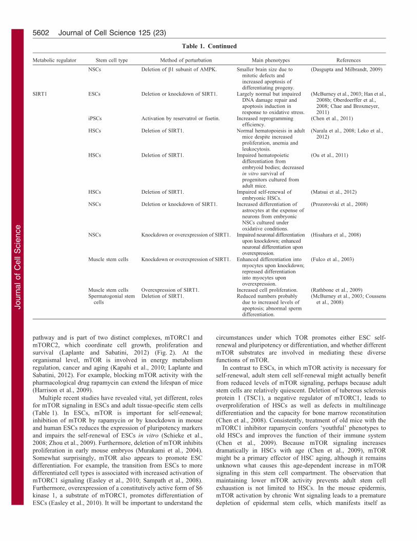
Metabolic regulator Stem cell type Method of perturbation Main phenotypes References
NSCs Deletion of b1 subunit of AMPK. Smaller brain size due tomitotic defects andincreased apoptosis ofdifferentiating progeny.
(Dasgupta and Milbrandt, 2009)
SIRT1 ESCs Deletion or knockdown of SIRT1. Largely normal but impairedDNA damage repair andapoptosis induction inresponse to oxidative stress.
(McBurney et al., 2003; Han et al.,2008b; Oberdoerffer et al.,2008; Chae and Broxmeyer,2011)
iPSCs Activation by reservatrol or fisetin. Increased reprogrammingefficiency.
(Chen et al., 2011)
HSCs Deletion of SIRT1. Normal hematopoiesis in adultmice despite increasedproliferation, anemia andleukocytosis.
(Narala et al., 2008; Leko et al.,2012)
HSCs Deletion of SIRT1. Impaired hematopoieticdifferentiation fromembryoid bodies; decreasedin vitro survival ofprogenitors cultured fromadult mice.
(Ou et al., 2011)
HSCs Deletion of SIRT1. Impaired self-renewal ofembryonic HSCs.
(Matsui et al., 2012)
NSCs Deletion or knockdown of SIRT1. Increased differentiation ofastrocytes at the expense ofneurons from embryonicNSCs cultured underoxidative conditions.
(Prozorovski et al., 2008)
NSCs Knockdown or overexpression of SIRT1. Impaired neuronal differentiationupon knockdown; enhancedneuronal differentiation uponoverexpression.
(Hisahara et al., 2008)
Muscle stem cells Knockdown or overexpression of SIRT1. Enhanced differentiation intomyocytes upon knockdown;repressed differentiationinto myocytes uponoverexpression.
(Fulco et al., 2003)
Muscle stem cells Overexpression of SIRT1. Increased cell proliferation. (Rathbone et al., 2009)Spermatogonial stem
cellsDeletion of SIRT1. Reduced numbers probably
due to increased levels ofapoptosis; abnormal spermdifferentiation.
(McBurney et al., 2003; Coussenset al., 2008)
Table 1. Continued
Journal of Cell Science 125 (23)5602
Journ
alof
Cell
Scie
nce
premature loss of hair (Castilho et al., 2009). Although
maintaining lower TOR activity is beneficial for adult stem
cells, a complete loss of mTOR activity is detrimental. Indeed,
deletion of Raptor, a key component of the mTORC1 complex,
leads to defects in HSC function upon transplantation (Kalaitzidis
et al., 2012). Finally, although mTOR appears to affect stem cells
mostly by acting in a cell-autonomous manner (Kalaitzidis et al.,
2012; Magri et al., 2011), it can also influence adult stem cells
(e.g. intestinal stem cells) in a non-cell-autonomous manner by
acting on the stem cell niche (Yilmaz et al., 2012).
How does mTOR hyper- or hypoactivation affect stem cell
metabolism? mTOR hyperactivation is associated with increased
numbers of mitochondria and higher ROS levels in adult HSCs
(Chen et al., 2008). Lowering ROS levels by treatment with the
antioxidant NAC partially rescues these HSC defects (Chen et al.,
2008), which are reminiscent of HSC phenotypes that lack
FOXOs (Tothova et al., 2007). Hypoactivation of mTOR owing
to loss of Raptor is associated with changes in metabolism, in
particular, the lipid and cholesterol metabolism in HSCs
(Kalaitzidis et al., 2012). The relative contributions of mTOR
targets, such as ribosomal S6 protein kinase 1 (S6K1, also known
as RPS6KB1), eukaryotic translation initiation factor 4E-binding
protein 1 [(4E-BP1), also known as EIF4EBP1] and SGK1
(Fig. 2), to these metabolic phenotypes in stem cells remain to be
determined.
Together, these studies highlight that mTOR signaling is essential
for ESC growth and proliferation, yet excessive mTOR activation can
be ultimately detrimental for adult stem cell pools and lead to early
aging phenotypes. Both mTOR and FOXO are downstream mediators
of insulin signaling, which leads to mTOR activation and inhibition of
FOXOs. Consistently, FOXOs and mTOR have largely antagonistic
functions in stem cells; FOXOs promote the maintenance of stem
cells in adulthood and help minimize oxidative stress, whereas
overactive mTOR signaling leads to premature stem cell depletion
and accumulation of oxidative stress. Collectively, these observations
support the idea that a certain degree of cellular quiescence is required
to preserve the pool of adult stem cells (Kippin et al., 2005; Morshead
et al., 1994). Although stem cell populations are considered
‘immortal’, there might be a limit in the number of times a stem
cell can undergo cell division before losing its self-renewal capacity in
vivo. These findings also support the idea that high levels of oxidative
stress contribute to stem cell dysfunction. Minimizing oxidative
damage is likely to be more important in stem cells than in
differentiated cells, as stem cells give rise to daughter stem cells and
therefore must maintain overall cellular integrity, including genomic,
protein and organelle content in order to ensure normal tissue function
and avoid tumor development.
AMPK in stem cell mitosis and mitochondrial homeostasis
AMPK is a central ‘fuel gauge’ that is activated by a wide range
of stimuli, including low energy or cellular stress. AMPK
becomes active when intracellular levels of AMP or ADP are
higher than that of ATP, and its activation requires the presence
of one of several upstream kinases, including the tumor
Glycolysis
Oxidative stress resistance,glucose metabolism, cell quiescence
Insulin Glucose
Insulin–IGF1R
AKT
FOXO
LKB1
[AMP]:ATP
TCA
Nucleus
[NAD+]:[NADH]ETC
Cytosol
AMPKHIF1
Amino acids High energy
SIRT1
PTEN
mTOR
PI3K
ATP
Protein synthesis, lipid synthesis, cell growth, proliferation, survival
Mitosis,oxidative phosphorylation?
S6K1Glycolysis, glucose transport, self-renewal
Genomic stability, cell cycle control
Substrates?
Mitochondrion
High [O2]
STEM CELL SELF-RENEWAL AND POTENCY
Substrates?
Substrates?
SGK14E-BP1
ATP
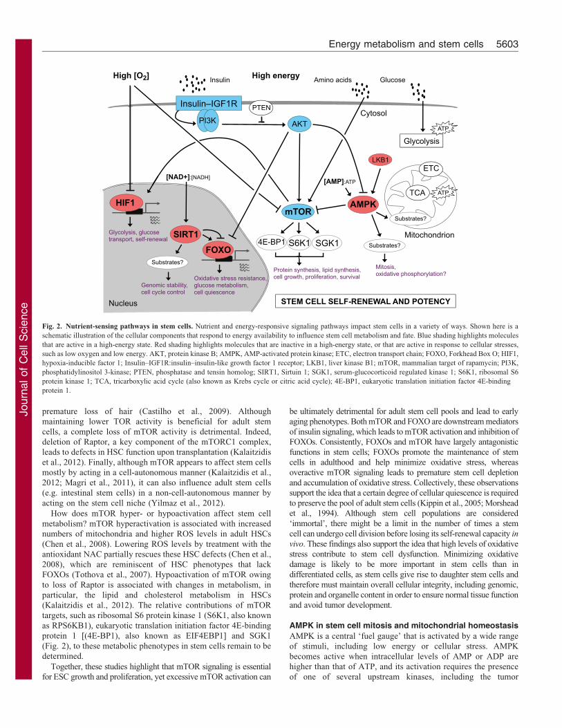
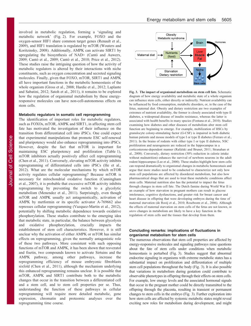
Fig. 2. Nutrient-sensing pathways in stem cells. Nutrient and energy-responsive signaling pathways impact stem cells in a variety of ways. Shown here is a
schematic illustration of the cellular components that respond to energy availability to influence stem cell metabolism and fate. Blue shading highlights molecules
that are active in a high-energy state. Red shading highlights molecules that are inactive in a high-energy state, or that are active in response to cellular stresses,
such as low oxygen and low energy. AKT, protein kinase B; AMPK, AMP-activated protein kinase; ETC, electron transport chain; FOXO, Forkhead Box O; HIF1,
hypoxia-inducible factor 1; Insulin–IGF1R:insulin–insulin-like growth factor 1 receptor; LKB1, liver kinase B1; mTOR, mammalian target of rapamycin; PI3K,
phosphatidylinositol 3-kinase; PTEN, phosphatase and tensin homolog; SIRT1, Sirtuin 1; SGK1, serum-glucocorticoid regulated kinase 1; S6K1, ribosomal S6
protein kinase 1; TCA, tricarboxylic acid cycle (also known as Krebs cycle or citric acid cycle); 4E-BP1, eukaryotic translation initiation factor 4E-binding
protein 1.
Energy metabolism and stem cells 5603
Journ
alof
Cell
Scie
nce
suppressor liver kinase B1 (LKB1, also known as STK11) and
Ca2+-calmodulin-dependent protein kinase kinase b (CAMKKb)(Kahn et al., 2005) (Fig. 2). AMPK switches off energy-consuming pathways and triggers energy-producing pathways
by phosphorylating many substrates that are involved in glucoseand lipid metabolism, autophagy and mitophagy, transcription,and cell cycle regulation (Banko et al., 2011; Mihaylova andShaw, 2011). AMPK has been shown to be crucial for longevity
(Greer et al., 2007; Mair et al., 2011), the prevention of type IIdiabetes (Li et al., 2011) and tumor suppression (Shaw et al.,2004).
Although the importance of AMPK in stem cells has not yetbeen extensively studied, it is known that AMPK functions inadult HSCs to influence mitochondrial homeostasis. HSCs thatlack both catalytic a-subunits of AMPK have decreased levels of
ATP and mitochondrial DNA (Nakada et al., 2010). Numbers ofHSCs in the bone marrow are substantially reduced severalmonths after AMPK activity is abrogated (Nakada et al., 2010),
suggesting that the regulation of mitochondrial homeostasis byAMPK contributes to the control of HSC proliferation. However,AMPK activity is not necessary for HSCs to reconstitute the
blood in bone marrow transplantation assays (Nakada et al.,2010). Furthermore, AMPK does not appear to mediate themajority of the actions of its upstream regulator LKB1 in the
maintenance of HSCs, suggesting that AMPK-related kinases areimportant for the function of LKB1 in this context (Gan et al.,2010; Gurumurthy et al., 2010; Nakada et al., 2010). Given thatAMPK activity relies on a high ratio between [AMP] and [ATP],
the functional relevance of the observed AMPK-dependentphenotypes might depend on the nutrient status of the organism.
AMPK can also regulate stem cell mitosis in the nervous
system. For example, in the developing mouse brain, abrogatingnormal AMPK activity leads to defective mitosis of neuralprogenitor cells and abnormal brain development (Dasgupta andMilbrandt, 2009). This possible effect is consistent with the
recent identification of a role for AMPK substrates in mitosis(Banko et al., 2011). Because AMPK is active under low energyconditions, it appears counterintuitive that it promotes stem cell
proliferation given the high-energy demands of cellular division.It is possible that under low energy conditions, AMPK activityensures the completion of mitosis, because cell cycle arrest at this
stage could have disastrous consequences for the genomicstability of a cell. Much still remains to be determined withregard to the roles of AMPK in stem cells, the metabolic
conditions under which it is most important, and the substratesthat mediate its actions in stem cells.
SIRT1 in the proliferation, differentiation and genomicintegrity of stem cells
The protein deacetylase Sirtuin 1 (SIRT1) is one of sevenmammalian Class III deacetylases (also called Sirtuins) thatbecome active when the ratio between [NAD+] and [NADH] is
high, a state associated with low energy as well as oxidativestress (Imai and Guarente, 2010; Webster et al., 2012). SIRT1 isthe Sirtuin with the greatest homology to the yeast Sir2
deacetylase, which is known to extend the replicative lifespanof yeast (Kaeberlein et al., 1999). In mammals, SIRT1 is a crucialregulator of cellular and organismal metabolism (Yu and
Auwerx, 2009), improves various markers of health (Bordoneet al., 2007; Herranz et al., 2010), provides neuroprotection in avariety of neurodegenerative diseases (Zhang et al., 2011a), and
has also been shown to have tumor-suppressive functions in
some, but not all, contexts (Fang and Nicholl, 2011).
The effects of SIRT1 perturbation are not identical in all stemcells (Table 1). SIRT1 is highly expressed in human and mouseESCs, and its expression declines during multilineage
differentiation, suggesting that SIRT1 has a more importantfunction in ESCs than in differentiated progeny (Calvanese et al.,2010; Saunders et al., 2010). Yet, under basal conditions, mouse
ESCs that lack SIRT1 show no obvious defects (McBurney et al.,2003). However, exposure to mild oxidative stress activatesSIRT1 to both promote apoptosis (Chae and Broxmeyer, 2011;
Han et al., 2008) and minimize the accumulation of chromosomalabnormalities in mouse ESCs (Oberdoerffer et al., 2008).Together, these studies indicate that SIRT1 carries out anti-tumorigenic activities by regulating nuclear translocation of
FOXO and p53, as well as promoting the repair of DNA damage.These findings contrast with previously demonstrated roles forSIRT1 in preventing apoptosis in response to oxidative stress in
mouse embryonic fibroblasts, cerebellar granule neurons andhuman cancer cell lines (Brunet et al., 2004; Motta et al., 2004).It is possible that the ability of SIRT1 to induce or prevent
apoptosis depends on the amount and source of oxidative stresspresent, or the specific cell type (stem versus differentiated).
The function of SIRT1 in HSCs is also age and context-specific. In vitro differentiation of mouse ESCs into the
hematopoietic lineage is defective in the absence of SIRT1 (Ouet al., 2011), yet adult mice lacking SIRT1 display virtuallynormal hematopoiesis (Leko et al., 2012; Narala et al., 2008). It is
possible that other histone deacetylases compensate for loss ofSIRT1 in adulthood, allowing the blood constituents to formproperly after development, even in the absence of SIRT1.
Intriguingly, HSC maintenance in old mice is not affected bythe deletion of SIRT1 despite increased proliferation levels,suggesting that HSC self-renewal can be maintained in theabsence of SIRT1 (Leko et al., 2012; Narala et al., 2008). The
mechanisms and specific substrates (histones or non-histoneproteins) underlying the ability of SIRT1 to either inhibit orpromote the expansion of a cell population in the blood lineage
remain to be elucidated.
SIRT1 activity is highly responsive to changes in oxidativestate given its dependence on a high [NAD+] to [NADH] ratio,and this has large implications for stem cell fate under varying
oxidative conditions. For example, SIRT1 promotes proliferationof adult rat muscle stem cells (Rathbone et al., 2009) andrepresses their differentiation into myocytes (Fulco et al., 2008;
Fulco et al., 2003). Indeed, a reduction in [NAD+]:[NADH], astate inhibitory for SIRT1 activity, is associated with mousemuscle cell differentiation and, by this means, is likely to relieve
SIRT1-mediated repression of genes that promote muscledifferentiation and maturation (Fulco et al., 2003). In addition,under oxidative conditions, SIRT1 skews the fate of embryonic
neural progenitors towards astrocytes at the expense of neuronsby repressing transcription of the proneural transcription factorASCL1 (also known as MASH1) (Prozorovski et al., 2008). Suchan activity might be relevant in neurodegenerative conditions that
are associated with high levels of oxidative stress, such asamyotrophic lateral sclerosis (ALS), Parkinson’s Disease andAlzheimer’s Disease (Emerit et al., 2004).
Interestingly, pathways that respond to nutrient availability,such as those involving insulin–FOXO, mTOR, SIRT1 andAMPK, interact with each other and with other factors that are
Journal of Cell Science 125 (23)5604
Journ
alof
Cell
Scie
nce
involved in metabolic regulation, forming a ‘signaling and
metabolic network’ (Fig. 2). For example, FOXO and the
oxygen-sensor HIF1 share common target genes (Renault et al.,
2009), and HIF1 translation is regulated by mTOR (Wouters and
Koritzinsky, 2008). Additionally, AMPK can activate SIRT1 by
upregulating the biosynthesis of NAD+ (Canto and Auwerx,
2009; Canto et al., 2009; Canto et al., 2010; Price et al., 2012).
These studies raise the intriguing question of how the activity of
metabolic regulators is altered by their interactions with niche
constituents, such as oxygen concentration and secreted signaling
molecules. Finally, given that FOXO, mTOR, SIRT1 and AMPK
all have important functions in the metabolic homeostasis of the
whole organism (Gross et al., 2008; Hardie et al., 2012; Laplante
and Sabatini, 2012; Satoh et al., 2011), it remains to be explored
how the regulation of organismal metabolism by these energy-
responsive molecules can have non-cell-autonomous effects on
stem cells.
Metabolic regulators in somatic cell reprogramming
The identification of important roles for metabolic regulators,
such as FOXOs, mTOR, AMPK and SIRT1, in affecting stem cell
fate has motivated the investigation of their influence on the
transition from differentiated cell into iPSCs. One could expect
that experimental manipulations that promote ESC self-renewal
and pluripotency would also enhance reprogramming into iPSCs.
However, despite the fact that mTOR is important for
maintaining ESC pluripotency and proliferation (Table 1),
mTOR inhibitors actually positively affect cell reprogramming
(Chen et al., 2011). Conversely, elevating mTOR activity inhibits
reprogramming of differentiated cells into iPSCs (He et al.,
2012). What are the molecular mechanisms by which mTOR
activity regulates cellular reprogramming? Because mTOR is
necessary for mitochondrial oxidative function (Cunningham
et al., 2007), it is probable that excessive mTOR activity inhibits
reprogramming by preventing the switch to a glycolytic
metabolism (Menendez et al., 2011). Surprisingly, even though
mTOR and AMPK usually act antagonistically, activation of
AMPK by metformin or its specific activator A-769662 also
represses cellular reprogramming (Vazquez-Martin et al., 2012),
potentially by shifting metabolic dependence towards oxidative
phosphorylation. These studies contribute to the emerging idea
that metabolic state, in particular, the balance between glycolysis
and oxidative phosphorylation, crucially impacts the
establishment of stem cell characteristics. However, it is still
unclear why the activation of either AMPK or mTOR has similar
effects on reprogramming, given the normally antagonistic role
of these two pathways. More consistent with such opposing
functions of mTOR and AMPK, it has been shown that resveratol
and fisetin, two compounds known to activate Sirtuins and the
AMPK pathway, among other pathways, increase the
reprogramming efficiency of mouse embryonic fibroblasts
sixfold (Chen et al., 2011), although the mechanism underlying
this enhanced reprogramming remains unclear. It is possible that
mTOR, AMPK and SIRT1 contribute both to the metabolic
changes that occur in the transition between a differentiated cell
and a stem cell, and to stem cell properties per se. Thus,
understanding the function of these pathways in cellular
reprogramming will require more detailed metabolic, gene
expression, chromatin and proteomic analyses over the
reprogramming time course.
Concluding remarks: implications of fluctuations inorganismal metabolism for stem cells
The numerous observations that stem cell properties are affected by
energy-responsive molecules and signaling pathways raise questions
about the fate of stem cells under conditions when metabolic
homeostasis is perturbed (Fig. 3). Studies suggest that abnormalendocrine signaling in organisms with extreme metabolic states has a
substantial impact on proliferation and differentiation of multiple
stem cell populations throughout the body (Fig. 3). It is also possible
that variations in metabolism during gestation could contribute to
observable phenotypes in offspring through their effects on stem cells.For example, low energy levels and the associated hormonal signals
that occur in the pregnant mother could be directly transmitted to the
offspring through the placenta, resulting in transient or permanent
changes to stem cells in the embryo (Fig. 3). Further exploration intohow stem cells are affected by systemic metabolic states might reveal
exciting new roles for metabolism during development, and might
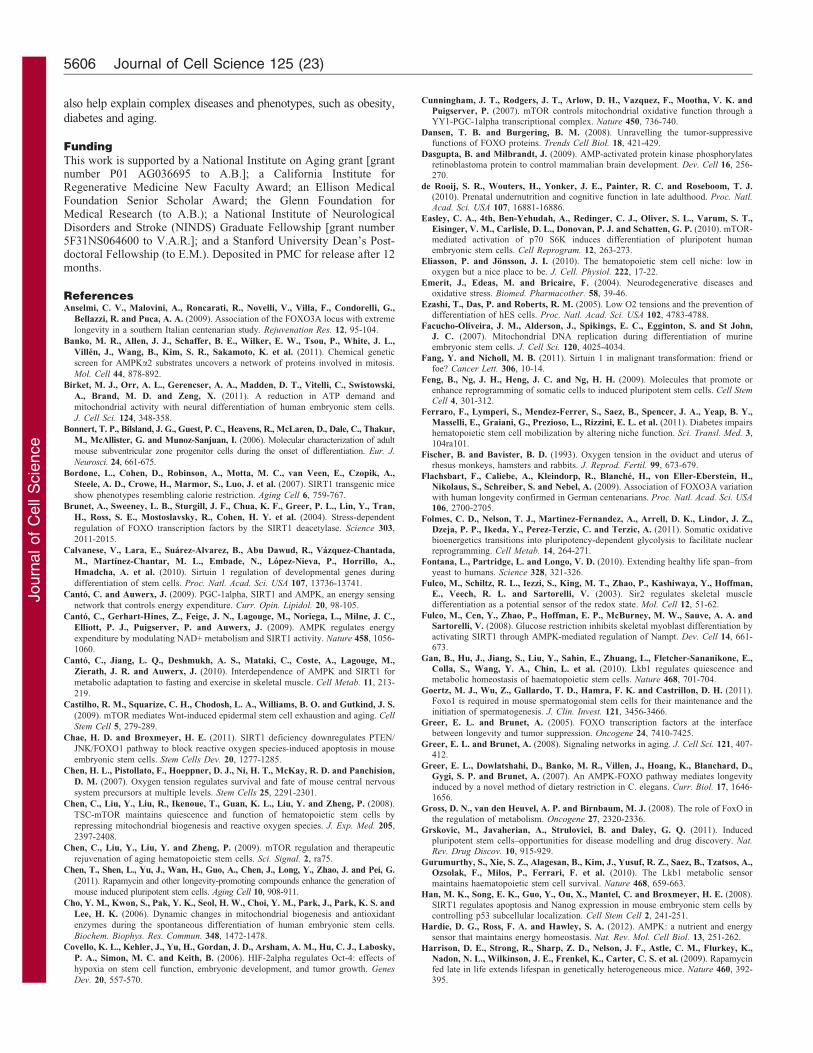
Fig. 3. The impact of organismal metabolism on stem cell fate. Schematic
diagram of how energy availability and metabolic state of a whole organism
can influence stem cells, either directly or indirectly. Nutrient availability can
be influenced by food consumption, metabolic disorders, or, in the case of the
fetus, maternal diet. Obesity and dietary restriction are two examples of
extremes of nutrient availability, the former is closely associated with type II
diabetes, a widespread disease of insulin resistance, whereas the latter is
associated with health benefits in many species (Fontana et al., 2010). Studies
examining how diabetes and other diseases of metabolism alter stem cell
function are beginning to emerge. For example, mobilization of HSCs by
granulocyte colony-stimulating factor (G-CSF) is impaired in both diabetic
human patients and mouse models of type I or type II diabetes (Ferraro et al.,
2011). In the brains of rodents with either type I or type II diabetes, NSC
proliferation and neurogenesis are reduced in the hippocampus in a
corticosterone-dependent manner (Rafalski and Brunet, 2011; Stranahan et
al., 2008). Conversely, dietary restriction (30% reduction in calorie intake
without malnutrition) enhances the survival of newborn neurons in the adult
rodent hippocampus (Lee et al., 2000). These studies highlight how stem cells
can respond dramatically to organismal changes in metabolic homeostasis and
argue that more studies need to be conducted to characterize not only how
stem cell populations are affected by disordered metabolism, but also how
pharmaceutical drugs that are used to treat these metabolic conditions affect
stem cells. Maternal nutrition also has the potential to impact the fetus
through changes in stem cell fate. The Dutch famine during World War II is
an example of how starvation in pregnant mothers can result in glucose
intolerance, cognitive dysfunctions, and greater risk for breast cancer and
heart disease in offspring that were developing embryos during the time of
maternal starvation (de Rooij et al., 2010; Roseboom et al., 2006). Although
the effects of maternal starvation on stem cells of the fetus are not known, in
utero changes in metabolism are likely to have a key function in the
regulation of stem cells and the tissues that develop from them.
Energy metabolism and stem cells 5605
Journ
alof
Cell
Scie
nce
also help explain complex diseases and phenotypes, such as obesity,
diabetes and aging.
FundingThis work is supported by a National Institute on Aging grant [grantnumber P01 AG036695 to A.B.]; a California Institute forRegenerative Medicine New Faculty Award; an Ellison MedicalFoundation Senior Scholar Award; the Glenn Foundation forMedical Research (to A.B.); a National Institute of NeurologicalDisorders and Stroke (NINDS) Graduate Fellowship [grant number5F31NS064600 to V.A.R.]; and a Stanford University Dean’s Post-doctoral Fellowship (to E.M.). Deposited in PMC for release after 12months.
ReferencesAnselmi, C. V., Malovini, A., Roncarati, R., Novelli, V., Villa, F., Condorelli, G.,
Bellazzi, R. and Puca, A. A. (2009). Association of the FOXO3A locus with extreme
longevity in a southern Italian centenarian study. Rejuvenation Res. 12, 95-104.
Banko, M. R., Allen, J. J., Schaffer, B. E., Wilker, E. W., Tsou, P., White, J. L.,
Villen, J., Wang, B., Kim, S. R., Sakamoto, K. et al. (2011). Chemical genetic
screen for AMPKa2 substrates uncovers a network of proteins involved in mitosis.
Mol. Cell 44, 878-892.
Birket, M. J., Orr, A. L., Gerencser, A. A., Madden, D. T., Vitelli, C., Swistowski,
A., Brand, M. D. and Zeng, X. (2011). A reduction in ATP demand and
mitochondrial activity with neural differentiation of human embryonic stem cells.
J. Cell Sci. 124, 348-358.
Bonnert, T. P., Bilsland, J. G., Guest, P. C., Heavens, R., McLaren, D., Dale, C., Thakur,
M., McAllister, G. and Munoz-Sanjuan, I. (2006). Molecular characterization of adult
mouse subventricular zone progenitor cells during the onset of differentiation. Eur. J.
Neurosci. 24, 661-675.
Bordone, L., Cohen, D., Robinson, A., Motta, M. C., van Veen, E., Czopik, A.,
Steele, A. D., Crowe, H., Marmor, S., Luo, J. et al. (2007). SIRT1 transgenic mice
show phenotypes resembling calorie restriction. Aging Cell 6, 759-767.
Brunet, A., Sweeney, L. B., Sturgill, J. F., Chua, K. F., Greer, P. L., Lin, Y., Tran,
H., Ross, S. E., Mostoslavsky, R., Cohen, H. Y. et al. (2004). Stress-dependent
regulation of FOXO transcription factors by the SIRT1 deacetylase. Science 303,
2011-2015.
Calvanese, V., Lara, E., Suarez-Alvarez, B., Abu Dawud, R., Vazquez-Chantada,
M., Martınez-Chantar, M. L., Embade, N., Lopez-Nieva, P., Horrillo, A.,
Hmadcha, A. et al. (2010). Sirtuin 1 regulation of developmental genes during
differentiation of stem cells. Proc. Natl. Acad. Sci. USA 107, 13736-13741.
Canto, C. and Auwerx, J. (2009). PGC-1alpha, SIRT1 and AMPK, an energy sensing
network that controls energy expenditure. Curr. Opin. Lipidol. 20, 98-105.
Canto, C., Gerhart-Hines, Z., Feige, J. N., Lagouge, M., Noriega, L., Milne, J. C.,
Elliott, P. J., Puigserver, P. and Auwerx, J. (2009). AMPK regulates energy
expenditure by modulating NAD+ metabolism and SIRT1 activity. Nature 458, 1056-
1060.
Canto, C., Jiang, L. Q., Deshmukh, A. S., Mataki, C., Coste, A., Lagouge, M.,
Zierath, J. R. and Auwerx, J. (2010). Interdependence of AMPK and SIRT1 for
metabolic adaptation to fasting and exercise in skeletal muscle. Cell Metab. 11, 213-
219.
Castilho, R. M., Squarize, C. H., Chodosh, L. A., Williams, B. O. and Gutkind, J. S.
(2009). mTOR mediates Wnt-induced epidermal stem cell exhaustion and aging. Cell
Stem Cell 5, 279-289.
Chae, H. D. and Broxmeyer, H. E. (2011). SIRT1 deficiency downregulates PTEN/
JNK/FOXO1 pathway to block reactive oxygen species-induced apoptosis in mouse
embryonic stem cells. Stem Cells Dev. 20, 1277-1285.
Chen, H. L., Pistollato, F., Hoeppner, D. J., Ni, H. T., McKay, R. D. and Panchision,
D. M. (2007). Oxygen tension regulates survival and fate of mouse central nervous
system precursors at multiple levels. Stem Cells 25, 2291-2301.
Chen, C., Liu, Y., Liu, R., Ikenoue, T., Guan, K. L., Liu, Y. and Zheng, P. (2008).
TSC-mTOR maintains quiescence and function of hematopoietic stem cells by
repressing mitochondrial biogenesis and reactive oxygen species. J. Exp. Med. 205,
2397-2408.
Chen, C., Liu, Y., Liu, Y. and Zheng, P. (2009). mTOR regulation and therapeutic
rejuvenation of aging hematopoietic stem cells. Sci. Signal. 2, ra75.
Chen, T., Shen, L., Yu, J., Wan, H., Guo, A., Chen, J., Long, Y., Zhao, J. and Pei, G.
(2011). Rapamycin and other longevity-promoting compounds enhance the generation of
mouse induced pluripotent stem cells. Aging Cell 10, 908-911.
Cho, Y. M., Kwon, S., Pak, Y. K., Seol, H. W., Choi, Y. M., Park, J., Park, K. S. and
Lee, H. K. (2006). Dynamic changes in mitochondrial biogenesis and antioxidant
enzymes during the spontaneous differentiation of human embryonic stem cells.
Biochem. Biophys. Res. Commun. 348, 1472-1478.
Covello, K. L., Kehler, J., Yu, H., Gordan, J. D., Arsham, A. M., Hu, C. J., Labosky,
P. A., Simon, M. C. and Keith, B. (2006). HIF-2alpha regulates Oct-4: effects of
hypoxia on stem cell function, embryonic development, and tumor growth. Genes
Dev. 20, 557-570.
Cunningham, J. T., Rodgers, J. T., Arlow, D. H., Vazquez, F., Mootha, V. K. and
Puigserver, P. (2007). mTOR controls mitochondrial oxidative function through aYY1-PGC-1alpha transcriptional complex. Nature 450, 736-740.
Dansen, T. B. and Burgering, B. M. (2008). Unravelling the tumor-suppressivefunctions of FOXO proteins. Trends Cell Biol. 18, 421-429.
Dasgupta, B. and Milbrandt, J. (2009). AMP-activated protein kinase phosphorylatesretinoblastoma protein to control mammalian brain development. Dev. Cell 16, 256-270.
de Rooij, S. R., Wouters, H., Yonker, J. E., Painter, R. C. and Roseboom, T. J.(2010). Prenatal undernutrition and cognitive function in late adulthood. Proc. Natl.
Acad. Sci. USA 107, 16881-16886.
Easley, C. A., 4th, Ben-Yehudah, A., Redinger, C. J., Oliver, S. L., Varum, S. T.,
Eisinger, V. M., Carlisle, D. L., Donovan, P. J. and Schatten, G. P. (2010). mTOR-mediated activation of p70 S6K induces differentiation of pluripotent humanembryonic stem cells. Cell Reprogram. 12, 263-273.
Eliasson, P. and Jonsson, J. I. (2010). The hematopoietic stem cell niche: low inoxygen but a nice place to be. J. Cell. Physiol. 222, 17-22.
Emerit, J., Edeas, M. and Bricaire, F. (2004). Neurodegenerative diseases andoxidative stress. Biomed. Pharmacother. 58, 39-46.
Ezashi, T., Das, P. and Roberts, R. M. (2005). Low O2 tensions and the prevention ofdifferentiation of hES cells. Proc. Natl. Acad. Sci. USA 102, 4783-4788.
Facucho-Oliveira, J. M., Alderson, J., Spikings, E. C., Egginton, S. and St John,J. C. (2007). Mitochondrial DNA replication during differentiation of murineembryonic stem cells. J. Cell Sci. 120, 4025-4034.
Fang, Y. and Nicholl, M. B. (2011). Sirtuin 1 in malignant transformation: friend orfoe? Cancer Lett. 306, 10-14.
Feng, B., Ng, J. H., Heng, J. C. and Ng, H. H. (2009). Molecules that promote orenhance reprogramming of somatic cells to induced pluripotent stem cells. Cell Stem
Cell 4, 301-312.
Ferraro, F., Lymperi, S., Mendez-Ferrer, S., Saez, B., Spencer, J. A., Yeap, B. Y.,
Masselli, E., Graiani, G., Prezioso, L., Rizzini, E. L. et al. (2011). Diabetes impairshematopoietic stem cell mobilization by altering niche function. Sci. Transl. Med. 3,104ra101.
Fischer, B. and Bavister, B. D. (1993). Oxygen tension in the oviduct and uterus ofrhesus monkeys, hamsters and rabbits. J. Reprod. Fertil. 99, 673-679.
Flachsbart, F., Caliebe, A., Kleindorp, R., Blanche, H., von Eller-Eberstein, H.,Nikolaus, S., Schreiber, S. and Nebel, A. (2009). Association of FOXO3A variationwith human longevity confirmed in German centenarians. Proc. Natl. Acad. Sci. USA
106, 2700-2705.
Folmes, C. D., Nelson, T. J., Martinez-Fernandez, A., Arrell, D. K., Lindor, J. Z.,Dzeja, P. P., Ikeda, Y., Perez-Terzic, C. and Terzic, A. (2011). Somatic oxidativebioenergetics transitions into pluripotency-dependent glycolysis to facilitate nuclearreprogramming. Cell Metab. 14, 264-271.
Fontana, L., Partridge, L. and Longo, V. D. (2010). Extending healthy life span–fromyeast to humans. Science 328, 321-326.
Fulco, M., Schiltz, R. L., Iezzi, S., King, M. T., Zhao, P., Kashiwaya, Y., Hoffman,
E., Veech, R. L. and Sartorelli, V. (2003). Sir2 regulates skeletal muscledifferentiation as a potential sensor of the redox state. Mol. Cell 12, 51-62.
Fulco, M., Cen, Y., Zhao, P., Hoffman, E. P., McBurney, M. W., Sauve, A. A. andSartorelli, V. (2008). Glucose restriction inhibits skeletal myoblast differentiation byactivating SIRT1 through AMPK-mediated regulation of Nampt. Dev. Cell 14, 661-673.
Gan, B., Hu, J., Jiang, S., Liu, Y., Sahin, E., Zhuang, L., Fletcher-Sananikone, E.,
Colla, S., Wang, Y. A., Chin, L. et al. (2010). Lkb1 regulates quiescence andmetabolic homeostasis of haematopoietic stem cells. Nature 468, 701-704.
Goertz, M. J., Wu, Z., Gallardo, T. D., Hamra, F. K. and Castrillon, D. H. (2011).Foxo1 is required in mouse spermatogonial stem cells for their maintenance and theinitiation of spermatogenesis. J. Clin. Invest. 121, 3456-3466.
Greer, E. L. and Brunet, A. (2005). FOXO transcription factors at the interfacebetween longevity and tumor suppression. Oncogene 24, 7410-7425.
Greer, E. L. and Brunet, A. (2008). Signaling networks in aging. J. Cell Sci. 121, 407-412.
Greer, E. L., Dowlatshahi, D., Banko, M. R., Villen, J., Hoang, K., Blanchard, D.,Gygi, S. P. and Brunet, A. (2007). An AMPK-FOXO pathway mediates longevityinduced by a novel method of dietary restriction in C. elegans. Curr. Biol. 17, 1646-1656.
Gross, D. N., van den Heuvel, A. P. and Birnbaum, M. J. (2008). The role of FoxO inthe regulation of metabolism. Oncogene 27, 2320-2336.
Grskovic, M., Javaherian, A., Strulovici, B. and Daley, G. Q. (2011). Inducedpluripotent stem cells–opportunities for disease modelling and drug discovery. Nat.
Rev. Drug Discov. 10, 915-929.
Gurumurthy, S., Xie, S. Z., Alagesan, B., Kim, J., Yusuf, R. Z., Saez, B., Tzatsos, A.,Ozsolak, F., Milos, P., Ferrari, F. et al. (2010). The Lkb1 metabolic sensormaintains haematopoietic stem cell survival. Nature 468, 659-663.
Han, M. K., Song, E. K., Guo, Y., Ou, X., Mantel, C. and Broxmeyer, H. E. (2008).SIRT1 regulates apoptosis and Nanog expression in mouse embryonic stem cells bycontrolling p53 subcellular localization. Cell Stem Cell 2, 241-251.
Hardie, D. G., Ross, F. A. and Hawley, S. A. (2012). AMPK: a nutrient and energysensor that maintains energy homeostasis. Nat. Rev. Mol. Cell Biol. 13, 251-262.
Harrison, D. E., Strong, R., Sharp, Z. D., Nelson, J. F., Astle, C. M., Flurkey, K.,
Nadon, N. L., Wilkinson, J. E., Frenkel, K., Carter, C. S. et al. (2009). Rapamycinfed late in life extends lifespan in genetically heterogeneous mice. Nature 460, 392-395.
Journal of Cell Science 125 (23)5606
Journ
alof
Cell
Scie
nce
He, J., Kang, L., Wu, T., Zhang, J., Wang, H., Gao, H., Zhang, Y., Huang, B., Liu,
W., Kou, Z. et al. (2012). An elaborate regulation of Mammalian target of rapamycin
activity is required for somatic cell reprogramming induced by defined transcription
factors. Stem Cells Dev. 21, 2630-2641.
Herranz, D., Munoz-Martin, M., Canamero, M., Mulero, F., Martinez-Pastor, B.,
Fernandez-Capetillo, O. and Serrano, M. (2010). Sirt1 improves healthy ageing
and protects from metabolic syndrome-associated cancer. Nat Commun. 1, 3.
Hsu, P. P. and Sabatini, D. M. (2008). Cancer cell metabolism: Warburg and beyond.
Cell 134, 703-707.
Imai, S. and Guarente, L. (2010). Ten years of NAD-dependent SIR2 family
deacetylases: implications for metabolic diseases. Trends Pharmacol. Sci. 31, 212-
220.
Ito, K., Hirao, A., Arai, F., Matsuoka, S., Takubo, K., Hamaguchi, I., Nomiyama,
K., Hosokawa, K., Sakurada, K., Nakagata, N. et al. (2004). Regulation of
oxidative stress by ATM is required for self-renewal of haematopoietic stem cells.
Nature 431, 997-1002.
Ito, K., Carracedo, A., Weiss, D., Arai, F., Ala, U., Avigan, D. E., Schafer, Z. T.,
Evans, R. M., Suda, T., Lee, C. H. et al. (2012). A PML-PPARdelta pathway for
fatty acid oxidation regulates hematopoietic stem cell maintenance. Nat. Med. 18,
1350-1358.
Jasper, H. and Jones, D. L. (2010). Metabolic regulation of stem cell behavior and
implications for aging. Cell Metab. 12, 561-565.
Joseph, N. M. and Morrison, S. J. (2005). Toward an understanding of the
physiological function of Mammalian stem cells. Dev. Cell 9, 173-183.
Kaeberlein, M., McVey, M. and Guarente, L. (1999). The SIR2/3/4 complex and SIR2
alone promote longevity in Saccharomyces cerevisiae by two different mechanisms.
Genes Dev. 13, 2570-2580.
Kahn, B. B., Alquier, T., Carling, D. and Hardie, D. G. (2005). AMP-activated
protein kinase: ancient energy gauge provides clues to modern understanding of
metabolism. Cell Metab. 1, 15-25.
Kalaitzidis, D., Sykes, S. M., Wang, Z., Punt, N., Tang, Y., Ragu, C., Sinha, A. U.,
Lane, S. W., Souza, A. L., Clish, C. B. et al. (2012). mTOR complex 1 plays critical
roles in hematopoiesis and Pten-loss-evoked leukemogenesis. Cell Stem Cell 11, 429-
439.
Kang, J., Shakya, A. and Tantin, D. (2009). Stem cells, stress, metabolism and cancer:
a drama in two Octs. Trends Biochem. Sci. 34, 491-499.
Kapahi, P., Chen, D., Rogers, A. N., Katewa, S. D., Li, P. W., Thomas, E. L. and
Kockel, L. (2010). With TOR, less is more: a key role for the conserved nutrient-
sensing TOR pathway in aging. Cell Metab. 11, 453-465.
Kenyon, C. J. (2010). The genetics of ageing. Nature 464, 504-512.
Kippin, T. E., Martens, D. J. and van der Kooy, D. (2005). p21 loss compromises the
relative quiescence of forebrain stem cell proliferation leading to exhaustion of their
proliferation capacity. Genes Dev. 19, 756-767.
Knobloch, M., Braun, S. M., Zurkirchen, L., von Schoultz, C., Zamboni, N.,
Arauzo-Bravo, M. J., Kovacs, W. J., Karalay, O., Suter, U., Machado, R. A. et al.
(2013). Metabolic control of adult neural stem cell activity by Fasn-dependent
lipogenesis. Nature 493, 226-230.
Kobayashi, C. I. and Suda, T. (2012). Regulation of reactive oxygen species in stem
cells and cancer stem cells. J. Cell. Physiol. 227, 421-430.
Laplante, M. and Sabatini, D. M. (2012). mTOR signaling in growth control and
disease. Cell 149, 274-293.
Lee, J., Duan, W., Long, J. M., Ingram, D. K. and Mattson, M. P. (2000). Dietary
restriction increases the number of newly generated neural cells, and induces BDNF
expression, in the dentate gyrus of rats. J. Mol. Neurosci. 15, 99-108.
Leko, V., Varnum-Finney, B., Li, H., Gu, Y., Flowers, D., Nourigat, C., Bernstein,
I. D. and Bedalov, A. (2012). SIRT1 is dispensable for function of hematopoietic
stem cells in adult mice. Blood 119, 1856-1860.
Li, Y., Xu, S., Mihaylova, M. M., Zheng, B., Hou, X., Jiang, B., Park, O., Luo, Z., Lefai,
E., Shyy, J. Y. et al. (2011). AMPK phosphorylates and inhibits SREBP activity to
attenuate hepatic steatosis and atherosclerosis in diet-induced insulin-resistant mice. Cell
Metab. 13, 376-388.
Magri, L., Cambiaghi, M., Cominelli, M., Alfaro-Cervello, C., Cursi, M., Pala, M.,
Bulfone, A., Garcıa-Verdugo, J. M., Leocani, L., Minicucci, F. et al. (2011).
Sustained activation of mTOR pathway in embryonic neural stem cells leads to
development of tuberous sclerosis complex-associated lesions. Cell Stem Cell 9, 447-
462.
Mair, W., Morantte, I., Rodrigues, A. P., Manning, G., Montminy, M., Shaw, R. J.
and Dillin, A. (2011). Lifespan extension induced by AMPK and calcineurin is
mediated by CRTC-1 and CREB. Nature 470, 404-408.
Mannello, F., Medda, V. and Tonti, G. A. (2011). Hypoxia and neural stem cells: from
invertebrates to brain cancer stem cells. Int. J. Dev. Biol. 55, 569-581.
Mathieu, J., Zhang, Z., Zhou, W., Wang, A. J., Heddleston, J. M., Pinna, C. M.,
Hubaud, A., Stadler, B., Choi, M., Bar, M. et al. (2011). HIF induces human
embryonic stem cell markers in cancer cells. Cancer Res. 71, 4640-4652.
McBurney, M. W., Yang, X., Jardine, K., Bieman, M., Th’ng, J. and Lemieux, M.
(2003). The absence of SIR2alpha protein has no effect on global gene silencing in
mouse embryonic stem cells. Mol. Cancer Res. 1, 402-409.
Menendez, J. A., Vellon, L., Oliveras-Ferraros, C., Cufı, S. and Vazquez-Martin, A.
(2011). mTOR-regulated senescence and autophagy during reprogramming of
somatic cells to pluripotency: a roadmap from energy metabolism to stem cell
renewal and aging. Cell Cycle 10, 3658-3677.
Miharada, K., Karlsson, G., Rehn, M., Rorby, E., Siva, K., Cammenga, J. and
Karlsson, S. (2011). Cripto regulates hematopoietic stem cells as a hypoxic-niche-related factor through cell surface receptor GRP78. Cell Stem Cell 9, 330-344.
Mihaylova, M. M. and Shaw, R. J. (2011). The AMPK signalling pathway coordinatescell growth, autophagy and metabolism. Nat. Cell Biol. 13, 1016-1023.
Miyamoto, K., Araki, K. Y., Naka, K., Arai, F., Takubo, K., Yamazaki, S.,
Matsuoka, S., Miyamoto, T., Ito, K., Ohmura, M. et al. (2007). Foxo3a is essentialfor maintenance of the hematopoietic stem cell pool. Cell Stem Cell 1, 101-112.
Mohyeldin, A., Garzon-Muvdi, T. and Quinones-Hinojosa, A. (2010). Oxygen instem cell biology: a critical component of the stem cell niche. Cell Stem Cell 7, 150-161.
Morshead, C. M., Reynolds, B. A., Craig, C. G., McBurney, M. W., Staines, W. A.,
Morassutti, D., Weiss, S. and van der Kooy, D. (1994). Neural stem cells in theadult mammalian forebrain: a relatively quiescent subpopulation of subependymalcells. Neuron 13, 1071-1082.
Motta, M. C., Divecha, N., Lemieux, M., Kamel, C., Chen, D., Gu, W., Bultsma, Y.,McBurney, M. and Guarente, L. (2004). Mammalian SIRT1 represses forkheadtranscription factors. Cell 116, 551-563.
Murakami, M., Ichisaka, T., Maeda, M., Oshiro, N., Hara, K., Edenhofer, F.,
Kiyama, H., Yonezawa, K. and Yamanaka, S. (2004). mTOR is essential forgrowth and proliferation in early mouse embryos and embryonic stem cells. Mol. Cell.
Biol. 24, 6710-6718.
Nakada, D., Saunders, T. L. and Morrison, S. J. (2010). Lkb1 regulates cell cycle andenergy metabolism in haematopoietic stem cells. Nature 468, 653-658.
Nakada, D., Levi, B. P. and Morrison, S. J. (2011). Integrating physiologicalregulation with stem cell and tissue homeostasis. Neuron 70, 703-718.
Narala, S. R., Allsopp, R. C., Wells, T. B., Zhang, G., Prasad, P., Coussens, M. J.,
Rossi, D. J., Weissman, I. L. and Vaziri, H. (2008). SIRT1 acts as a nutrient-sensitive growth suppressor and its loss is associated with increased AMPK andtelomerase activity. Mol. Biol. Cell 19, 1210-1219.
Oberdoerffer, P., Michan, S., McVay, M., Mostoslavsky, R., Vann, J., Park, S. K.,Hartlerode, A., Stegmuller, J., Hafner, A., Loerch, P. et al. (2008). SIRT1redistribution on chromatin promotes genomic stability but alters gene expressionduring aging. Cell 135, 907-918.
Okita, K., Ichisaka, T. and Yamanaka, S. (2007). Generation of germline-competentinduced pluripotent stem cells. Nature 448, 313-317.
Ou, X., Chae, H. D., Wang, R. H., Shelley, W. C., Cooper, S., Taylor, T., Kim, Y. J.,Deng, C. X., Yoder, M. C. and Broxmeyer, H. E. (2011). SIRT1 deficiencycompromises mouse embryonic stem cell hematopoietic differentiation, andembryonic and adult hematopoiesis in the mouse. Blood 117, 440-450.
Paik, J. H., Ding, Z., Narurkar, R., Ramkissoon, S., Muller, F., Kamoun, W. S.,
Chae, S. S., Zheng, H., Ying, H., Mahoney, J. et al. (2009). FoxOs cooperativelyregulate diverse pathways governing neural stem cell homeostasis. Cell Stem Cell 5,540-553.
Panopoulos, A. D., Yanes, O., Ruiz, S., Kida, Y. S., Diep, D., Tautenhahn, R.,
Herrerıas, A., Batchelder, E. M., Plongthongkum, N., Lutz, M. et al. (2012). Themetabolome of induced pluripotent stem cells reveals metabolic changes occurring insomatic cell reprogramming. Cell Res. 22, 168-177.
Pawlikowska, L., Hu, D., Huntsman, S., Sung, A., Chu, C., Chen, J., Joyner, A. H.,Schork, N. J., Hsueh, W. C., Reiner, A. P. et al. (2009). Association of commongenetic variation in the insulin/IGF1 signaling pathway with human longevity. Aging
Cell 8, 460-472.
Pistollato, F., Chen, H. L., Schwartz, P. H., Basso, G. and Panchision, D. M. (2007).Oxygen tension controls the expansion of human CNS precursors and the generationof astrocytes and oligodendrocytes. Mol. Cell. Neurosci. 35, 424-435.
Price, N. L., Gomes, A. P., Ling, A. J., Duarte, F. V., Martin-Montalvo, A., North,
B. J., Agarwal, B., Ye, L., Ramadori, G., Teodoro, J. S. et al. (2012). SIRT1 isrequired for AMPK activation and the beneficial effects of resveratrol onmitochondrial function. Cell Metab. 15, 675-690.
Prozorovski, T., Schulze-Topphoff, U., Glumm, R., Baumgart, J., Schroter, F.,Ninnemann, O., Siegert, E., Bendix, I., Brustle, O., Nitsch, R. et al. (2008). Sirt1contributes critically to the redox-dependent fate of neural progenitors. Nat. Cell Biol.
10, 385-394.
Rafalski, V. A. and Brunet, A. (2011). Energy metabolism in adult neural stem cellfate. Prog. Neurobiol. 93, 182-203.
Ramalho-Santos, M., Yoon, S., Matsuzaki, Y., Mulligan, R. C. and Melton, D. A.
(2002). ‘‘Stemness’’: transcriptional profiling of embryonic and adult stem cells.Science 298, 597-600.
Rando, T. A. (2006). Stem cells, ageing and the quest for immortality. Nature 441,1080-1086.
Rathbone, C. R., Booth, F. W. and Lees, S. J. (2009). Sirt1 increases skeletal muscleprecursor cell proliferation. Eur. J. Cell Biol. 88, 35-44.
Renault, V. M., Rafalski, V. A., Morgan, A. A., Salih, D. A., Brett, J. O., Webb,A. E., Villeda, S. A., Thekkat, P. U., Guillerey, C., Denko, N. C. et al. (2009).FoxO3 regulates neural stem cell homeostasis. Cell Stem Cell 5, 527-539.
Roseboom, T., de Rooij, S. and Painter, R. (2006). The Dutch famine and its long-termconsequences for adult health. Early Hum. Dev. 82, 485-491.
Rubakhin, S. S., Romanova, E. V., Nemes, P. and Sweedler, J. V. (2011). Profilingmetabolites and peptides in single cells. Nat. Methods 8 Suppl., S20-S29.
Salih, D. A. and Brunet, A. (2008). FoxO transcription factors in the maintenance ofcellular homeostasis during aging. Curr. Opin. Cell Biol. 20, 126-136.
Sampath, P., Pritchard, D. K., Pabon, L., Reinecke, H., Schwartz, S. M., Morris,
D. R. and Murry, C. E. (2008). A hierarchical network controls protein translation
Energy metabolism and stem cells 5607
Journ
alof
Cell
Scie
nce
during murine embryonic stem cell self-renewal and differentiation. Cell Stem Cell 2,448-460.
Satoh, A., Stein, L. and Imai, S. (2011). The role of mammalian sirtuins in theregulation of metabolism, aging, and longevity. Handb. Exp. Pharmacol. 206, 125-162.
Saunders, L. R., Sharma, A. D., Tawney, J., Nakagawa, M., Okita, K., Yamanaka,S., Willenbring, H. and Verdin, E. (2010). miRNAs regulate SIRT1 expressionduring mouse embryonic stem cell differentiation and in adult mouse tissues. Aging
(Albany NY) 2, 415-431.Schieke, S. M., Ma, M., Cao, L., McCoy, J. P., Jr, Liu, C., Hensel, N. F., Barrett, A.
J., Boehm, M. and Finkel, T. (2008). Mitochondrial metabolism modulatesdifferentiation and teratoma formation capacity in mouse embryonic stem cells.J. Biol. Chem. 283, 28506-28512.
Shakya, A., Cooksey, R., Cox, J. E., Wang, V., McClain, D. A. and Tantin, D. (2009).Oct1 loss of function induces a coordinate metabolic shift that opposestumorigenicity. Nat. Cell Biol. 11, 320-327.
Shaw, R. J., Bardeesy, N., Manning, B. D., Lopez, L., Kosmatka, M., DePinho, R. A.
and Cantley, L. C. (2004). The LKB1 tumor suppressor negatively regulates mTORsignaling. Cancer Cell 6, 91-99.
Shyh-Chang, N., Locasale, J. W., Lyssiotis, C. A., Zheng, Y., Teo, R.,Ratanasirintrawoot, S., Zhang, J., Onder, T., Unternaehrer, J. J., Zhu, H. et al.
(2013). Influence of Threonine Metabolism on S-Adenosylmethionine and HistoneMethylation. Science 339, 222-226.
Silver, I. and Erecinska, M. (1998). Oxygen and ion concentrations in normoxic andhypoxic brain cells. Adv. Exp. Med. Biol. 454, 7-16.
Simsek, T., Kocabas, F., Zheng, J., Deberardinis, R. J., Mahmoud, A. I., Olson,
E. N., Schneider, J. W., Zhang, C. C. and Sadek, H. A. (2010). The distinctmetabolic profile of hematopoietic stem cells reflects their location in a hypoxicniche. Cell Stem Cell 7, 380-390.
St John, J. C., Ramalho-Santos, J., Gray, H. L., Petrosko, P., Rawe, V. Y., Navara,C. S., Simerly, C. R. and Schatten, G. P. (2005). The expression of mitochondrialDNA transcription factors during early cardiomyocyte in vitro differentiation fromhuman embryonic stem cells. Cloning Stem Cells 7, 141-153.
Steinhauser, M. L., Bailey, A. P., Senyo, S. E., Guillermier, C., Perlstein, T. S.,
Gould, A. P., Lee, R. T. and Lechene, C. P. (2012). Multi-isotope imaging massspectrometry quantifies stem cell division and metabolism. Nature 481, 516-519.
Stranahan, A. M., Arumugam, T. V., Cutler, R. G., Lee, K., Egan, J. M. and Mattson,M. P. (2008). Diabetes impairs hippocampal function through glucocorticoid-mediatedeffects on new and mature neurons. Nat. Neurosci. 11, 309-317.
Studer, L., Csete, M., Lee, S. H., Kabbani, N., Walikonis, J., Wold, B. and McKay,R. (2000). Enhanced proliferation, survival, and dopaminergic differentiation of CNSprecursors in lowered oxygen. J. Neurosci. 20, 7377-7383.
Suda, T., Takubo, K. and Semenza, G. L. (2011). Metabolic regulation ofhematopoietic stem cells in the hypoxic niche. Cell Stem Cell 9, 298-310.
Suhr, S. T., Chang, E. A., Tjong, J., Alcasid, N., Perkins, G. A., Goissis, M. D.,
Ellisman, M. H., Perez, G. I. and Cibelli, J. B. (2010). Mitochondrial rejuvenationafter induced pluripotency. PLoS ONE 5, e14095.
Takahashi, K. and Yamanaka, S. (2006). Induction of pluripotent stem cells frommouse embryonic and adult fibroblast cultures by defined factors. Cell 126, 663-676.
Takahashi, K., Tanabe, K., Ohnuki, M., Narita, M., Ichisaka, T., Tomoda, K. and
Yamanaka, S. (2007). Induction of pluripotent stem cells from adult humanfibroblasts by defined factors. Cell 131, 861-872.
Takubo, K., Goda, N., Yamada, W., Iriuchishima, H., Ikeda, E., Kubota, Y., Shima,
H., Johnson, R. S., Hirao, A., Suematsu, M. et al. (2010). Regulation of the HIF-1alpha level is essential for hematopoietic stem cells. Cell Stem Cell 7, 391-402.
Thomson, J. A., Itskovitz-Eldor, J., Shapiro, S. S., Waknitz, M. A., Swiergiel, J. J.,Marshall, V. S. and Jones, J. M. (1998). Embryonic stem cell lines derived fromhuman blastocysts. Science 282, 1145-1147.
Tomita, S., Ueno, M., Sakamoto, M., Kitahama, Y., Ueki, M., Maekawa, N.,Sakamoto, H., Gassmann, M., Kageyama, R., Ueda, N. et al. (2003). Defectivebrain development in mice lacking the Hif-1alpha gene in neural cells. Mol. Cell.
Biol. 23, 6739-6749.Tothova, Z., Kollipara, R., Huntly, B. J., Lee, B. H., Castrillon, D. H., Cullen, D. E.,
McDowell, E. P., Lazo-Kallanian, S., Williams, I. R., Sears, C. et al. (2007). FoxOsare critical mediators of hematopoietic stem cell resistance to physiologic oxidativestress. Cell 128, 325-339.
Unwin, R. D., Smith, D. L., Blinco, D., Wilson, C. L., Miller, C. J., Evans, C. A.,
Jaworska, E., Baldwin, S. A., Barnes, K., Pierce, A. et al. (2006). Quantitativeproteomics reveals posttranslational control as a regulatory factor in primaryhematopoietic stem cells. Blood 107, 4687-4694.
Varum, S., Momcilovic, O., Castro, C., Ben-Yehudah, A., Ramalho-Santos, J. andNavara, C. S. (2009). Enhancement of human embryonic stem cell pluripotencythrough inhibition of the mitochondrial respiratory chain. Stem Cell Res. 3, 142-156.
Varum, S., Rodrigues, A. S., Moura, M. B., Momcilovic, O., Easley, C. A., 4th,
Ramalho-Santos, J., Van Houten, B. and Schatten, G. (2011). Energy metabolism
in human pluripotent stem cells and their differentiated counterparts. PLoS ONE 6,
e20914.
Vazquez-Martin, A., Vellon, L., Quiros, P. M., Cufi, S., Ruiz de Galarreta, E.,
Oliveras-Ferraros, C., Martin, A. G., Martin-Castillo, B., Lopez-Otin, C. and
Menendez, J. A. (2012). Activation of AMP-activated protein kinase (AMPK)
provides a metabolic barrier to reprogramming somatic cells into stem cells. Cell
Cycle 11, 974-989.
Vilchez, D., Boyer, L., Morantte, I., Lutz, M., Merkwirth, C., Joyce, D., Spencer, B.,
Page, L., Masliah, E., Berggren, W. T. et al. (2012). Increased proteasome activity
in human embryonic stem cells is regulated by PSMD11. Nature 489, 304-308.
Wang, P. and Na, J. (2011). Mechanism and methods to induce pluripotency. Protein
Cell 2, 792-799.
Wang, J., Alexander, P., Wu, L., Hammer, R., Cleaver, O. and McKnight, S. L.
(2009). Dependence of mouse embryonic stem cells on threonine catabolism. Science
325, 435-439.
Wang, J., Alexander, P. and McKnight, S. L. (2011). Metabolic specialization of
mouse embryonic stem cells. Cold Spring Harb. Symp. Quant. Biol. 76, 183-193.
Warburg, O. (1956). On the origin of cancer cells. Science 123, 309-314.
Webster, B. R., Lu, Z., Sack, M. N. and Scott, I. (2012). The role of sirtuins in
modulating redox stressors. Free Radic. Biol. Med. 52, 281-290.
Willcox, B. J., Donlon, T. A., He, Q., Chen, R., Grove, J. S., Yano, K., Masaki,
K. H., Willcox, D. C., Rodriguez, B. and Curb, J. D. (2008). FOXO3A genotype is
strongly associated with human longevity. Proc. Natl. Acad. Sci. USA 105, 13987-
13992.
Wouters, B. G. and Koritzinsky, M. (2008). Hypoxia signalling through mTOR and
the unfolded protein response in cancer. Nat. Rev. Cancer 8, 851-864.
Yalcin, S., Zhang, X., Luciano, J. P., Mungamuri, S. K., Marinkovic, D., Vercherat,
C., Sarkar, A., Grisotto, M., Taneja, R. and Ghaffari, S. (2008). Foxo3 is essential
for the regulation of ataxia telangiectasia mutated and oxidative stress-mediated
homeostasis of hematopoietic stem cells. J. Biol. Chem. 283, 25692-25705.
Yalcin, S., Marinkovic, D., Mungamuri, S. K., Zhang, X., Tong, W., Sellers, R. and
Ghaffari, S. (2010). ROS-mediated amplification of AKT/mTOR signalling pathway
leads to myeloproliferative syndrome in Foxo3(-/-) mice. EMBO J. 29, 4118-4131.
Yanes, O., Clark, J., Wong, D. M., Patti, G. J., Sanchez-Ruiz, A., Benton, H. P.,
Trauger, S. A., Desponts, C., Ding, S. and Siuzdak, G. (2010). Metabolic oxidation
regulates embryonic stem cell differentiation. Nat. Chem. Biol. 6, 411-417.
Yilmaz, O. H., Katajisto, P., Lamming, D. W., Gultekin, Y., Bauer-Rowe, K. E.,
Sengupta, S., Birsoy, K., Dursun, A., Yilmaz, V. O., Selig, M. et al. (2012).
mTORC1 in the Paneth cell niche couples intestinal stem-cell function to calorie
intake. Nature 486, 490-495.
Yoshida, Y., Takahashi, K., Okita, K., Ichisaka, T. and Yamanaka, S. (2009).
Hypoxia enhances the generation of induced pluripotent stem cells. Cell Stem Cell 5,
237-241.
Yu, J. and Auwerx, J. (2009). The role of sirtuins in the control of metabolic
homeostasis. Ann. N. Y. Acad. Sci. 1173 Suppl. 1, E10-E19.
Yu, J., Vodyanik, M. A., Smuga-Otto, K., Antosiewicz-Bourget, J., Frane, J. L.,
Tian, S., Nie, J., Jonsdottir, G. A., Ruotti, V., Stewart, R. et al. (2007). Induced
pluripotent stem cell lines derived from human somatic cells. Science 318, 1917-
1920.
Zhang, F., Wang, S., Gan, L., Vosler, P. S., Gao, Y., Zigmond, M. J. and Chen, J.
(2011a). Protective effects and mechanisms of sirtuins in the nervous system. Prog.
Neurobiol. 95, 373-395.
Zhang, J., Khvorostov, I., Hong, J. S., Oktay, Y., Vergnes, L., Nuebel, E., Wahjudi,
P. N., Setoguchi, K., Wang, G., Do, A. et al. (2011b). UCP2 regulates energy
metabolism and differentiation potential of human pluripotent stem cells. EMBO J.
30, 4860-4873.
Zhang, X., Yalcin, S., Lee, D. F., Yeh, T. Y., Lee, S. M., Su, J., Mungamuri, S. K.,
Rimmele, P., Kennedy, M., Sellers, R. et al. (2011c). FOXO1 is an essential
regulator of pluripotency in human embryonic stem cells. Nat. Cell Biol. 13, 1092-
1099.
Zhou, J., Su, P., Wang, L., Chen, J., Zimmermann, M., Genbacev, O., Afonja, O.,
Horne, M. C., Tanaka, T., Duan, E. et al. (2009). mTOR supports long-term self-
renewal and suppresses mesoderm and endoderm activities of human embryonic stem
cells. Proc. Natl. Acad. Sci. USA 106, 7840-7845.
Zhou, W., Choi, M., Margineantu, D., Margaretha, L., Hesson, J., Cavanaugh, C.,
Blau, C. A., Horwitz, M. S., Hockenbery, D., Ware, C. et al. (2012). HIF1ainduced switch from bivalent to exclusively glycolytic metabolism during ESC-to-
EpiSC/hESC transition. EMBO J. 31, 2103-2116.
Journal of Cell Science 125 (23)5608