Embed Size (px)
Citation preview

Enhanced Effectivity of an ALK5-Inhibitor after Cell-Specific Delivery to Hepatic Stellate Cells in Mice withLiver InjuryMarike Marjolijn van Beuge1*¤a, Jai Prakash1¤b, Marie Lacombe2, Eduard Post1, Catharina Reker-Smit1,
Leonie Beljaars1, Klaas Poelstra1
1 Department of Pharmacokinetics, Toxicology and Targeting, University of Groningen, Groningen, The Netherlands, 2 Kreatech Diagnostics, Amsterdam, The Netherlands
Abstract
Transforming growth factor-b (TGF-b) is a major pro-fibrotic cytokine, causing the overproduction of extracellular matrixmolecules in many fibrotic diseases. Inhibition of its type-I receptor (ALK5) has been shown to effectively inhibit fibrosis inanimal models. However, apart from its pro-fibrotic effects, TGF-b also has a regulatory role in the immune system andinfluences tumorigenesis, which limits the use of inhibitors. We therefore explored the cell-specific delivery of an ALK5-inhibitor to hepatic stellate cells, a key cell in the development of liver fibrosis. We synthesized a conjugate of the ALK5-inhibitor LY-364947 coupled to mannose-6-phosphate human serum albumin (M6PHSA), which binds to the insulin-likegrowth factor II receptor on activated HSC. The effectivity of the conjugate was evaluated in primary HSC and in an acuteliver injury model in mice. In vitro, the free drug and the conjugate significantly inhibited fibrotic markers in HSC. Inhepatocytes, TGF-b-dependent signaling was inhibited by free drug, but not by the conjugate, thus showing its cell-specificity. In vivo, the conjugate localized in desmin-positive cells in the liver and not in hepatocytes or immune cells. In theacute liver injury model in mice, the conjugate reduced fibrogenic markers and collagen deposition more effectively thanfree drug. We conclude that we can specifically deliver an ALK5-inhibitor to HSC using the M6PHSA carrier and that thistargeted drug reduces fibrogenic parameters in vivo, without affecting other cell-types.
Citation: van Beuge MM, Prakash J, Lacombe M, Post E, Reker-Smit C, et al. (2013) Enhanced Effectivity of an ALK5-Inhibitor after Cell-Specific Delivery to HepaticStellate Cells in Mice with Liver Injury. PLoS ONE 8(2): e56442. doi:10.1371/journal.pone.0056442
Editor: Wing-Kin Syn, Institute of Hepatology, Foundation for Liver Research, United Kingdom
Received March 22, 2012; Accepted January 13, 2013; Published February 18, 2013
Copyright: � 2013 van Beuge et al. This is an open-access article distributed under the terms of the Creative Commons Attribution License, which permitsunrestricted use, distribution, and reproduction in any medium, provided the original author and source are credited.
Funding: This work was made possible by a grant from the European Framework program FP6 (LSHB-CT-2007-036644). The funders had no role in study design,data collection and analysis, decision to publish, or preparation of the manuscript.
Competing Interests: KP is scientific advisor and shareholder (,5%) of BiOrion Technologies B.V., a company dedicated to the commercialization of drugcarriers, including the carrier presented here. ML is an employee of Kreatech Diagnostics. This does not alter the authors’ adherence to all the PLOS ONE policieson sharing data and materials.
* E-mail: [email protected]
¤a Current address: Department of Pathology and Medical Biology, Division of Medical Biology, University Medical Center Groningen, Groningen, The Netherlands¤b Current address: Department of Targeted Therapeutics, MIRA Institute for Biomedical Technology and Technical Medicine, University of Twente, Enschede, TheNetherlands
Introduction
Transforming growth factor b (TGF-b) is a major pro-
fibrogenic cytokine during liver fibrosis, playing an important role
in various cellular processes such as cell proliferation, apoptosis,
differentiation, migration, stimulation of extracellular matrix
(ECM) synthesis, and downregulation of ECM degradation [1].
TGF-b binds to the TGF-b type-II receptor on the cell surface,
which then heterotetramerizes with a type-I receptor, in most
cases activin-like kinase 5 (ALK5) [2]. The signal via ALK5 is
further propagated by phosphorylation of Smad 2/3 transcription
factors. The translocation of phosphorylated Smad 2/3 to the
nucleus, together with co-transcription factors, leads to transcrip-
tion of pro-fibrotic genes [1]. Additionally, TGF-b activates many
other pathways which may have pro-fibrotic effects [3].
The inhibition of the TGF-b pathway directly by small molecule
inhibitors or via indirect strategies has been investigated as a
potential strategy for the treatment of fibrotic diseases. Since TGF-
b is a key regulator of fibrogenesis, it is an attractive target for anti-
fibrotic treatments. In animal models for liver fibrosis and
pulmonary fibrosis, inhibition of the TGF-b pathway has been
shown to have anti-fibrotic effects [4,5,6], reducing extracellular
matrix deposition and pro-fibrotic cytokines.
Although inhibition of the TGF-b receptor seems a rational
strategy, it might cause serious side-effects, since TGF-b signaling
also plays an important role in tumor suppression, immune
regulation and many physiological functions involving cell
differentiation [7]. For this reason we propose to deliver the
ALK5-inhibitor specifically to the key fibrogenic cells, in this case
the HSC in the liver. By coupling it to mannose-6-phosphate
human serum albumin (M6PHSA), specific uptake of the drug by
activated HSC occurs [8].
During liver fibrosis, hepatic stellate cells (HSC) are primarily
activated by TGF-b in addition to other pro-fibrotic cytokines.
Upon activation, HSC proliferate and differentiate into myofibro-
blasts which secrete several extracellular matrix constituents,
including collagens, laminin and fibronectin, [9,10]. Furthermore,
TGF-b induces other pro-fibrotic factors, such as connective tissue
growth factor (CTGF) [11], which in turn enhances the effects of
PLOS ONE | www.plosone.org 1 February 2013 | Volume 8 | Issue 2 | e56442

TGF-b. All together, the activated HSC are the key cells involved
in the progression of liver fibrosis.
During activation of HSC, the mannose-6-phosphate/insulin-
like growth factor II (M6P/IGFII) receptor is highly upregulated
on the plasma membrane of these cells [12,13]. The M6PHSA-
conjugate binds to this receptor and is taken up into the cell
through endocytosis [8]. The multifunctional M6P/IGFII-recep-
tor traffics between the Golgi and the endosomal-lysosomal
network and also shuttles to the plasma membrane [14]. A drug
coupled to the carrier protein will be therefore taken up
preferentially by the activated HSC.
We hypothesize that coupling of an ALK5-inhibitor to
M6PHSA will increase its uptake in HSC and prevent unwanted
effects in hepatocytes and immune cells. We examined this
approach in vitro and in vivo to establish whether cell-specific
inhibition of ALK5 in HSC can be a potential strategy to treat
liver fibrosis. We established the characteristics of the conjugate
and found in vitro HSC-specific effects. In vivo, two different doses of
conjugate gave specific effects in an acute model of CCl4-induced
liver injury, where our target receptor was upregulated, with an
increase in effect compared to the free drug.
Materials and Methods
MaterialsALK5-inhibitor 3-(Pyridin-2-yl)-4-(4-quinonyl)]-1H-pyrazole,
also known as LY-364947, was purchased from Calbiochem
(Merck Chemicals, Darmstadt, Germany). Recombinant human
TGF-b1 was purchased from Roche Diagnostics (Mannheim,
Germany). Primary antibodies used are mouse anti-a-smooth
muscle actin, mouse anti-b-actin, mouse anti-fibronectin and
mouse anti-desmin (Sigma, St.Louis, MO), rat anti-CD68 (AbD
Serotec, Oxford, UK), rat anti-CD31 (BD Pharmingen, San
Diego, CA), goat anti-human serum albumin and rabbit anti-
human serum albumin (Cappel, Zoetermeer, Netherlands), goat
anti-collagen I and goat anti-collagen III (Southern Biotech,
Birmingham, AL), rabbit anti-phosphorylated Smad 2 (Ser 465/
467) (Cell Signaling, Beverly, MA), goat anti-Smad 2 (S-20) and
goat anti-CTGF (L-20) (both Santa Cruz Biotechnology, Santa
Cruz, CA). Species-specific HRP or TRITC-coupled secondary
antibodies were purchased from DAKO (Glostrup, Denmark).
Synthesis of LY-364947-ULS-M6PHSA (LY-conjugate)The Universal Linkage System (ULSTM) - developed by
Kreatech Diagnostics, Amsterdam, The Netherlands - is a
platinum based linkage technology which facilitates the coupling
of molecules directly to each other through the formation of a
coordinative bond. The ULSTM technology has been proven to
have important applications in the area of genomics, proteomics,
diagnostics, and therapeutics. The inhibitor was conjugated to the
ULS-linker as previously reported [15]. M6P28HSA was synthe-
sized and characterized as described elsewhere [16]. ULS – LY-
364947 (12,5 mmol) was subsequently reacted with M6PHSA
(0,83 mmol) in tricine buffer at 37uC. After overnight reaction the
conjugate was extensively dialyzed and purified.
Characterization of LY-conjugateThe amount of LY-364947 coupled to M6PHSA was deter-
mined by HPLC analysis after chemically displacing the drug from
the carrier by overnight incubation with 200 mM sodium
dithiocarbamate at 80uC. M6PHSA protein concentration was
determined by Lowry assay (Bio-Rad, Hercules, CA). The stability
of the LY-conjugate was also determined after 1 or 2 freeze-thaw
cycles. HPLC analysis was performed on a C18 reversed-phase
SunFire column (Waters, Milford, MA) using a mobile phase of
water-acetonitrile-trifluoroacetic acid (91:9:0.1, vol/vol/vol;
pH 2.0) at a flow-rate of 1 ml/min. LY-364947 was detected at
320 nm and eluted after circa 6.0 minutes. Quantitation of the
drug levels was done by analyzing peak areas using calibration
curves.
Cell cultureHSC were isolated from the livers of male Wistar rats (.500 g,
Harlan, Netherlands) according to previously published methods
[17]. After isolation, HSC were cultured on plastic for 7 days until
activation, and then used for experiments. HepG2 cells were
cultured in DMEM (Gibco, Invitrogen, Carlsbad, CA) containing
10% fetal calf serum and penicillin/streptomycin. For conjugate
binding assays cells were incubated for 2 h at 37uC with 0.1 mg/
ml of the LY-conjugate, pre-incubation with antibody was 30 min.
For the Smad-reporter assay Mv1Lu cells were used, stably
transfected with a SBE-Luc reporter plasmid as described
previously [18,19]. These cells express the M6P/IGFII receptor,
and the Mv1Lu model is a well-defined system to assess the effect
of interventions on the TGF/Smad signaling pathway. Luciferase
assay was performed using a Luciferase Assay System (Promega,
Madison, WI) according to the manufacturer’s instructions. Cell
viability was determined by fluorescent alamarBlue assay (Serotec,
Oxford, UK), according to the manufacturer’s instructions.
Animal experimentsAll protocols for animal experiments were approved by the
Institutional Animal Care and Use Committee of the University of
Groningen, the Netherlands (permit number 5232C). All animals
were purchased from Harlan (Zeist, Netherlands) and kept at 12 h
light/12 h dark cycles with ad libitum chow and water.
CCl4 -induced acute liver injuryFor in vivo studies in the acute (72 h) model, male C57/Bl6 mice
(20–22 g) received a single intraperitoneal injection of 1 ml/kg
CCl4 diluted in olive oil. Control mice received only olive oil. The
mice were then divided into 5 groups (4 animals per group): 1)
CCl4+vehicle (PBS), 2) CCl4+LY-conjugate (equivalent to 650 mg/
kg/day LY-364947), 3) CCl4+LY-conjugate (equivalent to
1300 mg/kg/day LY-364947), 4) CCl4+LY-364947 (650 mg/kg/
day), 5) CCl4+LY-364947 (1300 mg/kg/day). All treatment groups
received 2 i.v. injections, 24 and 48 h after CCl4 and were
sacrificed 24 h after the last injection. A blood cell count was
performed and serum markers were determined according to
standard clinical procedures at the University Medical Center
Groningen.
Real time RT- PCRTotal RNA was isolated from HSC using the Absolutely RNA
Microprep Kit (Stratagene, La Jolla, CA) and from tissue
homogenates using the RNeasy Mini kit (Qiagen, Hilden,
Germany). The amount of RNA was determined using a
NanoDrop UV-detector (Nano Drop Technologies, Wilmington,
DE). Synthesis of cDNA was performed using random primers; for
isolated HSCs, the Superscript III first-strand synthesis kit
(Invitrogen, Carlsbad, CA) was used, while for tissue samples
AMV Reverse Transcriptase (Promega, Madison, WI) was used.
All primers were purchased from Sigma Genosys (Haverhill, UK).
Gene expression levels were measured by real-time quantitative
PCR on an ABI 7900HT apparatus (Applied Biosystems, Foster
City, CA) with SYBR-Green PCR Master Mix (Applied Biosys-
tems). The formation of single products was confirmed by
Enhanced Activity of Cell-Specific ALK5-Inhibitor
PLOS ONE | www.plosone.org 2 February 2013 | Volume 8 | Issue 2 | e56442

analyzing the dissociation step at the end of each PCR reaction.
Data were analyzed using the SDS 2.3 software program (Applied
Biosystems). The relative amount of product was calculated using
a calibration curve, normalizing for the expression of the
household gene GAPDH and related to the control treatment.
Western blotCollagen I expression in the livers of CCl4-mice and Smad2
phosphorylation in HepG2 and HSC cells were determined using
Western blot analysis. Fifty mg of protein from each sample was
applied on a SDS-PAGE gel en the proteins were transferred to a
polyvinylidene fluoride membrane electrophoretically. Mem-
branes were blocked with 5% nonfat milk in Tris-buffered saline
containing 0.5% Tween-20 and then incubated with primary
antibody overnight at 4uC. After washing horseradish peroxidase-
conjugated secondary antibody was applied for 2 h. Protein bands
were developed with ECL detection reagent (Perkin-Elmer Life
Sciences, Boston, MA) and quantified using the GeneSnap
program (SynGene, Synoptics, Cambridge, UK). Collagen I
expression was normalized to b-actin levels and Smad phosphor-
ylation to total Smad2 levels.
ImmunohistochemistryImmunohistochemistry and immunofluorescence were per-
formed on 4 mm cryo- and paraffin sections. Immunocytochem-
istry was performed on cells fixated in methanol-acetone. Stainings
were visualized using 3, 39-diamino-benzidine tetrahydrochloride
or 3-amino-9-ethylcarbazole. When necessary, immunofluorescent
staining in sections was visualized using a M.O.M.-kit (Vector
Laboratories, Burlingame, CA) according to the manufacturer’s
instructions. Nuclei were counterstained with Mayer’s hematox-
ylin or DAPI. Immunohistochemical stainings were quantitated
using the Cell D computer program (Olympus, Hamburg,
Germany), according to the parameters described in the respective
figure legends.
Statistical analysisResults are expressed as the mean 6 SD, unless otherwise
specified. Statistical analyses were performed using Student’s t test
or one-way ANOVA with post-hoc Bonferroni test. p,0.05 was
considered as the minimum level of significance.
Results
Characterization of LY- conjugateThe ALK5-inhibitor LY-364947 was successfully conjugated to
M6PHSA using the ULS linker. To determine the amount of LY-
364947 (Fig. 1A) in the LY-M6PHSA conjugate, the concentra-
tions of both protein and drug in the conjugate were determined
using a protein assay and the HPLC method, respectively. The
average ratio of drug to protein was 10 : 1 and there were no
major differences between different batches synthesized in the
course of this study. HPLC analysis also showed that the drug can
be released from the conjugate in release buffer containing di-
thiocarbamate (Fig. 1B), but there was no release after repeated
freeze-thawing (results not shown) indicating the stability of the
conjugate. The release of drug by thiol (– SH) containing groups
indicates that the drug can be displaced from the carrier by
intracellular components like glutathione.
In vitro anti-fibrotic effects of the LY-conjugateIn order to examine the anti-fibrotic activity of the LY-
conjugate, we used primary rat HSC that are spontaneously
activated after 7 days of culture. We found that treatment with
LY-conjugate resulted in a profound inhibition of both collagen
type I and III deposition (Fig. 2A). Parallel experiments
demonstrated that at the mRNA level, two key markers of fibrosis,
i.e. a-smooth muscle actin (a-SMA) and collagen 1A1, were also
significantly reduced both by the free drug and the conjugated
drug (Fig. 2B). The free drug was more potent in this in vitro
system, presumably because the free drug enters into the cells
rapidly, providing high intracellular drug levels, in contrast to the
targeted construct which enters through a receptor-mediated
endocytosis process.
Effects of LY-conjugate on TGF-b signaling in vitroWe also assessed whether conjugation of LY-364947 to the
carrier protein altered the inhibitory effects mediated via the TGF
pathway. To that end, we examined the effect of the conjugate on
the expression of luciferase in mink cells that were stably
transfected with the luciferase reporter gene. The latter is
controlled by a promoter containing Smad-binding elements
(SBE), and is thus responsive to p-Smad 2/3 in combination with
Smad4. Both free LY-364947 and the conjugate significantly
inhibited TGF-b1-induced Smad signaling, measured as luciferase
activity (Fig. 2C), whereas the carrier itself did not have any effect.
The reduction in luciferase expression was not due to toxicity of
the drug or the conjugate, as measured by AlamarBlue assay (data
not shown).
Specificity of the LY-conjugate in vitroThe binding of the conjugate to primary rat HSC was examined
by immunofluorescent anti-HSA staining on cells incubated with
the conjugate. The conjugate bound to activated rat HSC and this
binding was strongly reduced by pre-incubation of the cells with a
specific antibody against the M6P/IGFII-receptor, confirming
that the conjugate binds specifically to the target receptor (Fig. 3A).
The specificity of the conjugate was also determined by
examining the effect of both free LY364947 and conjugate on
HepG2 hepatocytes. The conjugate did not bind to these cells
(Fig. 3B). In addition to this, we found that free drug could almost
completely inhibit TGF-b-induced Smad phosphorylation (90%
reduction) in hepatocytes, whereas the conjugate did not have a
significant effect on phosphorylation levels in these cells. In HSC
in contrast, which do express the M6P/IGFII-receptor abundant-
ly, both the free inhibitor and the conjugate significantly inhibited
TGF-b1-induced Smad phosphorylation (Fig. 3C).
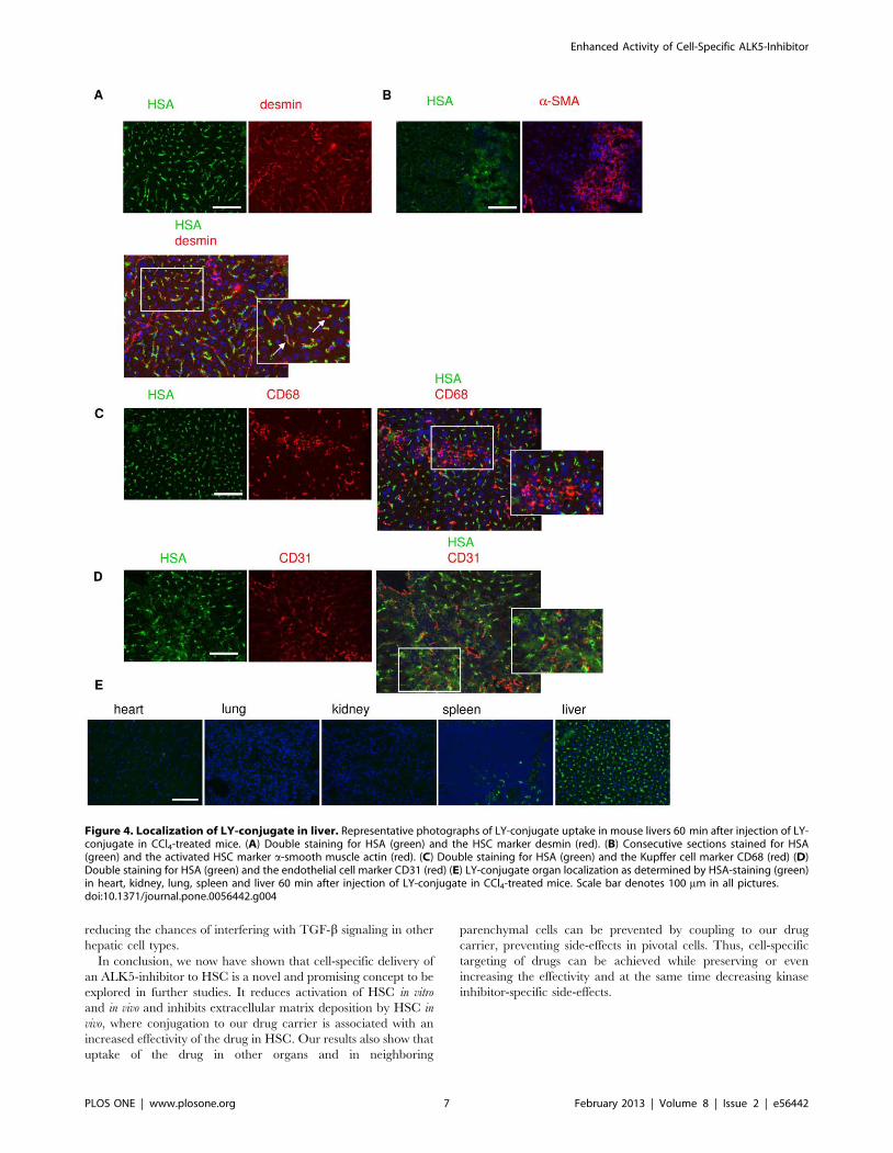
Biodistribution of the LY-conjugate in vivoWe furthermore examined whether the LY-conjugate accumu-
lated into the target cells in vivo. The intrahepatic distribution of
the LY-conjugate was determined 60 minutes after systemic
administration in CCl4-treated mice. The HSA-staining (green)
localized to desmin-positive HSC (red) in the liver (Fig. 4A). HSA
staining was also found in the same area as a-smooth muscle actin-
positive cells, a marker for activated HSC (Fig. 4B). There was no
co-localization of HSA (green) and CD68-positive (Kupffer) cells
(red) (Fig. 4C) or HSA (green) and CD31-positive endothelial cells
(Fig. 4D). No staining for HSA was observed at all in hepatocytes
(Fig. 4A–D). To demonstrate organ-specificity, we performed an
anti-HSA-staining in other major organs. The staining for HSA
showed no accumulation of LY-conjugate in heart, kidney or lung
60 min after injection, whereas there was a small amount present
in the spleen (Fig. 4E), where it was confined to the marginal zone.
No staining was found in the red or white pulp.
Enhanced Activity of Cell-Specific ALK5-Inhibitor
PLOS ONE | www.plosone.org 3 February 2013 | Volume 8 | Issue 2 | e56442

Effects of the LY-conjugate in vivoEffectivity of the free drug and the HSC-specific conjugate was
subsequently examined in vivo in an acute CCl4-induced liver
injury model. We tested two different doses of unmodified LY-
364947 and equimolar doses of LY-conjugate. Collagen I
expression, as assessed by western blot (Fig. 5A) was significantly
decreased by both the low and the high doses of conjugate, while
the free drug had less effect on collagen I expression. Stainings for
other extracellular matrix molecules showed further differences
between free drug and conjugate. Deposition of both collagen III
and fibronectin was significantly inhibited by the high dose of LY-
conjugate but not by the free drug (Fig. 5B & C). These effects
were not due to a difference in CCl4-induced damage, since all
treatment groups displayed a similar amount of damage, as
reflected by the PAS-staining and ALT and AST levels (data not
shown). The carrier alone did not affect collagen deposition levels
(Fig. S1). As the acute liver injury model also causes considerable
inflammation in the liver, we examined the effects of the
treatments on liver inflammatory cells, but found no effects on
influx of T-cells or activation of resident macrophages (data not
shown). The reduction in collagen deposition induced by our
conjugate was therefore not caused by an effect on immune cells.
In order to study whether these reductions in extracellular
matrix deposition coincided with a decrease in TGF-b-induced
pro-fibrotic cytokines, mRNA levels of the downstream mediator
CTGF were measured in mice livers. Both free LY-364947 and
the HSC-specific conjugate significantly reduced CTGF mRNA
levels in liver (Fig. 5D). Immunohistochemical stainings showed
that CTGF expression was localized in the portal areas, and was
strongly inhibited by the conjugate, but not by free LY-364947
(Fig. 5D).
Discussion
In the present study, we demonstrated that local inhibition of
TGF-b receptor type I (ALK5) in HSC using our cell-specific
targeting approach in vivo strongly inhibits early liver fibrogenesis.
Selective inhibition of ALK5 in HSC is of high interest as
prolonged ALK5 inhibition elsewhere in the body or even in other
cell types in the liver may induce severe adverse effects, such as
cardiac problems, tumorigenesis or immune system deregulation.
To achieve cell-selective delivery, we conjugated ALK5 inhibitor
LY-364947 to HSC-targeting carrier M6PHSA. The LY-conju-
gate specifically accumulated into the target cells in vitro and in vivo.
Figure 1. Synthesis and characterization of LY-364947-ULS-M6PHSA. (A) Structure of LY-364947. (B) HPLC analysis of LY-conjugate: free LY-364947 (upper panel), LY-conjugate without treatment to release drug (middle panel) and LY-conjugate after treatment with 200 mM sodiumdithiocarbamate to release the drug from the carrier (lower panel).doi:10.1371/journal.pone.0056442.g001
Enhanced Activity of Cell-Specific ALK5-Inhibitor
PLOS ONE | www.plosone.org 4 February 2013 | Volume 8 | Issue 2 | e56442

Within HSC, it blocked the ALK5 pathway and induced a strong
anti-fibrogenic effect compared to equivalent doses of the free
drug. These data show that selective blocking of ALK5 in HSC
may result in a cell-specific therapeutic strategy.
Experimental drugs that were very effective in vitro or in
experimental animal models have often failed to be effective in
subsequent studies [20]. Exploration of drug effects after a cell-
specific approach might explain why drugs fail to have the
expected effect. Failure in a (pre)-clinical setting may be caused by
several factors, ranging from impaired delivery in diseased tissue to
dose-limiting side-effects, and these factors can be modulated by a
cell-specific delivery approach. If targeted drugs are not effective,
the target pathway within the target cell is of minor importance.
Here we have shown that TGF-b-signaling via the ALK5-receptor
in HSC is of great importance in early liver fibrogenesis.
In the current study we have demonstrated specific targeting of
the LY-conjugate to HSC both in vitro and in vivo. In vitro, the LY-
conjugate was taken up by the primary rat HSC, while blocking of
the uptake with a specific antibody showed the specificity to the
receptor. The conjugate was fully biologically active as it inhibited
the spontaneous activation of primary HSC and it reduced Smad
2/3 signaling profoundly. Even though the conjugate was proven
to be active in HSC, it did not inhibit Smad phosphorylation in
the most abundantly present cell type in liver, hepatocytes, which
is consistent with the fact that the conjugate did not bind to these
cells. The fact that there is no effect on TGF-b signaling in
hepatocytes, nor binding (in vitro or in vivo) implies a reduced risk of
pro-tumorigenic effects [21] of targeted TGF-b inhibition, which is
particularly relevant in hepatocytes that reside in the pro-
tumorigenic fibrotic environment.
In vivo, specific localization of the conjugate in HSC but not in
other cell types in the liver or in other organs, as demonstrated by
double immunofluorescent staining, revealed the cell-specific
accumulation of the conjugate. It was not possible to directly
measure concentrations of this drug within the liver after
treatment due to rapid metabolism of the released drug, but
previous studies with a similar kinase inhibitor-conjugate have
shown up to 76 higher levels of drug in the liver after treatment
with conjugate as compared to treatment with free drug [22].
Furthermore, studies in kidney fibrosis using the same drug and
linker have shown sustained high levels of drug within the target
organ [15]. Therefore it is probable that increased efficacy of this
conjugate in vivo is mainly due to its more favorable pharmaco-
kinetic profile. The targeting method thus leads to a high
accumulation in the target cell, increasing effectivity compared
to an equimolar dose of free drug.
In vivo the targeted ALK5-inhibitor significantly reduced the
deposition of extracellular matrix constituents, that is, collagen I
and III and fibronectin. Previous experiments have shown no
effects of the M6PHSA carrier in this in vivo model [22], so the
Figure 2. In vitro effects of LY-conjugate. (A) Collagen deposition by HSC incubated with LY-conjugate (equivalent to 10 mM free drug), LY-364947 (10 mM) or carrier (molar equivalent). Cells were stained for collagen I and III. Scale bar denotes 100 mm. (B) Effect of LY-conjugate (equivalentto 10 mM free drug), LY-364947 (10 mM) and carrier (molar equivalent) on the fibrotic markers a-SMA and collagen 1A1 in isolated rat HSCs after 48 hincubation. * p,0.05 vs. control by Student’s t-test. (C) LY-conjugate and LY-364947 reduce luciferase expression in mink epithelial cells with a SBE-Luc reporter. *** p,0.001 vs. TGF-b1 by Student’s t-test.doi:10.1371/journal.pone.0056442.g002
Enhanced Activity of Cell-Specific ALK5-Inhibitor
PLOS ONE | www.plosone.org 5 February 2013 | Volume 8 | Issue 2 | e56442

anti-fibrotic effects are due to the targeted ALK5-inhibitor.
Furthermore, the expression of the TGF-b dependent cytokine
CTGF was also inhibited by the conjugate, indicating a TGF-b-
inhibiting activity of the conjugate. Immunohistochemistry showed
that CTGF protein expression was localized near portal tracts,
most likely within the portal tract fibroblasts, as found in earlier
studies [23]. This CTGF protein expression was reduced by the
HSC-specific ALK5-inhibitor, but not by free drug, possibly
reflecting uptake and pharmacological effects of our conjugate in
the portal fibroblasts as well.
Inhibition of ALK5 has been shown to be a valuable antifibrotic
strategy in animal models for fibrosis in different organs
[5,15,24,25], since TGF-b plays a crucial role in most fibrotic
diseases. Despite the anti-fibrotic effects of TGF-b inhibitors, their
use is considered unsafe due to critical side-effects [7,21,26,27,28].
Since the ALK5 is expressed ubiquitously nearly on all cell types,
the inhibition of this receptor may induce many effects. Small
molecule ALK5-inhibitors have been shown to cause heart valve
lesions in animal models [28]. TGF-b is also known to be an
important regulator of the immune system, as mice lacking TGF-
b1 die from a multi-organ inflammatory syndrome [7]. Deregu-
lation of the immune system in immune-compromised cirrhotic
patients or patients with viral hepatitis poses a risk for the patient.
Furthermore, TGF-b is a suppressor of early tumor growth [21].
Pre-clinical evidence suggests that inhibition of ALK5 in rats
predisposed to developing renal cell carcinoma may elicit tumor
development [29]. Since cirrhosis patients are at a higher risk for
hepatocellular carcinoma [30] the use of ALK5-inhibitors for the
treatment of liver fibrosis might therefore pose an extra risk. In this
study we showed that there is no effect of the targeted conjugate in
hepatocytes or uptake of the conjugate in Kupffer cells, thus
Figure 3. Binding of LY-conjugate and effect in hepatocytes. (A) HSA staining showing the binding of LY-conjugate to HSC: control cells (left),LY-conjugate-incubated cells (middle), and LY-conjugate-incubated cells pretreated with a M6P/IGFII receptor-specific antibody (right). Note thatblocking of the receptor reduces binding of the conjugate to the cells. Scale bar denotes 100 mm. (B) HSA staining showing the binding of LY-conjugate to HepG2 cells: control cells (left), LY-conjugate incubated cells (right). (C) TGF-b1-induced phosphorylation of Smad2 in HepG2 cells and inHSC after incubation with LY-364947, conjugate, carrier or HSA. Representative western blots and quantitative analysis of blot density (n = 3),* p,0,05 vs. TGF-b1, ** p,0.01 vs. TGF-b1 by Student’s t-test.doi:10.1371/journal.pone.0056442.g003
Enhanced Activity of Cell-Specific ALK5-Inhibitor
PLOS ONE | www.plosone.org 6 February 2013 | Volume 8 | Issue 2 | e56442
reducing the chances of interfering with TGF-b signaling in other
hepatic cell types.
In conclusion, we now have shown that cell-specific delivery of
an ALK5-inhibitor to HSC is a novel and promising concept to be
explored in further studies. It reduces activation of HSC in vitro
and in vivo and inhibits extracellular matrix deposition by HSC in
vivo, where conjugation to our drug carrier is associated with an
increased effectivity of the drug in HSC. Our results also show that
uptake of the drug in other organs and in neighboring
parenchymal cells can be prevented by coupling to our drug
carrier, preventing side-effects in pivotal cells. Thus, cell-specific
targeting of drugs can be achieved while preserving or even
increasing the effectivity and at the same time decreasing kinase
inhibitor-specific side-effects.
Figure 4. Localization of LY-conjugate in liver. Representative photographs of LY-conjugate uptake in mouse livers 60 min after injection of LY-conjugate in CCl4-treated mice. (A) Double staining for HSA (green) and the HSC marker desmin (red). (B) Consecutive sections stained for HSA(green) and the activated HSC marker a-smooth muscle actin (red). (C) Double staining for HSA (green) and the Kupffer cell marker CD68 (red) (D)Double staining for HSA (green) and the endothelial cell marker CD31 (red) (E) LY-conjugate organ localization as determined by HSA-staining (green)in heart, kidney, lung, spleen and liver 60 min after injection of LY-conjugate in CCl4-treated mice. Scale bar denotes 100 mm in all pictures.doi:10.1371/journal.pone.0056442.g004
Enhanced Activity of Cell-Specific ALK5-Inhibitor
PLOS ONE | www.plosone.org 7 February 2013 | Volume 8 | Issue 2 | e56442

Enhanced Activity of Cell-Specific ALK5-Inhibitor
PLOS ONE | www.plosone.org 8 February 2013 | Volume 8 | Issue 2 | e56442

Supporting Information
Figure S1 M6PHSA carrier does not affect collagendeposition in livers of CCl4 mice. Representative pictures of
immunohistochemical stainings for collagen I and collagen III on
liver sections of C57Bl/6 mice, after one injection of CCl4 and
treated with M6PHSA carrier. Original magnification 4006.
(TIF)
Acknowledgments
Dr. L.E. Deelman (Dept. Clinical Pharmacology, University of Groningen)
is kindly acknowledged for providing mink cells.
Author Contributions
Conceived and designed the experiments: MMVB JP LB KP. Performed
the experiments: MMVB ML EP CRS. Analyzed the data: MMVB JP LB
KP. Contributed reagents/materials/analysis tools: JP ML. Wrote the
paper: MMVB JP KP.
References
1. Lee U, Friedman S (2011) Mechanisms of hepatic fibrogenesis. Best Pract Res
Clin Gastroenterol 25(2): 195–206.
2. Derynck R, Zhang YE (2003) Smad-dependent and smad-independent
pathways in TGF-ß family signaling. Nature 425: 577–584.
3. Zhang YE (2009) Non-smad pathways in TGF-beta signaling. Cell Res 19(1):
128–139.
4. de Gouville AC, Huet S (2006) Inhibition of ALK5 as a new approach to treat
liver fibrotic diseases. Drug News Perspect 19(2): 85–90.
5. de Gouville AC, Boullay V, Krysa G, Pilot J, Brusq JM, et al. (2005) Inhibition of
TGF-beta signaling by an ALK5 inhibitor protects rats from dimethylnitrosa-
mine-induced liver fibrosis. Br J Pharmacol 145(2): 166–177.
6. Arribillaga L, Dotor J, Basagoiti M, Riezu-Boj J, Borrs-Cuesta F, et al. (2011)
Therapeutic effect of a peptide inhibitor of TGF-b on pulmonary fibrosis.
Cytokine 53(3): 327–333.
7. Prud’homme GJ (2007) Pathobiology of transforming growth factor ß in cancer,
fibrosis and immunologic disease, and therapeutic considerations. Laboratory
Investigation 87: 1077–1091.
8. Beljaars L, Molema G, Weert B, Bonnema H, Olinga P, et al. (1999) Albumin
modified with mannose 6-phosphate: A potential carrier for selective delivery of
antifibrotic drugs to rat and human hepatic stellate cells. Hepatology 29(5):
1486–1493.
9. Gressner AM, Weiskirchen R (2006) Modern pathogenetic concepts of liver
fibrosis suggest stellate cells and TGF-beta as major players and therapeutic
targets. J Cell Mol Med 10(1): 76–99.
10. Gressner AM, Weiskirchen R, Breitkopf K, Dooley S (2002) Roles of TGF-beta
in hepatic fibrosis. Front Biosci 7: 793–807.
11. Gressner OA, Gressner AM (2008) Connective tissue growth factor: A fibrogenic
master switch in fibrotic liver diseases. Liver Int 28(8): 1065–1079.
12. de Bleser P, Jannes P, van Buul-Offers SC, Hoogerbrugge CM, van
Schravendijk CFH, et al. (1995) Insulinlike growth factor-II/Mannose 6-
phosphate receptor is expressed on CCl4-exposed rat fat-storing cells and
facilitates activation of latent transfroming growth factor-ß in cocultures with
sinusoidal endothelial cells. Hepatology 21: 1429–1437.
13. Greupink R, Bakker HI, van Goor H, de Borst MH, Beljaars L, et al. (2006)
Mannose-6-phosphate/insulin-like growth factor-II receptors may represent a
target for the selective delivery of mycophenolic acid to fibrogenic cells. Pharm
Res 23(8): 1827–1834.
14. Ghosh P, Dahms NM, Kornfeld S (2003) Mannose 6-phosphate receptors: New
twists in the tale. Nat Rev Mol Cell Biol 4: 202–212.
15. Prakash J, de Borst MH, van Loenen-Weemaes AM, Lacombe M, Opdam F, et
al. (2008) Cell-specific delivery of a transforming growth factor-beta type I
receptor kinase inhibitor to proximal tubular cells for the treatment of renal
fibrosis. Pharm Res 25(10): 2427–2439.
16. Beljaars L, Olinga P, Molema G, de Bleser P, Geerts A, et al. (2001)Characteristics of the hepatic stellate cell-selective carrier mannose 6-phosphate
modified albumin (M6P28-HSA). Liver 21: 320–328.17. Geerts A, Niki T, Hellemans K, De Craemer D, Van Den Berg K, et al. (1998)
Purification of rat hepatic stellate cells by side scatter-activated cell sorting.Hepatology 27(2): 590–598.
18. Hageman J, Eggen B, Rozema T, Damman K, Kampinga H, et al. (2005)
Radiation and transforming growth factor-beta cooperate in transcriptionalactivation of the profibrotic plasminogen activator inhibitor-1 gene. Clinical
Cancer Research 11: 5956–5964.19. Jonk LJ, Itoh S, Heldin CH, ten Dijke P, Kruijer W (1998) Identification and
functional characterization of a smad binding element (SBE) in the JunB
promoter that acts as a transforming growth factor-beta, activin, and bonemorphogenetic protein-inducible enhancer. Journal of Biological Chemistry 273:
21145–21152.20. Pinzani M (2005) Fibrosis in chronic liver diseases: Diagnosis and management.
J Hepatol 42 Suppl 1(1): 22–36.21. Lahn M, Kloeker S, Berry B (2005) TGF-beta inhibitors for the treatment of
cancer. Expert Opin Investig Drugs 14(6): 629–643.
22. van Beuge MM, Prakash J, Lacombe M, Post E, Reker-Smit C, et al. (2011)Increased liver uptake and reduced hepatic stellate cell activation with a cell-
specific conjugate of the rho-kinase inhibitor Y27632. Pharm Res (8): 2045–2054.
23. Sedlaczek N, Jia JD, Bauer M, Herbst H, Ruehl M, et al. (2001) Proliferating
bile duct epithelial cells are a major source of connective tissue growth factor inrat biliary fibrosis. Am J Pathol 158(4): 1239–1244.
24. Fu K, Corbley M, Sun L, Friedman J, Shan F, et al. (2008) SM16, an orallyactive TGF-beta type I receptor inhibitor prevents myofibroblast induction and
vascular fibrosis in the rat carotid injury model. Arterioscler Thromb Vasc Biol28(4): 665–671.
25. Higashiyama H, Yoshimoto D, Kaise T, Matsubara S, Fujiwara M, et al. (2007)
Inhibition of activin receptor-like kinase 5 attenuates bleomycin-inducedpulmonary fibrosis. Exp Mol Pathol 83(1): 39–46.
26. Dooley S, Weng H, Mertens P (2009) Hypotheses on the role of transforminggrowth factor-beta in the onset and progression of hepatocellular carcinoma.
Digestive Diseases 27(2): 93–101.
27. Ling L, Lee WC (2011) TGF-beta type I receptor (Alk5) kinase inhibitors inoncology. Curr Pharm Biotechnol 12: 2202.
28. Anderton M, Mellor H, Bell A, Sadler C, Pass M, et al. (2011) Induction of heartvalve lesions by small-molecule ALK5 inhibitors. Toxicol Pathol 39(6): 916–924.
29. Laping N, Everitt J, Frazier K, Burgert M, Portis M, et al. (2007) Tumor-specific
efficacy of transforming growth factor-beta RI inhibition in eker rats. ClinCancer Res 13(10): 3087–3099.
30. Sherman M (2010) Hepatocellular carcinoma: New and emerging risks. DigLiver Dis 42 Suppl 3: S215–S222.
Figure 5. LY-conjugate reduces collagen deposition in livers of CCl4 mice. (A) Western blot analysis of collagen I expression in livers ofC57Bl/6 mice, after one injection of CCl4 and treated with vehicle (PBS), LY-conjugate (low and high dose) or LY-364947 (low and high dose). Figuresshow representative blots and quantitative analysis of western blots, n = 3–4 per group. * p,0.05 vs. CCl4-PBS by one way ANOVA with Bonferronipost-hoc test. (B) Representative pictures and quantitation of immunohistochemical stainings for collagen III on liver sections of C57Bl/6 mice, afterone injection of CCl4 and treated with vehicle (PBS), LY-conjugate (low and high dose) or LY-364947 (low and high dose). Stainings were quantitatedusing the Cell D software, calculating the total stained area in 18–24 fields per section at 1006magnification as a percentage of the stained area inthe control CCl4 sections. Data shown are the mean of 3–4 animals per group. * p,0.05 vs. CCl4-PBS by Student’s t-test. (C) Representative picturesand quantitation of immunohistochemical stainings for fibronectin on liver sections of C57Bl/6 mice, after one injection of CCl4 and treated withvehicle (PBS), LY-conjugate (low and high dose) or LY-364947 (low and high dose). Quantitation of the relative area stained positive for fibronectinwas performed as described above. (D) Representative pictures of immunohistochemical staining for connective tissue growth factor on livers ofC57Bl/6 mice, after one injection of CCl4 and treated with vehicle (PBS), LY-conjugate (high dose) or LY-364947 (high dose). Original magnification4006. Expression levels of connective tissue growth factor mRNA in livers of C57Bl/6 mice, after one injection of CCl4 and treated with vehicle (PBS),LY-conjugate (low and high dose) or LY-364947 (low and high dose). * p,0.05 vs. CCl4-PBS by Student’s t-test.doi:10.1371/journal.pone.0056442.g005
Enhanced Activity of Cell-Specific ALK5-Inhibitor
PLOS ONE | www.plosone.org 9 February 2013 | Volume 8 | Issue 2 | e56442