Embed Size (px)
Citation preview
From DEPT OF CLINICAL NEUROSCIENCE Karolinska Institutet, Stockholm, Sweden
ADVANCES IN SPINAL CORD STIMULATION
ENHANCEMENT OF EFFICACY, IMPROVED SURGICAL TECHNIQUE
AND A NEW INDICATION
Göran Lind
Stockholm 2012

All previously published papers were reproduced with permission from the publisher. Published by Karolinska Institutet. Printed by Larserics Digital Print AB, Bromma © Göran Lind, 2012 ISBN 978-91-7457-938-3

ABSTRACT Introduction and aim: Spinal cord stimulation (SCS) has been used for treatment of
otherwise therapy-resistant chronic neuropathic pain for about four decades. However, 30-40 % of the patients do not benefit from SCS, despite careful case selection and technical advances. In search of ways to improve the outcome mechanisms underlying the pain relieving effect of SCS have been extensively explored. Experimental findings suggest a possibility to enhance the effect of SCS by concomitant intrathecal (i.t.) administration of pharmaceuticals, such as baclofen, clonidine and adenosine.
Animal research has indicated that hypersensitivity to colonic dilatation can be attenuated by SCS. This finding, as well as related clinical observations, forms a basis for the possibility of treating irritable bowel syndrome (IBS) with SCS.
Implantation of an SCS system with a plate electrode requires extensive surgery. This can be painful and cumbersome for the patient, since finding an optimal electrode position demands patient cooperation with reporting of stimulation evoked sensations.
Aims of the thesis were to study: 1) if co-administration of baclofen (Study I and III), clonidine (Study III) or adenosine (Study I) can enhance the effect of SCS, 2) if long-term i.t. administration of a drug will continue to support the effect of SCS over time (Study II), 3) if implantation of plate electrodes can be performed in spinal anesthesia, retaining the possibility for the patient to feel and report stimulation evoked paresthesias and 4) if SCS can be used as a treatment option for IBS, otherwise resistant to therapy.
Methods: In Study I, 43 patients with neuropathic pain either experiencing diminished effect of previously efficacious SCS or with insufficient initial effect of SCS were recruited for trials of bolus i.t. injections of baclofen. Patients responding to the addition of baclofen were offered continued administration either i.t., via an implanted pump, or orally. Seven patients were also tested with i.t. adenosine. In Study II, the patients who continued with i.t. baclofen via a pump were assessed for long-term results. In Study III, 10 neuropathic pain patients with insufficient effect of SCS were recruited for a randomized double-blind trial, with i.t. injections of baclofen, clonidine and placebo. In Study IV, results from 20 implantations of plate electrodes in spinal anesthesia are reported. In Study V, 10 patients with IBS participated in a study of SCS, comparing randomly assigned periods of active stimulation versus a period without stimulation.
Results: In Study I, 20 patients responded to i.t. baclofen, with or without SCS. Three patients tested oral baclofen as an adjunct to SCS, but terminated treatment due to side effects. Eleven patients had pumps implanted, two of which were explanted during the trial period. Two patients opted for i.t. adenosine delivery via a pump, but discontinued due to side effects. In Study II, it was confirmed that all 9 patients with remaining working pumps continued to benefit from the therapy, albeit with a dose increase. In Study III, 5 patients responded to either baclofen or clonidine and 4 received pumps for i.t. delivery (2 baclofen, 2 clonidine). In Study IV, it was demonstrated that in all 20 implantations it was possible to perform successful intra-operative testing in spinal anesthesia. In Study V, 6 out of 9 patients responded beneficially to SCS as a treatment for IBS (1 patient left the study).
Conclusions: I.t. medication with baclofen or clonidine can enhance the effect of SCS. This enhancement remains over a long-term follow up. Implantations of plate electrodes can be performed with intra-operative testing in spinal anesthesia. SCS may alleviate pain in IBS, but studies in larger patient materials are needed to investigate effects on other IBS symptoms.
Key words: spinal cord stimulation, neuropathic pain, baclofen, clonidine, adenosine, intrathecal medication, IBS

LIST OF PUBLICATIONS The thesis is based on the following papers, which will be referred to in the
text by the roman numerals as given below.
I Lind, G., Meyerson, B. A., Winter, J., Linderoth, B., Intrathecal baclofen as adjuvant therapy to enhance the effect of spinal cord stimulation in neuropathic pain: a pilot study. Eur J Pain, 2004. 8(4): p. 377-83.
II Lind, G., Schechtmann, G., Winter, J., Meyerson, B. A., Linderoth, B., Baclofen-enhanced spinal cord stimulation and intrathecal baclofen alone for neuropathic pain. Long-term outcome of a pilot study. Eur J Pain, 2007. 12(1): p. 132-6.
III Schechtmann, G., Lind, G., Winter, J., Meyerson, B. A., Linderoth, B., Intrathecal clonidine and baclofen enhance the pain-relieving effect of spinal cord stimulation: a comparative placebo-controlled, randomized trial. Neurosurgery, 2010. 67(1): p. 173-81.
IV Lind, G., Meyerson, B. A., Winter, J., Linderoth, B., Implantation of laminotomy electrodes for spinal cord stimulation in spinal anesthesia with intraoperative dorsal column activation. Neurosurgery, 2003. 53(5): p. 1150-3; discussion 1153-4.
V Lind, G., Winter, J., Linderoth, B., Hellström, P.M., Spinal cord stimulation in the irritable bowel syndrome – a randomized cross-over study. (Submitted)

CONTENTS 1 Introduction ......................................................................................... 1
1.1 Pain and pain assessment .......................................................... 1 1.1.1 Classification of pain ....................................................... 1 1.1.2 Assessment of pain .......................................................... 3
1.2 Spinal cord stimulation – background ...................................... 5 1.3 Spinal cord stimulation – indications ....................................... 7
1.3.1 Present established indications ....................................... 8 1.3.2 Exploratory and experimental use of SCS .................... 11
1.4 Spinal cord stimulation – technique ....................................... 11 1.4.1 Implantation techniques ................................................ 12 1.4.2 Electrode and stimulator design .................................... 13 1.4.3 Stimulation parameters .................................................. 15 1.4.4 Computer modeling ....................................................... 15
1.5 Spinal cord stimulation – results ............................................. 16 1.6 Spinal cord stimulation – mechanisms of action. ................... 17
1.6.1 Human studies ............................................................... 18 1.6.2 Animal studies ............................................................... 18
2 Aims of the thesis ............................................................................. 24 2.1 Studies I-III: Pharmacological enhancement of SCS effect .. 24 2.2 Study IV: Technical improvement of SCS ............................. 24 2.3 Study V: New indication for SCS ........................................... 24
3 Materials and methods ...................................................................... 25 3.1 Patients .................................................................................... 25
3.1.1 Study I ............................................................................ 25 3.1.2 Study II .......................................................................... 25 3.1.3 Study III ......................................................................... 25 3.1.4 Study IV ......................................................................... 25 3.1.5 Study V .......................................................................... 26
3.2 Equipment ............................................................................... 26 3.2.1 Lumbar puncture ........................................................... 26 3.2.2 Implants ......................................................................... 26 3.2.3 Pharmaceutical agents ................................................... 28
3.3 Study outlines .......................................................................... 31 3.3.1 Study I ............................................................................ 31 3.3.2 Study II .......................................................................... 31 3.3.3 Study III ......................................................................... 31 3.3.4 Study IV ......................................................................... 33 3.3.5 Study V .......................................................................... 33
4 Results ............................................................................................... 35 4.1 Study I ..................................................................................... 35 4.2 Study II .................................................................................... 35 4.3 Study III ................................................................................... 35 4.4 Study IV ................................................................................... 36 4.5 Study V .................................................................................... 36
5 Discussion ......................................................................................... 37

5.1.1 Pharmacological enhancement of SCS effects ............. 37 5.1.2 Technical improvement ................................................ 41 5.1.3 New indication – IBS – ................................................. 43
6 Conclusions ...................................................................................... 45 7 Acknowledgements .......................................................................... 46 8 References ........................................................................................ 48

LIST OF ABBREVIATIONS
Ach Acetylcholine CGRP Calcitonin gene-related peptide CLI Critical limb ischemia CMM Conventional medical management CRPS Complex regional pain syndrome CSF Cerebrospinal fluid DH Dorsal horn EEG Electroencephalography FBSS Failed back surgery syndrome FDA Federal Drug Administration fMRI Functional magnetic resonance imaging i.t. Intra-thecal/intra-thecally i.v. Intra-venous IASP International Association for the Study of Pain IBS Irritable bowel syndrome IPG Implantable pulse generator L Lumbar MRI Magnetic resonance imaging NMDA N-methyl-d-aspartate (receptor) NRS Numeric rating scale O.D. Outer diameter PET Positron emission tomography PGIC Patient’s Global Impression of Change PROCESS Prospective RandOmised Controlled multicentre trial of the
Effectiveness of Spinal cord Stimulation PT Physiotherapy QOL Quality of life SCS Spinal cord stimulation SEP Somatosensory evoked potentials T Thoracic TCD Transcranial doppler flowmetry tcpO2 Transcutaneous oxygen partial pressure TENS Transcutaneous electrical nerve stimulation TMS Transcranial magnetic stimulation VAS Visual analogue scale VMR Visceromotor response VRS Verbal Rating Scale WDR Wide dynamic range neurons


1
1 INTRODUCTION Spinal cord stimulation (SCS) has been in clinical use for about four
decades. It has evolved as a useful, minimally invasive, cost-efficient and reversible therapy for certain forms of chronic pain, when pharmacological treatment has failed. SCS requires operative implantation of a stimulating electrode connected to a subcutaneous pulse generator. The individual patient can turn the stimulation on and off at will and adjust the stimulation intensity. Stimulation is accompanied by a tingling sensation, paresthesia.
For some pain indications the evidence for the SCS efficacy is satisfactory. The usage is wide-spread with, at present, about 30,000 implants performed globally each year199.
Many patients with severe pain, however, do not benefit from SCS, despite adequate indication and implant technique. It is important that we advance the knowledge of the mechanisms of action of SCS to enable the development of methods for improving the outcome.
1.1 PAIN AND PAIN ASSESSMENT
Pain is the result of a process of utmost importance for survival, namely the swift recognition and prompt reaction to potentially harmful influences on the body. The process itself is called nociception. Pain and nociception are, however, not identical entities. Nociception is a physiological process whilst pain is a phenomenon experienced by the sufferer. Pain can occur without nociception and nociception does not always result in pain, as can be the case when a patient uses an analgetic. Pain is described by the International Association for the Study of Pain (IASP) as: “an unpleasant sensory and emotional experience associated with actual or potential tissue damage, or described in terms of such damage”140.
Pain is the most common symptom among health-care seekers. In a Canadian survey ⅔ of the patients presenting at the emergency department had pain146 and in a Finnish primary health care study pain was recognized as the reason for 40 % of the visits238.
1.1.1 Classification of pain
The notion of pain comprises different subtypes. The most obvious subdivision of pain is based on time: acute and chronic pain. There is no universally accepted definition of these terms, but a classification of pain lasting less than 3 months “acute” and pain lasting more than three months “chronic” has been proposed166, as well as several other definitions.
Another subdivision of pain relates to the underlying mechanism believed to constitute the background for each form of pain. The kind of pain that arises from stimulation of nociceptors, the receptors specifically activated by noxious stimuli (i.e. an actually or potentially tissue damaging event), is called nociceptive pain. This type of pain is defined by IASP simply as “pain arising from activation of nociceptors”202. It is, however, clear that not all pain can be attributed to nociception, as many instances of pain do not have a demonstrable

2
nociceptive background. An illustrative example is phantom limb pain, which is independent of nociceptor activation. This is a type of non-nociceptive pain that is denoted as neuropathic. IASP has defined this as “pain initiated or caused by a primary lesion or dysfunction in the nervous system”. Recently a somewhat different and more restrictive definition has been proposed: “pain arising as a direct consequence of a lesion or disease affecting the somatosensory system”341. Even though this definition is not the one used in the publications of this thesis it would not have had any impact on patient recruitment for the studies presented, i.e. none of the patients with neuropathic pain involved in the studies would have been reclassified as not having neuropathic pain if the new definition had been applied.
Nociceptive and neuropathic pain are not necessarily mutually exclusive and may coexist. Nevertheless, it is of utmost importance to adequately analyze pain and to identify its nature and different components, because treatment options differ depending on the type of pain.
Both the terms neuropathic and nociceptive are generalized terms incorporating many subclassifications. Nociceptive pain may be subdivided depending on the character of the noxious stimulus, such as ischemic or inflammatory pain. Neuropathic pain may be of central or peripheral origin, and several other subdivisions exist, many of which have separate definitions in the IASP taxonomy140. Instead of neuropathic the term neurogenic is sometimes used.
Other pain classifications that have been in use, but less frequently during the past decade, are psychogenic and idiopathic pain. Psychogenic pain would be used for pain associated with psychological or psychiatric factors, e.g. defined in “Bonica’s management of pain” as “report of pain attributable primarily to psychological factors usually in the absence of any objective physical pathology that could account for pain”344. The entity of psychogenic pain has been much questioned and distinguishing it from other forms of pain can be difficult336,337. Idiopathic as a medical term is used to describe an entity of obscure or unknown origin or spontaneous appearance. Idiopathic pain in that sense can be used as a term for pain of unknown origin. The term, however, has been much used for specific syndromes frequently accompanied with pain, where the pain component of the syndrome is less readily fully explained, such as temporomandibular joint disorders, chronic headaches, whiplash-associated disorders etc.59 Neither psychogenic pain nor idiopathic pain is presently included in the IASP pain taxonomy.
Other subdivisions of pain relate to the part of the body where the pain occurs, such as headache, abdominal pain, elbow pain, etc. Pain can also be classified according to its severity or from underlying pathology, such as rheumatological pain and cancer pain.
An important step in the field of pain classification is the ongoing process of developing a classification based on mechanisms. In an editorial in the journal “Pain” an expert group has proposed foundations for such a classification, with the hope that a mechanism-based classification will lead to better medical treatment of pain tailored to mechanisms361. They list three major categories: transient pain - pain as a response to a passing noxious stimulus that does not produce a lasting impact, such as a pin prick, tissue injury pain and nervous

3
system injury pain, with subclassifications based on underlying mechanisms. A non-mechanism based classification may lead to misinterpretation of symptoms and signs, for example allodynia can occur not only from nervous system injury but may appear with tissue injury as well361.
1.1.2 Assessment of pain
Since pain is an exclusively subjective entity its intensity and character can only be described by the sufferer. This is not easily performed (nor easily standardized) and Virginia Woolf described this in an often cited passage from her 1930 book On being ill:”let a sufferer try to describe a pain in his head to a doctor, and language runs at once dry".
In 1947 Hardy, Wolff and Goodell presented experiments on the perception of heat induced pain (with themselves as subjects)110. They claimed that in between the slightest stimulus perceived as pain and the ceiling intensity of stimulation (above which an increase in stimulation intensity did not lead to a more intense perception of pain) no more than 21 steps could be discriminated. They proposed the unit “dol”, corresponding to two such steps, as a measurement for pain. Furthermore, they devised an apparatus, the “dolorimeter”, which could be used for the purpose of producing a graded painful stimulus to a patient in pain so that he or she could report if the pain induced by the dolorimeter corresponded to the original pain (yielding a specified number of “dols”). The method was tried in some scientific studies but was soon abandoned118. Patients were hesitant to experience the pain twice and sometimes even hostile to the experimenters9.
Another early attempt to standardize reports on pain intensity was “The pain chart”, described by Keele in Lancet 1948157. The chart was set up with grades (0-4) corresponding to different pain intensities, each grade described by a defined word (“nil”, “slight", " moderate", " severe" and " agonising"). In the classic human experiments on pain-producing substances of Keele and Armstrong the subjects moved a pointer along a scale marked with these numbers (0-4) during the experiments, yielding a continuous analogue recording of perceived pain intensity156.
In 1965 a case of phantom limb pain was presented, where the patient had been instructed to repeatedly assess his pain by making a pencil mark on a 10 cm line with the label “No pain at all” on the right side and “As painful as it could possibly be” on the left side266. By simply measuring the distance from the right end of the line to the mark a “pain score” was acquired. The notion of using graphic representations for assessments was adopted from the social sciences and psychology, where it had been in use since the 1920ies88,120, and the idea of putting a mark on a 10 cm line between a verbal description of extremes and in millimeters measure the distance to produce a score was presented from the fields of psychology and psychiatry39. This method of assessing pain was soon adopted by many researchers and during the subsequent decade the phrase “Visual Analogue Scale” (abbreviated VAS) was adopted as a term for this graphic pain scale2,139,147,360. The VAS scale has subsequently proven to be one of the most common ways of reporting pain intensity, either transferring the scale to numeric values by measuring in mm (0-100) or in cm (0-10).
4
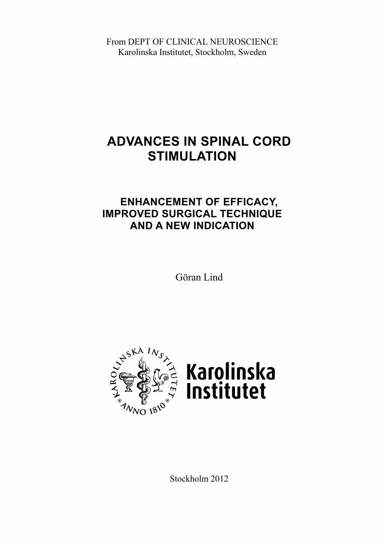
Other common scales for pain assessment are the Numeric Rating Scale (NRS) and the Verbal Rating Scale (VRS). VRS constitutes of a set number of verbal descriptors, each representing an increasingly intense pain and each corresponding to a number, e.g. 0-4 as in “The pain chart” mentioned previously. NRS is a scale, as the VAS, where only the extremes are anchored in words: 0 set to mean “no pain” and the highest number, be it 5, 10 or 100, corresponding to e.g. “worst pain imaginable”. NRS and VRS are frequently accompanied by a number indicating the total number of choices, e.g. NRS-11 (0-10), NRS-101 (0-100) or VRS-4 (such as “The pain chart”). In Figure 1 examples of VAS, NRS and VRS are given.
Numerous comparisons of these pain assessment scales have been published. It is evident that verbal rating cannot in an exact manner be transformed into numerical or visual rating or the opposite207, but nonetheless several studies indicate a fair correspondence between VAS and NRS78,269. In a recent comprehensive literature review of comparisons of VAS, VRS and NRS the authors conclude that:” the results show that NRS-11, VRS-7, or VAS all work quite well. … the most important choice is not the type of scale per se, but the conditions related to its use …”125. It should be noted that even though VAS, NRS and VRS may end up in figures that can be identical the methods are different. If a patient is asked to give a number to describe the intensity of pain the kind of scale used should be referred to as NRS and not VAS despite the fact that processing the data will yield similar figures. It is, however, not uncommon that inappropriate labeling of a scale is performed (and that in fact is also the case in some of the studies presented in this thesis).
A number of publications have been presented concerning changes in assessed pain, in order to determine the magnitude of change that is of importance for a patient. In absolute measures a change of 13 mm on the VAS
□ No pain □ Mild pain □ Moderate pain □ Severe pain □ Unbearable pain
VRS-5 Please indicate your present pain
PGIC-5
□ Much better □ Slightly better □ No change □ Slightly worse □ Much worse
Please indicate how your pain has changed since you entered the study
Worst pain imaginable
0 1 2 3 4 5 6 7 8 9 10
VAS Please mark your present pain
NRS-11 Please indicate your present pain
No pain Worst pain imaginable
No pain
Figure 1: Examples of commonly used pain scales

5
scale20,90,338 or of one unit in the NRS-11 scale289 has been proposed as a minimum requirement for an apparent true change. For a change of clinical significance, however, a relative change of 30 %76,144,289 or even 50 %84 has been advocated, based on clinical studies. There are also scales for assessing change per se and during the past decade many publications on pain have used Patient’s Global Impression of Change (PGIC) as a measurement of alteration of pain intensity76,289. The PGIC scale consists of verbal statements, frequently 5 or 7, where the patient is instructed to choose one to describe the change between to specified points in time (such as e.g. “since the start of the study until now”). The statements can be e.g. (for PGIC-7) ‘‘very much better’’, ‘‘much better’’, ‘‘slightly better’’, ‘‘no change’’, ‘‘slightly worse’’, ‘‘much worse’’ or ‘‘very much worse’’. One draw-back of this kind of scale can be that the patient may have difficulties in recalling a previous intensity of pain.
The aforementioned scales are all unidimensional, in that they solely measure the intensity of pain. There are also pain rating instruments taking other modalities into account. The McGill Pain Questionnaire, developed by Melzack at McGill University, is probably the one most commonly used222. The questionnaire allows for the patient to select a number of verbal descriptors related to pain (such as “dull”, ”vicious”, ”unbearable” – 102 words in total, subdivided in three classes and sixteen subclasses) apart from rating the pain intensity.
The VAS and NRS yield numbers and are by some viewed as ratio scales. Some studies concerning the distribution of VAS and NRS ratings support the claim that the scales are linear115,235,236, but this has been questioned265. These considerations are of importance for the choice of statistical analysis methods when applied for pain scales. Non-parametric statistical methods are presently generally recommended for such scales216,327, even though there are many publications where parametric statistics have been used (e.g.215).
1.2 SPINAL CORD STIMULATION – BACKGROUND
In the first issue of the journal “Pain” Kane and Taub give an interesting and commendable account of “A history of local electrical analgesia”148. Electrical stimulation has been used for the treatment of pain for thousands of years148. Natural sources of electricity seem to have been used, such as the torpedo ray (electric ray) or the Nile catfish. Even etymologically there is evidence of this since the greek word for torpedo ray is “narke”, meaning “numbing”, a word the root of which is used in narcosis148. The oldest written account of the use of electricity for pain dates back to the descriptions of Scribonius Largus, from around 46 A.D., on how the use of a live torpedo fish can ease headache or the pain of gout 148. Numerous accounts of the continued use of natural sources of electricity exist and since the 18th century apparatuses producing electricity came into use for medical purposes. There are many accounts of the use of electroanalgesia during the 19th century, even for anesthetic purposes during tooth extractions and surgical procedures, including amputations148. In the early 20th century a device for electrical stimulation, the “Electreat”, was marketed95. It had some resemblances to a modern TENS apparatus and was provided with a

6
small sign reading “Electreat relieves pain”. It has been estimated that about 300 000 of these devices were sold301.
The earliest attempts to treat pain with electrical stimulation by invasive techniques were performed with intracranial electrodes. During the late 50ies and early 60ies stimulation of the septal area was tried in a few patients99,121 as well as stimulation in the thalamus217. Thus Mazars used stimulation in the sensory thalamus to treat severe pain syndromes even before the advent of the gate theory, and this application was based on a considerably older theory launched by Head and Thompson199.
In 1965 Melzack and Wall presented a novel theory on the processing of pain, the “gate control theory of pain”, stipulating that pain, among other factors, is influenced by “the relative balance of activity in large versus small fibers”223. They suggested a new perspective to the understanding of pain transmission unifying the two prevailing, but mutually opposing, theories of pain transmission that had been debated during the early part of the 20th century158,223. One of them was called the specificity theory, originally proposed by von Frey in 1894, postulating that pain constitutes a modality all of its own with pain receptors and pain transmitting peripheral and central connections, in essence a direct line from pain impulse to the brain. The other theory was called the pattern or summation theory, suggested by Goldscheider, also in 1894, proposing that pain perception is the result of summation of impulses from receptors not by themselves primarily directed to react to pain. Neither of these theories could fully explain all features of how pain is experienced and during the 20th century modifications of these theories were brought forth158. The “gate control theory”, though much criticized199, has however, serve as an important foundation that has generated a wealth of pain research. In an editorial from 2001 Dickenson summarizes the impact of this theory by stating that the “Gate control theory of pain stands the test of time”60.
The “gate control theory” spurred the interest in electrical stimulation for pain relief. In January 1967 Wall and Sweet presented eight patients treated for pain with different ways of stimulation of peripheral nerves or nerve roots, either with implanted electrodes or electrodes on the skin surface347. Stimulation was performed with square-wave pulses at 100 Hz and a pulse width of 0.1 msec. The patients reported a tingling sensation and temporary abolition or reduction of pain. Later the same year Shealy et al proposed that a logical site for stimulation would be the spinal dorsal columns. They presented the results with 50 Hz electrical stimulation of the dorsal columns of the cervical cord in cats and later in 1967 the first use of such stimulation in a human subject302,303. The method evolved quickly and already in 1968 the first commercially available equipment for spinal cord stimulation was introduced96. During the first years transcutaneous electrical stimulation (TENS) was used for screening patients suitable for implantation of spinal cord stimulation (at that time referred to as dorsal column stimulation), but soon evolved into a successful pain treatment modality by itself205. It has been reported that Shealy initially used the Electreat for the purpose of screening patients for SCS, but soon commissioned a more versatile and reliable contemporary stimulator95,300,301.

7
1.3 SPINAL CORD STIMULATION – INDICATIONS
During the first decades of SCS the method was applied to a number of pain conditions as well as other pathologies, such as spasticity. Over time it became evident that not all types of pain responded to SCS and some forms of pain related to injury or malfunction of the nervous system – neuropathic pain - evolved as the main indications96. Already in one of the earliest surveys of SCS, by Nashold and Friedman, it was noted that pain at the site of the surgical wound for SCS implantation was not ameliorated even though the effect was excellent for the pain targeted for treatment240. One patient was reported to suffer an intercurrent bone fracture, the pain of which was not relieved by SCS241.
Observations of autonomic changes in patients treated with SCS lead Cook and coworkers to successfully try SCS for vascular disease of the extremities47. The same year Dooley and Kasprak, based on the fact that patients frequently reported a feeling of warmth accompanying SCS, examined the effect of SCS on the peripheral vasculature. They reported that SCS induced arterial dilatation and soon tried it for patients with extremity vascular disease64,65. The observation that SCS could be an effective treatment of peripheral vascular disease by improving circulation in the limbs5,27,38,136 spurred the interest in applying the method to other conditions of ischemia. After initial reports of beneficial effect on angina pectoris using TENS213, SCS was shown to be effective for the treatment of angina pectoris as well234. When used for treatment of ischemic conditions it is likely that the effect of SCS is not primarily a direct pain reducing effect, but rather produces pain relief secondary to reduction of tissue ischemia produced by the stimulation194,225.
Clinical studies of SCS have been hampered by the difficulty to blind the stimulation to the patient, because of stimulation-induced paresthesias. The fact that paresthesias are evoked during effective SCS is not only based on experience, but is also a natural result of orthodromic activation of dorsal column fibers, in accordance with the gate control theory. The necessity for paresthesias covering the painful area for a benificial effect was noted early in the evolution of SCS240. In a study on SCS effects on sensory modalities by Lindblom and Meyerson in 1975 it was noted that a few patients reported some pain reduction with stimulation intensities below the threshold for paresthesias, but the pain reduction was less than that obtained with paresthetic stimulation193. Cases with subthreshold SCS but still with marked increases of cutaneous blood flow, as demonstrated by thermography, were reported by Linderoth in 1995194, and in a recent case study on treatment of Raynaud’s phenomenon subthreshold stimulation appeared to have some effect17. Subthreshold stimulation has also been tested in two clinical trials of SCS for angina. One study showed equal effects of SCS on functional status and angina symptoms by paresthetic stimulation and a stimulation of 85 % of the intensity yielding paresthesias, but there was no effect of sham stimulation66. In another study, the paresthetic stimulation group had a significantly better outcome than both the sham stimulation group and the subliminal stimulation group185. In both these studies it was thus possible to apply a blinded stimulation design. On the other hand, it is in all cases necessary to ascertain the proper placement of the stimulating electrode as well as to determine the perception threshold with at

8
least a short stimulation session. Recently, a different stimulation paradigm has been presented in a small trial with “burst stimulation (40-Hz bursts with 5 spikes at 500 Hz per burst)”, which produced good pain relief without any subjective sensations55.
Numerous studies have reported on the value of SCS for a number of indications and during the past decades some prospective randomized, but non-blinded, controlled trials have been presented. The effectiveness of SCS has been demonstrated for pain associated with lumbosacral rhizopathy (often referred to as failed back surgery syndrome, “FBSS”)182,211,246, CRPS (type 1)160, limb ischemia145,170 and angina pectoris54,66,214,220.
1.3.1 Present established indications
1.3.1.1 Lumbosacral radicular pain (FBSS)
In a study, presented in 2005 by North et al., 50 patients with recurring or persistent radicular pain after lumbosacral spinal surgery were randomized either to reoperation or SCS246. Eligible patients had a history of one or several previous surgical spinal interventions and suffered from radicular pain exceeding or equal to their low back pain as well as radiological findings of nerve root compression. Patients were followed for three years and were allowed to cross-over to the other treatment arm if they considered the effect unsatisfactory. Patients randomized to SCS were at first subject to a trial stimulation, with permanent implantation provided that at least 50 % pain reduction was achieved. At three year follow-up, the SCS group had a significantly higher rate of participants achieving a pain reduction exceeding 50 % than that with reoperation (47 % and 12 % respectively). The opioid usage was significantly lower in the SCS group than in the reoperation group. Fifty-four % of the patients in the reoperation group chose to cross-over to reoperation whereas only 21% of the patients randomized to SCS opted for a reoperation.
In another study, the PROCESS study (Prospective Randomised Controlled Multicentre Trial of the Effectiveness of Spinal Cord Stimulation), 100 patients with persistent radicular pain after lumbar disc hernia surgery with successful anatomical result were randomized either to conventional medical management (CMM) or CMM and SCS. Patients were allowed to cross-over between treatments after 6 months. In this study as well, SCS patients started with a trial stimulation and were permanently implanted only if they experienced at least 50 % reduction of the leg pain. Results were reported after one year182, with the proportion of patients receiving ≥ 50 % pain relief at 6 month as the primary outcome, and again after two years follow-up181. 10 % of the SCS patients chose to cross over to CMM and 73 % of the CMM patients demanded to cross over to SCS. At 6 month follow-up (i.e. before any cross-over took place) 48 % of the SCS patients reported the primary outcome of at least 50 % pain reduction but only 9 % of the control group attained that outcome. At the two years follow-up the effects of SCS were sustained with a statistically significant difference between treatments both in a modified intention-to-treat analysis as well as in a final treatment analysis.

9
1.3.1.2 Complex Regional Pain Syndrome (CRPS)
The effects of SCS on CRPS (Complex Regional Pain Syndrome) have been explored in a Dutch study presented as a two years follow-up160 supplemented by a final five-year evaluation161. The term CRPS (Table 1) was introduced by IASP as an umbrella diagnosis containing conditions previously referred to as: reflex sympathetic dystrophy, algodystrophy, Sudeck’s dystrophy, causalgia, etc.. In the Dutch study 54 CRPS-1 patients (no patients with CRPS-2 were part of this study) were recruited and randomized in a 2:1 fashion to either SCS and physiotherapy (PT) or physiotherapy alone. At two years follow-up the reported pain intensity was significantly lower in the SCS group than in the PT group. However, at five years there was no longer any statistically detectable difference between the treatments (p=0.06), in an intention-to-treat analysis. It is worth noting that there had been cross-over between groups, with 22 % of the patients in the PT group crossing over to SCS and in total 9 % of patients lost to follow-up. Even at 5 years most SCS patients wanted to continue treatment and found it useful.
1.3.1.3 Limb ischemia
A number of prospective randomized studies have addressed the effect of SCS on limb ischemia. A meta-analysis of these studies can be retrieved from the Cochrane Collaboration database345. Six studies comprising almost 450 patients having chronic critical limb ischemia (CLI), deemed not treatable with arterial reconstruction, were evaluated. Patients had resting pain due to ischemia or ulcerations smaller than 3 cm in diameter or both and were at risk for amputation. For all studies the primary end-point was the limb-salvage rate at twelve months (amputation of a foot or higher was considered as non-limb salvage). The meta-analysis showed no significant difference between treatment groups concerning ulcer healing, but a pooled analysis disclosed a significantly higher limb-salvage rate for SCS-treatment as compared to conventional medical management. Pain measurements could not be pooled, but in several studies patients with SCS-treatment appeared to show significantly better pain scores145,319 and less analgesic medication than in the control group318. However, a highly significant pain reduction occurred after amputation in patients for
Diagnostic criteria for CRPS-1224 1) presence of an initiating noxious event or cause of immobilization 2) continuing pain, allodynia, or hyperalgesia with which the pain is disproportionate to the inciting event 3) evidence at some time of edema, changes in skin blood flow, or abnormal sudomotor activity in the region of pain 4) absence of conditions that would otherwise account for the degree of pain and dysfunction Diagnostic criteria for CRPS-2 For CRPS-2 there must be a, partial, nerve injury as well as the criteria for CRPS-1 Table 1: Diagnostic criteria for CRPS
10
whom that was inevitable, and pain relief was better for amputated patients than for non-amputated patients regardless of treatment318. In a more recently published non-randomized study, the long-term effect of SCS for CLI has been reported beyond the 12 months scope of the randomized trials. Eightyseven CLI patients were permanently implanted with SCS after an initial trial period with requirements of both substantial pain reduction and an increase of tcpO2 (transcutaneous oxygen partial pressure) in the foot. At an average of 48 months the beneficial effects of SCS remained and after the second year major amputations became infrequent93.
1.3.1.4 Angina pectoris
SCS treatment for angina pectoris has been used since 1987/1988212,234 and several randomized control studies have been performed. A concise review of these studies was presented in 200828, and there is a recent additional randomized control trial from 2010185. The evidence that SCS can improve quality of life, reduce number of angina attack and increase capacity on treadmill was classified as being on a high level. In the largest of the included studies with 104 patients, the Swedish ESBY-study (Electrical Stimulation versus Coronary Artery Bypass Surgery in Severe Angina Pectoris), SCS was compared to bypass surgery214. This study showed equal effects on angina symptoms for both study groups and a lower mortality in the SCS group.
1.3.1.5 Abdominal pain
Already in a publication from 1975 SCS treatment of a gastrointestinal ailment is described21. In a study of evoked potentials during SCS the 10 participating patients are presented in a table and one of them was treated for chronic pancreatitis with a “fair” result 21. In 1981 and 1982 an Italian group reported on SCS influence on colonic motility. Two patients, one with bifid spine and one with MS, had SCS applied at the T8/9 level for their neurological disorder, and they reported having a beneficial effect of SCS on their severe constipation as well263,264. Later on additional reports on gastrointestinal functions of SCS, both beneficial effects and unintended side effects, have appeared. Several publications cover treatment of different types of abdominal pain (such as chronic pancreatitis, post-surgical intra-abdominal adhesions and other forms of post-surgical visceral pain149), where at present over 70 patients have been treated successfully, in general after rigorous screening and testing149-
152,163. Additionally two cases of successful treatment of abdominal angina and mesenteric ischemia with SCS have been reported31,34. It might be that in these two cases the beneficial effects are the result of the same mechanisms that are responsible for the increased blood flow/diminished ischemia produced by SCS in other ischemic conditions. Some reports also include information about gastrointestinal side effects of SCS, also in situations where the electrodes were implanted at other locations than the mid-thoracic level where abdominal effects could be expected183,334. For example one patient who was successfully treated with cervical SCS for CRPS in the arm chose to have the system removed because the stimulation repeatedly evoked relapses of, pre-existing but until then mild, symptoms of ulcerative colitis159.
11
In the literature only four case reports on SCS applied for irritable bowel syndrome (IBS) are available. The first is from 2001 and SCS was without effect209. In 2004, on the basis of previously published animal studies102, Krames and Moussad reported on a single patient with highly successful SCS treatment for IBS173. Finally, in 2012 an additional two case reports with beneficial effects of SCS for IBS have been published253,272.
1.3.1.6 Other forms of pain
Pain after peripheral nerve injury is an important indication for SCS. Even though no RCTs have been performed it is seen as an indication with a high likelihood of success155,187,305.
SCS is also used for a number of other forms of neuropathic pain. This includes phantom limb pain230, post-herpetic neuralgia111,221, diabetic polyneuropathic pain267 and pain after partial injury to nervous plexus26. No RCTs have been performed for these indications, but many non-randomized retrospective, as well as some prospective, case series, demonstrating varying success have been presented.
For some other ischemic conditions such as Reynaud’s phenomenon17,285 and frostbite4 SCS has appeared to be effective in non-randomized, mainly retrospective, studies.
1.3.2 Exploratory and experimental use of SCS
In the early years of SCS the technique was also used for a number of other indications. Especially it was used for spasticity, e.g. in conjunction with MS43,141 and spinal cord injury281,282, torticollis97,349 and bladder dysfunction46,169. A publication exists were it was tested, with short term success, for amyotrophic lateral sclerosis45. SCS has also been tried, with some success, for cerebral vasospasm in conjunction with subarachnoid hemorrhage331. There are also a few reports were SCS has been tested as an adjuvant to spinal cord injury rehabilitation with improvement of motor function13,30,113,124,169. Furthermore, SCS has been tried, successfully, for orthostatic tremor175 and in a recent publication improvement of motor function in a patient with Parkinson’s disease with SCS was reported 77.
1.4 SPINAL CORD STIMULATION – TECHNIQUE
Originally the electrodes used for SCS were placed subdurally and anchored to the inner surface of the dura via a laminectomy. With time, cable electrodes were constructed, which could be implanted percutaneously, using a Tuohy-needle, enabling a much more patient-friendly technique. Over time electrode configurations have evolved further. Modern electrodes have up to 16 individual contacts, allowing for stimulation more easily tailored to the individual patient. Both percutaneous techniques with cable-type electrodes and surgical implantations with plate-electrodes are in use, depending on patient needs and implanters’ preference.

12
Modern stimulators are fully implantable, with internal electronic circuitry and battery, producing either constant voltage or constant current stimulation. Typically stimulation is performed using square wave pulses in a frequency range between 30 and 70 Hz and using pulse widths ranging from about 210 to 450 µsec. The stimulation intensity is set by the individual patient to a level yielding comfortable paresthesias covering the painful area and stimulation is turned on and off by the patient at will.
A positioning of the electrode resulting in paresthesias covering the painful area appears to be necessary for a pain relieving effect240. The proper positioning of an electrode cannot be precisely deduced from the anatomical distribution of pain. Only the patient’s own report of the spread of paresthesias can confirm an optimal placement of the electrode. It is therefore difficult to obtain a good position of an electrode if the patient is not cooperable, e.g. is under general anesthesia. However general anesthesia is often unavoidable if more extensive surgery is necessary for implantation, as can be the case for plate electrodes.
1.4.1 Implantation techniques
1.4.1.1 Percutaneous technique – cable-type electrodes
During the first years with SCS a need for testing a patient’s response before extensive surgery became apparent. One such technique was introduced in 1972 by Hosobuchi et al., who tested the patient’s response to dorsal column stimulation using an electrode introduced into the spinal cord at the C1/2 level137. Stimulation was however only performed during the surgical procedure. The technique was modified so that a cable-type electrode could be introduced into the epidural space through a Tuohy-needle and testing performed for a prolonged time period74. If the result of the testing was satisfactory a laminotomy electrode was implanted. Quite soon some implanters instead chose to retain the cable-type electrode and to utilize it permanently, only connecting it to an implantable pulse generator (IPG) after the trial period44,47.
Typically, implantation of a percutaneous cable-type electrode is done in local anesthesia with the patient in the prone position, but a sitting position can also be used. Intraoperative fluoroscopy aides in locating a proper position of the electrode corroborated by the patient’s report of paresthesias perceived. Usually the Tuohy-needle is introduced in a paramedian oblique fashion some vertebral levels below the spinal level at which an optimal position is expected.
Percutaneous implantation is the method of choice for the majority of SCS implanters, due to its ease of performance and minor infliction of surgically related pain. Evident draw-backs are, however, a possible need for higher stimulation amplitude as the electrode contacts are smaller, and a higher risk of electrode dislocation, especially at the cervical level247,248.
1.4.1.2 Laminotomy technique – plate electrodes
The original technique for implantation of an SCS electrode not only required a laminectomy, but also opening of the dura. This had the advantage of quite low current demand, but concerns with post-operative CSF leaks. The

13
technique evolved into usage of an epidural positioning of the electrode, eliminating the problem of CSF leaks. Even so the operation is associated with substantial operative and post-operative pain. The extent of removal of lamina and spinous processes necessary varies depending on the amount of epidural adhesions and if extensive removal is necessary the pain caused by the procedure will increase. In most cases only a minor laminotomy is necessary, but in a few cases laminectomy of several vertebral levels has to be performed.
Typically, the procedure is performed with the patient in the prone position under fluoroscopic control of the position of the electrode. The procedure can be done with the patient under general anesthesia, effectively diminishing operative pain but with the disadvantage of precluding intraoperative confirmation of a positioning of the electrode yielding paresthesias covering the painful area. Alternatively the procedure can be performed under local anesthesia, allowing for intra-operative control of adequate positioning, but with much more discomfort for the patient.
For many implanters laminotomy electrodes are the first choice of technique based on a number of advantages. Laminotomy (plate) electrodes (paddle leads) are typically larger and also insulated on the side not facing the dura resulting in lower current demand and wider paresthesia coverage247. Furthermore, the size generally makes the position of these electrodes in the epidural space more stable248.
1.4.1.3 Implantation of IPG and connections
A permanent SCS system also requires an implanted pulse generator (IPG) that can deliver current. There are many possible sites for placement of an IPG, such as the abdominal wall, the buttocks3, infraclavicularly or in the lateral lumbar region in the back232, close to the electrode insertion. Some expert groups favour the abdominal wall, based on more advantageous measurements of tensile load on the electrode and the connecting cable123,177, but the choice must anyway be based on the implanter’s experience and foremost on the patient’s preference.
Depending on the electrode length and the position chosen for the IPG a connector cable is necessary in most cases.
1.4.2 Electrode and stimulator design
Electrical stimulation requires a negative and a positive contact for a closed circuit current. Initially, SCS electrodes either were monopolar with the cathode at the spinal cord and the anode elsewhere (harbored inside the IPG or in a different location, such as the thoracic wall) or bipolar with the negative and the positive contacts adjacent to each other on the electrode. Later, as a means to diminish the negative consequences of electrode dislocation or suboptimal initial electrode positioning, multipolar electrodes were produced. At first they included four contacts. These were distributed in line with each other for cable-type electrodes and the spacing between electrodes varied between electrode types. For plate electrodes configurations with contacts in line as well as diamond shape were manufactured.

14
Modern electrodes have even more contacts, at present up to twenty, and with elaborate configurations for plate electrodes.
The first IPGs were passive receivers relaying current produced by an external stimulator via an RF antenna. A clear advantage was that battery exchange was a minor problem, as they were not internalized in the patient. Battery exchange was as simple as for a transistor radio. Disadvantages were on the other hand that it was difficult to use this setup with multipolar electrodes. Furthermore patients often felt that the process of daily attaching the antenna to the skin at the stimulator site was cumbersome and skin reactions to the antenna or its adhesive were not uncommon.
The next generation of IPGs has internalized batteries allowing more freedom for the patient. Using a remote control the patient can turn the stimulation on or off and increase or decrease the intensity of stimulation. Recent generations of IPGs allow for even more intricate control of the IPG function, with multi-programmable settings of stimulation parameters. The disadvantage of the battery-operated IPG is its limited capacity that eventually will lead to depletion and the need for replacement. As the battery is internalized an operative procedure is thus necessitated, in a few cases within an even shorter time-period than a year.
The most recent development of IPGs has been the introduction of rechargeable IPGs. These have, as so many modern electrical appliances, rechargeable batteries and the patient will repeatedly recharge his/her battery through the skin. Even a rechargeable battery will need replacement eventually, but far less frequently than the non-rechargeable ones. As the more elaborate programming capabilities of recent generation IPGs drain battery power, rechargeable batteries are becoming more and more in use, despite higher cost and the necessity for the patient to recharge regularly.
Figure 2: Contemporary electrodes and implantable pulse generators from three manufacturers of SCS systems: Medtronic (A), Boston Scientific (B) and Saint Jude Medical (C).

15
IPGs have also evolved into smaller size, imposing less discomfort to the patient at the implant site. Examples of contemporary electrodes and IPGs are given in Figure 2.
1.4.3 Stimulation parameters
The first SCS treatments were performed with variants of commercially available stimulators that were present at that time. Medtronic Inc. then manufactured two types of stimulators for carotid sinus stimulation, the Barostat (since 1963) for hypertension and the Angiostat (since 1965) for angina, which were modified95. The stimulator produced square wave pulses and the amplitude, the frequency and the pulse-width could be modified. Shealy et al state that during the pioneering early years of SCS other modes of stimulation were also tested, such as sine wave stimulation, triangular biphasic square wave stimulation and also increased frequency up to 2 000 Hz, with no improvement in pain relief299.
Modern stimulators also allow for modification of frequency, pulse-width and amplitude yielding square wave pulses either with constant voltage or constant current. Settings are individually tried out to result in the spread of comfortable paresthesias in the painful area.
Changes in pulse width can alter the spread of paresthesias, with higher pulse width often resulting in a wider spreading. Computer modeling and a patient trial suggest that a higher pulse width than 450 µs, however, will not result in further increase of covered area129. The increase in coverage with increased pulse-width may be the result of smaller nerve fibers being more easily recruited129 or, alternatively, that more deep-seated fibers in the dorsal columns are activated .
Variation of frequency has been tested in a study from 20101. Frequencies from 10 to 100 Hz were tried in increments of 10 in a study population of 72 patients with SCS for various indications. As frequency increased the spread of paresthesias also did, but the quality of the paresthesias changed as well. The patients graded the quality of paresthesias on a 5-degree scale and the best score was achieved at 50 Hz.
The amplitude is set by the patient to yield comfortable paresthesias. It is a common practice to determine, for the individual patient, the usage range. This is the range between the perception threshold and the discomfort level, i.e. the range from the lowest stimulation intensity that the patient can perceive and the highest stimulation intensity that the patient considers comfortable249. The patient also decides when to turn the stimulation on and off. The amount of time that the patient has the stimulation turned on varies individually, some patients only using stimulation occasionally, but most patients repeatedly every day and some patients continuously. No systematic study on the significance of stimulation time patterns has been published.
1.4.4 Computer modeling
With a knowledge of the electrical properties of the different tissues and fluids inside the spinal canal12 it is possible to set up computer models of current

16
spread and the resulting electrical impact on the spinal cord. This has been performed and reported since 1980 (Coburn)41 with increasingly better models presented over the years, especially by Holsheimer and collaborators126. Based on these models it has been possible to predict optimal electrode design in terms of contact spacing and configuration128,130-133 as well as stimulation parameters129. Computer models also help us to determine which neuronal structures that are primarily stimulated in clinical SCS, i.e. mainly axons in the dorsal columns127.
1.5 SPINAL CORD STIMULATION – RESULTS
Not all eligible patients respond to SCS in spite of adequate electrode positioning. Typically, the response to SCS is a pain reduction that usually outlasts the stimulation for up to an hour or more. Often a 50 % pain reduction is required for a neuropathic pain patient to be classified as a responder243. In
Figure 3. A schematic diagram showing a lumbar slice of the spinal cord with SCS applied just rostrally to this level. The antidromic impulses generated in the dorsal columns activate inhibitory interneurons – among them some GABAergic which reduce the activation (and release of excitatory transmitters) of the hyperexcitable second order neurons and among these the WDR cells. Another major impulse path is orthodromic to the brain, activating circuitry in the brain stem ultimately giving rise to descending impulses via the dorsolateral funiculi (DLF) amplifying the inhibitory processes at the spinal level (Ach=acetylcholine, Aden=adenosine, 5‐HT=serotonin, DC=dorsal column, X=as yet unknown mediators). Reprinted, with permission, and slightly modified from Linderoth and Meyerson. Anesthesiology, 2010200.

17
neuropathic pain the responder rate may vary between 50 and 70 %, whereas it is higher for ischemic pain conditions, especially for angina pectoris. A characteristic feature of SCS is that in many cases a good pain relieving effect may persist for many years, even decades305.
It is a common procedure to test the effect of SCS for a limited time period, such as 1- 4 weeks, before a decision is taken to fully implant a system243,245. Unfortunately a reduction of SCS effect may occur, even after several years of successful stimulation, as well as technical problems such as failure of a component in the SCS system or electrode dislocation.
Among hardware related complications to SCS electrode migration is the most common and electrode breakage the second most common. In a meta-analysis the risks were calculated to 13.2 % for dislocation and 9.1 % for breakage29. Dislocation may require electrode repositioning or even exchange of a cable-type electrode to a plate electrode, less prone to dislocate due to its larger size248. Other types of hard-ware related malfunction, such as battery failure (battery depletion due to intended use of SCS is not considered a complication) or loose connection were less common, in total about 4.9 %29.
Other important complications are infections, with the risk estimated to 3.4 % in a meta-analysis, and dural puncture, pain over implant, undesirable paresthesias, hematomas and seroma formation all of whom carry a low risk29. The risk of permanent spinal cord injury with persisting neurological deficits is very low, estimated to 0.03 %29. Formation of scarring around the electrode, causing spinal cord compression, has been reported in a few cases53,279,346.
1.6 SPINAL CORD STIMULATION – MECHANISMS OF ACTION.
The mechanisms of action of spinal cord stimulation are still incompletely understood. In order to elucidate the mechanisms underlying the pain relieving effects a number of studies have been published in the last 30 years, many of which emanate from the Karolinska laboratories195. Although some of these investigations have been performed on patients, most data have been collected from experiments on animals subjected to e.g. peripheral nerve injury or myocardial ischemia, i. e. mimicking known pathological conditions for which SCS treatment is used195. There is evidence that the mechanisms of action in neuropathic pain differ from those in ischemic pain225 (see Figure 3 for a schematic representation of putative mechanisms of SCS in the treatment of neuropathic pain) . Opioid mechanisms seem not to be involved, since injection of naloxone does not diminish the pain-relieving effect of SCS in humans87.
When SCS has been tested in animal models of neuropathy it appears that like human patients not all animals with signs of neuropathy respond to SCS195. The involvement of a number of neurotransmitters in the SCS effects has been explored and these levels have been correlated to the animals’ response to SCS. Further information has been gathered through administration of different receptor agonists or antagonists as well as other pharmaceutically active substances. There is good evidence that GABAergic49, serotonergic315 and cholinergic295 mechanisms are involved in the effect of SCS, predominantly mediated via GABAB, muscarinic M4 and a few identified serotonergic receptors. Furthermore, in these experiments a number of pharmacological

18
agents have been demonstrated to enhance the pain relieving effect of SCS, such as the GABAB-agonist baclofen49, the adenosine A1 receptor agonist R-PIA52, the α2-adrenoceptor agonist clonidine296, etc. It was also found that addition of one of these drugs, even in a per se ineffective dose, could convert a SCS non-responsive animal into a responder.
1.6.1 Human studies
A number of studies to elucidate the underlying mechanisms have been presented, where patients with SCS have been investigated, utilizing e.g. sensory testing63,162,193,276,351, CSF sampling189,226,340, EEG328 and SEP257,268, functional magnetic resonance imaging (fMRI)167,277,320, transcranial Doppler flowmetry (TCD)40,284, transcranial magnetic stimulation (TMS)297 and PET168.
1.6.2 Animal studies
In the laboratory setting experiments have been executed both on intact animals and on animals subject to injuries and modifications mimicking pathological conditions195. Different types of spinal cord stimulation equipment modified to suit the experimental settings have been used. Experiments have been performed acutely, on anesthetized animals, as well as with miniature SCS systems chronically implanted in freely moving animals. Stimulation parameters used have varied extensively and in many cases they have been quite different to normal parameters used for human SCS, e.g. stimulation lasting only a few seconds.
When experimental SCS has been used in an attempt to resemble clinical settings stimulation sessions of 20-30 minutes often have been utilized and stimulation amplitude frequently set to a percentage (e.g. 66, 80 or 90 %) of the intensity necessary for a motor response228. This may well correspond to stimulation intensities in humans. In a study, presented in 1980, Dimitrijevic et al reported both the perception threshold and the threshold for muscle twitches with SCS and the ratio was in average 77 %61. During awake stimulation in freely moving rats animals are reported to typically react to initiation of SCS with “a short disruption of their ongoing activities, but did not display signs of unpleasant sensations”321. This is in compliance with a perceivable but not disagreeable experience of the stimulation, just as clinical SCS.
1.6.2.1 Animal studies in models of neuropathic pain
For a better understanding of the mechanisms specific for the treatment of neuropathic pain experimental studies on nerve lesioned animals have been performed227. Frequently this has involved testing of sensitivity after a partial nerve lesion, making it possible to subdivide the animals into those showing withdrawal responses to normally innocuous sensory stimulation and those displaying normal reaction patterns. The former are often classified as “hypersensitive” or “allodynic” animals (since the pathological hypersensitivity resembles clinical allodynia as observed in some neuropathic patients). Sensory testing has involved e.g. von Frey filaments, focused radiant heat and cold spray
19
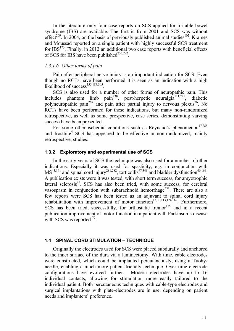
in animals moving freely, awake, inside a cage. In the vast majority of published experiments rats have been used.
In a neurophysiological study of nerve injured rats the presence of dorsal horn (DH) neurons displaying increased spontaneous discharge, increased responsiveness to pressure and brush stroke and prolonged afterdischarge was noted, especially among animals classified as “allodynic”366. The position in the DH of each of the 140 neurons recorded in the study could not be determined, but when calculated from the recording probe depth it was concluded that they were situated in lamina III-V366. Specifically wide-dynamic range neurons (WDR, i.e. neurons showing increasing responses to many intensities of stimulation, such as brush►pressure►pinch) were tested. When SCS was applied a decrease of afterdischarge as well as a decrease of an exaggerated primary response appeared. In Figure 4 the responses of two individual WDR neurons in an “allodynic” animal to mild paw pressure before and after SCS are depicted. It is conceivable that this SCS response in animals could correspond to the effect SCS can have on pain and allodynia in humans112,225. The electrophysiological findings on SCS effects in the DH have been confirmed and explored further in a recent publication demonstrating that SCS can reduce DH neuronal excitability in nerve injured rats106.
In experiments where SCS was applied and microdialysis of the DH was performed an increase of GABA and a reduction of the excitatory amino acids glutamate and aspartate could be demonstrated51. The GABA-effect appeared only in animals that also had responded to SCS with a suppression of the nerve
Figure 4: An example of electrophysiological studies of SCS mechanisms showing responses to innocuous pressure in two separate dorsal horn neurons (A,B) in a nerve lesioned, hypersensitive “allodynic” rat. The bottom histograms show responses before SCS. Horizontal bars under the histograms indicate the duration of the innocuous pressure applied to the paw. Reprinted, with permission, from Yakhnitsa et al. Pain, 1999.366.

20
lesion induced hypersensitivity in the hind paw322. Since it has been observed that GABA may decrease in the spinal cord after peripheral nerve injury32, a finding confirmed also using microdialysis that assessed the extracellular GABA release, it seems reasonable that one mechanism of action for SCS may be a restoration of GABA levels in the DH. In a recent study it has been shown that in nerve injured rats responding to SCS an immediate decrease in intracellular GABA-immunoreactivity in DH neurons appears after SCS142. Furthermore it has been shown, by double-fluorescence of C-Fos and GABA, that SCS induced activation of GABA-immunoreactive cells142.
The effect of pharmaceuticals acting on GABA-receptors has also been explored experimentally and it has been shown that i.t. injection of a GABAB antagonist could abolish the SCS effect on tactile “allodynia” in neuropathic animals; bicuculline (a GABAA antagonist) had a lesser effect49,51. When i.t. injections of baclofen (a GABAB-agonist) or GABA were used the effect of SCS was instead substantially enhanced, to an extent that even animals who had not responded to SCS could be converted to responders with an i.t. dose of baclofen so low that it did not by itself demonstrate any effect49. In a similar way it has been shown that R-PIA (an adenosine A1-receptor agonist) also can enhance the effect of SCS and even turn SCS non-responding animals into responders, with a per se sub-effective dose50,52.
The role of acetylcholine (Ach) has also been explored in conjunction with SCS. Using microdialysis in the DH it was demonstrated that animals responding to SCS exhibited an increase in the release of Ach as a result of SCS, whereas this did not appear in non-responding animals295. I.t. administration of nicotinic and muscarinic Ach receptor antagonist revealed that M4 and M2 receptors were essential for the SCS-effect and an immunohistochemical study has supported the importance of the M4 receptor for the SCS response in animals that were subject to peripheral nerve injury313. Administration of a muscarinic receptor agonist (oxotremorine) i.t. exhibited a dose-dependent
Figure 5. An example of experimental results demonstrating enhancing effect of i.t. medication on SCS effect. The graphs show withdrawal thresholds to tactile stimulation during and after SCS with or without concomitant i.t. administration of clonidine (in an individually predetermined subeffective dose). Reprinted, with permission, from Schechtmann et al. Anesth Analg, 2004 296.

21
suppression of tactile hypersensitivity and an enhancement of SCS effects. Combining a subeffective dose of oxotremorine with SCS induced a substantial improvement of the SCS effect, a result appearing even in animals that had not at first displayed any appreciable treatment effect with SCS alone313.
Levels of serotonin in the DH have been measured in rats both on the ipsilateral and the contralateral side to an induced nerve injury315. Serotonin appeared to increase in the ipsilateral DH as a result of SCS and only in those animals that had previously responded to SCS with normalized withdrawal threshold but not in the animals that despite an “allodynic” result of nerve injury did not respond to SCS. Furthermore, it was shown that a combined treatment with SCS and i.t. delivery of a low dose of serotonin (that in itself exerted no effect) could render an animal not responsive to SCS a clear responder315.
Clonidine, an adrenergic alpha-2 receptor agonist mainly used for treatment of hypertension, has evolved to be used for pain treatment as well. Cholinergic mechanisms seem to be involved in the pain reducing effects of clonidine250,255,256, possibly also involving nitric oxide (NO) transmission365. I.t. clonidine has also been tested together with SCS and it has been shown that it can enhance the effect of SCS, using a dose lower than the dose necessary for clonidine to produce an effect on the withdrawal threshold by itself (Fig 5)296.
Animal experiments with SCS and i.t. pharmaceutical agents have also been performed with the anticonvulsants pregabalin and gabapentin, drugs that are frequently used for peroral treatment of neuropathic pain. Both drugs could produce a reduction of the hypersensitivity from experimental nerve injury, in a dose-dependent manner and a subeffective dose could, used together with SCS, augment the effect of SCS and even turn a SCS non-responding animal into a responder348.
Another group of pharmaceuticals with important use in the treatment of neuropathic pain, namely antidepressants, have also been studied in animal experiments exploring SCS effects312. Doses subeffective for a drug effect on pain behavior per se were administered i.t. concomitantly with SCS. Of the drugs tested, amitryptilin (a tricyclic antidepressant) and milnacipran (a dual serotonin/noradrenalin reuptake inhibitor) were shown to enhance the effect of SCS on the withdrawal threshold. In consideration of the previously mentioned cholinergic mechanisms involved in SCS the effect of amitryptilin would appear unexpected, since it is a drug with well-known cholinergic side effects. As for the third drug tested, fluoxetine (a selective serotonin reuptake inhibitor) no impact on the effect of SCS could be detected, with the doses used.
It has been suggested that one of the glutamate receptors, n-methyl-d-aspartate (NMDA) is involved in the central sensitization associated with neuropathic pain231,362. Based on this animal experiments using individually titrated sub-effective doses of i.t. ketamine (an NMDA receptor antagonist) concomitantly with SCS have been performed showing a potential for ketamine to transform a non-responding animal into a responder343.
SCS does not only operate via spinal segmental effects but its mode of action also involves supraspinal mechanisms. In an experimental setting transection of the dorsal columns between an examined spinal level and a more rostrally applied SCS does not abolish the SCS effect11,288. In a series of experiments, a supraspinal loop, going from the dorsal columns to the brainstem

22
and back to the spinal cord, has been implicated in the effects of SCS72,287,288. This loop may well, but not exclusively, involve the nucleus raphe magnus. The previously mentioned experiments concerning serotonin release as a result of SCS also support a supraspinal influence on SCS effect, since serotonin in the spinal cord is of supra-spinal origin315.
1.6.2.2 Animal studies in models of ischemic pain
It is unlikely that the effect of SCS on ischemic pain predominately is a result of a reduction of pain generation and transmission225. An improved balance between tissue demand and supply of oxygen is more likely the reason for the reduction of ischemic pain by SCS197. An important factor seems to be an SCS-induced reduction of sympathetic activity and the effect of SCS on blood flow has been shown to be abolished if a sympathectomy had been performed198. Antidromic activiation with release of vasoactive substances, such as CGRP (calcitonin gene-related peptide) has also been implicated in the effect of SCS333. SCS may not only result in an increased oxygen supply, but might also reduce the tissue demand. In a study on skin flaps to which the arterial supply was occluded the survival was substantially improved if the animal had been pretreated with SCS94.
1.6.2.3 Animal studies in models of bowel disorders
A few animal studies on SCS mechanisms demonstrate effects on the gastrointestinal system, pertinent to IBS. One publication, from 2003, concerns the visceromotor response (VMR – measured by a strain gauge force transducer to the external oblique muscle) to balloon distention of the distal portion of the bowel102. It was shown that SCS, in settings corresponding to its clinical use, markedly reduced the VMR (see Figure 6). This reduction was maintained even after cessation of SCS, for more than an hour. If animals had been pretreated with a slow low concentration intracolonic acetic acid infusion a sensitization occurred, resulting in a marked VMR to an otherwise innocuous distension. SCS abolished this reaction completely. These results are of importance for IBS treatment, as it has been shown that IBS patients have lower thresholds to distension for pain, discomfort and perception in the distal colon as well as in the rectum and the oesophagus283,342.
In another study, from 2005, an animal model of post-inflammatory colonic hypersensitivity was used. An enema of trinitrobenzenesulfonic acid was given and thirty days later, when the mucosa was normalized, a pronounced reaction to colonic distention remained. SCS, however, normalized this reaction103. Again these results are important for IBS-treatment, as there is an increased risk of developing IBS after gastroenteritis286.

23
In animal studies SCS has been shown to influence the transmission of visceroreceptor information in the spinal cord270,271. Qin et al have presented experiments where extra-cellular signaling from 28 single spinal neurons was recorded in the rat L6-S2 spinal segments. For a number of the neurons that responded to colorectal distention the response was inhibited by SCS both at the L2-L3 level and at the C1-C2 level. Furthermore, this inhibition was maintained after spinal cord transection of the cervicomedullary junction, but the effect of the C1-C2 SCS was abolished by transaction of the dorsal columns at the C7-C8 level. At the end of each recording an electrolytic lesion was performed allowing for localization of the neurons. The responding neurons were found to be located in laminae I–III, VI, VII and X. A neurophysiological substrate for the SCS effect on the reaction to colonic dilatation has thus been demonstrated, similar to the effect SCS has been shown to exert on WDR cells in the experiments of SCS for neuropathic pain106,366.
Figure 6. An example of the effect of SCS on the viscero‐motor response, VMR, to distension of a balloon in the colon to 60 mm Hg. After three consecutive 10 minute distensions a 30 minute SCS session (90 % motor threshold) was performed and followed by an additional 10 minute balloon distension. Reprinted, with permission, from Greenwood‐Van Meerveld et al. Auton Neurosci, 2003102.
24
2 AIMS OF THE THESIS The general aim of the studies in this thesis is to improve the use of spinal
cord stimulation in humans with pain, specifically building, for Studies I-III and V, on the results from previous experimental studies in animals. A major part of the thesis thus exemplifies translational research “from bench to bedside”.
2.1 STUDIES I-III: PHARMACOLOGICAL ENHANCEMENT OF SCS EFFECT
To investigate if the beneficial enhancement of SCS effects that has been demonstrated in animal experiments with baclofen (study I and III), adenosine (study I) and clonidine (study III) also can be reproduced in human patients.
To explore if this can be used to increase and/or restore the effect of SCS for patients with neuropathic pain, if the stimulation does not yield an optimal treatment result.
To verify if a beneficial effect of pharmaceutical enhancement of SCS will remain over time (study II).
2.2 STUDY IV: TECHNICAL IMPROVEMENT OF SCS
To investigate if implantation of plate electrodes by laminotomy can be performed without discomfort using spinal anesthesia but still retaining the patient’s ability to experience and report paresthesias.
2.3 STUDY V: NEW INDICATION FOR SCS
To test if a different painful indication, namely IBS, can be effectively treated with SCS, again based on previous promising results in animal experiments.
25
3 MATERIALS AND METHODS
3.1 PATIENTS
3.1.1 Study I
43 patients with neuropathic pain participated in a trial with SCS combined with intrathecal (i.t.) bolus administration of baclofen or adenosin. 35 of these patients were new to spinal cord stimulation, but during the test stimulation period they either had insufficient pain reduction (less than 50 %) or a poststimulatory pain reduction (after 30-40 min session of SCS) lasting for less than 45 minutes (or both). Eight patients had previously obtained SCS with good effect, but had experienced a diminishing pain relief with time. Five more patients were tested with i.t. drug administration only. They had either had SCS earlier but had had their equipment removed (3 patients, unwilling to receive new implants) or were unsuitable for SCS due to previous extensive spinal surgery (2 patients).
Average age at the time of the trial was 51 years (range 25-75 years), 16 men and 32 women.
3.1.2 Study II
Nine patients from study I, i.e. the remaining five patients having both an SCS-system and a pump for i.t. drug delivery pump and the four patients who were implanted with a pump only.
3.1.3 Study III
Ten patients with neuropathic pain were recruited. As in study I the patients either had used SCS for some time (in average 8 years; range 0.6-15 years), with intended effect initially, but diminishing pain relief over time (nine patients) or with insufficient pain reduction during test stimulation with a temporary electrode (one patient). For inclusion in the study the present situation should be, as in study I, that the pain reduction from SCS was less than 50 % as compared to baseline or that the pain reduction did not outlast cessation of a 30 to 40 minutes long session of SCS (or both) with ≥ 45 minutes. Patients with 1) mental disability or other communication problems, 2) ongoing infection, 3) CNS disease, 4) heart disease and other vascular disease or 5) ongoing medication with which clonidine or baclofen can interact were excluded from the trial.
Average age was 56 years (range 39-68 years), 5 men and 5 women.
3.1.4 Study IV
This is a retrospective study of twenty consecutive procedures with implantation of plate electrodes in spinal anesthesia, of which 18 were new implants and 2 (plate electrode) replacements. All patients had neuropathic pain that had been successfully treated with SCS using standard cable-type electrodes

26
in the low thoracic region. Due to repeated dislocations and/or inadequate paresthesia coverage implant of a plate electrode had been suggested, a common routine in these circumstances.
Average age was 48 years (range 23-67 years), 7 men and 12 women.
3.1.5 Study V
Ten patients with IBS were recruited. Patients had a definite diagnosis of IBS according to Rome II criteria (Table 2)335. They were to have abdominal pain that, at least episodically, reached more than 4 on a 0-10 VAS scale and the symptoms should have been stable during the past 2 years. Patients were not to have, at the time of inclusion, other significant somatic or psychiatric illnesses. Routine blood and electrolyte samples, hepatic laboratory test, coloscopy or colon x-ray as well as lactose tolerance test were performed.
Average age was 39 years (range 26-56 years), 3 men and 7 women.
3.2 EQUIPMENT
3.2.1 Lumbar puncture
For lumbar punctures (Studies I and III) thin pencil-point needles (27 G, O.D.: 0.4 mm, Whitacre spinal set, USA) were used. By using a non-cutting thin blunt point the risk for post-dural puncture headache was expected to diminish. This claim is supported both by experimental findings134 as well as several randomized trials81,184,186,258 and long-time general experience. The pencil-point needles were introduced by Hart and Whitacre in October 1951114, based on ideas presented by Greene already in 1926101. Historically the blunt needle tip lumbar puncture needle was actually first presented by Sixten Haraldsson in May 1951, with a design quite similar to that of the Whitacre needle109.
3.2.2 Implants
3.2.2.1 SCS equipment
In Studies I and III different types of SCS equipment were used (Figure 7) involving bipolar test electrodes, 4- and 8-polar cable type leads (Quad® and Octad®) as well as plate electrodes (Resume®). Implanted electrodes were
Diagnostic criteria for IBS (Rome II) At least 12 weeks, which need not be consecutive, in the preceding 12 months of abdominal discomfort or pain that has two of three features: (1) Relieved with defecation. (2) Onset associated with a change in frequency of stool. (3) Onset associated with a change in form (appearance) of stool.
Table 2: Diagnostic criteria for IBS, Rome II

27
connected to implantable pulse generators, IPGs, Itrel 3®, Itrel 2®, Synergy® or Xtrel® (all manufactured by Medtronic Inc.).
In Study IV either plate electrode Resume® or Symmix® (Medtronic Inc.) were utilized.
In Study V patients were operated with cable type electrodes with wide spacing, Quad Plus®, and IPG Itrel 3® (Medtronic Inc.).
3.2.2.2 Pumps for intrathecal drug administration
In Study I Synchromed® pumps (Medtronic Inc) were implanted for i.t. baclofen administration. Synchromed® pumps are designed for slow continuous drug delivery and have been in use for treatment of spasticity since 1985. For administration of adenosine Algomed® pumps (Medtronic Inc.) were used. These are subcutaneous patient-operated pumps enabling the individual patient to perform bolus infusions of 1 ml at his/her discretion. Bolus delivery is important for adenosine as it is rapidly metabolized, making this kind of pump suitable for adenosine administration.
In Study III pump implants were Synchromed II® (Medtronic Inc.), a newer more slender version of the Synchromed pumps used in Study I (Figure 8).
Figure 7. Various SCS implants used in the studies photographed on a background of 1 cm x 1 cm grey squares. A: Itrel‐3®. B: Resume®. C: Symmix®. D: Quad‐plus®.
Figure 8: Pump implant, Synchromed II®, photographed on a background of 1 cm x 1 cm grey squares.

28
3.2.3 Pharmaceutical agents
3.2.3.1 Baclofen
Baclofen (Figure 9) was first synthesized in 1962 and was used for peroral treatment of spasticity. It acts on the GABAB-receptor367. GABA (Figure 9) is the foremost inhibitory transmitter of the mammalian brain and one has estimated that about 40 % of synapses in the central nervous system are GABAergic367. GABA does not penetrate the blood-brain barrier, which baclofen does to some extent. In animal studies CSF concentration is about 10 % of that in plasma290 and in a human study quite variable, but low (≤10-20 %)171. By administering baclofen directly into the spinal canal much higher concentrations can be achieved260. In 1978 an animal study on i.t. baclofen was presented, showing an antinociceptive effect358. Later, in 1984, it was reported that i.t. baclofen could reduce spasticity in an animal model176 and in humans259. Pumps for continuous i.t. administration were being developed in the early 1980ies, initially for opiate treatment of cancer pain252,261, and soon studies on i.t. baclofen administration via implantable pumps were presented260,262. Since then i.t. baclofen therapy has been used for spasticity with abundant experience25,138,251, and also for pain307. It is clear that the individual response is variable and it is customary to assess the individual response by single injection before pump implant. Already in one of the earliest studies the doses 25, 50 and 75 µg were used for this purpose262. These are the doses our clinic has been using for screening of spasticity patients and therefore we chose these doses for our studies of i.t. baclofen as adjunct to SCS.
Side-effects of i.t. baclofen include dizziness, muscular hypotonia, drowsiness as well as constipation and overdose can cause respiratory depression, coma and death138. There is no specific antidote to baclofen for human use, but increasing cholinergic transmission by i.v. administration of physostigmin has been recommended if serious symptoms of overdose appears237. Present recommendations, however, mainly emphasize stopping the pump, replacing its baclofen content with saline solution (in severe cases CSF withdrawal) and continued monitoring and supportive care (including ventilator support) in an intensive care setting353. Sudden withdrawal of baclofen may also be dangerous, with the possibility of severe reactions, including severe aggravation of spasticity138. Deaths have been reported100.
Figure 9: Structural formulas of baclofen and GABA

29
3.2.3.2 Adenosine
Adenosine (Figure 10) is an endogenous compound, found both intra- and extracellularly85. Adenosine receptors are widespread and found throughout the body with high concentration in the brain, dorsal horn of spinal cord, spleen, thymus, colon, heart, platelets and lymphocytes amongst other regions86. Adenosine thus affects a number of biological processes, including the immune system, but its main clinical use has been, since the 1980ies, within the field of cardiology116. Adenosine, however, has important effects on pain processing and animal experiments have shown reduction of pain behavior with i.t. adenosine293,316, especially with a neurogenic pain model317. It has also been reported that levels of blood and CSF adenosine is lower for patients with neuropathic pain than for controls107. In a case report from 1994 a patient with sural neuropathy and severe allodynia to touch and vibration was free from spontaneous pain for 10 days and from allodynia for over six months following an i.t. injection of the adenosine receptor A1 agonist R-PIA153. Adenosine has been explored for human i.t. use in a number of studies, in volunteers67,70,273, in pain patients16,71,311 and as an adjunct in anesthesiological practice16,71,274,275,311. Exogenous adenosine has a very rapid turnover both in plasma22 and CSF273, half-life within 10-20 minutes . Side-effects are mainly local lumbal pain at the level of injection and headache. The doses of adenosine in Study I were the same as in a parallel study in volunteers, referred to above273: 500 µg, 1 000 µg and 2 000 µg.
3.2.3.3 Clonidine
Clonidine (Figure 11) has been in use for treatment of hypertension since 1966143. It is an adrenergic alpha-2 receptor agonist104, for which several other uses have evolved, such as pain treatment and opiate withdrawal symptoms143. Already in 1949 an analgesic effect of adrenalin was presented188 and in 1974 animal experimental data indicated an antinociceptive effect of clonidine, both when administered intraperitoneally and in the lateral ventricle298. An involvement of cholinergic mechanisms in the pain-reducing effect of clonidine was reported already in 1980201, a notion supported by a number of publications since135,250,255,256,365. In 1984 Tamsen and Gordh reported that epidurally
Figure 10: Structural formula of adenosine

30
administered clonidine could have analgetic action in man and a potentiating effect on coadministered epidural morphine332. Coombs et al reported in 1985 on the successful use of clonidine i.t. alone as a treatment for cancer pain in a patient with morphine tolerance48. Over the ensuing years numerous publications supporting the use of i.t. clonidine have appeared, either as a single treatment or in combination with other drugs8,62,80,117. Elimination half-life i.t. has been studied in sheep, yielding a value of in average 64 minutes33. There are no human studies on elimination after i.t. injection, but after epidural injection average half-life of clonidine in the CSF has been reported to be 66 minutes98. Side-effects of i.t. clonidine include hypotension, malaise, headache, confusion and lethargy117. In published trials doses for single i.t. injections varying from 15 µg to 450 µg have been used8,62,79. We have chosen the doses 25, 50 and 75 µg for the bolus trials in Study III. In the first two cases a 100 µg dose was used, but this was abandoned as one patient demonstrated hypotension.
3.2.3.4 Bupivacaine
Bupivacaine (Figure 12) was synthesized in the late 50ies by the Swedish company Bofors and was introduced in clinical practice in the mid 1960ies187,
188. Its foremost advantage was a long-lasting anesthetic effect, however accompanied by a higher toxicity. It came into abundant use, also for i.t. administration324. Serious side-effects of spinal anesthesia are uncommon, but include neurologic injuries, seizures, hypotension and cardiac arrest6. Several reports concerning increased risk for cardiac arrest with bupivacaine have been published, but no such increased risk could be seen in a very large French study summarizing the results of >40 000 spinal anesthesias6. The doses used in
Figure 11: Structural formula of clonidine
Figure 12: Structural formula of bupivacaine
31
Study IV were decided individually by the anesthesiologist in charge and were in accordance with current guidelines18.
3.3 STUDY OUTLINES
3.3.1 Study I
On separate days single doses of Baclofen (25-75 µg, on some occasions 100 µg) or placebo (saline) were injected via lumbar puncture in a single-blinded fashion, the first dose always being 25 µg, to check for side-effects, and higher doses in random order. Patients evaluated pain on a VAS scale (10 cm) at baseline and again after 30, 60 and 90 minutes. At 90 minutes after the injection of i.t. drug a 30 min session of SCS was started, after which patients recorded pain again. Patients were also asked to record for how long the SCS-effect lasted, if such was present, as well as any side-effects. A pain reduction of at least 50 %, with either i.t. drug or SCS or both, was considered a positive response, with only spontaneously appearing pain being assessed.
A separate testing of adenosine was performed in a similar manner in a subset of patients (seven patients) (bolus doses of 500, 1000 or 2000 µg). In this procedure, however, SCS was started already after 30 minutes. Apart from this the procedure and pain assessment was the same as for baclofen.
If i.t. drug administration substantially improved the effect of SCS patients were offered implantation of a pump for i.t. drug delivery as well as an SCS system. If the pain reduction was foremost attributed to the i.t. drug implantation of only a pump was suggested. For a few patients with exceptionally good response to a low dose of a drug, peroral drug administration was tried, as an adjunct to SCS.
Follow-up investigation was performed on all patients who continued to use baclofen, either i.t. or perorally, with or without SCS. Patients were contacted for telephone interviews, utilizing a structured protocol (Table 3) and hospital records were reviewed for dosages, reoperations and adverse events. Follow-up was performed in average 30 months after pump implantation (or after initiation of medication for the three patients receiving per-oral baclofen), range 1-72 months.
3.3.2 Study II
The second study constituted a late follow-up of all patients with remaining pumps at the end of Study I. Again structured telephone interviews were performed, with the same protocol as for Study I (table 3). Follow-up was performed at in average 67 months after pump implant, range 37-103 months. Hospital records were also reviewed for present dosages, reoperation etc. until in average 73 months, range 45-111 months.
3.3.3 Study III
On separate days single injections of baclofen (doses 25, 50 and 75 µg), clonidine (25, 50 and 75 µg – in two cases instead 100 µg) or placebo were
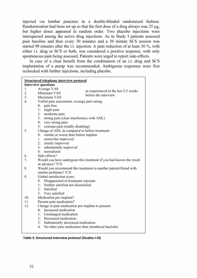
32
injected via lumbar puncture in a double-blinded randomized fashion. Randomization had been set up so that the first dose of a drug always was 25 µg, but higher doses appeared in random order. Two placebo injections were interspersed among the active drug injections. As in Study I patients assessed pain baseline and then every 30 minutes and a 30 minute SCS session was started 90 minutes after the i.t. injection. A pain reduction of at least 30 %, with either i.t. drug or SCS or both, was considered a positive response, with only spontaneous pain being assessed. Patients were urged to report side-effects.
In case of a clear benefit from the combination of an i.t. drug and SCS implantation of a pump was recommended. Ambiguous responses were first rechecked with further injections, including placebo.
Structured telephone interview protocol Interview questions 1. Average VAS as experienced in the last 2-3 weeks
before the interview 2. Minimum VAS 3. Maximum VAS 4. Verbal pain assessment, average pain rating:
0: pain free 1: slight pain 2: moderate pain 3: strong pain (clear interference with ADL) 4: very strong pain 5: extreme pain (totally disabling)
5. Change of ADL as compared to before treatment: 0: similar or worse than before implant 1 somewhat improved 2: clearly improved 3: substantially improved 4: normalized
6. Side-effects? 7. Would you have undergone this treatment if you had known the result
in advance? Y/N 8. Would you recommend this treatment to another patient/friend with
similar problems? Y/N 9. Global satisfaction score:
0: Disappointed of treatment outcome 1: Neither satisfied nor dissatisfied 2: Satisfied 3: Very satisfied
10. Medication pre-implant? 11. Present pain medication? 12. Change in pain medication pre-implant to present:
0: Increased medication 1: Unchanged medication 2: Decreased medication 3: Substantially decreased medication 4: No other pain medication than intrathecal baclofen
Table 3: Structured interview protocol (Studies I‐III)

33
In Study III, as well, a follow-up was performed on the patients that received a pump, using the structured interview protocol from Studies I and II (table 3) and hospital record reviews.
3.3.4 Study IV
Patients undergoing scheduled implantations of a plate electrode in spinal anesthesia were interviewed regarding problems during surgery. Ease of performing operation, operative time, thresholds for paresthesias during surgery as well as in the prone position postoperatively, amounts of anesthetic agents and i.t. injection level were noted.
At operations spinal anesthesia was induced via injection of bupivacaine (see section 3.2.3.4) at the L2/3 level. Supplementary local anesthetic was used at the site of incision. A small laminotomy was performed at the intended level, including part of the spinous process and the electrode was introduced into the epidural space. Intraoperatively testing for adequate paresthesias was performed utilizing a standard external stimulator (DualScreen®, Model 362, Medtronic Inc.). If paresthesias were not adequate repositioning of the electrode was carried out. Before closure the electrode position was checked with anteroposterior fluoroscopy.
3.3.5 Study V
Electrodes with widely spaced stimulating poles (Quad plus®, Medtronic Inc.) were percutaneously implanted at T11/12 and positioned to yield paresthesias in the abdominal area (with or without paresthesias in the lower extremities), with the tip of the electrode at the T5-T8 level. According to the protocol patients did not use stimulation the first 2 weeks after surgery. They were then randomized to either start a 6 week period with stimulation or a 6 week period without. At the end of these six weeks, patients crossed over to not stimulate for another six weeks or vice versa. Subsequently, all patients continued with a 12 week long period of stimulation (Figure 13 illustrates the
Figure 13: Flow chart for the SCS‐IBS study (Study V)

34
trial outline). During periods of stimulation patients turned the stimulation on or off at will, but were instructed to stimulate at least 8-12 hours out of 24, with a comfortable intensity and preferably at least 12 hours. Compliance was checked from the implanted stimulator memory at regular out-patient visits. Participants noted daily 1) number of pain attacks during the day, 2) number of diarrheas during the day, 3) average pain level during the day and 4) average evaluation of quality of life (QOL). For items 3 and 4 patients were asked to utilize a numeric rating scale from 0-10 (10 implying worst possible pain or best possible quality of life respectively). At the end of the study patients could choose to keep the stimulating system or have it removed.
All patients were contacted for follow-up at the end of the study using a structured telephone interview. Among questions asked were present pain level, usage of stimulation system (if present), medication, side-effects and global satisfaction. Each patient was also evaluated using a Swedish version of the hospital anxiety and depression scale (HAD)370 and a modified gastrointestinal symptom rating scale (GSRS-IBS)355.
35
4 RESULTS
4.1 STUDY I
Twenty patients responded to Baclofen, either in combination with SCS (16 patients) or baclofen alone (4 patients). These patients were offered continued baclofen treatment with i.t. (with an implantable pump) or oral administration (the oral route was offered to those who had an exceptionally good response to baclofen at the lowest i.t. dose, 25 µg) either with SCS or with the drug alone (if SCS was not possible). Seven patients received a baclofen pump as an adjunct to SCS, and four patients received pumps for i.t. drug delivery alone, without SCS. In two of the seven patients with SCS and i.t. drug treatment, the pumps had to be explanted due to technical problems and diminished pain relieving effect.
Two patients opted for i.t. administration of adenosine, requiring a different type of pump that does not yield a constant flow, but has a patient operated mechanism for single boluses. Delivering adenosine by this pump unfortunately did not function satisfactorily. Due to side effects, back-pain and headache, a local anesthetic had to be coadministered with the adenosine solution. This mixture was, however, not stable in the pump and most of the pain relieving effect was lost within a week. One patient with such a pump chose to have it removed and eventually all adenosine treatments were terminated (within ≈ 1 year).
Follow-up on the patients with baclofen was performed in average 30 months after implant using a formal telephone interview protocol. The patients with SCS + i.t. baclofen reported a drop in VAS from 76 (70-90) before the bolus trials to 33 (0-80) at follow-up. The patients with SCS and peroral medication reduced the pre-trial VAS from 70 to 27 whereas the patients with i.t. baclofen alone reported a reduction from 63 (40-90) to 33 (20-80). During the follow-up period the baclofen dose had to be increased in all patients from an average of 72 µg/24 h to 157 µg/24 h.
4.2 STUDY II
Patients reported some increase in pain as compared to the early follow-up, but still it was significantly lower than the pre-trial values. In most patients it was necessary to further increase the baclofen dose by in average 30 % compared to the early follow-up. A number of surgical interventions had been necessary (four exchanges of pumps due to battery depletion, one replacement of a fractured i.t. catheter, seven exchanges of SCS stimulators and three SCS electrode revisions); nonetheless, all patients continued to use the treatment and wanted to keep it.
4.3 STUDY III
For both drugs a significant dose dependant enhancement of pain reduction was recorded. Five patients were deemed as clear responders and four of them

36
chose to have a pump implanted. Based upon individual patient response two pumps were used for administration of baclofen and two pumps for clonidine.
4.4 STUDY IV
In all patients a complete analgesia could be achieved, using bolus injections of 12.5 to 20 mg bupivacaine, and a small laminotomy performed at the T9/10, T10/11 or T11/12 level. For one patient it was necessary to perform a second i.t. injection. A plate electrode was introduced and tested intraoperatively, and in all cases paresthesias could be produced, in several instances resulting in a repositioning of the electrode. For all patients an optimal paresthesia coverage could be achieved. Operative times were between 70 and 195 minutes, in average 112 minutes. The thresholds for paresthesias during operation were similar or only slightly higher than thresholds recorded after surgery when tested in the same supine position (mean 3.1 V during surgery and 2.1 V after surgery). At interview after surgery none of the patients reported that the interventions had been painful or uncomfortable.
4.5 STUDY V
One patient chose to leave the study after ten weeks and her SCS system was removed. The remaining nine patients completed the entire study. Three patients did not achieve any benefits from stimulation and chose to have their SCS-system removed. Six of the participants reported a satisfactory result with stimulation and have continued the stimulation treatment beyond the trial period.
Periods with stimulation were compared to periods without stimulation. Pain scores as well as number of pain attacks and diarrhea episodes daily were lower during stimulation periods than during non-stimulation, but only the difference in pain scores reached statistical significance (p=0.04). The differences for the other parameters were not significant and for quality of life measurement no differences could be detected.
On follow-up half of the patients who had decided to keep their stimulation stated that they still used it. For two of the patients who had ceased to use it a gradual decrease of pain had appeared over the years to a degree that they had chosen not to use it. They kept the stimulation system anyhow, in case pain would recur. One patient had to have the equipment removed, due to need for an MRI scan.

37
5 DISCUSSION
5.1.1 Pharmacological enhancement of SCS effects
These studies are entirely based on findings derived from experiments on animal models of neuropathic pain performed at the laboratory of the Department of Neurosurgery at the Karolinska University Hospital/Karolinska Institutet. Thus they are typical examples of a translational approach with a direct transfer of knowledge from “bench to bedside”.
There are several limitations to the studies, most importantly the small number of patients. In Study I the injections were only single-blinded, but in the preparations for Study III it was decided to use double-blinded and randomized injections. In none of the studies SCS could be blinded to the patients, which is a limitation to almost all studies on SCS (compare section 1.3). Even with blinded testing of i.t. injections the comparison of results only concerns the outcomes of bolus injections, not the long-term results. For a decisive investigation of the long-term usefulness of adding i.t. drug delivery to suboptimally acting SCS a proper (randomized) control group with SCS but no pump should be included (or better pump with saline and periods with active drug). The protocol for follow-up telephone interviews is not validated, but it was considered that comparisons could be facilitated if the same protocol was utilized. The usage of telephone interviews implies that an NRS scale has been employed, but it has been inappropriately named VAS in the publications. There are, however, some studies indicating a fair correspondence between VAS and NRS78,269 implying that this practice is acceptable.
Based on the laboratory findings on the pivotal role of the GABAB-receptor in the SCS effect49-52 it was logical to start investigating the possibility of enhancing the response to SCS with i.t. drug delivery of baclofen since it is a drug already registered for i.t. use, accompanied with a substantial clinical experience. This not only facilitated ethical approval, but also general handling of the patients concerning dosages, side effects, etc. The choice to continue with clonidine (strongly supported by both the animal findings with SCS enhanced by clonidine296 and by the importance of cholinergic mechanisms in SCS295) was also based on the fact that an abundance of clinical experience – manifested through a number of scientific publications – existed as to its use as an i.t. treatment. The use of clonidine i.t. is, however, off-label, and, at least in Sweden where the studies took place, an individual approval from a state authority (Swedish Medical Products Agency) was necessary for continued treatment with i.t. clonidine for each patient receiving a pump. The results from Study I were unfavorable for adenosine and no further attempts to explore adenosine i.t. in continued bolus administration, via an implanted pump, seem reasonable until a solution to the side effects with low-back pain has been established.
In none of the trials reported in these studies (I-III) it was possible to convert a patient who showed no response to SCS into a responder. This is in contrast to the results of the preceding animal studies50,296, where a non-responding animal could be transformed into a responder with a dose of an i.t. drug that in itself was without effect. The animal models are, however, models of reactions

38
associated with neuropathic pain. All testing in animals subjected to nerve injury consisted of stimulus-induced pain-like behavior, i.e. evoked pain, whereas in practically all the human studies only ongoing spontaneous pain was assessed. A notable concern here is that only 20-40 %, at most, of neuropathic pain patients present with mechanical allodynia108. Only in a study from 2004, Harke et al. reported SCS effects on both spontaneous pain and allodynia112. Or obvious reasons it is difficult to know if experimental animals suffer spontaneous ongoing pain after nerve injury because they rarely exhibit behavioral changes that may be interpreted as indicative of pain. It has also been debated if behavioral changes after nerve injury, such as holding a paw in a protected position, necessarily reflect ongoing pain196,233,354. Not only sensory but also motor disturbances can contribute to an abnormal posture239.
With the exception of not being able to convert SCS non-responders into responders, the human studies were able to essentially replicate the findings in the animal studies to some degree. This supports the use of animal studies of this kind as a base for exploring underlying mechanisms of SCS effect in humans and to find new ways to increase the efficacy and usefulness of this treatment.
Even though the animal trials have demonstrated successful i.t. administration of pharmaceuticals augmenting the effect of SCS it is not necessary to exclusively use i.t. administration in clinical trials of adjuvant therapy to SCS. As mentioned in Study I a few patients tested oral baclofen instead of i.t. administration. The treatment effect was good, but due to side effects they all chose to discontinue. In future clinical trials of some of the previously mentioned substances oral treatment can still be considered.
The method of combining SCS and pharmacotherapy could be further explored by using other drugs beside baclofen and clonidine. As stated in section 1.6.2.1 several drugs have already been tested in a laboratory setting. These include drugs already in use for peroral medication, such as gabapentin, pregabalin, milnacipran and amitryptilin. These drugs are in such frequent use among pain patients that it is likely that a substantial number of patients with SCS also have tried concomitant medication perorally with one or several of the drugs. However, no systematic trials or even retrospective studies have been performed.
Most of the drugs specified in section 1.6.2.1 are not in regular use for human i.t. administration. Gabapentin has been used i.t. in a number of animal studies, but no publication exists concerning human use. The Polyanalgesic Consensus Conference58 has an expert panel on i.t. medication for pain that has regularly published recommendations. The latest report, from 2012, not only holds a summary of animal trials with i.t. gabapentin, but also informs of an ongoing clinical study on human i.t. gabapentin pain treatment58. When more experience with i.t. gabapentin is available clinical trials with i.t. gabapentin as an adjuvant to SCS may be a possibility.
Another drug that has been experimentally tested is ketamine343. However, this drug is not available for regular oral use, but it has been in use since long i.v. for anesthetic purposes and reports on oral administration exist23. The use of ketamine is also hampered by its potential for drug abuse and serious side effects. Ketamine is not approved for i.t. use, but has been used off-label for that purpose since many years, the first report appearing already in 198419. A number
39
of single-patient reports on i.t. ketamine exist, often co-administered with other substances – mainly opiates –, and it has successfully been in use long-term for i.t. delivery via an implanted pump292,364. There are also, however, reports of toxicity154,323,363 and further safety studies are necessary before a potential full scale trial with i.t. ketamine as an adjunct to SCS can be carried out.
Amitryptiline has been tested i.t. in several animal studies since 198324. It has several modes of action, including NMDA-receptor antagonism68 (as ketamine) and Na+-channel blocking325. The latter characteristic implies actions as a local anesthetic. The effect as a spinal anesthetic has been tested in several animal studies37,92 as well as effects on pain, both acting alone35 or as an adjuvant69,330. Unfortunately reports of serious side effects of i.t. amitryptiline, including severe arachnoiditis, in different laboratory animal species have appeared89,326. This precludes human testing of i.t. amitryptiline until the risks have been further elucidated.
One of the drugs for i.t. use as a pain treatment that has authority approval (FDA, Swedish Medical Products Agency, etc.), namely ziconotide, has not been tested in conjunction with SCS – neither in a laboratory setting nor in human clinical trials. Ziconotide blocks N-type voltage-sensitive calcium channels356. It is only used for i.t. administration and it is approved both for nociceptive and neuropathic pain. Ziconotide is, however, more difficult both to test and to use long-term, since it has slow onset both of actions and side effects and a rather narrow therapeutic window356. Side effects include orthostatic hypotension, dizziness, ataxia, abnormal gait, memory impairment, confusion and hallucinations309,356. A trial period is generally recommended utilizing an i.t. catheter in an in-patient setting, but there is no consensus as to the appropriate technique58. If animal studies would demonstrate a suitable facilitating effect of ziconitide in SCS clinical trials would be feasible, since the drug is approved for i.t. administration. Such a study would have to involve an implanted i.t. catheter and continuous drug infusion, instead of the i.t. bolus injections used in Study I and III, due to the different time-scale of ziconitide effects and side effects as compared to that of baclofen and clonidine.
The most commonly used type of medication for i.t. pain treatment is opioids309. In at least one published study, i.t. opioids have been administered concomitantly to SCS via implanted pumps339. In that study the purpose of the i.t. administration was not to enhance the effect of SCS, but to treat separate symptoms, i.e. midline low-back pain, not improved by SCS. Even though there is presently no evidence that opioid mechanisms are involved in the mode of action of SCS87, systematic studies of a possible enhancing effect of opioids, as well as of a potential counteracting effect of opioid receptor antagonists, are warranted.
Despite the limitations of Studies I-III they provide support to the notion that neuropathic pain patients with insufficient pain reduction from SCS should be considered for trials with adjuvant pharmacological treatment. A prerequisite of course is that the SCS system in itself functions properly, yielding comfortable paresthesias covering the painful area. As oral alternatives frequently already have been tried in this setting it seems reasonable to commence such a trial with i.t. baclofen, because this drug is already approved

40
for i.t. administration. If this fails clonidine should instead be tested. In the future it is likely that other i.t. drugs will also prove useful (see Figure 14).
Studies I-III have been cited in 30 publications: Study I42,58,82,105,122,172,192,195,218,243,244,294,295,297,309,310,312,314,315,329,339,368 Study II105,142,278,294,295,306,308,309,313-315,329,339,350,368 Study III36,73,105,142,191,312
+
Neuropathic pain
Revision/ additional
lead(s)
Permanent implant
Trials with low dose peroral drugs + SCS (see text)
Adequateparesthesia
< 45 min > 50 % VAS
+
‐‐ SCS trial
Diminished therapeutic
effect of SCS
Continue peroral therapy
Trials with alternative
stimulation setup
(or side effects) ‐
(or side effects) ‐
SCS + drug 1 (e.g.: i.t. baclofen
25-100 µg)
SCS + drug 2 (e.g.: i.t. clonidin
25-75 µg)
(or side effects) ‐
i.t. infusion pump
p.o. administration (if applicable)
i.t. infusion pump
p.o. administration (if applicable)
+
SCS + drug 3 (see text)
i.t. infusion pump
p.o. administration (if applicable)
+
Alternative therapy
+
< 45 min> 50 % VAS Choice
between neuromodulation
paradigms
(or side effects)‐
(or side effects)‐
‐
Figure 14: Suggested algorithm for possible trials of adjuvant pharmacological treatment in SCS with insufficient relief of neuropathic pain.

41
5.1.2 Technical improvement
Implantation of plate electrodes in an appropriate position is a challenge since a procedure under local anesthesia may be demanding and painful to the patient and surgery in general anesthesia precludes the use of intra-operative testing.
The finding that intra-operative test stimulation could be performed during spinal anesthesia is not only interesting from a technical point of view but also concerning the information it conveys on the mechanisms of spinal anesthesia. These findings demonstrate that anesthetic agents for spinal anesthesia act more on spinal rootlets than on spinal cord afferent pathways. Study IV shows that plate electrodes (paddle leads) can be implanted in the low thoracic and lumbar region with ease both for implanter and patient using spinal anesthesia. Following this publication several other groups have adopted this technique, and together with a group in Regina, Saskatchewan, Canada, we have recently presented a total of 53 patients undergoing successful plate electrode implantation in spinal anesthesia179. Subsequently other implanters have also presented their experiences with paddle lead implants in spinal anesthesia, combining it with minimally invasive laminotomy techniques291,352. The technique cannot, however, be used for implants above the mid-thoracic level, as higher spinal anesthesia evidently may affect respiratory function.
The possibility of using epidural anesthesia instead has been explored for paddle lead implants in the low thoracic region91,369. In these studies epidural anesthesia was obtained with a catheter inserted into the appropriate level. One or several injections of a local anesthetic were performed. Once sufficient regional anesthesia was achieved the catheter was removed prior to the surgical procedure. An expected advantage compared to spinal anesthesia was the possibility to easily iterate injections of the anesthetic if the effect was insufficient, avoidance of meningeal puncture and possibly improved hemodynamic stability91. A disadvantage, however, could be that if epidural adhesions exist they might limit the spread of the anesthetic agent resulting in insufficient anesthetic effect. The presence of epidural adhesions is not uncommon in patients eligible for paddle lead insertion and in one of the cited studies induction of epidural anesthesia actually failed in 7 out of 31 patients, necessitating general anesthesia91. In this respect spinal anesthesia has the advantage that intrathecal adhesions rarely restrain the spread of the anesthetic. The earliest attempt to use epidural anesthesia for SCS implant actually dates from 1996, when it was used to facilitate insertion of a cable-type electrode in a case were passage of the electrode itself in the epidural space was felt painful by the patient280. The first report of a series of patients subject to paddle lead implantation in epidural anesthesia is that by Meyerson et al. in 1998229.
A different strategy to diminish the pain associated with plate electrode insertion in local anesthesia is to minimize the procedure. Beems and van Dongen have presented a technique were a series of dilators are used to spread paraspinal muscle fibers and enable key-hole entrance to the epidural space for paddle lead implant15.
Another development has been the introduction of a hybrid percutaneous and paddle-lead. This is a lead with insulation on the side not facing the dura. It

42
is thicker and wider than a regular cable-type lead but more slender than plate electrodes and it can be introduced percutaneously using a specially designed needle and a Seldinger-type technique is used for widening of the introductory path. Some experience has accumulated in recent years of this technique, indicating that the technique works well, with the longest median follow-up of one year56,57,165,203,204,359.
Yet another strategy when implanting paddle leads is to increase the likelihood of a proper placement of the electrode by using intra-operative neurophysiology, as described in some recent publications10,75,210,304. Without any other information in a fully anesthetized patient the laterality of a lead can only be decided from intra-operative fluoroscopy. The radiological midline may, however, differ from the physiological. By using either EMG75,210,304 or SSEP10 further information on the laterality can be obtained. With a physiological midline placement of the electrode an equal size of the EMG response is to be expected from both sides. If a lateral electrode position is needed a larger response is aimed for on the side where the electrode should be. SSEP is expected to diminish on the intended side, when SCS is turned on, due to impulse collision10. These techniques are described in detail in the publications, but it is worth noting that at present these techniques only help in determining the laterality of an electrode and not the proper positioning in the caudal-cephalad dimension.
Modern electrode design can also be of importance. Study IV took place at a time when SCS electrodes had four contact areas at most. Modern electrodes have up to 16 electrode contacts, which should increase the likelihood that simple reprogramming of electrical contacts can solve the problems of inadequate paresthesia coverage. No scientific publications substantiating this hypothesis exist as yet and the modern multipolar electrodes have not been implanted for a sufficient number of years for reliable experience to accumulate. In a recent retrospective investigation there was a slight trend that in patients with 16 contact electrodes loss of paresthesia coverage could more easily be recaptured than in patients with 4 contact electrodes, but the difference was not statistically significant164.
In conclusion the usefulness of spinal anesthesia with intraoperative paresthesia testing for implantation of plate electrodes is now well established and several other groups have adopted the practice. Although implantation techniques have evolved, it is likely that spinal anesthesia will continue to be useful in this context. A further development is the use of epidural anesthesia for electrode implantation at higher levels than mid-thoracic because the risk of respiratory depression should be less. The usefulness of a catheter, as applied in the publications concerning paddle lead implant in epidural anesthesia91,369, could easily be transferred to the setting of spinal anesthesia, facilitating repeat injections if the anesthetic effect diminishes. Repeat spinal injections can, however, be performed without an i.t. catheter.
Study IV has been cited in 17 publications: 10,14,15,75,82,91,119,177,178,180,190,208,210,219,291,352,369.
43
5.1.3 New indication – IBS –
IBS is a very common disorder206 with insufficient therapeutic options83 and much need for new treatments. As stated in sections 1.3.1.5 and 1.6.2.3, SCS effects on gut function have been demonstrated repeatedly and it is logical that SCS has been considered for IBS treatment as well. A few beneficial case reports on the use of SCS for IBS have been presented173,253,272 - and one report demonstrating no effect209 - but Study V, though comprising a small number of patients, is the first randomized prospective study.
There are many possible routes by which SCS could influence gastrointestinal disorders such as IBS. The nervous system regulation of the gastrointestinal tract is intricate. There is both an intrinsic and an extrinsic neural control of the gut7. The intrinsic enteric nervous system is highly complex and controls basic gastrointestinal functions. Extrinsic nervous control consists of parasympathetic innervation via the vagus nerve and the sacral nerves and sympathetic innervations via the splanchnic nerves. Both sympathetic and parasympathetic nerves contain afferent and efferent fibres. Afferent sympathetic information is carried in the spinal cord through many pathways, including not only the spinothalamic tract, but also the dorsal columns as well as the spinoreticular, spinomesencephalic and spinosolitary tracts7. Complex brain circuitry modulates the gut innervations, together constituting the “brain-gut axis”7. Evidence has accumulated that visceral pain is transmitted both in the spinothalamic tracts - that normally convey pain impulses - and in postsynaptic neurons in the dorsal columns174,242. The dorsal columns have been implicated in the transmission of visceral pain in a series of studies based on the prominent relieving effect on pain from pelvic cancer by limited midline punctuate myelotomies357. The postsynaptic dorsal column neuron pathway may also have an important role in conveying pain from inflammatory visceral processes and in a rat study lesions of the pathway did not diminish the reaction to colorectal distension under normal circumstances, but under inflammatory conditions such a lesion would return the response to normal254. Krames and Foreman have hypothesized that SCS exerts its influence on gastrointestinal pain by modifying these pathways174. As reported in section 1.6.2.4 animal studies have shown that spinal cord neurons firing in response to colonic distension show a reduced response after SCS. Another type of mechanism that also may be involved is antidromic activation and peripheral release of active substances. This has been shown to take place in the use of SCS for peripheral ischemia, where antidromic release of CGRP has been implicated in the beneficial effect333.
Study V indicates that SCS appears to exert beneficial effects in the majority of patients with IBS, with a responder rate in the vicinity of that for patients with neuropathic pain. There are, however, several limitations of the study, the most important being the small size. Larger studies must be performed to verify the results, preferably as a multi-center effort. The results of Study V may help in performing an adequate power calculation. Another limitation is the lack of patient-blinding, since SCS-induced paresthesias were perceived by the patients. Also, the method of assessing quality of life, using an NRS, may have been insensitive and a more appropriate validated contemporary scale should be utilized in future studies.

44
In our study, one patient chose a low intensity stimulation and reported a good treatment effect also with stimulation subthreshold to paresthesias. He was, however, urged to continue with paresthetic stimulation, as demanded by the study protocol. Furthermore, in a personal communication Dr E. Krames has reported that the stimulation for his pioneer case eventually was subliminal, but with remaining treatment effect. Suddenly the symptoms of IBS, however, reappeared and the patient contacted the clinic. On examination the stimulator was found to have been turned off (possibly due to exposure to an external magnetic field) and when again turned on the beneficial effects returned. As described in section 1.3 there have been a few studies presented where SCS at an intensity below that needed to produce paresthesias has been used17,66,185,193,194. Thus, it is foreseeable that in future studies of SCS for the treatment of IBS subliminal treatment could be included.
45
6 CONCLUSIONS Insufficient effects by SCS in neuropathic pain, where appropriate coverage
of the painful region with paresthesias indicate a proper lead position, may be improved with the addition of i.t. medication.
A neuropathic pain patient with insufficient pain relieving effect of SCS, or with a diminished previously satisfactory effect, could be subjected to a trial with i.t administration of baclofen and/or clonidine; at present, there is more evidence for the clinical usefulness of the former drug.
The effect of combined therapy is likely to last for many years (as demonstrated for baclofen), even though a gradual increase of the dosage required to maintain a beneficial effect can be anticipated.
A patient who does not have any appreciable relief of neuropathic pain by SCS alone cannot be expected to benefit from adjunctive i.t. pharmacotherapy.
Plate electrodes can reliably – and comfortably – be implanted under spinal anesthesia, which permits intraoperative testing of paresthesia distribution.
SCS may be a treatment option for IBS when conventional therapy has proven ineffective. Further studies with larger patient groups are needed to confirm the observations of the present small pilot study.

46
7 ACKNOWLEDGEMENTS The completion of this thesis would not have been possible without the
support of a number of people. I would like to express my deep gratitude to everyone who has supported me during over the years, and in particular I would like to acknowledge:
Prof Bengt Linderoth, my main supervisor and close friend, who got me
started in the scientific work. With your vast knowledge, kindness, generosity and sense of humor you have helped me and guided me throughout all these years. You make hard work joyous and worth-while.
Prof Björn Meyersson, my co-supervisor, for introducing me to the field of
pain research and for being my mentor through my career. For being there even many years after your own retirement, and encouraging me to go forward at a point when I could have chosen to let go instead.
My co-authors: Jaleh Winter, who has cared for all the patients and
meticulously audited the patient data in the papers, apart from always being kind and supportive. Gastón Schechtmann, my friend and colleague both in science and clinical work, who has helped me so much both in research and every-day work. Prof Per Hellström, who has introduced me to another field of medicine, gastroenterology, and who has supervised the IBS project with clarity and wit.
My collegue Per Almqvist for being such a good friend, for numerous
scientific discussions and for being generously supportive. All the members of the neurosurgical research lab, of whom I especially
wish to thank those working with functional neurosurgery, Zhiyang Song, Johan Wallin and Camilla Ultenius, for your interest in the field and for all your help during the years. I also wish to express my sincere gratitude to Britt Meijer, who has helped me with numerous things over the years.
Members of other collaborating research groups, especially Mia Jönhagen,
Lars Wahlberg and Åke Seiger, for the fruitful and productive scientific environment that you build and for kindly supporting a place for me in it.
All colleagues, former and present, at the Department of Neurosurgery, for
your profound interest in caring for the patients, in excellence in surgical skills and in bringing research forward. I especially wish to thank those who have been heads of the Department during my research years, Prof Iver Langmoen, Lars Wallstedt, Prof Mikael Svensson and Inti Peredo, for your support. I also want to express my gratitude to all the other colleagues, among them Peter Alpkvist, Lisa Arvidsson, Bo-Michael Bellander, Jörgen Boethius, Ernest Dodoo, Göran Edner, Erik Edström, Adrian Elmi, Michael Fagerlund, Anders Fytagoridis, Petter Förander, Bengt Gustavsson, Simon Hilliges, Hans von Holst, Lars Kihlström, Bodo Lippitz, Tiit Mathiesen, Ilias

47
Nikaolaidis, Ingrid Ohlsson-Lindblom, Marcus Ohlsson, Martin Olsson, Kyrre Pedersen, Jenny Pettersson-Segerlind, Tiit Rähn, Ingvar Ólafsson, Ulrika Sandvik, Georges Sinclair, Halldór Skúlason, Annika Suneson, Elfar Úlfarsson, Theresa Wangerid and Ulf Westerlund. I am proud to have worked and to keep working with you. I also wish to thank my former colleague Prof Bo Levander for your consideration and support over the years and your keen interest in my work.
Maj-Britt Andersson and the whole staff at ward R-14 for your
professionalism, dedication and excellent care of the patients, and for always being so kind and supportive to me. I always feel happy and among friends at the ward. I also wish to thank Karin Persliden, who has joined the functional neurosurgery team with such enthusiasm and competence, for your sympathy and interest.
Carina Colliander-Holst, Kerstin Johansson, Barbro Sköld and all the
staff at the operating ward, as well as the colleagues at the Department of Anesthesiology, for taking care of the patients and for always being kind and helpful to me.
Collegues and friends at the Departments of Neurology, Neurophysiology
and Neuroradiology and at the Pain Centre, for caring for the patients with pain and for your support.
I want to thank Medtronic Europe for the continuous support of our
translational studies over the years. I am especially thankful to Keith Mullet and his successor Ali Sarem-Aslani for this support.
My family, my mother Elisabeth, my sister Kerstin and my aunt Ruth, for
all your help and kindness. My dearly beloved children Sofia, Susanna, Carl-Fredrik and Stella and
my wonderful wife Charlotta for your unconditional love and support. You are the meaning and joy of my life.
The studies upon which this thesis was based were supported by Magnus
Bergvalls Fond, Karolinska Institutet Fonder and Medtronic Inc.

48
8 REFERENCES
1. Abejón D, Cameron T, Feler C, Pérez-Cajaraville J: Electric parameters optimization in spinal cord stimulation. Study in conventional nonrechargeable systems. Neuromodulation, 13:281-287, 2010
2. Aitken RC: Measurement of feelings using visual analogue scales. Proc R Soc Med, 62:989-993, 1969
3. Andaluz N, Taha JM: Implantation of the Synergy pulse generator in the gluteal area: surgical technique. Neuromodulation, 5:72-74, 2002
4. Arregui R, Morandeira JR, Martinez G, Gomez A, Calatayud V: Epidural neurostimulation in the treatment of frostbite. Pacing Clin Electrophysiol, 12:713-717, 1989
5. Augustinsson LE, Linderoth B, Mannheimer C, Eliasson T: Spinal cord stimulation in cardiovascular disease, in Gildenberg P (ed): Neurosurg Clin N Am, 1995, Vol 6, pp 157-166
6. Auroy Y, Narchi P, Messiah A, Litt L, Rouvier B, Samii K: Serious complications related to regional anesthesia: results of a prospective survey in France. Anesthesiology, 87:479-486, 1997
7. Aziz Q, Thompson DG: Brain-gut axis in health and disease. Gastroenterology, 114:559-578, 1998
8. Baker A, Klimscha W, Eisenach JC, Li XH, Wildling E, Menth-Chiari WA, et al: Intrathecal clonidine for postoperative analgesia in elderly patients: the influence of baricity on hemodynamic and analgesic effects. Anesth Analg, 99:128-134, 2004
9. Ball C, Westhorpe RN: The history of pain measurement. Anaesth Intensive Care, 39:529, 2011
10. Balzer JR, Tomycz ND, Crammond DJ, Habeych M, Thirumala PD, Urgo L, et al: Localization of cervical and cervicomedullary stimulation leads for pain treatment using median nerve somatosensory evoked potential collision testing Clinical article. J Neurosurg, 114:200-205, 2011
11. Barchini J, Tchachaghian S, Shamaa F, Jabbur SJ, Meyerson BA, Song Z, et al: Spinal segmental and supraspinal mechanisms underlying the pain-relieving effects of spinal cord stimulation: An experimental study in a rat model of neuropathy. Neuroscience, 215:196-208, 2012
12. Barolat G: Epidural spinal cord stimulation: Anatomical and electrical properties of the intraspinal structures relevant to spinal cord stimulation and clinical correlations. Neuromodulation, 1:63-71, 1998
13. Barolat G, Myklebust JB, Wenninger W: Enhancement of voluntary motor function following spinal cord stimulation--case study. Appl Neurophysiol, 49:307-314, 1986
14. Beems T, Beekwilder JP, van Dongen RTM: The use of laminectomy for the placement of a percutaneous spinal cord stimulation electrode. Neuromodulation, 14:142-145, 2011
15. Beems T, van Dongen RT: Use of a tubular retractor system as a minimally invasive technique for epidural plate electrode placement under local anesthesia for spinal cord stimulation: technical note. Neurosurgery, 58:ONS-E177; discussion ONS-E177, 2006

49
16. Belfrage M, Segerdahl M, Arner S, Sollevi A: The safety and efficacy of intrathecal adenosine in patients with chronic neuropathic pain. Anesth Analg, 89:136-142, 1999
17. Benyamin R, Kramer J, Vallejo R: A case of spinal cord stimulation in Raynaud's Phenomenon: can subthreshold sensory stimulation have an effect? Pain Physician, 10:473-478, 2007
18. Bernards CM: Epidural and Spinal Anesthesia, in Barash PG, Cullen BF, Stoelting RK, Cahalan MK, Stock MC (eds): Clin Anesth, ed 6th. Philadelphia: Lippincott Williams & Wilkins, 2009
19. Bion JF: Intrathecal ketamine for war surgery. A preliminary study under field conditions. Anaesthesia, 39:1023-1028, 1984
20. Bird SB, Dickson EW: Clinically significant changes in pain along the visual analog scale. Ann Emerg Med, 38:639-643, 2001
21. Blair RDG, Lee RG, Vanderlinden G: Dorsal columns stimulation-its effect on somatosensory evoked-response. Arch Neurol, 32:826-829, 1975
22. Blardi P, Laghi Pasini F, Urso R, Frigerio C, Volpi L, De Giorgi L, et al: Pharmacokinetics of exogenous adenosine in man after infusion. Eur J Clin Pharmacol, 44:505-507., 1993
23. Blonk MI, Koder BG, van den Bemt PMLA, Huygen FJPM: Use of oral ketamine in chronic pain management: A review. Eur J Pain, 14:466-472, 2010
24. Botney M, Fields HL: Amitriptyline potentiates morphine analgesia by a direct action on the central nervous system. Ann Neurol, 13:160-164, 1983
25. Brennan PM, Whittle IR: Intrathecal baclofen therapy for neurological disorders: a sound knowledge base but many challenges remain. Br J Neurosurg, 22:508-519, 2008
26. Brill S, Aryeh IG: Neuromodulation in the management of pain from brachial plexus injury. Pain Physician, 11:81-85, 2008
27. Broseta J, Garcia-March G, Sanchez MJ, Goncales J: Influence of spinal cord stimulation on peripheral blood flow. Appl Neurophysiol, 48:367-370, 1985
28. Börjesson M, Andrell P, Lundberg D, Mannheimer C: Spinal cord stimulation in severe angina pectoris - A systematic review based on the Swedish Council on Technology assessment in health care report on long-standing pain. Pain, 140:501-508, 2008
29. Cameron T: Safety and efficacy of spinal cord stimulation for the treatment of chronic pain: a 20-year literature review. J Neurosurg, 100:254-267, 2004
30. Carhart MR, He JP, Herman R, D'Luzansky S, Willis WT: Epidural spinal-cord stimulation facilitates recovery of functional walking following incomplete spinal-cord injury. IEEE Trans Neural Syst Rehabil Eng, 12:32-42, 2004
31. Caruso C, Lo Sapio D, Ragosa V, Lo Sapio S, Cafora C, Romano L: Abdominal angina due to obstruction of mesenteric artery treated with spinal cord stimulation: a clinical case. Neuromodulation, 14:146-149; discussion 149-150, 2011
32. Castro-Lopes JM, Tavares I, Coimbra A: GABA decreases in the spinal cord dorsal horn after peripheral neurectomy. Brain Res, 620:287-291, 1993
33. Castro MI, Eisenach JC: Pharmacokinetics and dynamics of intravenous, intrathecal, and epidural clonidine in sheep. Anesthesiology, 71:418-425, 1989
34. Ceballos A, Cabezudo L, Bovaira M, Fenollosa P, Moro B: Spinal cord stimulation: a possible therapeutic alternative for chronic mesenteric ischaemia. Pain, 87:99-101, 2000
35. Cerda SE, Tong C, Deal DD, Eisenach JC: A physiologic assessment of intrathecal amitriptyline in sheep. Anesthesiology, 86:1094-1103, 1997

50
36. Chaparro LE, Wiffen PJ, Moore RA, Gilron I: Combination pharmacotherapy for the treatment of neuropathic pain in adults. Cochrane Database of Systematic Reviews, 2012
37. Chen Y-W, Huang K-L, Liu S-Y, Tzeng J-I, Chu K-S, Lin M-T, et al: Intrathecal tri-cyclic antidepressants produce spinal anesthesia. Pain, 112:106-112, 2004
38. Claeys LGY: Improvement of microcirculatory blood flow under epidural spinal cord stimulation in patients with nonreconstructible peripheral arterial occlusive disease. Artif Organs, 21:201-206, 1997
39. Clarke P, Spear F: Reliability and sensitivity in the self assessment of well being. Bull Br Psychol Soc, 17:A18, 1964
40. Clavo B, Robaina F, Catalá L, Valcárcel B, Morera J, Caramés MÁ, et al: Increased locoregional blood flow in brain tumors after cervical spinal cord stimulation. J Neurosurg, 98:1263-1270, 2003
41. Coburn B: Electrical stimulation of the spinal cord: two-dimensional finite element analysis with particular reference to epidural electrodes. Med Biol Eng Comput, 18:573-584, 1980
42. Cohen SP, Dragovich A: Intrathecal analgesia. Med Clin North Am, 91:251-+, 2007
43. Cook AW: Electrical stimulation in multiple sclerosis. Hosp Pract, 11:51-58, 1976
44. Cook AW: Percutaneous trial for implantable stimulating devices. J Neurosurg, 44:650-651, 1976
45. Cook AW: Stimulation of the spinal cord in motor-neurone disease. The Lancet, 304:230-231, 1974
46. Cook AW, Abbate A, Atallah M, Pacheco S, Kleriga E, Saada S, et al: Neurogenic bladder. Reversal by stimulation of thoracic spinal cord. N Y State J Med, 79:255-258, 1979
47. Cook WW, Oygar A, Baggenstos P, Pacheco S, Kleriga E: Vascular disease of extremities: electrical stimulation of spinal cord and posterior roots. N Y State J Med, 76:366-368, 1976
48. Coombs DW, Saunders RL, Lachance D, Savage S, Ragnarsson TS, Jensen LE: Intrathecal morphine tolerance: use of intrathecal clonidine, DADLE, and intraventricular morphine. Anesthesiology, 62:358-363, 1985
49. Cui JG, Linderoth B, Meyerson BA: Effects of spinal cord stimulation on touch-evoked allodynia involve GABAergic mechanisms. An experimental study in the mononeuropathic rat. Pain, 66:287-295, 1996
50. Cui JG, Meyerson BA, Sollevi A, Linderoth B: Effect of spinal cord stimulation on tactile hypersensitivity in mononeuropathic rats is potentiated by simultaneous GABA(B) and adenosine receptor activation. Neurosci Lett, 247:183-186, 1998
51. Cui JG, O'Connor WT, Ungerstedt U, Linderoth B, Meyerson BA: Spinal cord stimulation attenuates augmented dorsal horn release of excitatory amino acids in mononeuropathy via a GABAergic mechanism. Pain, 73:87-95, 1997
52. Cui JG, Sollevi A, Linderoth B, Meyerson BA: Adenosine receptor activation suppresses tactile hypersensitivity and potentiates spinal cord stimulation in mononeuropathic rats. Neurosci Lett, 223:173-176, 1997
53. Dam-Hieu P, Magro E, Seizeur R, Simon A, Quinio B: Cervical cord compression due to delayed scarring around epidural electrodes used in spinal cord stimulation. J Neurosurg Spine, 12:409-412, 2010
54. de Jongste MJ, Hautvast RW, Hillege HL, Lie KI: Efficacy of spinal cord stimulation as adjuvant therapy for intractable angina pectoris: a prospective,
51
randomized clinical study. Working Group on Neurocardiology. J Am Coll Cardiol, 23:1592-1597, 1994
55. De Ridder D, Vanneste S, Plazier M, van der Loo E, Menovsky T: Burst spinal cord stimulation: toward paresthesia-free pain suppression. Neurosurgery, 66:986-990, 2010
56. de Vos CC, Dijkstra C, Lenders MWPM, Holsheimer J: Spinal Cord Stimulation With Hybrid Lead Relieves Pain in Low Back and Legs. Neuromodulation, 15:118-123, 2012
57. Deer T, Bowman R, Schocket SM, Kim C, Ranson M, Amirdelfan K, et al: The prospective evaluation of safety and success of a new method of introducing percutaneous paddle leads and complex arrays with an epidural access system. Neuromodulation, 15:21-30, 2012
58. Deer TR, Prager J, Levy R, Rathmell J, Buchser E, Burton A, et al: Polyanalgesic consensus conference 2012: recommendations for the management of pain by intrathecal (intraspinal) drug delivery: report of an interdisciplinary expert panel. Neuromodulation, 15:436-466, 2012
59. Diatchenko L, Nackley AG, Slade GD, Fillingim RB, Maixner W: Idiopathic pain disorders – Pathways of vulnerability. Pain, 123:226-230, 2006
60. Dickenson AH: Gate control theory of pain stands the test of time. Br J Anaesth, 88:755-757, 2002
61. Dimitrijevic MR, Faganel J, Sharkey PC, Sherwood AM: Study of sensation and muscle twitch responses to spinal cord stimulation. Int Rehabil Med, 2:76-81, 1980
62. Dobrydnjov I, Samarutel J: Enhancement of intrathecal lidocaine by addition of local and systemic clonidine. Acta Anaesthesiol Scand, 43:556-562, 1999
63. Doerr M, Krainick JU, Thoden U: Pain perception in man after long term spinal cord stimulation. J Neurol, 217:261-270, 1978
64. Dooley DM, Kasprak M: Modification of blood flow to the extremities by electrical stimulation of the nervous system. South Med J, 69:1309-1311, 1976
65. Dooley DM, Sharkey J: Electrostimulation of the nervous system for patients with demyelinating and degenerative diseases of the nervous system and vascular diseases of the extremities. Appl Neurophysiol, 40:208-217, 1977
66. Eddicks S, Maier-Hauff K, Schenk M, Muller A, Baumann G, Theres H: Thoracic spinal cord stimulation improves functional status and relieves symptoms in patients with refractory angina pectoris: the first placebo-controlled randomised study. Heart, 93:585-590, 2007
67. Eisenach JC, Curry R, Hood DD: Dose response of intrathecal adenosine in experimental pain and allodynia. Anesthesiology, 97:938-942, 2002
68. Eisenach JC, Gebhart GF: Intrathecal amitriptyline acts as an N-methyl-D-aspartate receptor antagonist in the presence of inflammatory hyperalgesia in rats. Anesthesiology, 83:1046-1054, 1995
69. Eisenach JC, Gebhart GF: Intrathecal amitriptyline. Antinociceptive interactions with intravenous morphine and intrathecal clonidine, neostigmine, and carbamylcholine in rats. Anesthesiology, 83:1036-1045, 1995
70. Eisenach JC, Hood DD, Curry R: Preliminary efficacy assessment of intrathecal injection of an American formulation of adenosine in humans. Anesthesiology, 96:29-34, 2002
71. Eisenach JC, Rauck RL, Curry R: Intrathecal, but not intravenous adenosine reduces allodynia in patients with neuropathic pain. Pain, 105:65-70, 2003
72. El-Khoury C, Hawwa N, Baliki M, Atweh SF, Jabbur SJ, Saade NE: Attenuation of neuropathic pain by segmental and supraspinal activation of the dorsal column system in awake rats. Neuroscience, 112:541-553, 2002
52
73. Elkamil AI, Andersen GL, Hagglund G, Lamvik T, Skranes J, Vik T: Prevalence of hip dislocation among children with cerebral palsy in regions with and without a surveillance programme: a cross sectional study in Sweden and Norway. BMC Muscoskel Disord, 12, 2011
74. Erickson DL: Percutaneous trial of stimulation for patient selection for implantable stimulating devices. J Neurosurg, 43:440-444, 1975
75. Falowski SM, Celii A, Sestokas AK, Schwartz DM, Matsumoto C, Sharan A: Awake vs. asleep placement of spinal cord stimulators: a cohort analysis of complications associated with placement. Neuromodulation, 14:130-135, 2011
76. Farrar JT, Young JP, Jr., LaMoreaux L, Werth JL, Poole RM: Clinical importance of changes in chronic pain intensity measured on an 11-point numerical pain rating scale. Pain, 94:149-158, 2001
77. Fenelon G, Goujon C, Gurruchaga JM, Cesaro P, Jarraya B, Palfi S, et al: Spinal cord stimulation for chronic pain improved motor function in a patient with Parkinson's disease. Parkinsonism Relat Disord, 18:213-214, 2012
78. Ferreira-Valente MA, Pais-Ribeiro JL, Jensen MP: Validity of four pain intensity rating scales. Pain, 152:2399-2404, 2011
79. Filos KS, Goudas LC, Patroni O, Polyzou V: Hemodynamic and analgesic profile after intrathecal clonidine in humans. A dose-response study. Anesthesiology, 81:591-601; discussion 527A-528A, 1994
80. Filos KS, Goudas LC, Patroni O, Polyzou V: Intrathecal clonidine as a sole analgesic for pain relief after cesarean section. Anesthesiology, 77:267-274, 1992
81. Flaatten H, Felthaus J, Kuwelker M, Wisborg T: Postural post-dural puncture headache. A prospective randomised study and a meta-analysis comparing two different 0.40 mm O.D. (27 g) spinal needles. Acta Anaesthesiol Scand, 44:643-647, 2000
82. Foletti A, Durrer A, Buchser E: Neurostimulation technology for the treatment of chronic pain: a focus on spinal cord stimulation. Expert Rev Med Dev, 4:201-214, 2007
83. Ford AC, Vandvik PO: Irritable bowel syndrome. Clin Evid (Online), 2012, 2012
84. Forouzanfar T, Weber WE, Kemler M, van Kleef M: What is a meaningful pain reduction in patients with complex regional pain syndrome type 1? Clin J Pain, 19:281-285, 2003
85. Fredholm BB: Adenosine, an endogenous distress signal, modulates tissue damage and repair. Cell Death Differ, 14:1315-1323, 2007
86. Fredholm BB, AP IJ, Jacobson KA, Klotz KN, Linden J: International Union of Pharmacology. XXV. Nomenclature and classification of adenosine receptors. Pharmacol Rev, 53:527-552, 2001
87. Freeman TB, Campbell JN, Long DM: Naloxone does not affect pain relief induced by electrical stimulation in man. Pain, 17:189-195, 1983
88. Freyd M: The Graphic Rating Scale. J Educ Psychol, 14:83-102, 1923 89. Fukushima FB, Barros GA, Marques ME, Vidal EI, Ganem EM: The neuraxial
effects of intraspinal amitriptyline at low concentrations. Anesth Analg, 109:965-971, 2009
90. Gallagher EJ, Liebman M, Bijur PE: Prospective validation of clinically important changes in pain severity measured on a visual analog scale. Ann Emerg Med, 38:633-638, 2001
91. Garcia-Perez ML, Badenes R, Garcia-March G, Bordes V, Belda FJ: Epidural anesthesia for laminectomy lead placement in spinal cord stimulation. Anesth Analg, 105:1458-1461, table of contents, 2007

53
92. Gerner P, Haderer AE, Mujtaba M, Sudoh Y, Narang S, Abdi S, et al: Assessment of differential blockade by amitriptyline and its N-methyl derivative in different species by different routes. Anesthesiology, 98:1484-1490, 2003
93. Gersbach PA, Argitis V, Gardaz JP, von Segesser LK, Haesler E: Late outcome of spinal cord stimulation for unreconstructable and limb-threatening lower limb ischemia. Eur J Vasc Endovasc Surg, 33:717-724, 2007
94. Gherardini G, Lundeberg T, Cui JG, Eriksson SV, Trubek S, Linderoth B: Spinal cord stimulation improves survival in ischemic skin flaps: an experimental study of the possible mediation by calcitonin gene-related peptide. Plast Reconstr Surg, 103:1221-1228, 1999
95. Gildenberg PL: Evolution of neuromodulation. Stereotact Funct Neurosurg, 83:71-79, 2005
96. Gildenberg PL: History of electrical neuromodulation for chronic pain. Pain med, 7:S7-S13, 2006
97. Gildenberg PL: Treatment of spasmodic torticollis by dorsal column stimulation. Appl Neurophysiol, 41:113-121, 1978
98. Glynn CJ, Jamous MA, Teddy PJ: Cerebrospinal fluid kinetics of epidural clonidine in man. Pain, 49:361-367, 1992
99. Gol A: Relief of pain by electrical stimulation of the septal area. J Neurol Sci, 5:115-120, 1967
100. Green LB, Nelson VS: Death after acute withdrawal of intrathecal baclofen: case report and literature review. Arch Phys Med Rehabil, 80:1600-1604, 1999
101. Greene HM: Lumbar puncture and the prevention of postpuncture headache. JAMA: The Journal of the American Medical Association, 86:391-392, 1926
102. Greenwood-Van Meerveld B, Johnson AC, Foreman RD, Linderoth B: Attenuation by spinal cord stimulation of a nociceptive reflex generated by colorectal distention in a rat model. Auton Neurosci, 104:17-24, 2003
103. Greenwood-Van Meerveld B, Johnson AC, Foreman RD, Linderoth B: Spinal cord stimulation attenuates visceromotor reflexes in a rat model of post-inflammatory colonic hypersensitivity. Auton Neurosci, 122:69-76, 2005
104. Griffith RK: Adrenergics and Adrenergic-Blocking Agents, in Burger's Medicinal Chemistry and Drug Discovery: John Wiley & Sons, Inc., 2003
105. Guan Y: Spinal cord stimulation: neurophysiological and neurochemical mechanisms of action. Curr Pain Headache Rep, 16:217-225, 2012
106. Guan Y, Wacnik PW, Yang F, Carteret AF, Chung CY, Meyer RA, et al: Spinal cord stimulation-induced analgesia: electrical stimulation of dorsal column and dorsal roots attenuates dorsal horn neuronal excitability in neuropathic rats. Anesthesiology, 113:1392-1405, 2010
107. Guieu R, Peragut JC, Roussel P, Hassani H, Sampieri F, Bechis G, et al: Adenosine and neuropathic pain. Pain, 68:271-274, 1996
108. Hansson P: Difficulties in stratifying neuropathic pain by mechanisms. Eur J Pain, 7:353-357, 2003
109. Haraldson S: Headache after spinal anesthesia: experiments with a new spinal needle. Anesthesiology, 12:321-327, 1951
110. Hardy JD, Wolff HG, Goodell H: Studies on pain; discrimination of differences in intensity of a pain stimulus as a basis of a scale of pain intensity. J Clin Invest, 26:1152-1158, 1947
54
111. Harke H, Gretenkort P, Ladleif HU, Koester P, Rahman S: Spinal cord stimulation in postherpetic neuralgia and in acute herpes zoster pain. Anesth Analg, 94:694-700; table of contents, 2002
112. Harke H, Gretenkort P, Ladleif HU, Rahman S: Spinal cord stimulation in sympathetically maintained complex regional pain syndrome type I with severe disability. A prospective clinical study. Eur J Pain, 9:363-373, 2005
113. Harkema S, Gerasimenko Y, Hodes J, Burdick J, Angeli C, Chen Y, et al: Effect of epidural stimulation of the lumbosacral spinal cord on voluntary movement, standing, and assisted stepping after motor complete paraplegia: a case study. Lancet, 377:1938-1947, 2011
114. Hart JR, Whitacre RJ: Pencil-point needle in prevention of postspinal headache. JAMA: The Journal of the American Medical Association, 147:657-658, 1951
115. Hartrick CT, Kovan JP, Shapiro S: The Numeric Rating Scale for Clinical Pain Measurement: A Ratio Measure? Pain pract, 3:310-316, 2003
116. Hasko G, Linden J, Cronstein B, Pacher P: Adenosine receptors: therapeutic aspects for inflammatory and immune diseases. Nat Rev Drug Discov, 7:759-770, 2008
117. Hassenbusch SJ, Gunes S, Wachsman S, Willis KD: Intrathecal clonidine in the treatment of intractable pain: a phase I/II study. Pain Med, 3:85-91, 2002
118. Haugen FP, Livingston WK: Experiences with the Hardy-Wolff-Goodell dolorimeter. Anesthesiology, 14:109-116, 1953
119. Hayek SM, Veizi IE, Stanton-Hicks M: Four-limb neurostimulation with neuroelectrodes placed in the lower cervical epidural space. Anesthesiology, 110:681-684, 2009
120. Hayes MHS, Patterson DG: Experimental development of the graphic rating method. Psychol Bull, 18:98-99, 1921
121. Heath RG, Mickle WA: Evaluation of seven years' experience with depth electrode studies in human patients, in Ramey ER, O'Doherty DS (eds): Electrical studies on the unanesthetized brain ; a symposium with 49 participants. [New York]: P. B. Hoeber, 1960, pp 214-247
122. Hejtmanek MR, Harvey TD, Bernards CM: Measured density and calculated baricity of custom-compounded drugs for chronic intrathecal infusion. Reg Anesth Pain Med, 36:7-11, 2011
123. Henderson JM, Schade CM, Sasaki J, Caraway DL, Oakley JC: Prevention of mechanical failures in implanted spinal cord stimulation systems. Neuromodulation, 9:183-191, 2006
124. Herman R, He J, D'Luzansky S, Willis W, Dilli S: Spinal cord stimulation facilitates functional walking in a chronic, incomplete spinal cord injured. Spinal Cord, 40:65-68, 2002
125. Hjermstad MJ, Fayers PM, Haugen DF, Caraceni A, Hanks GW, Loge JH, et al: Studies comparing Numerical Rating Scales, Verbal Rating Scales, and Visual Analogue Scales for assessment of pain intensity in adults: a systematic literature review. J Pain Symptom Manage, 41:1073-1093, 2011
126. Holsheimer J: Computer modelling of spinal cord stimulation and its contribution to therapeutic efficacy. Spinal Cord, 36:531-540, 1998
127. Holsheimer J: Which neuronal elements are activated directly by spinal cord stimulation. Neuromodulation, 5:25-31, 2002
128. Holsheimer J, Barolat G: Spinal geometry and paresthesia coverage in spinal cord stimulation. Neuromodulation, 1:129-136, 1998

55
129. Holsheimer J, Buitenweg JR, Das J, de Sutter P, Manola L, Nuttin B: The effect of pulse width and contact configuration on paresthesia coverage in spinal cord stimulation. Neurosurgery, 68:1452-1461; discussion 1461, 2011
130. Holsheimer J, Struijk JJ, Rijkhoff NJ: Contact combinations in epidural spinal cord stimulation. A comparison by computer modeling. Stereotact Funct Neurosurg, 56:220-233, 1991
131. Holsheimer J, Struijk JJ, Tas NR: Effects of electrode geometry and combination on nerve fibre selectivity in spinal cord stimulation. Med Biol Eng Comput, 33:676-682, 1995
132. Holsheimer J, Struijk JJ, Wesselink WA: Analysis of spinal cord stimulation and design of epidural electrodes by computer modeling. Neuromodulation, 1:14-18, 1998
133. Holsheimer J, Wesselink WA: Optimum electrode geometry for spinal cord stimulation: the narrow bipole and tripole. Med Biol Eng Comput, 35:493-497, 1997
134. Holst D, Mollmann M, Ebel C, Hausman R, Wendt M: In vitro investigation of cerebrospinal fluid leakage after dural puncture with various spinal needles. Anesth Analg, 87:1331-1335, 1998
135. Honda K, Koga K, Moriyama T, Koguchi M, Takano Y, Kamiya H-o: Intrathecal α2 adrenoceptor agonist clonidine inhibits mechanical transmission in mouse spinal cord via activation of muscarinic M1 receptors. Neurosci Lett, 322:161-164, 2002
136. Horsch S, Claeys L: Epidural spinal cord stimulation in the treatment of severe peripheral arterial occlusive disease. Ann Vasc Surg, 8:468-474, 1994
137. Hosobuchi Y, Adams JE, Weinstein PR: Preliminary percutaneous dorsal column stimulation prior to permanent implantation. Technical note. J Neurosurg, 37:242-245, 1972
138. Hsieh JC, Penn RD: Intrathecal baclofen in the treatment of adult spasticity. Neurosurg Focus, 21:e5, 2006
139. Huskisson EC: Measurement of pain. The Lancet, 304:1127-1131, 1974 140. IASP Task Force on Taxonomy: Part III: Pain Terms, A Current List with
Definitions and Notes on Usage, in Merskey H, Bogduk N (eds): Classification of Chronic Pain, Second Edition. Seattle: IASP Press, 1994, pp 209-214
141. Illis LS, Oygar AE, Sedgwick EM, Awadalla MA: Dorsal-column stimulation in the rehabilitation of patients with multiple sclerosis. Lancet, 1:1383-1386, 1976
142. Janssen SP, Gerard S, Raijmakers ME, Truin M, Van Kleef M, Joosten EA: Decreased intracellular GABA levels contribute to spinal cord stimulation-induced analgesia in rats suffering from painful peripheral neuropathy: The role of KCC2 and GABAA receptor-mediated inhibition. Neurochem Int, 60:21-30, 2012
143. Jarrott B, Conway EL, Maccarrone C, Lewis SJ: Clonidine: understanding its disposition, sites and mechanism of action. Clin Exp Pharmacol Physiol, 14:471-479, 1987
144. Jensen MP, Chen C, Brugger AM: Interpretation of visual analog scale ratings and change scores: a reanalysis of two clinical trials of postoperative pain. The Journal of Pain, 4:407-414, 2003
145. Jivegård LEH, Augustinsson LE, Holm J, Risberg B, Örtenwall P: Effects of spinal cord stimulation (SCS) in patients with inoperable severe lower limb ischaemia: A prospective randomised controlled study. Eur J Vasc Endovasc Surg, 9:421-425, 1995

56
146. Johnston CC, Gagnon AJ, Fullerton L, Common C, Ladores M, Forlini S: One-week survey of pain intensity on admission to and discharge from the emergency department: a pilot study. The Journal of Emergency Medicine, 16:377-382, 1998
147. Joyce CR, Zutshi DW, Hrubes V, Mason RM: Comparison of fixed interval and visual analogue scales for rating chronic pain. Eur J Clin Pharmacol, 8:415-420, 1975
148. Kane K, Taub A: A history of local electrical analgesia. Pain, 1:125-138, 1975 149. Kapural L, Deer T, Yakovlev A, Bensitel T, Hayek S, Pyles S, et al: Technical
aspects of spinal cord stimulation for managing chronic visceral abdominal pain: the results from the national survey. Pain Med, 2010
150. Kapural L, Nagem H, Tlucek H, Sessler DI: Spinal cord stimulation for chronic visceral abdominal pain. Pain med, 11:347-355, 2010
151. Kapural L, Narouze SN, Janicki TI, Mekhail N: Spinal cord stimulation is an effective treatment for the chronic intractable visceral pelvic pain. Pain Med, 7:440-443, 2006
152. Kapural L, Rakic M: Spinal cord stimulation for chronic visceral pain secondary to chronic non-alcoholic pancreatitis. J Clin Gastroenterol, 42:750-751, 2008
153. Karlsten R, Gordh T, Jr.: An A1-selective adenosine agonist abolishes allodynia elicited by vibration and touch after intrathecal injection. Anesth Analg, 80:844-847, 1995
154. Karpinski N, Dunn J, Hansen L, Masliah E: Subpial vacuolar myelopathy after intrathecal ketamine: report of a case. Pain, 73:103-105, 1997
155. Kay D, McIntyre WA, MacRae TRK, Varma A: Spinal cord stimulation - a long-term evaluation in patients with chronic pain. Br J Neurosurg, 15:335-341, 2001
156. Keele CA, Armstrong D: Substances producing pain and itch. London: Edward Arnold Ltd, 1964
157. Keele KD: The pain chart. Lancet, 2:6-8, 1948 158. Keller T, Krames ES: "On the shoulders of Giants": a history of the
understandings of pain, leading to the understandings of neuromodulation. Neuromodulation, 12:77-84, 2009
159. Kemler MA, Barendse GA, Van Kleef M: Relapsing ulcerative colitis associated with spinal cord stimulation. Gastroenterology, 117:215-217, 1999
160. Kemler MA, De Vet HCW, Barendse GAM, Van den Wildenberg F, Van Kleef M: The effect of spinal cord stimulation in patients with chronic reflex sympathetic dystrophy: Two years' follow-up of the randomized controlled trial. Ann Neurol, 55:13-18, 2004
161. Kemler MA, de Vet HCW, Barendse GAM, van den Wildenberg FAJM, van Kleef M: Effect of spinal cord stimulation for chronic complex regional pain syndrome Type I: five-year final follow-up of patients in a randomized controlled trial. J Neurosurg, 108:292-298, 2008
162. Kemler MA, Reulen JP, Barendse GA, van Kleef M, de Vet HC, van den Wildenberg FA: Impact of spinal cord stimulation on sensory characteristics in complex regional pain syndrome type I: a randomized trial. Anesthesiology, 95:72-80, 2001
163. Khan YN, Raza SS, Khan EA: Spinal cord stimulation in visceral pathologies. Pain med, 7:S121-S125, 2006
164. Kim DD, Vakharyia R, Kroll HR, Shuster A: Rates of lead migration and stimulation loss in spinal cord stimulation: a retrospective comparison of

57
laminotomy versus percutaneous implantation. Pain Physician, 14:513-524, 2011
165. Kinfe TM, Schu S, Quack FJ, Wille C, Vesper J: Percutaneous implanted paddle lead for spinal cord stimulation: technical considerations and long-term follow-up. Neuromodulation, 15:402-407, 2012
166. King W: Acute pain, subacute pain and chronic pain, in Schmidt RR, Willis WD (eds): Encyclopedia of Pain. Berlin Heidelberg New York: Springer-Verlag, 2007, pp 35-36
167. Kiriakopoulos ET, Tasker RR, Nicosia S, Wood ML, Mikulis DJ: Functional magnetic resonance imaging: a potential tool for the evaluation of spinal cord stimulation: technical case report. Neurosurgery, 41:501-504, 1997
168. Kishima H, Saitoh Y, Oshino S, Hosomi K, Ali M, Maruo T, et al: Modulation of neuronal activity after spinal cord stimulation for neuropathic pain; H2
15O PET study. Neuroimage, 49:2564-2569, 2010
169. Kiwerski J: Stimulation of the spinal cord in the treatment of traumatic injuries of cervical spine. Appl Neurophysiol, 49:166-171, 1986
170. Klomp HM, Spincemaille GH, Steyerberg EW, Habbema JD, van Urk H: Spinal-cord stimulation in critical limb ischaemia: a randomised trial. ESES Study Group. Lancet, 353:1040-1044, 1999
171. Knutsson E, Lindblom U, Mårtensson A: Plasma and cerebrospinal fluid levels of baclofen (lioresal®) at optimal therapeutic responses in spastic paresis. J Neurol Sci, 23:473-484, 1974
172. Krach LE: Intrathecal baclofen use in adults with cerebral palsy. Dev Med Child Neurol, 51:106-112, 2009
173. Krames E, Mousad D: Spinal cord stimulation reverses pain and diarrheal episodes of irritable bowel syndrome:a case report. Neuromodulation, 7:82–88, 2004
174. Krames ES, Foreman R: Spinal cord stimulation modulates visceral nociception and hyperalgesia via the spinothalamic tracts and the postsynaptic dorsal column pathways: A literature review and hypothesis. Neuromodulation, 10:224-237, 2007
175. Krauss JK, Weigel R, Blahak C, Bäzner H, Capelle H-H, Grips E, et al: Chronic spinal cord stimulation in medically intractable orthostatic tremor. J Neurol Neurosurg Psychiatry, 77:1013-1016, 2006
176. Kroin JS, Penn RD, Beissinger RL, Arzbaecher RC: Reduced spinal reflexes following intrathecal baclofen in the rabbit. Exp Brain Res, 54:191-194, 1984
177. Kumar K, Buchser E, Linderoth B, Meglio M, Van Buyten J-P: Avoiding complications from spinal cord stimulation: practical recommendations from an international panel of experts. Neuromodulation, 10:24-33, 2007
178. Kumar K, Hunter G, Demeria D: Spinal cord stimulation in treatment of chronic benign pain: Challenges in treatment planning and present status, a 22-year experience. Neurosurgery, 58:481-494, 2006
179. Kumar K, Lind G, Winter J, Gupta S, Bishop S, Linderoth B: Spinal Cord Stimulation: Placement of Surgical Leads via Laminotomy—Techniques and Benefits, in Krames E, Hunter P, Rezai A (eds): Neuromodulation. London, Burlington, San Diego: Elsevier, 2009, pp 1005-1011
180. Kumar K, Rizvi S, Bnurs SB: Spinal cord stimulation is effective in management of complex regional pain syndrome I: fact or fiction. Neurosurgery, 69:566-578, 2011
181. Kumar K, Taylor RS, Jacques L, Eldabe S, Meglio M, Molet J, et al: The effects of spinal cord stimulation in neuropathic pain are sustained: a 24-month follow-up of the prospective randomized controlled multicenter trial of the

58
effectiveness of spinal cord stimulation. Neurosurgery, 63:762-770; discussion 770, 2008
182. Kumar K, Taylor RS, Jacques L, Eldabe S, Meglio M, Molet J, et al: Spinal cord stimulation versus conventional medical management for neuropathic pain: a multicentre randomised controlled trial in patients with failed back surgery syndrome. Pain, 132:179-188, 2007
183. La Grua M: Rare Side-effects during Spinal Cord Stimulation: Gastrointestinal Symptoms. Neuromodulation, 12:161-163, 2009
184. Lambert DH, Hurley RJ, Hertwig L, Datta S: Role of needle gauge and tip configuration in the production of lumbar puncture headache. Reg Anesth, 22:66-72, 1997
185. Lanza GA, Grimaldi R, Greco S, Ghio S, Sarullo F, Zuin G, et al: Spinal cord stimulation for the treatment of refractory angina pectoris: a multicenter randomized single-blind study (the SCS-ITA trial). Pain, 152:45-52, 2011
186. Lavi R, Yarnitsky D, Rowe JM, Weissman A, Segal D, Avivi I: Standard vs atraumatic Whitacre needle for diagnostic lumbar puncture: a randomized trial. Neurology, 67:1492-1494, 2006
187. Lazorthes Y, Siegfried J, Verdie JC, Casaux J: [Chronic spinal cord stimulation in the treatment of neurogenic pain. Cooperative and retrospective study on 20 years of follow-up]. Neurochirurgie, 41:73-88, 1995
188. Leimdorfer A, Metzner WR: Analgesia and anesthesia induced by epinephrine. Am J Physiol, 157:116-121, 1949
189. Levin BE, Hubschmann OR: Dorsal column stimulation: Effect on human cerebrospinal fluid and plasma catecholamines. Neurology, 30:65-71, 1980
190. Levy R, Henderson J, Slavin K, Simpson BA, Barolat G, Shipley J, et al: Incidence and avoidance of neurologic complications with paddle type spinal cord stimulation leads. Neuromodulation, 14:412-422, 2011
191. Levy RM: The failed and future promise of intraspinal drug administration for neurologic disorders. Neuromodulation, 15:165-170, 2012
192. Lind G, Schechtmann G, Winter J, Meyerson BA, Linderoth B: Baclofen-enhanced spinal cord stimulation and intrathecal baclofen alone for neuropathic pain: Long-term outcome of a pilot study. Eur J Pain, 12:132-136, 2008
193. Lindblom U, Meyerson BA: Influence on touch, vibration and cutaneous pain of dorsal column stimulation in man. Pain, 1:257-270, 1975
194. Linderoth B: Spinal cord stimulation in ischemia and ischemic pain. Possible mechanisms of actions., in Horsch S, Claeys L (eds): Spinal cord stimulation II - An innovative method in the treatment of PVD and angina. Darmstadt: Steinkopff, 1995, pp 19-35
195. Linderoth B, Foreman R: Mechanisms of spinal cord stimulation in painful syndromes: Role of animal models. Pain med, 7:S14–S26, 2006
196. Linderoth B, Foreman R, Meyerson B: Mechanisms of spinal cord stimulation in neuropathic and ischemic pain syndromes. Chpt 25, in Krames E, Peckham P, Rezai A (eds): Neuromodulation. London: Academic Press/Elsevier, 2009
197. Linderoth B, Foreman RD: Physiology of spinal cord stimulation: review and update. Neuromodulation, 2:150-164, 1999
198. Linderoth B, Herregodts P, Meyerson B: Sympathetic mediation of peripheral vasodilatation induced by spinal cord stimulation: animal studies of the role of cholinergic and adrenergic receptor subtypes. Neurosurgery, 35:711-719, 1994
199. Linderoth B, Meyerson B: Spinal cord stimulation. Techniques, indications and outcome. Chpt 137., in Lozano AM, Gildenberg PL, Tasker RR (eds): Textbook of Stereotactic and Functional Neurosurgery, ed 2nd. Berlin-Heidelberg: Springer Verlag, 2009, Vol 3288, pp 2305-2340

59
200. Linderoth B, Meyerson BA: Spinal cord stimulation: exploration of the physiological basis of a widely used therapy. Anesthesiology, 113:1265-1267, 2010
201. Lipman JJ, Spencer PSJ: A comparison of muscarinic cholinergic involvement in the antinociceptive effects of morphine and clonidine in the mouse. Eur J Pharmacol, 64:249-258, 1980
202. Loeser JD, Treede RD: The Kyoto protocol of IASP Basic Pain Terminology. Pain, 137:473-477, 2008
203. Logé D, De Coster O, Pollet W, Vancamp T: A novel percutaneous technique to implant plate-type electrodes. Minim Invasive Neurosurg, 54:219-222, 2011
204. Logé D, De Coster O, Washburn S: Technological Innovation in Spinal Cord Stimulation: Use of a Newly Developed Delivery Device for Introduction of Spinal Cord Stimulation Leads. Neuromodulation, 15:392-401, 2012
205. Long DM, Hagfors N: Electrical stimulation in the nervous system: the current status of electrical stimulation of the nervous system for relief of pain. Pain, 1:109-123, 1975
206. Lovell RM, Ford AC: Global prevalence of and risk factors for irritable bowel syndrome: a meta-analysis. Clin Gastroenterol Hepatol, 2012
207. Lund I, Lundeberg T, Sandberg L, Budh CN, Kowalski J, Svensson E: Lack of interchangeability between visual analogue and verbal rating pain scales: a cross sectional description of pain etiology groups. BMC Med Res Methodol, 5:31, 2005
208. MacDonald JD, Fisher KJ: Technique for steering spinal cord stimulator electrode. Neurosurgery, 69:83-86, 2011
209. Malcolm A, Phillips SF, Kellow JE, Cousins MJ: Direct clinical evidence for spinal hyperalgesia in a patient with irritable bowel syndrome. Am J Gastroenterol, 96:2427-2431, 2001
210. Mammis A, Mogilner AY: The use of intraoperative electrophysiology for the placement of spinal cord stimulator paddle leads under general anesthesia. Neurosurgery, 70:230-236, 2012
211. Manca A, Kumar K, Taylor RS, Jacques L, Eldabe S, Meglio M, et al: Quality of life, resource consumption and costs of spinal cord stimulation versus conventional medical management in neuropathic pain patients with failed back surgery syndrome (PROCESS trial). Eur J Pain, 12:1047-1058, 2008
212. Mannheimer C, Augustinsson LE, Carlsson CA, Manheim K, Wilhelmsson C: Epidural spinal electrical stimulation in severe angina pectoris. Br Heart J, 59:56-61, 1988
213. Mannheimer C, Carlsson CA, Ericson K, Vedin A, Wilhelmsson C: Transcutaneous electrical nerve stimulation in severe angina pectoris. Eur Heart J, 3:297-302, 1982
214. Mannheimer C, Eliasson T, Augustinsson LE, Blomstrand C, Emanuelsson H, Larsson S, et al: Electrical stimulation versus coronary artery bypass surgery in severe angina pectoris: the ESBY study. Circulation, 97:1157-1163, 1998
215. Mantha S, Thisted R, Foss J, Ellis JE, Roizen MF: A proposal to use confidence intervals for visual analog scale data for pain measurement to determine clinical significance. Anesth Analg, 77:1041-1047, 1993
216. Manuguerra M, Heller GZ: Ordinal regression models for continuous scales. Int J Biostat, 6:Article 14, 2010
217. Mazars G, Merienne L, Ciolocca C: [Intermittent analgesic thalamic stimulation. Preliminary note]. Rev Neurol (Paris), 128:273-279, 1973

60
218. McCarson KE, Duric V, Reisman SA, Winter M, Enna SJ: GABA(B) receptor function and subunit expression in the rat spinal cord as indicators of stress and the antinociceptive response to antidepressants. Brain Res, 1068:109-117, 2006
219. McDonagh DL, Allen IN, Keifer JC, Warner DS: Induction of hypothermia after intraoperative hypoxic brain insult. Anesth Analg, 103:180-181, 2006
220. McNab D, Khan SN, Sharples LD, Ryan JY, Freeman C, Caine N, et al: An open label, single-centre, randomized trial of spinal cord stimulation vs. percutaneous myocardial laser revascularization in patients with refractory angina pectoris: the SPiRiT trial. Eur Heart J, 27:1048-1053, 2006
221. Meglio M, Cioni B, Prezioso A, Talamonti G: Spinal cord stimulation (SCS) in the treatment of postherpetic pain. Acta Neurochir Suppl (Wien), 46:65-66, 1989
222. Melzack R: The McGill Pain Questionnaire: Major properties and scoring methods. Pain, 1:277-299, 1975
223. Melzack R, Wall PD: Pain mechanisms: a new theory. Science, 150:971-979, 1965
224. Merskey H, Bogduk N: Descriptions of Chronic Pain Syndromes and Definitions of Pain Terms, in Classification of Chronic pain, ed 2. Seattle: IASP Press, 1994, pp 40-42
225. Meyerson B, Linderoth B: Spinal cord stimulation - mechanisms of action in neuropathic and ischemic pain, in Simpson B (ed): Electrical Stimulation and the Relief of Pain. Pain Research and Clinical Management, vol. 15. Amsterdam: Elsevier, 2003, pp 161-182
226. Meyerson BA, Brodin E, Linderoth B: Possible neurohumoral mechanisms in CNS stimulation for pain suppression. Appl Neurophysiol, 48:175-180, 1985
227. Meyerson BA, Herregodts P, Linderoth B, Ren B: An experimental animal model of spinal cord stimulation for pain. Stereotact Funct Neurosurg, 62:256-262, 1994
228. Meyerson BA, Ren B, Herregodts P, Linderoth B: Spinal cord stimulation in animal models of mononeuropathy: effects on the withdrawal response and the flexor reflex. Pain, 61:229-243, 1995
229. Meyerson BA, Wolff T, Linderoth B: Implantation of laminotomy electrodes in spinal or epidural anesthesia aided by per-operative coarse fibre-evoked paresthesias. Abstract from the 13th Congress of the European Society for Stereotactic and Functional Neurosurgery in Freiburg, Germany, on 20–23 September, 1998. Acta Neurochir (Wien), 140:852, 1998
230. Miles J, Lipton S: Phantom limb pain treated by electrical-stimulation. Pain, 5:373-382, 1978
231. Millan MJ: The induction of pain: an integrative review. Prog Neurobiol, 57:1-164, 1999
232. Mironer YE: Response to Henderson et al. "Prevention of mechanical failures in implanted spinal cord stimulation systems". Neuromodulation, 10:82-83, 2007
233. Mogil JS: Animal models of pain: progress and challenges. Nat Rev Neurosci, 10:283-294, 2009
234. Murphy DF, Giles KE: Dorsal column stimulation for pain relief from intractable angina pectoris. Pain, 28:365-368, 1987
235. Myles PS, Troedel S, Boquest M, Reeves M: The pain visual analog scale: is it linear or nonlinear? Anesth Analg, 89:1517-1520, 1999
236. Myles PS, Urquhart N: The linearity of the visual analogue scale in patients with severe acute pain. Anaesth Intensive Care, 33:54-58, 2005

61
237. Müller-Schwefe G, Penn RD: Physostigmine in the treatment of intrathecal baclofen overdose. J Neurosurg, 71:273-275, 1989
238. Mäntyselkä P, Kumpusalo E, Ahonen R, Kumpusalo A, Kauhanen J, Viinamäki H, et al: Pain as a reason to visit the doctor: a study in Finnish primary health care. Pain, 89:175-180, 2001
239. Na HS, Yoon YW, Chung JM: Both motor and sensory abnormalities contribute to changes in foot posture in an experimental rat neuropathic model. Pain, 67:173-178, 1996
240. Nashold BS, Jr., Friedman H: Dorsal column stimulation for control of pain. Preliminary report on 30 patients. J Neurosurg, 36:590-597, 1972
241. Nashold BS, Somjen G, Friedman H: Paresthesias and EEG potentials evoked by stimulation of the dorsal funiculi in man. Exp Neurol, 36:273-287, 1972
242. Ness TJ: Evidence for ascending visceral nociceptive information in the dorsal midline and lateral spinal cord. Pain, 87:83-88, 2000
243. North R, Shipley J: Practice parameters for the use of spinal cord stimulation in the treatment of chronic neuropathic pain. Pain med, 8:S200-S275, 2007
244. North RB: Neural interface devices: Spinal cord stimulation technology. Proc IEEE, 96:1108-1119, 2008
245. North RB: SCS Trial Duration. Neuromodulation, 6:4-5, 2003 246. North RB, Kidd DH, Farrokhi F, Piantadosi SA: Spinal cord stimulation versus
repeated lumbosacral spine surgery for chronic pain: a randomized, controlled trial. Neurosurgery, 56:98-106; discussion 106-107, 2005
247. North RB, Kidd DH, Olin JC, Sieracki JM: Spinal cord stimulation electrode design: Prospective, randomized, controlled trial comparing percutaneous and laminectomy electrodes - Part I: Technical outcomes. Neurosurgery, 51:381-389, 2002
248. North RB, Kidd DH, Petrucci L, Dorsi MJ: Spinal cord stimulation electrode design: A prospective, randomized, controlled trial comparing percutaneous with laminectomy electrodes: Part II - Clinical outcomes. Neurosurgery, 57:990-995, 2005
249. Oakley JC, Prager JP: Spinal cord stimulation - Mechanisms of action. Spine, 27:2574-2583, 2002
250. Obata H, Li X, Eisenach JC: alpha2-Adrenoceptor activation by clonidine enhances stimulation-evoked acetylcholine release from spinal cord tissue after nerve ligation in rats. Anesthesiology, 102:657-662, 2005
251. Ochs G, Struppler A, Meyerson BA, Linderoth B, Gybels J, Gardner BP, et al: Intrathecal baclofen for long-term treatment of spasticity: a multi-centre study. J Neurol Neurosurg Psychiatry, 52:933-939, 1989
252. Onofrio BM, Yaksh TL, Arnold PG: Continuous low-dose intrathecal morphine administration in the treatment of chronic pain of malignant origin. Mayo Clin Proc, 56:516-520, 1981
253. Oshodi D, Tamilmani K, Walsh R, Murphy P: Spinal cord stimulation reversing diarrhea and abdominal pain due to irritable bowel syndrome. Neuromodulation, 15:62, 2012
254. Palecek J, Willis WD: The dorsal column pathway facilitates visceromotor responses to colorectal distention after colon inflammation in rats. Pain, 104:501-507, 2003
255. Pan HL, Chen SR, Eisenach JC: Intrathecal clonidine alleviates allodynia in neuropathic rats: interaction with spinal muscarinic and nicotinic receptors. Anesthesiology, 90:509-514, 1999

62
256. Paqueron X, Li X, Bantel C, Tobin JR, Voytko ML, Eisenach JC: An obligatory role for spinal cholinergic neurons in the antiallodynic effects of clonidine after peripheral nerve injury. Anesthesiology, 94:1074-1081, 2001
257. Paradiso C, De Vito L, Rossi S, Setacci C, Battistini N, Cioni R, et al: Cervical and scalp recorded short latency somatosensory evoked potentials in response to epidural spinal cord stimulation in patients with peripheral vascular disease. Electroencephalogr Clin Neurophysiol/Evoked Potentials Section, 96:105-113, 1995
258. Pedersen ON: Use of a 22-gauge Whitacre needle to reduce the incidence of side effects after lumbar myelography: a prospective randomised study comparing Whitacre and Quincke spinal needles. Eur Radiol, 6:184-187, 1996
259. Penn RD, Kroin JS: Intrathecal baclofen alleviates spinal cord spasticity. The Lancet, 323:1078, 1984
260. Penn RD, Kroin JS: Long-term intrathecal baclofen infusion for treatment of spasticity. J Neurosurg, 66:181-185, 1987
261. Penn RD, Paice JA, Gottschalk W, Ivankovich AD: Cancer pain relief using chronic morphine infusion. Early experience with a programmable implanted drug pump. J Neurosurg, 61:302-306, 1984
262. Penn RD, Savoy SM, Corcos D, Latash M, Gottlieb G, Parke B, et al: Intrathecal baclofen for severe spinal spasticity. N Engl J Med, 320:1517-1521, 1989
263. Pescatori M, Meglio M, Cioni B, Colagrande C: Colonic motility in 2 constipated neurological patients treated by spinal-cord stimulation. Z Gastroenterol, 19:442-443, 1981
264. Pescatori M, Meglio M, Cioni B, Colagrande C: Colonic motility in two constipated neurological patients treated by spinal cord stimulation, in Wienbeck M (ed): Motility of the digestive tract. New York: Raven Press, 1982, pp 541-547
265. Pesudovs K, Craigie MJ, Roberton G: The visual analogue scale for the measurement of pain is not linear. Anaesth Intensive Care, 33:686-687; author reply 687, 2005
266. Pilowsky I, Kaufmann A: An experimental study of atypical phantom pain. The British Journal of Psychiatry, 111:1185-1187, 1965
267. Pluijms WA, Slangen R, Joosten EA, Kessels AG, Merkies IS, Schaper NC, et al: Electrical spinal cord stimulation in painful diabetic polyneuropathy, a systematic review on treatment efficacy and safety. Eur J Pain, 15:783-788, 2011
268. Poláček H, Kozák J, Vrba I, Vrána J, Stančák A: Effects of spinal cord stimulation on the cortical somatosensory evoked potentials in failed back surgery syndrome patients. Clin Neurophysiol, 118:1291-1302, 2007
269. Price DD, Bush FM, Long S, Harkins SW: A comparison of pain measurement characteristics of mechanical visual analogue and simple numerical rating scales. Pain, 56:217-226, 1994
270. Qin C, Farber JP, Linderoth B, Shahid A, Foreman RD: Neuromodulation of thoracic intraspinal visceroreceptive transmission by electrical stimulation of spinal dorsal column and somatic afferents in rats. J Pain, 9:71-78, 2008
271. Qin C, Lehew RT, Khan KA, Wienecke GM, Foreman RD: Spinal cord stimulation modulates intraspinal colorectal visceroreceptive transmission in rats. Neurosci Res, 58:58-66, 2007
272. Rana MV, Knezevic NN: Tripolar spinal cord stimulation for the treatment of abdominal pain associated with irritable bowel syndrome. Neuromodulation:[Epub ahead of print], 2012

63
273. Rane K, Segerdahl M, Goiny M, Sollevi A: Intrathecal adenosine administration: a phase 1 clinical safety study in healthy volunteers, with additional evaluation of its influence on sensory thresholds and experimental pain. Anesthesiology, 89:1108-1115; discussion 1109A, 1998
274. Rane K, Sollevi A, Segerdahl M: Intrathecal adenosine administration in abdominal hysterectomy lacks analgesic effect. Acta Anaesthesiol Scand, 44:868-872, 2000
275. Rane K, Sollevi A, Segerdahl M: A randomised double-blind evaluation of adenosine as adjunct to sufentanil in spinal labour analgesia. Acta Anaesthesiol Scand, 47:601-603, 2003
276. Rasche D, Ruppolt MA, Kress B, Unterberg A, Tronnier VM: Quantitative sensory testing in patients with chronic unilateral radicular neuropathic pain and active spinal cord stimulation Neuromodulation, 9:239-247, 2006
277. Rasche D, Siebert S, Stippich C, Kress B, Nennig E, Sartor K, et al: [Spinal cord stimulation in Failed-Back-Surgery-Syndrome. Preliminary study for the evaluation of therapy by functional magnetic resonance imaging (fMRI)]. Schmerz, 19:497-500, 502-495, 2005
278. Rekand T, Gronning M: Treatment of spasticity related to multiple sclerosis with intrathecal baclofen: a long-term follow-up. J Rehabil Med, 43:511-514, 2011
279. Reynolds AF, Shetter AG: Scarring around cervical epidural stimulating electrode. Neurosurgery, 13:63-65, 1983
280. Richardson J: Facilitation of spinal cord stimulator implantation with epidural analgesia. Pain, 65:277-278, 1996
281. Richardson RR, Cerullo LJ, McLone DG, Gutierrez FA, Lewis V: Percutaneous epidural neurostimulation in modulation of paraplegic spasticity. Six case reports. Acta Neurochir (Wien), 49:235-243, 1979
282. Richardson RR, McLone DG: Percutaneous epidural neurostimulation for paraplegic spasticity. Surg Neurol, 9:153-155, 1978
283. Ritchie J: Pain from distension of the pelvic colon by inflating a balloon in the irritable colon syndrome. Gut, 14:125-132, 1973
284. Robaina F, Clavo B, Catalá L, Caramés MÁ, Morera J: Blood flow increase by cervical spinal cord stimulation in middle cerebral and common carotid arteries. Neuromodulation, 7:26-31, 2004
285. Robaina FJ, Dominguez M, Diaz M, Rodriguez JL, de Vera JA: Spinal cord stimulation for relief of chronic pain in vasospastic disorders of the upper limbs. Neurosurgery, 24:63-67, 1989
286. Rodriguez LA, Ruigomez A: Increased risk of irritable bowel syndrome after bacterial gastroenteritis: cohort study. BMJ, 318:565-566, 1999
287. Saadé NE, Atweh SF, Privat A, Jabbur SJ: Inhibitory effects from various types of dorsal column and raphe magnus stimulations on nociceptive withdrawal flexion reflexes. Brain Res, 846:72-86, 1999
288. Saadé NE, Tabet MS, Banna NR, Atweh SF, Jabbur SJ: Inhibition of nociceptive evoked activity in spinal neurons through a dorsal column-brainstem-spinal loop. Brain Res, 339:115-158, 1985
289. Salaffi F, Stancati A, Silvestri CA, Ciapetti A, Grassi W: Minimal clinically important changes in chronic musculoskeletal pain intensity measured on a numerical rating scale. Eur J Pain, 8:283-291, 2004
290. Sallerin B, Lazorthes Y: [Intrathecal baclofen. Experimental and pharmacokinetic studies]. Neurochirurgie, 49:271-275, 2003
291. Sarubbo S, Latini F, Tugnoli V, Quatrale R, Granieri E, Cavallo MA: Spinal anesthesia and minimal invasive laminotomy for paddle electrode placement in

64
spinal cord stimulation: technical report and clinical results at long-term followup. ScientificWorldJournal, 2012:201053, 2012
292. Sator-Katzenschlager S, Deusch E, Maier P, Spacek A, Kress HG: The long-term antinociceptive effect of intrathecal S(+)-ketamine in a patient with established morphine tolerance. Anesth Analg, 93:1032-1034, table of contents, 2001
293. Sawynok J, Sweeney MI, White TD: Classification of adenosine receptors mediating antinociception in the rat spinal cord. Br J Pharmacol, 88:923-930, 1986
294. Schechtmann G, Lind G, Winter J, Meyerson BA, Linderoth B: Intrathecal clonidine and baclofen enhance the pain-relieving effect of spinal cord stimulation: a comparative placebo-controlled, randomized trial. Neurosurgery, 67:173-181, 2010
295. Schechtmann G, Song Z, Ultenius C, Meyerson BA, Linderoth B: Cholinergic mechanisms involved in the pain relieving effect of spinal cord stimulation in a model of neuropathy. Pain, 139:136-145, 2008
296. Schechtmann G, Wallin J, Meyerson BA, Linderoth B: Intrathecal clonidine potentiates suppression of tactile hypersensitivity by spinal cord stimulation in a model of neuropathy. Anesth Analg, 99:135-139, 2004
297. Schlaier JR, Eichhammer P, Langguth B, Doenitz C, Binder H, Hajak G, et al: Effects of spinal cord stimulation on cortical excitability in patients with chronic neuropathic pain: A pilot study. Eur J Pain, 11:863-868, 2007
298. Schmitt H, Le Douarec JC, Petillot N: Antinociceptive effects of some alpha-sympathomimetic agents. Neuropharmacology, 13:289-294, 1974
299. Shealy CN: Dorsal column electrohypalgesia. Headache, 9:99-102, 1969 300. Shealy CN: Transcutaneous electrical nerve stimulation: the treatment of choice
for pain and depression. J Altern Complement Med, 9:619-623, 2003 301. Shealy CN: Transcutaneous electrical stimulation for control of pain. Clin
Neurosurg, 21:269-277, 1974 302. Shealy CN, Mortimer JT, Reswick JB: Electrical inhibition of pain by
stimulation of the dorsal columns: preliminary clinical report. Anesth Analg, 46:489-491, 1967
303. Shealy CN, Taslitz N, Mortimer JT, Becker DP: Electrical inhibition of pain: experimental evaluation. Anesth Analg, 46:299-305, 1967
304. Shils JL, Arle JE: Intraoperative neurophysiologic methods for spinal cord stimulator placement under general anesthesia. Neuromodulation, 2012
305. Simpson B, Meyerson B, Linderoth B: Spinal cord and brain stimulation (Chpt 37), in McMahon S, Kolzenburg M (eds): Wall and Melzack's textbook of pain. Philadephia: Elsevier/Churchill Livingstone, 2005, Vol 5, pp 563-590
306. Simpson EL, Duenas A, Holmes MW, Papaioannou D, Chilcot tJ: Spinal cord stimulation for chronic pain of neuropathic or ischaemic origin: systematic review and economic evaluation. Health Technol Assess, 13, 2009
307. Slonimski M, Abram SE, Zuniga RE: Intrathecal baclofen in pain management. Reg Anesth Pain Med, 29:269-276, 2004
308. Smith HS: Chirality counts? Pain Physician, 15:E355-E357, 2012 309. Smith HS, Deer TR, Staats PS, Singh V, Sehgal N, Cordner H: Intrathecal drug
delivery. Pain Physician, 11:S89-S104, 2008 310. Smits H, Ultenius C, Deumens R, Koopmans GC, Honig WMM, van Kleef M,
et al: Effect of spinal cord stimulation in an animal model of neuropathic pain relates to degree of tactile “allodynia”. Neuroscience, 143:541-546, 2006
311. Sollevi A: Adenosine for pain control. Acta Anaesthesiologica Scandinavica, 41:135-136, 1997

65
312. Song Z, Meyerson BA, Linderoth B: The interaction between antidepressant drugs and the pain-relieving effect of spinal cord stimulation in a rat model of neuropathy. Anesth Analg, 113:1260-1265, 2011
313. Song Z, Meyerson BA, Linderoth B: Muscarinic receptor activation potentiates the effect of spinal cord stimulation on pain-related behavior in rats with mononeuropathy. Neurosci Lett, 436:7-12, 2008
314. Song Z, Meyerson BA, Linderoth B: Spinal 5-HT receptors that contribute to the pain-relieving effects of spinal cord stimulation in a rat model of neuropathy. Pain, 152:1666-1673, 2011
315. Song Z, Ultenius C, Meyerson BA, Linderoth B: Pain relief by spinal cord stimulation involves serotonergic mechanisms: an experimental study in a rat model of mononeuropathy. Pain, 147:241-248, 2009
316. Sosnowski M, Stevens CW, Yaksh TL: Assessment of the role of A1/A2 adenosine receptors mediating the purine antinociception, motor and autonomic function in the rat spinal cord. J Pharmacol Exp Ther, 250:915-922, 1989
317. Sosnowski M, Yaksh TL: Role of spinal adenosine receptors in modulating the hyperesthesia produced by spinal glycine receptor antagonism. Anesth Analg, 69:587-592, 1989
318. Spincemaille GH, Klomp HM, Steyerberg EW, Habbema JD: Pain and quality of life in patients with critical limb ischaemia: results of a randomized controlled multicentre study on the effect of spinal cord stimulation. ESES study group. Eur J Pain, 4:173-184, 2000
319. Spincemaille GH, Klomp HM, Steyerberg EW, Habbema JDF: Spinal cord stimulation in patients with critical limb ischernia: a preliminary evaluation of a multicentre trial (Rückenmarkstimulation in der Behandlung bei Patienten mit kritischer Beinischämie: Vorläufige Evaluierung einer Multizenterstudie). Acta Chir Austriaca, 32:49-51, 2000
320. Stančák A, Kozák J, Vrba I, Tintěra J, Vrána J, Poláček H, et al: Functional magnetic resonance imaging of cerebral activation during spinal cord stimulation in failed back surgery syndrome patients. Eur J Pain, 12:137-148, 2008
321. Stiller C-O, Linderoth B, O´Connor W, Frank J, Falkenberg T, Ungerstedt U, et al: Repeated spinal stimulation decreases the extracellular level of gamma-aminobutyric acid in the periaqueductal grey matter of freely moving rats. Brain Res, 699:231-241, 1995
322. Stiller CO, Cui JG, O'Connor WT, Brodin E, Meyerson BA, Linderoth B: Release of γ-aminobutyric acid in the dorsal horn and suppression of tactile allodynia by spinal cord stimulation in mononeuropathic rats. Neurosurgery, 39:367-374; discussion 374-365, 1996
323. Stotz M, Oehen HP, Gerber H: Histological findings after long-term infusion of intrathecal ketamine for chronic pain: a case report. J Pain Symptom Manage, 18:223-228, 1999
324. Stratmann D, Gotte A, Meyer-Hamme K, Watermann WF: [Spinal anaesthesia using bupivacain--clinical experience of more than 6000 cases (author's transl)]. Anaesthesist, 28:49-56, 1979
325. Strumper D, Durieux ME: Antidepressants as long-acting local anesthetics. Reg Anesth Pain Med, 29:277-285, 2004
326. Sudoh Y, Desai SP, Haderer AE, Sudoh S, Gerner P, Anthony DC, et al: Neurologic and histopathologic evaluation after high-volume intrathecal amitriptyline. Reg Anesth Pain Med, 29:434-440, 2004
327. Svensson E: Ordinal invariant measures for individual and group changes in ordered categorical data. Stat Med, 17:2923-2936, 1998

66
328. Svoboda L, Stancak A, Sovka P: Detection of cortical oscillations induced by SCS using power spectral density. Radioengineering, 16:38-45, 2007
329. Taira T, Hori T: Intrathecal baclofen therapy. Neurol Surg, 36:573-590, 2008 330. Taiwo YO, Fabian A, Pazoles CJ, Fields HL: Potentiation of morphine
antinociception by monoamine reuptake inhibitors in the rat spinal cord. Pain, 21:329-337, 1985
331. Takanashi Y, Shinonaga M: Spinal cord stimulation for cerebral vasospasm as prophylaxis. Neurol Med Chir (Tokyo), 40:352-356; discussion 356-357, 2000
332. Tamsen A, Gordh T: Epidural clonidine produces analgesia. The Lancet, 324:231-232, 1984
333. Tanaka S, Barron KW, Chandler MJ, Linderoth B, Foreman RD: Low intensity spinal cord stimulation may induce cutaneous vasodilation via CGRP release. Brain Res, 896:183-187, 2001
334. Thakkar N, Connelly NR, Vieira P: Gastrointestinal symptoms secondary to implanted spinal cord stimulators. Anesth Analg, 97:547-549, 2003
335. Thompson WG, Longstreth GF, Drossman DA, Heaton KW, Irvine EJ, Muller-Lissner SA: Functional bowel disorders and functional abdominal pain. Gut, 45 Suppl 2:II43-47, 1999
336. Toda K: The Term “Psychogenic Pain” should be Abolished or Changed to “Braingenic Pain” (Pain Whose Affected Area is in the Brain). Pain pract, 11:421-421, 2011
337. Toda K: The terms neurogenic pain and psychogenic pain complicate clinical practice. Clin J Pain, 23:380-381, 2007
338. Todd KH, Funk KG, Funk JP, Bonacci R: Clinical significance of reported changes in pain severity. Ann Emerg Med, 27:485-489, 1996
339. Tomycz ND, Ortiz V, Moossy JJ: Simultaneous intrathecal opioid pump and spinal cord stimulation for pain management: analysis of 11 patients with failed back surgery syndrome. J Pain Palliat Care Pharmacother, 24:374-383, 2010
340. Tonelli L, Setti T, Falasca A, Martignoni E, Torcia E, Calcaterra FM, et al: Investigation on cerebrospinal fluid opioids and neurotransmitters related to spinal cord stimulation. Appl Neurophysiol, 51:324-332, 1988
341. Treede RD, Jensen TS, Campbell JN, Cruccu G, Dostrovsky JO, Griffin JW, et al: Neuropathic pain: redefinition and a grading system for clinical and research purposes. Neurology, 70:1630-1635, 2008
342. Trimble K, Farouk R, Pryde A, Douglas S, Heading R: Heightened visceral sensation in functional gastrointestinal disease is not site-specific. Dig Dis Sci, 40:1607-1613, 1995
343. Truin M, Janssen SPM, van Kleef M, Joosten EAJ: Successful pain relief in non-responders to spinal cord stimulation: The combined use of ketamine and spinal cord stimulation. Eur J Pain, 15:1049.e1041-1049.e1049, 2011
344. Turk DC, Okifuji A: Pain terms and taxonomies of pain, in Fishman SM, Ballantyne JC, P. RJ (eds): Bonica's Management of Pain. Baltimore: Lippincott, Williams & Wilkins, 2010
345. Ubbink DT, Vermeulen H: Spinal cord stimulation for non-reconstructable chronic critical leg ischaemia. Cochrane Database of Systematic Reviews, 2005
346. Wada E, Kawai H: Late onset cervical myelopathy secondary to fibrous scar tissue formation around the spinal cord stimulation electrode. Spinal Cord, 48:646-648, 2010
347. Wall PD, Sweet WH: Temporary abolition of pain in man. Science, 155:108-109, 1967

67
348. Wallin J, Cui JG, Yakhnitsa V, Schechtmann G, Meyerson BA, Linderoth B: Gabapentin and pregabalin suppress tactile allodynia and potentiate spinal cord stimulation in a model of neuropathy. Eur J Pain, 6:261-272, 2002
349. Waltz JM: Computerized percutaneous multi-level spinal cord stimulation in motor disorders. Appl Neurophysiol, 45:73-92, 1982
350. Van Buyten JP: Radiofrequency or neuromodulation treatment of chronic pain, when is it useful? Eur J Pain:57-66, 2008
351. Van Eijs F, Smits H, Geurts JW, Kessels AGH, Kemler MA, Van Kleef M, et al: Brush-evoked allodynia predicts outcome of spinal cord stimulation in Complex Regional Pain Syndrome type 1. Eur J Pain, 14:164-169, 2010
352. Vangeneugden J: Implantation of surgical electrodes for spinal cord stimulation: classical midline laminotomy technique versus minimal invasive unilateral technique combined with spinal anaesthesia. Acta Neurochir Suppl, 97:111-114, 2007
353. Watve SV, Sivan M, Raza WA, Jamil FF: Management of acute overdose or withdrawal state in intrathecal baclofen therapy. Spinal Cord, 50:107-111, 2012
354. Vierck CJ, Hansson PT, Yezierski RP: Clinical and pre-clinical pain assessment: are we measuring the same thing? Pain, 135:7-10, 2008
355. Wiklund IK, Fullerton S, Hawkey CJ, Jones RH, Longstreth GF, Mayer EA, et al: An irritable bowel syndrome-specific symptom questionnaire: development and validation. Scand J Gastroenterol, 38:947-954, 2003
356. Williams JA, Day M, Heavner JE: Ziconotide: an update and review. Expert Opin Pharmacother, 9:1575-1583, 2008
357. Willis WD, Al-Chaer ED, Quast MJ, Westlund KN: A visceral pain pathway in the dorsal column of the spinal cord. Proc Natl Acad Sci U S A, 96:7675-7679, 1999
358. Wilson PR, Yaksh TL: Baclofen is antinociceptive in the spinal intrathecal space of animals. Eur J Pharmacol, 51:323-330, 1978
359. Vonhögen LH, Vancamp T, Vanneste S, Pollet W, Dirksen R, Bakker P, et al: Percutaneously implanted plates in failed back surgery syndrome (FBSS). Neuromodulation, 14:319-325, 2011
360. Woodforde JM, Merskey H: Some relationships between subjective measures of pain. J Psychosom Res, 16:173-178, 1972
361. Woolf C, Bennett G, Doherty M, Dubner R, Kidd B, Koltzenburg M, et al: Editorial: Towards a mechanism-based classification of pain? Pain, 77:227-229, 1998
362. Woolf CJ, Thompson SWN: The induction and maintenance of central sensitization is dependent on N-methyl-d-aspartic acid receptor activation; implications for the treatment of post-injury pain hypersensitivity states. Pain, 44:293-299, 1991
363. Vranken JH, Troost D, Wegener JT, Kruis MR, van der Vegt MH: Neuropathological findings after continuous intrathecal administration of S(+)-ketamine for the management of neuropathic cancer pain. Pain, 117:231-235, 2005
364. Vranken JH, Van Der Vegt MH, Kal JE, Kruis MR: Treatment of neuropathic cancer pain with continuous intrathecal administration of S (+)-ketamine. Acta Anaesthesiol Scand, 48:249-252, 2004
365. Xu Z, Chen S-R, C. Eisenach J, Pan H-L: Role of spinal muscarinic and nicotinic receptors in clonidine-induced nitric oxide release in a rat model of neuropathic pain. Brain Res, 861:390-398, 2000

68
366. Yakhnitsa V, Linderoth B, Meyerson BA: Spinal cord stimulation attenuates dorsal horn neuronal hyperexcitability in a rat model of mononeuropathy. Pain, 79:223-233, 1999
367. Yogeeswari P, Ragavendran JV, Sriram D: An update on GABA analogs for CNS drug discovery. Recent Pat CNS Drug Discov, 1:113-118, 2006
368. Zhang GH, Chen WL, Marvizon JCG: Src family kinases mediate the inhibition of substance P release in the rat spinal cord by mu-opioid receptors and GABA(B) receptors, but not alpha(2) adrenergic receptors. Eur J Neurosci, 32:963-973, 2010
369. Zhang K, Bhatia S, Oh M, Whiting D: Epidural anesthesia for placement of spinal cord stimulators with paddle-type electrodes. Stereotact Funct Neurosurg, 87:292-296, 2009
370. Zigmond AS, Snaith RP: The hospital anxiety and depression scale. Acta Psychiatr Scand, 67:361-370, 1983