Embed Size (px)
Citation preview
ENT Considerations in Down’s Syndrome
Prasad John Thottam, DOPediatric Otolaryngology Fellow
Children’s Hospital of Pittsburgh of UPMC
General information/ definitions/ considerations Otologic manifestations Airway and sleep considerations General surgical and management pearls
Outline
Identified as a syndrome in 1886 by John Landon Down Microgenia, macroglossia, epicanthal folds, upslanting
palpebral fissures, shorter limbs, single transverse palmar crease, poor muscle tone, mental retardation and learning disabilities
Originally described as “Mongolian idiocy” until 1961 Lancet publication changing name to Down’s Syndrome
Chromosomal abnormality/ chromosome 21 trisomy was identified in 1959 by Jerome Lejeune
History
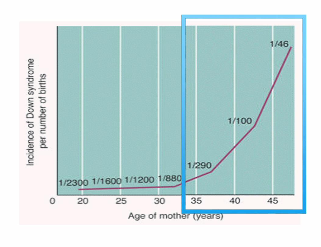
Trisomy 21 (47 chromosomes; 3‐ chrms 21) 94% of Down’s Risk increases with maternal age
Robertsonian translocation involving chrm 21 3‐4% of cases Not associated with maternal age
Trisomy 21 Mosaicism 1‐2% of cases
Genetics
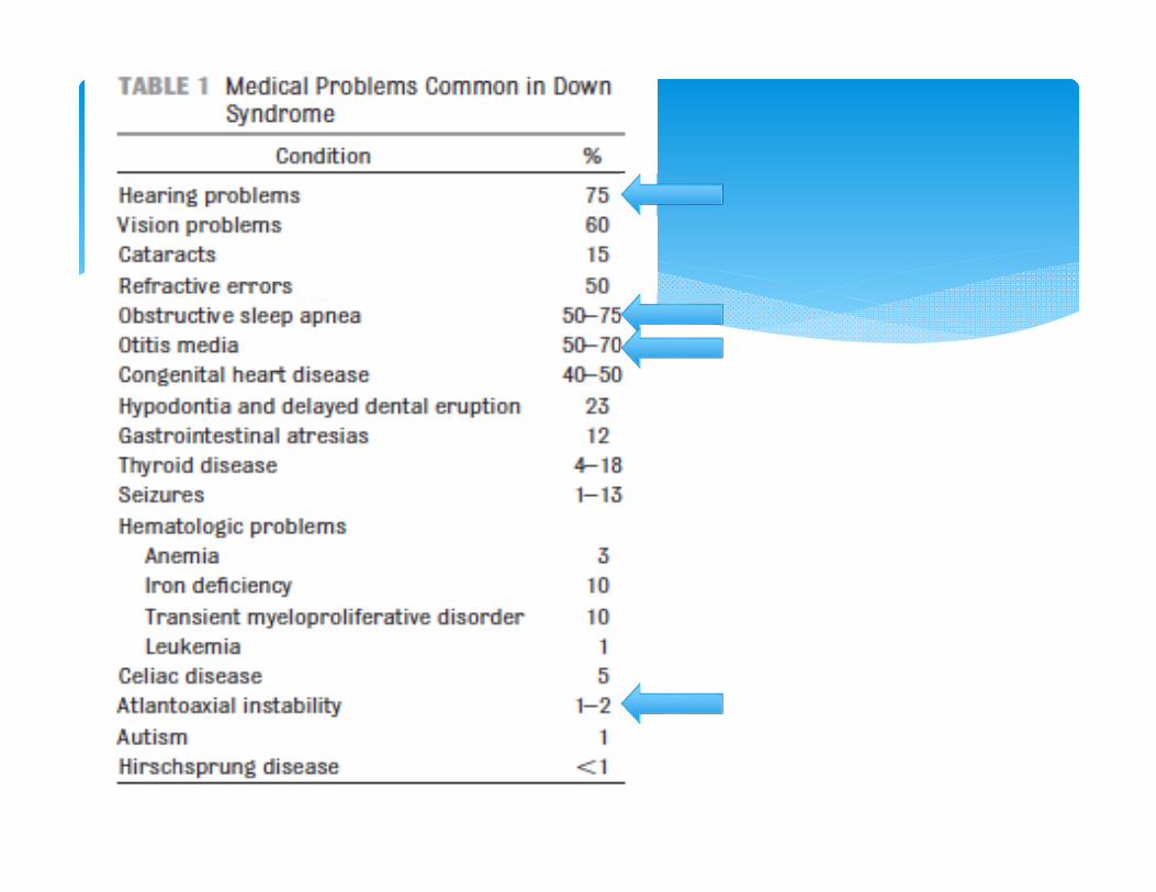
Most common congenital chromosomal abnormality 1 of 700 live birth1
Massive gains life expectancy over the past 40 years Life expectancy in 1983 – 25 years2
Life expectancy in 2014 – 50 to 60 years3
Primary reasoning – congenital heart surgical advancement2,3
> 50% report seeing an otolaryngologist regularly4
Epidemiology
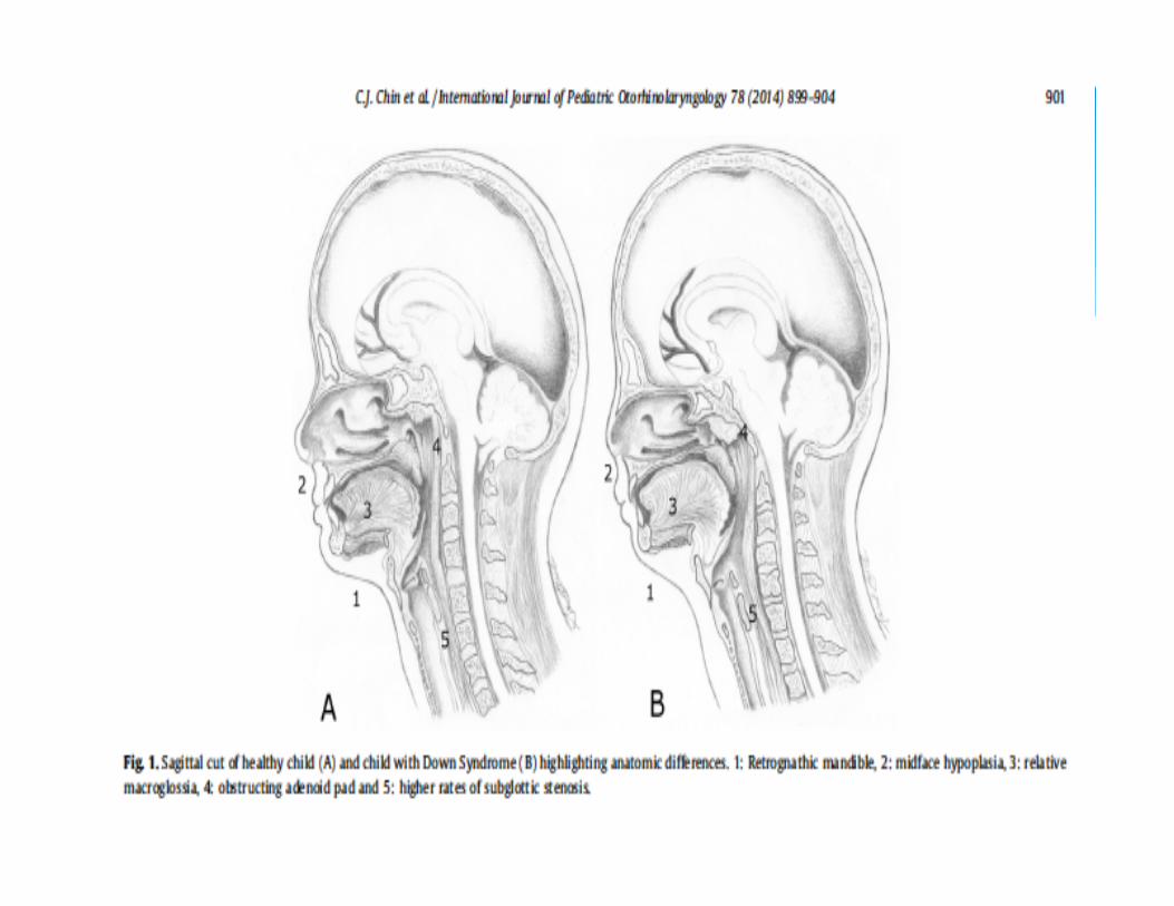
Anatomical Midface hypoplasia Shortened palate Relative Macroglossia Narrowed oropharynx and nasopharynx Hypotonia Paranasal sinus abnormalities
Systemic Immunologic deficiency Ciliary dyskinesia
Predisposition to ENT related Problems

Congenital heart disease Pulmonary hypertension GERD Subglottic stenosis Cervical instability
Comorbidities
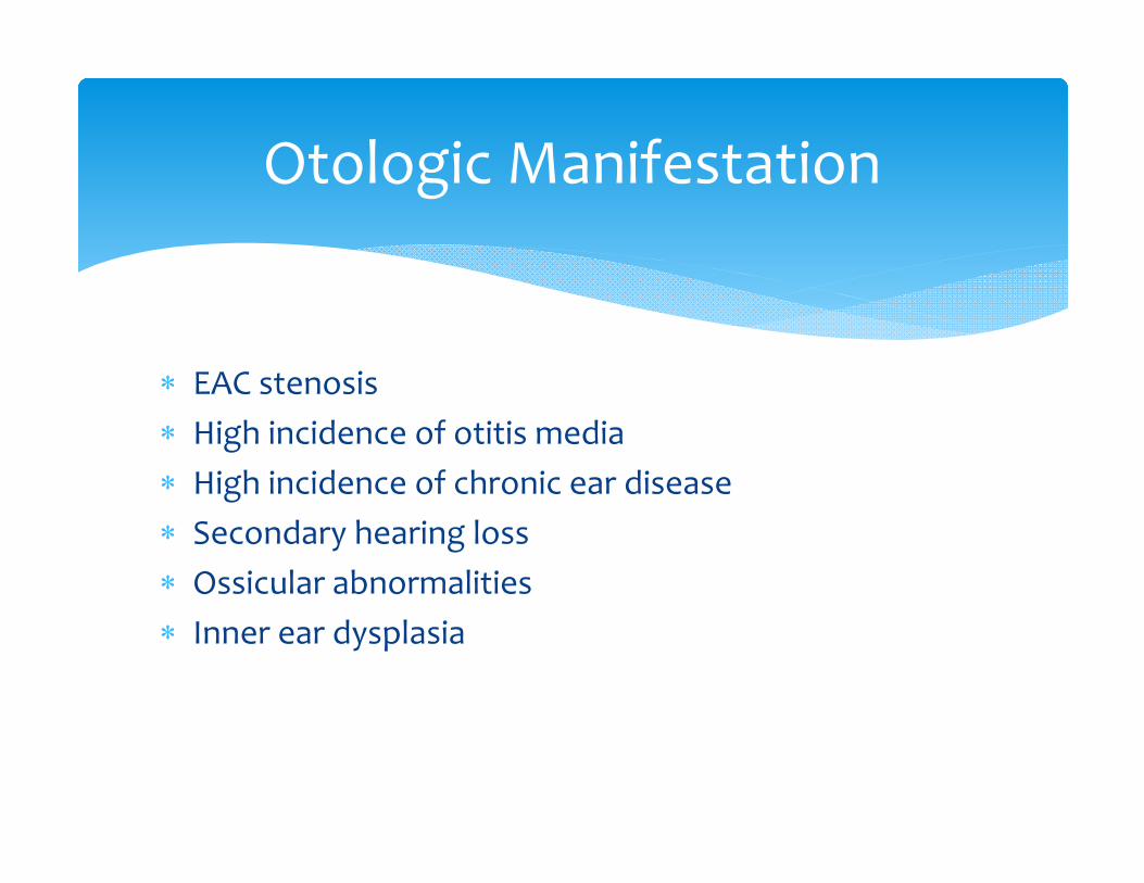
EAC stenosis High incidence of otitis media High incidence of chronic ear disease Secondary hearing loss Ossicular abnormalities Inner ear dysplasia
Otologic Manifestation
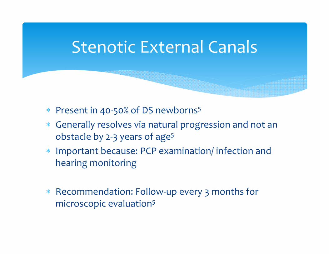
Present in 40‐50% of DS newborns5
Generally resolves via natural progression and not an obstacle by 2‐3 years of age5
Important because: PCP examination/ infection and hearing monitoring
Recommendation: Follow‐up every 3 months for microscopic evaluation5
Stenotic External Canals
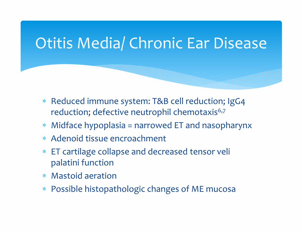
Reduced immune system: T&B cell reduction; IgG4 reduction; defective neutrophil chemotaxis6,7
Midface hypoplasia = narrowed ET and nasopharynx Adenoid tissue encroachment ET cartilage collapse and decreased tensor velipalatini function
Mastoid aeration Possible histopathologic changes of ME mucosa
Otitis Media/ Chronic Ear Disease
The Skull Base & Nasopharynx in DS in Relation to Hearing8
28 DS/ 33 non‐syndromic: age & sex matched All underwent pneumatic otoscopy, audio, lateral x‐ray DS patients demonstrated smaller nasopharyngeal area & less acute angle between skull base & hard palate
Resulted in soft tissue encroachment and less acute angle in children with DS and hearing loss
Midface Hypoplasia/ Nasopharynx

Congenital anomalies of the ET in DS: Histopathology9
DS ET smaller, collapsed in midcartilaginous, isthmus and poorly developed lateral cartilage
Temporal bone morphometric study on ET & assoc. structures in patients with chromosomal aberrations10
Chromosomal aberration patients had smaller volume of lateral lamina cartilage, reduced tensor veli palatini m. attachment
Chrom. aberration patients reduced LL to ML ratio
Eustachian Tube
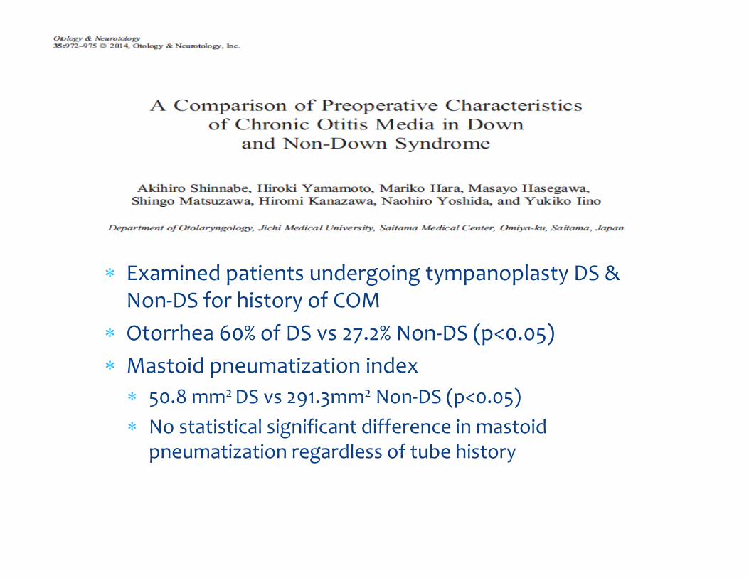
Examined patients undergoing tympanoplasty DS & Non‐DS for history of COM
Otorrhea 60% of DS vs 27.2% Non‐DS (p<0.05) Mastoid pneumatization index 50.8 mm2 DS vs 291.3mm2 Non‐DS (p<0.05) No statistical significant difference in mastoid
pneumatization regardless of tube history
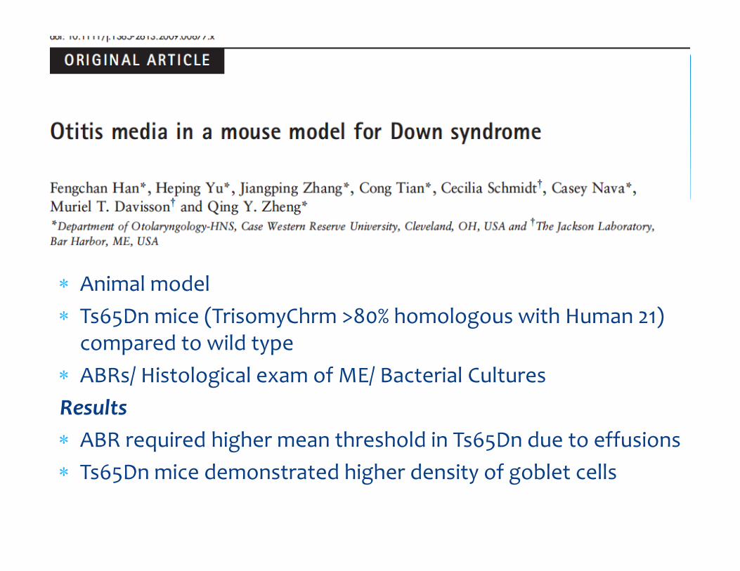
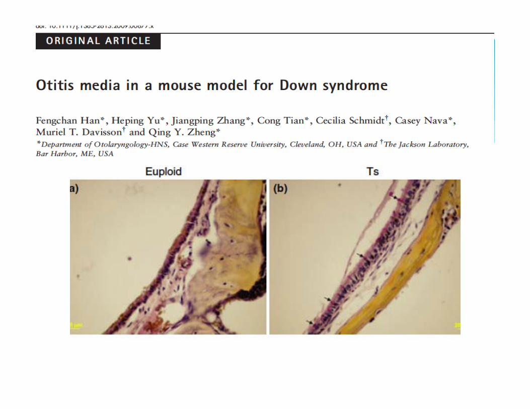
Animal model Ts65Dn mice (TrisomyChrm >80% homologous with Human 21) compared to wild type
ABRs/ Histological exam of ME/ Bacterial CulturesResults ABR required higher mean threshold in Ts65Dn due to effusions Ts65Dn mice demonstrated higher density of goblet cells
Higher prevalence of hearing loss in DS regardless of CHL/ Mixed/ SNHL12
Estimated 50‐90% of DS children dx with hearing impairment vs 4‐9% general population12,13
Monitoring is paramount as hearing loss can be dismissed as natural course/ intellectual impairment
Hearing Loss
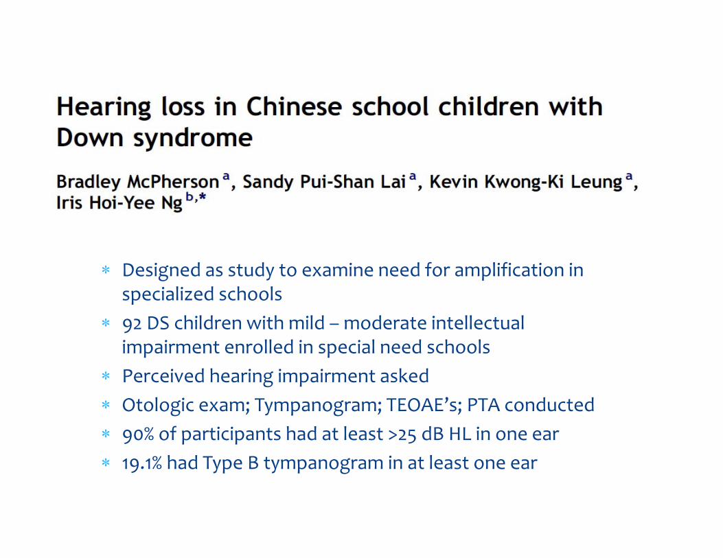
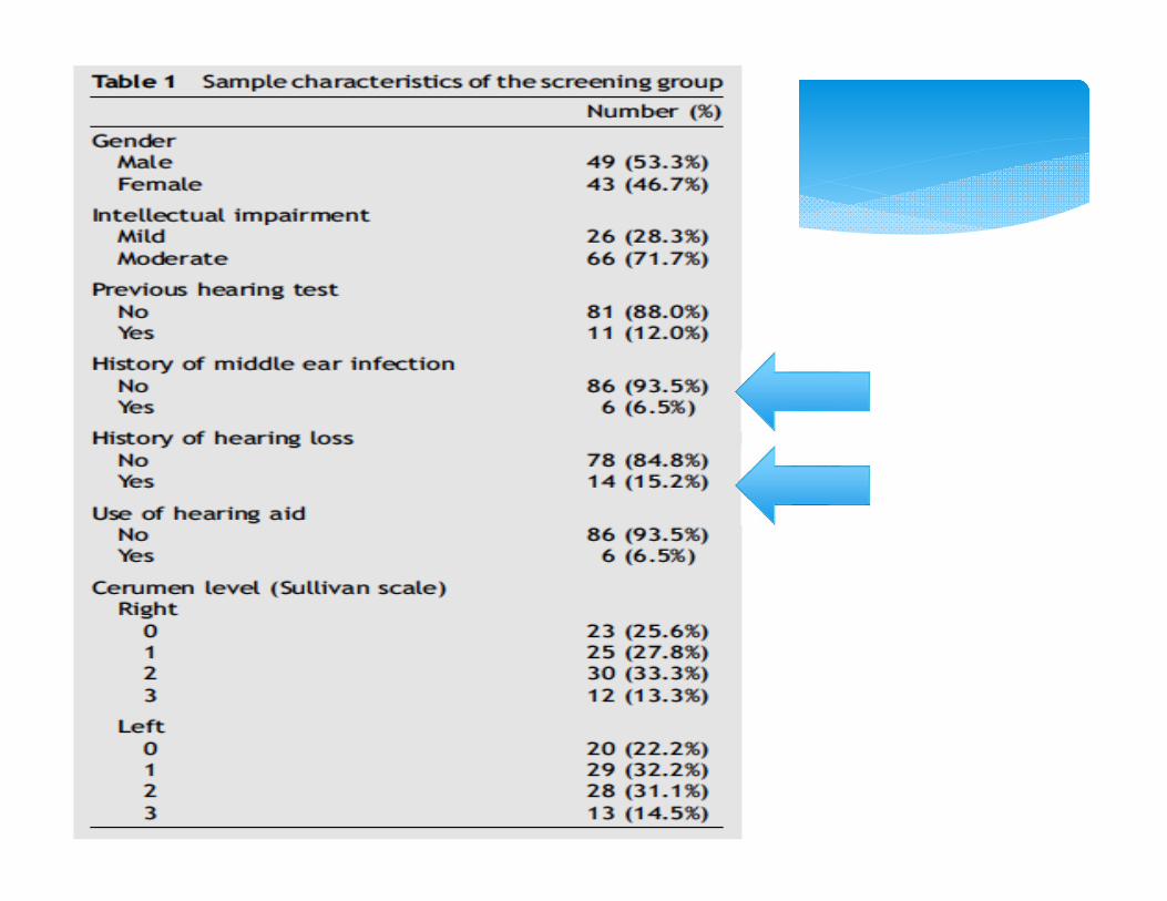
Designed as study to examine need for amplification in specialized schools
92 DS children with mild – moderate intellectual impairment enrolled in special need schools
Perceived hearing impairment asked Otologic exam; Tympanogram; TEOAE’s; PTA conducted 90% of participants had at least >25 dB HL in one ear 19.1% had Type B tympanogram in at least one ear
Hearing loss is masked by various delays seen in DS (speech; intellectual)14
Early detection and associated maintenance critical Effect of hearing loss is greater on children with developmental delay compared to non‐delayed (critical)14
Hearing Loss
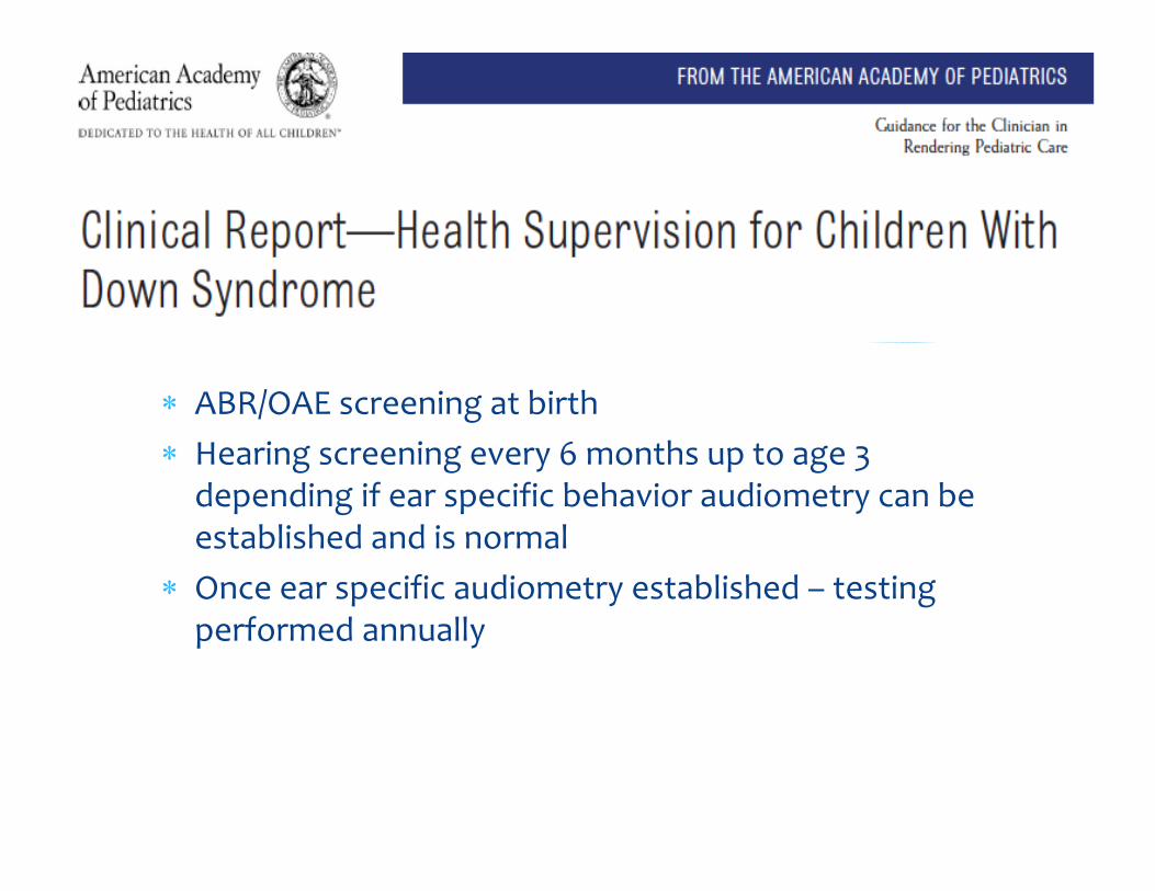
ABR/OAE screening at birth Hearing screening every 6 months up to age 3 depending if ear specific behavior audiometry can be established and is normal
Once ear specific audiometry established – testing performed annually

Surprisingly controversial
Short‐term efficacy of tympanostomy tubes for secretory OM in children with DS15
24 DS vs 21 non‐DS/ All with secretory OM and CHL/ Age matched Audiogram performed 6‐9 wks post BMT 60% of DS vs 91% in non‐DS reported improvement NOTE: all patients over the age of 6 ‐> delay of treatment
Tympanostomy Tubes
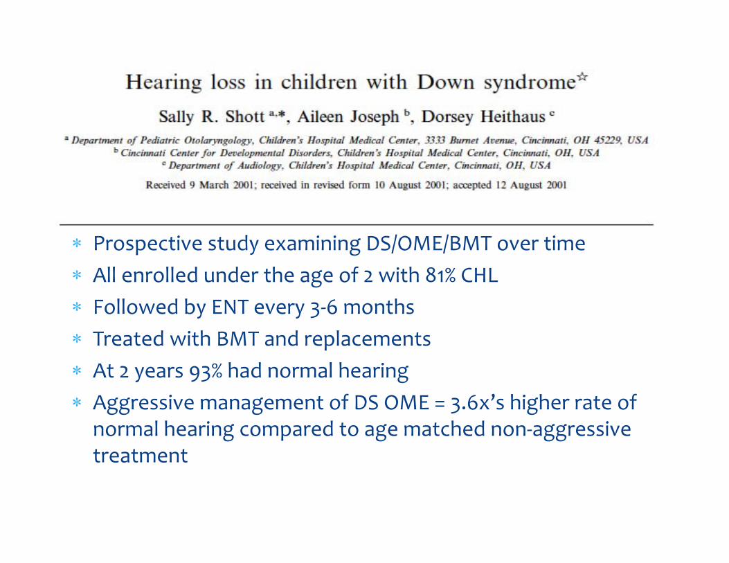
Prospective study examining DS/OME/BMT over time All enrolled under the age of 2 with 81% CHL Followed by ENT every 3‐6 months Treated with BMT and replacements At 2 years 93% had normal hearing Aggressive management of DS OME = 3.6x’s higher rate of normal hearing compared to age matched non‐aggressive treatment

Age requiring first set of tubes
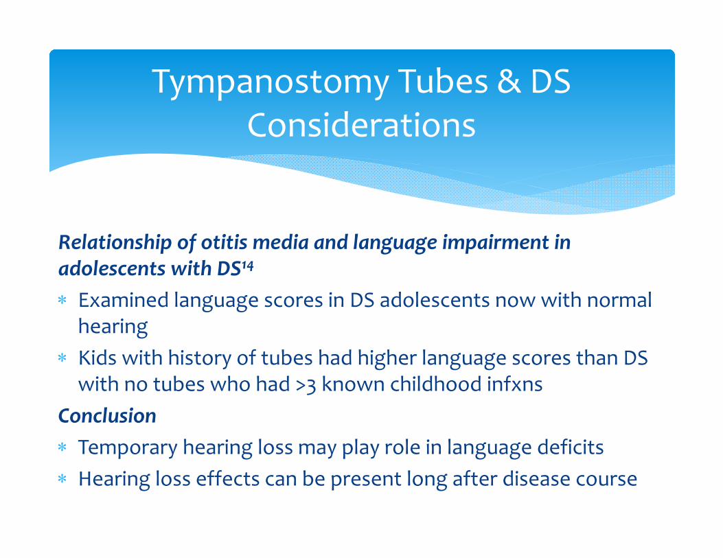
Relationship of otitis media and language impairment in adolescents with DS14
Examined language scores in DS adolescents now with normal hearing
Kids with history of tubes had higher language scores than DS with no tubes who had >3 known childhood infxns
Conclusion Temporary hearing loss may play role in language deficits Hearing loss effects can be present long after disease course
Tympanostomy Tubes & DS Considerations
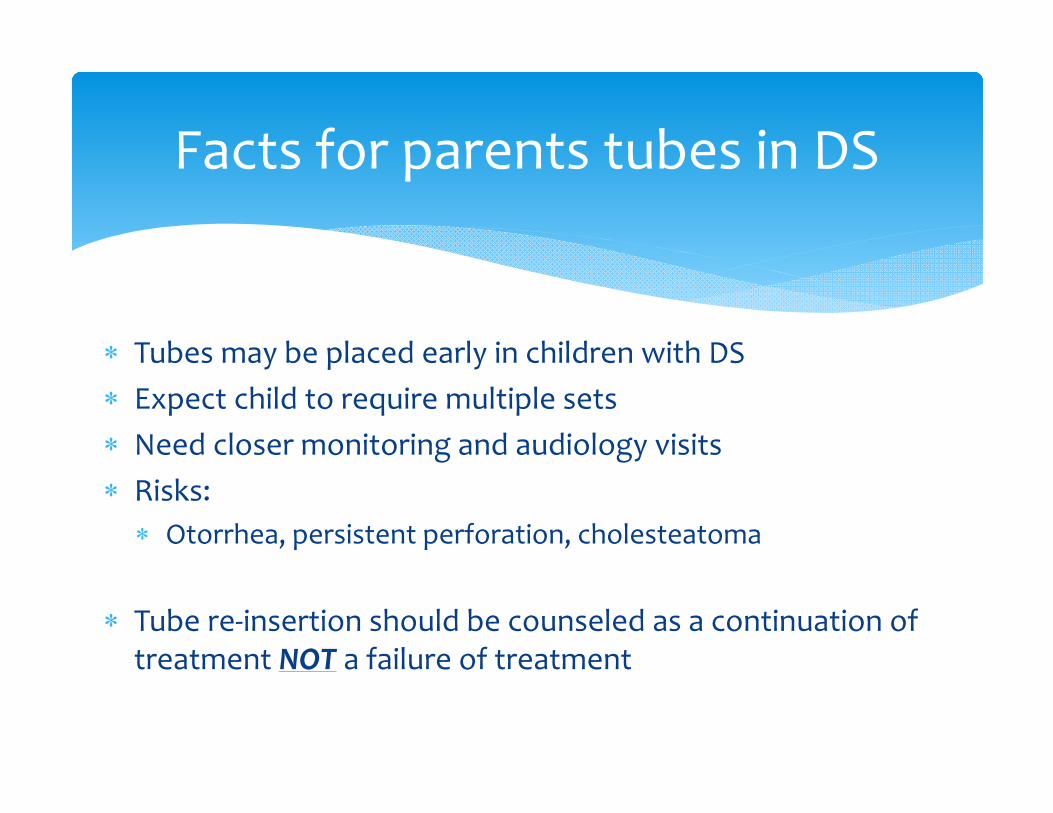
Tubes may be placed early in children with DS Expect child to require multiple sets Need closer monitoring and audiology visits Risks: Otorrhea, persistent perforation, cholesteatoma
Tube re‐insertion should be counseled as a continuation of treatment NOT a failure of treatment
Facts for parents tubes in DS
Estimated as high as 80% in DS vs 1‐2% non‐syndromic15,16
Many predisposing factors that contribute Single modality treatment often not curative Can lead to further neurocognitive delay in the already delayed
Pulmonary HTN in children predisposed to cardiac anomalies
Obstructive Sleep Apnea
Midfacial and mandibular hypoplasia Relative macroglossia Glossoptosis Smaller upper airway prone to adenotonsillar encroachment Lingual tonsil hypertrophy Laryngomalacia Increased secretions Increased obesity Generalized hypotonia
Predisposing Factors
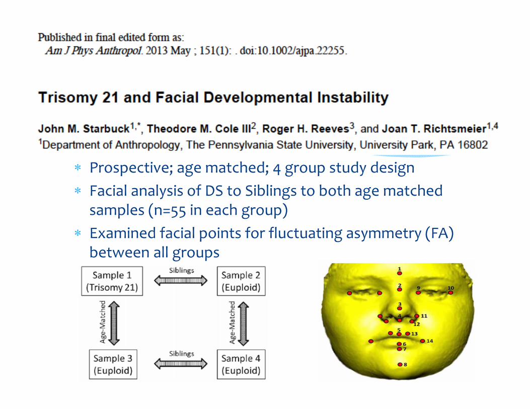
Prospective; age matched; 4 group study design Facial analysis of DS to Siblings to both age matched samples (n=55 in each group)
Examined facial points for fluctuating asymmetry (FA) between all groups
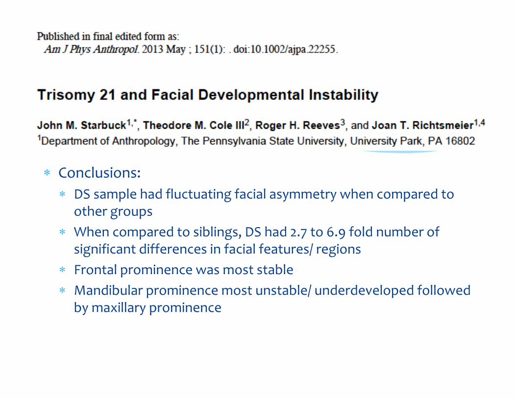
Conclusions: DS sample had fluctuating facial asymmetry when compared to
other groups When compared to siblings, DS had 2.7 to 6.9 fold number of
significant differences in facial features/ regions Frontal prominence was most stable Mandibular prominence most unstable/ underdeveloped followed
by maxillary prominence
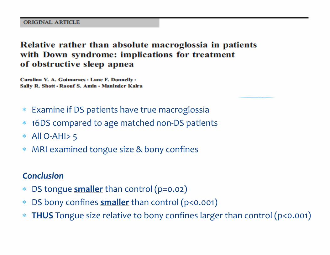
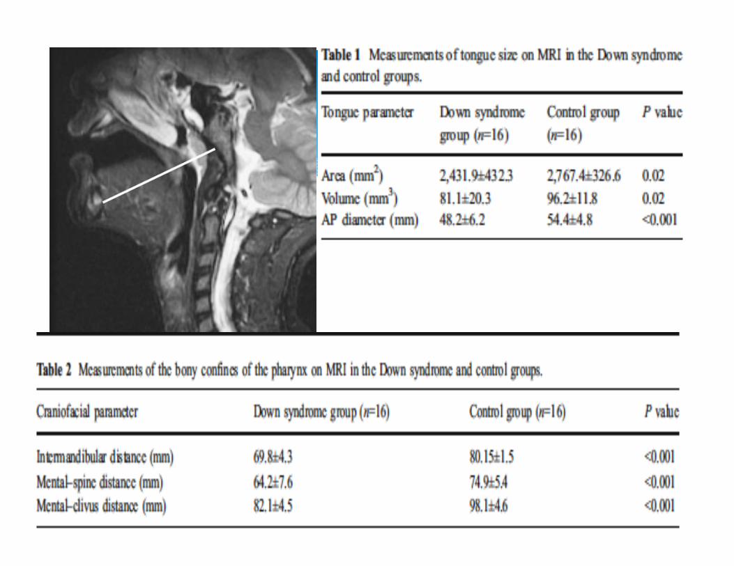
Examine if DS patients have true macroglossia 16DS compared to age matched non‐DS patients All O‐AHI> 5 MRI examined tongue size & bony confines
Conclusion DS tongue smaller than control (p=0.02) DS bony confines smaller than control (p<0.001) THUS Tongue size relative to bony confines larger than control (p<0.001)
Thought to be secondary narrowing of nasopharynx and oropharynx
Contribution by relative hypotonia Adenotonsillectomy only curative 27% of DS patients17
Ongoing CHP study on TA, PSG and DS (Thottam, Choi, Kitsko) CSA decrease post TA (p=0.02) 88% reduction in disease severity (p<0.001) TA size correlated with surgical response (p=0.02)
Adenotonsillar Encroachment
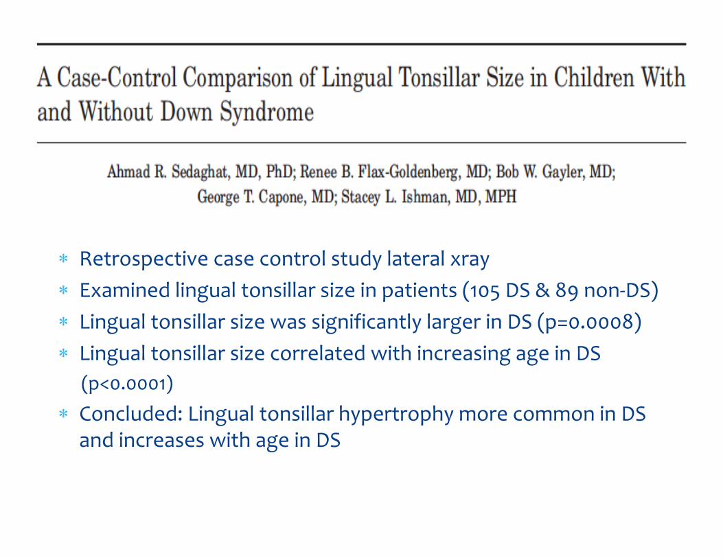
Retrospective case control study lateral xray Examined lingual tonsillar size in patients (105 DS & 89 non‐DS) Lingual tonsillar size was significantly larger in DS (p=0.0008) Lingual tonsillar size correlated with increasing age in DS
(p<0.0001) Concluded: Lingual tonsillar hypertrophy more common in DS and increases with age in DS
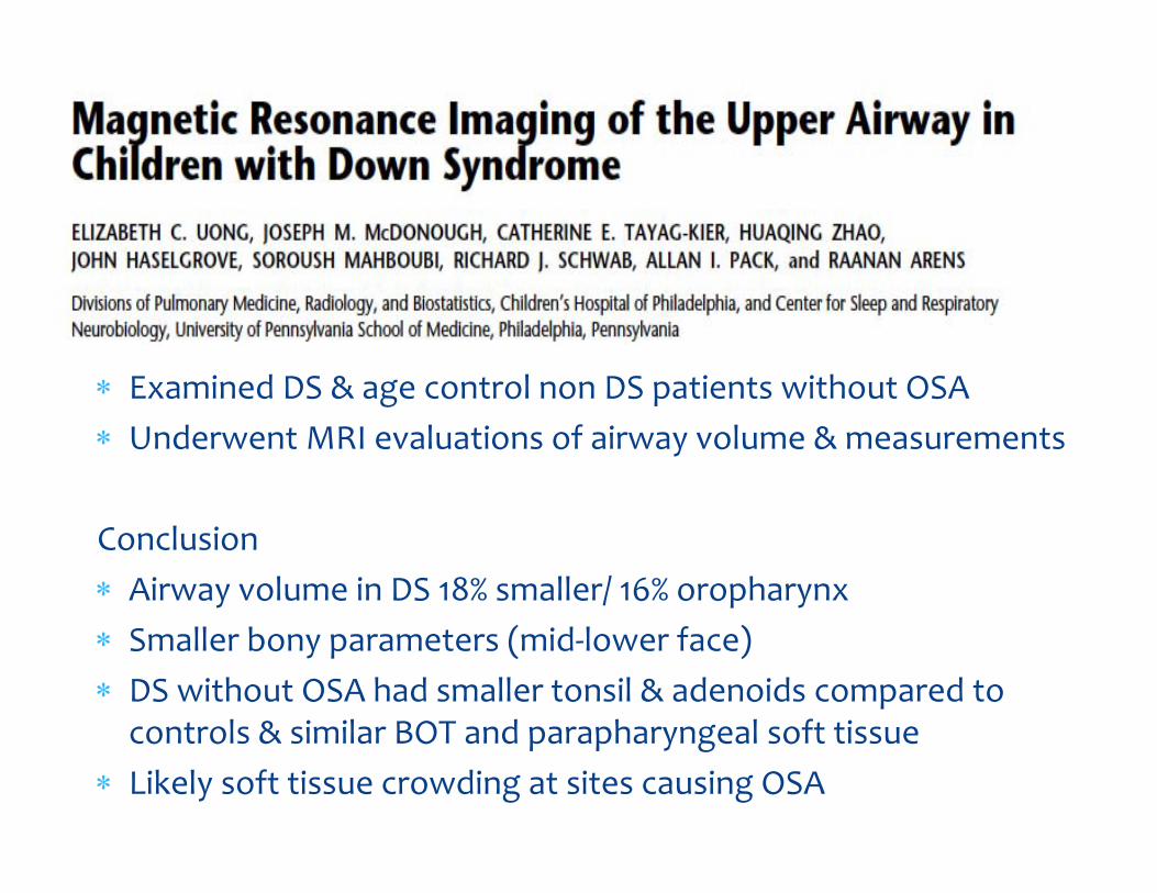
Examined DS & age control non DS patients without OSA Underwent MRI evaluations of airway volume & measurements
Conclusion Airway volume in DS 18% smaller/ 16% oropharynx Smaller bony parameters (mid‐lower face) DS without OSA had smaller tonsil & adenoids compared to controls & similar BOT and parapharyngeal soft tissue
Likely soft tissue crowding at sites causing OSA
So, who needs to be evaluated and when?
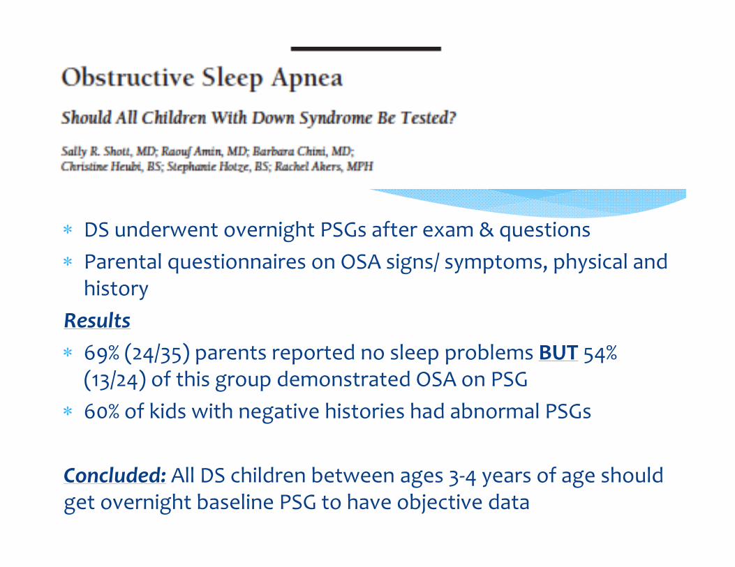
DS underwent overnight PSGs after exam & questions Parental questionnaires on OSA signs/ symptoms, physical and history
Results 69% (24/35) parents reported no sleep problems BUT 54% (13/24) of this group demonstrated OSA on PSG
60% of kids with negative histories had abnormal PSGs
Concluded: All DS children between ages 3‐4 years of age should get overnight baseline PSG to have objective data
HAS TO BE OVERNIGHT STUDY
Nap studies tend to underestimate severity18
Nap studies have demonstrated less sensitivity (75%) of patients with OSA; compared to full night (100%) in previous study18
So what kind of study?
CLINICAL PRACTICE GUIDELINE19
All children with DS get PSG before undergoing TA
“Parents just don’t understand” – it’s under reported Give them objective evidence
May require more than just a TA Can follow results and progression (baseline)
SO why get a PSG in DS
Adenotonsillectomy alone initially No data to support more aggressive surgery initially20
TA+lateral pharyngoplasty vs TA alone20
1. No statistical difference in OSA post‐operatively with roughly 50‐60% both having residual OSA
Next place to look ‐> BOT/ lingual tonsils Genioglossus advancement + BOT coblation post TA21
1. 63% of DS patients AHI < 5 post procedure
Surgical Treatment DS & OSA
PSG needed before surgery TA often not curative so often set realistic goals
25% cure rate but a much higher reduction rate22
Reduce CPAP settings/ increase compliance
If obese BMI reduction always helps Increased risk and have to stay overnight23,24
Longer hospital stay; decreased PO; 5x’s increase in respiratory event
Increased risk of VPI and hypernasal speech24,25
High arched palate, hypotonia, Levator dysfunction More surgery/ further interventions and PSG’s are common
Points for Parents on OSA/DS/Surgery
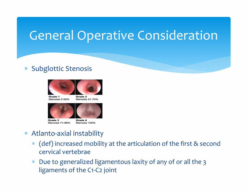
Subglottic Stenosis
Atlanto‐axial instability (def) increased mobility at the articulation of the first & second
cervical vertebrae Due to generalized ligamentous laxity of any of or all the 3
ligaments of the C1‐C2 joint
General Operative Consideration
Subglottic Stenosis and DS26
4% of DS population required LTRS vs. 0.15% of non‐DS Secondary to congenital narrowing and acquired
LTP for SGS in DS: The Cincinnati Experience27
Higher rate of intubation 2ndary to cardiac surgery Severe respiratory infections requiring intubation The above occurs at a young age = increased risk of SGS
Subglottic Stenosis
Prospectively evaluated DS airway size in DS (42) compared to control (32)
Leak tests and MRIs (evaluate diameter) Concluded DS kids required ETT 2‐3 sizes smaller
Recommended: ETT in DS be 2 sizes smaller for intubation and critical to check for air leak at 10‐30 cm H20
Was first brought to wide attention in 1983 Special Olympics Incidence reported to be around 14% BUT only 1.5%27
determined to symptomatic Catastrophic injury can occur at extension and rotation BUT has been demonstrated in patients with long standing history of signs (abnormal gait; limited neck mobility etc)28
Recommendations: 1. History of neurological signs greater priority than radiography2. Support head with rotation for BMT and limited extension
AA Instability in DS
For stenotic ear canals hearing and cerumen should be monitored closely
DS child should undergo behavioral audiologic testing q6 months or q3 if canals are stenotic until able to tolerate ear specific testing
Treat OME aggressively & prepare for multiple tubes High rate of OSA & get PSG at 3‐4 y/o regardless TA is first treatment but only 25% successful Intubate with tube 2 sizes smaller Careful when turning head and history is most important
Summary

Thank You
1. Centers for Disease Control and Prevention (CDC). Down syndrome prevalence at birth: United States, 1983‐1990. MMWR Morb Mortal Wkly Rep 1994;43: 617–22.2. Yang Q, Rasmussen S, Friedman J. Mortality associated with Down’s syndrome in the USA from 1983 to 1997: a population based study. Lancet 2002;359:1019–25.3. Bittles AH, Bower C, Hussain R, et al. The four ages of Down syndrome. Eur J Public Health 2007;17(2):221–5.4. Hans PS, Belloso A, Sheehan PZ. Parental satisfaction with health services provided to children with Down syndrome in north‐west England: an ENT perspective. J Laryngol Otol
2006;121:382–6.5. Shott SR. Down syndrome: common otolaryngologic manifestations. Am J Med Genet C Semin Med Genet 2006;142C:131–40.6. Chaushu S, Yefenof E, Becker A, et al. A link between parotid salivary Ig level and recurrent respiratory infections in young Down’s syndrome patients. Oral Microbiol Immunol 2002;17:172–
6.7. Nespoli L, Burgio GR, Ugazio AG, et al. Immunological feature of Down’s syndrome: a review. J Intellect Disabil Res 1993;37:543–51.8. Brown PM, Lewis GT, Parker AJ, et al. The skull base and nasopharynx in relation in Down’s syndrome in relation to hearing impairment. Clin Otolaryngol 1989;14:241–6.9. Yamaguchi N, Sando I, Hashida Y, et al. Histologic study of eustachian tube cartilage with and without congenital anomalies: a preliminary study. Ann Otol Rhinol Laryngol 1990;99:984–7.10. Miura M, Sando I, Balaban CD, et al. Temporal bone morphometric study on the eustachian tube and its associated structures in patients with chromosomal aberrations. Ann Otol Rhinol
Laryngol 2002:111;8:722‐729.11. Han F, Yu H, Zhang J, et al. Otitis media in a mouse model for Down syndrome. Int J Exp Pathol 2009;90:480–8.12. Cunningham C, McArthur K. Hearing loss and treatment in young Downs syndrome children. Child Care Health Dev 1982;7:357–74.13. McPherson B, Lai S, Leung K, et al. Hearing loss in Chinese school children with Down syndrome. Int J Pediatr Otorhinolaryngol 2007;71:1905–15.14. Balkany TJ, Dows MP, Jafek BW, Krajicek MJ. Hearing loss in Downs syndrome: a treatable handicap more common than generally recognized. Clinical Pediatrics. 1979;18:116‐118. 15. Marcus C, Keens T, Bautista D, Pechmann W, Davidson S. Obstructive Sleep Apnea in Children with Down Syndrome. Pediatrics. 1991;88(1):132‐139 16. . Shott S, Amin R, Chini B, Heubi C, Hotze S, Akers R. Obstructive sleep apnea: should all children with Down syndrome be tested? Arch Otolaryngol Head Neck Surg 2006;132:432‐436. 17. Shete M, Stocks R, Sebelik M, et al. Effects of adeno‐tonsillectomy on polysomnography patterns in Down syndrome children with obstructive sleep apnea: a comparative study with
children without Down syndrome. Int J Pediatr Otorhinolaryngol 2010;74:241–4.18. Marcus C, Keens T, Bautista D, Pechmann W, Davidson S. Obstructive Sleep Apnea in Children with Down Syndrome. Pediatrics. 1991;88(1):132‐139 19. Roland P, Rosenfield R, Brooks L, et al. Polysomnography for sleep disordered breathing prior to tonsillectomy in children. Otolaryngol Head Neck Surg 2011; 145(1):S1–15.20. Mitchell RB. Adenotonsillectomy for obstructive sleep apnea in children: outcome evaluated by pre‐and postoperative polysomnography. Laryngoscope 2007;117(10):1844–54.21. Wootten C, Shott S. Evolving therapies to treat retroglossal and base of tongue obstruction in pediatric obstructive sleep apnea. Arch Otolaryngol Head Neck Surg 2010;136(10):983–7.22. Donnelly L, Shott S, LaRose C, Chini B, Amin R. Causes of persistent obstructive sleep apnea despite previous tonsillectomy and adenoidectomy in children with Down syndrome as
depicted on static and dynamic cine MRI. Am Journal Roentgenology 1994;183:175‐181. 23. Kavanagn K, Kahane J, Kordan B. Risks and benefits of adenotonsillectomy for children with Down syndrome. American Journal of Mental Deficiency 1986;91(1)22‐29 24. Gelder LV. Open nasal speech following adenoidectomy and tonsillectomy. Journal of Communications Disorders, 1974:4:263‐267 69.25. Gibb AG. Hypernasality (rhinolalia aperta) following tonsil and adenoid removal. Journal of Laryngology and Otology 1958;72:433‐451 26. Miller R, Gray S, Cotton R, Myer C, Netterville J. Sunglottic stenosis and Down syndrome. Pediatric Otolaryngology, Am J Otolaryngology. 1990;11:274‐277. 27. Miller J, Bapusten B, Lampard R. Changes at the base of skull and cervical spine in Down syndrome. Journal of the Canadian Association of Radiologists. 1986;37:85‐89. 28. American Academy of Pediatrics, Committee on Sports Medicine and Fitness. Atlantoaxial instability in Down syndrome: Subject Review. Pediatrics 1995;96:151‐154.
Citations