Embed Size (px)
Citation preview
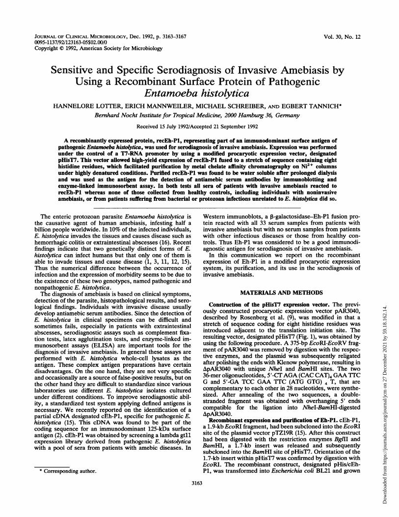
JOURNAL OF CLINICAL MICROBIOLOGY, Dec. 1992, p. 3163-31670095-1137/92/123163-05$02.00/0Copyright C 1992, American Society for Microbiology
Vol. 30, No. 12
Sensitive and Specific Serodiagnosis of Invasive Amebiasis byUsing a Recombinant Surface Protein of Pathogenic
Entamoeba histolyticaHANNELORE LOITER, ERICH MANNWEILER, MICHAEL SCHREIBER, AND EGBERT TANNICH*
Bernhard Nocht Institute for Tropical Medicine, 2000 Hamburg 36, Germany
Received 15 July 1992/Accepted 21 September 1992
A recombinantly expressed protein, recEh-Pl, representing part of an immunodominant surface antigen ofpathogenic Entamoeba histolytica, was used for serodiagnosis of invasive amebiasis. Expression was performedunder the control of a T7-RNA promoter by using a modified procaryotic expression vector, designatedpHisT7. This vector allowed high-yield expression of recEh-Pl fused to a stretch of sequence containing eighthistidine residues, which facilitated purification by metal chelate affinity chromatography on Ni2+ columnsunder highly denatured conditions. Purified recEh-Pl was found to be water soluble after prolonged dialysisand was used as the antigen for the detection of antiamebic serum antibodies by immunoblotting andenzyme-linked immunosorbent assay. In both tests all sera of patients with invasive amebiasis reacted torecEh-Pl whereas none of those collected from healthy controls, including individuals with noninvasiveamebiasis, or from patients suffering from bacterial or protozoan infections unrelated to E. histolytica did so.
The enteric protozoan parasite Entamoeba histolytica isthe causative agent of human amebiasis, infesting half abillion people worldwide. In 10% of the infected individuals,E. histolytica invades the tissues and causes disease such ashemorrhagic colitis or extraintestinal abscesses (16). Recentfindings indicate that two genetically distinct forms of E.histolytica can infect humans but that only one of them isable to invade tissues and cause disease (1, 3, 11, 12, 15).Thus the numerical difference between the occurrence ofinfection and the expression of morbidity seems to be due tothe existence of these two genotypes, named pathogenic andnonpathogenic E. histolytica.The diagnosis of amebiasis is based on clinical symptoms,
detection of the parasite, histopathological results, and sero-logical findings. Individuals with invasive disease usuallydevelop antiamebic serum antibodies. Since the detection ofE. histolytica in clinical specimens can be difficult andsometimes fails, especially in patients with extraintestinalabscesses, serodiagnostic assays such as complement fixa-tion tests, latex agglutination tests, and enzyme-linked im-munosorbent assays (ELISA) are important tools for thediagnosis of invasive amebiasis. In general these assays areperformed with E. histolytica whole-cell lysates as theantigen. These complex antigen preparations have certaindisadvantages. On the one hand, they are not very specificand occasionally are a source of false-positive results, but onthe other hand they are difficult to standardize since variouslaboratories use different E. histolytica isolates culturedunder different conditions. To improve serodiagnostic abil-ity, a standardized test system applying defined antigens isnecessary. We recently reported on the identification of apartial cDNA designated cEh-Pl, specific for pathogenic E.histolytica (15). This cDNA was found to be part of thecoding sequence for an immunodominant 125-kDa surfaceantigen (2). cEh-Pl was obtained by screening a lambda gtllexpression library derived from pathogenic E. histolyticawith a pool of sera from patients with amebic diseases. In
* Corresponding author.
Western immunoblots, a 3-galactosidase-Eh-P1 fusion pro-tein reacted with all 33 serum samples from patients withinvasive amebiasis but with no serum samples from patientswith other infectious diseases or those from healthy con-trols. Thus Eh-P1 was considered to be a good immunodi-agnostic antigen for serodiagnosis of invasive amebiasis.
In this communication we report on the recombinantexpression of Eh-P1 in a modified procaryotic expressionsystem, its purification, and its use in the serodiagnosis ofinvasive amebiasis.
MATERIALS AND METHODS
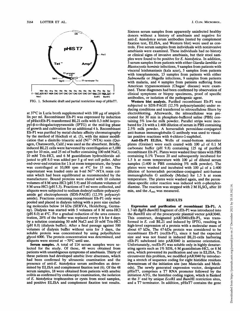
Constrction of the pHisT7 expression vector. The previ-ously constructed procaryotic expression vector pAR3040,described by Rosenberg et al. (9), was modified in that astretch of sequence coding for eight histidine residues wasintroduced adjacent to the translation initiation site. Theresulting vector, designated pHisT7 (Fig. 1), was obtained byusing the following procedure. A 375-bp EcoRI-EcoRV frag-ment of pAR3040 was removed by digestion with the respec-tive enzymes, and the plasmid was subsequently religatedafter polishing the ends with Klenow polymerase, resulting inApAR3040 with unique NheI and BamHI sites. The two36-mer oligonucleotides, 5'-CTAGA (CAC CAT)4 GAATTCG and 5'-GA TCC GAA TTC (ATG GTG) 4 T, that arecomplementary to each other in 28 nucleotides, were synthe-sized. After annealing of the two sequences, a double-stranded fragment was obtained with overhanging 5' endscompatible for the ligation into NAeI-BamHI-digestedApAR3040.Recombinant expression and purification of Eh-P1. cEh-P1,
a 1.9-kb EcoRI fragment, had been subcloned into the EcoRIsite of the plasmid vector pTZ19R (15). After this constructhad been digested with the restriction enzymes BglII andBamHI, a 1.7-kb insert was released and subsequentlysubcloned into the BamHI site of pHisT7. Orientation of the1.7-kb insert within pHisT7 was confirmed by digestion withEcoRI. The recombinant construct, designated pHis/cEh-P1, was transformed into Escherichia coli BL21 and grown
3163
Dow
nloa
ded
from
http
s://j
ourn
als.
asm
.org
/jour
nal/j
cm o
n 27
Dec
embe
r 20
21 b
y 59
.18.
162.
14.
3164 LOTTER ET AL.
Pst
Met Ala Arg His His His His His His His His Glu Phe Gly Ser5'- CALAIG GCT AGA CAC CAT CAC CAT CAC CAT CAC CAT GMATTC GCALEC - 3'
NdeI EcoRI BamHI
FIG. 1. Schematic draft and partial restriction map of pHisT7.
at 37°C in Luria broth supplemented with 100 ,ug of ampicil-lin per ml. Recombinant Eh-P1 was expressed by inductionof pHis/cEh-P1-transformed BL21 cells with 0.3 mM isopro-pyl-f-D-thiogalactopyranoside (IPTG) at the mid-log phaseof growth and cultivation for an additional 4 h. RecombinantEh-Pl was purified by metal chelate affinity chromatographyby the method of Hochuli et al. (5), with the minor modifi-cation that a dinitrilo triacetic acid (Ni2+-NTA) resin (Qui-agen, Chatsworth, Calif.) was used as the absorbent. Briefly,induced BL21 cells were harvested by centrifugation at 5,000rpm for 10 min, and 25 ml of buffer containing 100 mM NaCl,10 mM Tris-HCl, and 6 M guanidinium hydrochloride ad-justed to pH 8.0 was added per 5 g of wet cell pellet. Afterend-over-end rotation for 1 h at room temperature, the lysatewas centrifuged at 10,000 rpm at 4°C for 15 min. Thesupernatant was loaded onto an 8-ml Ni2+-NTA resin col-umn which had been equilibrated as recommended by themanufacturer. Bound proteins were eluted with 10 columnvolumes of 8 M urea-HCl (pH 5.9) and 10 column volumes of8 M urea-HCl (pH 5.3). Fractions of 5 ml were collected, andaliquots were subjected to sodium dodecyl sulfate-polyacryl-amide gel electrophoresis (SDS-PAGE) (12.5% polyacryl-amide). Fractions containing recombinant Eh-Pl only were
pooled and placed in dialysis tubing with a pore size exclud-ing molecules below 14 kDa (SERVA, Heidelberg, Germa-ny). Dialysis was started with 5 volumes of 8 M urea-HCl(pH 8.0) at 4°C. For a gradual reduction of the urea concen-tration, 20% of the buffer was replaced every 8 h for 4 daysby a solution containing 50 mM NaCl and 10 mM Tris-HCl(pH 8.0) (dialysis buffer). After a final passage against 500volumes of dialysis buffer without urea for 3 days, thesoluble protein was concentrated by using polyethyleneglycol 6000. The protein concentration was determined, andaliquots were stored at -70°C until use.Serum samples. A total of 114 serum samples were se-
lected for the study. Of these, 48 were obtained frompatients with unambiguous symptoms of amebiasis. Thirty ofthese patients had developed amebic liver abscesses, whichhad been confirmed by ultrasonic examination and thepresence of anti-E. histolytica serum antibodies as deter-mined by ELISA and complement fixation test (6). Of the 48serum samples, 18 were obtained from patients with amebiccolitis as confirmed by endoscopic examination, the isolationof E. histolytica trophozoites or cysts from stool samples,and positive ELISA and complement fixation test results.
Sixteen serum samples from apparently uninfected healthydonors without a history of amebiasis and negative foranti-E. histolytica serum antibodies (tested by complementfixation test, ELISA, and Western blot) were used as con-trols. Five serum samples from individuals with noninvasiveamebiasis were examined. These individuals had no historyor clinical signs of invasive amebiasis, but their stool sam-ples were found to be positive for E. histolytica. In addition,5 serum samples from patients with either Giardia lamblia orBlastocystis hominis infections, 9 samples from patients withvisceral leishmaniasis (kala azar), 5 samples from patientswith toxoplasmosis, 13 samples from patients with eitherSalmonella or Shigella infections, 9 samples from patientswith malaria, and 4 samples from patients suffering fromAmerican trypanosomiasis (Chagas' disease) were exam-ined. These diagnoses had been confirmed by observation ofclinical symptoms or biopsy specimens, proof of specificantibodies, or isolation of the pathogenic agent.Western blot analysis. Purified recombinant Eh-P1 was
subjected to SDS-PAGE (12.5% polyacrylamide) under re-ducing conditions and transferred to nitrocellulose filters byelectroblotting. Afterwards, the nitrocellulose was pre-coated for 30 min in phosphate-buffered saline (PBS) con-taining 5% low-fat milk powder. Parallel strips were incu-bated for 2 h with a 1:400 dilution of serum in PBS containing2.5% milk powder. A horseradish peroxidase-conjugatedanti-human immunoglobulin G antibody was used to visual-ize positive reactions with 4-chloro-1-naphthol.Anti-Eh-Pl ELISA. Wells of round-bottom microtiter
plates (Greiner) were each coated with 100 pl of 0.1 Mcarbonate buffer (pH 9.8) containing 125 ng of purifiedrecombinant Eh-P1. Plates were washed four times with PBScontaining 0.1% Tween 20 and subsequently incubated for1.5 h at room temperature with 100 pl of diluted serumsamples (1:400 in PBS containing 5% milk powder). Theplates were washed and incubated with 100 ,ul of a 1:400dilution of horseradish peroxidase-conjugated anti-humanimmunoglobulin G antibody (Medac) for 1.5 h at roomtemperature. The plates were washed again, and the perox-idase-specific color reaction was induced with o-phenylen-diamine. The reaction was stopped with 2 M H2SO4 after 10min, and the A492 was measured.
RESULTS
Expression and purification of recombinant Eh-Pl. A1.7-kb BglII-BamHI fragment of cEh-P1 was introduced intothe BamHI site of the procaryotic plasmid vector pAR3040.This construct, designated pAR3040/cEh-P1, was trans-formed in E. coli BL21 and induced by IPTG, resulting inhigh-level expression of a protein with a molecular mass ofabout 67 kDa. The 67-kDa protein was considered to berecombinant Eh-P1 (recEh-P1), since it had the expectedsize and was not found in induced BL21-cells harboringcEh-P1 subcloned into pAR3040 in antisense orientation.Unfortunately, recEh-P1 was soluble only in highly denatur-ating agents such as 1% SDS, 6 M guanidinium-HCl, or 8 Murea, which prevented its purification and use in ELISA. Tocircumvent this problem, we modified pAR3040 by introduc-ing a stretch of sequence coding for eight histidine residuesdownstream of the initiation site (see Materials and Meth-ods). The newly generated expression vector, designatedpHisT7, comprises a T7 RNA promoter followed by theinitiation ATG, the histidine coding region, which is flankedat the 3' end by unique EcoRI and BamHI restriction sites,and a T7 terminator. In addition, pHisT7 contains the gene
J. CLIN. MICROBIOL.
Dow
nloa
ded
from
http
s://j
ourn
als.
asm
.org
/jour
nal/j
cm o
n 27
Dec
embe
r 20
21 b
y 59
.18.
162.
14.

SERODIAGNOSIS OF AMEBIASIS WITH E. HISTOLYTICA PROTEIN 3165
1 2 3 4
k[Da kDa
94-
67-
45
3*
FIG. 2. Expression and purification of recEh-Pl as monitored byCoomassie blue-stained SDS-PAGE. Lanes: 1, molecular mass
markers; 2, lysate of E. ccli transformed with recombinant pHisT7/cEh-Pl in which cEh-Pl was subcloned in the antisense orientation
(control); 3, lysate of E. coli transformed with pHisT/cEh-Pl in
which cEh-Pl was subcloned in the sense orientation; 4, purifiedrecEh-Pl after affinity chromatography of E. coli lysate on an
Ni2"-NTA column.
for ampicillin resistance and an origin of replication for E.
coli (Fig. 1).Induction of BL21 cells transformed with pHisT7/cEH-P1
again yielded high-level expression of a 67-kDa protein onlywhen cEh-Pl was introduced into pHisT7 in the sense
orientation (Fig. 2). The 67-kDa protein was purified bymetal chelate affinity chromatography. Induced E. coli cells
were lysed in 6 M guanidinium hydrochloride and applied to
an Ni column. Bound material was eluted by using 8 M
urea with a decreasing pH step gradient. The 67-kDa proteinwas obtained at pH 5.8 and 5.2. Extensive dialysis resulted
in a water-soluble protein which was subjected to protein
sequencing. The sequence disclosed that the 67-kDa proteinwas identical to recEh-Pl, as it was deduced from DNA
sequencing. The yield of soluble recEh-Pl was found to be 4
to 5 mg/500 ml of bacterial culture.
Detection of anti-Eh-Pl antibodies in human sera. (i) Immu-
noblotting. Approximately 100 pLg of recEh-Pl was subjectedto preparative SDS-PAGE and transferred to nitrocellulose.
Serum samples from 24 individuals were tested on parallel
strips at a 1:400 dilution for reactivity against recEh-Pl.Serum samples being used were collected from three pa-
tients with invasive amebiasis, three with malaria, three with
Chagas' disease, three with syphilis, three with toxoplasmo-sis, four with kala azar, one with salmonellosis, and one with
shigellosis; in addition, serum samples from three healthycontrols were included. The results obtained demonstrated
that only the three serum samples from patients with inva-
sive amebiasis were reactive to recEh-Pl (Fig. 3).(ii) ELISA. To -investigate the use of recEh-Pl as an
immunodiagnostic reagent for the detection of antiamebic
antibodies in a more standardizable test system, we used an
ELISA. Wells of microtiter plates were coated with 125 ng of
recEh-Pl each and incubated with human sera at a 1:400
dilution. A total of 114 individual serum samples were
1 2 3 4 5 6 7 8 9 1011 12 13 14 15 16 17 18 19 20 21 222 3 24 25
=
94 -
67 -
45 - ..
30 -
20- W.
\. _ w
*. .2.:.i .f
.;,,* i4: ti it 9 r:.E "@*S,,...........
X 4 -4;E 1t ':FIG. 3. Immunoblots of subject sera with purified recombinant
Eh-Pl. The lanes represent immunoblots of sera from patients withmalaria (lanes 2 to 4), Chagas' disease (lanes 5 to 7), syphilis (lanes8 to 10), salmonellosis (lane 11), shigellosis (lane 12), toxoplasmosis(lanes 13 to 15), kala azar (lanes 16 to 19), and invasive amebiasis(lanes 20 to 22). Immunoblots of sera from apparently uninfectedhealthy donors are shown in lanes 23 to 25. Molecular mass markersare added on the left (lane 1).
tested, comprising those from apparently healthy donors (n= 16), those from individuals with noninvasive amebiasis (n= 5), and those from patients with amebic colitis (n = 18),amebic liver abscess (n = 30), kala azar (n = 9), toxoplas-mosis (n = 5), salmonellosis or shigellosis (n = 13), malaria(different Plasmodium spp.) (n = 9), Chagas' disease (n = 4),and G. lamblia or B. hominis infections (n = 5). The amountof reactive antibody was determined by measurement ofA492, as indicated by the color reaction of specific boundhorseradish peroxidase-conjugated anti-human immunoglob-ulin G. All 48 serum samples from patients with invasiveamebiasis exhibit A492 values above 0.4. No statistical dif-ferences were found between samples from patients withamebic liver abscesses and those from patients with amebiccolitis. In contrast, all of the other 66 serum samples testedrevealed A492 values below 0.4 (Fig. 4). The mean of
1.4 -
1.2 -
1.0-SYS 0.8 -
8 -0.6
0.4 -
0.2 _
w
TaI
± J+
I
A B C D E F G H
FIG. 4. Detection of serum antibodies to recombinant Eh-Pl byELISA. Each point represents the A492 of a serum sample from oneindividual. The lanes represent reactivity of sera from differentgroups of infected or uninfected subjects with the following diag-noses: invasive amebiasis (n = 48) (A), healthy controls (n = 16) (B),kala azar (n = 9) (C), toxoplasmosis (n = 5) (D), salmonellosis orshigellosis (n = 13) (E), malaria (different Plasmodium spp.) (n = 9)(F), Chagas' disease (n = 4) (G), G. lamblia or B. hominis infections(n = 5) (H), and noninvasive amebiasis (n = 5) (I).
VOL. 30, 1992
*"1a.
Dow
nloa
ded
from
http
s://j
ourn
als.
asm
.org
/jour
nal/j
cm o
n 27
Dec
embe
r 20
21 b
y 59
.18.
162.
14.

3166 LOTTER ET AL.
extinction of the 66 control serum samples was calculated tobe 0.158 with a standard deviation of 0.082. Thus, the cutoffoptimal density of 0.4 represents the mean of extinction plusthree times the standard deviation of the number of controls.
DISCUSSION
Immunodiagnosis of invasive amebiasis is usually per-formed with whole-cell lysates or membrane fractions ofcultured E. histolytica trophozoites. The E. histolyticastrains and culture methods used vary among differentlaboratories. To standardize the diagnostic assays, a reduc-tion of the protein number as well as a characterization of theapplied antigens is desired.The use of recombinant proteins offers the advantage of
testing with defined antigens, which might improve specific-ity and should facilitate standardization of serological as-says. In addition, infinite amounts of the proteins can begenerated, and costly culturing of a potential pathogen aswell as changes in antigenicity during culturing are avoided.The first serological investigation applying a recombinant
protein of E. histolytica was reported by Stanley et al. (10).They used a protein, designated SREHP (serine-rich E.histolytica protein), which was found to possess multipletandem repeats. As described for other immunodominantproteins of E. histolytica, the native correlate of SREHP isassociated with the ameba membrane. A total of 65 serumsamples from patients with invasive amebiasis were testedby immunoblotting, and 82% reacted with SREHP, indicat-ing that a number of patients fail to produce detectableamounts of anti-SREHP antibodies reacting with the dena-tured recombinant protein.
During this study, we investigated the use of anotherrecombinant protein, recEh-P1, for the immunodiagnosis ofinvasive amebiasis. Eh-P1 represents part of an immuno-dominant 125-kDa surface protein of pathogenic E. histolyt-ica (2).
Expression of recEh-P1 was performed by using a modi-fied procaryotic expression vector, pHisT7. This vector wasderived from the well-established plasmid pAR3040, whichallows high-yield expression of recombinant proteins (9).Overexpression in procaryotic expression systems oftenresults in recombinant proteins that are soluble in highlydenaturing agents only, preventing further purification ofthese proteins. pHisT7 offers the advantage of circumvent-ing this problem. The fusion of eight histidine residues toEh-P1 allowed purification by Ni2" affinity chromatographyin the presence of 6 M guanidinium-HCl or 8 M urea withina short period. The serological data obtained with recEh-P1as the antigen suggested that human sera do not react withthe histidine tail. Thus, there is no need to eliminate thehistidine residues in recombinant proteins used for serodi-agnosis.Another advantage was the finding that urea-solubilized,
purified recEh-P1 was water soluble after prolonged dialysis,allowing its application in more automatic standard operat-ing procedures such as ELISA.To our knowledge, only one purified E. histolytica antigen
has been studied for recognition by human immune sera inan ELISA. Ravdin et al. (8) have shown that 99% of serumsamples from patients with invasive amebiasis recognized apurified surface antigen, which is known as the Gal/GalNAc-inhibitable lectin of E. histolytica. Although the primarystructure of this lectin has recently been determined bycloning the corresponding gene (7, 13, 14), no data on the use
of recombinant expressed lectin in serodiagnosis of amebia-sis are available.Our serological investigations of the use of purified re-
cEh-P1 in an ELISA demonstrated 100% sensitivity andspecificity to sera of patients with invasive amebiasis. Incontrast, previous Western blot analyses with trophozoiteextract revealed that the 125-kDa natural correlate of Eh-P1reacted only with 73% of serum samples collected frompatients with amebic liver abscesses (2). Several reasons canbe put forward to explain the higher sensitivity of the partialrecombinant protein: (i) epitopes of antigenic relevance maybe covered in the natural full-length protein; (ii) relevantepitopes are destroyed during denaturation for Westernblotting; (iii) epitopes are discovered which are normallydisguised by glycosylation in the natural protein but arerecognized in recEh-P1 expressed in E. coli as a result of thelack of glycosylation in this system; and (iv) a higherconcentration of the recombinant protein is applied in ourtest systems, and this allows us to detect low levels ofspecific antibodies.
Introduction of new antigen preparations to replace al-ready existing ones in serological assays must maintain orincrease the sensitivity of the test system. Assays being usedare able to identify 95 to 100% of patients with amebic liverabscesses and 85 to 95% of patients with amebic colitis (4).So far, recEh-P1 fulfills the criteria demanded for the appli-cation of a single protein in a serological test system. On theother hand, only very few of the sera we have examinedwere collected from residents of areas endemic for amebia-sis. With the purified lectin, Ravdin et al. (8) found antiame-bic serum antibodies in 25% of serum samples from appar-ently uninfected individuals from the Durban area, Republicof South Africa. From statistical analyses it was suggestedthat this result most probably represents persistence ofantibodies after self-limited infections with pathogenic E.histolytica (8). In a preliminary study we have analyzedsera collected from three patients with amebic liver ab-scesses after successful treatment. The antibody response torecEh-P1 was found to be negative in the sera from two ofthem, one after 3 months and one after 9 months, but is stillpositive in the third after more than 1 year (data not shown).Further investigations are needed to analyze more thor-oughly the time course of the antibody response againstrecEh-P1 after E. histolytica infections.
REFERENCES1. Clark, C. G., and L. S. Diamond. 1991. Ribosomal RNA genes
of "pathogenic" and "nonpathogenic" Entanoeba histolyticaare distinct. Mol. Biochem. Parasitol. 49:297-302.
2. Edman, U., M. A. Meraz, S. Rausser, N. Agabian, and I. Meza.1990. Characterization of an immunodominant variable surfaceantigen from pathogenic and nonpathogenic Entamoeba his-tolytica. J. Exp. Med. 172:879-888.
3. Garfinkel, L. I., M. Giladi, M. Huber, C. Gitler, D. Mirelman,M. Revel, and S. Rozenblatt. 1989. DNA probes specific forEntamoeba histolytica possessing pathogenic and nonpatho-genic zymodemes. Infect. Immun. 57:926-931.
4. Healy, G. R. 1974. Immunologic tools in the diagnosis ofamebiasis: epidemiology in the United States. Rev. Infect. Dis.8:239-246.
5. Hochuli, E., W. S. Bannwarth, H. Dobeli, R. Gentz, and D.Stuiber. 1988. Genetic approach to facilitate purification ofrecombinant proteins with a novel metal chelate adsorbent.Biol. Technol. 6:1321-1325.
6. Knobloch, J., and E. Manaweiler. 1983. Development andpersistence of antibodies to Entamoeba histolytica in patientswith amebic liver abscess. Analysis of 216 cases. Am. J. Trop.Med. Hyg. 27:882-887.
J. CLIN. MICROBIOL.
Dow
nloa
ded
from
http
s://j
ourn
als.
asm
.org
/jour
nal/j
cm o
n 27
Dec
embe
r 20
21 b
y 59
.18.
162.
14.
SERODIAGNOSIS OF AMEBIASIS WITH E. HISTOLYTICA PROTEIN 3167
7. Mann, B. J., B. E. Torian, T. S. Vedvick, and W. A. Petri, Jr.1991. Sequence of a cysteine-rich galactose-specific lectin ofEntamoeba histolytica. Proc. Natl. Acad. Sci. USA 88:3248-3252.
8. Ravdin, J. I., T. F. H. G. Jackson, W. A. Petry, Jr., C. F.Murphy, B. L. P. Ungar, V. Gathiram, J. Skilogiannis, and E.Simjee. 1990. Association of serum antibodies to adherencelectin with invasive amebiasis and asymptomatic infections withpathogenic Entamoeba histolytica. J. Infect. Dis. 162:768-772.
9. Rosenberg, A., B. N. Lade, C. Dao-shan, L. Shu-Wha, J. J.Dunn, and F. W. Studier. 1987. Vectors for selective expressionof cloned T7 RNA polymerase. Gene 56:125-135.
10. Stanley, S. L., Jr., T. F. H. G. Jackson, S. L. Reed, J. Calderon,C. Kunz-Jenkins, V. Gathiram, and E. Li. 1991. Serodiagnosisof invasive amebiasis using a recombinant Entamoeba histolyt-ica protein. JAMA 266:1984-1986.
11. Tachibana, H., S. Ihara, S. Kobayashi, Y. Kaneda, T. Takeuchi,and Y. Watanabe. 1991. Differences in genomic DNA sequencesbetween pathogenic and nonpathogenic isolates of Entamoebahistolytica identified by polymerase chain reaction. J. Clin.
Microbiol. 29:2234-2239.12. Tannich, E., and G. D. Burchard. 1991. Differentiation of
pathogenic from nonpathogenic Entamoeba histolytica by re-striction fragment analysis of a single gene amplified in vitro. J.Clin. Microbiol. 29:250-255.
13. Tannich, E., F. Ebert, and R. D. Horstmann. 1991. Primarystructure of the 170-kDa surface lectin of pathogenic Ent-amoeba histolytica. Proc. Natl. Acad. Sci. USA 88:1849-1853.
14. Tannich, E., F. Ebert, and R. D. Horstmann. Molecular cloningof cDNA and genomic sequences coding for the 35-kDa subunitof the galactose inhibitable lectin of pathogenic Entamoebahistolytica. Mol. Biochem. Parasitol., in press.
15. Tannich, E., R. D. Horstmann, J. Knobloch, and H. H. Arnold.1989. Genomic DNA differences between pathogenic and non-
pathogenic Entamoeba histolytica. Proc. Natl. Acad. Sci. USA86:5118-5122.
16. Walsh, J. A. 1986. Problems in recognition and diagnosis ofamebiasis: estimation of the global magnitude of morbidity andmortality. Rev. Infect. Dis. 8:228-238.
VOL. 30, 1992
Dow
nloa
ded
from
http
s://j
ourn
als.
asm
.org
/jour
nal/j
cm o
n 27
Dec
embe
r 20
21 b
y 59
.18.
162.
14.


![Entamoeba histolytica / E. dispar - qu.edu.iqqu.edu.iq/vmjou/wp-content/uploads/2015/01/Vol.-111-8-14.pdf · [Entamoeba histolytica / E. dispar] ... Entamoeba histolytica trophozoite](https://img.pdfslide.net/doc/110x75/5aa7ee767f8b9ab8228ce260/entamoeba-histolytica-e-dispar-queduiqqueduiqvmjouwp-contentuploads201501vol-111-8-14pdfentamoeba.jpg)