Embed Size (px)
Citation preview
Korean J Radiol 9(3), June 2008 275
Enterobiliary Fistula as a Complication ofEosinophilic Gastroenteritis: a CaseReport
Eosinophilic gasteroenteritis is an uncommon disease with variable clinical fea-tures characterized by eosinophilic infiltration. Clinical manifestations range fromnon-specific gastrointestinal complaints such as nausea, vomiting, crampyabdominal pain, and diarrhea to specific findings such as malabsorption, proteinloosing enteropathy, luminal obstruction, eosinophilic ascites and effusion. Wereport here on a case of eosinophilic gastroenteritis causing enterobiliary fistulawhich is an extremely unusual complication.
osinophilic gastroenteritis is a relatively uncommon disease that’s charac-terized by eosinophilic infiltration of the gastrointestinal tract, peripheraleosinophilia and abnormal gastrointestinal function. Since it was first
recognized by Kaijer in 1937, more than 400 cases of eosinophilic gastroenteritis havebeen reported in the literature (1). Most of the clinical signs and symptoms are relatedto the gastrointestinal tract, e.g., vomiting, abdominal pain, diarrhea, malabsorption,obstruction and ascites. Although various signs and symptoms have been previouslyreported, to the best of our knowledge, enterobiliary fistula as a complication ofeosinophilic gastroenteritis has not yet been reported on. We report here on a case ofenterobiliary fistula that was complicated with eosinophilic gastroenteritis, and thiswas demonstrated by an upper gastrointestinal series (UGI) and magnetic resonancecholangiopancreatography (MRCP).
CASE REPORT
A 68-year-old man presented with a 3-week history of abdominal discomfort,nausea and skin eruption on his entire body. There was no past medial history ofhepatolithiasis, recurrent peptic ulcer, abdominal operation, allergic disease, general-ized skin eruption or food sensitivity. The laboratory investigations revealed a whiteblood cell count of 12.86 103 / l. There was peripheral eosinophilia ranging from 7%to 52% (normal: 0 7.2%). The total IgE was 593 IU/ml (normal: 0 170 IU/ml). Stoolexamination and an enzyme-linked immunosorbent assay (ELISA) test to detectparasite infestation showed negative results. The blood urea nitrogen (BUN) was 40mg/dl (normal: 10 26 mg/dl) and the serum creatine was 2.1 mg/dl (normal: 0.6 1.3mg/dl). An ultrasound scan for evaluation of acute renal failure yielded negativeresults except for pneumobilia. Because of the absence of prior sphincterotomy, asurgical bypass procedure or recent endoscopic retrograde cholangiopancreatography(ERCP), we inferred the existence of enterobiliary fistula. The acute renal failure wasslightly improved and we then performed multidetector computed tomography
Han Myun Kim, MDJi Young Woo, MD
Index terms:Eosinophilic gastroenteritisEnterobiliary fistulaGastrointestinal tract,
inflammationMagnetic resonance (MR) Cholangiopancreatography
DOI:10.3348/kjr.2008.9.3.275
Korean J Radiol 2008;9:275-278Received October 19, 2005; accepted after revision February 9, 2006.
All authors: Department of Radiology,Kangnam Sacred Heart Hospital, HallymUniversity College of Medicine, Seoul150-950, Korea
Address reprint requests to:Ji Young Woo, MD, Department ofRadiology, Kangnam Sacred HeartHospital, Hallym University College ofMedicine, 948-1, Daerim 1-dong,Yungdungpo-gu, Seoul 150-950, Korea.Tel. (822) 829-5241Fax. (822) 832-1845e-mail: [email protected]
E
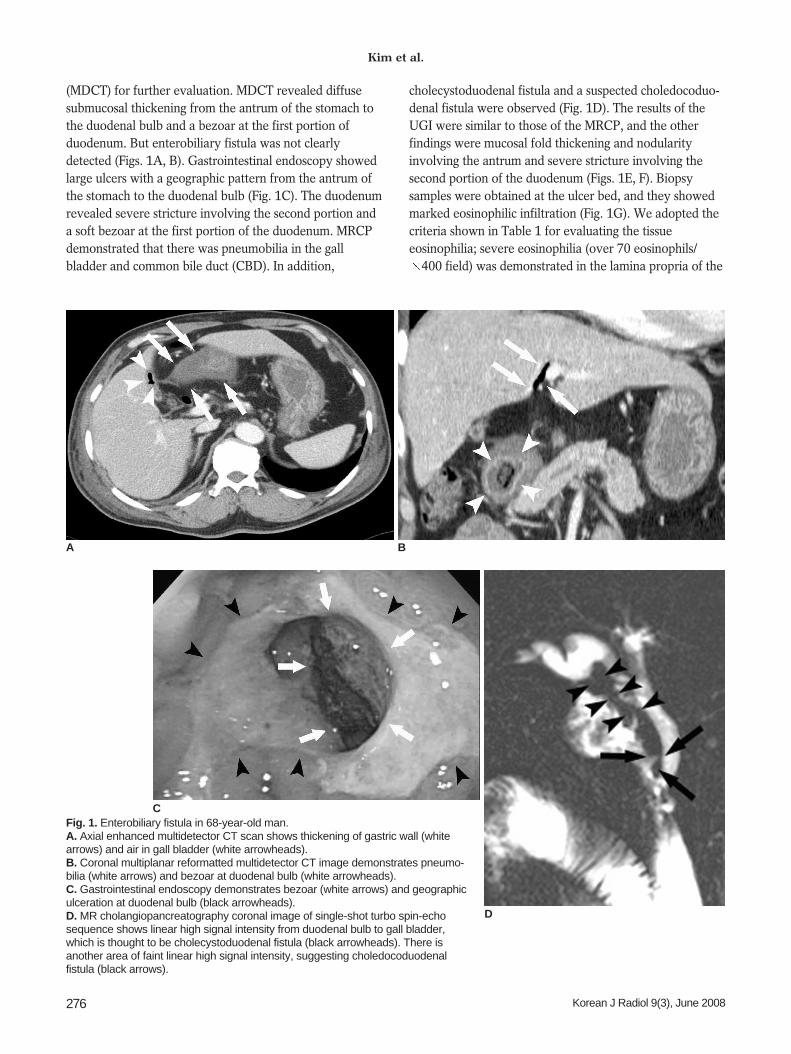
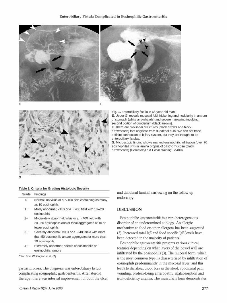
(MDCT) for further evaluation. MDCT revealed diffusesubmucosal thickening from the antrum of the stomach tothe duodenal bulb and a bezoar at the first portion ofduodenum. But enterobiliary fistula was not clearlydetected (Figs. 1A, B). Gastrointestinal endoscopy showedlarge ulcers with a geographic pattern from the antrum ofthe stomach to the duodenal bulb (Fig. 1C). The duodenumrevealed severe stricture involving the second portion anda soft bezoar at the first portion of the duodenum. MRCPdemonstrated that there was pneumobilia in the gallbladder and common bile duct (CBD). In addition,
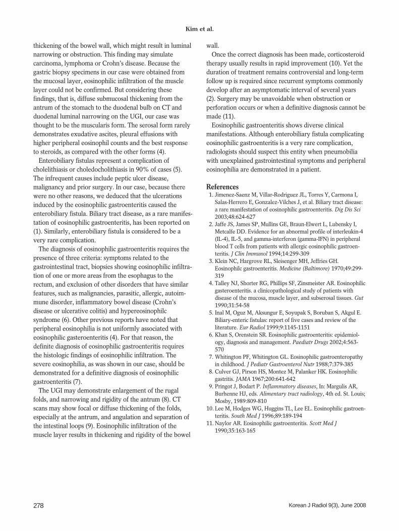
cholecystoduodenal fistula and a suspected choledocoduo-denal fistula were observed (Fig. 1D). The results of theUGI were similar to those of the MRCP, and the otherfindings were mucosal fold thickening and nodularityinvolving the antrum and severe stricture involving thesecond portion of the duodenum (Figs. 1E, F). Biopsysamples were obtained at the ulcer bed, and they showedmarked eosinophilic infiltration (Fig. 1G). We adopted thecriteria shown in Table 1 for evaluating the tissueeosinophilia; severe eosinophilia (over 70 eosinophils/
400 field) was demonstrated in the lamina propria of the
Kim et al.
276 Korean J Radiol 9(3), June 2008
A B
Fig. 1. Enterobiliary fistula in 68-year-old man.A. Axial enhanced multidetector CT scan shows thickening of gastric wall (whitearrows) and air in gall bladder (white arrowheads).B. Coronal multiplanar reformatted multidetector CT image demonstrates pneumo-bilia (white arrows) and bezoar at duodenal bulb (white arrowheads).C. Gastrointestinal endoscopy demonstrates bezoar (white arrows) and geographiculceration at duodenal bulb (black arrowheads).D. MR cholangiopancreatography coronal image of single-shot turbo spin-echosequence shows linear high signal intensity from duodenal bulb to gall bladder,which is thought to be cholecystoduodenal fistula (black arrowheads). There isanother area of faint linear high signal intensity, suggesting choledocoduodenalfistula (black arrows).
C
D
gastric mucosa. The diagnosis was enterobiliary fistulacomplicating eosinophilic gastroenteritis. After steroidtherapy, there was interval improvement of both the ulcer
and duodenal luminal narrowing on the follow upendoscopy.
DISCUSSION
Eosinophilic gastroenteritis is a rare heterogeneousdisorder of an undetermined etiology. An allergicmechanism to food or other allergens has been suggested(2). Increased total IgE and food specific IgE levels havebeen detected in the majority of patients.
Eosinophilic gastroenteritis presents various clinicalfeatures depending on what layers of the bowel wall areinfiltrated by the eosinophils (3). The mucosal form, whichis the most common type, is characterized by infiltration ofeosinophils predominantly in the mucosal layer, and thisleads to diarrhea, blood loss in the stool, abdominal pain,vomiting, protein-losing enteropathy, malabsorption andiron-deficiency anemia. The muscularis form demonstrates
Enterobiliary Fistula Complicated in Eosinophilic Gastroenteritis
Korean J Radiol 9(3), June 2008 277
Fig. 1. Enterobiliary fistula in 68-year-old man.E. Upper GI reveals mucosal fold thickening and nodularity in antrumof stomach (white arrowheads) and severe narrowing involvingsecond portion of duodenum (black arrows).F. There are two linear structures (black arrows and blackarrowheads) that originate from duodenal bulb. We can not tracedefinite connection to biliary system, but they are thought to beenterobiliary fistulas.G. Microscopic finding shows marked eosinophilic infiltration (over 70eosinophils/HPF) in lamina propria of gastric mucosa (blackarrowheads) (Hematoxylin & Eosin staining, 400).
E F
Table 1. Criteria for Grading Histologic Severity
Grade Findings
0 Normal; no villus or a 400 field containing as many as 10 eosinophils
1+ Mildly abnormal; villus or a 400 field with 10 20eosinophils
2+ Moderately abnormal; villus or a 400 field with20 50 eosinophils and/or focal aggregates of 10 orfewer eosinophils
3+ Severely abnormal; villus or a 400 field with morethan 50 eosinophils and/or aggregates or more than10 eosinophils
4+ Extremely abnormal; sheets of eosinophils oreosinophilic tumors
Cited from Whitington et al. (7)
G
thickening of the bowel wall, which might result in luminalnarrowing or obstruction. This finding may simulatecarcinoma, lymphoma or Crohn’s disease. Because thegastric biopsy specimens in our case were obtained fromthe mucosal layer, eosinophilic infiltration of the musclelayer could not be confirmed. But considering thesefindings, that is, diffuse submucosal thickening from theantrum of the stomach to the duodenal bulb on CT andduodenal luminal narrowing on the UGI, our case wasthought to be the muscularis form. The serosal form rarelydemonstrates exudative ascites, pleural effusions withhigher peripheral eosinophil counts and the best responseto steroids, as compared with the other forms (4).
Enterobiliary fistulas represent a complication ofcholelithiasis or choledocholithiasis in 90% of cases (5).The infrequent causes include peptic ulcer disease,malignancy and prior surgery. In our case, because therewere no other reasons, we deduced that the ulcerationsinduced by the eosinophilic gastroenteritis caused theenterobiliary fistula. Biliary tract disease, as a rare manifes-tation of eosinophilic gastroenteritis, has been reported on(1). Similarly, enterobiliary fistula is considered to be avery rare complication.
The diagnosis of eosinophilic gastroenteritis requires thepresence of three criteria: symptoms related to thegastrointestinal tract, biopsies showing eosinophilic infiltra-tion of one or more areas from the esophagus to therectum, and exclusion of other disorders that have similarfeatures, such as malignancies, parasitic, allergic, autoim-mune disorder, inflammatory bowel disease (Crohn’sdisease or ulcerative colitis) and hypereosinophilcsyndrome (6). Other previous reports have noted thatperipheral eosinophilia is not uniformly associated witheosinophilic gasteroenteritis (4). For that reason, thedefinite diagnosis of eosinophilic gastroenteritis requiresthe histologic findings of eosinophilic infiltration. Thesevere eosinophilia, as was shown in our case, should bedemonstrated for a definitive diagnosis of eosinophilicgastroenteritis (7).
The UGI may demonstrate enlargement of the rugalfolds, and narrowing and rigidity of the antrum (8). CTscans may show focal or diffuse thickening of the folds,especially at the antrum, and angulation and separation ofthe intestinal loops (9). Eosinophilic infiltration of themuscle layer results in thickening and rigidity of the bowel
wall.Once the correct diagnosis has been made, corticosteroid
therapy usually results in rapid improvement (10). Yet theduration of treatment remains controversial and long-termfollow up is required since recurrent symptoms commonlydevelop after an asymptomatic interval of several years(2). Surgery may be unavoidable when obstruction orperforation occurs or when a definitive diagnosis cannot bemade (11).
Eosinophilic gastroenteritis shows diverse clinicalmanifestations. Although enterobiliary fistula complicatingeosinophilic gastroenteritis is a very rare complication,radiologists should suspect this entity when pneumobiliawith unexplained gastrointestinal symptoms and peripheraleosinophilia are demonstrated in a patient.
References1. Jimenez-Saenz M, Villar-Rodriguez JL, Torres Y, Carmona I,
Salas-Herrero E, Gonzalez-Vilches J, et al. Biliary tract disease:a rare manifestation of eosinophilic gastroenteritis. Dig Dis Sci2003;48:624-627
2. Jaffe JS, James SP, Mullins GE, Braun-Elwert L, Lubensky I,Metcalfe DD. Evidence for an abnormal profile of interleukin-4(IL-4), IL-5, and gamma-interferon (gamma-IFN) in peripheralblood T cells from patients with allergic eosinophilic gastroen-teritis. J Clin Immunol 1994;14:299-309
3. Klein NC, Hargrove RL, Sleisenger MH, Jeffries GH.Eosinophilc gastroenteritis. Medicine (Baltimore) 1970;49:299-319
4. Talley NJ, Shorter RG, Phillips SF, Zinsmeister AR. Eosinophilicgasteroenteritis. a clinicopathological study of patients withdisease of the mucosa, muscle layer, and subserosal tissues. Gut1990;31:54-58
5. Inal M, Oguz M, Aksungur E, Soyupak S, Boruban S, Akgul E.Biliary-enteric fistulas: report of five cases and review of theliterature. Eur Radiol 1999;9:1145-1151
6. Khan S, Orenstein SR. Eosinophilic gastroenteritis: epidemiol-ogy, diagnosis and management. Paediatr Drugs 2002;4:563-570
7. Whitington PF, Whitington GL. Eosinophilic gastroenteropathyin childhood. J Pediatr Gastroenterol Nutr 1988;7:379-385
8. Culver GJ, Pirson HS, Montez M, Palanker HK. Eosinophilicgastritis. JAMA 1967;200:641-642
9. Pringot J, Bodart P. Inflammatory diseases, In: Margulis AR,Burhenne HJ, eds. Alimentary tract radiology, 4th ed. St. Louis;Mosby, 1989:809-810
10. Lee M, Hodges WG, Huggins TL, Lee EL. Eosinophilic gastroen-teritis. South Med J 1996;89:189-194
11. Naylor AR. Eosinophilic gastroenteritis. Scott Med J1990;35:163-165
Kim et al.
278 Korean J Radiol 9(3), June 2008