Embed Size (px)
Citation preview
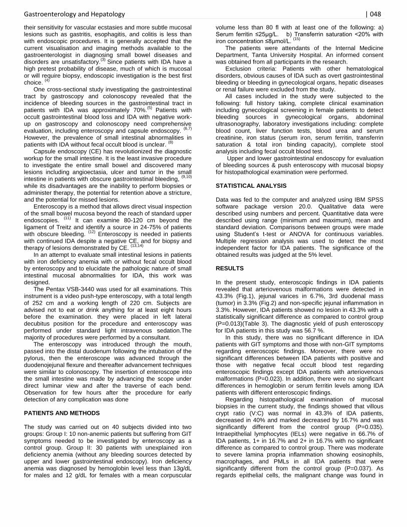
ISSN: 2986-9994
www.grantmedicaljournals.org
March, 2017; 02(03): 047-056 gah/16/1620/gmj Gastroenterology and Hepatology
Original Article
Enteroscopic Study for Evaluation of Small Intestinal Lesions
in Patients with Unexplained Iron Deficiency Anemia
Ahmed M. Ramadan1, Mohamed A. Tawfik1, Eiman A. Hasby2, Sahar S. Bessa1
1 Department of Internal Medicine, Tanta University, Egypt
2 Department of Pathology, Faculty of Medicine, Tanta University, Egypt
*Corresponding author(s): Mohamed A.Tawfik, Department of Internal Medicine, Tanta University, Egypt
Email: [email protected]
Published: 2017.03.10.
ABSTRACT Background: Iron deficiency anemia (IDA) is the most common cause of anemia worldwide. It is widely accepted that chronic occult blood loss from the gastrointestinal tract is a major cause of IDA. This study aimed to evaluate small intestinal lesions in patients with unexplained iron deficiency anemia, with or without fecal occult blood by enteroscopy and to elucidate the pathologic nature of small intestinal mucosal abnormalities for iron deficiency anemia. Methods: This study included 40 subjects; 10 non-anemic patients but suffering from GIT symptoms needed to be investigated by enteroscopy as a control group and 30 patients with unexplained iron deficiency anemia (without any bleeding sources detected by upper and lower gastrointestinal endoscopy). Complete blood count, iron status, and fecal occult blood test were measured. The small intestine was evaluated by push enteroscopy and histopathological examination for duodenum and jejunum mucosal biopsies was done. Results: Enteroscopic findings in IDA patients revealed that arteriovenous malformations were detected in 43.3%, jejunal varices in 6.7%, 3rd duodenal mass (tumor) in 3.3% and non-specific jejunal inflammation in 3.3%. There were moderate to severe lamina propria inflammation in all IDA patients that was significantly different from the control group. Logistic regression analysis showed that IDA was related to body mass index, enteroscopic and histopathologic lesions. Conclusions: Subjects with iron deficiency anemia after negative workup on the bleeding sources by conventional upper and lower endoscopies should undergo a further investigation of the small intestine by enteroscopic study, irrespective of their fecal occult blood status. Keywords: Unexplained iron deficiency anemia , Enteroscopy , Small bowel
INTRODUCTION
Iron deficiency anemia (IDA) is the most common cause of anemia worldwide, causing significant disease-related morbidity, and has a negative impact on patient's well-being and overall outcome. The prevalence of IDA in developed countries is estimated to be 2-5% of adult men and post-
menopausal women. (1)
It is widely accepted that chronic blood loss from the gastrointestinal tract is a major cause of IDA.
(2)
The evaluating methods for IDA patients are endoscopic and/or radiographic. Radiographic studies are generally effective for detecting masses and large ulcerating lesions, but
Gastroenterology and Hepatology | 048
their sensitivity for vascular ecstasies and more subtle mucosal lesions such as gastritis, esophagitis, and colitis is less than with endoscopic procedures. It is generally accepted that the current visualisation and imaging methods available to the gastroenterologist in diagnosing small bowel diseases and disorders are unsatisfactory.
(3) Since patients with IDA have a
high pretest probability of disease, much of which is mucosal or will require biopsy, endoscopic investigation is the best first choice.
(4)
One cross-sectional study investigating the gastrointestinal tract by gastroscopy and colonoscopy revealed that the incidence of bleeding sources in the gastrointestinal tract in patients with IDA was approximately 70%.
(5) Patients with
occult gastrointestinal blood loss and IDA with negative work- up on gastroscopy and colonoscopy need comprehensive evaluation, including enteroscopy and capsule endoscopy.
(6,7)
However, the prevalence of small intestinal abnormalities in patients with IDA without fecal occult blood is unclear.
(8)
Capsule endoscopy (CE) has revolutionized the diagnostic workup for the small intestine. It is the least invasive procedure to investigate the entire small bowel and discovered many lesions including angioectasia, ulcer and tumor in the small intestine in patients with obscure gastrointestinal bleeding,
(9,10)
while its disadvantages are the inability to perform biopsies or administer therapy, the potential for retention above a stricture, and the potential for missed lesions.
Enteroscopy is a method that allows direct visual inspection of the small bowel mucosa beyond the reach of standard upper endoscopies.
(11) It can examine 80-120 cm beyond the
ligament of Treitz and identify a source in 24-75% of patients with obscure bleeding.
(12) Enteroscopy is needed in patients
with continued IDA despite a negative CE, and for biopsy and therapy of lesions demonstrated by CE.
(13,14)
In an attempt to evaluate small intestinal lesions in patients with iron deficiency anemia with or without fecal occult blood by enteroscopy and to elucidate the pathologic nature of small intestinal mucosal abnormalities for IDA, this work was designed.
The Pentax VSB-3440 was used for all examinations. This instrument is a video push-type enteroscopy, with a total length of 252 cm and a working length of 220 cm. Subjects are advised not to eat or drink anything for at least eight hours before the examination. they were placed in left lateral decubitus position for the procedure and enteroscopy was performed under standard light intravenous sedation.The majority of procedures were performed by a consultant.
The enteroscopy was introduced through the mouth, passed into the distal duodenum following the intubation of the pylorus, then the enteroscope was advanced through the duodenojejunal flexure and thereafter advancement techniques were similar to colonoscopy. The insertion of enteroscope into the small intestine was made by advancing the scope under direct luminar view and after the traverse of each bend. Observation for few hours after the procedure for early detection of any complication was done PATIENTS AND METHODS
The study was carried out on 40 subjects divided into two groups: Group I: 10 non-anemic patients but suffering from GIT symptoms needed to be investigated by enteroscopy as a control group. Group II: 30 patients with unexplained iron deficiency anemia (without any bleeding sources detected by upper and lower gastrointestinal endoscopy). Iron deficiency anemia was diagnosed by hemoglobin level less than 13g/dL for males and 12 g/dL for females with a mean corpuscular
volume less than 80 fl with at least one of the following: a) Serum ferritin ≤25μg/L. b) Transferrin saturation <20% with iron concentration ≤8μmol/L.
(15)
The patients were attendants of the Internal Medicine Department, Tanta University Hospital. An informed consent was obtained from all participants in the research.
Exclusion criteria: Patients with other hematological disorders, obvious causes of IDA such as overt gastrointestinal bleeding or bleeding in gynecological organs, hepatic diseases or renal failure were excluded from the study.
All cases included in the study were subjected to the following: full history taking, complete clinical examination including gynecological screening in female patients to detect bleeding sources in gynecological organs, abdominal ultrasonography, laboratory investigations including: complete blood count, liver function tests, blood urea and serum creatinine, iron status (serum iron, serum ferritin, transferrin saturation & total iron binding capacity), complete stool analysis including fecal occult blood test.
Upper and lower gastrointestinal endoscopy for evaluation of bleeding sources & push enteroscopy with mucosal biopsy for histopathological examination were performed. STATISTICAL ANALYSIS
Data was fed to the computer and analyzed using IBM SPSS software package version 20.0. Qualitative data were described using numbers and percent. Quantitative data were described using range (minimum and maximum), mean and standard deviation. Comparisons between groups were made using Student’s t-test or ANOVA for continuous variables. Multiple regression analysis was used to detect the most independent factor for IDA patients. The significance of the obtained results was judged at the 5% level. RESULTS
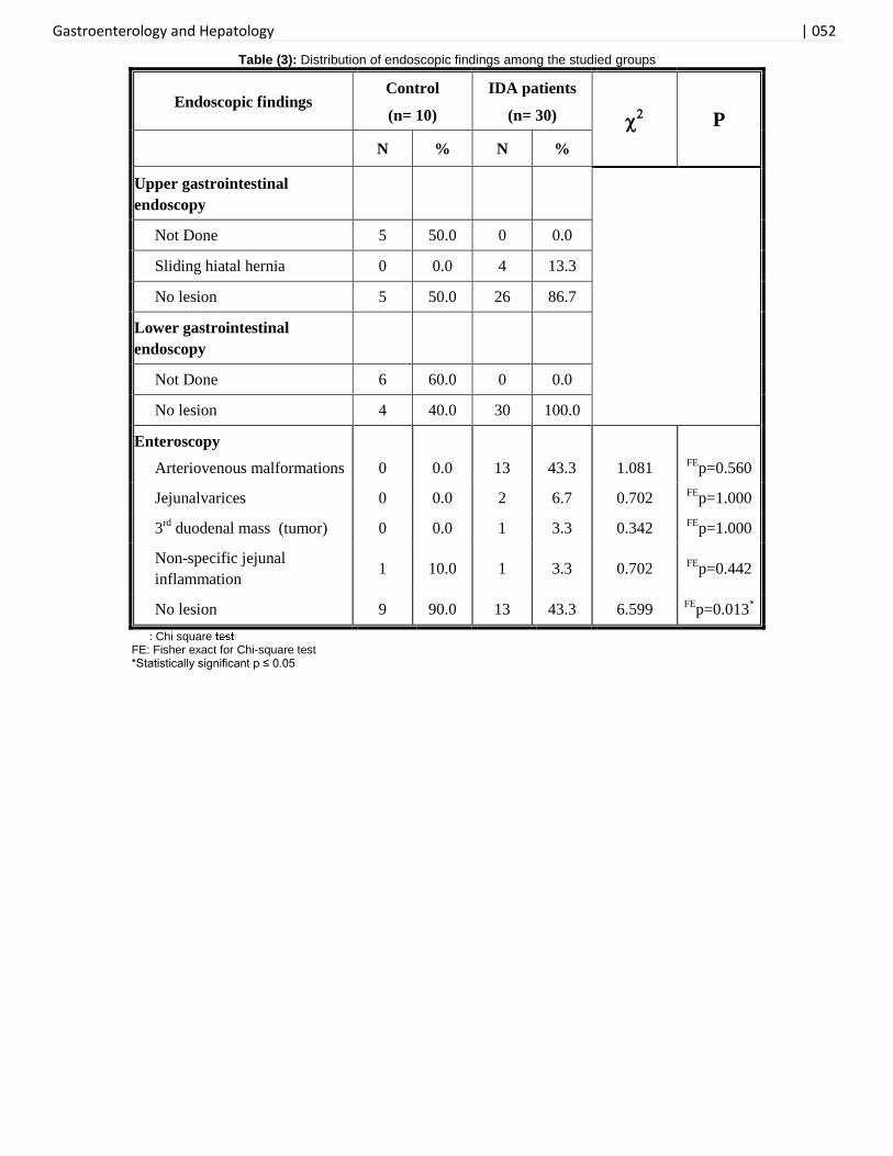
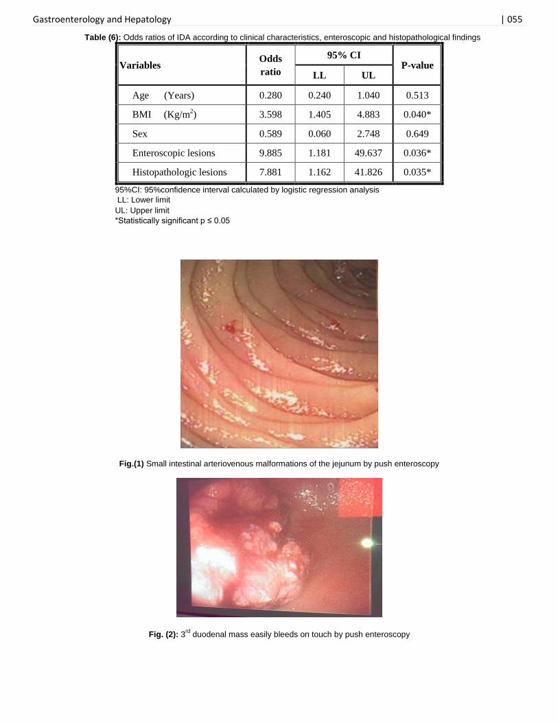
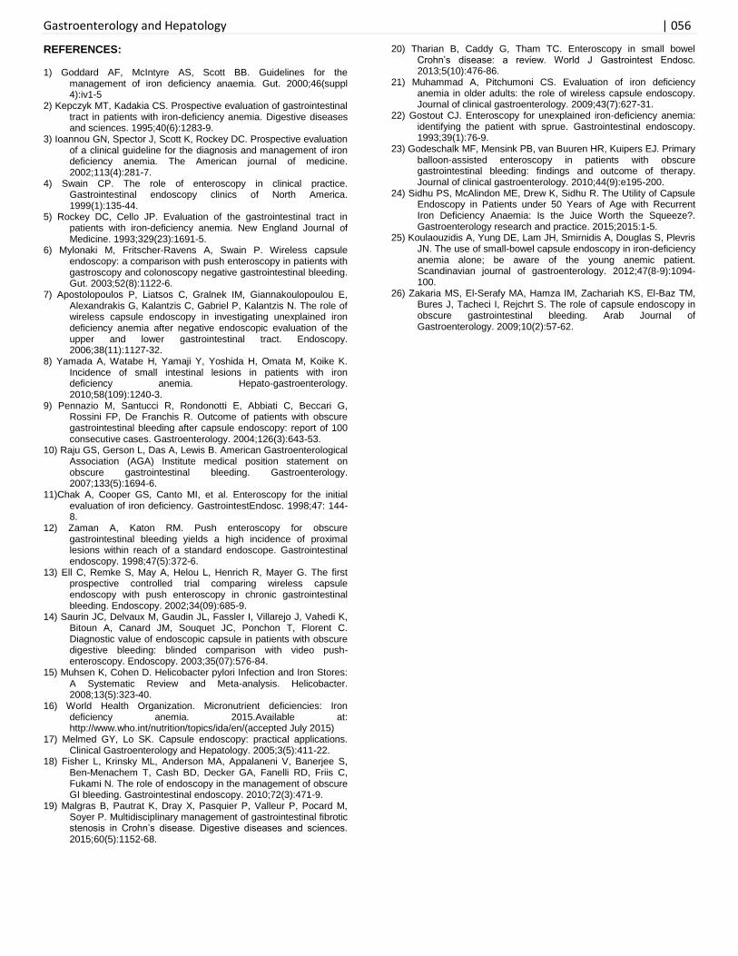
In the present study, enteroscopic findings in IDA patients revealed that arteriovenous malformations were detected in 43.3% (Fig.1), jejunal varices in 6.7%, 3rd duodenal mass (tumor) in 3.3% (Fig.2) and non-specific jejunal inflammation in 3.3%. However, IDA patients showed no lesion in 43.3% with a statistically significant difference as compared to control group (P=0.013)(Table 3). The diagnostic yield of push enteroscopy for IDA patients in this study was 56.7 %.
In this study, there was no significant difference in IDA patients with GIT symptoms and those with non-GIT symptoms regarding enteroscopic findings. Moreover, there were no significant differences between IDA patients with positive and those with negative fecal occult blood test regarding enteroscopic findings except IDA patients with arteriovenous malformations (P=0.023). In addition, there were no significant differences in hemoglobin or serum ferritin levels among IDA patients with different enteroscopic findings.
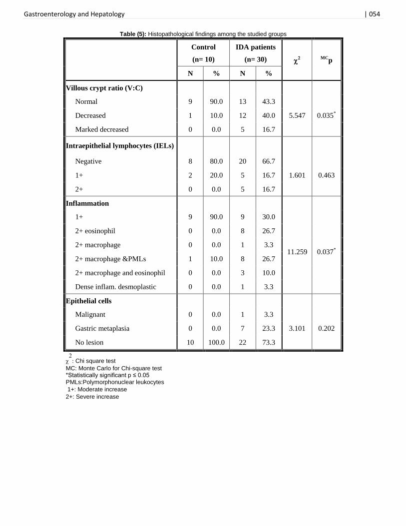
Regarding histopathological examination of mucosal biopsies in the current study, the findings showed that villous crypt ratio (V:C) was normal in 43.3% of IDA patients, decreased in 40% and marked decreased by 16.7% and was significantly different from the control group (P=0.035). Intraepithelial lymphocytes (IELs) were negative in 66.7% of IDA patients, 1+ in 16.7% and 2+ in 16.7% with no significant difference as compared to control group. There was moderate to severe lamina propria inflammation showing eosinophils, macrophages, and PMLs in all IDA patients that were significantly different from the control group (P=0.037). As regards epithelial cells, the malignant change was found in
Gastroenterology and Hepatology | 049
3.3% of IDA patients, gastric metaplasia in 23.3% and no lesion was detected in 73.3 % with no significant difference as compared to control group (Table 5).
In this study, there was no significant difference in IDA patients with GIT symptoms and those with non-GIT symptoms regarding histopathological findings. Also, there was no significant difference in the villous crypt ratio or intraepithelial lymphocytes among IDA patients with different enteroscopic findings. Regarding inflammation and epithelial cells, there were significant differences in IDA patients with 3rd duodenal mass (tumor) by enteroscopy (P=0.034 & P=0.039, respectively). On the other hand, there were no significant differences between IDA patients with positive and those with negative fecal occult blood test regarding histopathological examination. Logistic regression analysis showed that IDA was related to body mass index (p=0.040), enteroscopic lesions (p=0.036) and histopathologic lesions (p=0.035) (Table 6).
DISCUSSION
Iron deficiency is the most common cause of anemia, with the World Health Organization estimating it affects about 30% of the population worldwide (WHO, 2015).(16) It is often secondary to chronic occult bleeding from the gastrointestinal tract and is a common reason for referral of patients to gastroenterologists.
(17)
In clinical practice, esophagogastroduodenoscopy (EGD) and colonoscopy are performed in the initial evaluation of IDA to exclude a source of chronic blood loss from the GI tract. Despite undergoing standard endoscopic evaluation, up to 30% of patients with IDA have no definitive diagnosis.
(17,18)
It is generally accepted that the current visualization and imaging methods available to the gastroenterologist in diagnosing small bowel diseases and disorders are unsatisfactory.
(3) Since patients with IDA have a high pretest
probability of disease, much of which is mucosal or will require biopsy, the endoscopic investigation is the best first choice. (4)
Enteroscopy, a rapidly evolving field, has had a significant Renaissance recently and the small bowel is no longer the black box for the endoscopist or the final frontier.
(19,20) This
method allows direct visual inspection of the small bowel mucosa beyond the reach of standard upper endoscopies. It can examine 80- 120 cm beyond the ligament of Treitz and identify a source in 24- 75% of patients with obscure bleeding. (12)
Enteroscopy is needed in patients with continued IDA despite negative capsule endoscopy (CE), and for biopsy and therapy of lesions demonstrated by CE.
(20)
This study was conducted to evaluate small intestinal lesions in patients with unexplained iron deficiency anemia, with or without fecal occult blood by enteroscopy after negative work- up on the bleeding source by conventional upper and lower endoscopies and to elucidate the pathologic nature of small intestinal mucosal abnormalities for iron deficiency anemia.
In the present study, enteroscopic findings in IDA patients revealed that arteriovenous malformations were detected in 43.3%, jejunal varices in 6.7%, 3rd duodenal mass (tumor) in 3.3% and non-specific jejunal inflammation in 3.3%. However, IDA patients showed no lesion in 43.3% with a statistically significant difference as compared to control group (P=0.013). There were no significant differences between IDA patients with positive and those with negative fecal occult blood test regarding enteroscopic findings except IDA patients with arteriovenous malformations (P=0.023). The present study
indicates that even IDA patients with negative fecal occult blood test should undergo enteroscopy.
These findings are in agreement with Yamada et al.(2011), (8)
who revealed that the prevalence of small intestinal lesions including angioectasia, ulcers, and tumors in Japanese patients with IDA was approximately 50% and that there was no significant association between small intestinal lesions and fecal occult blood. Also, one European prospective study reported that 57% of IDA patients had small intestinal lesions.
(7) Moreover, Muhammed et al. (2009),
(21)
retrospectively investigated IDA patients in the United States and reported that the incidence of small intestinal lesions was 59-69%. The current data suggest that the incidence of small intestinal lesions in IDA is globally similar.
Furthermore, Chak et al. (1998), (11)
found that performing push enteroscopy in combination with esophagogastroduodenoscopy (EGD) increased the diagnostic yield of the upper evaluation in asymptomatic patients with iron deficiency from 41% to 68%. They concluded that performance of push enteroscopy should be the preferred initial diagnostic test when evaluating the gastrointestinal tract of patients with iron deficiency anemia. Notably, Gostout (1993)
(22) has
recommended small bowel biopsies in all patients with iron deficiency to diagnose sprue.
Reportedly, a retrospective study showed no recurrence of anemia in 71% with endoscopic treatment performed by push enteroscopy after 18 months of follow-up. However, if push enteroscopy showed no lesion, further explorations were unnecessary because the anemia did not recur in the majority (63%) of cases.
(23)
Recently, Sidhu et al. (2015), (24)
have demonstrated that the diagnostic yield of capsule endoscopy in patients under 50 years of age with recurrent IDA was 28%. Significant pathology identified in this group included small bowel tumors, ulcers, and angioectasia. They reported that angioectasia is the second commonest finding after erosions and ulcers and that small bowel tumor were found in 3% of patients which is similar to findings by Koulaouzidis et al. (2012).
(25)
Interestingly, Zakaria et al. (2009), (26)
evaluated the role of capsule endoscopy (CE) in obscure gastrointestinal bleeding (OGIB) and found that the diagnostic yield of CE in cases of OGIB was 56.1% and that the commonest lesions were angiodysplasias (17.5%), examinations were negative for lesions in 35.1% while capsule retention occurred in 3.5%. They concluded that capsule examination should be done after competent exhaustion of other diagnostic tools including upper and lower endoscopic examinations after proper bowel preparation in order to increase the diagnostic yield. They recommended repeat upper endoscopy and full colonoscopy with terminal ileoscopy before proceeding to capsule examination due to the high cost of capsule examination in our country. CONCLUSION
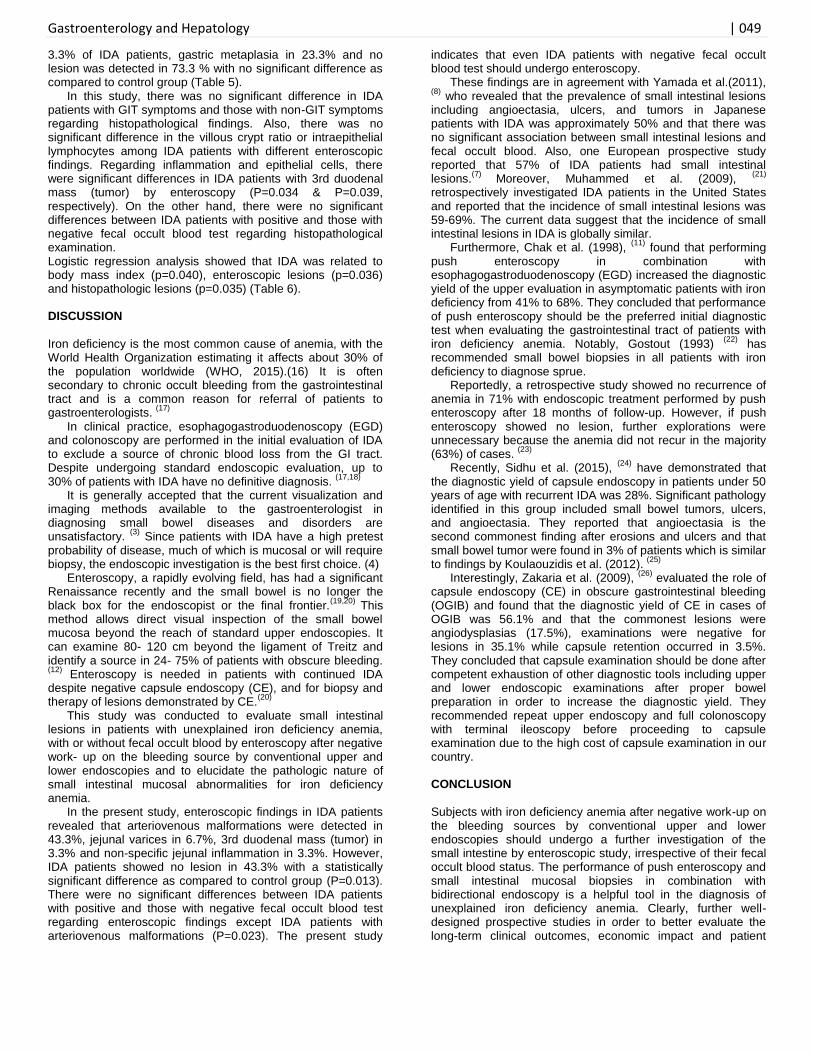
Subjects with iron deficiency anemia after negative work-up on the bleeding sources by conventional upper and lower endoscopies should undergo a further investigation of the small intestine by enteroscopic study, irrespective of their fecal occult blood status. The performance of push enteroscopy and small intestinal mucosal biopsies in combination with bidirectional endoscopy is a helpful tool in the diagnosis of unexplained iron deficiency anemia. Clearly, further well-designed prospective studies in order to better evaluate the long-term clinical outcomes, economic impact and patient
Gastroenterology and Hepatology | 050
satisfaction levels of the small bowel diagnostic tests that are currently available would be warranted. ACKNOWLEDGEMENTS
We would like to thank all participants who helped during this study.
CONFLICT OF INTEREST
The authors declare that there is no conflict of interest associated with this work.
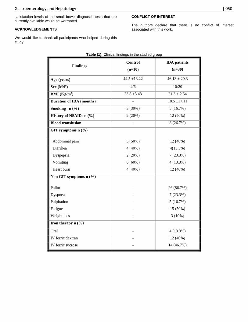
Table (1): Clinical findings in the studied group
Findings Control
(n=10)
IDA patients
(n=30)
Age (years) 44.5 ±13.22 46.13 ± 20.3
Sex (M/F) 4/6 10/20
BMI (Kg/m2) 23.8 ±3.43 21.3 ± 2.54
Duration of IDA (months) - 18.5 ±17.11
Smoking n (%) 3 (30%) 5 (16.7%)
History of NSAIDs n (%) 2 (20%) 12 (40%)
Blood transfusion - 8 (26.7%)
GIT symptoms n (%)
Abdominal pain 5 (50%) 12 (40%)
Diarrhea 4 (40%) 4(13.3%)
Dyspepsia 2 (20%) 7 (23.3%)
Vomiting 6 (60%) 4 (13.3%)
Heart burn 4 (40%) 12 (40%)
Non GIT symptoms n (%)
Pallor - 26 (86.7%)
Dyspnea - 7 (23.3%)
Palpitation - 5 (16.7%)
Fatigue - 15 (50%)
Weight loss - 3 (10%)
Iron therapy n (%)
Oral - 4 (13.3%)
IV ferric dextran - 12 (40%)
IV ferric sucrose - 14 (46.7%)
Gastroenterology and Hepatology | 051
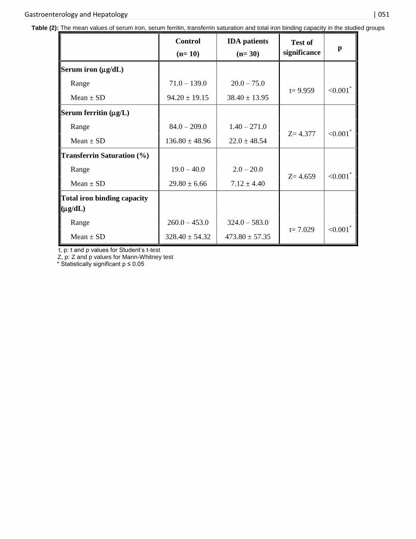
Table (2): The mean values of serum iron, serum ferritin, transferrin saturation and total iron binding capacity in the studied groups
Control
(n= 10)
IDA patients
(n= 30)
Test of
significance p
Serum iron (g/dL)
Range 71.0 – 139.0 20.0 – 75.0 t= 9.959 <0.001
*
Mean ± SD 94.20 ± 19.15 38.40 ± 13.95
Serum ferritin (g/L)
Range 84.0 – 209.0 1.40 – 271.0 Z= 4.377 <0.001
*
Mean ± SD 136.80 ± 48.96 22.0 ± 48.54
Transferrin Saturation (%)
Range 19.0 – 40.0 2.0 – 20.0 Z= 4.659 <0.001
*
Mean ± SD 29.80 ± 6.66 7.12 ± 4.40
Total iron binding capacity
(g/dL)
Range 260.0 – 453.0 324.0 – 583.0 t= 7.029 <0.001
*
Mean ± SD 328.40 ± 54.32 473.80 ± 57.35
t, p: t and p values for Student’s t-test Z, p: Z and p values for Mann-Whitney test
* Statistically significant p ≤ 0.05
Gastroenterology and Hepatology | 052
Table (3): Distribution of endoscopic findings among the studied groups
Endoscopic findings Control
(n= 10)
IDA patients
(n= 30)
P
N % N %
Upper gastrointestinal
endoscopy
Not Done 5 50.0 0 0.0
Sliding hiatal hernia 0 0.0 4 13.3
No lesion 5 50.0 26 86.7
Lower gastrointestinal
endoscopy
Not Done 6 60.0 0 0.0
No lesion 4 40.0 30 100.0
Enteroscopy
Arteriovenous malformations
0
0.0
13
43.3
1.081
FEp=0.560
Jejunalvarices 0 0.0 2 6.7 0.702 FE
p=1.000
3rd
duodenal mass (tumor) 0 0.0 1 3.3 0.342 FE
p=1.000
Non-specific jejunal
inflammation 1 10.0 1 3.3 0.702
FEp=0.442
No lesion 9 90.0 13 43.3 6.599 FE
p=0.013*
Chi square FE: Fisher exact for Chi-square test *Statistically significant p ≤ 0.05
Gastroenterology and Hepatology | 053
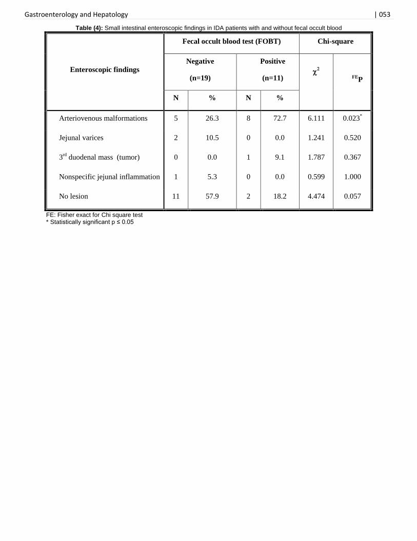
Table (4): Small intestinal enteroscopic findings in IDA patients with and without fecal occult blood
Enteroscopic findings
Fecal occult blood test (FOBT) Chi-square
Negative
(n=19)
Positive
(n=11)
FEP
N % N %
Arteriovenous malformations 5 26.3 8 72.7 6.111 0.023*
Jejunal varices 2 10.5 0 0.0 1.241 0.520
3rd
duodenal mass (tumor) 0 0.0 1 9.1 1.787 0.367
Nonspecific jejunal inflammation 1 5.3 0 0.0 0.599 1.000
No lesion 11 57.9 2 18.2 4.474 0.057
FE: Fisher exact for Chi square test * Statistically significant p ≤ 0.05
Gastroenterology and Hepatology | 054
Table (5): Histopathological findings among the studied groups
Control
(n= 10)
IDA patients
(n= 30)
MCp
N % N %
Villous crypt ratio (V:C)
Normal 9 90.0 13 43.3
5.547 0.035* Decreased 1 10.0 12 40.0
Marked decreased 0 0.0 5 16.7
Intraepithelial lymphocytes (IELs)
Negative 8 80.0 20 66.7
1.601 0.463 1+ 2 20.0 5 16.7
2+ 0 0.0 5 16.7
Inflammation
1+ 9 90.0 9 30.0
11.259 0.037*
2+ eosinophil 0 0.0 8 26.7
2+ macrophage 0 0.0 1 3.3
2+ macrophage &PMLs 1 10.0 8 26.7
2+ macrophage and eosinophil 0 0.0 3 10.0
Dense inflam. desmoplastic 0 0.0 1 3.3
Epithelial cells
Malignant 0 0.0 1 3.3
3.101 0.202 Gastric metaplasia 0 0.0 7 23.3
No lesion 10 100.0 22 73.3
: Chi square test MC: Monte Carlo for Chi-square test *Statistically significant p ≤ 0.05 PMLs:Polymorphonuclear leukocytes
1+: Moderate increase 2+: Severe increase
Gastroenterology and Hepatology | 055
Table (6): Odds ratios of IDA according to clinical characteristics, enteroscopic and histopathological findings
Variables Odds
ratio
95% CI P-value
LL UL
Age (Years) 0.280 0.240 1.040 0.513
BMI (Kg/m2) 3.598 1.405 4.883 0.040*
Sex 0.589 0.060 2.748 0.649
Enteroscopic lesions 9.885 1.181 49.637 0.036*
Histopathologic lesions 7.881 1.162 41.826 0.035*
95%CI: 95%confidence interval calculated by logistic regression analysis
LL: Lower limit
UL: Upper limit *Statistically significant p ≤ 0.05
Fig.(1) Small intestinal arteriovenous malformations of the jejunum by push enteroscopy
Fig. (2): 3
rd duodenal mass easily bleeds on touch by push enteroscopy
Gastroenterology and Hepatology | 056
REFERENCES: 1) Goddard AF, McIntyre AS, Scott BB. Guidelines for the
management of iron deficiency anaemia. Gut. 2000;46(suppl 4):iv1-5
2) Kepczyk MT, Kadakia CS. Prospective evaluation of gastrointestinal tract in patients with iron-deficiency anemia. Digestive diseases and sciences. 1995;40(6):1283-9.
3) Ioannou GN, Spector J, Scott K, Rockey DC. Prospective evaluation of a clinical guideline for the diagnosis and management of iron deficiency anemia. The American journal of medicine. 2002;113(4):281-7.
4) Swain CP. The role of enteroscopy in clinical practice. Gastrointestinal endoscopy clinics of North America. 1999(1):135-44.
5) Rockey DC, Cello JP. Evaluation of the gastrointestinal tract in patients with iron-deficiency anemia. New England Journal of Medicine. 1993;329(23):1691-5.
6) Mylonaki M, Fritscher-Ravens A, Swain P. Wireless capsule endoscopy: a comparison with push enteroscopy in patients with gastroscopy and colonoscopy negative gastrointestinal bleeding. Gut. 2003;52(8):1122-6.
7) Apostolopoulos P, Liatsos C, Gralnek IM, Giannakoulopoulou E, Alexandrakis G, Kalantzis C, Gabriel P, Kalantzis N. The role of wireless capsule endoscopy in investigating unexplained iron deficiency anemia after negative endoscopic evaluation of the upper and lower gastrointestinal tract. Endoscopy. 2006;38(11):1127-32.
8) Yamada A, Watabe H, Yamaji Y, Yoshida H, Omata M, Koike K. Incidence of small intestinal lesions in patients with iron deficiency anemia. Hepato-gastroenterology. 2010;58(109):1240-3.
9) Pennazio M, Santucci R, Rondonotti E, Abbiati C, Beccari G, Rossini FP, De Franchis R. Outcome of patients with obscure gastrointestinal bleeding after capsule endoscopy: report of 100 consecutive cases. Gastroenterology. 2004;126(3):643-53.
10) Raju GS, Gerson L, Das A, Lewis B. American Gastroenterological Association (AGA) Institute medical position statement on obscure gastrointestinal bleeding. Gastroenterology. 2007;133(5):1694-6.
11)Chak A, Cooper GS, Canto MI, et al. Enteroscopy for the initial evaluation of iron deficiency. GastrointestEndosc. 1998;47: 144-8.
12) Zaman A, Katon RM. Push enteroscopy for obscure gastrointestinal bleeding yields a high incidence of proximal lesions within reach of a standard endoscope. Gastrointestinal endoscopy. 1998;47(5):372-6.
13) Ell C, Remke S, May A, Helou L, Henrich R, Mayer G. The first prospective controlled trial comparing wireless capsule endoscopy with push enteroscopy in chronic gastrointestinal bleeding. Endoscopy. 2002;34(09):685-9.
14) Saurin JC, Delvaux M, Gaudin JL, Fassler I, Villarejo J, Vahedi K, Bitoun A, Canard JM, Souquet JC, Ponchon T, Florent C. Diagnostic value of endoscopic capsule in patients with obscure digestive bleeding: blinded comparison with video push-enteroscopy. Endoscopy. 2003;35(07):576-84.
15) Muhsen K, Cohen D. Helicobacter pylori Infection and Iron Stores: A Systematic Review and Meta‐analysis. Helicobacter. 2008;13(5):323-40.
16) World Health Organization. Micronutrient deficiencies: Iron deficiency anemia. 2015.Available at: http://www.who.int/nutrition/topics/ida/en/(accepted July 2015)
17) Melmed GY, Lo SK. Capsule endoscopy: practical applications. Clinical Gastroenterology and Hepatology. 2005;3(5):411-22.
18) Fisher L, Krinsky ML, Anderson MA, Appalaneni V, Banerjee S, Ben-Menachem T, Cash BD, Decker GA, Fanelli RD, Friis C, Fukami N. The role of endoscopy in the management of obscure GI bleeding. Gastrointestinal endoscopy. 2010;72(3):471-9.
19) Malgras B, Pautrat K, Dray X, Pasquier P, Valleur P, Pocard M, Soyer P. Multidisciplinary management of gastrointestinal fibrotic stenosis in Crohn’s disease. Digestive diseases and sciences. 2015;60(5):1152-68.
20) Tharian B, Caddy G, Tham TC. Enteroscopy in small bowel Crohn’s disease: a review. World J Gastrointest Endosc. 2013;5(10):476-86.
21) Muhammad A, Pitchumoni CS. Evaluation of iron deficiency anemia in older adults: the role of wireless capsule endoscopy. Journal of clinical gastroenterology. 2009;43(7):627-31.
22) Gostout CJ. Enteroscopy for unexplained iron-deficiency anemia: identifying the patient with sprue. Gastrointestinal endoscopy. 1993;39(1):76-9.
23) Godeschalk MF, Mensink PB, van Buuren HR, Kuipers EJ. Primary balloon-assisted enteroscopy in patients with obscure gastrointestinal bleeding: findings and outcome of therapy. Journal of clinical gastroenterology. 2010;44(9):e195-200.
24) Sidhu PS, McAlindon ME, Drew K, Sidhu R. The Utility of Capsule Endoscopy in Patients under 50 Years of Age with Recurrent Iron Deficiency Anaemia: Is the Juice Worth the Squeeze?. Gastroenterology research and practice. 2015;2015:1-5.
25) Koulaouzidis A, Yung DE, Lam JH, Smirnidis A, Douglas S, Plevris JN. The use of small-bowel capsule endoscopy in iron-deficiency anemia alone; be aware of the young anemic patient. Scandinavian journal of gastroenterology. 2012;47(8-9):1094-100.
26) Zakaria MS, El-Serafy MA, Hamza IM, Zachariah KS, El-Baz TM, Bures J, Tacheci I, Rejchrt S. The role of capsule endoscopy in obscure gastrointestinal bleeding. Arab Journal of Gastroenterology. 2009;10(2):57-62.