Embed Size (px)
Citation preview

JOURNAL OF VIROLOGY, Jan. 2006, p. 51–61 Vol. 80, No. 10022-538X/06/$08.00�0 doi:10.1128/JVI.80.1.51–61.2006
Entry of Vaccinia Virus and Cell-Cell Fusion Require a Highly ConservedCysteine-Rich Membrane Protein Encoded by the A16L Gene
Suany Ojeda, Tatiana G. Senkevich, and Bernard Moss*Laboratory of Viral Diseases, National Institute of Allergy and Infectious Diseases,
National Institutes of Health, Bethesda, Maryland 20892-0445
Received 5 August 2005/Accepted 26 September 2005
The vaccinia virus A16L open reading frame encodes a 378-amino-acid protein with a predicted C-terminaltransmembrane domain and 20 invariant cysteine residues that is conserved in all sequenced members of thepoxvirus family. The A16 protein was expressed late in infection and incorporated into intracellular virusparticles with the N-terminal segment of the protein exposed on the surface. The cysteine residues weredisulfide bonded via the poxvirus cytoplasmic redox system. Unsuccessful attempts to isolate a mutant viruswith the A16L gene deleted suggested that the protein is essential for replication. To study the role of the A16protein, we made a recombinant vaccinia virus that has the Escherichia coli lac operator system regulatingtranscription of the A16L gene. In the absence of inducer, A16 synthesis was repressed and plaque size andvirus yield were greatly reduced. Nevertheless, virus morphogenesis occurred and normal-looking intracellularand extracellular virus particles formed. Purified virions made in the presence and absence of inducer wereindistinguishable, though the latter had 60- to 100-fold-lower specific infectivity. A16-deficient virions boundto cells, but their cores did not penetrate into the cytoplasm. Furthermore, A16-deficient virions were unableto induce low-pH-triggered syncytium formation. The phenotype of the inducible A16L mutant was similar tothose of mutants in which synthesis of the A21, A28, H2, or L5 membrane protein was repressed, indicating thatat least five conserved viral proteins are required for entry of poxviruses into cells as well as for cell-cell fusion.
Vaccinia virus (VACV) is a member of the poxvirus familyof large, double-stranded DNA viruses that replicate entirelyin the cytoplasm (17). VACV strain Western Reserve (WR),the prototype member of the orthopoxvirus genus, containsapproximately 200 genes of which 44 have a putative trans-membrane domain (Poxvirus Bioinformatics Resource Center[www.poxvirus.org]). VACV membrane proteins have diverseessential and nonessential roles in virus-host interactions,virion assembly, intracellular movement, and cell-to-cell spread.A novel class of poxvirus membrane proteins, required for cellentry but not for virion assembly, was recently identified (20–22, 27, 28). These four VACV proteins (A21, A28, H2, and L5)are conserved in all sequenced poxviruses, have common fea-tures including N-terminal or near N-terminal transmembranedomains and cysteines that form two to four intramoleculardisulfide bonds, and are located on the surfaces of infectiousintracellular mature virions (IMVs). Each of the four proteinsis also required for cell-cell fusion, implying that cell entryinvolves a related or identical fusion mechanism. Fusion ofIMVs with the plasma membrane was previously suggested onthe basis of biochemical and microscopic studies (4, 7, 8, 11,15). Cellular receptor proteins that participate in cell entry,however, remain to be identified.
Following virus morphogenesis, IMVs are completely envel-oped by an additional membrane containing an entirely differ-ent set of viral proteins; these proteins play roles in intracel-lular transport, egress, and cell-to-cell spread (reviewed inreference 25). The presence of multiple membranes has com-
plicated models of VACV entry. However, the finding that thesame proteins required for IMV entry are also required forcell-to-cell spread by extracellular virions necessitates the re-moval of the additional membrane prior to entry of the IMV(20–22, 27, 28). Disruption of the extra membrane in the low-pHenvironment of endosomes has been suggested (14, 29).
As a continuation of our studies of VACV proteins withputative transmembrane domains, we analyzed the product ofthe A16R (VACV WR 136) open reading frame (ORF). TheA16R ORF is conserved in all poxviruses and has 20 cysteineresidues predicted to form disulfide bonds and a C-terminaltransmembrane domain (19). A previous study had determinedthat the A16 ORF is expressed late in infection as a soluble,myristylated protein (16). Here we show that the A16 proteinis associated with the membrane of intracellular mature virionsand that virions lacking A16 were unable to enter cells orinduce low-pH-triggered cell-cell fusion. During the course ofthis analysis, A16 was detected as part of a complex containingthe four other entry/fusion proteins (19b). These results, indi-cating that A16 is an additional member of the group of pox-virus entry/fusion proteins, was unanticipated because its pre-dicted structural features differ from those of A21, A28, H2,and L5.
(This work was done to partially fulfill the Ph.D. thesis require-ments of S. Ojeda at the University of Chile, Santiago, Chile.)
MATERIALS AND METHODS
Cells and viruses. BS-C-1 cells (ATCC CCL6), grown in Eagle’s minimumessential medium (Quality Biological, Inc.) supplemented with 2.5% fetal bovineserum, were used for all experiments unless otherwise indicated. The VACV WRstrain and the recombinant viruses vT7acOI (1) and vA16Li were propagated inHeLa S3 cells as described previously (12) and stored at �80°C.
* Corresponding author. Mailing address: Laboratory of Viral Dis-eases, National Institutes of Health, 4 Center Dr., MSC 0445, Bethesda,MD 20892-0445. Phone: (301) 496-9869. Fax: (301) 480-1147. E-mail:[email protected].
51
Dow
nloa
ded
from
http
s://j
ourn
als.
asm
.org
/jour
nal/j
vi o
n 14
Nov
embe
r 20
21 b
y 21
8.15
0.17
9.24
3.

Generation of vA16Li. VT7lacOI, the parental virus of vA16Li, contains theEscherichia coli lac repressor gene and the bacteriophage T7 RNA polymerasegene regulated by a VACV late promoter and the Escherichia coli lac operator(1). DNA containing (i) the left and right flanking regions of the A16L ORF, (ii)a copy of the A16L gene regulated by a T7 promoter and lac operator, and (iii)the gene encoding �-glucuronidase under a synthetic early or late VACV pro-moter was prepared by overlapping PCR. The arrangement of genes is shownbelow (see Fig. 2A). A second inducible virus, vV5A16Li, which expresses aninducible A16 with a V5 epitope tag at the N terminus, was constructed in thesame manner.
Antibodies. Rabbit anti-A16 serum was produced by immunizing a rabbit witha synthetic peptide corresponding to amino acids 364 to 378 (SRPKIKTNDINVRRR) of the predicted A16L ORF with an additional cysteine residue forprotein conjugation (Covance Research Products, Inc., Denver, Pa.). Antisera to
A27 and A28, made by immunizing rabbits with recombinant proteins, wereprovided by Gary Cohen and Gretchen Nelson, respectively. Rabbit polyclonalantibodies against the following VACV proteins were used: anti-A21 and anti-L5(27, 28), anti-A14 (5), anti-A4 (10), and anti-p4b/4b (R. Doms and B. Moss,unpublished). Mouse monoclonal antibody to L1 was prepared from a hybrid-oma kindly provided by Alan Schmaljohn (31).
Western blot analysis. Cells were lysed in sodium dodecyl sulfate (SDS) gelloading buffer (Invitrogen) containing reducing agent unless otherwise specifiedand analyzed by SDS-polyacrylamide gel electrophoresis (PAGE). Proteins weretransferred to a polyvinylidene difluoride membrane (Invitrogen), and the mem-brane was blocked overnight in 5% nonfat dry milk in phosphate-buffered salinecontaining Tween 20 (9 g/liter of NaCl and 0.01% [vol/vol] Tween 20). Themembranes were incubated with a 1:1,000 dilution of anti-A16 serum, and pro-teins were detected using a chemiluminescence kit (West-Pico; Pierce).
FIG. 1. Multiple-sequence alignment of A16 orthologs in poxviruses. One representative sequence from each genus of Chordopoxvirinae andthe two complete sequences from each genus of Entomopoxvirinae are included in the alignment. MCV, molluscum contagiosum (Molluscipox-virus); FPV, fowlpox virus (Avipoxvirus); LSDV, lumpy skin disease virus (Capripoxvirus); SPV, swinepox virus (Suipoxvirus); MYX, myxoma virus(Leporipoxvirus); YLDV, Yaba-like disease virus (Yatapoxvirus); VAC, vaccinia virus; MSV, Melanoplus sanguinipes entomopoxvirus (Entomopox-virus B); AMV, Amsacta moorei entomopoxvirus (Entomopoxvirus B). Conserved cysteines are indicated by white letters on black background;other conserved residues are shown on a shaded background. Gaps introduced to maximize alignment (dashes) and the predicted transmembranedomain are indicated.
52 OJEDA ET AL. J. VIROL.
Dow
nloa
ded
from
http
s://j
ourn
als.
asm
.org
/jour
nal/j
vi o
n 14
Nov
embe
r 20
21 b
y 21
8.15
0.17
9.24
3.

Disulfide bond analysis. Cells were collected by centrifugation, solubilized innonreducing SDS gel loading buffer (Invitrogen) containing 20 mM 4-acetamido-4�-maleimidylstilbene-2,2�-disulfonic acid (AMS; Molecular Probes) or N-ethyl-maleimide (NEM; Sigma). In some cases, the proteins were reduced with Tris-(2-carboxyethyl) phosphine (Invitrogen). Lysates were sonicated, heated to100°C and analyzed by SDS-PAGE (10% polyacrylamide) in Tris-glycine buffer(Invitrogen). The proteins were transferred to a nitrocellulose membrane, incu-bated with mouse anti-V5 immunoglobulin G (IgG) conjugated to horseradishperoxidase (Invitrogen), and detected by chemiluminescence.
Electron microscopy. Infected-cell monolayers were fixed with 2% glutaralde-hyde, embedded in Epon resin, and viewed by transmission electron microscopy.
Immunofluorescence microscopy. HeLa cells were infected with 5 PFU ofVACV per cell. After 12 h, the cells were washed and then fixed in 4% para-formaldehyde for 30 min at 4°C. The coverslips were washed in phosphate-buffered saline, and the cells were permeabilized in 0.1% Triton X-100. The cellswere incubated with A16 peptide antibody (1:500) followed by an anti-rabbit IgGfluorescein isothiocyanate conjugate. Diamidino-2-phenylindole dihydrochloride(DAPI; Molecular Probes) staining was used to visualize DNA in nuclei and viralfactories. Images were collected on a Leica TCS NT laser-scanning confocalmicroscope.
Partial trypsin digestion of virions. Purified virions were incubated with L-(tosylamido-2-phenyl)ethyl chloromethyl ketone-treated trypsin (Sigma) in 10mM Tris-HCl (pH 9) without or with 1% NP-40 and 140 mM NaCl. Sampleswere incubated for 1 h at 37°C, and then 1 mM phenylmethylsulfonyl fluoridewas added to stop the reaction. The supernatant and pellet fractions wereseparated by centrifugation for 30 min at 4°C and immediately boiled. Proteinswere analyzed by Western blotting as described above.
Northern blotting. RNA was extracted by using the RNeasy Mini kit (QIAGEN),resolved by electrophoresis on a 1% agarose glyoxal gel, and transferred to anylon membrane. DNA probes were labeled with [�-32P]dCTP using the DECAprime kit (Ambion) and analyzed with a phosphorimager.
RESULTS
A16R is a highly conserved essential gene. The A16R openreading frame (VACV WR 136) is predicted to encode a43.4-kDa protein. Orthologs of the protein are present in allpoxviruses sequenced to date, but no nonpoxvirus homologswere detected by a position-specific iterative BLAST search. A
multiple-sequence alignment of A16R orthologs shows severalconserved features, including a penultimate N-terminal gly-cine, 20 invariant cysteines, and a C-terminal hydrophobic do-main (Fig. 1). The conserved glycine is consistent with previousevidence that the A16 protein is myristylated (16).
The conservation of A16R orthologs in all poxviruses, in-cluding a VACV strain that was highly attenuated by in vitropassage (3), suggested that the gene is essential for virus rep-lication. Indeed, our attempts to isolate a deletion mutant byinsertion of DNA encoding green fluorescent protein into theA16R ORF were unsuccessful. As an alternative, we made arecombinant virus called vA16Li by using the E. coli lac oper-ator system to regulate A16R transcription as previously de-scribed for other VACV genes (23, 32). As depicted in Fig. 2A,vA16Li contains the lac repressor expressed continuously by anearly/late VACV promoter and the T7 RNA polymerase geneadjacent to a late VACV promoter, which is regulated by thelac operator. The A16R gene was modified so that it is drivenby a bacteriophage T7 promoter and regulated by the lac opera-tor. In the absence of isopropyl-�-D-thiogalactopyranoside(IPTG), the lac repressor is expected to inhibit expression bybinding to lac operators adjacent to the promoters of both theT7 RNA polymerase and the A16R ORFs. In the presence ofIPTG, however, the repressor should be inactivated to allowexpression of T7 RNA polymerase and transcription of theA16R ORF.
vA16Li was clonally purified in the presence of 50 �MIPTG. The virus made tiny plaques in the absence of inducerand nearly normal-size plaques in its presence (Fig. 2B). In aone-step growth experiment, replication of vA16Li was de-layed and reduced by about 1.5 log units in the absence ofinducer (Fig. 2C). Since a mutant with the deleted gene could
FIG. 2. Construction of an inducible A16L VACV mutant. (A) Genome of vA16Li. Abbreviations: P11, a vaccinia virus late promoter; P7.5, avaccinia virus early-late promoter; lacO, E. coli lac operator; lacI, E. coli lac repressor gene; T7Pol, bacteriophage T7 RNA polymerase gene; PT7,bacteriophage T7 promoter; PA16, A16 promoter; GUS, �-glucuronidase. (B) IPTG dependence of plaque formation. Cell monolayers wereinfected with approximately 100 and 15 PFU of vA16Li and overlaid with medium containing 0 or 50 �M IPTG. After 37°C for 48 h, the plateswere stained with crystal violet. (C) Time course of virus production. BS-C-1 cells were infected with VACV WR or vA16Li in the presence (�)or absence (�) of 50 �M IPTG. Cells were harvested at the indicated time (hours postinfection [p.i.]). Virus titers were determined by plaque assayin the presence of 50 �M IPTG.
VOL. 80, 2006 POXVIRUS ENTRY AND FUSION 53
Dow
nloa
ded
from
http
s://j
ourn
als.
asm
.org
/jour
nal/j
vi o
n 14
Nov
embe
r 20
21 b
y 21
8.15
0.17
9.24
3.
not be isolated, the low degree of replication could be due toincomplete repression of A16.
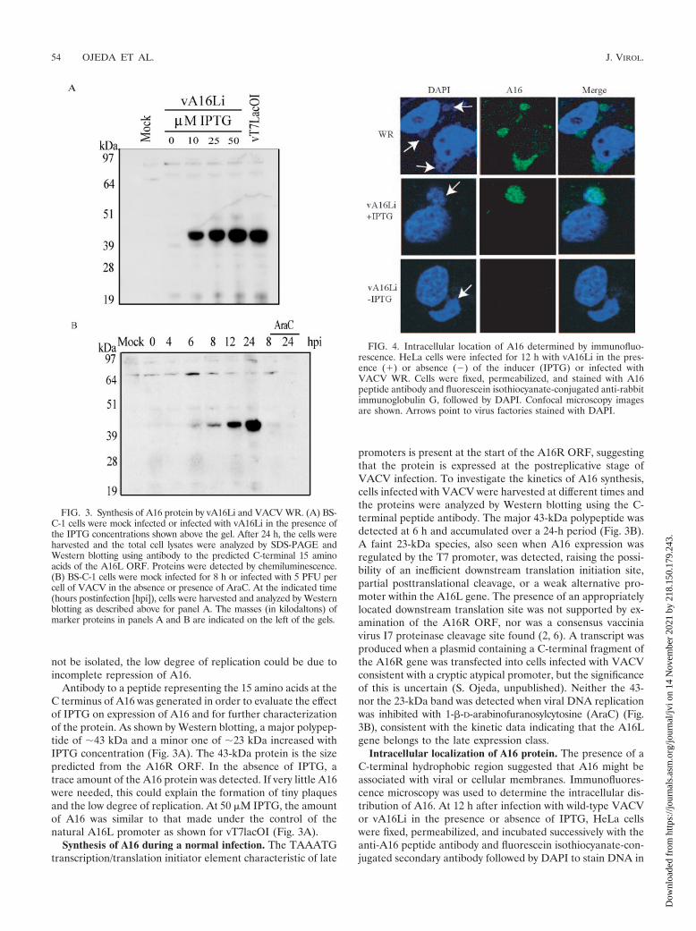
Antibody to a peptide representing the 15 amino acids at theC terminus of A16 was generated in order to evaluate the effectof IPTG on expression of A16 and for further characterizationof the protein. As shown by Western blotting, a major polypep-tide of �43 kDa and a minor one of �23 kDa increased withIPTG concentration (Fig. 3A). The 43-kDa protein is the sizepredicted from the A16R ORF. In the absence of IPTG, atrace amount of the A16 protein was detected. If very little A16were needed, this could explain the formation of tiny plaquesand the low degree of replication. At 50 �M IPTG, the amountof A16 was similar to that made under the control of thenatural A16L promoter as shown for vT7lacOI (Fig. 3A).
Synthesis of A16 during a normal infection. The TAAATGtranscription/translation initiator element characteristic of late
promoters is present at the start of the A16R ORF, suggestingthat the protein is expressed at the postreplicative stage ofVACV infection. To investigate the kinetics of A16 synthesis,cells infected with VACV were harvested at different times andthe proteins were analyzed by Western blotting using the C-terminal peptide antibody. The major 43-kDa polypeptide wasdetected at 6 h and accumulated over a 24-h period (Fig. 3B).A faint 23-kDa species, also seen when A16 expression wasregulated by the T7 promoter, was detected, raising the possi-bility of an inefficient downstream translation initiation site,partial posttranslational cleavage, or a weak alternative pro-moter within the A16L gene. The presence of an appropriatelylocated downstream translation site was not supported by ex-amination of the A16R ORF, nor was a consensus vacciniavirus I7 proteinase cleavage site found (2, 6). A transcript wasproduced when a plasmid containing a C-terminal fragment ofthe A16R gene was transfected into cells infected with VACVconsistent with a cryptic atypical promoter, but the significanceof this is uncertain (S. Ojeda, unpublished). Neither the 43-nor the 23-kDa band was detected when viral DNA replicationwas inhibited with 1-�-D-arabinofuranosylcytosine (AraC) (Fig.3B), consistent with the kinetic data indicating that the A16Lgene belongs to the late expression class.
Intracellular localization of A16 protein. The presence of aC-terminal hydrophobic region suggested that A16 might beassociated with viral or cellular membranes. Immunofluores-cence microscopy was used to determine the intracellular dis-tribution of A16. At 12 h after infection with wild-type VACVor vA16Li in the presence or absence of IPTG, HeLa cellswere fixed, permeabilized, and incubated successively with theanti-A16 peptide antibody and fluorescein isothiocyanate-con-jugated secondary antibody followed by DAPI to stain DNA in
FIG. 3. Synthesis of A16 protein by vA16Li and VACV WR. (A) BS-C-1 cells were mock infected or infected with vA16Li in the presence ofthe IPTG concentrations shown above the gel. After 24 h, the cells wereharvested and the total cell lysates were analyzed by SDS-PAGE andWestern blotting using antibody to the predicted C-terminal 15 aminoacids of the A16L ORF. Proteins were detected by chemiluminescence.(B) BS-C-1 cells were mock infected for 8 h or infected with 5 PFU percell of VACV in the absence or presence of AraC. At the indicated time(hours postinfection [hpi]), cells were harvested and analyzed by Westernblotting as described above for panel A. The masses (in kilodaltons) ofmarker proteins in panels A and B are indicated on the left of the gels.
FIG. 4. Intracellular location of A16 determined by immunofluo-rescence. HeLa cells were infected for 12 h with vA16Li in the pres-ence (�) or absence (�) of the inducer (IPTG) or infected withVACV WR. Cells were fixed, permeabilized, and stained with A16peptide antibody and fluorescein isothiocyanate-conjugated anti-rabbitimmunoglobulin G, followed by DAPI. Confocal microscopy imagesare shown. Arrows point to virus factories stained with DAPI.
54 OJEDA ET AL. J. VIROL.
Dow
nloa
ded
from
http
s://j
ourn
als.
asm
.org
/jour
nal/j
vi o
n 14
Nov
embe
r 20
21 b
y 21
8.15
0.17
9.24
3.
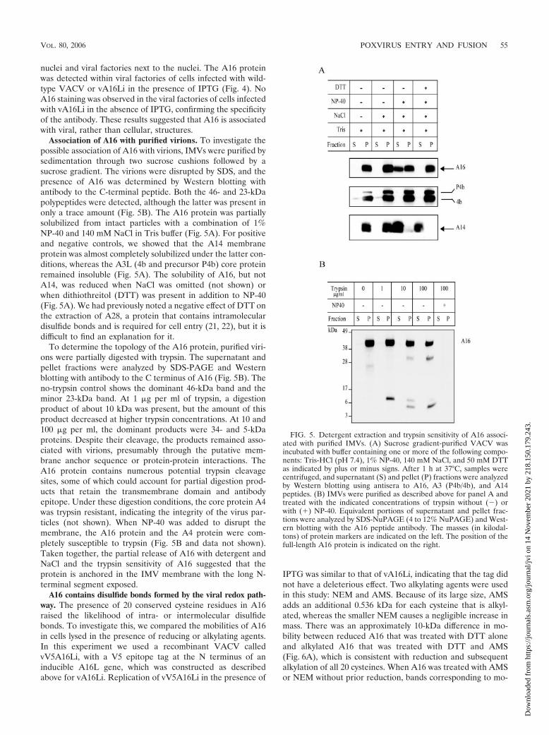
nuclei and viral factories next to the nuclei. The A16 proteinwas detected within viral factories of cells infected with wild-type VACV or vA16Li in the presence of IPTG (Fig. 4). NoA16 staining was observed in the viral factories of cells infectedwith vA16Li in the absence of IPTG, confirming the specificityof the antibody. These results suggested that A16 is associatedwith viral, rather than cellular, structures.
Association of A16 with purified virions. To investigate thepossible association of A16 with virions, IMVs were purified bysedimentation through two sucrose cushions followed by asucrose gradient. The virions were disrupted by SDS, and thepresence of A16 was determined by Western blotting withantibody to the C-terminal peptide. Both the 46- and 23-kDapolypeptides were detected, although the latter was present inonly a trace amount (Fig. 5B). The A16 protein was partiallysolubilized from intact particles with a combination of 1%NP-40 and 140 mM NaCl in Tris buffer (Fig. 5A). For positiveand negative controls, we showed that the A14 membraneprotein was almost completely solubilized under the latter con-ditions, whereas the A3L (4b and precursor P4b) core proteinremained insoluble (Fig. 5A). The solubility of A16, but notA14, was reduced when NaCl was omitted (not shown) orwhen dithiothreitol (DTT) was present in addition to NP-40(Fig. 5A). We had previously noted a negative effect of DTT onthe extraction of A28, a protein that contains intramoleculardisulfide bonds and is required for cell entry (21, 22), but it isdifficult to find an explanation for it.
To determine the topology of the A16 protein, purified viri-ons were partially digested with trypsin. The supernatant andpellet fractions were analyzed by SDS-PAGE and Westernblotting with antibody to the C terminus of A16 (Fig. 5B). Theno-trypsin control shows the dominant 46-kDa band and theminor 23-kDa band. At 1 �g per ml of trypsin, a digestionproduct of about 10 kDa was present, but the amount of thisproduct decreased at higher trypsin concentrations. At 10 and100 �g per ml, the dominant products were 34- and 5-kDaproteins. Despite their cleavage, the products remained asso-ciated with virions, presumably through the putative mem-brane anchor sequence or protein-protein interactions. TheA16 protein contains numerous potential trypsin cleavagesites, some of which could account for partial digestion prod-ucts that retain the transmembrane domain and antibodyepitope. Under these digestion conditions, the core protein A4was trypsin resistant, indicating the integrity of the virus par-ticles (not shown). When NP-40 was added to disrupt themembrane, the A16 protein and the A4 protein were com-pletely susceptible to trypsin (Fig. 5B and data not shown).Taken together, the partial release of A16 with detergent andNaCl and the trypsin sensitivity of A16 suggested that theprotein is anchored in the IMV membrane with the long N-terminal segment exposed.
A16 contains disulfide bonds formed by the viral redox path-way. The presence of 20 conserved cysteine residues in A16raised the likelihood of intra- or intermolecular disulfidebonds. To investigate this, we compared the mobilities of A16in cells lysed in the presence of reducing or alkylating agents.In this experiment we used a recombinant VACV calledvV5A16Li, with a V5 epitope tag at the N terminus of aninducible A16L gene, which was constructed as describedabove for vA16Li. Replication of vV5A16Li in the presence of
IPTG was similar to that of vA16Li, indicating that the tag didnot have a deleterious effect. Two alkylating agents were usedin this study: NEM and AMS. Because of its large size, AMSadds an additional 0.536 kDa for each cysteine that is alkyl-ated, whereas the smaller NEM causes a negligible increase inmass. There was an approximately 10-kDa difference in mo-bility between reduced A16 that was treated with DTT aloneand alkylated A16 that was treated with DTT and AMS(Fig. 6A), which is consistent with reduction and subsequentalkylation of all 20 cysteines. When A16 was treated with AMSor NEM without prior reduction, bands corresponding to mo-
FIG. 5. Detergent extraction and trypsin sensitivity of A16 associ-ated with purified IMVs. (A) Sucrose gradient-purified VACV wasincubated with buffer containing one or more of the following compo-nents: Tris-HCl (pH 7.4), 1% NP-40, 140 mM NaCl, and 50 mM DTTas indicated by plus or minus signs. After 1 h at 37°C, samples werecentrifuged, and supernatant (S) and pellet (P) fractions were analyzedby Western blotting using antisera to A16, A3 (P4b/4b), and A14peptides. (B) IMVs were purified as described above for panel A andtreated with the indicated concentrations of trypsin without (�) orwith (�) NP-40. Equivalent portions of supernatant and pellet frac-tions were analyzed by SDS-NuPAGE (4 to 12% NuPAGE) and West-ern blotting with the A16 peptide antibody. The masses (in kilodal-tons) of protein markers are indicated on the left. The position of thefull-length A16 protein is indicated on the right.
VOL. 80, 2006 POXVIRUS ENTRY AND FUSION 55
Dow
nloa
ded
from
http
s://j
ourn
als.
asm
.org
/jour
nal/j
vi o
n 14
Nov
embe
r 20
21 b
y 21
8.15
0.17
9.24
3.
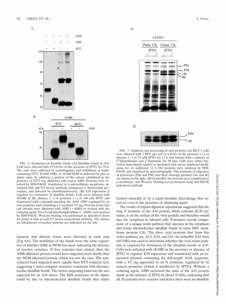
nomeric and dimeric forms were detected in each case(Fig. 6A). The mobilities of the bands were the same regard-less of whether AMS or NEM was used, indicating the absenceof reactive cysteines. If free cysteines had existed, then theAMS-alkylated protein would have migrated more slowly thanthe NEM-alkylated protein, which was not the case. The non-reduced band migrated more rapidly than DTT-reduced A16,suggesting a more-compact structure consistent with intramo-lecular disulfide bonds. The slower-migrating band was the sizeexpected for an A16 dimer. The SDS resistance of the dimercould be due to intermolecular disulfide bonds that either
formed naturally or by a rapid disulfide interchange that oc-curred even in the presence of alkylating agent.
The results of trypsin digestion experiments suggested that thelong N terminus of the A16 protein, which contains all 20 cys-teines, is on the surface of the virus particle and therefore wouldface the cytoplasm in infected cells. Poxviruses encode compo-nents of a unique redox pathway that operates in the cytoplasmand forms intramolecular disulfide bonds in some IMV mem-brane proteins (24). The three viral proteins that form thisredox pathway are A2.5, E10, and G4. An inducible E10 virus(vE10Ri) was used to determine whether the viral redox path-way is required for formation of the disulfide bonds of A16.Cells were infected with vE10Ri in the presence or absence ofIPTG to regulate E10 expression and transfected with an ex-pression plasmid containing the full-length A16L sequence,with a V5 tag appended to its N terminus, regulated by itsnatural promoter (which is unaffected by IPTG). Without areducing agent, AMS increased the mass of the A16 proteinmade in the absence of IPTG by about 10 kDa, indicating thatall 20 cysteines were reactive and hence there were no disulfide
FIG. 6. Formation of disulfide bonds. (A) Disulfide bonds in A16.Cells were infected with vV5A16Li in the presence of IPTG for 18 h.The cells were collected by centrifugation and solubilized in buffercontaining DTT, 20 mM AMS, or 10 nM NEM as indicated by plus orminus signs. In addition, a portion of the extract solubilized in thepresence of DTT was alklylated with excess AMS. Proteins were re-solved by SDS-PAGE, transferred to a nitrocellulose membrane, in-cubated with anti-V5 mouse antibody conjugated to horseradish per-oxidase, and detected by chemiluminescence. (B) E10 expression isrequired for formation of disulfide bonds. Cells were infected withvE10Ri in the absence (�) or presence (�) of 100 �M IPTG andtransfected with a plasmid encoding the A16L ORF regulated by itsown promoter and containing a C-terminal V5 tag. Proteins from totalcell extracts were alkylated with AMS (�AMS) or treated with thereducing agent Tris-(2-carboxyethyl)phosphine (�AMS) and resolvedby SDS-PAGE. Western blotting was performed as described abovefor panel A with an anti-V5 mouse monoclonal antibody. The masses(in kilodaltons) of marker proteins are indicated on the left.
FIG. 7. Synthesis and processing of viral proteins. (A) BS-C-1 cellswere infected with 5 PFU per cell of vA16Li in the presence (�) orabsence (�) of 50 �M IPTG for 12 h and labeled with a mixture of[35S]methionine and [35S]cysteine for 30 min. Cells were either har-vested immediately (pulse) or incubated with excess unlabeled methi-onine for an additional 12 h The proteins were analyzed by SDS-PAGE and visualized by autoradiography. The positions of migrationof precursors (P4a and P4b) and their cleavage products (4a and 4b)are shown on the right. (B) In parallel, the proteins were transferred toa membrane, and Western blotting was performed using anti-P4b/4bpolyclonal antibody.
56 OJEDA ET AL. J. VIROL.
Dow
nloa
ded
from
http
s://j
ourn
als.
asm
.org
/jour
nal/j
vi o
n 14
Nov
embe
r 20
21 b
y 21
8.15
0.17
9.24
3.
bonds (Fig. 6B). In contrast, AMS increased the mass of theA16 protein made in the presence of IPTG by about 2 kDa,which corresponds to four reactive cysteines and indicates that8 of the 10 disulfide bonds had formed (Fig. 6B). Incompletedisulfide bond formation in the presence of IPTG is probablyrelated to the somewhat artificial induction and transfectionsystem, as noted previously for the A28 protein under similarconditions (21). The formation of disulfide bonds in A16 viathe viral redox pathway is consistent with the deduced mem-brane topology in which the long N terminus of the membrane-anchored protein is exposed to the cytoplasm.
Virus morphogenesis is unaffected by repression of A16.Some viral membrane proteins are required for particle assem-bly and morphogenesis; consequently, processing of core pro-teins fails to occur in their absence. To investigate such a rolefor the A16 protein, a pulse-chase experiment was carried out.Cells were infected with vA16Li in the presence or absence ofIPTG, pulse-labeled at 12 h (when host protein synthesis isturned off) with [35S]methionine, and then chased with excessmethionine for an additional 12 h. Processing of core proteins,determined by SDS-PAGE and autoradiography, was unaf-fected by IPTG, suggesting that A16 is not required for earlysteps in assembly (Fig. 7A). Processing of the P4b protein wasalso demonstrated by Western blotting (Fig. 7B). The relativeamounts of precursor and product were similar in the presenceand absence of A16 expression.
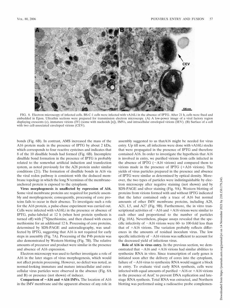
Electron microscopy was used to further investigate a role ofA16 in the later stages of virus morphogenesis, which wouldnot affect protein processing. However, no defect was noted, asnormal-looking immature and mature intracellular and extra-cellular virus particles were observed in the absence (Fig. 8Aand B) or presence (not shown) of inducer.
Comparison of �A16 and �A16 IMVs. The location of A16in the IMV membrane and the apparent absence of any role in
assembly suggested to us thatA16 might be needed for virusentry. Up till now, all infections were done with vA16Li stocksthat were propagated in the presence of IPTG and thereforecontained A16. In order to investigate the hypothesis that A16is involved in entry, we purified virions from cells infected inthe absence of IPTG (�A16 virions) and compared them tovirions made in the presence of IPTG (�A16 virions). Theyields of virus particles prepared in the presence and absenceof IPTG were similar as determined by optical density. More-over, the two types of particles were indistinguishable by elec-tron microscopy after negative staining (not shown) and bySDS-PAGE and silver staining (Fig. 9A). Western blotting ofproteins from virions formed with and without IPTG indicatedthat the latter contained only a trace of A16 but normalamounts of other IMV membrane proteins, including A28,A21, L5, and A27 (Fig. 9B). Furthermore, the in vitro tran-scriptional activities of �A16 and �A16 virions were similar toeach other and proportional to the number of particles(Fig. 10A). Nevertheless, plaque assays revealed that the spe-cific infectivity of �A16 virions were 60- to 100-fold less thanthat of �A16 virions. The variation probably reflects differ-ences in the amounts of residual inoculum virus. The lowspecific infectivity of �A16 virions was sufficient to account forthe decreased yield of infectious virus.
Role of A16 in virus entry. In the previous section, we dem-onstrated that �A16 and �A16 virions had similar abilities tosynthesize RNA in vitro. Since transcription of early genes isinitiated soon after the delivery of cores into the cytoplasm,failure of �A16 virus to synthesize RNA would suggest a blockin entry. To evaluate viral early RNA synthesis, cells wereinfected with equal amounts of purified �A16 or �A16 virionsin the presence of AraC to prevent DNA replication and late-stage RNA synthesis. Total RNA was extracted, and Northernblotting was performed using a radioactive probe complemen-
FIG. 8. Electron microscopy of infected cells. BS-C-1 cells were infected with vA16Li in the absence of IPTG. After 21 h, cells were fixed andembedded in Epon. Ultrathin sections were prepared for transmission electron microscopy. (A) A low-power image of a viral factory regiondisplaying crescents (c), immature virions (IV) (some with nucleoids [n]), IMVs, and intracellular enveloped virions (IEV). (B) Surface of a cellwith two cell-associated enveloped virions (CEV).
VOL. 80, 2006 POXVIRUS ENTRY AND FUSION 57
Dow
nloa
ded
from
http
s://j
ourn
als.
asm
.org
/jour
nal/j
vi o
n 14
Nov
embe
r 20
21 b
y 21
8.15
0.17
9.24
3.

tary to the viral A20R early mRNA. We observed an intenseband of 0.5 kb corresponding to the full-length transcript fromcells infected with �A16 virions but a much weaker one from cellsinfected with �A16 virions (Fig. 10B). �-Actin mRNA was ana-lyzed as a control for RNA integrity and loading (Fig. 10B).
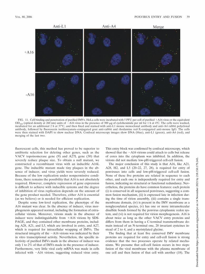
To directly examine the ability of �A16 virions to entercells, we used an assay originally described by Vanderplasschenet al. (29). In our adaptation, purified virions were adsorbed tocells for 1 h at 4°C and then the temperature was raised to 37°Cfor 2 h to allow penetration. The protein synthesis inhibitorcycloheximide was present in order to prevent cytopathic ef-fects and core disassembly. The cells were stained with anti-bodies to the L1 membrane protein and the A4 core protein todetect virions on the surfaces of cells and cores in the cyto-plasm, respectively. As shown in Fig. 11, anti-L1-stained viri-ons were associated with cells infected with �A16 or �A16virions, indicating that A16 is not required for binding. Nu-merous stained cores were seen in the cytoplasm of cells in-fected with �A16 virions but were infrequent in cells infectedwith �A16 virions (Fig. 11). The few cores detected under thelatter conditions colocalized with L1 staining, suggesting thatthey were located on the surface of the cell (Fig. 11, merge).Thus, �A16 virions exhibited a defect in a step of virus entryafter binding.
Low-pH-induced fusion from within and without. Cell-cellfusion can be triggered by briefly exposing cells to low pH atlate times after VACV infection, when progeny virions are onthe cell surface (called fusion from within) or soon after in-fecting cells with large numbers of IMV particles (called fusionfrom without) (11, 13). Previous studies with conditional lethalmutants impaired in expression of the A21, A28, H2, and L5
proteins showed a correlation between the inability of virionsto enter cells and the inability to induce cell-cell fusion (20, 22,27, 28).
To determine whether A16 expression is required for fusionfrom within, we infected cells with vA16Li in the presence orabsence of IPTG and briefly exposed the cells to pH 5.5 at 18 hafter infection. Large syncytia were observed only in the pres-ence of IPTG (Fig. 12A), indicating that the expression of A16is essential for fusion. Similarly, low-pH-triggered fusion fromwithout occurred when cells were infected with purified �A16virions but not with �A16 virions (Fig. 12B). As expected,fusion from within or without did not occur with �A16 virionswhen the low-pH treatment was omitted (not shown).
DISCUSSION
Our inability to isolate a deletion mutant during attemptsto knock out the A16 gene by insertion of DNA encodinggreen fluorescent protein strongly suggested that A16 ex-pression is essential for virus replication. Because of theability to identify tiny plaques comprised of only a few green
FIG. 9. Protein composition of purified virions. IMVs were purifiedby sucrose gradient sedimentation from cells infected with VACV WRor with vA16Li in the presence (�A16) or absence (�A16) of IPTG.Equal numbers of particles (determined by optical density at 260 nm)were analyzed by SDS-PAGE and silver staining. The masses (in kilo-daltons) of marker proteins are shown on the left. (B) Western blottingof samples prepared as described above for panel A and probed withantibodies to the A16, A28, A21, L5, and A27 proteins. FIG. 10. In vitro and in vivo RNA synthesis. (A) In vitro transcrip-
tion by permeabilized �A16 and �A16 virions. Lysates were madefrom cells infected with vA16Li in the presence (�) or absence (�) ofIPTG, and virions were purified by sucrose gradient centrifugation.The indicated number of OD260 (optical density at 260 nm) units ofpurified virions were incubated in a reaction mixture containing[�-32P]UTP. Incorporation of radioactivity into RNA was determined.(B) Northern blot analysis. BS-C-1 cells were treated with 40 �g per mlof AraC for 1 h. Total RNA was extracted from cells at 3 h after mockinfection or infection with 5 PFU of purified �A16 virions or theequivalent OD260 units of �A16 virions. The RNA was resolved byagarose gel electrophoresis, transferred to a membrane, probed withradioactively labeled DNA complementary to the A20R early vacciniavirus gene or �-actin, and analyzed by autoradiography.
58 OJEDA ET AL. J. VIROL.
Dow
nloa
ded
from
http
s://j
ourn
als.
asm
.org
/jour
nal/j
vi o
n 14
Nov
embe
r 20
21 b
y 21
8.15
0.17
9.24
3.
fluorescent cells, this method has proved to be superior toantibiotic selection for deleting other genes, such as theVACV topoisomerase gene (9) and A27L gene (30) thatseverely reduce plaque size. To obtain a null mutant, weconstructed a recombinant virus with an inducible A16Lgene. The inducible mutant made tiny plaques in the ab-sence of inducer, and virus yields were severely reduced.Because of the low replication under nonpermissive condi-tions, there remains the possibility that A16 is not absolutelyrequired. However, complete repression of gene expressionis difficult to achieve with inducible systems and the degreeof inhibition of virus replication depends on the amount ofthe gene product needed. Therefore, either A16 is essential(as we believe) or is needed for efficient replication.
Despite some low-level replication, the phenotype of theA16 mutant was clear. In the absence of inducer, virus mor-phogenesis appeared normal, including the formation of extra-cellular virions. Moreover, virions made in the absence ofinducer were indistinguishable from �A16 virions by SDS-PAGE, and they contained other membrane proteins, includ-ing A28, A21, and L5, which are involved in entry, and A27,which is required for intracellular wrapping of IMVs. Thestructural integrity of the �A16 virions was indicated by theirin vitro transcriptional activity. Nevertheless, the specific in-fectivity of purified IMVs made in the absence of inducer wasonly 1 to 2% of that of IMVs made in the presence of inducer.Furthermore, very little viral early mRNA was made in cellsinfected with �A16 virions, suggesting reduced virus entry.
This entry block was confirmed by confocal microscopy, whichshowed that the �A16 virions could attach to cells but releaseof cores into the cytoplasm was inhibited. In addition, thevirions did not mediate low-pH-triggered cell-cell fusion.
The major conclusion of this study is that A16, like A21,A28, H2, and L5 (20–22, 27, 28), is required for entry ofpoxviruses into cells and low-pH-triggered cell-cell fusion.None of these five proteins are related in sequence to eachother, and each one is independently required for entry andfusion, indicating no structural or functional redundancy. Nev-ertheless, the proteins do have common features: each protein(i) is conserved in all sequenced poxviruses, suggesting a com-mon fusion mechanism, (ii) is expressed late in infection dur-ing the time of virion assembly, (iii) contains a single trans-membrane domain, (iv) is present in the IMV membrane as anonglycosylated species, (v) has one or more intramoleculardisulfide bonds formed by the poxvirus cytoplasmic redox sys-tem, and (vi) is not required for virion morphogenesis. A16 isabout twice as long as the other VACV entry proteins anddiffers from them in having a C-terminal transmembrane do-main instead of an N-terminal one, 20 invariant cysteines in-stead of 2 to 4, and a myristylated glycine.
The finding that at least five conserved IMV membraneproteins are required for entry and cell-cell fusion is strongevidence that the two processes operate by related mecha-nisms. We presume that cell-cell fusion occurs in two steps:fusion of the viral membrane with the plasma membrane ofone cell and then fusion of that cell with another (18). The
FIG. 11. Cell binding and penetration of purified IMVs. HeLa cells were incubated with 5 PFU per cell of purified �A16 virus or the equivalentOD260 (optical density at 260 nm) units of �A16 virus in the presence of 300 �g of cycloheximide per ml for 1 h at 4°C. The cells were washed,incubated for an additional 2 h at 37°C, and then fixed and stained with anti-L1 mouse monoclonal antibody and anti-A4 rabbit polyclonalantibody, followed by fluorescein isothiocyanate-conjugated goat anti-rabbit and rhodamine red-X-conjugated anti-mouse IgG. The cellswere then stained with DAPI to show nuclear DNA. Confocal microscopy images show DNA (blue), anti-L1 (green), anti-A4 (red), andmerging of the last two.
VOL. 80, 2006 POXVIRUS ENTRY AND FUSION 59
Dow
nloa
ded
from
http
s://j
ourn
als.
asm
.org
/jour
nal/j
vi o
n 14
Nov
embe
r 20
21 b
y 21
8.15
0.17
9.24
3.
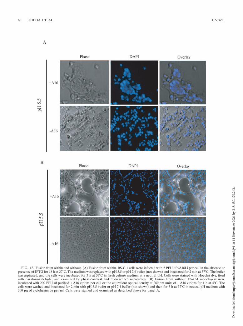
FIG. 12. Fusion from within and without. (A) Fusion from within. BS-C-1 cells were infected with 2 PFU of vA16Li per cell in the absence orpresence of IPTG for 18 h at 37°C. The medium was replaced with pH 5.5 or pH 7.4 buffer (not shown) and incubated for 2 min at 37°C. The bufferwas aspirated, and the cells were incubated for 3 h at 37°C in fresh culture medium at a neutral pH. Cells were stained with Hoechst dye, fixedwith paraformaldehyde, and examined by phase-contrast and fluorescence microscopy. (B) Fusion from without. BS-C-1 monolayers wereincubated with 200 PFU of purified �A16 virions per cell or the equivalent optical density at 260 nm units of �A16 virions for 1 h at 4°C. Thecells were washed and incubated for 2 min with pH 5.5 buffer or pH 7.4 buffer (not shown) and then for 3 h at 37°C in neutral pH medium with300 �g of cycloheximide per ml. Cells were stained and examined as described above for panel A.
60 OJEDA ET AL. J. VIROL.
Dow
nloa
ded
from
http
s://j
ourn
als.
asm
.org
/jour
nal/j
vi o
n 14
Nov
embe
r 20
21 b
y 21
8.15
0.17
9.24
3.

reason why so many poxvirus proteins are required for entryand fusion is perplexing. Two of the entry/fusion proteins werereported to associate with each other (20), and there is evi-dence that the others, including A16, are part of the samecomplex (19b). Nevertheless, as found here for A16, A21, A28,and L5 and elsewhere for A28 and H2 (20), the entry proteinsseem capable of trafficking independently to the viral mem-brane. It is possible that the proteins in the complex havemultiple, nonredundant roles, including membrane fusion perse, activation of fusion, receptor recognition, and a scaffoldingfunction. Because the number of proteins involved in poxvirusentry seems to be higher than for other viruses, determinationof the mechanism is expected to be challenging. Only membersof the Herpesviridae family approach poxviruses with regard tothe number of proteins involved in entry (26). Orthologs ofthree glycoproteins, designated gB, gH, and gL, are essentialfor entry of all herpesviruses. In addition to the three basicfusion proteins, some herpesviruses require additional noncon-served receptor-binding proteins, such as gD for most alpha-herpesviruses.
ACKNOWLEDGMENTS
We thank Andrea Weisberg for electron microscopy, Owen Schwartzfor help with confocal microscopy, and Norman Cooper for cell cultures.
This research was supported in part by the Intramural ResearchProgram of the NIAID of the NIH.
REFERENCES
1. Alexander, W. A., B. Moss, and T. R. Fuerst. 1992. Regulated expression offoreign genes in vaccinia virus under the control of bacteriophage T7 RNApolymerase and the Escherichia coli lac repressor. J. Virol. 66:2934–2942.
2. Ansarah-Sobrinho, C., and B. Moss. 2004. Role of the I7 protein in proteo-lytic processing of vaccinia virus membrane and core components. J. Virol.78:6335–6343.
3. Antoine, G., F. Scheiflinger, F. Dorner, and F. G. Falkner. 1998. The com-plete genomic sequence of the modified vaccinia Ankara strain: comparisonwith other orthopoxviruses. Virology 244:365–396.
4. Armstrong, J. A., D. H. Metz, and M. R. Young. 1973. The mode of entry ofvaccinia virus into L cells. J. Gen. Virol. 21:533–537.
5. Betakova, T., E. J. Wolffe, and B. Moss. 1999. Regulation of vaccinia virusmorphogenesis: phosphorylation of the A14L and A17L membrane proteinsand C-terminal truncation of the A17L protein are dependent on the F10Lprotein kinase. J. Virol. 73:3534–3543.
6. Byrd, C. M., T. C. Bolken, and D. E. Hruby. 2002. The vaccinia virus I7Lgene product is the core protein proteinase. J. Virol. 76:8973–8976.
7. Carter, G. C., M. Law, M. Hollinshead, and G. L. Smith. 2005. Entry of thevaccinia virus intracellular mature virion and its interactions with glycosami-noglycans. J. Gen. Virol. 86:1279–1290.
8. Chang, A., and D. H. Metz. 1976. Further investigations on the mode of entryof vaccinia virus into cells. J. Gen. Virol. 32:275–282.
9. Da Fonseca, F., and B. Moss. 2003. Poxvirus DNA topoisomerase knockoutmutant exhibits decreased infectivity associated with reduced early transcrip-tion. Proc. Natl. Acad. Sci. USA 100:11291–11296.
10. Demkowicz, W. E., J. S. Maa, and M. Esteban. 1992. Identification andcharacterization of vaccinia virus genes encoding proteins that are highlyantigenic in animals and are immunodominant in vaccinated humans. J. Vi-rol. 66:386–398.
11. Doms, R. W., R. Blumenthal, and B. Moss. 1990. Fusion of intra- andextracellular forms of vaccinia virus with the cell membrane. J. Virol. 64:4884–4892.
12. Earl, P. L., N. Cooper, S. Wyatt, B. Moss, and M. W. Carroll. 1998. Prepa-ration of cell cultures and vaccinia virus stocks, p. 16.16.1–16.16.3. In F. M.Ausubel, R. Brent, R. E. Kingston, D. D. Moore, J. G. Seidman, J. A. Smith,and K. Struhl (ed.), Current protocols in molecular biology, vol. 2. JohnWiley and Sons, New York, N.Y.
13. Gong, S. C., C. F. Lai, and M. Esteban. 1990. Vaccinia virus induces cellfusion at acid pH and this activity is mediated by the N-terminus of the14-kDa virus envelope protein. Virology 178:81–91.
14. Ichihashi, Y. 1996. Extracellular enveloped vaccinia virus escapes neutral-ization. Virology 217:478–485.
15. Janeczko, R. A., J. F. Rodriguez, and M. Esteban. 1987. Studies on themechanism of entry of vaccinia virus into animal cells. Arch. Virol. 92:135–150.
16. Martin, K. H., D. W. Grosenbach, C. A. Franke, and D. E. Hruby. 1997.Identification and analysis of three myristylated vaccinia virus late proteins.J. Virol. 71:5218–5226.
17. Moss, B. 2001. Poxviridae: the viruses and their replication, p. 2849–2883. InD. M. Knipe and P. M. Howley (ed.), Fields virology, 4th ed., vol. 2. Lip-pincott Williams & Wilkins, Philadelphia, Pa.
18. Moss, B. Poxvirus entry and membrane fusion. Virology, in press.19. Senkevich, T. G., E. V. Koonin, J. J. Bugert, G. Darai, and B. Moss. 1997.
The genome of molluscum contagiosum virus: analysis and comparison withother poxviruses. Virology 233:19–42.
19b.Senkevich, T. G., S. Ojeda, A. Townsley, G. E. Nelson, and B. Moss. Poxvirusmultiprotein entry-fusion complex. Proc. Natl. Acad. Sci. USA, in press.
20. Senkevich, T. G., and B. Moss. 2005. Vaccinia virus H2 protein is an essentialcomponent of a complex involved in virus entry and cell-cell fusion. J. Virol.79:4744–4754.
21. Senkevich, T. G., B. M. Ward, and B. Moss. 2004. Vaccinia virus A28L geneencodes an essential protein component of the virion membrane with in-tramolecular disulfide bonds formed by the viral cytoplasmic redox pathway.J. Virol. 78:2348–2356.
22. Senkevich, T. G., B. M. Ward, and B. Moss. 2004. Vaccinia virus entry intocells is dependent on a virion surface protein encoded by the A28L gene.J. Virol. 78:2357–2366.
23. Senkevich, T. G., A. Weisberg, and B. Moss. 2000. Vaccinia virus E10Rprotein is associated with the membranes of intracellular mature virions andhas a role in morphogenesis. Virology 278:244–252.
24. Senkevich, T. G., C. L. White, E. V. Koonin, and B. Moss. 2002. Completepathway for protein disulfide bond formation encoded by poxviruses. Proc.Natl. Acad. Sci. USA 99:6667–6672.
25. Smith, G. L., and M. Law. 2004. The exit of vaccinia virus from infected cells.Virus Res. 106:189–197.
26. Spear, P. G., and R. Longnecker. 2003. Herpesvirus entry: an update. J. Vi-rol. 77:10179–10185.
27. Townsley, A., T. G. Senkevich, and B. Moss. 2005. The product of thevaccinia virus L5R gene is a fourth membrane protein encoded by all pox-viruses that is required for cell entry and cell-cell fusion. J. Virol. 79:10988–10998.
28. Townsley, A., T. G. Senkevich, and B. Moss. 2005. Vaccinia virus A21 virionmembrane protein is required for cell entry and fusion. J. Virol. 79:9458–9469.
29. Vanderplasschen, A., M. Hollinshead, and G. L. Smith. 1998. Intracellularand extracellular vaccinia virions enter cells by different mechanisms. J. Gen.Virol. 79:877–887.
30. Ward, B. M. 2005. Visualization and characterization of the intracellularmovement of vaccinia virus intracellular mature virions. J. Virol. 79:4755–4763.
31. Wolffe, E. J., S. Vijaya, and B. Moss. 1995. A myristylated membrane proteinencoded by the vaccinia virus L1R open reading frame is the target of potentneutralizing monoclonal antibodies. Virology 211:53–63.
32. Zhang, Y., and B. Moss. 1991. Inducer-dependent conditional-lethal mutantanimal viruses. Proc. Natl. Acad. Sci. USA 88:1511–1515.
VOL. 80, 2006 POXVIRUS ENTRY AND FUSION 61
Dow
nloa
ded
from
http
s://j
ourn
als.
asm
.org
/jour
nal/j
vi o
n 14
Nov
embe
r 20
21 b
y 21
8.15
0.17
9.24
3.