Embed Size (px)
Citation preview

Indian Journal of Experimental Biology Vol. 44, August 2006, pp. 597-617
Review Article
Environmental contaminants in pathogenesis of breast cancer Shyamali Mukherjee
Department of Pharmacology, Meharry Medical College, 1005 Dr. D. B. Todd Jr. Blvd., Nashville, TN 37208, USA
Bidhan Chandra Koner* Department of Biochemistry, JIPMER, Pondicherry 605 006, India
Sanhita Ray Division of Reproductive and Developmental Biology, Vanderbilt University School of
Medicine, Nashville, TN 37232, USA Amitabha Ray
The Hormel Institute, University of Minnesota, 801-16th Avenue NE, Austin, MN 55912, USA
This review is an attempt to comprehend the diverse groups of environmental chemical contaminants with a potential for pathogenesis of breast cancer, their probable sources and the possible mechanisms by which these environmental contaminants act and interplay with other risk factors. Estrogens are closely related to the pathogenesis of breast cancer. Oxidative catabolism of estrogen, mediated by various cytochrome P450 enzymes, generates reactive free radicals that can cause oxidative damage. The same enzymes of estrogenic metabolic pathways catalyze biological activation of several environmental (xenobiotic) chemicals. Xenobiotic chemicals may exert their pathological effects through generation of reactive free radicals. Breast tissue can be a target of several xenobiotic agents. DNA-reactive metabolites of different xenobiotic compounds have been detected in breast tissue. Many phase I and II xenobiotic metabolizing enzymes are expressed in both normal and cancerous breast tissues. These enzymes play a significant role in the activation/detoxification of xenobiotic and endogenous compounds including estrogens. More than 30 carcinogenic chemicals are present in tobacco smoke; many of them are fat-soluble, resistant to metabolism and can be stored in breast adipose tissue. Similarly, pesticides are also known to cause oxidative stress; while some act as endocrine disruptor, some are shown to suppress apoptosis in estrogen sensitive cell lines. Reports have shown an association of smoking (both active and passive) and pesticides with breast cancer risk. However, the issues have remained controversial. Different mutagenic substances that are generated in the cooking process e.g., heterocyclic amines and polycyclic aromatic hydrocarbons (PAHs) can be a threat to breast tissue. PAHs and dioxins exert their adverse effects through the aryl hydrocarbon receptor (AhR), which activates several genes involved in the metabolisms of xenobiotic compounds and endogenous estrogens. These chemicals also induce AhR-dependent mitochondrial dysfunction. Many of the environmental pollutants suppress the immune system, which are implicated to risk. A better understanding about the biological effects of different environmental carcinogenic compounds and determination of their impact on rising incidence of breast cancer will be beneficial in improving preventive policy against breast cancer.
Keywords: Aryl hydrocarbon receptor, Breast cancer, Immunotoxicant, Oxidative metabolism, Xenoestrogens
Breast cancer is the most commonly occurring neoplastic disease in women worldwide and is second only to lung cancer as a cause of cancer death in women1. There is a gradual increase in breast cancer incidence in most developed countries and in societies which became westernized recently or are in the process. Breast cancer is the second most common cancer among Indian women; however, it is the leading cancer in Mumbai and Kolkata, and an
increasing incidence has been recorded in urban females2,3. Among the population based cancer registries functioning in various parts of the country (India), the Mumbai Registry is a highly efficient system; and a major portion of the country’s epidemiological data have been derived from Mumbai. However, aspects related with different environmental risk factors are not clear in those published reports. Studies on Mumbai population showed highest breast cancer incidence rates among Parsis and Christians, followed by Hindus and Muslims, and lowest rates among Jains and Buddhists2,4,5. Interestingly, data from most of the registries indicate that Christians in India have the
______________ *Correspondent author Phone: 0413-2273116 Fax: 0091-413-2272067 e-mail: [email protected]

INDIAN J EXP BIOL, AUGUST 2006
598
greatest risk of breast cancer6. A more westernized lifestyle in both Christian and Parsi community is probably the reason for high incidence of breast cancer. Furthermore, higher incidence among Parsis is partly due to more consanguineous marriages (genetic etiology), a large number of spinsters or delayed marriage/first child birth and less number of children7.
Epidemiological observations indicate a close association between the process of westernization and an increase in breast cancer incidence. Studies on the populations which migrated to the Western countries showed a higher breast cancer risk8,9; the phenomenon suggests an environmental influence. It is obvious that the effects of physical environment within a geographical area are more or less similar on the inhabitants; whereas lifestyle factors differ among individuals. Apart from reproduction-related factors, a westernized lifestyle is intimately associated with some particular forms of food processing/cooking practices and dietary habits such as frequent use of fast foods. In this article (under the section ‘Role of food mutagens’), an attempt has been made to describe the known food related mutagens which are linked with the westernized dietary habits as well as mammary tumor development in experimental animals. The results of various epidemiological studies on different dietary factors (carcinogenic or chemopreventive) are ambiguous and conclusions have been derived mainly from the laboratory experiments. However, evidence is accumulating, which indicates an association between the westernized dietary habits and risk of breast cancer10.
Several lines of research suggest that estrogens are closely associated with the pathological process of breast cancer. The disease risk is increased by longer duration of estrogen exposure; therefore, early menarche and/or late menopause increase the risk. Similarly, age at first pregnancy, nulliparity, obesity, estrogen replacement therapy or oral contraceptives are considered to be related with the higher risk11,12. Further, alcohol intake can increase risk by elevating the levels of circulating estrogen13. Different mechanisms may be responsible for the carcinogenicity of estrogens in the human breast such as receptor-mediated hormonal activity leading to cellular proliferation and more chances for accumulation of the genetic errors; the induction of aneuploidy by estrogen; and a cytochrome P450-mediated metabolic activation that can cause genotoxic effects. There is evidence that oxidative catabolism of estrogens, mediated by
various cytochrome P450 complexes, constitutes a pathway of their metabolic activation and generates reactive free radicals that can cause oxidative stress and genomic damage14. There are plenty of indirect evidences to show that environmental contaminants could contribute to pathogenesis of breast cancer.
Enzymes linked with both estrogen and xenobiotic metabolisms
In the metabolism of xenobiotics (foreign chemicals), cytochrome P450s or monooxygenases perform an important function by catalyzing the hydroxylation reaction. Overall, the goal of xenobiotic metabolism is to increase the water solubility of various foreign chemicals in order to eliminate them from our physiological system; and this process occurs in two phases. Cytochrome P450 enzymes are involved in phase I reaction, which sometimes convert biologically inactive compounds into active or toxic metabolites. Subsequently, in phase II, products of phase I reaction are conjugated with various molecules such as glucuronic acid, sulfate, glutathione, acetyl or methyl groups, leading to excretion from the body. Several phase I and II enzymes are associated with estrogen metabolism (Fig. 1 and Table 1). In human breast tissue, estrogens are mainly hydroxylated by cytochrome P4501A1 (CYP1A1) and CYP1B1 into 2-hydroxyestrogens and 4-hydroxyestrogens, respectively. Higher ratio of 4-hydroxyestrogens:2-hydroxyestrogens was detected in breast cancer; and the ratio was reported to be 4:1 in breast tumor tissue extract15,16. It is believed that 4-hydroxyestrogens may act as a carcinogen17.
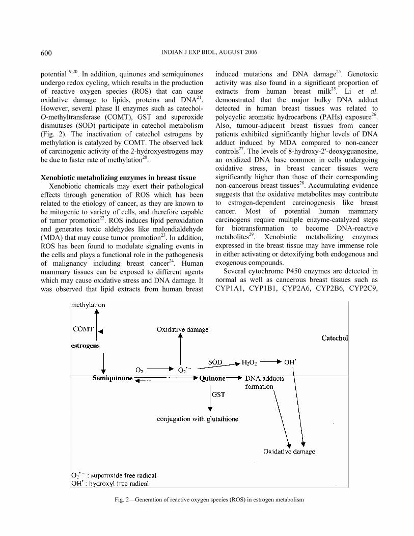
The metabolisms of 2- and 4-hydroxyestrogens (catechol estrogens) are produced in a series of linked oxidation reactions that form the oxidative estrogen metabolism pathway18. Metabolism of the catechol estrogens leads to the formation of unstable semiquinone which is an intermediate in both oxidation and reduction reactions, and can react with molecular oxygen to form superoxide radicals and quinone. Superoxide radicals may be reduced to hydrogen peroxide and then, to hydroxyl radical in presence of metal ions (Fig. 2). In general, quinones can be conjugated with glutathione by glutathione-S-transferase (GST) or can form adducts with guanine and adenine base in DNA19. Catechol estrogen 2,3-quinone can bind stably to DNA, whereas, 3,4-quinone forms depurinating adducts with guanine and adenine, which are lost from DNA by cleavage of the glucosidic bond leaving apurinic sites with mutagenic

MUKHERJEE et al.: ENVIRONMENTAL CONTAMINANTS & PATHOGENESIS OF BREAST CANCER
599
Table 1⎯Important estrogen biosynthesis/metabolism-related cytochrome P450 enzymes and their xenobiotic substrates
Enzyme Function Steps of catalysis Xenobiotic substrate
CYP11A biosynthesis cholesterol to pregnenolone — CYP17 (17α-hydroxylase, C17-20 lyase)
biosynthesis pregnenolone to 17α-hydroxypregnenolone to dehydroepiandrosterone (DHEA)
—
progesterone to 17α-hydroxyprogesterone to androstenedione
CYP19 (aromatase) biosynthesis androstenedione to estrone (E1) testosterone to estradiol (E2)
—
CYP1A1 metabolism estrogens to 2-hydroxyestrogens PAHs CYP1A2 metabolism estrogens to 2-hydroxyestrogens heterocyclic amines CYP1B1 (4-estrogen hydroxylase) metabolism estrogens to 4-hydroxyestrogens PAHs, heterocyclic amines CYP2C9 metabolism estrone sulfate to 16-hydroxysulfate drug-paclitaxel CYP3A3 metabolism estrogens to 2-hydroxyestrogens — CYP3A4 metabolism estrogens to 2- and 16-hydroxyestrogens PAHs, aflatoxin CYP3A5 metabolism estrogens to 16α-hydroxyestrogens paclitaxel, vinca alkaloids,
tamoxifen PAHs – polycyclic aromatic hydrocarbons (N.B.: CYP is ‘cytochrome P450 enzyme’, the next numeral indicates the ‘family’; the capital letter denotes the ‘subfamily’, and the last number designates the individual ‘member’)
Fig. 1—Principal pathways of estrogen biosynthesis and metabolism
INDIAN J EXP BIOL, AUGUST 2006
600
potential19,20. In addition, quinones and semiquinones undergo redox cycling, which results in the production of reactive oxygen species (ROS) that can cause oxidative damage to lipids, proteins and DNA21. However, several phase II enzymes such as catechol-O-methyltransferase (COMT), GST and superoxide dismutases (SOD) participate in catechol metabolism (Fig. 2). The inactivation of catechol estrogens by methylation is catalyzed by COMT. The observed lack of carcinogenic activity of the 2-hydroxyestrogens may be due to faster rate of methylation20. Xenobiotic metabolizing enzymes in breast tissue
Xenobiotic chemicals may exert their pathological effects through generation of ROS which has been related to the etiology of cancer, as they are known to be mitogenic to variety of cells, and therefore capable of tumor promotion22. ROS induces lipid peroxidation and generates toxic aldehydes like malondialdehyde (MDA) that may cause tumor promotion23. In addition, ROS has been found to modulate signaling events in the cells and plays a functional role in the pathogenesis of malignancy including breast cancer24. Human mammary tissues can be exposed to different agents which may cause oxidative stress and DNA damage. It was observed that lipid extracts from human breast
induced mutations and DNA damage25. Genotoxic activity was also found in a significant proportion of extracts from human breast milk25. Li et al. demonstrated that the major bulky DNA adduct detected in human breast tissues was related to polycyclic aromatic hydrocarbons (PAHs) exposure26. Also, tumour-adjacent breast tissues from cancer patients exhibited significantly higher levels of DNA adduct induced by MDA compared to non-cancer controls27. The levels of 8-hydroxy-2′-deoxyguanosine, an oxidized DNA base common in cells undergoing oxidative stress, in breast cancer tissues were significantly higher than those of their corresponding non-cancerous breast tissues28. Accumulating evidence suggests that the oxidative metabolites may contribute to estrogen-dependent carcinogenesis like breast cancer. Most of potential human mammary carcinogens require multiple enzyme-catalyzed steps for biotransformation to become DNA-reactive metabolites29. Xenobiotic metabolizing enzymes expressed in the breast tissue may have immense role in either activating or detoxifying both endogenous and exogenous compounds.
Several cytochrome P450 enzymes are detected in normal as well as cancerous breast tissues such as CYP1A1, CYP1B1, CYP2A6, CYP2B6, CYP2C9,
Fig. 2—Generation of reactive oxygen species (ROS) in estrogen metabolism

MUKHERJEE et al.: ENVIRONMENTAL CONTAMINANTS & PATHOGENESIS OF BREAST CANCER
601
CYP2E1 and CYP3A430-32. Although phase I enzymes such as CYP1A2, CYP2C6 and CYP3A4 are involved in hepatic and extrahepatic estrogen oxidation, CYP1A1 and CYP1B1 display their leading expression in breast tissue21,29. The 4-hydroxylation activity of CYP1B1 has received particular attention because of experimental evidence that 4-hydroxy catechol estrogens are more carcinogenic than the 2-hydroxy isomers21. On the other hand, several anti-oxidant and phase II enzymes are expressed in breast tissue. Studies observed higher activities of SOD in breast cancer33,34. Er et al. noticed 1.5-fold increase of MnSOD expression in breast cancer tissues compared to non-cancerous tissues35. Further, Thomas et al. recorded higher expression of both MnSOD and CuZnSOD in breast tumours36. Genetic polymorphism in the MnSOD gene (Ala/Ala genotype) was found to have increased risk of breast cancer among Chinese women with high levels of oxidative stress or low intake of anti-oxidant dietary factors37. Like SOD, catalase showed higher expression and activity in breast cancer tissues34,36. On the contrary, Tas et al. found significantly decreased activity of catalase in tumour tissues33.
Human myeloperoxidase is expressed in neutrophils recruited to the lung; and the enzyme is also present in breast milk and blood29. Following immunological and/or chemical insults, neutrophils release myeloperoxidase and undergo a ‘respiratory burst’, which is characterized by a massive increase in oxygen consumption and a consequent NADPH-dependent production of superoxide and other free radicals38. Myeloperoxidase produces the potent bacteriotoxic oxidizing agent hypochlorous acid and ROS in physiological situations, which may also cause DNA damage38,39 (Table 2). Probably, myeloperoxidase is particularly significant in breast cancer, as suggested by association between this enzyme activity and estrogen levels40. Because of the presence of myeloperoxidase in normal breast and tumour tissues of the breast, its association with estrogen levels, and its ability to generate ROS, myeloperoxidase may be
important in the oxidative stress pathway in human breast cancer41. It is worthy to mention that methylation catalyzed by COMT is a necessary mechanism for preventing oxidative metabolites of catechol estrogens in order to protect DNA from oxidative damage. Expression of COMT is observed in the liver, kidney, breast and endometrium42. Further, the phase II enzyme GST is known to play a key role in the detoxification of both xenobiotic and endogenous compounds, and in the reduction of ROS and DNA adducts formation. Overall, GSTs form a group of multi-gene isoenzymes and on the basis of sequence similarity and immunological cross-reactivity43-45, they have been divided into a number of subclasses (e.g., α, δ, ζ, θ, μ, π, σ and τ). GST π(pi) and μ(mu) are the major isoforms expressed in the breast tissues32,46. Among other phase II enzymes, sulfotransferase converts estrogens into the biologically inactive estrogen sulfates. On the other hand, steroid sulfatase hydrolyzes inactive estrogen sulfates to estrogens. Further, sulfotransferases catalyze the biotransformation of xenobiotic compounds. Usually, sulfonation by sulfotransferases leads to the inactivation of parent compounds; however, formation of more toxic metabolites can occur (Table 2). Several studies detected various isoforms of sulfotransferases in both normal and malignant breast tissues47-49. Another important phase II enzyme expressed in breast tissue is N-acetyltransferases. Evaluation of N-acetyltransferases (NAT) and sulfotransferases (SULT) enzyme expression in the breast has shown that major isoforms are NAT1, SULT1A1 and SULT1A3, all of which can activate xenobiotic compounds into DNA-reactive metabolites38,50 (Table 2). Nevertheless, current evidence indicates a role of oxidative metabolites in the pathogenesis of breast cancer. Both in the xenobiotic and catechol estrogen metabolisms, there is a considerable inter-individual variability (polymorphism); and these person-to-person differences, which are attributed to polymorphisms in the genes encoding for the respective enzymes, may define subpopulations of women with risk for breast cancer20.
Table 2⎯Adverse effects of selected anti-oxidant/phase II enzymes expressed in breast tissues
Enzymes Common genotypes involved in xenobiotic metabolism (broadly classified)
Adverse effects/metabolic activation of xenobiotic compounds
Myeloperoxidase MPO (GG, GA, AA) heterocyclic amines, aromatic amines, PAHs Glutathione-S-transferase GSTM1, GSTM3, GSTP1, GSTT1 multi-drugs resistance of tumour cells Sulfotransferase SULT1, SULT2 PAHs, aromatic amines, heterocyclic amines N-acetyltransferase NAT1, NAT2 heterocyclic amines, aromatic amines
INDIAN J EXP BIOL, AUGUST 2006
602
Effects of tobacco/cigarette smoking Other than cancers of the directly affected organs
such as respiratory system51 and distant organs such as uterine cervix52
, smoking may increase the risk of breast cancer53. Various factors like duration and intensity of smoking54
, family history of breast cancer55, cigarette smoking during first pregnancy56
etc. are found to influence the smoking induced risk of breast cancer. Also, a direct relationship was observed between cigarette smoking and metastasis in breast cancer57. However, the association between tobacco smoking and breast cancer risk has remained controversial 58-60.
Many lipophilic carcinogens of tobacco smoke20
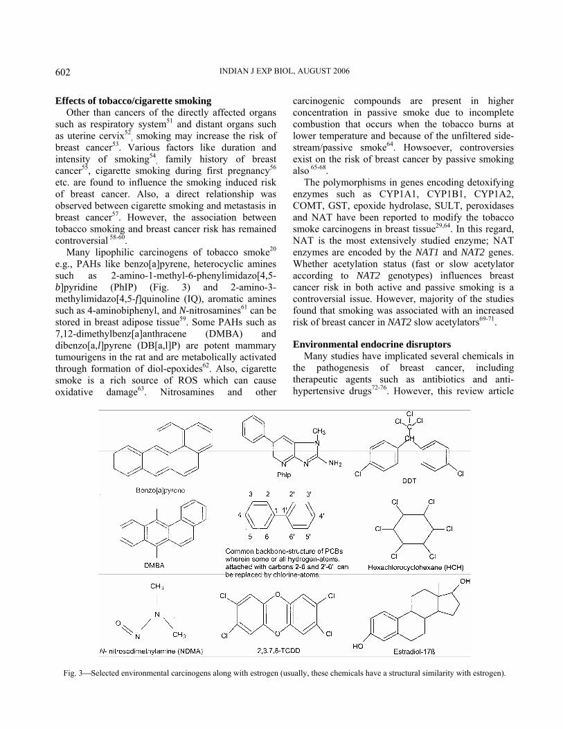
e.g., PAHs like benzo[a]pyrene, heterocyclic amines such as 2-amino-1-methyl-6-phenylimidazo[4,5-b]pyridine (PhIP) (Fig. 3) and 2-amino-3-methylimidazo[4,5-f]quinoline (IQ), aromatic amines such as 4-aminobiphenyl, and N-nitrosamines61 can be stored in breast adipose tissue59. Some PAHs such as 7,12-dimethylbenz[a]anthracene (DMBA) and dibenzo[a,l]pyrene (DB[a,l]P) are potent mammary tumourigens in the rat and are metabolically activated through formation of diol-epoxides62. Also, cigarette smoke is a rich source of ROS which can cause oxidative damage63. Nitrosamines and other
carcinogenic compounds are present in higher concentration in passive smoke due to incomplete combustion that occurs when the tobacco burns at lower temperature and because of the unfiltered side-stream/passive smoke64. Howsoever, controversies exist on the risk of breast cancer by passive smoking also 65-68.
The polymorphisms in genes encoding detoxifying enzymes such as CYP1A1, CYP1B1, CYP1A2, COMT, GST, epoxide hydrolase, SULT, peroxidases and NAT have been reported to modify the tobacco smoke carcinogens in breast tissue29,64. In this regard, NAT is the most extensively studied enzyme; NAT enzymes are encoded by the NAT1 and NAT2 genes. Whether acetylation status (fast or slow acetylator according to NAT2 genotypes) influences breast cancer risk in both active and passive smoking is a controversial issue. However, majority of the studies found that smoking was associated with an increased risk of breast cancer in NAT2 slow acetylators69-71. Environmental endocrine disruptors
Many studies have implicated several chemicals in the pathogenesis of breast cancer, including therapeutic agents such as antibiotics and anti-hypertensive drugs72-76. However, this review article
Fig. 3⎯Selected environmental carcinogens along with estrogen (usually, these chemicals have a structural similarity with estrogen).
MUKHERJEE et al.: ENVIRONMENTAL CONTAMINANTS & PATHOGENESIS OF BREAST CANCER
603
has tried to concentrate on a small number of chemical contaminants which exist in the environment. The concept of ‘endocrine disruptors’ is an emerging subject that has gained popularity among both public and scientific community following publication of the book ‘Our Stolen Future’ by Theo Colborn and colleagues77 in 1996. Endocrine disruptors are xenobiotic chemicals that adversely interfere with the natural functions of hormones (Table 3). Estrogenic endocrine disruptors or xenoestrogens are widely distributed in the environment78,79. Numerous chemicals such as pesticides, polychlorinated biphenyl congeners (both have been discussed in the next section), food-related mycotoxin zearalenone and its derivatives, ultraviolet screen 3-(4-methylbenzylidene)-camphor (4-MBC) and even some metals like cadmium can influence hormonal responses by binding to estrogen receptor (ER). This phenomenon is the most commonly studied mechanism by which environmental chemicals exert their effects on breast. The xenoestrogens interact with the binding pocket of the ER because they have chemical similarities to estrogen (usually a phenolic A-ring). However, reduced activity of xenoestrogens probably results from lack of fit of the remainder of the molecule within the binding pocket79. As discussed earlier, several xenobiotic chemicals may interact with the enzyme systems that metabolize estrogens; and by this process, they can modulate the endogenous steroid metabolism.
A number of chemicals such as organotin compound triphenyltin and fungicides fenarimol, procymidone, vinclozolin etc. inhibit steroid hormone synthesis. Alternatively, synthetic compound like diethylstilbestrol (DES) interfere the bio-availability and overall functions of estrogen by interacting with steroid hormone binding proteins in the blood such as sex hormone-binding globulin (SHBG) and albumin. Chemicals like retinoids, certain pyrethroid insecticides (e.g., sumithrin), pentachlorophenol and β-hexachlorocyclohexane alter steroid signaling pathways. Some compounds are able to disrupt estrogenic responses through several mechanisms. For instance, dioxins alter steroid hormone metabolism as well as there is a cross-talk between dioxin- and estrogen-mediated signaling pathways (vide ‘Aryl hydrocarbon receptor and breast cancer’ section). There are other mechanisms by which xenobiotic chemicals can influence the physiological functions of
estrogen such as adverse effects on release and excretion of hormones, disruption of regulatory feedback relationships between two endocrine organs, and modulation of non-genomic pathways.
Several investigators believe that endocrine disruptors may play a role in breast cancer incidence in the developed countries79-83. Perhaps this view is also true for the countries that are in the process of westernization. Recently, Kortenkamp has hypothesized the possibility of combination effects by a large number of xenobiotic chemicals, all may present at low levels in women’s body, in the pathogenesis of breast cancer83. Howsoever, exploring the mechanisms of pathological effects by different endocrine disruptors is a challenging field. Identifying
Table 3⎯ Man-made chemicals which can act as endocrine disruptors (related with estrogens)
Compound Principal use/Source Hormonal character
Atrazine herbicide estrogenic Bisphenol A manufacture of plastics estrogenic Butylhydroxyanisole manufacture of plastics estrogenic
Cyclotetrasiloxanes rubber, plastics, and shampoo
hormonal modulator
DDT insecticide estrogenic Dieldrin pesticide estrogenic DES synthetic estrogen/drug estrogenic Endosulphan pesticide estrogenic Ethynylestradiol synthetic estrogen/drug estrogenic Fenarimol fungicide anti-steroid Hexachlorophene disinfectant anti-estrogenic Kepone insecticide estrogenic 4-MBC organic sun screen estrogenic Methoxychlor insecticide estrogenic Menadione synthetic vitamin K
(K3) anti-estrogenic
Nonylphenol manufacture of rubber and plastics
estrogenic
o-phenyl phenol fungicide, dye, rubber and disinfectant
estrogenic
Phthalates plastics, fixatives for perfume
estrogenic
Pentachlorophenol pesticide and wood preservative
anti-estrogenic
PCBs plasticizers, dyes and coolants
estrogenic
PAHs burning of organic substances
anti-/estrogenic
Triphenyltin algicides and molluscicides
steroid inhibitor
Toxaphene insecticide estrogenic Vinclozolin fungicide anti-steroid

INDIAN J EXP BIOL, AUGUST 2006
604
the pathways of action is of great concern not only for preventive strategy but such discoveries may elucidate new signaling pathways leading to the development of selective ER modulators84. Already, this concept has become the working hypothesis for many researchers. It has been mentioned above that the aryl hydrocarbon receptor (AhR) or dioxin receptor has an involvement with the ER-mediated response pathways. Apart from dioxins, the AhR can be activated by a wide range of structurally diverse chemicals including different dietary substances. Therefore, modulation of the AhR activity in a favorable way may be possible by appropriate ligand(s). In fact, there is an effort to identify suitable ligands for the AhR in order to develop
chemotherapeutic agents against breast cancer85. In this connection, a well-classified list of ligands has been provided (Table 4). Pesticide residues and breast cancer
Pesticides have become a part of the environment as contaminants due to their widespread use in agriculture and disease control programmes (Table 5). In the previous section, their endocrine disruption activity has been mentioned. It has been thought that some pesticides and related chemicals may act as carcinogens86,87. These xenobiotic compounds have been shown to enhance oxidative stress and lipid peroxidation in various tissues88-90, and adversely affect the lymphocyte function91. It may be worthy to
Table 4⎯Different chemicals that can act as a ligand and modulate the AhR
Compounds Source
I. Environmental pollutants/toxicants Halogenated aromatic hydrocarbons (HAHs)/dioxins (eg, TCDD)
Machinery process involving chlorine and phenolic substrates
Polycyclic aromatic hydrocarbons (PAHs) benzo[a]pyrene, 3-MC)
Incomplete pyrolysis of organic substances
Polychlorinated biphenyls (PCBs) (e.g., congeners 77, 126, 156)
Electrical substances
Heterocyclic amines (Ref.: 180, 182) Cooking muscular tissues at high temperature II. Endogenous compounds 7-Ketocholesterol Oxidized cholesterol/oxysterol Lipoxin A4 Arachidonic acid metabolite Bilirubin and biliverdin Heme breakdown products Tryptophan-related compounds: Tryptamine and indole acetic acid Tryptophan metabolites 6-Formylindolo[3,2-b]carbazole (FICZ) Tryptophan photoproduct Indolo[3,2-b]carbazole and 3,3′-diindolylmethane Gastric indole-3-carbinol Indigo and indirubin Human urinary products III. Natural dietary compounds Flavonoids:
Flavones (e.g., apigenin, luteolin) Celery, sweet red pepper Flavonols (e.g., kaempferol, quercetin) Onions, apples, broccoli, grapes Flavanones (e.g., hesperetin, naringenin) Oranges, lemons, prunes Flavanols or catechins (e.g., EGCG) Green tea, black tea, plums Isoflavones (e.g., genistein, equol) Soya beans, chickpeas, legumes Carotinoids: (canthaxanthin, astaxanthin, lutein, Carrots, tomatoes, apricots, β-apo-8′-carotenal) pineapple, strawberry Phenolcarboxylic acid & related compounds: Resveratrol Grapes, berries, peanuts Curcumin (turmeric) Rhizome of Curcuma longa Other compounds: (e.g., piperine, rosmarinic acid) IV. Synthetic compounds
Synthetic flavones: (e.g., β-naphthoflavone) Drugs: (e.g., omeprazole, mevinolin, thiabendazole) Pesticides: (e.g., carbaryl, cypermethrin)

MUKHERJEE et al.: ENVIRONMENTAL CONTAMINANTS & PATHOGENESIS OF BREAST CANCER
605
mention that the immune surveillance mechanism mostly by T-cell mediated function continuously eliminates the cells transformed into malignancy; and many pesticides are immunotoxic and found to suppress the cell-mediated immunity. Burow et al.92 demostrated that pesticides such as DDT and alachlor can behave like endogenous estrogen and function to suppress apoptosis in ER-positive MCF-7 human breast cancer cells. Therefore, it is reasonable to consider a relationship between pesticide exposure and breast cancer risk93,94. The data of different investigators are inconsistent. Several studies did not support this hypothesis95-98. However, many investigators observed a positive association90,99-109.
Organochlorine compounds are a diverse group of synthetic chemicals such as dichlorodiphenyltrichloroethane (DDT), dieldrin, hexachlorocyclohexane isomers (HCH), toxaphene, polychlorinated biphenyls (PCBs) and dioxin (Fig 3). DDT was the first widely used and also the most widely studied pesticide. Although, use of DDT has been banned in the developed countries since 1970s, this pesticide use is continuing in some developing countries including South Asian region110,111. The most prevalent breakdown product of DDT is dichlorodiphenyldichloroethylene (DDE) that persists in the environment, concentrates up the food chain, is stored in fatty tissues of animals, fish, and human, is widely detected in breast milk and cow’s milk, and has been detected in household dust and air110. Estrogenic activities of some DDT metabolites are more than DDT itself, with reasonably long biological half-life. PCBs represent a large and diverse class of
organochlorine chemicals that include approximately 209 individual chlorinated compounds known as congeners112,113. PCBs have been used as coolants and lubricants in electric appliances such as transformers and capacitors. The manufacture of PCBs was stopped in the United States in 1977 because of evidence that they build up in the environment and can cause health hazards. PCBs enter the air, water and soil during their manufacture, use and disposal; humans consume PCBs when they eat contaminated foods113. The common features of all these above-mentioned compounds are their persistence in the environment, their bioaccumulation in adipose tissue and in food chains due to lipophilic character, and their resistance to metabolism. It has been hypothesized that the lipid-soluble carcinogens released from the adipose tissue in the human breast may influence the ductal epithelial cells, from which breast tumours commonly arise25. Therefore, deposition of these chemicals in the adipose tissue of the breast has been linked to its neoplastic transformation. Overall, the investigation on the association between pesticide exposure and breast cancer risk faces various issues, which may explain the inconsistency in the results amongst different studies such as ethnic groups or genetic predisposition of population, lifestyle factors, and other environmental aspects107. Role of food mutagens
It has been thought that diet has an influence on cancer development, and a part of the risk may be associated with the consumption of mutagenic
Table 5—Selected pesticides categorized by chemical class
Insecticides
Carbamate: carbaryl, carbosulfan, pirimicarb, aldicarb, methomyl, oxamyl, methiocarb Organochlorine: DDT, HCH, lindane, methoxychlor, pentachlorophenol, aldrin, chlordane, dieldrin, endosulfan, endrin,
heptachlor: Organophosphorous: dichlorvos, monocrotophos, malathion, dimethoate, omethoate, phoxim, chlorpyrifos, diazinon, pirimiphos-
methyl, quinalphos, fenitrothion, parathion, parathion-methyl, profenofos, trichlorfon Pyrethroid: bioallethrin, deltamethrin, fenpropathrin, fenvalerate, permethrin, sumithrin, cypermethrin Herbicides Amide: isoxaben, pentanochlor, alachlor, metolachlor, chlorthiamid Quaternary ammonium (Dipyridyl): paraquat, diquat Phenoxy: 2,4-dichlorophenoxy acetic acid (2,4-D), 2,4,5-trichlorophenoxy acetic acid (2,4,5-T), 2,4,5-trichlorophenoxy propionic acid (2,4,5-TP) Triazine: atrazine, simazine, cyanazine, atraton Unclassified group: oxadiazon, pentachlorophenol

INDIAN J EXP BIOL, AUGUST 2006
606
substances along with the foods. Several compounds, either present as dietary components or contaminants or formed during food processing, can play a role in cancer risk. For instance, some dairy products like whole milk and different types of cheese contain high levels of saturated fat, which may increase risk. Moreover, milk products may contain growth factors such as insulin-like growth factor-I, which have been shown to promote breast cancer cell growth, and pesticide residues114. Recently, Coyle et al.115 observed in their study that styrene was the most important environmental toxicant positively associated with invasive breast cancer incidence. Styrene is used as a building block for polymers in plastics, resins, coatings and paints; however, it can be found in a variety of vegetables, beverages and meats. Further, food related mycotoxin zearalenone and its derivatives such as α-zearalenol, β-zearalenol, α-zearalanol and β-zearalanol can bind to ER and exert estrogenic action116. Zearalenone is a non-steroidal compound produced by several Fusarium fungi species which contaminate dairy products and cereals such as barley, corn, maize, rice, wheat, etc. It has been hypothesized that zearalenone may be a potential promoter of breast tumourigenesis117. After consumption, food mutagens undergo metabolic activation or detoxification by different endogenous enzymes. Most mutagens begin their adverse effects at the DNA level by forming DNA adducts with carcinogenic metabolites118. Nevertheless, there are some non-genotoxic carcinogens like chloropropanols. In experimental animals, 3-monochloropropanediol (3-MCPD) and 1,3-dichloro-2-propanol (DCP) have been shown to produce neoplastic lesions in various sites including mammary gland119. These compounds are produced in roasted or toasted cereal products, bread and biscuits, cooked meats and cheese, and some soy sauces119,120. Usually, chloropropanols are present in savoury foods, which contain extracts of protein-rich vegetables, prepared at high temperature. Similarly, high levels of acrylamide have been detected mainly in carbohydrate-based foods that are cooked at high temperature deep-frying and baking such as in potato chips, French fries and crisps. Acrylamide in food is largely derived from heat-induced reactions between the amino group of the free amino acid asparagine and the carbonyl group of reducing sugars (linked to the Maillard reaction)121,122. Acrylamide is a water-soluble
compound and readily absorbed in the body. This compound is biotransformed in vivo to its chemically reactive epoxide, glycidamide, which form DNA adducts123. It has been shown that orally administered acrylamide increased the incidence of mammary tumours in experimental animals124.
The N-nitroso compounds are a large group of chemicals that have been linked with the pathogenesis of cancer. Broadly, N-nitroso compounds are classified into two groups: N-nitrosamines (Table 6) and nitrosamides (e.g., N-nitrosureas, N-nitrosocarbamates, N-nitrosoguanidines, etc.). Humans are exposed to N-nitrosamines in diet from a variety of cured meats and fish products; also, these compounds can be formed in vivo during simultaneous ingestion of nitrite or nitrogen oxides and a nitrosable substrate such as a secondary amine118,125-127. Usually, carcinogenic properties are present in volatile N-nitrosamines. However, non-volatile N-nitrosamines can be converted to volatile compounds. In diet, N-nitrosodimethylamine (NDMA) (Fig. 3) has been detected most frequently, among volatile N-nitrosamines128-130. NDMA undergoes enzymatic hydroxylation mainly by CYP2E1 and subsequent hydrolysis to an aldehyde and a monoalkylnitrosamine, which finally forms carbo-cation that is reactive to DNA bases121. Experimental animal models support the carcinogenic properties of N-nitrosamines, and cancer of various sites including mammary gland has been reported131. It has been mentioned earlier that tobacco smoke contains different N-nitrosamines. Tobacco-specific nitrosamines, 4-(methylnitrosamino)-1-(3-pyridyl)-1-butanone (NNK) and N-nitrosonornicotine (NNN), are by far the most prevalent strong carcinogens in unburned tobacco132. Interestingly, some pesticides like atrazine can be converted into genotoxic N-nitrosamines (N-nitrosoatrazine) in the environment or the digestive system133.
Polycyclic aromatic hydrocarbons (PAHs) are formed during the incomplete combustion of coal, oil, gas, garbage, and other organic substances such as tobacco and different food items. Grilling or broiling of meat, fish or other foods over a direct flame leads to fat dripping on the hot fire and yielding gleams containing of PAHs that deposit on the surface of the food materials. Moreover, formation of PAHs is directly related with the intensity of the heat121. PAHs are ubiquitous environmental contaminants; the presence of PAHs has been demonstrated in a wide

MUKHERJEE et al.: ENVIRONMENTAL CONTAMINANTS & PATHOGENESIS OF BREAST CANCER
607
variety of plants and aquatic organisms134,135. Leafy vegetables can be a significant source of PAHs in the human diet; the level of contamination is governed by where the vegetables are grown, those situated close to roads being particularly likely to be contaminated with PAHs136. Although few epidemiological studies have been conducted for chemical exposure, occupational studies show association between breast cancer and exposure to PAHs137. Nevertheless, benzo[a]pyrene is the best-characterized PAH compound available from the diet118 (Table 6). Metabolic activation of PAHs results in DNA binding products via diol-epoxide formation138, In general, PAHs occur in lower amount in cigarette smoke; human exposure is predominantly from dietary sources.
Observation at the later part of the 19th century that occupational exposure in the dye industry led to the development of urinary bladder cancer was a landmark discovery in order to understand the carcinogenic role of aromatic amines (arylamines). People can be exposed to different types of aromatic amines, which are present in the environment, diet and tobacco. Overall, aromatic amines can be classified into monocyclic aromatic amines (e.g., aniline, o-toluidine or 2-methylaniline and 4-chloro-o-toluidine), polycyclic aromatic amines (β-naphthylamine, 4-aminobiphenyl and benzidine) and heterocyclic aromatic amines (Table 6). Most heterocyclic amines, many polycyclic aromatic amines, and some monocyclic aromatic amines are mutagenic139. Cooking meat and fish at high temperature produces heterocyclic amines. Generally, heterocyclic amines are formed from creatine or creatinine, amino acids and carbohydrates, e.g., phenylalanine, creatinine and glucose are probable precursors of PhIP140. It has been observed that higher consumption of meat probably associated with an increased risk of breast cancer141,142. A case-control study in Uruguay showed a correlation between exposure to meat heterocyclic amines and breast cancer risk143. Studies of the amount of heterocyclic amines produced in foods of different cooking practices have revealed that PhIP and 2-amino-3,8-dimethylimidazo[4,5-f]quinoxaline (MeIQx) are the most abundant heterocyclic amines144. The International Agency for Research on Cancer (IARC) has classified IQ as a possible human carcinogen (Group 2A) and other heterocyclic amines such as 2-amino-2,4-dimethylimidazo[4,5-f]quinoline (MeIQ), 2-amino-3,8-dimethylimidazo[4,5-f]quinoxaline (MeIQ-x), PhIP, 2-amino-9H-pyrido[2,3-b]indole (AαC), 2-amino-3-methyl-9H-pyrido[2,3-b]indole (MeAαC), 3-amino-1,4-dimethyl-5H-pyrido[4,3-b]indole (Trp-P-1), 3-amino-1-methyl-5H-pyrido[4,3-b]indole (Trp-P-2), 2-amino-6-methyl-dipyrido[1,2-a:3′,2′-d]imidazole (Glu-P-1) and 2-amino-dipyrido[1,2-a:3′,2′-d]imidazole (Glu-P-2) as probable human carcinogens (Group 2B)145. The proposed bioactivation pathway of heterocyclic amines consists of N-hydroxylation by CYP1A2, and subsequent acetylation; the nitrenium ion (derived from the exocyclic amino group of the imidazo-moiety) is the likely ultimate carcinogen binding to the DNA bases118,121.
Aryl hydrocarbon receptor and breast cancer Aryl hydrocarbons such as dioxins, PCBs and
PAHs bind to the cellular aryl hydrocarbon receptor
Table 6—Dietary mutagens, which may have relation with the pathogenesis of breast cancer, and their metabolizing enzymes
Mutagenic chemicals Principal metabolizing
enzymes Acrylamide and glycidamide CYP2E1, epoxide
hydrolase, GST Heterocyclic aromatic amines CYP1A2, NAT, SULT,
GST Polar
Imidazoquinoline: IQ, MeIQ Imidazoquinoxaline: MeIQx Imidazopyridine: PhIP
Non-polar Pyridoimidazole/Pyridoindole: Trp-P-1, Trp-P-2, Glu-P-1, Glu-P-2, AαC MeAαC
N-nitrosamines CYP2E1, CYP2A6 Volatile
N-nitrosodimethylamine N-nitrosodiethylamine N-nitrosopyrrolidine N-nitrosopiperidine N-nitrosomethyl-benzylamine
Non-volatile N-nitrosoproline N-nitrosothiazolidine-4-carboxylic
acid
Polycyclic aromatic hydrocarbons (PAHs)
CYP1A1, CYP1B1, epoxide hydroxylase, GST,glucuronyl transferases
Benzo[a]pyrene Dibenz[a,h]anthracene Benzo[b]fluoranthene Indeno[1,2,3-cd]pyrene
Benzo[b]fluoranthene

INDIAN J EXP BIOL, AUGUST 2006
608
(AhR); and the activation of intracellular signaling subsequent to the AhR binding is highly correlated with the toxicity and carcinogenicity of these chemicals146. Dioxins belong to a group of halogenated aromatic hydrocarbons (HAHs), which have a similar chemical structure and biological effects. This group includes 7 polychlorinated dibenzo dioxins (PCDDs), 10 polychlorinated dibenzo furans (PCDFs) and 12 PCBs147. Dioxins are derived from combustion process (e.g., incineration and burning of fuels), during production and utilization of chlorinated compounds (e.g., PCBs) and bleaching of paper-pulp. Humans are exposed to dioxins mainly through the consumption of contaminated foods. Among these compounds, 2,3,7,8-tetrachlorodibenzo-para-dioxin (TCDD) (Fig. 3) is a persistent lipophilic environmental contaminant (half-life ~7years) and considered as the prototype chemical. Much attention has been focused on TCDD for several reasons. TCDD is the most toxic congener and an animal non-genotoxic carcinogen. Further, TCDD was used in Vietnam War (with Agent Orange) between 1962 and 1971; and there was a major industrial accident in Seveso of Italy in 1976, which resulted in the highest known population exposure to TCDD. It has been observed that increased serum TCDD levels were significantly related with breast cancer incidence among females in the Seveso Women’s Health Study (SWHS) cohort148. The median serum TCDD level for
women with breast cancer (median = 71.8 ppt, interquartile range 47.3–200.0) was greater than for women without breast cancer (median = 55.1 ppt, interquartile range 27.8–153.0). Cox proportional hazards modeling showed a 2-fold increase of risk for a 10-fold increase in serum TCDD. Moreover, approximately 20 years after the Seveso accident, the expression of AhR gene was found to be decreased in the exposed subjects and negatively correlated with blood TCDD levels149,150.
Earlier reports also showed an increased breast cancer incidence151 and mortality152,153 among female workers occupationally exposed to TCDD. Moreover, high levels of dioxins including TCDD were detected in the environment of Chapaevsk, Russia, due to industrial pollution. Also, it has been reported that Chapaevsk women have a higher risk for breast cancer154. In a study conducted by Brown et al.155, prenatal TCDD treatment resulted in an increased number of chemically induced mammary adenocarcinomas in experimental animals. Further, evidence suggests that exposure to dioxin-like PCBs increases risk for breast cancer156. The studies which showed a positive association between breast cancer risk and pollutants that are considered as ligands for the AhR have been summarized in the Table 799,101,102,106,148,151-154,156-165.
It has been mentioned earlier that TCDD exposure causes a persistent alteration in the AhR expression
Table 7—Salient findings of the epidemiological studies which showed a positive association between breast cancer risk and pollutants that are considered as ligands for the AhR (reports in chronological order).
Investigators Observations Manz et al.152 Increased breast cancer mortality among female workers occupationally exposed to TCDD. Kogevinas et al.153 Increased risk for cancer in workers exposed to dioxins. Flesch-Janys et al.151 Increased incidence of breast cancer in female workers exposed to dioxins. Revich et al.154 Higher risk of breast cancer in a Russian town contaminated with TCDD from industrial source. Warner et al.148 Increased breast cancer incidence among women with higher serum TCDD levels in the SWHS cohort. Wassermann et al.157 Higher PCBs in adipose tissue of breast cancer patients. Falck Jr et al.158 ,, Liljegren et al.159 Increased levels of PCB #77, 126 and 169 in adipose tissue of postmenopausal breast cancer. Guttes et al.160 Higher concentrations of PCB #118, 153 and 180 in breast cancer tissue. Aronson et al.99 Higher levels of PCBs in adipose tissue of breast cancer. Lucena et al.161 Higher adipose tissue levels of PCB #28 and 52 in breast cancer cases. Woolcott et al.101 Positive association between breast adipose tissue PCBs and poor prognosis. Demers et al.156 Higher plasma concentrations of PCB #99, 118 and 156 in breast cancer. Hoyer et al.102 Higher serum levels of PCBs among cases with mutant p53. Muscat et al.106 Association between adipose tissue PCB concentrations and tumor recurrence. Zhang et al.162 Modification of the association between PCB exposure and breast cancer risk by CYP1A1 m2 variant
genotype. Li et al.163 ,, Petralia et al.164 Association between PAH exposure and ER-positive breast cancer. Bonner et al.165 Association between PAH exposure in early life and postmenopausal breast cancer risk.
MUKHERJEE et al.: ENVIRONMENTAL CONTAMINANTS & PATHOGENESIS OF BREAST CANCER
609
among affected subjects in Seveso. Human studies examining the AhR are few, probably due to the fact that investigators are more interested in functional significance of AhR and the suitability of CYP1A1 or CYP1B1 as a biomarker in epidemiological studies related with the AhR147,166. However, remarkably little genetic variation has been detected in the human AhR gene167. In a study on breast cancer tissue, Dialyna et al. observed that AhR gene was frequently deregulated in an independent manner168. On the other hand, study on human breast cancer cell lines revealed that AhR expression levels were highly dependent on cell types169. Further, over-expression of the AhR was noticed in rat mammary tumor tissue170.
Toxic chemicals such as TCDD, benzo[a]pyrene and PCBs like congener 126 can activate AhR, which subsequently induce CYP1A1 and CYP1B1 expression171. Interestingly, estradiol is metabolized by CYP1A1 and CYP1B1, which also activate benzo[a]pyrene to reactive DNA-binding intermediates172,173. There is evidence of cross-talk between estrogen receptor-α (ERα) and AhR-mediated signaling in breast and endometrial cells174. On the other hand, oxidative stress caused by the induction of cytochrome P450 enzymes is one of the toxic effects of dioxin. Further, transforming growth factor-α (TGFα) plays a pivotal role in the dioxin-induced activation of the epidermal growth factor receptor (EGFR) and the extracellular signal-related kinase pathway, which acts as a signal to suppress apoptosis induced by cellular stress175. In addition, Angus et al.176 observed in ER-positive breast cancer cells that TCDD induced the expression of c-erbB-2 and c-erbB-3, the other members of EGFR family. Apart from the above-mentioned mechanisms, dioxins and related PCBs have immunosuppressive effects that may play an important role in carcinogenic process. TCDD exerts adverse effects by activating AhR in both B- and T-cells177,178. Overall, TCDD and structurally similar HAHs cause a broad range of immunological effects in experimental animals including decreased host resistance to infectious disease and suppressed humoral and cell-mediated immune response147. Similarly, many PAHs (e.g., benzo[a]pyrene and DMBA) are potent immunotoxic agents; and the AhR is known to mediate many effects of PAHs, including immunotoxic effects that could indirectly contribute to the carcinogenic properties of PAHs179.
The AhR is a cytoplasmic basic helix-loop-helix/Per-Arnt-Sim homology (bHLH/PAS) protein,
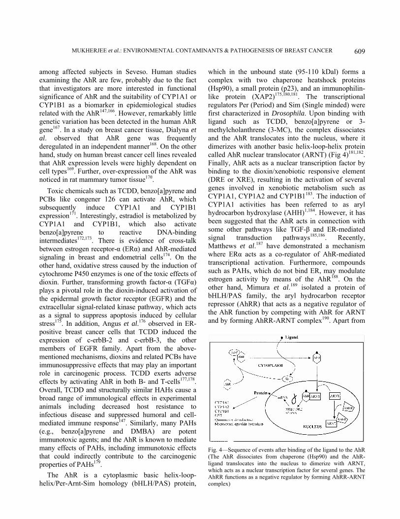
which in the unbound state (95-110 kDal) forms a complex with two chaperone heatshock proteins (Hsp90), a small protein (p23), and an immunophilin-like protein (XAP2)175,180,181. The transcriptional regulators Per (Period) and Sim (Single minded) were first characterized in Drosophila. Upon binding with ligand such as TCDD, benzo[a]pyrene or 3-methylcholanthrene (3-MC), the complex dissociates and the AhR translocates into the nucleus, where it dimerizes with another basic helix-loop-helix protein called AhR nuclear translocator (ARNT) (Fig 4)181,182. Finally, AhR acts as a nuclear transcription factor by binding to the dioxin/xenobiotic responsive element (DRE or XRE), resulting in the activation of several genes involved in xenobiotic metabolism such as CYP1A1, CYP1A2 and CYP1B1183. The induction of CYP1A1 activities has been referred to as aryl hydrocarbon hydroxylase (AHH)1,184. However, it has been suggested that the AhR acts in connection with some other pathways like TGF-β and ER-mediated signal transduction pathways185,186. Recently, Matthews et al.187 have demonstrated a mechanism where ERα acts as a co-regulator of AhR-mediated transcriptional activation. Furthermore, compounds such as PAHs, which do not bind ER, may modulate estrogen activity by means of the AhR188. On the other hand, Mimura et al.189 isolated a protein of bHLH/PAS family, the aryl hydrocarbon receptor repressor (AhRR) that acts as a negative regulator of the AhR function by competing with AhR for ARNT and by forming AhRR-ARNT complex190. Apart from
Fig. 4—Sequence of events after binding of the ligand to the AhR (The AhR dissociates from chaperone (Hsp90) and the AhR-ligand translocates into the nucleus to dimerize with ARNT, which acts as a nuclear transcription factor for several genes. The AhRR functions as a negative regulator by forming AhRR-ARNT complex)

INDIAN J EXP BIOL, AUGUST 2006
610
environmental contaminants such as dioxins and PAHs, the AhR can bind with a variety of structurally diverse chemicals191 (Table 4). Several investigators have observed that different dietary components, e.g., flavonoids, carotinoids, resveratrol, etc., bind to the AhR; and many of them exert antagonistic activity182,191,192. Some dietary components like indolo[3,2-b]carbazole closely resembles dioxin in structure. In acidic environment of the stomach, indolo[3,2-b]carbazole and 3,3′-diindolylmethane are produced from indole-3-carbinol derived from glucosinolates of consumed cruciferous vegetables such as broccoli, cabbage and cauliflower. Interestingly, Chen et al.193 commented that resveratrol can act as a potential chemopreventive against dioxin-induced human mammary carcinogenesis by blocking the reactive metabolite formation of the catechol estrogens and scavenging the ROS generated during their redox cycling. Overall, a dietary modification with the introduction of increased quantities of soybean-products, cruciferous vegetables, fruits, curcumin, tea and low fat could be beneficial in reducing the risk of developing cancer and possibly the adverse effects of different environmental contaminats194-196.
It is known that TCDD produces an AhR-dependent oxidative stress in mitochondria197. This phenomenon is associated with concomitant loss of mitochondrial membrane potential, a reduction in mitochondrial glutathione levels and mitochondrial DNA (mtDNA) damage198,199. Further, Banzet et al. observed that mitochondria could be a target for ROS-mediated effects of tobacco smoke exposure200. Alternatively, mitochondrial dysfunction may contribute to increased ROS production and play a role in tobacco carcinogenicity201. Lipophilic carcinogens such as PAHs accumulate in mitochondria and can cause mtDNA damage202. The incidence of mtDNA damage is 10- to 15-fold greater compared to nuclear DNA, since mtDNA has no protective histones. Nevertheless, it is possible to partially counteract the deleterious effects of benzo[a]pyrene, a prototypical PAH, on mitochondrion-related apoptotic process by blocking the AhR203. Mechanisms of toxicity of several pesticides such as 2,4-dichlorophenoxyacetic acid (2,4-D) and paraquat are associated with a disruption of mitochondrial membrane potential204,205. Growing evidence suggests that cancer cells exhibit increased intrinsic ROS stress due in part to mitochondrial
malfunction that also alters cellular architecture, signaling, metabolism, cell growth and differentiation, and apoptotic response to anticancer agents206,207.
Conclusions The recent increasing trend in the incidence of
breast cancer in urban population has drawn attention of all sections of the society. Attempts have been made to find out the causes of this increasing trend by different research initiatives. Many lifestyle factors have been incriminated in this regard. With the discovery and increasing use of pesticides, westernized food habits and due to other industrialized products, the levels of environmental contaminants are increasing day by day. As discussed in the review, these xenobiotics by various mechanisms are capable of contributing to pathogenesis of breast cancer. For many such chemicals, the potential of carcinogenicity and the mechanisms of action have not been evaluated till date. For some environmental contaminants, it is partially understood. Laboratory animal experimentation and reports of higher levels of the concerned environmental contaminants in breast cancer cases indicate that these environmental chemicals may be a confounding factor in the increasing prevalence of breast cancer. Knudson’s double hit hypothesis has shown how environmental insult/hit can lead to cancer in genetically susceptible subjects. There is no doubt that with increasing levels of environmental chemical contaminants, the chances of getting exposed to such environmental insults are more. Frequent exposure and higher doses of such insults can possibly induce cancer in subjects who are not even genetically susceptible. But how much is the real impact of these chemical contaminants in breast cancer pathogenesis is yet to be determined. This is the time to think about the inclusion of new parameters like AhR activation and other potential pathways of endocrine disruptors, apart from E-screen assay, in evaluation of toxicity profile of pesticides and other xenobiotics. Obviously, there is an urgent need for systematic study to evaluate the impact. Such studies can explore possible reasons for the difference in prevalence of breast cancer in rural and urban areas and in different ethnic groups. Also, this process may lead to discovery of complex interplay of xenobiotics in induction of cancer and new dimensions in gene-environment interactions. Only a clear understanding could lead to an efficient strategy of breast cancer prevention.

MUKHERJEE et al.: ENVIRONMENTAL CONTAMINANTS & PATHOGENESIS OF BREAST CANCER
611
References 1 Dumitrescu R G & Cotarla I, Understanding breast cancer risk
– where do we stand in 2005? J Cell Mol Med, 9 (2005) 208. 2 Report of the National Cancer Registry Programme (Indian
Council of Medical Research, New Delhi) 2001. 3 Sen U, Sankaranarayanan R, Mandal S, Ramanakumar A V,
Parkin D M & Siddiqi M, Cancer patterns in eastern India: The first report of the Kolkata Cancer Registry, Int J Cancer, 100 (2002) 86.
4 Jussaawala D J, Yeole B B & Natekar M V, Histological and epidemiological features of breast cancer in different religious groups in greater Bombay, J Surg Oncol, 18 (1981) 269.
5 Badwe R A, Gangawal S, Mittra I & Desai P B. Clinico-pathological features and prognosis of breast cancer in different religious communities in India, Indian J Cancer, 27 (1990) 220.
6 Yeole B B & Kurkure A P, An epidemiological assessment of increasing incidence and trends in breast cancer in Mumbai and other sites in India, during the last two decades, Asian Pac J Cancer Prev, 4 (2003) 51.
7 Paymaster T C & Gangadharan P, Cancer in Parsi community of Bombay, Int J Cancer, 5 (1970) 426.
8 Kliewer E V & Smith K R, Breast cancer mortality among immigrants in Australia and Canada, J Natl Cancer Inst, 87 (1995) 1154.
9 Winter H, Cheng K K, Cummins C, Maric R, Silcocks P & Varghese C, Cancer incidence in the south Asian population of England (1990-92), Br J Cancer, 79 (1999) 645.
10 Kotsopoulos J, Liede A, De Matsuda M L, Sun P & Narod S A, Method of cooking and risk of breast cancer in the Philippines, Cancer Causes Control, 17 (2006) 341.
11 Sharma B K, Ray A, Kaur S & Gupta S, Immunohistochemical co-expression of c-erbB-2/Neu oncoprotein, altered tumour suppressor (p53) protein, EGF-R and EMA in histological subtypes of infiltrating duct carcinoma of the breast, Indian J Exp Biol, 37 (1999) 223.
12 Kuhl H, Breast cancer risk in the WHI study: the problem of obesity, Maturitas, 51 (2005) 83.
13 Onland-Moret N C, Peeters P H, van der Schouw Y T, Grobbee D E & van Gils C H, Alcohol and endogenous sex steroid levels in postmenopausal women: a cross-sectional study, J Clin Endocrinol Metab, 90 (2005) 1414.
14 Russo J & Russo I H, Genotoxicity of steroidal estrogens, Trends Endocrinol Metab, 15 (2004) 211.
15 Castagnetta L A, Granata O M, Arcuri F P, Polito L M, Rosati F & Cartoni G P, Gas chromatography/mass spectrometry of catechol estrogens, Steroids, 57 (1992) 437.
16 Liehr J G, Is estradiol a genotoxic mutagenic carcinogen? Endocr Rev, 21 (2000) 40.
17 Rylander-Rudqvist T, Wedren S, Granath F, Humphreys K, Ahlberg S, Weiderpass E, Oscarson M, Ingelman-Sundberg M & Persson I, Cytochrome P4501B1 gene polymorphisms and postmenopausal breast cancer risk, Carcinogenesis, 24 (2003) 1533.
18 Yager J D & Liehr J G, Molecular mechanisms of estrogen carcinogenesis, Annu Rev Pharmacol Toxicol, 36 (1996) 203.
19 Jefcoate C R, Liehr J G, Santen R J, Sutter T R, Yager J D, Yue W, Santner S J, Tekmal R, Demers L, Pauley R, Naftolin F, Mor G & Berstein L, Tissue-specific synthesis and oxidative metabolism of estrogens, J Natl Cancer Inst Monogr, 27 (2000) 95.
20 Mitrunen K & Hirvonen A, Molecular epidemiology of sporadic breast cancer: The role of polymorphic genes involved in oestrogen biosynthesis and metabolism, Mutat Res, 544 (2003) 9.
21 Dawling S, Hachey D L, Roodi N & Parl F F, In vitro model of mammary estrogen metabolism: Structural and kinetic differences between catechol estrogens 2- and 4-hydroxyestradiol, Chem Res Toxicol, 17 (2004) 1258.
22 Emerit I, Reactive oxygen species, chromosome mutation, and cancer: possible role of clastogenic factors in carcinogenesis, Free Radic Biol Med, 16 (1994) 99.
23 Vaca C E, Wilhelm J & Harms-Ringdal M, Interaction of lipid peroxidation products with DNA. A review, Mutat Res, 195 (1988) 137.
24 Kumaraguruparan R, Kabalimoorthy J & Nagini S, Correlation of tissue lipid peroxidation and antioxidants with clinical stage and menopausal status in patients with adenocarcinoma of the breast, Clin Biochem, 38 (2005) 154.
25 Phillips D H, Martin F L, Williams J A, Wheat L M, Nolan L, Cole K J & Grover P L, Mutagens in human breast lipid and milk: the search for environmental agents that initiate breast cancer, Environ Mol Mutagen, 39 (2002) 143.
26 Li D, Wang M, Firozi P F, Chang P, Zhang W, Baer-Dubowska W, Moorthy B, Vulimiri S V, Goth-Goldstein R, Weyand E H & DiGiovanni J, Characterization of a major aromatic DNA adduct detected in human breast tissues, Environ Mol Mutagen, 39 (2002) 193.
27 Wang M, Dhingra K, Hittelman W N, Liehr J G, de Andrade M & Li D, Lipid peroxidation-induced putative malondialdehyde-DNA adducts in human breast tissues, Cancer Epidemiol Biomarkers Prev, 5 (1996) 705.
28 Matsui A, Ikeda T, Enomoto K, Hosoda K, Nakashima H, Omae K, Watanabe M, Hibi T & Kitajima M, Increased formation of oxidative DNA damage, 8-hydroxy-2′-deoxyguanosine, in human breast cancer tissue and its relationship to GSTP1 and COMT genotypes, Cancer Lett, 151 (2000) 87.
29 Williams J A & Phillips D H, Mammary expression of xenobiotic metabolizing enzymes and their potential role in breast cancer, Cancer Res, 60 (2000) 4667.
30 Oyama T, Morita M, Isse T, Kagawa N, Nakata S, So T, Mizukami M, Ichiki Y, Ono K, Sugaya M, Uramoto H, Yoshimatsu T, Hanagiri T, Sugio K, Kawamoto T & Yasumoto K, Immunohistochemical evaluation of cytochrome P450 (CYP) and p53 in breast cancer, Front Biosci, 10 (2005) 1156.
31 Modugno F, Knoll C, Kanbour-Shakir A & Romkes M, A potential role for the estrogen-metabolizing cytochrome P450 enzymes in human breast carcinogenesis, Breast Cancer Res Treat, 82 (2003) 191.
32 El-Rayes B F, Ali S, Heilbrun L K, Lababidi S, Bouwman D, Visscher D & Philip P A, Cytochrome P450 and glutathione transferase expression in human breast cancer, Clin Cancer Res, 9 (2003) 1705.
33 Tas F, Hansel H, Belce A, Ilvan S, Argon A, Camlica H & Topuz E, Oxidative stress in breast cancer, Med Oncol, 22 (2005) 11.
34 Portakal O, Ozkaya O, Inal M E, Bozan B, Kosan M & Sayek I, Coenzyme Q10 concentrations and antioxidant status in tissues of breast cancer patients, Clin Biochem, 33 (2000) 279.

INDIAN J EXP BIOL, AUGUST 2006
612
35 Er T K, Hou M F, Tsa E M, Lee J N & Tsai L Y, Differential expression of manganese containing superoxide dismutase in patients with breast cancer in Taiwan, Ann Clin Lab Sci, 34 (2004) 159.
36 Thomas P A, Oykutlu D, Pou B, Tyler D, Oberley L W, Robinson R A & Lenel J C, Immunohistochemical characterization of antioxidant enzymes in human breast cancer, Pathol Oncol Res, 3 (1997) 278.
37 Cai Q, Shu X O, Wen W, Cheng J R, Dai Q, Gao Y T & Zheng W, Genetic polymorphism in the manganese superoxide dismutase gene, antioxidant intake, and breast cancer risk: results from the Shanghai Breast Cancer Study, Breast Cancer Res, 6 (2004) R647.
38 Williams J A, Single nucleotide polymorphisms, metabolic activation and environmental carcinogenesis: why molecular epidemiologists should think about enzyme expression, Carcinogenesis, 22 (2001) 209.
39 Lin S C, Chou Y C, Wu M H, Wu C C, Lin W Y, Yu C P, Yu J C, You S L, Chen C J & Sun C A, Genetic variants of myeloperoxidase and catechol-O-methyltransferase and breast cancer risk, Eur J Cancer Prev, 14 (2005) 257.
40 Bekesi G, Kakucs R, Varbiro S, Feher J, Pazmany T, Magyar Z, Sprintz D & Szekacs B, Induced myeloperoxidase activity and related superoxide inhibition during hormone replacement therapy, BJOG, 108 (2001) 474.
41 Nowell S A, Ahn J & Ambrosone C B, Gene-nutrient interactions in cancer etiology, Nutr Rev, 62 (2004) 427.
42 Mannisto P T, Ulmanen I, Lundstrom K, Taskinen J, Tenhunen J, Tilgmann C & Kaakkola S, Characteristics of catechol-O-methyl-transferase (COMT) and properties of selective COMT inhibitors, Prog Drug Res, 39 (1992) 291.
43 Whalen R & Boyer T D, Human glutathione S-transferases, Semin Liver Dis, 18 (1998) 345.
44 Salinas A E & Wong M G, Glutathion S-transferases – a review, Curr Med Chem, 6 (1999) 279.
45 Sheweita S A & Tilmisany A K, Cancer and phase II drug-metabolizing enzymes, Curr Drug Metab, 4 (2003) 45.
46 Forrester L M, Hayes J D, Millis R, Barnes D, Harris A L, Schlager J J, Powis G & Wolf C R, Expression of glutathione S-transferases and cytochrome P450 in normal and tumor breast tissue, Carcinogenesis, 11 (1990) 2163.
47 Suzuki T, Miki Y, Nakata T, Shiotsu Y, Akinaga S, Inoue K, Ishida T, Kimura M, Moriya T & Sasano H, Steroid sulfatase and estrogen sulfotransferase in normal human tissue and breast carcinoma, J Steroid Biochem Mol Biol, 86 (2003) 449.
48 Yoshimura N, Harada N, Bukholm I, Karesen R, Borresen-Dale A L & Kristensen V N, Intratumoural mRNA expression of genes from the oestradiol metabolic pathway and clinical and histopathological parameters of breast cancer, Breast Cancer Res, 6 (2004) R46.
49 Aust S, Obrist P, Klimpfinger M, Tucek G, Jager W & Thalhammer T, Altered expression of the hormone- and xenobiotic-metabolizing sulfotransferase enzymes 1A2 and 1C1 in malignant breast tissue, Int J Oncol, 26 (2005) 1079.
50 Williams J A, Stone E M, Fakis G, Johnson N, Meinl W, Glatt H, Sim E & Phillips D H, Human mammary NAT and SULT enzymes metabolically activate N-hydroxylated heterocyclic amines, but NAT enzyme activity is not influenced by NAT genotype, Proc Am Assoc Cancer Res, 41 (2000) 551.
51 Mukherjee S, Nayyar T, Chytil F & Das S K, Mainstream and sidestream cigarette smoke exposure increases retinol in guinea pig lungs, Free Radic Biol Med, 18 (1995) 507.
52 Mukundan H, Bahadur A K, Kumar A, Sardana S, Naik S L D, Ray A & Sharma B K, Glutathione level and its relation to radiation therapy in patients with cancer of uterine cervix, Indian J Exp Biol, 37 (1999) 859.
53 Russo I H, Cigarette smoking and risk of breast cancer in women, Lancet, 360 (2002) 1033.
54 Terry P D, Miller A B & Rohan T E, Cigarette smoking and breast cancer risk: a long latency period? Int J Cancer, 100 (2002) 723.
55 Couch F J, Cerhan J R, Vierkant R A, Grabrick D M, Therneau T M, Pankratz V S, Hartmann L C, Olson J E, Vachon C M & Sellers T A, Cigarette smoking increases risk for breast cancer in high-risk breast cancer families, Cancer Epidemiol Biomarkers Prev, 10 (2001) 327.
56 Innes K E & Byers T E, Smoking during pregnancy and breast cancer risk in very young women (United States), Cancer Causes Control, 12 (2001) 179.
57 Murin S & Inciardi J, Cigarette smoking and the risk of pulmonary metastasis from breast cancer, Chest, 119 (2001) 1635.
58 Baron J A, Newcomb P A, Longnecker M P, Mittendorf R, Storer B E, Clapp R W, Bogdan G & Yuen J, Cigarette smoking and breast cancer, Cancer Epidemiol Biomarkers Prev, 5 (1996) 399.
59 Terry P D & Rohan T E, Cigarette smoking and the risk of breast cancer in women: a review of the literature, Cancer Epidemiol Biomarkers Prev, 11 (2002) 953.
60 Lash T L & Aschengrau A, A null association between active or passive cigarette smoking and breast cancer risk, Breast Cancer Res Treat, 75 (2002) 181.
61 Ambrosone C B & Shields P, Smoking as a risk factor for breast cancer, in Breast Cancer: Molecular Genetics, Pathogenesis, and Therapeutics, edited by A. Bowcock (Humana Press, Totowa, NJ) 2001, 519.
62 Hecht S S, Tobacco smoke carcinogens and breast cancer, Environ Mol Mutagen, 39 (2002) 119.
63 Pryor W A, Cigarette smoke radicals and the role of free radicals in chemical carcinogenicity, Environ Health Perspect, 105 (Suppl. 4) (1997) 875.
64 Morabia A, Smoking (active and passive) and breast cancer: epidemiologic evidence up to June 2001, Environ Mol Mutagen, 39 (2002) 89.
65 Wartenberg D, Calle E E, Thun M J, Heath C W Jr, Lally C & Woodruff T, Passive smoking exposure and female breast cancer mortality, J Natl Cancer Inst, 92 (2000) 1666.
66 Liu L, Wu K, Lin X, Yin W, Zheng X, Tang X, Mu L, Hu Z & Wang J, Passive smoking and other factors at different periods of life and breast cancer risk in Chinese women who have never smoked – a case-control study in Chongqing, People’s Republic of China, Asian Pac J Cancer Prev, 1 (2000) 131.
67 Shrubsole M J, Gao Y T, Dai Q, Shu XO, Ruan Z X, Jin F & Zheng W, Passive smoking and breast cancer risk among non-smoking Chinese women, Int J Cancer, 110 (2004) 605.
68 Hanaoka T, Yamamoto S, Sobue T, Sasaki S, Tsugane S & Japan Public Health Center-based Prospective Study on Cancer and Cardiovascular Disease Study Group, Active and passive smoking and breast cancer risk in middle-aged Japanese women, Int J Cancer, 114 (2005) 317.

MUKHERJEE et al.: ENVIRONMENTAL CONTAMINANTS & PATHOGENESIS OF BREAST CANCER
613
69 Ambrosone C B, Freudenheim J L, Graham S, Marshall J R, Vena J E, Brasure J R, Michalek A M, Laughlin R, Nemoto T, Gillenwater K A & Shields P G, Cigarette smoking, N-acetyltransferase 2 genetic polymorphisms, and breast cancer risk, JAMA, 276 (1996) 1494.
70 Chang-Claude J, Kropp S, Jager B, Bartsch H & Risch A, Differential effect of NAT2 on the association between active and passive smoke exposure and breast cancer risk, Cancer Epidemiol Biomarkers Prev, 11 (2002) 698.
71 van der Hel O L, Peeters P H M, Hein D W, Doll M A, Grobbee D E, Kromhout D & de Mesquita H B B, NAT2 slow acetylation and GSTM1 null genotypes may increase postmenopausal breast cancer risk in long-term smoking women, Pharmacogenetics, 13 (2003) 399.
72 Coyle Y M, The effect of environment on breast cancer risk, Breast Cancer Res Treat, 84 (2004) 273.
73 Rennix C P, Quinn M M, Amoroso P J, Eisen E A & Wegman D H, Risk of breast cancer among enlisted army women occupationally exposed to volatile organic compounds, Am J Ind Med, 48 (2005) 157.
74 Takkouche B, Etminan M & Montes-Martinez A, Personal use of hair dyes and risk of cancer: a meta-analysis, JAMA, 293 (2005) 2516.
75 Velicer C M, Heckbert S R, Rutter C, Lampe J W & Malone K, Association between antibiotic use prior to breast cancer diagnosis and breast tumour characteristics (United States), Cancer Causes Control, 17 (2006) 307.
76 Ray A, Ray S & Koner B C, Hypertension, cancer and angiogenesis: Relevant epidemiological and pharmacological aspects, Indian J Pharmacol, 36 (2004) 341.
77 Colborn T, Dumanoski D & Myers J P, Our Stolen Future (Penguin, New York) 1996.
78 Ray A, Husain S A & Sharma B K, Emerging areas: environmental biochemistry, in Proceedings of 9th National Conference of the Association of Medical Biochemists of India (New Delhi) 2000, 57.
79 Witorsch R J, Endocrine disruptors: can biological effects and environment risk be predicted? Regul Toxicol Pharmacol, 36 (2002) 118.
80 Safe S, Endocrine disruptors and human health – is there a problem? An update, Environ Health Perspect, 108 (2000) 487.
81 Recchia A G, Vivacqua A, Gabriele S, Carpino A, Fasanella G, Rago V, Bonofiglio D & Maggiolini M, Xenoestrogens and the induction of proliferative effects in breast cancer cells via direct activation of oestrogen receptor alpha, Food Addit Contam, 21 (2004) 134.
82 Harvey P W & Everett D J, Regulation of endocrine-disrupting chemicals: Critical overview and deficiencies in toxicology and risk assessment for human health, Best Pract Res Clin Endocrinol Metab, 20 (2006) 145.
83 Kortenkamp A, Breast cancer, oestrogens and environmental pollutants: a re-evaluation from a mixture perspective, Int J Androl, 29 (2006) 193.
84 Kaminuma T, Takai-Igarashi T, Nakano T & Nakata K, Modeling of signaling pathways for endocrine disruptors, BioSystems, 55 (2000) 23.
85 Safe S & McDougal A, Mechanism of action and development of selective aryl hydrocarbon receptor modulators for treatment of hormone-dependent cancers (Review), Int J Oncol, 20 (2002) 1123.
86 Figa-Talamanca I, Mearelli I, Valente P & Bascherini S, Cancer mortality in a cohort of rural licensed pesticide users in the province of Rome, Int J Epidemiol, 22 (1993) 579.
87 Wesseling C, Antich D, Hogstedt C, Rodriguez A C & Ahlbom A, Geographical differences of cancer incidence in Costa Rica in relation to environmental and occupational pesticide exposure, Int J Epidemiol, 28 (1999) 365.
88 Koner B C, Banerjee B D & Ray A, Organochlorine pesticide-induced oxidative stress and immune suppression in rats, Indian J Exp Biol, 36 (1998) 395.
89 Ray S, Studies on paraquat toxicity in rat, Ph.D. thesis, University of Calcutta, Kolkata, 2001.
90 Iscan M, Coban T, Cok I, Bulbul D, Eke B C & Burgaz S, The organochlorine pesticide residues and antioxidant enzyme activities in human breast tumors: is there any association? Breast Cancer Res Treat, 72 (2002) 173.
91 Banerjee B D, Koner B C & Ray A, Immunotoxicity of pesticides: perspectives and trends, Indian J Exp Biol, 34 (1996) 723.
92 Burow M E, Tang Y, Collins-Burow B M, Krajewski S, Reed J C, McLachlan J A & Beckman B S, Effects of environmental estrogens on tumor necrosis factor α-mediated apoptosis in MCF-7 cells, Carcinogenesis, 20 (1999) 2057.
93 Ray A & Mitra A B, Estrogen and breast cancer, ICMR Bull, 33 (2003) 13.
94 Starek A, Estrogens and organochlorine xenoestrogens and breast cancer risk, Int J Occup Med Environ Health, 16 (2003) 113.
95 Schecter A, Toniolo P, Dai L C, Thuy L T & Wolff M S, Blood levels of DDT and breast cancer risk among women living in the north of Vietnam, Arch Environ Contam Toxicol, 33 (1997) 453.
96 Wolff M S, Berkowitz G S, Brower S, Senie R, Bleiweiss I J, Tartter P, Pace B, Roy N, Wallenstein S & Weston A, Organochlorine exposures and breast cancer risk in New York city women, Environ Res, 84 (2000) 151.
97 Lopez-Cervantes M, Torres-Sanchez L, Tobias A & Lopez-Carrillo L, Dichlorodiphenyldichloroethane burden and breast cancer risk: a meta-analysis of the epidemiological evidence, Environ Health Perspect, 112 (2004) 207.
98 Raaschou-Nielsen O, Pavuk M, Leblanc A, Dumas P, Philippe Weber J, Olsen A, Tjonneland A, Overvad K & Olsen J H, Adipose organochlorine concentrations and risk of breast cancer among postmenopausal Danish women, Cancer Epidemiol Biomarkers Prev, 14 (2005) 67.
99 Aronson K J, Miller A B, Woolcott C G, Sterns E E, McCready D R, Lickley L A, Fish E B, Hiraki G Y, Holloway C, Ross T, Hanna W M, Sengupta S K & Weber J P, Breast adipose tissue concentrations of polychlorinated biphenyls and other organochlorines and breast cancer risk, Cancer Epidemiol Biomarkers Prev, 9 (2000) 55.
100 Stellman S D, Djordjevic M V, Britton J A, Muscat J E, Citron M L, Kemeny M, Busch E & Gong L, Breast cancer risk in relation to adipose concentrations of organochlorine pesticides and polychlorinated biphenyls in Long Island, New York, Cancer Epidemiol Biomarkers Prev, 9 (2000) 1241.
101 Woolcott C G, Aronson K J, Hanna W M, Sengupta S K, McCready D R, Sterns E E & Miller A B, Organochlorines and breast cancer risk by receptor status, tumor size, and grade (Canada), Cancer Causes Control, 12 (2001) 395.

INDIAN J EXP BIOL, AUGUST 2006
614
102 Hoyer A P, Gerdes A M, Jorgensen T, Rank F & Hartvig H B, Organochlorines, p53 mutations in relation to breast cancer risk and survival. A Danish cohort-nested case-controls study, Breast Cancer Res Treat, 71 (2002) 59.
103 Mathur V, Bhatnagar P, Sharma R G, Acharya V & Saxena R, Breast cancer incidence and exposure to pesticides among women originating from Jaipur, Environ Int, 28 (2002) 331.
104 Charlier C, Albert A, Herman P, Hamoir E, Gaspard U, Meurisse M & Plomteux G, Breast cancer and serum organochlorine residues, Occup Environ Med, 60 (2003) 348.
105 Pavuk M, Cerhan J R, Lynch C F, Kocan A, Petrik J & Chovancova J, Case-control study of PCBs, other organochlorines and breast cancer in eastern Slovakia, J Expo Anal Environ Epidemiol, 13 (2003) 267.
106 Muscat J E, Britton J A, Djordjevic M V, Citron M L, Kemeny M, Busch-Devereaux E, Pittman B & Stellman S D, Adipose concentrations of organochlorine compounds and breast cancer recurrence in Long Island, New York, Cancer Epidemiol Biomarkers Prev, 12 (2003) 1474.
107 Ibarluzea J M, Fernandez M F, Santa-Marina L, Olea-Serrano M F, Rivas A M, Aurrekoetxea J J, Exposito J, Lorenzo M, Torne P, Villalobos M, Pedraza V, Sasco A J & Olea N, Breast cancer risk and the combined effect of environmental estrogens, Cancer Causes Control, 15 (2004) 591.
108 Mills P K & Yang R, Breast cancer risk in Hispanic agricultural workers in California, Int J Occup Environ Health, 11 (2005) 123.
109 Cassidy R A, Natarajan S & Vaughan G M, The link between the insecticide heptachlor epoxide, estradiol, and breast cancer, Breast Cancer Res Treat, 90 (2005) 55.
110 Snedeker S M, Pesticides and breast cancer risk: a review of DDT, DDE, and dieldrin, Environ Health Perspect, 109 (Suppl 1) (2001) 35.
111 Ejaz S, Akram W, Lim C W, Lee J J & Hussain I, Endocrine disrupting pesticides: a leading cause of cancer among rural people in Pakistan, Exp Oncol, 26 (2004) 98.
112 Calle E E, Frumkin H, Henley S J, Savitz D A & Thun M J, Organochlorines and breast cancer risk, CA Cancer J Clin, 52 (2002) 301.
113 Mitra A K, Faruque F S & Avis A L, Breast cancer and environmental risks: where is the link? J Environ Health, 66 (2004) 24.
114 Moorman P G & Terry P D, Consumption of dairy products and the risk of breast cancer: a review of the literature, Am J Clin Nutr, 80 (2004) 5.
115 Coyle Y M, Hynan L S, Euhus D M & Minhajuddin A T M, An ecological study of the association of environmental chemicals on breast cancer incidence in Texas, Breast Cancer Res Treat, 92 (2005) 107.
116 Minervini F, Giannoccaro A, Cavallini A & Visconti A, Investigations on cellular proliferation induced by zearalenone and its derivatives in relation to the estrogenic parameters, Toxicol Lett, 159 (2005) 272.
117 Ahamed S, Foster J S, Bukovsky A & Wimalasena J, Signal transduction through the Ras/Erk pathway is essential for the mycoestrogen zearalenone-induced cell-cycle progression in MCF-7 cells, Mol Carcinog, 30 (2001) 88.
118 Goldman R & Shields P G, Food mutagens. J Nutr, 133 (2003) 965S.
119 Tritscher A M, Human health risk assessment of processing-related compounds in food, Toxicol Lett, 149 (2004) 177.
120 Crews C, Brereton P & Davies A, The effects of domestic cooking on the levels of 3-monochloropropanediol in foods, Food Addit Contam, 18 (2001) 271.
121 Jagerstad M & Skog K, Genotoxicity of heat-processed foods. Mutat Res, 574 (2005) 156.
122 Blank I, Current status of acrylamide research in food: measurement, safety assessment, and formation, Ann NY Acad Sci, 1043 (2005) 30.
123 Rice J M, The carcinogenicity of acrylamide, Mutat Res, 580 (2005) 3.
124 Erdreich L S & Friedman M A, Epidemiologic evidence for assessing the carcinogenicity of acrylamide, Regul Toxicol Phamacol, 39 (2004) 150.
125 Fiddler W, The occurrence and determination of N-nitroso compounds, Toxicol Appl Pharmacol, 31 (1975) 352.
126 Wogan G N & Tannenbaum S R, Environmental N-nitroso compounds: implications for public health, Toxicol Appl Pharmacol, 31 (1975) 375.
127 Bartsch H & Montesano R, Relevance of nitrosamines in human cancer, Carcinogenesis, 5 (1984) 1381.
128 Ahn H J, Jo C, Kim J H, Chung Y J, Lee C H & Byun M W, Monitoring of nitrite and N-nitrosamine levels in irradiated pork sausage, J Food Prot, 65 (2002) 1493.
129 Lin K, Shen Z Y, Lu S H & Wu Y N, Intake of volatile N-nitrosamines and their ability to exogenously synthesize in the diet of inhabitants from high-risk area of esophageal cancer in southern China, Biomed Environ Sci, 15 (2002) 277.
130 Lee S J, Shin J H, Sung N J, Kim J G & Hotchkiss J H, Effect of cooking on the formation of N-nitrosodimethylamine in Korean dried seafood products, Food Addit Contam, 20 (2003) 31.
131 Lijinsky W, In vivo testing for carcinogenicity, in Chemical Carcinogenesis and Mutagenesis I, edited by C S Cooper & P L Grover (Springer-Verlag, Berlin, Germany) 1990, 179.
132 Wogan G N, Hecht S S, Felton J S, Conney A H & Loeb L A, Environmental and chemical carcinogenesis, Semin Cancer Biol, 14 (2004) 473.
133 Gammon D W, Aldous C N, Carr W C Jr, Sanborn J R & Pfeifer K F, A risk assessment of atrazine use in California: human health and ecological aspects, Pest Manag Sci, 61 (2005) 331.
134 Meador J P, Stein J E, Reichert W L & Varanasi U, Bioaccumulation of polycyclic aromatic hydrocarbons by marine organisms, Rev Environ Contam Toxicol, 143 (1995) 79.
135 Guillen M D, Sopelana P & Partearroyo M A, Food as a source of polycyclic aromatic carcinogens, Rev Environ Health, 12 (1997) 133.
136 Phillips D H, Polycyclic aromatic hydrocarbons in the diet. Mutat Res, 443 (1999) 139.
137 Brody J G & Rudel R A, Environmental pollutants and breast cancer, Environ Health Perspect, 111 (2003) 1007.
138 Autrup H, Harris C C, Trump B F & Jeffrey A H, Metabolism of benzo(a)pyrene and identification of the major benzo(a)pyrene-DNA adducts in cultured human colon, Cancer Res, 38 (1978) 3689.
139 BeBruin L S & Josephy P D, Perspectives on the chemical etiology of breast cancer, Environ Health Perspect, 110 (Suppl 1) (2002) 119.
140 Murkovic M, Formation of heterocyclic aromatic amines in model systems, J Chromatogr B, 802 (2004) 3.

MUKHERJEE et al.: ENVIRONMENTAL CONTAMINANTS & PATHOGENESIS OF BREAST CANCER
615
141 Weisburger J H, Antimutagens, anticarcinogens, and effective worldwide cancer prevention, J Environ Pathol Toxicol Oncol, 18 (1999) 85.
142 Ferguson L R, Meat consumption, cancer risk and population groups within New Zealand, Mutat Res, 506-507 (2002) 215.
143 De Stefani E, Ronco A, Mendilaharsu M, Guidobono M & Deneo-Pellegrini H, Meat intake, heterocyclic amines, and risk of breast cancer: a case-control study in Uruguay, Cancer Epidemiol Biomarkers Prev, 6 (1997) 573.
144 Knize M G & Felton J S, Formation and human risk of carcinogenic heterocyclic amines formed from natural precursors in meat, Nutr Rev, 63 (2005) 158.
145 Monographs on the Evaluation of Carcinogenic Risk to Humans, Vol. 56 (International Agency for Research on Cancer, Lyon, France) 1993, 163.
146 Kawanishi M, Sakamoto M, Ito A, Kishi K & Yagi T, Construction of reporter yeasts for mouse aryl hydrocarbon receptor ligand activity, Mutat Res, 540 (2003) 99.
147 Mandal P K, Dioxin: a review of its environmental effects and its aryl hydrocarbon receptor biology, J Comp Physiol B, 175 (2005) 221.
148 Warner M, Eskenazi B, Mocarelli P, Gerthoux P M, Samuels S, Needham L, Patterson D & Brambilla P, Serum dioxin concentrations and breast cancer risk in the Seveso Women’s Health Study, Environ Health Perspect, 110 (2002) 625.
149 Landi M T, Bertazzi P A, Baccarelli A, Consonni D, Masten S, Lucier G, Mocarelli P, Needham L, Caporaso N & Grassman J, TCDD-mediated alterations in the AhR-dependent pathway in Seveso, Italy, 20 years after the accident, Carcinogenesis, 24 (2003) 673.
150 Baccarelli A, Pesatori A C, Masten S A, Patterson D G Jr, Needham L L, Mocarelli P, Caporaso N E, Consonni D, Grassman J A, Bertazzi P A & Landi M T, Aryl-hydrocarbon receptor-dependent pathway and toxic effects of TCDD in humans: a population-based study in Seveso, Italy, Toxicol Lett, 149 (2004) 287.
151 Flesch-Janys D, Becher H, Manz A, Morgenstern I, Nagel S, Steindorf K, Epidemiological investigation of breast cancer incidence in a cohort of female workers with high exposure to PCDD/F and HCH, Organohalogen Compounds, 44 (1999) 379.
152 Manz A, Berger J, Dwyer J H, Flesch-Janys D, Nagel S, Waltsgott H, Cancer mortality among workers in chemical plant contaminated with dioxin, Lancet, 338 (1991) 959.
153 Kogevinas M, Becher H, Benn T, Bertazzi P A, Boffetta P, Bueno-de-Mesquita H B, Coggon D, Colin D, Flesch-Janys D, Fingerhut M, Green L, Kauppinen T, Littorin M, Lynge E, Mathews J D, Neuberger M, Pearce N & Saracci R, Cancer mortality in workers exposed to phenoxy herbicides, chlorophenols, and dioxins. An expanded and updated international cohort study, Am J Epidemiol, 145 (1997) 1061.
154 Revich B, Aksel E, Ushakova T, Ivanova I, Zhuchenko N, Klyuev N, Brodsky B & Sotskov Y, Dioxin exposure and public health in Chapaevsk, Russia, Chemosphere, 43 (2001) 951.
155 Brown N M, Manzolillo P A, Zhang J X, Wang J & Lamartiniere C A, Prenatal TCDD and predisposition to mammary cancer in the rat, Carcinogenesis, 19 (1998) 1623.
156 Demers A, Ayotte P, Brisson J, Dodin S, Robert J & Dewailly E, Plasma concentrations of polychlorinated biphenyls and the risk of breast cancer: a congener-specific
analysis, Am J Epidemiol, 155 (2002) 629. 157 Wassermann M, Nogueira D P, Tomatis L, Mirra A, Shibata
H, Arie G, Cucos S & Wassermann D, Organochlorine compounds in neoplastic and adjacent apparently normal breast tissue, Bull Environ Contam Toxicol, 15 (1976) 478.
158 Falck F Jr, Ricci A, Wolff M S, Godbold J & Deckers P, Pesticides and polychlorinated biphenyl residues in human breast lipids and their relation to breast cancer, Arch Environ Health, 47 (1992) 143.
159 Liljegren G, Hardell L, Lindstrom G, Dahl P & Magnuson A, Case-control study on breast cancer and adipose tissue concentrations of congener specific polychlorinated biphenyls, DDE and hexachlorobenzene, Eur J Cancer Prev, 7 (1998) 135.
160 Guttes S, Failing K, Neumann K, Kleinstein J, Georgii S & Brunn H, Chlororganic pesticides and polychlorinated biphenyls in breast tissue of women with benign and malignant breast disease, Arch Environ Contam Toxicol, 35 (1998) 140.
161 Lucena R A, Allam M F, Costabeber I H, Villarejo M L & Navajas R F, Breast cancer risk factors: PCB congeners, Eur J Cancer Prev, 10 (2001) 117.
162 Zhang Y, Wise J P, Holford T R, Xie H, Boyle P, Zahm S H, Rusiecki J, Zou K, Zhang B, Zhu Y, Owens P H & Zheng T, Serum polychlorinated biphenyls, cytochrome P-450 1A1 polymorphisms, and risk of breast cancer in Connecticut women, Am J Epidemiol, 160 (2004) 1177.
163 Li Y, Millikan R C, Bell D A, Cui L, Tse C K, Newman B & Conway K, Polychlorinated biphenyls, cytochrome P450 1A1 (CYP1A1) polymorphisms, and breast cancer risk among African American women and white women in North Carolina: a population-based case-control study, Breast Cancer Res, 7 (2005) R12.
164 Petralia S A, Vena J E, Freudenheim J L, Dosemeci M, Michalek A, Goldberg M S, Brasure J & Graham S, Risk of premenopausal breast cancer in association with occupational exposure to polycyclic aromatic hydrocarbons and benzene, Scand J Work Environ Health, 25 (1999) 215.
165 Bonner M R, Han D, Nie J, Rogerson P, Vena J E, Muti P, Trevisan M, Edge S B & Freudenheim J L, Breast cancer risk and exposure in early life to polycyclic aromatic hydrocarbons using total suspended particulates as a proxy measure, Cancer Epidemiol Biomarkers Prev, 14 (2005) 53.
166 Khorram O, Garthwaite M & Golo S, Uterine and ovarian aryl hydrocarbon receptor (AHR) and aryl hydrocarbon receptor nuclear translocator (ARNT) mRNA expression in benign and malignant gynaecological conditions, Mol Hum Reprod, 8 (2002) 75.
167 Wong J M, Harper P A, Meyer U A, Bock K W, Morike K, Lagueux J, Ayotte P, Tyndale R F, Sellers E M, Manchester D K & Okey A B, Ethnic variability in the allelic distribution of human aryl hydrocarbon receptor codon 554 and assessment of variant receptor function in vitro, Pharmacogenetics, 11 (2001) 85.
168 Dialyna I A, Arvanitis D A & Spandidos D A, Genetic polymorphisms and transcriptional pattern analysis of CYP1A1, AhR, GSTM1, GSTP1 and GSTT1 genes in breast cancer, Int J Mol Med, 8 (2001) 79.
169 Angus W G, Larsen M C & Jefcoate C R, Expression of CYP1A1 and CYP1B1 depends on cell-specific factors in human breast cancer cell lines: role of estrogen receptor status, Carcinogenesis, 20 (1999) 947.

INDIAN J EXP BIOL, AUGUST 2006
616
170 Trombino A F, Near R I, Matulka R A, Yang S, Hafer L J, Toselli P A, Kim D W, Rogers A E, Sonenshein G E & Sherr D H, Expression of the aryl hydrocarbon receptor/transcription factor (AhR) and AhR-regulated CYP1 gene transcripts in a rat model of mammary tumorigenesis, Breast Cancer Res Treat, 63 (2000) 117.
171 Lin P, Chang J T, Ko J L, Liao S H & Lo W S, Reduction of androgen receptor expression by benzo[alpha]pyrene and 7,8-dihydro-9,10-epoxy-7,8,9,10-tetrahydrobenzo[alpha]pyrene in human lung cells, Biochem Pharmacol, 67 (2004) 1523.
172 Uno S, Dalton T P, Shertzer H G, Genter M B, Warshawsky D, Talaska G & Nebert D W, Benzo[a]pyrene-induced toxicity: paradoxical protection in Cyp1a1(-/-) knockout mice having increased hepatic BaP-DNA adduct levels, Biochem Biophys Res Commun, 289 (2001) 1049.
173 Russo J, Tahin Q, Lareef M H, Hu Y F & Russo I H, Neoplastic transformation of human breast epithelial cells by estrogens and chemical carcinogens, Environ Mol Mutagen, 39 (2002) 254.
174 Spink D C, Katz B H, Hussain M M, Pentecost B T, Cao Z & Spink B C, Estrogen regulates Ah responsiveness in MCF-7 breast cancer cells, Carcinogenesis, 24 (2003) 1941.
175 Matsumura F, On the significance of the role of cellular stress response reactions in the toxic actions of dioxin, Biochem Pharmacol, 66 (2003) 527.
176 Angus W G, Larsen M C & Jefcoate C R, TCDD elevates erbB2 expression and signaling in T47D cells by reversing serum potentiation of estrogen receptor activity, independent of estrogen levels and enhanced ER down-regulation, Mol Cell Endocrinol, 170 (2000) 1.
177 Kerkvliet N I, Shepherd D M & Baecher-Steppan L, T lymphocytes are direct, aryl hydrocarbon receptor (AhR)-dependent targets of 2,3,7,8-tetrachlorodibenzo-p-dioxin (TCDD): AhR expression in both CD4+ and CD8+ T cells is necessary for full suppression of a cytotoxic T lymphocyte response by TCDD, Toxicol Appl Pharmacol, 185 (2002) 146.
178 Doi H, Baba T, Tohyama C & Nohara K, Functional activation of aryl hydrocarbon receptor (AhR) in primary T cells by 2,3,7,8-tetrachlorodibenzo-p-dioxin, Chemosphere, 52 (2003) 655.
179 Laupeze B, Amiot L, Sparfel L, Ferrec E L, Fauchet R & Fardel O, Polycyclic aromatic hydrocarbons affect functional differentiation and maturation of human monocyte-derived dendritic cells, J Immunol, 168 (2002) 2652.
180 Coumailleau P, Poellinger L, Gustafsson J A & Whitelaw M L, Definition of a minimal domain of the dioxin receptor that is associated with Hsp90 and maintains wild type ligand binding affinity and specificity, J Biol Chem, 270 (1995) 25291.
181 Pocar P, Fischer B, Klonisch T & Klonisch-Hombach S, Molecular interactions of the aryl hydrocarbon receptor and its biological and toxicological relevance for reproduction, Reproduction, 129 (2005) 379.
182 Fukuda I, Sakane I, Yabushita Y, Kodol R, Nishiumi S, Kakuda T, Sawamura S, Kanazawa K & Ashida H, Pigments in green tea leaves (Camellia sinensis) suppress transformation of the aryl hydrocarbon receptor induced by dioxin, J Agric Food Chem, 52 (2004) 2499.
183 Gonzalez F J & Fernandez-Salguero P, The aryl hydrocarbon receptor: studies using the AHR-null mice, Drug Metab Dispos, 26 (1998) 1194.
184 Schmidt J V & Bradfield C A, Ah receptor signaling pathways, Annu Rev Cell Dev Biol, 12 (1996) 55.
185 Kohle C, Hassepass I, Bock-Hennig B S, Bock K W, Poellinger L & McGuire J, Conditional expression of a constitutively active aryl hydrocarbon receptor in MCF-7 human breast cancer cells, Arch Biochem Biophys, 402 (2002) 172.
186 Puga A, Tomlinson C R & Xia Y, Ah receptor signals cross-talk with multiple developmental pathways, Biochem Pharmacol, 69 (2005) 199.
187 Matthews J, Wihlen B, Thomsen J & Gustafsson J A, Aryl hydrocarbon receptor-mediated transcription: ligand-dependent recruitment of estrogen receptor alpha to 2,3,7,8-tetrachlorodibenzo-p-dioxin-responsive promoters, Mol Cell Biol, 25 (2005) 5317.
188 Pillon A, Boussioux A M, Escande A, Ait-Aissa S, Gomez E, Fenet H, Ruff M, Moras D, Vignon F, Duchesne M J, Casellas C, Nicolas J C & Balaguer P, Binding of estrogenic compounds to recombinant estrogen receptor-alpha: application to environmental analysis, Environ Health Perspect, 113 (2005) 278.
189 Mimura J, Ema M, Sogawa K & Fujii-Kuriyama Y, Identification of a novel mechanism of regulation of Ah (dioxin) receptor function, Genes Dev, 13 (1998) 20.
190 Tsuchiya Y, Nakajima M, Itoh S, Iwanari M & Yokoi T, Expression of aryl hydrocarbon receptor repressor in normal human tissues and inducibility by polycyclic aromatic hydrocarbons in human tumor-derived cell lines, Toxicol Sci, 72 (2003) 253.
191 Denison M S & Nagy S R, Activation of the aryl hydrocarbon receptor by structurally diverse exogenous and endogenous chemicals, Annu Rev Pharmacol Toxicol, 43 (2003) 309.
192 Amakura Y, Tsutsumi T, Sasaki K, Yoshida T & Maitani T, Screening of the inhibitory effect of vegetable constituents on the aryl hydrocarbon receptor-mediated activity induced by 2,3,7,8-tetrachlorodibenzo-p-dioxin, Biol Pharm Bull, 26 (2003) 1754.
193 Chen Z H, Hurh Y J, Na H K, Kim J H, Chun Y J, Kim D H, Kang K S, Cho M H & Surh Y J, Resveratrol inhibits TCDD-induced expression of CYP1A1 and CYP1B1 and catechol estrogen-mediated oxidative DNA damage in cultured human mammary epithelial cells, Carcinogenesis, 25 (2004) 2005.
194 Jaga K & Duvvi H, Risk reduction for DDT toxicity and carcinogenesis through dietary modification, R Soc Health, 121 (2001) 107.
195 Weisburger J H, Comments on the history and importance of aromatic and heterocyclic amines in public health, Mutat Res, 506-507 (2002) 9.
196 Ray A, Cancer preventive role of selected dietary factors, Indian J Cancer, 42 (2005) 15.
197 Senft A P, Dalton T P, Nebert D W, Genter M B, Puga A, Hutchinson R J, Kerzee J K, Uno S & Shertzer H G, Mitochondrial reactive oxygen production is dependent on the aromatic hydrocarbon receptor, Free Radic Biol Med, 33 (2002) 1268.
198 Fisher M T, Nagarkatti M & Nagarkatti P S, Aryl hydrocarbon receptor-dependent induction of loss of mitochondrial membrane potential in epididydimal spermatozoa by 2,3,7,8-tetrachlorodibenzo-p-dioxin (TCDD), Toxicol Lett, 157 (2005) 99.

MUKHERJEE et al.: ENVIRONMENTAL CONTAMINANTS & PATHOGENESIS OF BREAST CANCER
617
199 Shen D, Dalton T P, Nebert D W & Shertzer H G, Glutathione redox state regulates mitochondrial reactive oxygen production, J Biol Chem, 280 ( 2005) 25305.
200 Banzet N, Francois D & Polla B S, Tobacco smoke induces mitochondrial depolarization along with cell death: effects of antioxidants, Redox Rep, 4 (1999) 229.
201 Miro O, Alonso J R, Jarreta D, Casademont J, Urbano-Marquez A & Cardellach F, Smoking disturbs mitochondrial respiratory chain function and enhances lipid peroxidation on human circulating lymphocytes, Carcinogenesis, 20 (1999) 1331.
202 LeDoux S P & Wilson G L, Mitochondrial DNA: a critical target for genotoxic agents, in AACR Education Book (Am Assoc Cancer Res, Anaheim, CA) 2005, 261.
203 van Grevenynghe J, Bernard M, Langouet S, Le Berre C, Fest
T & Fardel O, Human CD34-positive hematopoietic stem cells constitute targets for carcinogenic polycyclic aromatic hydrocarbons, J Pharmacol Exp Ther, 314 (2005) 693.
204 Tuschl H & Schwab C, Cytotoxic effects of the herbicide 2,4-dichlorophenoxyacetic acid in HepG2 cells, Food Chem Toxicol, 41 (2003) 385.
205 McCarthy S, Somayajulu M, Sikorska M, Borowy-Borowski H & Pandey S, Paraquat induces oxidative stress and neuronal cell death; neuroprotection by water-soluble Coenzyme Q 10, Toxicol Appl Pharmacol, 201 (2004) 21.
206 Delsite R, Kachhap S, Anbazhagan R, Gabrielson E & Singh K K, Nuclear genes involved in mitochondria-to-nucleus communication in breast cancer cells, Mol Cancer, 1 (2002) 6.
207 Pelicano H, Carney D & Huang P, ROS stress in cancer cells and therapeutic implications, Drug Resist Updat, 7 (2004) 97.


![[Micro] pathogenesis](https://img.pdfslide.net/doc/110x75/55d6fc34bb61eb0d2b8b47a6/micro-pathogenesis-55d98896d0eb8.jpg)