Embed Size (px)
Citation preview
J Pediatr (Rio J). 2019;95(S1):S85---S94
www.jped.com.br
REVIEW ARTICLE
Environmental enteric dysfunction and growth�
Mauro Batista de Morais a,∗, Giselia Alves Pontes da Silva b
a Universidade Federal de São Paulo (UNIFESP), Escola Paulista de Medicina, Disciplina de Gastroenterologia Pediátrica, SãoPaulo, SP, Brazilb Universidade Federal de Pernambuco (UFPE), Departamento Materno-Infantil, Recife, PE, Brazil
Received 9 November 2018; accepted 23 November 2018Available online 7 January 2019
KEYWORDSDysfunction;Enteric;Environmental;Enteropathy;Malnutrition, growth
AbstractObjective: To describe the current indicators of environmental enteric dysfunction and itsassociation with linear growth deficit and the height-for-age anthropometric indicator.Data sources: Narrative review with articles identified in PubMed and Scopus databases usingcombinations of the following words: environmental, enteric, dysfunction, enteropathy, andgrowth, as well as the authors’ personal records.Data synthesis: In the last 15 years, new non-invasive markers have been investigated to charac-terize environmental enteric dysfunction; however, the best tests to be used have not yet beenidentified. There is evidence that, in environmental enteric dysfunction, a systemic inflamma-tory process may also occur as a consequence of increased intestinal permeability, in additionto intestinal mucosa abnormalities. Bacterial overgrowth in the small intestine and changesin fecal microbiota profile have also been identified. There is evidence indicating that envi-ronmental enteric dysfunction can impair not only full growth but also the neuropsychomotordevelopment and response to orally administered vaccines. It is important to emphasize thatthe environmental enteric dysfunction is not a justification for not carrying out vaccination,which must follow the regular schedule. Another aspect to emphasize is the greater risk forthose children who had height impairment in early childhood, possibly associated with environ-mental enteric dysfunction, to present overweight and obesity in adulthood when exposed toa high calorie diet, which has been called ‘‘triple burden.’’Conclusions: According to the analyzed evidence, the control of environmental enteric dysfunc-tion is very important for the full expression of growth, development, and vaccine response in
the pediatric age group.© 2018 Sociedade Brasileira de Pediatria. Published by Elsevier Editora Ltda. This is an openaccess article under the CC BY-NC-ND license (http://creativecommons.org/licenses/by-nc-nd/4.0/).� Please cite this article as: Morais MB, Silva GA. Environmental enteric dysfunction and growth. J Pediatr (Rio J). 2019;95:S85---S94.∗ Corresponding author.
E-mail: [email protected] (M.B. Morais).
https://doi.org/10.1016/j.jped.2018.11.0040021-7557/© 2018 Sociedade Brasileira de Pediatria. Published by Elsevier Editora Ltda. This is an open access article under the CC BY-NC-NDlicense (http://creativecommons.org/licenses/by-nc-nd/4.0/).
S86 Morais MB, Silva GA
PALAVRAS-CHAVEDisfuncão;Entérica;Ambiental;Enteropatia;Desnutricão,crescimento
Disfuncão entérica ambiental e crescimento
ResumoObjetivo: Descrever os indicadores atuais da disfuncão entérica ambiental e sua relacão comdéficit de crescimento linear e com o indicador antropométrico estatura-idade.Fontes dos dados: Revisão narrativa com artigos identificados no PubMed e Scopus com o usode combinacões das seguintes palavras: environmental, enteric, dysfunction, enteropathy egrowth e dos artigos dos arquivos pessoais dos autores.Síntese dos dados: Nos últimos 15 anos, vem sendo pesquisados novos marcadores não invasivospara caracterizar disfuncão entérica ambiental. No entanto, ainda não foram identificados osmelhores testes a serem usados. Existem evidências de que na disfuncão entérica ambien-tal, além das anormalidades da mucosa intestinal, pode ocorrer também processo inflamatóriosistêmico em consequência da maior permeabilidade intestinal. Sobrecrescimento bacterianono intestino delgado e mudanca no perfil da microbiota fecal também estão sendo identificados.Evidências indicam que a disfuncão entérica ambiental pode comprometer não somente o plenocrescimento como também comprometer o desenvolvimento neuropsicomotor e a resposta devacinas administradas por via oral. É importante destacar que a disfuncão entérica ambien-tal não é justificativa para não fazer a vacinacão, que deve seguir o calendário normal. Umoutro aspecto a ser ressaltado é o risco maior dessas criancas que tiveram comprometimentoda estatura na infância precoce, possivelmente associado à disfuncão entérica ambiental, apre-sentarem na idade adulta excesso de peso e obesidade quando expostas a uma dieta rica emcalorias, o que tem sido chamado ‘‘triple burden’’.Conclusões: De acordo com as evidências analisadas, o controle da disfuncão entérica ambientalé muito importante para plena expressão do crescimento, desenvolvimento e resposta vacinalna faixa etária pediátrica.© 2018 Sociedade Brasileira de Pediatria. Publicado por Elsevier Editora Ltda. Este e um artigoOpen Access sob uma licenca CC BY-NC-ND (http://creativecommons.org/licenses/by-nc-nd/4.0/).
I
Itpalpcicswsbrtwsh
ttYtira
too
eacrcslto
wmtocaptnt
ntroduction
n the first half of the 1960s, it was observed in Thailandhat adult individuals without gastrointestinal symptomsresented reduced intestinal absorption of d-xylose andbnormalities in the small intestine mucosa (reduction in vil-us height and increase in lymphocyte infiltrate in the laminaropria) in comparison with individuals living in developedountries.1 In other tropical countries, the same morpholog-cal and functional abnormalities were described, and theondition became known as tropical enteropathy.2 Evidencehowed that these abnormalities were acquired, i.e., theyere not present in the newborn and appeared after the first
emester of life.3---5 Considering that the geographic distri-ution of tropical enteropathy was not restricted to tropicalegions, in the 1980s, authors from South America proposedhe term environmental enteropathy.6,7 In this context, it isorth noting that, in some countries in the tropical region,
uch as Qatar and Singapore, these types of abnormalitiesave not been identified in the small intestine.8
Another fundamental aspect, known since the 1970s, ishe spontaneous reversibility of environmental enteropa-hy. In Pakistani and Indian patients who moved to Nework and did not receive any type of therapeutic interven-
ion, an improvement was observed in intestinal functionndicators.9 The opposite phenomenon, that is, the occur-ence of small intestine functional alterations, was observedfew months after North-Americans individuals moved from
c
et
he United States to Pakistan.10 For decades, it has also beenbserved that the intensity of environmental enteropathyscillates over time.
In the 1980s, it was suggested defining environmentalnteropathy as a set of nonspecific small intestinal alter-tions, both morphological and functional, with or withoutlinical exteriorization, which potentially show spontaneouseversion after moving to a healthy environment.3 In thisontext, the environmental enteropathy was considered aocietal disease that compromises the health and quality ofife of children and adults living under environmental condi-ions that are below the dignity deserved by all individualsf a civilized society.11
At that time, according to the existing knowledge, itas very difficult to specifically associate the intestinalorphology and function abnormalities with environmen-
al enteropathy, since there was often a coexistence withther common problems of populations living in unfavorableonditions, such as an inappropriate diet regarding macrond micronutrients, energy-protein malnutrition, intestinalarasites, and repeated outbreaks of acute and persis-ent diarrhea. Although suspected, in the 20th century theegative effect of intestinal malabsorption of environmen-al enteropathy on linear growth impairment was yet not
lear.12In the 1990s, the idea was established that repeatedxposure to environment microbial agents, usually of a bac-erial nature, had the potential to harm intestinal health
eh(tadalmitdaia
Rd
Tr
eateimii
odtiti
winpisiotp
itIaiootciamt
Environmental enteric dysfunction and growth
and was the most important factor in the genesis of envi-ronmental enteropathy.2,3,5,8 Associated alterations in themicrobiota of the proximal intestine leading to bacterialovergrowth in the small intestine could also occur. Consider-ing that, similar to obtaining a small intestine biopsy sample,the collection of enteric fluid for microbiological analysis isan invasive procedure, the hydrogen breath test was used asa non-invasive alternative for the characterization of bac-terial overgrowth.13---16
From the 1990s to date, other indicators for thecharacterization of environmental enteropathy are beinginvestigated, especially the intestinal permeability testof lactulose and mannitol. The d-xylose absorption test,although feasible, has been practically abandoned.4,5,8
In 2013, the term ‘‘environmental enteric dysfunction’’was proposed as synonymous to the term environmentalenteropathy.17 The interest in environmental enteric dys-function was also increased due to its association withthe linear growth deficit (stunting) frequently observed inchildren living under unfavorable socioenvironmental con-ditions. Pediatrician started to take into consideration thepotential effect of environmental enteric dysfunction on fullgrowth impairment, which occurs faster in the first years oflife. However, it must be emphasized that environmentalenteric dysfunction can occur at any age group.18---20
The objective of this review was to describe the cur-rent environmental enteric dysfunction indicators and itsassociation with linear growth deficit and the height-for-ageanthropometric indicator.
Methods
For this narrative review, articles were retrieved inthe PubMed and Scopus databases using combinations ofthe following words: environmental, enteric, dysfunction,enteropathy, and growth. Not only original articles, but alsoreview articles were considered for the review. Referencesfrom the selected articles and the authors’ own records werealso used.
Results
Environmental enteric dysfunction and otherenteropathies
Environmental enteric dysfunction is currently defined as anacquired, reversible subclinical disease associated with par-tial atrophy of intestinal villi, increased crypt depth, andinfiltration of T-lymphocytes into the lamina propria andsmall intestinal epithelium, as a result from repeated bowelexposure to exogenous pathogens.17,20,21 It is also associatedwith increased intestinal permeability and influx of inflam-matory elements through the intestinal epithelium.22
In turn, tropical sprue is characterized by more extensiveinvolvement of the small intestine, including the termi-nal portion. The most frequent descriptions are from theCaribbean and Southeast Asia. In the past, the disease wasmistaken by tropical (environmental) enteropathy, but laterit became clear that they were different diseases. Unlikeenvironmental enteric dysfunction, tropical sprue manifestsas severe diarrhea associated with abrupt weight loss. In
the past, the presence of steatorrhea and vitamin B12 mal-absorption were taken into account. There is no specificlaboratory indicator for its diagnosis, such as the serologicalmarkers of celiac disease.3,5shw
S87
At present, the most common form of chronic protein-nergy malnutrition is the linear growth deficit witheight-for-age Z-scores below −2.0 standard deviationsstunting). Environmental enteric dysfunction is consideredo be one of the determining factors of stunting. In turn,cute protein-energy malnutrition with significant weighteficit can lead to a diarrheal process, secondary to thetrophy of intestinal villi and systemic inflammation. Theseesions are also found in kwashiorkor. In these children,alnutrition reversal is difficult, and the risk of death
s high.5 In addition to protein-energy enteropathy andropical sprue malnutrition, other enteropathies must beifferentiated from environmental enteric dysfunction, suchs celiac disease, autoimmune enteropathy, enteropathynduced by non-steroidal anti-inflammatory drugs (NSAIDs),mong others.5
ole of biomarkers in environmental entericysfunction
o date, the criteria used for the diagnosis of enteric envi-onmental dysfunction are yet to be established.
The fact that the signs and symptoms are shared by sev-ral diseases makes the search for pathogenicity biomarkers
challenge. The same alterations observed in environmen-al enteric dysfunction (increased intestinal permeability,nterocyte lesion, mucosal inflammation) are also presentn relatively common diseases in the same age group: inflam-atory bowel disease, celiac disease, food allergy, and even
n some cases of irritable bowel syndrome, where mucosalnflammation predominates.23,24
To facilitate understanding and guide the interpretationf the biomarkers associated with environmental entericysfunction, different approaches have been proposed withhe objective of evaluating: (a) damage and repair of thentestinal mucosa; (b) intestinal permeability; (c) absorp-ion; (d) bacterial translocation; and (e) systemic andntestinal mucosa inflammation.
An ideal biomarker would have high specificity, whichould allow diagnostic refinement. Due to the complex-
ty of the interaction of involved factors, this biomarker isot yet available. However, if it is possible to identify theathophysiological alteration that underlies the process, its possible to identify intestinal dysfunction in its initial,ubclinical phase, and at a time when interventions can benitiated with greater chance of effectiveness. From the the-retical standpoint, the histopathological study would behe gold standard; however, the performance of an invasiverocedure (intestinal biopsy) is a limiting factor.23,24
Considering that tissue changes correlate with increasedntestinal permeability, the performance of a diagnosticest capable of measuring it is useful in clinical practice.n this context, the lactulose/mannitol test performs well,lthough it has some interpretation difficulties, especiallyn children and when there is an association with bacterialvergrowth in the small intestine and/or dysbiosis. The lackf standardization and validation studies in relation to theechnique, the absence of a reference standard are diffi-ulties that still need to be overcome.25,26 The difficultiesnclude the need for fasting, complete urine collection for
prolonged period, and the measurement of lactulose andannitol, which require the availability of complex labora-
ory procedures.8
Under normal circumstances, mannitol is absorbed in themall intestine, whereas lactulose is minimally absorbed byumans and, therefore, remains in the intestinal lumen,here it undergoes bacterial fermentation. Thus, if there is
S
amina
htlpaube
crsrsidpiie
av2infi
bcpcioiti
(ottmiatdcbtd
rtotmwcii
pmm
awhaes
E
TtEaarfomoim
cetrh
itgoi
icpamrenin
tehirpomtnptpI
88
ny intestinal area impairment, there is a reduction in theonosaccharide absorption and, consequently, reduction in
ts urinary excretion. In turn, if there is an increase in intesti-al permeability, lactulose can also reach the bloodstreamnd also be excreted by the kidney.23,24
A systematic review of studies from several regionsas shown that when the probability of environmen-al enteric dysfunction occurrence is high, the modifiedactulose/mannitol test shows good association with theresence of stunting. Thus, it has been used not only as
marker of the disease but also as a parameter to eval-ate intervention effectiveness.26 Table 1 presents the mainiomarkers that are being investigated in environmentalnteric dysfunction.20,23,24,27
The function of alpha-1-antitrypsin (AAT) is to protectells from the deleterious effects of proteolytic enzymeseleased by stimulated neutrophils. This protein is notynthesized in the intestine and its presence in stooleflects increased intestinal permeability and protein loss,econdary to mucosal inflammation. Fecal calprotectins a protein present in the neutrophil cytoplasm and isetected in feces when there is an active inflammatoryrocess. The presence of high concentrations of zonulinn feces also suggests an increase in intestinal permeabil-ty, since its function is to modulate the junctions betweennterocytes.23,24,27
Citrulline is an amino acid produced by enterocytes,nd its levels are low in the presence of flattening of theilli and consequent reduction of the intestinal area. GLP-
(glucagon-like peptide 2) is an important trophic factorn intestinal cell repair, stimulating the growth of intesti-al villi, the integrity of the mucosal barrier, absorptiveunctions and has an anti-inflammatory role, reducing thentestinal mucosa inflammation.23,24,27
As a biomarker of bacterial translocation, two structuralacterial components have been studied, the lipopolysac-harides (LPS) and flagellin. As one explanation for theresence of inflammation is systemic exposure to LPS, Endo-ab measures this exposure and is increased when theres increased intestinal permeability and endotoxemia sec-ndary to inflammation. Other inflammation markers ofnterest are myeloperoxidase (MPO) --- an enzyme present inhe cytoplasm of neutrophils --- and neopterin (NEO), whichs produced by macrophages and dendritic cells.23,24,27
The Bangladesh Environmental Enteric DysfunctionBEED) project24 aims to compare the performancef different biomarkers often used in the investiga-ion of environmental enteric dysfunction in relation tohe histopathological examination of the small intestineucosa, which is considered the gold standard. The project
s still underway and has recruited 400 children with stuntingnd 400 children at risk of linear growth retardation. One ofhe expectations is the creation of a score that will allowiagnostic refinement. One of the difficulties is that a typi-al pattern of histological alterations of the small intestinaliopsy has not yet been identified to allow the definition ofhe gold standard for the diagnosis of environmental entericysfunction.24
In another study carried out in the same geographicegion with a high risk of environmental enteric dysfunc-ion, involving approximately 180 infants in the second yearf life, the concordance between the lactulose/mannitolest and a set of biomarkers indicative of intestinalucosal structure impairment and systemic inflammation
as assessed. The results were disappointing, and it wasoncluded that the presence of other factors that canmpair intestinal permeability might explain the difficultyn interpreting the tests. Considering the complexity of thecatf
Morais MB, Silva GA
athogenic mechanisms involved in the genesis of environ-ental enteric dysfunction, it is necessary to search forarkers with greater specificity.28
Therefore, a biomarker or set of biomarkers shown to beccurate in refining the diagnostic probability in a patientith clinical suspicion of environmental enteric dysfunctionas yet to be found. As the pathophysiological mechanismsnd the pathophysiology are common to several other dis-ases, it can be speculated that this problem will not beolved in the short term.
nvironmental enteric dysfunction and growth
he final height of humans depends primarily on the geno-ype and environmental factors that influence growth.18,19
ven after reducing its prevalence, stunting continues to ben important public health problem present in practicallyll regions of the world.29 In Brazil, there was a significanteduction in the prevalence of stunting, which decreasedrom 37% in 1974 to 7% in 2007 in children under 5 yearsf age.30 However, worldwide, it is estimated that approxi-ately 25% of children exhibit stunting in the first five years
f life, characterized by a Z-score <−2.0 standard deviationsn relation to the World Health Organization growth curve,ainly in Africa and Southeast Asia.18,19
The mechanism for the development of height deficit isomplex and not fully understood. It is considered that nutri-nt deficits in intrauterine and postnatal life may contributeo its development. In recent years, the role of enteric envi-onmental dysfunction in the onset of linear growth deficitas also been evaluated.18,19
In the past, subclinical intestinal malabsorption observedn environmental enteric dysfunction was suspected of beinghe most important mechanism of nutritional status androwth impairment. More recently, not only the involvementf the small intestine but also the occurrence of systemicnflammation has been taken into consideration.
Systemic inflammation may be secondary to increasedntestinal permeability, which allows bacterial translo-ation and/or the passage of substances with theotential to generate an inflammatory response. Anotherspect is the presence of alterations in the intestinalicrobiota. Thus, the following mechanisms have been
ecently presented to explain the interference of entericnvironmental dysfunction in growth: (1) increased intesti-al permeability; (2) bacterial translocation; (3) intestinalnflammation; (4) systemic inflammation; (5) dysbiosis; (6)utrient malabsorption.18
A recent systematic review of the literature analyzedhe publications between 2010 and 2018 that associatednvironmental enteric dysfunction indicators with growth oreight deficit.20 Of the five published articles that analyzedndicators of injury and/or repair of the small intestine (cit-ulline, intestinal fatty acid binding protein, regeneratingrotein family [REG], and glucagon-like peptide 2 [GLP-2]),nly two identified a positive association between environ-ental enteric dysfunction and growth deficit.20 Regarding
he analysis of the ten articles that evaluated intesti-al permeability (lactulose/mannitol or rhamnose ratio;ercentage of lactulose, zonulin, claudulin-4 and 15 absorp-ion), an association was found between increased intestinalermeability and growth deficit in half of the publications.20
n turn, of the eleven studies that assessed bacterial translo-
ation indicators (anti-lipopolysaccharide [LPS] antibodynd anti-flagellin antibody), only four showed an associa-ion with linear growth impairment. Similar results wereound in association with intestinal inflammation indicators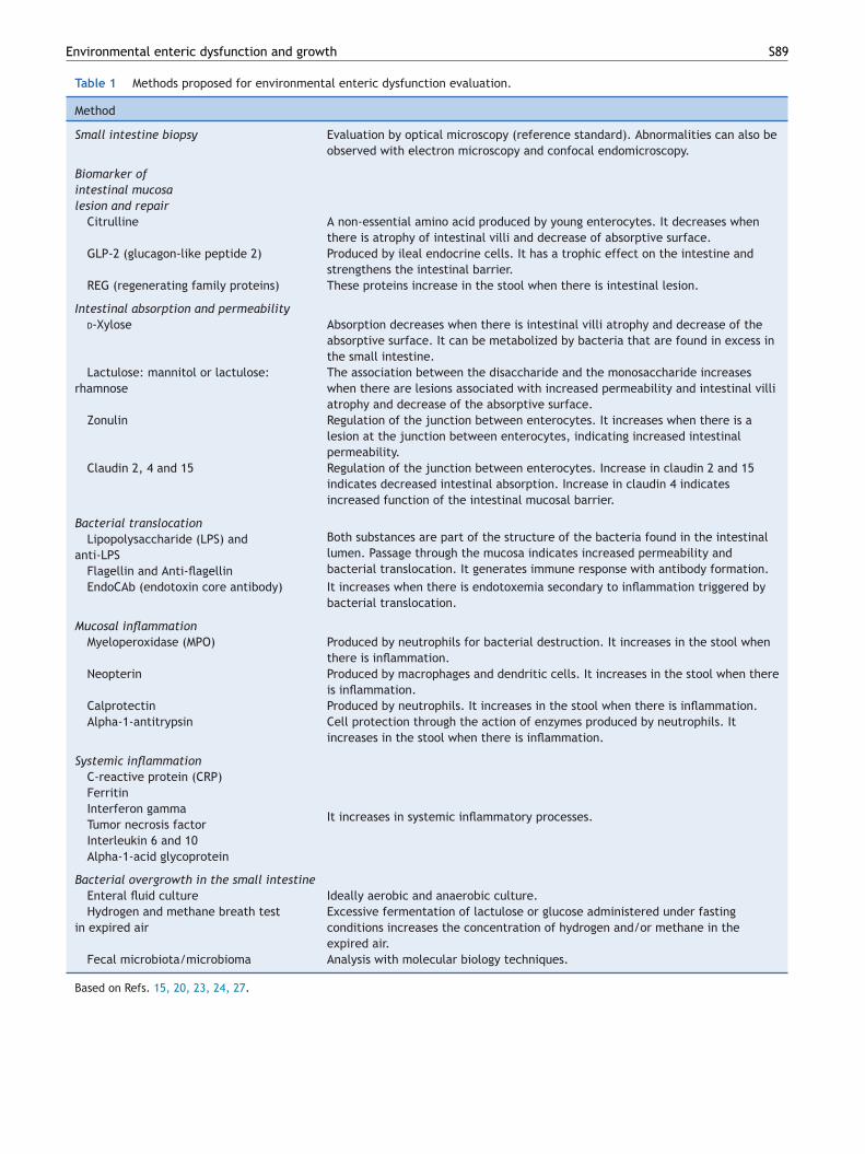
Environmental enteric dysfunction and growth S89
Table 1 Methods proposed for environmental enteric dysfunction evaluation.
Method
Small intestine biopsy Evaluation by optical microscopy (reference standard). Abnormalities can also beobserved with electron microscopy and confocal endomicroscopy.
Biomarker ofintestinal mucosalesion and repair
Citrulline A non-essential amino acid produced by young enterocytes. It decreases whenthere is atrophy of intestinal villi and decrease of absorptive surface.
GLP-2 (glucagon-like peptide 2) Produced by ileal endocrine cells. It has a trophic effect on the intestine andstrengthens the intestinal barrier.
REG (regenerating family proteins) These proteins increase in the stool when there is intestinal lesion.
Intestinal absorption and permeabilityd-Xylose Absorption decreases when there is intestinal villi atrophy and decrease of the
absorptive surface. It can be metabolized by bacteria that are found in excess inthe small intestine.
Lactulose: mannitol or lactulose:rhamnose
The association between the disaccharide and the monosaccharide increaseswhen there are lesions associated with increased permeability and intestinal villiatrophy and decrease of the absorptive surface.
Zonulin Regulation of the junction between enterocytes. It increases when there is alesion at the junction between enterocytes, indicating increased intestinalpermeability.
Claudin 2, 4 and 15 Regulation of the junction between enterocytes. Increase in claudin 2 and 15indicates decreased intestinal absorption. Increase in claudin 4 indicatesincreased function of the intestinal mucosal barrier.
Bacterial translocationLipopolysaccharide (LPS) and
anti-LPSBoth substances are part of the structure of the bacteria found in the intestinallumen. Passage through the mucosa indicates increased permeability andbacterial translocation. It generates immune response with antibody formation.Flagellin and Anti-flagellin
EndoCAb (endotoxin core antibody) It increases when there is endotoxemia secondary to inflammation triggered bybacterial translocation.
Mucosal inflammationMyeloperoxidase (MPO) Produced by neutrophils for bacterial destruction. It increases in the stool when
there is inflammation.Neopterin Produced by macrophages and dendritic cells. It increases in the stool when there
is inflammation.Calprotectin Produced by neutrophils. It increases in the stool when there is inflammation.Alpha-1-antitrypsin Cell protection through the action of enzymes produced by neutrophils. It
increases in the stool when there is inflammation.
Systemic inflammationC-reactive protein (CRP)
It increases in systemic inflammatory processes.
FerritinInterferon gammaTumor necrosis factorInterleukin 6 and 10Alpha-1-acid glycoprotein
Bacterial overgrowth in the small intestineEnteral fluid culture Ideally aerobic and anaerobic culture.Hydrogen and methane breath test
in expired airExcessive fermentation of lactulose or glucose administered under fastingconditions increases the concentration of hydrogen and/or methane in theexpired air.
Fecal microbiota/microbioma Analysis with molecular biology techniques.
Based on Refs. 15, 20, 23, 24, 27.
S
(iaiCitdiabw
iifcteatbro13oi
oeacwwmsswbiT(issgooprfiottwgtsimcwbha
i
cmtidfoaitBMliIwlethobgwt
snjeirliimr
bwmodabdNtvampuaccaFoutcct
90
myeloperoxidase, neopterin, and alpha-1-antitrypsin),.e., no association was observed between linear growthnd inflammation in four of the five publications.20 Regard-ng the indicators of systemic inflammation (cytokines,-reactive protein, and ferritin, among others) conflicting
nformation was also retrieved. The attempt to associatehe different environmental enteric dysfunction indicatorsid not show any relevant association.20 Thus, up to date,t is not possible to choose a noninvasive marker that willllow the diagnosis of environmental enteric dysfunction toe established with accuracy and to define its associationith the occurrence of linear growth deficit.20
Regarding the changes in the intestinal microbiotan environmental enteric dysfunction, most of the stud-es evaluated small intestine bacterial overgrowth andecal microbiota composition. Currently, the microbiologi-al study of duodenal secretion is the most accurate methodo evaluate small intestine bacterial overgrowth. How-ver, considering the complexity of specimen collectionnd processing, the Exhaled Breath Test has been used inhe investigation of small intestine bacterial overgrowth,ecause it is a noninvasive and low-cost test.14,15 A study car-ied out in South Asia evaluated 430 children under 5 yearsf age and showed small intestine bacterial overgrowth in2.5% of children in the first year of life and approximately0% in the subsequent years. The distribution of bacterialvergrowth in relation to age was similar to that observedn the number of episodes of acute diarrhea.13
In Brazil, the breath test was used to investigate bacterialvergrowth in school-aged children living under unfavorablenvironmental conditions. A study published in 2007 evalu-ted 50 children aged 5---11 years who lived in a slum in theountryside of the state of São Paulo, Brazil.31 The resultsere compared with those obtained from the control group,hich comprised children from a private school in the sameunicipality. The breath test was carried out utilizing two
ubstrates: glucose and lactulose. Glucose did not present aignificant hydrogen production in children living in a slumhen compared with the controls. In turn, small intestineacterial overgrowth was more prevalent in the children liv-ng in a slum (37.5%) than in the controls (2.1%; p < 0.001).31
he mean height-for-age Z-score of the control children+0.19 ± 0.84) was higher (p < 0.05) than in children livingn a slum, with (−0.63 ± 0.91) or without (−0.81 ± 1.19)mall intestine bacterial overgrowth. In those living in slums,mall intestine bacterial overgrowth was not associated withreater height impairment. Two other studies were carriedut at different times in a slum in the metropolitan regionf the city of São Paulo, Brazil.32,33 The data were com-ared with those of children of good socioeconomic statusecruited from a private school in the same region. Therst showed a higher prevalence of small intestine bacterialvergrowth in the children living in the slum (30.9%) than inhe ones from the private school (2.4%; p = 0.0007). Amonghe children living in a slum, the anthropometric indicatorsere similar in children with and without bacterial over-rowth. Conversely, they were lower than those observed inhe children from the private school, including height.32 Theecond study showed small intestine bacterial overgrowthn 61.0% of 100 children who lived in a slum.33 A lowerean height-for-age (−0.48 ± 0.90 vs. −0.11 ± 0.97) and
apillary hemoglobin (12.6 ± 1.0 vs. 13.4 ± 1.2 g/dL) Z-scoreere verified in the group of children with small intestineacterial overgrowth when compared to those who did not
ave it (p < 0.05).33 Unfortunately, that study did not evalu-te systemic inflammation indicators.The development of small intestine bacterial overgrowthn residents of areas with unsatisfactory environmental
Cbwa
Morais MB, Silva GA
onditions was attributed to the inhibition of the migratingotor complex (MMC), secondary to repeated exposure of
he digestive tract to lipopolysaccharides (LPS) throughngestion of water or soil contaminated with bacteria.14 Aecrease in peristalsis increases intestinal transit time andacilitates microbial proliferation. This hypothesis is basedn animal models, in which a decrease in the frequencynd vigor of peristaltic contractions was shown in the smallntestine induced by LPS produced by Escherichia coli. Addi-ionally, in germ-free mice, Lactobacillus acidophilus andifidobacterium bifidus cause an increase in the MMC, whileicrococcus luteus and E. coli cause a decrease. There is
ittle evidence to demonstrate effective nutritional statusmpairment due to small intestine bacterial overgrowth.n Brazil, it was verified that, in children aged 5---11 yearsho lived in a slum, those with bacterial overgrowth had
ower values of height-for-age Z-score.33 These data can bexplained by intestinal malabsorption, as characterized inhe 1990s in a community in Burma/Myanmar.34,35 Using theydrogen breath test, the authors found lower absorptionf a test meal with rice in children with small intestineacterial overgrowth. They observed longitudinally that therowth rate in height of children with malabsorption of riceas lower.34,35 This is the only longitudinal study showing
hat bacterial overgrowth can impair growth.Another possible mechanism to explain the effect of
mall intestine bacterial overgrowth is the increased intesti-al permeability.14 Thus, functional impairment of the tightunction between enterocytes and consequent endotox-mia and systemic inflammation may occur. The role of thentestinal microbiota in the intestinal homeostasis must beemembered. It is well known that the microbiota regu-ates not only the intestinal barrier but also the intestinalmmune function, with a complex interaction that resultsn the maintenance of a minimum basal level of inflam-ation and controls the occurrence of a full inflammatory
esponse.14
From the microbiological standpoint, the fecal micro-iota of children and adults living in slums in Bangladesh,here environmental enteropathy is prevalent, showsarked differences when compared with those of residents
f the United States.5 Samples from a small number of chil-ren and adults (four to six) were evaluated monthly forpproximately six months. It was observed that the micro-iota of the individuals living in Bangladesh showed greateriversity and lower stability than those observed in theorth-American individuals. In contrast to the Americans,he feces of the Bangladesh sample were richer in Pre-otella, Butyrivibrio, and Oscillospira and had a greaterbundance of Bacteroides.36 Differences in the microbiotaay be related to the type of diet in communities with a highrevalence of environmental enteric dysfunction, which aresually rich in starch and dietary fiber and low in proteinnd fat from animal sources.5 One example is the studyomparing the microbiota of 11 African children from ruralommunities in Burkina Faso with 13 children from urbanreas of the same country and 13 Italian children fromlorence.37,38 It was observed that, when food of animalrigin and simple carbohydrates are included in the diet inrban areas of the African country, the microbiota startso show an increased abundance of bacteria with a higherapacity to metabolize these foods, as observed in Europeanhildren. This process is associated with a reduced produc-ion of short-chain fatty acids by the intestinal microbiota.
hildren who live in rural communities persist with a micro-iota richer in Prevotella, Treponema, and Succinivibrio,hich have greater specificity for foods rich in dietary fibernd other complex carbohydrates found in vegetables.37,38
mtdgrGtrie
oetSgduaftrrriciatr
nrtcait
En
Ecianmtc(d
RMDdBTisic
Environmental enteric dysfunction and growth
A project carried out in the metropolitan region of SãoPaulo compared the microbiota of 100 school-aged childrenliving in a slum with 30 children living under good environ-mental conditions recruited from private schools. Real-timePCR was used to quantify the selected phyla, genus andspecies. Children living in unfavorable conditions had ahigher number of bacteria, organisms from the Firmicutesand Bacteroidetes phyla, the Escherichia and Lactobacillusgenus, and lower counts of Salmonella. A lower prevalenceand counts of Clostridium difficile were also observed. Com-paratively, it was speculated that a greater prevalence ofSalmonella and C. difficile could indicate negative aspectsin the microbiota of children living in good environmentalconditions.39 It is interesting to mention that a larger amountof Archaea Methanobrevibacter smithii was observed in thechildren living in a slum, accompanied by a larger productionof methane in the exhaled air, indicating a different pat-tern of bacterial metabolism.40 In children living in a slumwith small intestine bacterial overgrowth, according to therespiratory test with lactulose, it was found that the fecalmicrobiota had a lower count of bacteria and Firmicutes anda higher count of bacteria of the Salmonella genus.
However, there are still many questions about the role ofthe microbiota on the development of environmental entericdysfunction, such as the stability of the microbiota profileof children at risk of stunting, whether there are particular-ities in the microbiota of children without stunting that livein non-industrialized countries, and whether interventionsto correct any eventual intestinal microbiota deviations arepossible.41 Another question is about the validity of consid-ering fecal microbiota as an indicator of abnormalities thatoccur in the small intestine, including bacterial overgrowth.Studies using molecular biology techniques can contributeto a better understanding of the microbiota in the proxi-mal small intestine. However, it should be remembered thatcollecting samples from this portion of the intestine is a dif-ficult and invasive procedure, such as the collection of tissuefragments for histological studies.
Environmental enteric dysfunction and response tooral immunization
There is evidence that the local and systemic inflammatorystate described in environmental enteric dysfunction mayalso decrease the response to oral vaccines, as well as beingthe main cause of stunting.42,43
In this context, decreased response to the rotavirus vac-cine, oral polio vaccine, and attenuated cholera vaccineobserved primarily in Africa and Asia have been associatedwith environmental enteric dysfunction. Several hypothe-ses have been raised to explain the phenomenon, fromthe heterogeneity of strains, which would make it difficultto develop a vaccine that had the same universal perfor-mance, to factors associated with host and environment.42,43
A recent editorial44 discussed the hypothesis that the char-acteristics of the intestinal microbiota, changes in theintestinal mucosa caused by the inflammatory process thataffects mainly the innate immune response, and the sys-temic proinflammatory state could be involved in theimpaired response to oral vaccines documented in childrenliving in regions with poor environmental conditions.
Case---control studies nested in clinical trials carried outin Ghana and India showed that the intestinal microbiota
analyzed before the children received the rotavirus vac-cine was qualitatively different among the children whoresponded and those who did not respond to the vaccinechallenge.45,46 These studies also compared the intestinalaoaa
S91
icrobiota of children from Ghana and India who respondedo the vaccine challenge with a group of German chil-ren, and found that the microbiota profile of the tworoups was similar. Conversely, the microbiota of the non-esponder children showed differences in comparison to theerman children.45,46 These findings cannot be extrapolatedo the entire child population living in contaminated envi-onments, as this hypothesis has not been corroboratedn other contexts where the prevalence of environmentalnteric dysfunction is high.47
A recent review article48 analyzed eight studies carriedut in Africa, Asia, and South America with the aim ofvaluating the response to oral vaccines in regions wherehe frequency of enteric environmental dysfunction is high.everal difficulties were found when carrying out the inte-rated interpretation of these studies, including: differentiagnostic criteria for enteric environmental dysfunction,se of biomarkers that can be altered by other diseases,nd other methodology limitations of these articles. There-ore, it was not possible to reach robust conclusions. Ofhe eight articles, four showed evidence that the vaccineesponse was lower in children with high probability of envi-onmental enteric dysfunction, while two found an oppositeesult, i.e., higher vaccine immunogenicity, and two stud-es did not find a statistically significant association. Theyoncluded that, although biologically plausible, there is stillnsufficient empirical data to allow a definitive conclusionbout the possibility that environmental enteric dysfunc-ion reduces oral vaccine responsiveness in underdevelopedegions.
However, it is important to note that these results shouldot discourage the use of these vaccines in populations atisk for enteric environmental dysfunction, albeit less effec-ive, immunization is able to protect a significant number ofhildren and contributes to the reduction of hospitalizationsnd deaths.42 That is, these data should be considered as anndication of the urgent need to control enteric environmen-al dysfunction.
nvironmental enteric dysfunction andeurocognitive development
xperimental studies have shown that an inflammatory statean induce neurocognitive alterations. It is theorized that,n addition to the inflammatory state, metabolites associ-ted with intestinal dysbiosis stimulate the production ofeurotransmitters that interfere with the brain develop-ent process. In humans, it is more difficult to evaluate
his process, because inflammatory processes can be asso-iated with other factors, such as micronutrient deficiencyiron), lack of environmental stimuli, poverty, and intestinalysbiosis.8
A multicenter study entitled MAL-ED (The Etiology,isk Factors, and Interactions of Enteric Infections andalnutrition and the Consequences for Child Health andevelopment) is following-up a cohort of 1600 chil-ren recruited at birth in eight countries (South Africa,angladesh, Brazil, India, Nepal, Pakistan, Peru, andanzania). Data on environmental exposures, intestinalnflammation, intestinal permeability assessment, expo-ure to enteropathogens, periodic measurements of foodntake, including micronutrients, response to oral vaccines,ognitive development, and growth monitoring are being
nalyzed. This project allows the study of the consequencesf environmental enteric dysfunction in different geographicnd epidemiological contexts.27 Fig. 1 outlines the etiologynd consequences of environmental enteric dysfunction.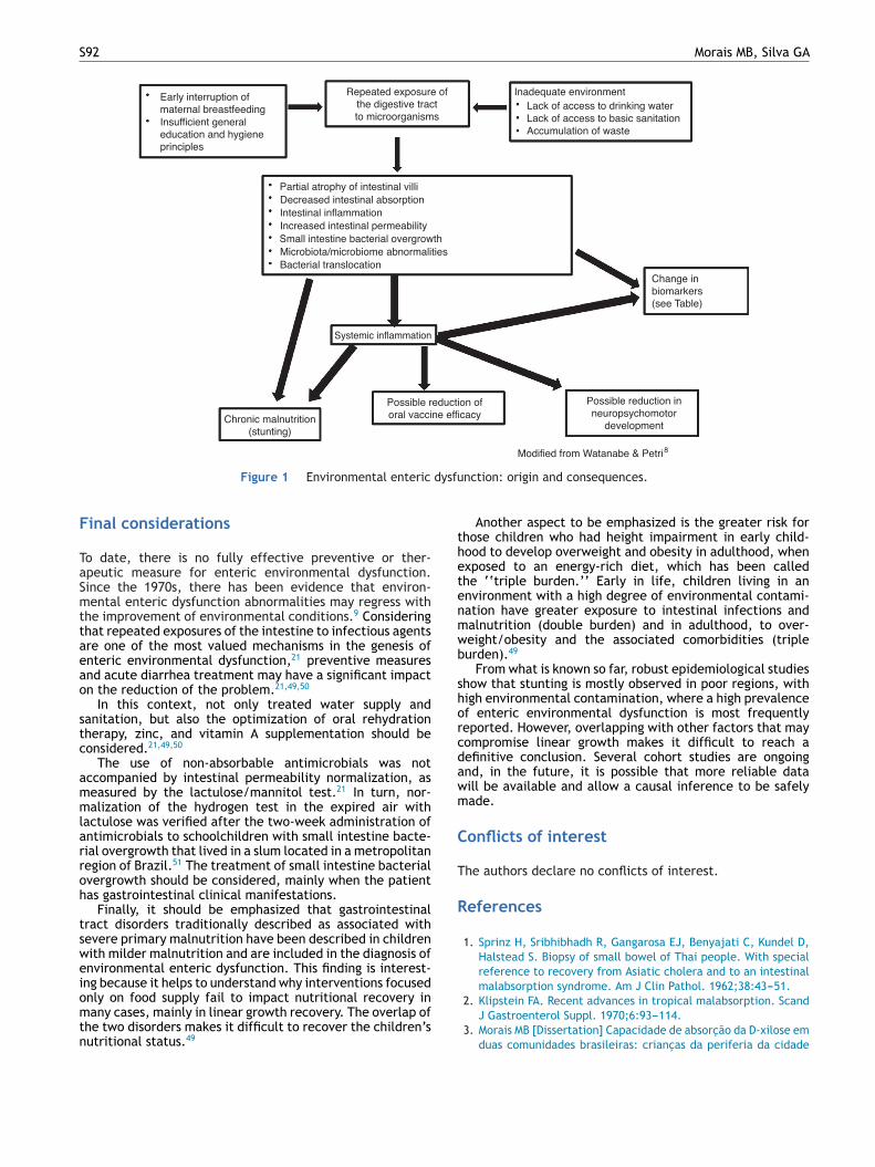
S92 Morais MB, Silva GA
dysfu
F
TaSmttaeao
stc
ammlarroh
tsweiomtn
thetenmwb
shorcdawm
C
T
R
2. Klipstein FA. Recent advances in tropical malabsorption. Scand
Figure 1 Environmental enteric
inal considerations
o date, there is no fully effective preventive or ther-peutic measure for enteric environmental dysfunction.ince the 1970s, there has been evidence that environ-ental enteric dysfunction abnormalities may regress with
he improvement of environmental conditions.9 Consideringhat repeated exposures of the intestine to infectious agentsre one of the most valued mechanisms in the genesis ofnteric environmental dysfunction,21 preventive measuresnd acute diarrhea treatment may have a significant impactn the reduction of the problem.21,49,50
In this context, not only treated water supply andanitation, but also the optimization of oral rehydrationherapy, zinc, and vitamin A supplementation should beonsidered.21,49,50
The use of non-absorbable antimicrobials was notccompanied by intestinal permeability normalization, aseasured by the lactulose/mannitol test.21 In turn, nor-alization of the hydrogen test in the expired air with
actulose was verified after the two-week administration ofntimicrobials to schoolchildren with small intestine bacte-ial overgrowth that lived in a slum located in a metropolitanegion of Brazil.51 The treatment of small intestine bacterialvergrowth should be considered, mainly when the patientas gastrointestinal clinical manifestations.
Finally, it should be emphasized that gastrointestinalract disorders traditionally described as associated withevere primary malnutrition have been described in childrenith milder malnutrition and are included in the diagnosis ofnvironmental enteric dysfunction. This finding is interest-ng because it helps to understand why interventions focusednly on food supply fail to impact nutritional recovery in
any cases, mainly in linear growth recovery. The overlap ofhe two disorders makes it difficult to recover the children’sutritional status.49
nction: origin and consequences.
Another aspect to be emphasized is the greater risk forhose children who had height impairment in early child-ood to develop overweight and obesity in adulthood, whenxposed to an energy-rich diet, which has been calledhe ‘‘triple burden.’’ Early in life, children living in annvironment with a high degree of environmental contami-ation have greater exposure to intestinal infections andalnutrition (double burden) and in adulthood, to over-eight/obesity and the associated comorbidities (tripleurden).49
From what is known so far, robust epidemiological studieshow that stunting is mostly observed in poor regions, withigh environmental contamination, where a high prevalencef enteric environmental dysfunction is most frequentlyeported. However, overlapping with other factors that mayompromise linear growth makes it difficult to reach aefinitive conclusion. Several cohort studies are ongoingnd, in the future, it is possible that more reliable dataill be available and allow a causal inference to be safelyade.
onflicts of interest
he authors declare no conflicts of interest.
eferences
1. Sprinz H, Sribhibhadh R, Gangarosa EJ, Benyajati C, Kundel D,Halstead S. Biopsy of small bowel of Thai people. With specialreference to recovery from Asiatic cholera and to an intestinalmalabsorption syndrome. Am J Clin Pathol. 1962;38:43---51.
J Gastroenterol Suppl. 1970;6:93---114.3. Morais MB [Dissertation] Capacidade de absorcão da D-xilose em
duas comunidades brasileiras: criancas da periferia da cidade
2
2
2
2
2
2
3
3
3
3
3
3
3
3
3
3
distinct socioeconomic levels living in the same urban area in
Environmental enteric dysfunction and growth
de São Paulo e criancas índias do Alto Xingu. Escola Paulista deMedicina; 1984.
4. Morais MB, Fagundes Neto U. Enteropatia ambiental. Estud Av.2003;17:137---49.
5. Louis-Auguste J, Kelly P. Tropical enteropathies. Curr Gastroen-terol Rep. 2017;19:29---37.
6. Fagundes-Neto U, Viaro T, Wehba J, Patrício FR, MachadoNL. Tropical enteropathy (environmental enteropathy) in earlychildhood: a syndrome caused by contaminated environment. JTrop Pediatr. 1984;30:204---9.
7. Brunser O, Araya M, Espinoza J, Figueroa G, Pacheco I, LoisI. Chronic environmental enteropathy in a temperate climate.Hum Nutr Clin Nutr. 1987;41:251---61.
8. Watanabe K, Petri WA Jr. Environmental enteropathy: elu-sive but significant subclinical abnormalities in developingcountries. EBioMedicine. 2016;10:25---32.
9. Gerson CD, Kent TH, Saha JR, Siddiqi N, Lindenbaum J. Recoveryof small-intestinal structure and function after residence in thetropics. II. Studies in Indians and Pakistanis living in New YorkCity. Ann Intern Med. 1971;75:41---8.
10. Lindenbaum J, Kent TH, Sprinz H. Malabsorption and jejunitis inAmerican Peace Corps volunteers in Pakistan. Ann Intern Med.1966;65:1201---9.
11. Fagundes Neto U. Enteropatia ambiental. Rio de Janeiro: Revin-ter; 1996.
12. Brown KH, Khatun M, Ahmed G. Relationship of the xyloseabsorption status of children in Bangladesh to their absorp-tion of macronutrients from local diets. Am J Clin Nutr.1981;34:1540---7.
13. Pereira SP, Khin-Maung U, Bolin TD, Duncombe VM, Nyunt NW,Myo K, et al. A pattern of breath hydrogen excretion suggestingsmall bowel bacterial overgrowth in Burmese village children.J Pediatr Gastroenterol Nutr. 1991;13:32---8.
14. Donowitz JR, Petri WA Jr. Pediatric small intestine bacte-rial overgrowth in low-income countries. Trends Mol Med.2015;21:6---15.
15. Sieczkowska A, Landowski P, Kaminska B, Lifschitz C. Smallbowel bacterial overgrowth in children. J Pediatr GastroenterolNutr. 2016;62:196---207.
16. Denno DM, Van Buskirk KM, Nelson ZC, Musser CA, Tarr PI.Environmental enteric dysfunction: advancing current knowl-edge. St. Louis, MO: Washington University Libraries; 2016,http://dx.doi.org/10.7936/K7WQ0228 [cited 09.11.18].
17. Keusch GT, Rosenberg IH, Denno DM, Duggan C, Guerrant RL,Lavery JV, et al. Implications of acquired environmental entericdysfunction for growth and stunting in infants and childrenliving in low- and middle-income countries. Food Nutr Bull.2013;34:357---64.
18. Owino V, Ahmed T, Freemark M, Kelly P, Loy A, Manary M, et al.Environmental enteric dysfunction and growth failure/stuntingin global child health. Pediatrics. 2016;138:e20160641.
19. Millward DJ. Nutrition, infection and stunting: the roles of defi-ciencies of individual nutrients and foods, and of inflammation,as determinants of reduced linear growth of children. Nutr ResRev. 2017;30:50---72.
20. Harper KM, Mutasa M, Prendergast AJ, Humphrey J, Manges AR.Environmental enteric dysfunction pathways and child stunting:a systematic review. PLoS Negl Trop Dis. 2018;12:e0006205.
21. Ali A, Iqbal NT, Sadiq K. Environmental enteropathy. Curr OpinGastroenterol. 2016;32:12---7.
22. Attia S, Feenstra M, Swain N, Cuesta M, Bandsma RHJ. Starvedguts: morphologic and functional intestinal changes in malnu-trition. J Pediatr Gastroenterol Nutr. 2017;65:491---5.
23. Prendergast AJ, Humphrey JH, Mutasa K, Majo FD, Rukobo S,
Govha M, et al. Assessment of environmental enteric dysfunc-tion in the SHINE trial: methods and challenges. Clin Infect Dis.2015;61:S726---32.4
S93
4. Mahfuz M, Das S, Mazumder RN, Masudur RM, Haque R, BhuiyanMM, et al. Bangladesh Environmental Enteric Dysfunction (BEED)study: protocol for a community-based intervention study tovalidate non-invasive biomarkers of environmental enteric dys-function. BMJ Open. 2017;7:e017768.
5. Lifschitz C, Sieczkowska A. New insights into the fecal micro-biota of children living in a slum: association with small bowelbacterial overgrowth. J Pediatr (Rio J). 2018;94:455---7.
6. Denno DM, VanBuskirk K, Nelson ZC, Musser CA, Hay Burgess DC,Tarr PI. Use of the lactulose to mannitol ratio to evaluate child-hood environmental enteric dysfunction: a systematic review.Clin Infect Dis. 2014;59:S213---9.
7. Kosek M, Guerrant RL, Kang G, Bhutta Z, Yori PP, GratzJ, et al. Assessment of environmental enteropathy in theMAL-ED cohort study: theoretical and analytic framework. ClinInfec Dis. 2014;59:S239---47.
8. Campbell RK, Schulze KJ, Shaikh S, Mehra S, Ali H, Wu L,et al. Biomarkers of environmental enteric dysfunction amongchildren in rural Bangladesh. J Pediatr Gastroenterol Nutr.2017;65:40---6.
9. Black RE, Victora CG, Walker SP, Bhutta ZA, Christian P, deOnis M, et al. Maternal and child undernutrition and over-weight in low-income and middle-income countries. Lancet.2013;382:427---51.
0. Monteiro CA, Benicio MH, Conde WL, Konno S, Lovadino AL,Barros AJ, et al. Narrowing socioeconomic inequality in childstunting: the Brazilian experience, 1974---2007. Bull WorldHealth Organ. 2010;88:305---11.
1. dos Reis JC, de Morais MB, Oliva CA, Fagundes-Neto U. Breathhydrogen test in the diagnosis of environmental enteropathy inchildren living in an urban slum. Dig Dis Sci. 2007;52:1253---8.
2. Mello CS, Tahan S, Melli LC, Rodrigues MS, de Mello RM, Scalet-sky IC, et al. Methane production and small intestinal bacterialovergrowth in children living in a slum. World J Gastroenterol.2012;18:5932---9.
3. Mello CS, Rodrigues MS, Araújo-Filho HB, Melli LC, Tahan S, Pig-natari AC, et al. Fecal microbiota analysis of children with smallintestinal bacterial overgrowth among residents of an urbanslum in Brazil. J Pediatr (Rio J). 2018;94:483---90.
4. Khin-Maung-U, Bolin TD, Duncombe VM, Myo-Khin, Nyunt-Nyunt-Wai, Pereira SP, et al. Epidemiology of small bowelbacterial overgrowth and rice carbohydrate malabsorption inBurmese (Myanmar) village children. Am J Trop Med Hyg.1992;47:298---304.
5. Khin-Maung-U, Pereira SP, Bolin TD, Duncombe VM, Myo-Khin,Nyunt-Nyunt-Wai, et al. Malabsorption of carbohydrate fromrice and child growth: a longitudinal study with the breath-hydrogen test in Burmese village children. Am J Clin Nutr.1990;52:348---52.
6. Lin A, Bik EM, Costello EK, Dethlefsen L, Haque R, Relman DA,et al. Distinct distal gut microbiome diversity and compositionin healthy children from Bangladesh and the United States. PLOSONE. 2013;8:e53838.
7. De Filippo C, Cavalieri D, Di Paola M, Ramazzotti M, PoulletJB, Massart S, et al. Impact of diet in shaping gut microbiotarevealed by a comparative study in children from Europe andrural Africa. Proc Natl Acad Sci U S A. 2010;107:14691---6.
8. De Filippo C, Di Paola M, Ramazzotti M, Albanese D, PieracciniG, Banci E, et al. Diet, environments, and gut microbiota. Apreliminary investigation in children living in rural and urbanBurkina Faso and Italy. Front Microbiol. 2017;8:1979.
9. Mello CS, Carmo-Rodrigues MS, Filho HB, Melli LC, Tahan S, Pig-natari AC, et al. Gut microbiota differences in children from
Brazil. J Pediatr Gastroenterol Nutr. 2016;63:460---5.0. de Araujo Filho HB, Carmo-Rodrigues MS, Mello CS, Melli
LC, Tahan S, Pignatari AC. Children living near a sanitary
S
4
4
4
4
4
4
4
4
4
5
5
94
landfill have increased breath methane and Methanobrevibac-ter smithii in their intestinal microbiota. Archaea. 2014:576249.
1. Syed S, Ali A, Duggan C. Environmental enteric dysfunction inchildren. J Pediatr Gastroenterol Nutr. 2016;63:6---14.
2. Velasquez DE, Parashar UD, Jiang B. Strain diversity plays nomajor role in the varying efficacy of rotavirus vaccines: anoverview. Infect Genet Evol. 2014;28:561---71.
3. Parker EP, Ramani S, Lopman BA, Church JA, Iturriza-GomaraM, Prendergast AJ, et al. Causes of impaired oral vaccineefficacy in developing countries. Future Microbiol. 2018;13:97---118.
4. Iturriza-Gómara MI, Cunliffe NA. The gut microbiome as pos-sible key to understanding and improving rotavirus vaccineperformance in high-disease burden settings. J Infect Dis.2017;215:8---10.
5. Harris VC, Armah G, Fuentes S, Korpela KE, Parashar U, VictorJC, et al. Significant correlation between the infant gut micro-
biome and rotavirus vaccine response in rural Ghana. J InfectDis. 2017;215:34---41.6. Harris V, Alic A, Fuentes S, Korpelad K, Kazic M, Tatef J,et al. Rotavirus vaccine response correlates with the infant
Morais MB, Silva GA
gut microbiota composition in Pakistan. Gut Microbes. 2018;9:93---101.
7. Parker EPK, Praharaj I, Zekavati A, Lazarus RP, Giri S, OperarioDJ, et al. Influence of the intestinal microbiota on the immuno-genicity of oral rotavirus vaccine given to infants in south India.Vaccine. 2018;36:264---72.
8. Church JA, Parker EPK, Kosek MN, Kang G, Grassly NC, KellyP, et al. Exploring the relationship between environmentalenteric dysfunction and oral vaccine responses. Future Micro-biol. 2018;13:1055---70.
9. Guerrant RL, De Boer MD, Moore SR, Scharf RJ, Lima AA.The impoverished gut --- a triple burden of diarrhoea, stunt-ing and chronic disease. Nat Rev Gastroenterol Hepatol.2013;10:220---9.
0. Trehan I, Kelly P, Shaikh N, Manary MJ. New insights intoenvironmental enteric dysfunction. Arch Dis Child. 2016;101:741---4.
1. Tahan S, Melli LC, Mello CS, Rodrigues MS, Bezerra Filho H,
de Morais MB. Effectiveness of trimethoprim---sulfamethoxazoleand metronidazole in the treatment of small intestinal bacterialovergrowth in children living in a slum. J Pediatr GastroenterolNutr. 2013;57:316---8.