Embed Size (px)
Citation preview
National Institutes of HealthU.S. Department of Health and Human Services
ENVIRONMENTALHEALTHPERSPECTIVES
ENVIRONMENTALHEALTHPERSPECTIVES
ehponline.org
Oridonin Confers Protection Against Arsenic-induced Toxicity Through Activation of the Nrf2-
mediated Defensive Response
Yu Du, Nicole F. Villeneuve, Xiao-Jun Wang, Zheng Sun, Weimin Chen, Jixue Li, Hongxiang Lou,
Pak Kin Wong, and Donna D. Zhang
doi:10.1289/ehp.11464 (available at http://dx.doi.org/)Online 21 May 2008
1
Oridonin Confers Protection Against Arsenic-induced Toxicity Through Activation of the Nrf2-mediated Defensive Response Yu Du1, 2^, Nicole F. Villeneuve1^, Xiao-Jun Wang1, Zheng Sun1, Weimin Chen1,
Jixue Li1, Hongxiang Lou2, and Pak Kin Wong3, and Donna D. Zhang1*
1. Department of Pharmacology and Toxicology, College of Pharmacy, University of Arizona,Tucson, AZ, 85721 2. Department of Natural Products, School of Pharmaceutical Sciences, Shandong University,44 Wenhua Xilu, Jinan, Shandong, P.R. China, 250012 3. Aerospace & Mechanical Engineering Department, University of Arizona,
Tucson, Arizona 85721
*Address correspondence to: Donna D. Zhang, Ph.D University of Arizona College of Pharmacy 1703 East Mabel Tucson, AZ 85721 USA [email protected] Telephone: 520-626-9918 FAX: 520-626-2466
^These two authors contributed equally
2
Running Title:
Identification of Oridonin As A Novel Nrf2 Activator
Key words:
Antitumor, ARE, arsenic, chemoprevention, diterpenoid, Keap1, Nrf2, oridonin,
oxidative stress, rubescensin.
Acknowledgments and grant information
This work was supported by research grants from NIEHS (1 R01 ES015010-01)
and American Cancer Society (RSG-07-154-01 -CNE) to D. D. Zhang, and by
China State Scholarship Fund to Y. Du.
Abbreviations
ARE antioxidant responsive element
GST glutathione S transferases
Keap1 Kelch-like ECH-associated protein 1
NQO1 NAD(P)H quinone oxidoreductase 1
Nrf2 NF-E2-related factor 2
ROS reactive oxygen specie
Tbhq tert-butylhydroquinone
γ-GCS γ-glutamylcysteine synthetase
3
Section headers
Abstract
Introduction
Materials and Methods
Chemicals and cell cultures
Establishment of a reporter cell line, and the luciferase reporter gene assay
mRNA extraction and Real time RT-PCR
Antibodies, immunoblot analysis, ubiquitination assay, and protein half-life
measurement
Transient transfection of siRNA, glutathione concentration, ROS detection,
cell viability, and cell death detection
Results
Identification of oridonin as an Nrf2 activator
Oridonin activated the ARE-dependent response primarily through
upregulation of the Nrf2 protein level
Oridonin blocked Nrf2 ubiquitination whereas enhanced Keap1
ubiquitination.
Efficacy of oridonin in protecting against As(III)-induced toxicity
Discussion
References
Figure Legends
Figures
4
Abstract
BACKGROUND: Ground water contaminated with arsenic imposes a big
challenge to human health worldwide. Using natural compounds to subvert the
detrimental effects of arsenic represents an attractive strategy. The transcription
factor, Nrf2, is a critical regulator of the cellular antioxidant response as well as
xenobiotic metabolism. Recently, Activation of the Nrf2 signaling pathway has
been reported to confer protection against arsenic-induced toxicity using a cell
culture model.
OBJECTIVES: The goal of the present work was to identify a potent Nrf2
activator from plants as a chemopreventive compound and to demonstrate the
efficacy of the compound in battling against arsenic-induced toxicity.
RESULTS: Oridonin activated the Nrf2 signaling pathway at a low subtoxic dose
and was able to stabilize Nrf2 by blocking Nrf2 ubiquitination and degradation,
leading to accumulation of the Nrf2 protein and activation of the Nrf2-dependent
cytoprotective response. Pretreatment of UROtsa cells with 1.4 µM oridonin
significantly enhanced the cellular redox capacity, reduced formation of reactive
oxygen species (ROS), and improved survival of UROtsa cells following arsenic
challenge.
CONCLUSIONS: We have identified oridonin as a novel class of Nrf2 activators
and illustrated the mechanism by which the Nrf2 pathway is activated.
Furthermore, we have demonstrated the feasibility of using natural compounds
targeting Nrf2 as a therapeutic approach to protect humans from various
environmental insults that we have to face every day.
5
Introduction
Arsenic is one of the major environmental pollutants. It exists in soil and minerals
and readily enters the ground water system, contaminating drinking water. The
concentration of arsenic in the ground water varies significantly in different
geographic areas. Arsenic concentrations are highest in East Asia, including
Bangladesh, West Bengal, India, and China (Kumagai and Sumi 2007; Smith et
al. 2000; Tchounwou et al. 2003). Many efforts have been made in an attempt to
reduce arsenic damage as exemplified by the guideline for arsenic in drinking
water set by the World Health Organization and by local governments.
Nevertheless, a large number of populations are still at risk of arsenic exposure
and are suffering from arsenic-induced adverse effects, such as hypertension,
arteriosclerosis, diabetes, hyperkeratosis, neuropathy, and cancer in the skin,
liver, bladder, and lung (Kumagai and Sumi 2007; Smith et al. 2006; Steinmaus
et al. 2000; Tseng 2002). Clearly, the best way to protect humans from arsenic-
induced damage is to reduce arsenic intake. However, it is not always practical,
because many people have no choice but to live off from drinking water and rice
that are heavily contaminated with arsenic, as these are their only sources of
food and water. Therefore, an alternative choice, of equal importance, is to
subvert the detrimental effects of arsenic by modulating the body’s defense
system.
6
Nrf2 is a critical transcription factor that regulates a cytoprotective response.
Many of its downstream target genes are important in maintaining the cellular
antioxidant response as well as xenobiotic metabolism. For example, γ-
glutamylcysteine synthetase (γ-GCS) and the xCT cysteine antiporter are the key
enzymes for synthesis of glutathione and maintenance of cellular redox
homeostasis (Chan and Kwong 2000; Sasaki et al. 2002; Wild et al. 1999);
Conjugating enzymes, such as glutathione S transferases (GSTs) and UDP-
glucuronosyl transferase, facilitate the removal of toxic and carcinogenic
chemicals by increasing their solubility and excretion (Kobayashi and Yamamoto
2006; Zhang 2006); Many transporters such as multidrug resistance proteins and
p-glycoprotein are important in uptake and removal of xenobiotics (Hayashi et al.
2003; Maher et al. 2005; Vernhet et al. 2001; Xu et al. 2005). Activation of the
Nrf2 signaling pathway is tightly regulated by Keap1 according to changes in the
intracellular redox state when cells are exposed to exogenous stimuli. Under
normal conditions, cells maintain low constitutive levels of Nrf2-target genes
through constant ubiquitination and degradation of Nrf2, which is accomplished
by the Keap1-dependent E3 ubiquitin ligase complex. Upon induction, Nrf2 is
stabilized due to impaired Keap1-E3 ubiquitin ligase activity, resulting in
activation of the Nrf2 signaling pathway (Cullinan et al. 2004; Furukawa and
Xiong 2005; Kobayashi et al. 2004; Sun et al. 2007; Zhang et al. 2004).
Chemopreventive compounds are able to activate the Nrf2-dependent adaptive
response, thus confer protection against subsequent toxic or carcinogenic
damage (Jeong et al. 2006; Yates and Kensler 2007). Intriguingly, in addition to
7
the beneficial antioxidants and many chemopreventive compounds, the Nrf2
signaling pathway can also be induced by many harmful chemicals such as
arsenic, hydrogen peroxide, and even anticancer drugs including cisplatin (Aono
et al. 2003; He et al. 2006; Massrieh et al. 2006; Pi et al. 2003; Purdom-
Dickinson et al. 2007; Wang et al. 2006). This paradox may be explained by the
balance between the induction of the Nrf2 defensive response and the toxic
outcome elicited by a particular compound. The most attractive chemopreventive
compounds are those that potentially induce the Nrf2-dependent defensive
response without eliciting toxic effects, that is, those that tip the balance toward
the Nrf2-dependent beneficial response. In accordance with this notion, many
chemopreventive compounds, extracted from dietary sources or plants, have
been shown to activate the Nrf2-dependent response at low doses and do not
elicit detectable toxic effects. Nrf2 activators identified so far can be classified
into categories including phenolic antioxidants (caffeic acid, epigallocatechin-3-
gallate, butylated hydroxyanisole), dithiolethiones (oltipraz, 3H-1,2-dithiole-3-
thione), isothiocyanates (sulforaphane), and triterpenoids (1-(2-cyano-3,12-
dioxooleane-1,9[11]-dien-28-oyl)imidozole) (Yates and Kensler 2007; Zhang
2006). Recently, upregulation of the Nrf2-dependent defense response has
proved to be beneficial in reducing arsenic-induced toxicity in a cell culture model
(Wang et al. 2007). Stable knockdown of endogenous Nrf2 using Nrf2-shRNA,
rendered cells more sensitive to arsenic-induced cell death. On the other hand,
pretreatment with chemicals that activate Nrf2 enhanced cell resistance to
arsenic-induced cell death. This study provides the framework of using natural
8
compounds to activate the Nrf2-dependent protective pathway to counteract
arsenic-induced damage.
Here, we report the identification of a novel class of Nrf2 activators. Oridonin,
also known as rubesecensin A, is a diterpenoid purified from the Chinese
medicinal herb Rabdosia rubescens (RR). As one of the important traditional
Chinese medicines, RR has been used by Chinese doctors to treat swelling of
the throat, insect bites, snake bites, inflammation of the tonsils, and cancer of the
esophagus, stomach, liver, prostate, and breast (Zhou et al. 2007). The active
ingredients of RR are rubesecensin A (oridonin) and rubesecensin B. Currently
the major research focus on oridonin is in its antiproliferation and antitumor
activities. The anticancer activity of oridonin was thought to rely on its ability to
inhibit cell growth, reduce angiogenesis, and enhance apoptosis (Chen et al.
2005; Ikezoe et al. 2003; Liu et al. 2006; Liu et al. 2004; Meade-Tollin et al. 2004;
Zhang et al. 2004). Oridonin has been shown to inhibit cell growth and induce
apoptotic cell death in many cancer cell lines, including leukemia (NB4, HL-60,
HPB-ALL, Kasumi-1), glioblastoma (U118, U138), melanoma (A375-S2), cervical
carcinoma (HeLa), ovarian carcinoma (A2780, PTX10), prostate carcinoma
(LNCap, Du145, PC3), breast carcinoma (MCF-7, MDA-MB231), murine
fibrosarcoma (L929), and non-small cell lung carcinoma (NCI-H520, NCI-H460,
NCI-H1299) (Chen et al. 2005; Ikezoe et al. 2003; Liu et al. 2006; Liu et al. 2004;
Zhang et al. 2004). The reported doses needed for growth inhibition and
apoptosis varied significantly amongst different groups using different cell lines,
9
ranging from 0.5 µM (0.18 µg/ml) in Kasumi-1 cells to 56 µM (20.4 µg/ml) in HPB-
ALL cells (Liu et al. 2006; Zhou et al. 2007). In addition, oridonin was also shown
to enhance the efficacy of the cancer drug cisplatin in mouse sarcoma cells (Gao
et al. 1993). Mechanistic studies have provided a molecular basis by which
oridonin inhibits cell growth and induces apoptosis. Oridonin induced p21
expression resulting in cell cycle arrest in LNCaP and NCI-H520 cells (Ikezoe et
al. 2003). Oridonin activated the caspase 3-dependent apoptotic pathway
through upregulation of Bax and downregulation of Bcl-2 which promotes release
of cytochrome c (Chen et al. 2005; Liu et al. 2006). Inhibition of telomerase
activity was reported to be another mechanism that contributes to the anti-cancer
function of oridonin (Liu et al. 2004). Since telomerase activity is absent in normal
somatic cells, but is upregulated in cancer cells or tumor tissues, this allows
oridonin to specifically target abnormal tissue. In addition, the total tyrosine
kinase activity was also reduced in response to oridonin treatment (Li et al.
2007). In addition to cancer cell lines, the efficacy of oridonin in vivo was
demonstrated in a colorectal carcinoma cell HT29-inoculated mouse model (Zhu
et al. 2007). More significantly, a recent report has demonstrated that oridonin
displayed a great antitumor activity specifically in acute myeloid leukemia with
the t(8;21) translocation between AML1 and ETO genes using both cell culture
and mouse models. Mechanistically, oridonin was shown to induce the caspase
3-dependent cleavage of the AML1-ETO fusion protein, leading to an accelerated
apoptotic response (Zhou et al. 2007).
10
Here, we report a novel function of oridonin. It is identified as a novel class of
Nrf2 activators. Similar to tert-butylhydroquinone (tBHQ), it inhibits ubiquitination
and degradation of Nrf2, resulting in stabilization of Nrf2 and activation of the
Nrf2 signaling pathway. Furthermore, the chemopreventive activity of oridonin
was demonstrated using a previously established arsenic-UROtsa cell model.
Pretreatment of UROtsa cells with 1.4 µM oridonin significantly enhanced the
cellular redox capacity, reduced formation of reactive oxygen species (ROS), and
improved survival of UROtsa cells following arsenic exposure.
Materials and Methods
Chemicals and cell cultures. Most chemicals, including sodium arsenite, tBHQ,
and Hoechst 33258, were from Sigma Chemical Co. (St. Louis, MO).
Rubescensin A (oridonin) was purchased from LKT laboratories, Inc (St. Paul,
MN). Human MDA-MB-231 breast carcinoma cells were from ATCC (Manassas,
VA) and cultured in Eagle’s minimal essential medium supplemented with 10%
fetal bovine serum, 2 mM HEPES, and 6 ng/mL bovine insulin from Sigma
Chemical Co. (St. Louis, MO). UROtsa cells were generously provided by Drs.
Mary Ann and Donald Sens (University of North Dakota). UROtsa cells were
grown in DMEM medium enriched with 5% FBS. All mammalian cells were
incubated at 37 oC in a humidified incubator containing 5% CO2.
Establishment of a reporter cell line, and the luciferase reporter gene
assay. The luciferase plasmid, pGL4.22[luc2CP/Puro], was purchased from
Promega (Madison, WI). A 39 bp ARE-containing sequence from the promoter
11
region of human NAD(P)H quinone oxidoreductase (NQO1) gene was inserted
into the cloning site of the luciferase plasmid. The ARE-luciferase plasmid was
transfected into MDA-MB-231 cells using Lipofectamine Plus from Invitrogen
(Grand Island, NY), according to the manufacturer’s instructions. At 48 h post-
transfection, cells were grown in medium containing 3 µg/ml puromycin for
selection. Stable cell lines were considered established once all the cells in the
negative control plate were killed. Stable cell lines were continuously grown in
the MEM medium containing 3 µg/ml puromycin. For the reporter gene assay, the
ARE-luciferase stable reporter cells were seeded the day before and treated with
different doses of test compounds for 24 h. Cells were lysed in extraction buffer
[0.1 M potassium phosphate and 1 mM dithiothreitol (DTT)] by freeze and thaw
three times and luciferase activities were measured in an assay buffer (25 mM
glycylglycine, 15 mM MgSO4, 500 µM ATP, 250 µM luciferin, and 250 µM CoA)
using BioTek Synergy 2. The reporter gene assay was carried out in triplicate
and mean ± SD was calculated. For the dual luciferase reporter gene assay,
MDA-MB-231 cells were transfected with the same ARE-luciferase plasmid along
with the renilla luciferase expression plasmid, pGL4.74[hRluc/TK] from Promega,
(Madison, WI). At 24 h post-transfection, the transfected cells were treated with
compounds for 24 h and both firefly and renilla luciferase activities were
measured with the dual luciferase reporter assay system from Promega,
(Madison, WI). Firefly luciferase activity was normalized to renilla luciferase
activity. The experiment was carried out in triplicate and expressed as mean ±
SD.
12
mRNA extraction and Real time RT-PCR. Total mRNA was extracted from cells
using TRIZOL reagent (Invitrogen, Grand Island, NY) and equal amounts of RNA
was reverse-transcripted to cDNA using Transcriptor First Strand cDNA
synthesis Kit (Roche, Indianapolis, IN). The PCR condition, Taqman probes and
primers for Nrf2, NQO1, heme oxygenase-1 (HO-1), and GAPDH were reported
previously (Wang et al. 2007). Briefly, Taqman probes were from the universal
probe library (Roche, Indianapolis, IN): hNrf2 (#70), hNQO1 (#87), hHO-1 (#25),
and hGAPDH (#25). Primers were synthesized by IDT (Integrated DNA
Technologies, Coralville, IA).
hNrf2: forward (acacggtccacagctcatc) and reverse (tgtcaatcaaatccatgtcctg);
hNQO1: forward (atgtatgacaaaggacccttcc) and reverse (tcccttgcagagagtacatgg);
hHO-1: forward (aactttcagaagggccaggt) and reverse (ctgggctctccttgttgc);
hGAPDH: forward (ctgacttcaacagcgacacc) and reverse (tgctgtagccaaattcgttgt).
The real-time PCR condition was as follows: one cycle of initial denaturation
(95°C for 10 min), 40 cycles of amplification (95°C for 10 sec and 60°C for 20
sec), and a cooling period (50°C for 5 sec). The data presented are relative
mRNA levels normalized to GAPDH and the value from the untreated cells was
set as 1. Triplicate samples were used to get mean ± SD.
Antibodies, immunoblot analysis, ubiquitination assay, and protein half-life
measurement. The antibodies for Nrf2, Keap1, and β-actin were purchased from
Santa Cruz Biotechnology (Santa Cruz, CA). Cells were lysed in a sample buffer
(50 mM Tris-HCl [pH 6.8], 2% SDS, 10% Glycerol, 100 mM DTT, 0.1%
bromophenol blue). Following sonication, cell lysates were electrophoresed
13
through a SDS-polyacrylamide gel and subjected to immunoblot analysis. For
detection of the ubiquitinated Nrf2 in vivo, cells were transfected with expression
vectors for HA-ubiquitin, Keap1, and Gal4-Neh2 (the N-terminal domain of Nrf2
containing the ubiquitin conjugating sites). The transfected cells were either left
untreated or treated with chemicals along with 10 µM MG132 (Sigma Chemical
Co., Saint Louis, MO) for 4 hours. Cells were lysed by boiling in a buffer
containing 2% SDS, 150 mM NaCl, 10 mM Tris-HCl and 1 mM DTT. These
lysates were then diluted five-fold in buffer lacking SDS and incubated with anti-
Nrf2 or anti-Keap1 antibodies. Immunoprecipitated proteins were analyzed by
immunoblot with antibodies directed against the HA epitope (Zhang and Hannink
2003). To detect endogenous Nrf2 ubiquitination, the UROtsa cells were treated
with 10 µM MG 132 and lysed and diluted in the same way. Nrf2 was
immunoprecipitated with an anti-Nrf2 antibody and subjected to immunoblot
analysis with an anti-ubiquitin antibody (Sigma Chemical Co., Saint Louis, MO).
To measure the Nrf2 half-life, cells were either left untreated or treated with
oridonin for 4 h. 50 µM cycloheximide was added to block protein synthesis. Total
cell lysates were collected at different time points and subjected to immunoblot
analysis with an anti-Nrf2 antibody. The relative intensity of bands was quantified
by the ChemiDoc CRS gel documentation system and Quantity One software
from BioRad (Hercules, CA).
Transient transfection of siRNA, glutathione concentration, ROS detection,
cell viability, and cell death detection. The Nrf2-siRNA and the control siRNA
14
were purchased from Qiagen (Valencia, CA). Transient transfection of siRNA
was performed using HiPerFect Transfection Reagent according to the
manufacturer’s protocol (Qiagen, Valencia, CA). Intracellular glutathione
concentration was measured using the QuantiChrom glutathione assay kit from
BioAssay Systems. All the procedures were followed according to the
manufacturer’s instructions. For detection of ROS, cells were pretreated with 1.4
µM oridonin for 24 h, followed by As(III) treatment or As(III) plus oridonin
cotreatment for another 24 h. ROS levels were measured using
dichlorofluorescein (Sigma Chemical Co., 10 µg/ml final concentration) and flow
cytometry. Cell viability was measured by MTT [3-(4, 5 dimethylthiazol-2-yl)-2,5-
diphenyl-2H-tetrazolium bromide] (Wang et al. 2007) and colony formation
assays. Colony formation assay was performed in 35 mm plates with 200
UROtsa cells. Attached cells were left untreated or treated with oridonin for 24 h,
followed by treatment with different doses of As(III) for another 48 h. Following
exposure, medium was replaced with fresh medium and cells were incubated for
12-14 days. The cells were then fixed and stained with crystal violet (0.5% in
95% ethanol). Colonies in each plate were counted. For detection of apoptotic
cell death, two different methods were used: (i) Annexin V-FITC apoptosis
detection (Sigma Chemical Co.), (ii) Hoechst staining (1 µg/ml) for detection of
the condensed nuclei. All experiments were conducted in triplicate and
expressed as mean ± SD. The statistical significance was determined by the
student t-test and labeled in the figure with asterisks (*p<0.05, **p<0.01).
15
Results
Identification of oridonin as an Nrf2 activator. Using the stable ARE luciferase
reporter cell line, derived from the MDA-MB-231 cells, combined with a 96 well
high-throughput screening system established in our laboratory, we identified a
novel Nrf2 activator that belongs to the class of diterpenoids (Figure 1A). The
MDA-MB-231 cell line was used to show Nrf2 activation for a couple reasons: (1)
MDA-MB-231 cells can be easily transfected. (2) The Nrf2 pathway is most
sensitive in this cell line in response to Nrf2-inducers. Oridonin induced
transcription of the ARE-dependent luciferase gene in a dose dependent manner
in the stable cell line (Figure 1B). To confirm oridonin activation of Nrf2 using the
high-throughput screening method, a dual luciferase reporter gene assay was
also performed in which a renilla luciferase gene is included as an internal control
for transfection efficiency and for toxicity induced during oridonin exposure.
Consistent with the data obtained from the high-throughput screening, oridonin
induced the ARE-dependent luciferase activity in a dose dependent manner
(Figure 1C). Slight induction (1.5 fold) was observed at as low as 1.4 µM and
reached maximum induction (11.3 fold) at 14 µM. There was no obvious toxicity
at 14 µM, as judged by cell morphology and renilla luciferase activity.
Oridonin activated the ARE-dependent response primarily through
upregulation of the Nrf2 protein level. Previous studies have demonstrated
that the ARE-dependent reporter gene activity correlated very well with the
16
protein level of Nrf2. Therefore, the same cell lysates from the dual luciferase
reporter gene assay were used for immunoblot analysis for detection of Nrf2,
Keap1 and β-actin. While the Keap1 levels remained constant, oridonin
enhanced the levels of Nrf2 protein in a dose dependent manner with the highest
induction at 14 µM (Figure 2A). During the reporter gene assay, it should be
noted that any doses higher than 14 µM caused marked toxicity as indicated by
an increased number of rounded and floating cells. A large body of literature
indicates that oridonin’s antitumor activity relies on its ability to inhibit cell growth
and to induce cell death. Since Nrf2 regulates a cellular survival response, it is
envisioned that treatment with high doses of oridonin may inhibit Nrf2, allowing
cells to undergo cell death. Therefore, Nrf2 protein levels in response to high
doses of oridonin were tested. Following treatment of MDA-MB-231 cells with
different doses of oridonin for 24 h, all cells including floating cells were collected.
Equal amounts of proteins were subjected to immunoblot analysis with Nrf2,
Keap1 and β-actin antibodies. Interestingly, at doses higher than 28 µM, Nrf2
protein levels decreased in a dose-dependent manner, while the expression of
Keap1 or β-actin had no significant change (Figure 2B, lanes 7-9). Previously, it
has been demonstrated that Nrf2 activators, including tBHQ, induce the Nrf2
signaling pathway primarily through stabilization of the Nrf2 protein, rather than
upregulation of its mRNA. Next, mRNA expression of Nrf2 and its target genes,
NQO1 and HO-1, in response to oridonin was measured using real time RT-
PCR. Nrf2 mRNA increased slightly in a dose dependent manner in response to
oridonin, whereas tBHQ had no effect (Figure 2C, upper panel). As expected,
17
mRNA of NQO1 or HO-1 was induced significantly by oridonin in a dose
dependent manner (Figure 2C, middle and lower panels). These results
demonstrate that oridonin is able to induce the Nrf2 signaling pathway mainly
through upregulation of Nrf2 at the protein level.
Oridonin blocked Nrf2 ubiquitination whereas enhanced Keap1
ubiquitination. tBHQ enhances the Nrf2 protein level by interfering with the
Keap1-dependent ubiquitin conjugation process. Therefore, the ability of oridonin
in modulating Nrf2 ubiquitination was tested. For this assay, Gal4-Neh2, a model
fusion protein previously used for the Nrf2 ubiquitination test, was used (Zhang
and Hannink 2003). Similar to tBHQ, oridonin suppressed Nrf2 ubiquitination
(Figure 3A, left Gal4-Neh2 panel). Furthermore, it was previously shown that
tBHQ caused a shift of ubiquitination from the substrate Nrf2 to the substrate
adaptor Keap1 (Zhang et al. 2005). Similar to tBHQ, oridonin treatment was also
effective in enhancing ubiquitination of Keap1 (Figure 3A, left Keap1 panel).
These results demonstrate that oridonin is able to induce a shift of ubiquitination
from Nrf2 to Keap1. One of the major roles for ubiquitin conjugation onto a
protein is to target the protein for 26S proteasome-mediated degradation. Next,
the half-life of Nrf2 in the absence or presence of oridonin was measured. Half-
life of the endogenous Nrf2 protein in MDA-MB-231 cells was 19 min, while
treatment with oridonin increased half-life to 51 min (Figure 3B, left panel). Taken
together, these results indicate that oridonin activates the Nrf2 pathway by
inhibiting ubiquitination and degradation of Nrf2, leading to an increase in Nrf2
protein level and activation of the Nrf2-dependent response.
18
Efficacy of oridonin in protecting against As(III)-induced toxicity. To test the
feasibility of using oridonin as a chemopreventive compound to elicit the Nrf2-
mediated protective response to defend against environmental insults, the
UROtsa cell line, an established model system for arsenic toxicity, was used.
First, activation of the Nrf2 pathway by oridonin was determined in this cell line.
Ubiquitination of endogenous Nrf2 in UROtsa cells was blocked by oridonin or
tBHQ treatment (Figure 3A, right panel). Consistent with a decrease in
ubiquitination of Nrf2 in response to oridonin, the half life of Nrf2 was increased
from 10 min in the untreated condition to 16 min in response to oridonin
treatment (Figure 3B, right panel). Next, the oridonin dose range that induces the
Nrf2 protein was determined in UROtsa cells. Compared to MDA-MB-231 cells,
UROtsa cells had a narrow range of Nrf2 induction, from 1.4 µM to 14 µM
(Figure 4A, lanes 3-6, compare with Figure 2B, lanes 2-7). At doses higher than
14 µM, toxicity was observed and induction of Nrf2 was decreased (Figure 4A,
lanes 7-9). At dose of 56 or 112 µg/ml, there was a decrease even in the level of
β-actin due to cytotoxicity (Figure 4A, lane 8 and lane 9). Nevertheless, reduction
of the Nrf2 protein was significantly more substantial, indicating that reduction of
Nrf2 at high doses may not solely be due to reduced cell number. Based on this
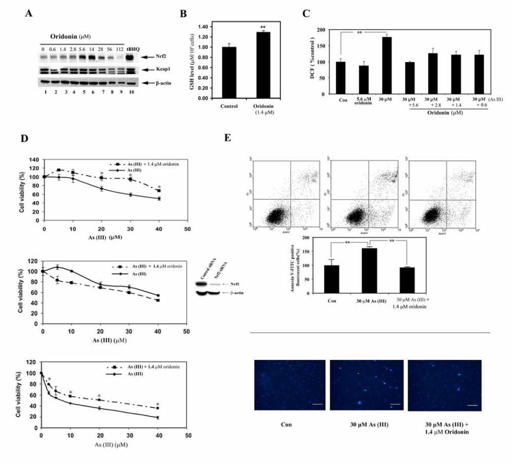
result, a low dose (0.5 µg/ml) was chosen for the protection assays. One of the
major functions of Nrf2 is to regulate an antioxidant response by upregulating
intracellular antioxidants and genes such as GCS and the xCT cysteine
antiporter that encode key enzymes in the synthesis of glutathione. To confirm
activation of the Nrf2-dependent response by 1.4 µM oridonin, the intracellular
19
glutathione level in the oridonin treated cells was compared to that in the
untreated cells. Oridonin treatment resulted in a significant increase in the
glutathione level (Figure 4B). Thus, oridonin is able to augment the cellular redox
capacity, which is the key mechanism in suppressing oxidative stress-induced
damage by environmental insults. In the protection assays, sodium arsenite
[As(III)] was used to treat UROtsa cells. The ability of oridonin in alleviating
As(III)-induced reactive oxygen species (ROS) was measured (Figure 4C). 30
µM As(III) treatment for 24 h increased the level of ROS significantly while 5.6
µM oridonin itself had no effect. Pretreatment of cells with several doses of
oridonin for 24 h and further cotreatment with As(III) for an additional 24 h
resulted in a significant reduction of the ROS levels, especially with 5.6 µM
oridonin. These data clearly demonstrate the efficacy of oridonin in suppressing
oxidative stress imposed by As(III) exposure. Finally, the effectiveness of
oridonin in protecting cells from acute cell death in response to As(III) was
assessed. UROtsa cells were left untreated or pretreated with 1.4 µM oridonin.
Following a 24 h pretreatment period, several doses of As(III) were added to both
groups and incubated for an additional 48 h before measuring total cell death
using both MTT and colony formation assays. Pretreatment followed by
cotreatment with oridonin significantly improved cell survival as judged by the
MTT assay (Figure 4D, top panel) and the colony formation assay (Fgiure 4D,
bottom panel). To confirm that the protection against As(III)-induced cell death is
attributed to the activation of Nrf2 by oridonin, the MTT assay was performed in
UROtsa cells that were treated with Nrf2-siRNA for 48 h. The immunoblot
20
analysis confirmed the effectiveness of Nrf2-siRNA in reducing Nrf2 expression
(Figure D, middle panel). Convincingly, inhibition of Nrf2 expression in UROtsa
cells reverted the MTT curve, i.e., oridonin lost its protection against As(III)
toxicity, rather, it aggravated the As(III)-induced cell death (Figure D, middle
panel). This result demonstrates that the oridonin-mediated protection requires
activation of the Nrf2 pathway. Next, apoptotic cell death was quantified using
Annexin V-FITC/flow cytometry. 30 µM As(III) treatment for 48 h increased the
percentage of apoptotic cells whereas pretreatment followed by cotreatment with
1.4 µM oridonin reduced apoptotic cell death to a level comparable to the
untreated cells (Figure 4E, upper panels). 1.4 µM oridonin itself did not increase
apoptotic cell death (data not shown). Next, Hoechst staining was used for
detection of condensed chromosomes in the apoptotic cells. 30 µM As(III)
increased the number of positive-stained cells, while pretreatment followed by
cotreatment with oridonin markedly reduced the number of apoptotic cells (Figure
4E, lower panel). Together, these results demonstrate that a low dose of oridonin
is able to protect cells from As(III)-induced damage as illustrated by reduced
ROS and increased survival in response to As(III).
Discussion
The pivotal role of Nrf2 in chemoprevention has clearly been shown in Nrf2 null
mice. These mice express lower basal levels of the Nrf2 target genes, such as
NQO1, GST, GCS, UDP-glucuronosyltransferase, glutathione peroxidase-2, and
HO-1 (Chan and Kwong 2000; Cho et al. 2002; Hayes et al. 2000; Kwak et al.
21
2001; McMahon et al. 2001). As a consequence, these mice are more
susceptible to toxic and carcinogenic challenges such as butylated
hydroxytoluene, benzo[a]pyrene, diesel exhaust, aflatoxin B1, N-nitrosobutyl (4-
hydroxybutyl) amino, pentachlorophenol, acetaminophen, ovalbumin, cigarette
smoke, and 4-vinyl cyclohexene diepoxide (Aoki et al. 2001; Chan et al. 2001;
Chan and Kan 1999; Enomoto et al. 2001; Hu et al. 2006; Iida et al. 2004; Iizuka
et al. 2005; Ramos-Gomez et al. 2001; Rangasamy et al. 2005; Umemura et al.
2006). These results provide the basis for chemopreventive intervention targeting
the Nrf2 signaling pathway. Many previously indentified naturally occurring
compounds, including sulforaphane, epigallocatechin-3-gallate, caffeic acid
phenethyl ester, and curcumin have proved to be Nrf2 activators, which further
implies the importance of Nrf2 in chemoprevention (Jeong et al. 2006; Nishinaka
et al. 2007; Zhang 2006). Identification, validation, and optimization of new Nrf2
activators are essential for the development of effective dietary supplements or
therapeutic drugs that can be used to boost the Nrf2-dependent adaptive system
to protect humans from various environmental insults.
Oridonin represents a novel class of Nrf2 activators that has not previously been
demonstrated. Mechanistic studies presented here indicate that oridonin induced
the Nrf2-dependent response primarily by enhancing the Nrf2 protein level. The
increase in the Nrf2 protein level in response to oridonin is attributed mainly to
the stabilization of Nrf2 with minor contribution from increased mRNA. Similar to
tBHQ, oridonin is able to block ubiquitination and degradation of Nrf2, resulting in
22
the prolonged half life of Nrf2. Furthermore, we have demonstrated the
effectiveness of a low dose of oridonin (1.4 µM) in eliciting the Nrf2-dependent
cytoprotective response in an As(III)-toxicity model. Low doses of oridonin are
able to enhance the cellular reducing capacity by significantly elevating the
reduced glutathione level, thus inhibiting the formation of ROS, resulting in
increased survival in response to As(III) exposure. Furthermore, glutathione is
able to conjugate arsenic to facilitate arsenic excretion, therefore reducing As(III)
toxicity (Shinkai et al. 2006). In addition to GCS that modulates intracellular
glutathione levels, other Nrf2 downstream genes, including GST, UDP-
glucuronosyl transferase and multidrug resistance proteins, also contribute to the
Nrf2-mediated protection against arsenic toxicity (Hayashi et al. 2003; Kobayashi
and Yamamoto 2006; Maher et al. 2005; Vernhet et al. 2001; Xu et al. 2005;
Zhang 2006). It is worth it to mention, that although the current study only shows
the protection of oridonin against acute As(III)-toxicity, it certainly can be applied
to other toxic and carcinogenic chemicals since oridonin induces the well
characterized Nrf2-dependent defensive response.
This cell-based study provides the evidence that oridonin can be used at a low
dose as a chemopreventive compound that specifically targets Nrf2. Further
studies on the chemopreventive activity of oridonin in animal models are needed.
If oridonin is shown to have great chemopreventive potential, then it has a great
economic advantage since it can easily be extracted from Rabdosia rubescens
(RR), “the Chinese grass”. In addition, identification of diterpenoids as a new
23
class of Nrf2 activators will broaden the choice for new chemopreventive
compounds. Moreover, the diterpenoid structure can serve as a scaffold for the
development of chemopreventive drugs. Identification of naturally occurring
diterpenoids or synthetic optimization of the diterpenoid, oridonin, that potently
and specifically induce the Nrf2 signaling pathway will greatly improve the
efficacy of chemopreventive drugs and decrease side effects, which will have a
profound impact on human health.
High doses of oridonin have been shown to have anticancer activity by causing
cell cycle arrest, inhibiting proliferation, and inducing the apoptotic cell death. The
dose range needed for oridonin to exhibit anticancer activities in these studies
conducted by different laboratories with a variety of cancer cell lines, is very
broad with 100 fold differences. Although this may be partially due to the purity of
oridonin used among groups, it largely indicates a difference in sensitivity of
cancer cells to the oridonin-induced apoptotic response. In this study, the effect
of oridonin in inducing the Nrf2 protein level was assessed in two different cell
lines, the breast carcinoma MDA-MB-231 and an immortalized but
nontransformed bladder urothelium UROtsa. UROtsa cells showed a narrower
window of Nrf2 induction in response to different doses of oridonin. It is very
interesting to note that oridonin induced the Nrf2 protein and the reporter gene
activity in a dose dependent manner to a certain point, then, the Nrf2 protein
level and the reporter gene activity dropped suddenly. The dose where an initial
decrease was observed in the Nrf2 protein in MDA-MB-231 cells or in UROtsa
24
cells is 56 µM or 28 µM respectively. This decrease in Nrf2 protein level in
response to high doses of oridonin is not solely due to cell toxicity because
Keap1 or β-actin levels only decreased slightly. Based on the important role of
Nrf2 in cell survival, It is conceivable that Nrf2 has to be repressed prior to the
execution of cell death. In support of this notion, Nrf2 has been reported as a
substrate of caspase 3 (Ohtsubo et al. 1999). The cleavage sites in Nrf2 for
caspase 3 have been identified. Based on the Nrf2 induction profile, it is
remarkable that oridonin functions as a chemopreventive compound at low doses
by activating the Nrf2 cytoprotective pathway, while at high doses, it activates
apoptotic cell death and concomitantly inhibits the Nrf2-dependent survival
pathway. Further studies are needed in understanding molecular events that
cause the switch between life and death.
References Aoki Y, Sato H, Nishimura N, Takahashi S, Itoh K, Yamamoto M. 2001. Accelerated
DNA adduct formation in the lung of the Nrf2 knockout mouse exposed to diesel
exhaust. Toxicol Appl Pharmacol 173(3):154-160.
Aono J, Yanagawa T, Itoh K, Li B, Yoshida H, Kumagai Y, et al. 2003. Activation of
Nrf2 and accumulation of ubiquitinated A170 by arsenic in osteoblasts. Biochem
Biophys Res Commun 305(2):271-277.
Chan JY, Kwong M. 2000. Impaired expression of glutathione synthetic enzyme genes in
mice with targeted deletion of the Nrf2 basic-leucine zipper protein. Biochim
Biophys Acta 1517(1):19-26.
25
Chan K, Han XD, Kan YW. 2001. An important function of Nrf2 in combating oxidative
stress: detoxification of acetaminophen. Proc Natl Acad Sci U S A 98(8):4611-
4616.
Chan K, Kan YW. 1999. Nrf2 is essential for protection against acute pulmonary injury
in mice. Proc Natl Acad Sci U S A 96(22):12731-12736.
Chen S, Gao J, Halicka HD, Huang X, Traganos F, Darzynkiewicz Z. 2005. The
cytostatic and cytotoxic effects of oridonin (Rubescenin), a diterpenoid from
Rabdosia rubescens, on tumor cells of different lineage. Int J Oncol 26(3):579-
588.
Cho HY, Jedlicka AE, Reddy SP, Kensler TW, Yamamoto M, Zhang LY, et al. 2002.
Role of NRF2 in protection against hyperoxic lung injury in mice. Am J Respir
Cell Mol Biol 26(2):175-182.
Cullinan SB, Gordan JD, Jin J, Harper JW, Diehl JA. 2004. The Keap1-BTB protein is an
adaptor that bridges Nrf2 to a Cul3-based E3 ligase: oxidative stress sensing by a
Cul3-Keap1 ligase. Mol Cell Biol 24(19):8477-8486.
Enomoto A, Itoh K, Nagayoshi E, Haruta J, Kimura T, O'Connor T, et al. 2001. High
sensitivity of Nrf2 knockout mice to acetaminophen hepatotoxicity associated
with decreased expression of ARE-regulated drug metabolizing enzymes and
antioxidant genes. Toxicol Sci 59(1):169-177.
Furukawa M, Xiong Y. 2005. BTB protein Keap1 targets antioxidant transcription factor
Nrf2 for ubiquitination by the Cullin 3-Roc1 ligase. Mol Cell Biol 25(1):162-171.
26
Gao ZG, Ye QX, Zhang TM. 1993. Synergistic effect of oridonin and cisplatin on
cytotoxicity and DNA cross-link against mouse sarcoma S180 cells in culture.
Zhongguo Yao Li Xue Bao 14(6):561-564.
Hayashi A, Suzuki H, Itoh K, Yamamoto M, Sugiyama Y. 2003. Transcription factor
Nrf2 is required for the constitutive and inducible expression of multidrug
resistance-associated protein 1 in mouse embryo fibroblasts. Biochem Biophys
Res Commun 310(3):824-829.
Hayes JD, Chanas SA, Henderson CJ, McMahon M, Sun C, Moffat GJ, et al. 2000. The
Nrf2 transcription factor contributes both to the basal expression of glutathione S-
transferases in mouse liver and to their induction by the chemopreventive
synthetic antioxidants, butylated hydroxyanisole and ethoxyquin. Biochem Soc
Trans 28(2):33-41.
He X, Chen MG, Lin GX, Ma Q. 2006. Arsenic induces NAD(P)H-quinone
oxidoreductase I by disrupting the Nrf2 x Keap1 x Cul3 complex and recruiting
Nrf2 x Maf to the antioxidant response element enhancer. J Biol Chem
281(33):23620-23631.
Hu X, Roberts JR, Apopa PL, Kan YW, Ma Q. 2006. Accelerated ovarian failure induced
by 4-vinyl cyclohexene diepoxide in Nrf2 null mice. Mol Cell Biol 26(3):940-
954.
Iida K, Itoh K, Kumagai Y, Oyasu R, Hattori K, Kawai K, et al. 2004. Nrf2 is essential
for the chemopreventive efficacy of oltipraz against urinary bladder
carcinogenesis. Cancer Res 64(18):6424-6431.
27
Iizuka T, Ishii Y, Itoh K, Kiwamoto T, Kimura T, Matsuno Y, et al. 2005. Nrf2-deficient
mice are highly susceptible to cigarette smoke-induced emphysema. Genes Cells
10(12):1113-1125.
Ikezoe T, Chen SS, Tong XJ, Heber D, Taguchi H, Koeffler HP. 2003. Oridonin induces
growth inhibition and apoptosis of a variety of human cancer cells. Int J Oncol
23(4):1187-1193.
Jeong WS, Jun M, Kong AN. 2006. Nrf2: a potential molecular target for cancer
chemoprevention by natural compounds. Antioxid Redox Signal 8(1-2):99-106.
Kobayashi A, Kang MI, Okawa H, Ohtsuji M, Zenke Y, Chiba T, et al. 2004. Oxidative
stress sensor Keap1 functions as an adaptor for Cul3-based E3 ligase to regulate
proteasomal degradation of Nrf2. Mol Cell Biol 24(16):7130-7139.
Kobayashi M, Yamamoto M. 2006. Nrf2-Keap1 regulation of cellular defense
mechanisms against electrophiles and reactive oxygen species. Adv Enzyme
Regul 46:113-140.
Kumagai Y, Sumi D. 2007. Arsenic: signal transduction, transcription factor, and
biotransformation involved in cellular response and toxicity. Annu Rev
Pharmacol Toxicol 47:243-262.
Kwak MK, Itoh K, Yamamoto M, Sutter TR, Kensler TW. 2001. Role of transcription
factor Nrf2 in the induction of hepatic phase 2 and antioxidative enzymes in vivo
by the cancer chemoprotective agent, 3H-1, 2-dimethiole-3-thione. Mol Med
7(2):135-145.
28
Li D, Wu LJ, Tashiro S, Onodera S, Ikejima T. 2007. Oridonin inhibited the tyrosine
kinase activity and induced apoptosis in human epidermoid carcinoma A431 cells.
Biol Pharm Bull 30(2):254-260.
Liu JJ, Huang RW, Lin DJ, Wu XY, Peng J, Pan XL, et al. 2006. Antiproliferation effects
of oridonin on HPB-ALL cells and its mechanisms of action. Am J Hematol
81(2):86-94.
Liu JJ, Wu XY, Lul HL, Pan XL, Peng J, Huang RW. 2004. Anti-proliferation effect of
oridonin on HL-60 cells and its mechanism. Chin Med Sci J 19(2):134-137.
Maher JM, Cheng X, Slitt AL, Dieter MZ, Klaassen CD. 2005. Induction of the
multidrug resistance-associated protein family of transporters by chemical
activators of receptor-mediated pathways in mouse liver. Drug Metab Dispos
33(7):956-962.
Massrieh W, Derjuga A, Blank V. 2006. Induction of endogenous Nrf2/small maf
heterodimers by arsenic-mediated stress in placental choriocarcinoma cells.
Antioxid Redox Signal 8(1-2):53-59.
McMahon M, Itoh K, Yamamoto M, Chanas SA, Henderson CJ, McLellan LI, et al.
2001. The Cap'n'Collar basic leucine zipper transcription factor Nrf2 (NF-E2 p45-
related factor 2) controls both constitutive and inducible expression of intestinal
detoxification and glutathione biosynthetic enzymes. Cancer Res 61(8):3299-
3307.
Meade-Tollin LC, Wijeratne EM, Cooper D, Guild M, Jon E, Fritz A, et al. 2004.
Ponicidin and oridonin are responsible for the antiangiogenic activity of Rabdosia
rubescens, a constituent of the herbal supplement PC SPES. J Nat Prod 67(1):2-4.
29
Nishinaka T, Ichijo Y, Ito M, Kimura M, Katsuyama M, Iwata K, et al. 2007. Curcumin
activates human glutathione S-transferase P1 expression through antioxidant
response element. Toxicol Lett 170(3):238-247.
Ohtsubo T, Kamada S, Mikami T, Murakami H, Tsujimoto Y. 1999. Identification of
NRF2, a member of the NF-E2 family of transcription factors, as a substrate for
caspase-3(-like) proteases. Cell Death Differ 6(9):865-872.
Pi J, Qu W, Reece JM, Kumagai Y, Waalkes MP. 2003. Transcription factor Nrf2
activation by inorganic arsenic in cultured keratinocytes: involvement of
hydrogen peroxide. Exp Cell Res 290(2):234-245.
Purdom-Dickinson SE, Lin Y, Dedek M, Morrissy S, Johnson J, Chen QM. 2007.
Induction of antioxidant and detoxification response by oxidants in
cardiomyocytes: evidence from gene expression profiling and activation of Nrf2
transcription factor. J Mol Cell Cardiol 42(1):159-176.
Ramos-Gomez M, Kwak MK, Dolan PM, Itoh K, Yamamoto M, Talalay P, et al. 2001.
Sensitivity to carcinogenesis is increased and chemoprotective efficacy of enzyme
inducers is lost in nrf2 transcription factor-deficient mice. Proc Natl Acad Sci U S
A 98(6):3410-3415.
Rangasamy T, Guo J, Mitzner WA, Roman J, Singh A, Fryer AD, et al. 2005. Disruption
of Nrf2 enhances susceptibility to severe airway inflammation and asthma in
mice. J Exp Med 202(1):47-59.
Sasaki H, Sato H, Kuriyama-Matsumura K, Sato K, Maebara K, Wang H, et al. 2002.
Electrophile response element-mediated induction of the cystine/glutamate
exchange transporter gene expression. J Biol Chem 277(47):44765-44771.
30
Shinkai Y, Sumi D, Fukami I, Ishii T, Kumagai Y. 2006. Sulforaphane, an activator of
Nrf2, suppresses cellular accumulation of arsenic and its cytotoxicity in primary
mouse hepatocytes. FEBS Lett 580(7):1771-1774.
Smith AH, Lingas EO, Rahman M. 2000. Contamination of drinking-water by arsenic in
Bangladesh: a public health emergency. Bull World Health Organ 78(9):1093-
1103.
Smith AH, Marshall G, Yuan Y, Ferreccio C, Liaw J, von Ehrenstein O, et al. 2006.
Increased mortality from lung cancer and bronchiectasis in young adults after
exposure to arsenic in utero and in early childhood. Environ Health Perspect
114(8):1293-1296.
Steinmaus C, Moore L, Hopenhayn-Rich C, Biggs ML, Smith AH. 2000. Arsenic in
drinking water and bladder cancer. Cancer Invest 18(2):174-182.
Sun Z, Zhang S, Chan JY, Zhang DD. 2007. Keap1 controls postinduction repression of
the Nrf2-mediated antioxidant response by escorting nuclear export of Nrf2. Mol
Cell Biol 27(18):6334-6349.
Tchounwou PB, Patlolla AK, Centeno JA. 2003. Carcinogenic and systemic health
effects associated with arsenic exposure--a critical review. Toxicol Pathol
31(6):575-588.
Tseng CH. 2002. An overview on peripheral vascular disease in blackfoot disease-
hyperendemic villages in Taiwan. Angiology 53(5):529-537.
Umemura T, Kuroiwa Y, Kitamura Y, Ishii Y, Kanki K, Kodama Y, et al. 2006. A crucial
role of Nrf2 in in vivo defense against oxidative damage by an environmental
pollutant, pentachlorophenol. Toxicol Sci 90(1):111-119.

31
Vernhet L, Seite MP, Allain N, Guillouzo A, Fardel O. 2001. Arsenic induces expression
of the multidrug resistance-associated protein 2 (MRP2) gene in primary rat and
human hepatocytes. J Pharmacol Exp Ther 298(1):234-239.
Wang XJ, Hayes JD, Wolf CR. 2006. Generation of a stable antioxidant response
element-driven reporter gene cell line and its use to show redox-dependent
activation of nrf2 by cancer chemotherapeutic agents. Cancer Res 66(22):10983-
10994.
Wang XJ, Sun Z, Chen W, Eblin KE, Gandolfi JA, Zhang DD. 2007. Nrf2 protects
human bladder urothelial cells from arsenite and monomethylarsonous acid
toxicity. Toxicol Appl Pharmacol 225(2):206-213.
Wild AC, Moinova HR, Mulcahy RT. 1999. Regulation of gamma-glutamylcysteine
synthetase subunit gene expression by the transcription factor Nrf2. J Biol Chem
274(47):33627-33636.
Xu C, Li CY, Kong AN. 2005. Induction of phase I, II and III drug metabolism/transport
by xenobiotics. Arch Pharm Res 28(3):249-268.
Yates MS, Kensler TW. 2007. Keap1 eye on the target: chemoprevention of liver cancer.
Acta Pharmacol Sin 28(9):1331-1342.
Zhang CL, Wu LJ, Tashiro S, Onodera S, Ikejima T. 2004. Oridonin induced A375-S2
cell apoptosis via bax-regulated caspase pathway activation, dependent on the
cytochrome c/caspase-9 apoptosome. J Asian Nat Prod Res 6(2):127-138.
Zhang DD. 2006. Mechanistic studies of the Nrf2-Keap1 signaling pathway. Drug Metab
Rev 38(4):769-789.
32
Zhang DD, Hannink M. 2003. Distinct cysteine residues in Keap1 are required for
Keap1-dependent ubiquitination of Nrf2 and for stabilization of Nrf2 by
chemopreventive agents and oxidative stress. Mol Cell Biol 23(22):8137-8151.
Zhang DD, Lo SC, Cross JV, Templeton DJ, Hannink M. 2004. Keap1 is a redox-
regulated substrate adaptor protein for a Cul3-dependent ubiquitin ligase complex.
Mol Cell Biol 24(24):10941-10953.
Zhang DD, Lo SC, Sun Z, Habib GM, Lieberman MW, Hannink M. 2005. Ubiquitination
of Keap1, a BTB-Kelch substrate adaptor protein for Cul3, targets Keap1 for
degradation by a proteasome-independent pathway. J Biol Chem 280(34):30091-
30099.
Zhou GB, Chen SJ, Wang ZY, Chen Z. 2007. Back to the future of oridonin: again,
compound from medicinal herb shows potent antileukemia efficacies in vitro and
in vivo. Cell Res 17(4):274-276.
Zhou GB, Kang H, Wang L, Gao L, Liu P, Xie J, et al. 2007. Oridonin, a diterpenoid
extracted from medicinal herbs, targets AML1-ETO fusion protein and shows
potent antitumor activity with low adverse effects on t(8;21) leukemia in vitro and
in vivo. Blood 109(8):3441-3450.
Zhu Y, Xie L, Chen G, Wang H, Zhang R. 2007. Effects of oridonin on proliferation of
HT29 human colon carcinoma cell lines both in vitro and in vivo in mice.
Pharmazie 62(6):439-444.
33
Figure legends
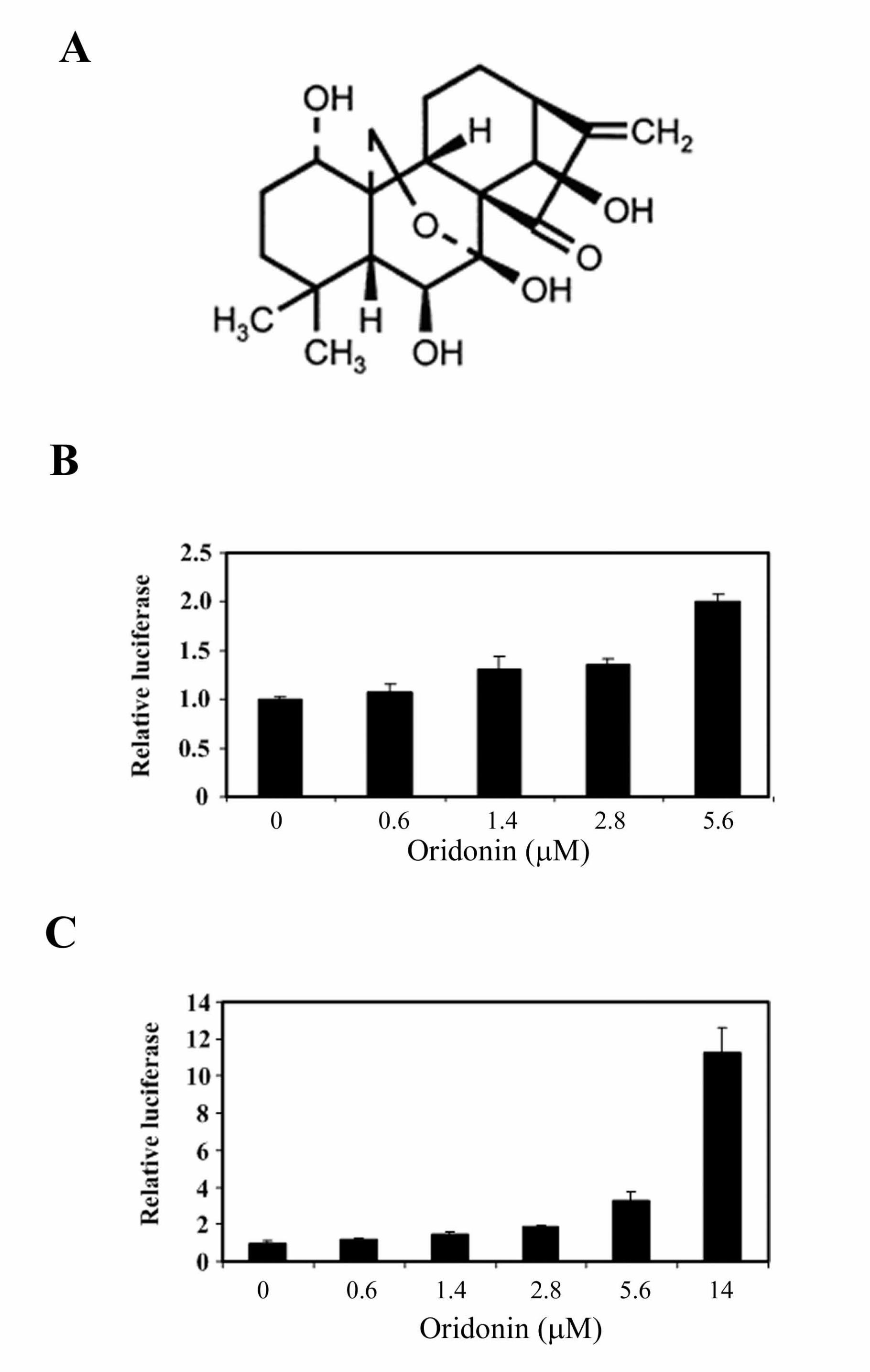
Figure 1. (A) Structure of the diterpenoid oridonin. (B) Identification of oridonin as
an Nrf2 activator using a high-throughput screening system. The stable MDA-
MB-231 cells expressing ARE-luciferase were seeded in 96 well plates. Cells
were grown to 90% confluence and treated with the indicated doses of oridonin
for 24 h before analysis of the luciferase activity. (C) MDA-MB-231 cells were
cotransfected with a plasmid containing an ARE-luciferase reporter gene and a
plasmid encoding renilla luciferase driven by the herpes simplex virus thymidine
kinase promoter. The transfected cells were treated with the indicated doses of
oridonin for 24 h prior to measurement of firefly and renilla luciferase activities in
cell lysates. All luciferase reporter gene assays were run in triplicate and
expressed as mean ± SD.
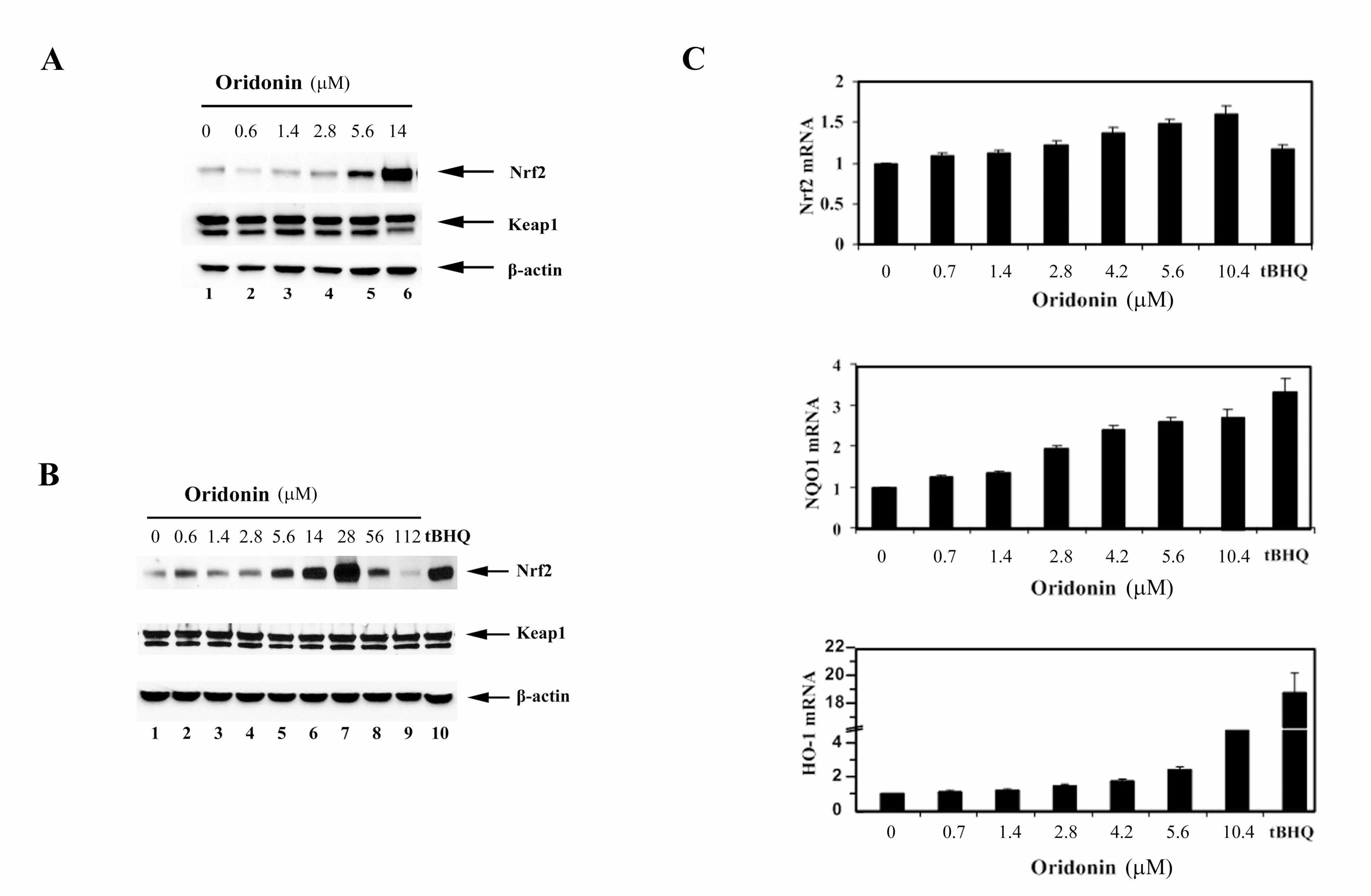
Figure 2. (A) An aliquot of cell lysates from the dual luciferase reporter gene
assay was used for immunoblot analysis for detection of Nrf2, Keap1, and β-
actin. (B) MDA-MB-231 cells were treated with the indicated doses of oridonin for
24 h. Total cell lysates were subjected to immunoblot analysis with anti-Nrf2,
anti-Keap1, and anti-β-actin antibodies. (C) mRNA from similarly treated cells
was extracted and reverse transcribed into cDNA prior to real-time PCR analysis
for detection of Nrf2, NQO1, and HO-1.
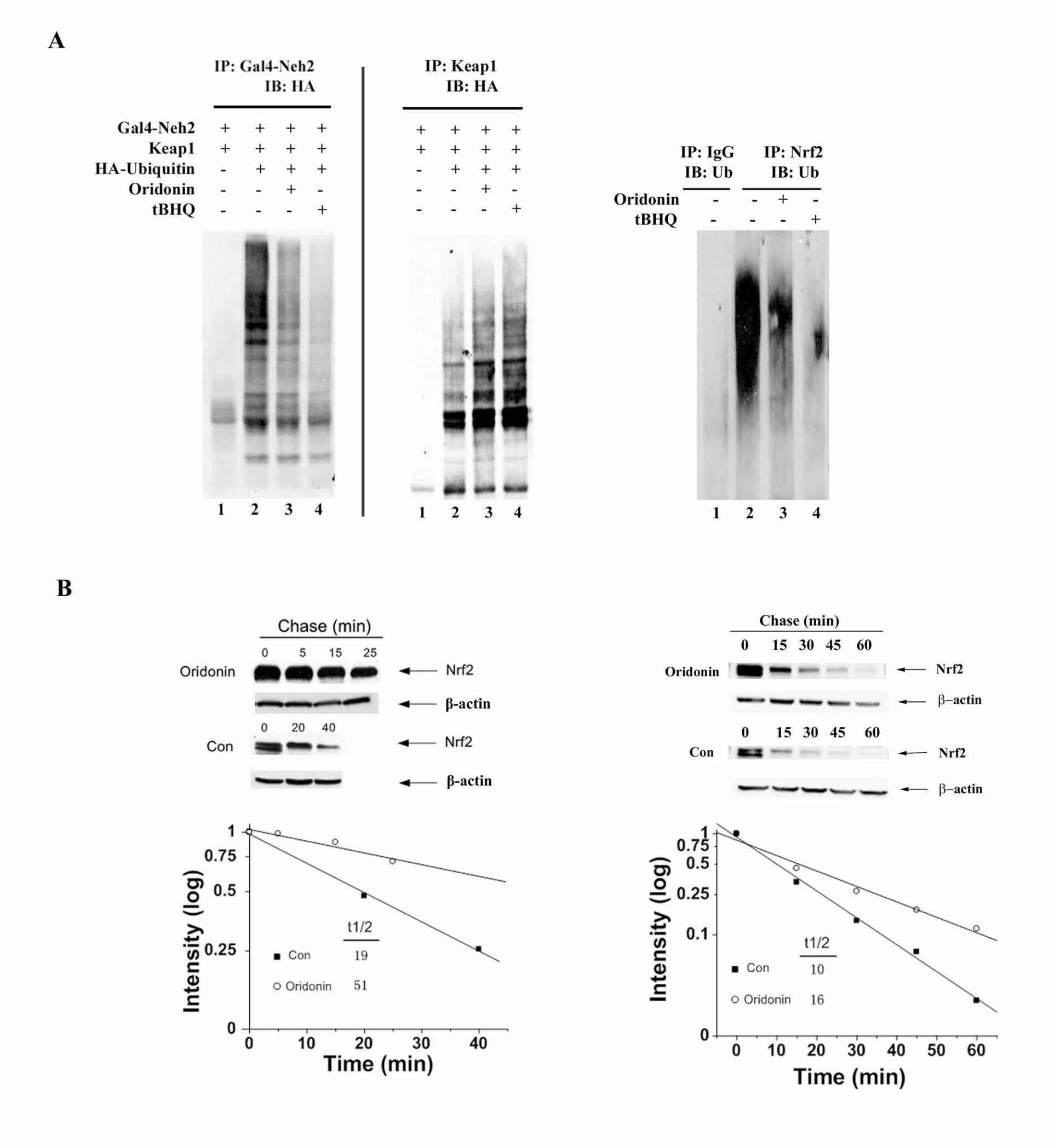
Figure 3. (A) MDA-MB-231 cells were cotransfected with expression vectors for
HA-ubiquitin, a Gal4-Neh2 fusion protein, and Keap1. The transfected cells were
34
left untreated or treated with 8.4 µM oridonin or 100 µM tBHQ for 4 h, along with
10 µM MG132. Cells were lysed in 2% SDS and immediately heated. Anti-Gal4
or anti-Keap1 immunoprecipitates were analyzed by immunoblot with anti-HA
antibodies for detection of the ubiquitin conjugated Neh2 or Keap1 (right panel).
Ubiquitination of endogenous Nrf2 was assessed in UROtsa cells treated with
DMSO, 8.4 µM oridonin or 100 µM tBHQ for 4 h, along with 10 µM MG132. Nrf2
was immunoprecipitated with an anti-Nrf2 antibody and ubiquitinated Nrf2 was
detected with an anti-ubiquitin antibody (left panel). (B) MDA-MB-231 cells (left
panel) and UROtsa cells (right panel) were either left untreated or treated with
8.4 µM oridonin for 4h. Cycloheximide (50 µM) was added to block protein
synthesis. Cells were lysed at the indicated time points and cell lysates were
subjected for immunoblot analysis with anti-Nrf2 and anti-β-actin antibodies. The
intensity of the bands was quantified using Quantity One software.
Figure 4. (A) UROtsa cells were treated with the indicated doses of oridonin for
24 h. Cell lysates were collected and subjected to immunoblot analysis with anti-
Nrf2, anti-Keap1, and anti-β-actin antibodies. (B) Intracellular glutathione
concentrations in UROtsa cells untreated or treated with 1.4 µM oridonin were
measured using the QuantiChrom glutathione assay kit. The experiment was
conducted in triplicate and expressed as mean ± SD. ** p<0.01. (C) UROtsa cells
were left untreated or pretreated with several doses of oridonin for 24 h. Then the
non-pretreated cells or the pretreated cells were further treated with As(III) or
As(III) plus oridonin respectively for another 24 h, followed by measurement of
35
ROS by dichlorofluorescein/flow cytometry. The experiment was run in triplicate
and mean ± SD was calculated. ** p<0.01. (D) UROtsa cells were left untreated
or pretreated with 1.4 µM oridonin for 24 h. Cells were then treated with the
indicated doses of As(III) in the absence (solid curve) or presence of 1.4 µM
oridonin (dashed curve) for another 48 h. Cell survival was measured by the MTT
assay (top panel). UROtsa cells were transfected with control siRNA or Nrf2-
siRNA for 48 h. Nrf2 protein levels were assessed by immunoblot analysis with
an anti-Nrf2 antibody to confirm knockdown of Nrf2 expression (small panel). The
Nrf2-siRNA transfected cells at 48 h post-transfection were used for the MTT
assay as described (middle panel). Two-hundred cells in 35 mm plates were
pretreated and cotreated in the same way as the MTT assay. Cell survival was
measured by the colony formation assay (bottom panel). All the experiments
were run in triplicate and mean ± SD was calculated. * p<0.05. (E) UROtsa cells
were pretreated and cotreated as described in the MTT assay. Apoptotic cell
death was detected using Annexin V-FITC and flow cytometry method. The
experiment was run in triplicate and mean ± SD was calculated. ** p<0.01. (upper
panel). UROtsa cells growing on cover slides were pretreated and cotreated in
the same way. Apoptotic cells were visualized by condensed nuclei using
Hoechst staining. Scale bars represent 25µm. The experiment was repeated and
similar results were obtained.


![Oridonin protects LPS-induced acute lung injury by ......and acute lung injury (ALI) [1, 2]. Lipopolysaccharide (LPS), from the outer membrane of gram-negative bacteria, has been widely](https://img.pdfslide.net/doc/110x75/608e9a4b0654131b49646243/oridonin-protects-lps-induced-acute-lung-injury-by-and-acute-lung-injury.jpg)