Embed Size (px)
Citation preview
Environmental Mercury Exposure and the Risk of Autism
SafeMinds, 2008 - Dan R Laks, M.S.
Environmental Mercury Exposure and Risk of Autism
Coalition for SafeMinds 1
Environmental Mercury Exposure and the Risk of Autism
SafeMinds, 2008 - Dan R Laks, M.S.
Section
1. Introduction 3
Page
1.1 Purpose 3
1.2 Mercury and Autism 3
2. Background 5
2.1 Source and Exposure 5
2.2 Organic Mercury 6
2.3 Elemental Mercury 8
2.4 Estimated Exposure 10
2.5 Recommended Exposure 12
2.6 Biotransformation 13
2.7 Enterohepatic Circulation 14
3. Toxicology 15
3.1 Cellular Toxicity 15
3.2 Oxidative Stress 16
4. Mechanism for Disease 16
4.1 Health Risks 16
4.2 Inflammation and Neurogenesis 17
4.3 Acetylcholine 18
4.4 Adrenal 18
4.5 Pituitary 19
Environmental Mercury Exposure and Risk of Autism
Coalition for SafeMinds 2
4.6 Immune Response 20
4.7 Autoimmune Response 20
4.8 Susceptible Subpopulation 21
4.9 Liver 22
4.10 Autocatalytic Origin of Disease 22
5. Policy 23
5.1 Policy Review 23
5.2 Recommendations 26
6. Conclusions 26
7. References 27
Environmental Mercury Exposure and Risk of Autism
Coalition for SafeMinds 3
Keywords and Abbreviations: Autism , environmental mercury exposure, inorganic mercury (I-Hg), methyl mercury (CH3
1.
Hg), total mercury (T-Hg), mercuric ions (Hg++), elemental mercury (Hg), National Health and Nutrition Survey (NHANES), Environmental Protection Agency (EPA).
1.1 Purpose:
Introduction
The purpose of this paper is to review the scientific evidence that links environmental mercury exposure and the risk of autism.
1.2 Mercury and Autism:
In 1968, both Sweden and Japan restricted mercury containing pesticides and Nature published this scientific opinion; “We feel that the example set by these two countries should be followed elsewhere before concentrations of mercury reach a point where methyl mercury is being titrated in humans as well as fish” [128]. Now, forty years after the warning in Nature, mercury in the blood of the U.S. population is being titrated. Elevated blood mercury levels, beyond what was considered “without increased risk of adverse neurodevelopmental effects associated with methyl mercury exposure”, were found in eight percent of the NHANES American survey population (1999-2000)[73]. This study estimated that due to mercury exposure, 300,000 to 600,000 American children would be born with elevated risks of neurodevelopmental disorders during those years.
At this time, a scientific consensus agrees that the global rate of atmospheric mercury deposition may be increasing over time [1]. The latest studies suggest that as the rate of global mercury deposition increases, the incidence of the most closely associated neurodegenerative diseases such as autism are rising as well [25]. Recent studies suggest that the incidence of autism may be rising in heavily industrialized countries around the world, in regions where unprecedented mercury levels have recently been found in women and children, fish and animals, rice and soil [60]. These global trends support the theory that both chronic mercury exposure and risks of associated, neurodegenerative disease may rise within the general U.S. population. In fact, a recent biomonitoring study of 1,811 residents in New York City found that one in four adults had mercury concentrations above reportable (safe) levels [80]. The mercury concentrations found in the New York study were three times the national average. These results are consistent with studies from around the world that are reporting elevated mercury levels within human populations [41,46,80,98].
In his paper “Autistic Disturbances of Affective Contact”, published in 1943, Dr. Leo Kanner first described “early infantile autism” as “the children’s inability to relate themselves in the ordinary way” [66,70]. Autism is a heterogenous, neurodevelopmental disorder with onset prior to 3 years of age. The symptoms include deficits in social interaction, speech, and behavior. Over the past two decades, the incidence of autism has increased ten-fold. While the etiology of the disease is
Environmental Mercury Exposure and Risk of Autism
Coalition for SafeMinds 4
unknown, many believe that it is caused by both environmental and genetic features. The Collaborative on Health and the Environment recently published a consensus statement linking environmental agents with neurodevelopmental disorders (http://www.iceh.org/LDDI.html).
With a comprehensive review of symptoms, traits, biological signs, and population characteristics of autism in comparison to those of mercury exposure, Bernard et al. propose a hypothesis that autism is a “novel form of mercury poisoning”[14]. The similarities between the two syndromes are comprehensive in both behavioral traits and biological symptoms. Mercury can induce all three of the autism diagnostic criteria: impairments in social interaction, communication difficulties, and repetitive and stereotyped patterns [15]. In fact, the Bernard group demonstrates that “every major characteristic of autism has been exhibited in at least several cases of documented mercury poisoning.” Mercury exposure and autism share symptoms such as sensory abnormalities, emotional changes, movement disorder, impaired thinking, severe sleep disturbances, self injurious behavior, gastrointestinal problems, autonomic nervous system disturbance, immune system alterations, irregularities in neurotransmitter systems, and non-specific brain lesions. Based on a thorough correspondence of defining characteristics, the Bernard group hypothesizes that the regressive form of autism is caused by mercury exposure.
Since the publication of the Bernard paper, there have been numerous studies which support the hypothesis that mercury exposure causes autism. In a retrospective study of 269 subjects, a specific biomarker for heavy metal toxicity, precoproporphyrin, was found to be significantly elevated in autistic patients relative to controls [85]. After application of a heavy metal chelator, to remove heavy metals, the levels of the porphyrin biomarkers subsided. This study gives compelling evidence that environmental heavy metal exposure is associated with autism in a causal relationship. In two other studies, these results were repeated [49,50]. In a prospective study, mercury toxicity and elevated urinary precoproporphyrin levels were again associated with autistic children as compared to controls [50]. The autistic children had two to three fold increases in these urinary biomarkers for mercury exposure relative to controls. And again, after chelation of heavy metals, these porphyrin levels subsided.
Perhaps an even more direct correlation is displayed in a study of blood mercury levels in autistic children which found an association between elevated blood mercury and a diagnosis of autism at children’s age averaging seven years old [39]. Because of the late age of these Autistic children, well past the onset of the disease, these results point to an impaired excretion of mercury as the source of their elevated blood levels. The correlation of elevated mercury body burden levels with the diagnosis of autism is reiterated in a study of baby teeth [4]. In this study, mercury in baby’s teeth represented cumulative exposure during the critical years of development as teeth are formed in utero and during the first years of life. Children with autism had 2.1 fold higher mean levels of mercury and 3.1 fold higher median levels of mercury in their teeth as compared to controls but similar levels of lead and zinc. These results suggest to the authors that the autistic patients had higher body burdens of mercury during several years of early development that may have produced the development of the disease. Mounting evidence
Environmental Mercury Exposure and Risk of Autism
Coalition for SafeMinds 5
points to a causal role for mercury in the development of autism. In a case series, exposure to mercury from vaccines preceded the development of autism and produced a dose response correlation between the severity of developmental symptoms and the amount of mercury exposure[47]. The severity of the disease was determined by how early in life the mercury exposure began and by how much total mercury exposure the patient incurred.
It was estimated that between 300,000 and 600,000 children each year are born with cord blood mercury concentrations above 5.8 ug/L; this a level associated in other studies with loss of IQ[113]. This estimation was used to calculate the resulting loss of productivity from these children and it amounted to roughly 9 billion dollars annually. Of this total, 1.3 billion dollars was attributable to U.S. power plants that emit mercury, primarily from the burning of coal. If the burning of coal increases, it is reasonable to assume that environmental mercury exposure and associated diseases will rise as well. Blood inorganic mercury concentration may be a good biomarker for chronic mercury exposure [116,117]. In the National Health and Nutrition Survey (NHANES), there appears a time dependent rise in the proportion of blood, inorganic mercury detection (values above the limit of detection). While there was only 3% of the NHANES population with detectable blood inorganic mercury (above the limit of detection) in 1999-2000, there was 6% of the NHANES population with detectable blood inorganic mercury (above the limit of detection) in 2001-2002. Whether this change in I-Hg detection was due to geographical differences in exposure, measurement error, or to a time dependent rise in the rate of chronic mercury exposure is unclear. While these observations do not verify a time dependent increase in chronic mercury exposure and deposition, they do support this possibility.
With sufficient evidence linking mercury exposure and autism, concerned scientists should ask whether rising rates of global atmospheric mercury deposition and chronic environmental mercury exposure are contributing to the rising rates of autism.
2.
2.1 Source and Exposure:
Background
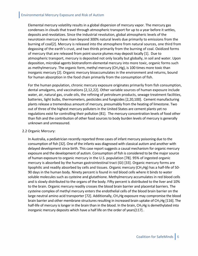
Mercury, commonly known as quicksilver, is the only metal that is liquid at room temperature. The element is volatile and vaporizes into a monatomic, mercury gas. On the basis of toxicological characteristics, there are three forms of mercury, elemental (Hg, metal and vapor), inorganic mercury (mercurous and mercuric ions) and organic compounds (methylmercury, dimethyl mercury, and ethylmercury found in thimerosal, a vaccine preservative).
CH3 CH3Hg ↔ CH3
Dimethyl Mercury Methyl Mercury Mercuric Ions Elemental (atomic) Mercury
Hg ↔ Hg++ ↔ Hg
Lipid soluble Lipid Soluble Water Soluble Liquid Metal/ Lipid Soluble Gas
Environmental Mercury Exposure and Risk of Autism
Coalition for SafeMinds 6
Elemental mercury volatility results in a global dispersion of mercury vapor. The mercury gas condenses in clouds that travel through atmospheric transport for up to a year before it settles, deposits and revolatizes. Since the industrial revolution, global atmospheric levels of the neurotoxin mercury have risen beyond 300% natural levels due primarily to emissions from the burning of coal[2]. Mercury is released into the atmosphere from natural sources, one third from degassing of the earth’s crust, and two thirds primarily from the burning of coal. Oxidized forms of mercury that are released from point source plumes may deposit locally [1]. Due to atmospheric transport, mercury is deposited not only locally but globally, in soil and water. Upon deposition, microbial agents biotransform elemental mercury into more toxic, organic forms such as methylmercury. The organic form, methyl mercury (CH3
For the human population, chronic mercury exposure originates primarily from fish consumption, dental amalgams, and vaccinations [2,12,22]. Other variable sources of human exposure include water, air, natural gas, crude oils, the refining of petroleum products, sewage treatment facilities, batteries, light bulbs, thermometers, pesticides and fungicides [2,20,100]. Cement manufacturing plants release a tremendous amount of mercury, presumably from the heating of limestone. Two out of three of the highest mercury polluters in the United States are cement plants yet no regulations exist for controlling their pollution [81]. The mercury concentration levels of food other than fish and the contribution of other food sources to body burden levels of mercury is generally unknown and unmeasured.
Hg), is 100 times more toxic than inorganic mercury [2]. Organic mercury bioaccumulates in the environment and returns, bound for human absorption in the food chain primarily from the consumption of fish.
2.2 Organic Mercury:
In Australia, a pediatrician recently reported three cases of infant mercury poisoning due to the consumption of fish [32]. One of the infants was diagnosed with classical autism and another with delayed development since birth. This case report suggests a causal mechanism for organic mercury exposure and the development of autism. Consumption of fish is considered to be the major source of human exposure to organic mercury in the U.S. population [78]. 95% of ingested organic mercury is absorbed by the human gastrointestinal tract (GI) [33]. Organic mercury forms are lipophilic and readily absorbed by cells and tissues. Organic mercury (CH3Hg) has a half-life of 50-90 days in the human body. Ninety percent is found in red blood cells where it binds to water soluble molecules such as cysteine and glutathione. Methylmercury accumulates in red blood cells and is slowly distributed to the organs of the body. Fifty percent is distributed to the liver and 10% to the brain. Organic mercury readily crosses the blood brain barrier and placental barriers. The cysteine complex of methyl mercury enters the endothelial cells of the blood brain barrier on the large neutral amino acid transporter [72]. Additionally, CH3Hg exposure may compromise the blood brain barrier and other membrane structures resulting in increased brain uptake of CH3Hg [116]. The half-life of mercury is longer in the brain than in the blood. In the brain, CH3Hg is demethylated into inorganic mercury deposits which have a half life on the order of years[117] .
Environmental Mercury Exposure and Risk of Autism
Coalition for SafeMinds 7
In studies to date, fish consumption is consistently a strong predictor of organic mercury concentration in the human body [36,46,53,80,98,115]. Maternal fish consumption contributes organic mercury to the fetus and cord blood levels may be twice those of maternal levels [7,115]. Several large epidemiological studies have found associations between prenatal mercury exposure and later cognitive development.
The Seychelles Child Development Study was “designed to study the developmental effects of prenatal MeHg exposure in a fish eating population” [36]. 779 infant-mother pairs were enrolled in the study and maternal hair mercury was measured to represent prenatal mercury exposure levels. Secondary analysis of the Seychelles cohort of children who were prenatally exposed to methylmercury from mother’s fish consumption found a delayed adverse effect on the children’s neurodevelopment at maternal hair mercury concentrations above 10-12 ppm.
In a similar, but more robust study on a cohort of 917 children from the Faroe islands, results showed cord blood mercury concentrations were associated with later childhood deficits in language, attention, and memory [53]. In addition, maternal hair mercury levels at the time of birth were inversely associated with the eventual IQ of the offspring [8]. This study suggested that the greatest susceptibility to exposure occurred at late gestation. The study concluded that “associations between prenatal mercury exposure and neurobehavioral decrements at age 7 years are likely to be causal.”
In a prospective cohort study of 135 mother-infant pairs designed to investigate maternal fish consumption during pregnancy and infant cognition, the benefits of fish consumption had positive effects on infant cognition while relatively higher mercury levels (as measured in maternal hair), had negative effects on infant cognition [87]. Significantly, the negative effects on infant cognition of mercury levels were still at low, chronic mercury exposure levels. The conclusions drawn by the author were that pregnant women should continue to eat varieties of fish that were low in mercury.
Without proper testing and consistent monitoring of mercury levels in fish, it is nearly impossible to ascertain which fish are safe to eat. The U.S. Environmental Protection Agency (EPA) has lowered their advisory on “acceptable” levels of mercury exposure and advises pregnant mothers against eating more than three fish a month for fear of neurological effects on the unborn child. In the U.S., geographic variability in the mercury concentrations of fish was found to affect the variability of mercury exposure between populations [108]. Another study showed that in fish from lakes around Western America, mercury concentration was directly associated with the atmospheric transport and deposition of mercury vapor [93]. Therefore, if global deposition of atmospheric mercury is increasing, it is reasonable to assume that human exposure from fish consumption will increase as well.
Recent reports from China indicate that rice bioaccumulates methylmercury as well [26,96]. Contaminated rice, when fed to rats, increased biomarkers for apoptosis (cell death) in areas associated with memory and learning. Perhaps it is this contaminated Chinese rice which
Environmental Mercury Exposure and Risk of Autism
Coalition for SafeMinds 8
accounts for the particularly high mercury levels found in Chinese Americans in the New York biomonitoring study [80] and in the rice eating population of Tehran [41].
Elimination of organic mercury from the human body is dependent upon gastrointestinal microflora. The excretion of organic mercury is almost exclusively through the gastrointestinal (GI) tract and is dependent on GI microbial flora populations. Blood mercury passes through the liver to the bile and returns into the GI tract. Under healthy conditions, flora (microbes) in the GI tract demethylate organic mercury to the poorly absorbed, water soluble, inorganic mercury, which is excreted in feces. Approximately 1% of the human body burden of CH3
The infant and developing fetus are particularly susceptible to the effects of mercury and face chronic mercury exposure from the mother, mother’s diet, and mother’s dental amalgams. After birth, the infant is further stressed due to the entire program of vaccines. Thimerosal, a vaccine preservative containing the organic mercury, ethylmercury, accounts for an estimated 100% increase in additional infant mercury exposure. Mercury from vaccine preservative is injected directly into the blood stream and not subject to excretion by the gut. Before the removal of thimerosal from most vaccines in the United States, the usual program of vaccinations resulted in infant mercury exposure that surpassed levels considered without health risks by the EPA [29]. Thimerosal is still used in flu vaccines in the United States and in routine vaccinations in developing countries worldwide at the recommendation of the World Health Organization.
Hg is excreted daily in feces. Much of the organic mercury in bile is bound to glutathione and cysteine and is reabsorbed by the gut in a process of enterohepatic circulation [13]. Organic mercury is also excreted through breast milk and passes from mothers to infants in that manner.
2.3 Elemental Mercury:
Elemental mercury exposure is primarily from dental amalgams containing elemental mercury. Elemental mercury in vapor form is lipophilic and 80% of inhaled elemental mercury is readily absorbed in the respiratory tract. Inhaled mercury vapor readily diffuses across the alveolar membranes and has an affinity for red blood cells. The vapor has high mobility and diffuses rapidly throughout the body and brain as a monatomic gas. Elemental mercury from the air is oxidized by the enzyme catalase to inorganic mercury ions [28]. But it still has time to pass through the placental and blood brain barrier before it deposits in tissue as inorganic ions. The half-life of elemental mercury in the human body is 45 days. Deposition occurs primarily in the kidney following inhalation [22]. Elemental mercury is excreted in exhaled air, sweat, saliva and as mercuric ions in feces and urine.
The mercury vapor released from dental amalgams is presumed to account for the majority of mercury detected in human urine [2]. The average daily uptake from inhalation of dental amalgams is estimated at 2 ug [71]. Studies on children have found both a moderate correlation between amalgams and urinary mercury [77] and a high correlation [129]. Presumably, some of the inorganic mercury excreted from urine is also as a result of the demethylation of organic mercury in tissues of the body. Inorganic mercury levels in the placenta are correlated with the mother’s amalgams [7].
Environmental Mercury Exposure and Risk of Autism
Coalition for SafeMinds 9
Mercury in both cord blood and maternal blood were correlated with the number of mother’s amalgams [115]. In a retrospective study with the largest cohort used to study dental amalgams (20,000 people from New Zealand), a small but significant association was found between amalgam surface and the risk of multiple sclerosis [12].
Global anthropogenic emissions of mercury are estimated to range between 2000 and 6000 metric tons per year (2,200 -6,600 tons). China alone is believed to emit about 1000 tons of mercury annually. In comparison, U.S. anthropogenic mercury emissions are estimated to be about 158 tons per year. Due to atmospheric transport of elemental mercury vapor, a major source of atmospheric mercury that deposits in California originates from Chinese coal burning plants [95]. Between 1995 and 2003, emissions from China increased at roughly 3% annually [131]. Could rising levels of airborne mercury indicate rising levels of associated diseases such as autism?
Recent studies have described the impact of airborne environmental mercury concentration on human population autism rates. In a case control analysis in California, results from a semi-ecologic study of 284 children with autism showed an association between autism prevalence and estimated metal concentrations in ambient air [127]. The first investigation to report an ecological association between autism and environmentally released mercury was by the Palmer group in San Antonio, at the University of Texas Health Science Center. In an ecological study of 1184 school districts in Texas representing approximately 4 million children, this study found that for every 1000lb of environmentally released mercury, there was a 61% increase in the rate of autism observed [88]. A more recent study by the Palmer group examined mercury-release data from 39 coal-fired power plants, 56 industrial facilities in Texas, and autism rates from 1,040 Texas school districts. This study showed for the first time that distance to sources of environmental mercury release was a significant predictor of autism risk [89]. In light of these studies, and as global emissions of mercury are increasing and the rate of atmospheric mercury deposition is increasing, it is logical to assume that rates of autism will increase as well.
The severity of airborne exposure to mercury is brought to light when one considers the amount of mercury that may be inhaled with proximity to point source plumes. In a comparison of urban versus rural air mercury measurements, the urban air mercury concentration in Minneapolis was 150% greater than rural levels [104]. In another study of air mercury concentration in a residential neighborhood of East St. Louis, impacted by industrial source plumes, the measurements of elemental mercury in the air were 100 times greater than the previously mentioned levels in urban Minneapolis [74]. Taking the worst case scenario of the residential neighborhood in East St. Louis, impacted by plumes of emissions, we can calculate the amount of Hg a person would breathe. An average human breathes in 8-20 liters/minute which averages to 20, 160 liters/day or 7 x 10^6 liters/year. The measurement in East St. Louis was 235 x 10^-9 g Hg/m^3. 1 m^3 = 1000 L. 7x10^6 liters of air breathed/year x 235 x 10^-12 g Hg/ Liter = 1.6 mg Hg/year of average inhalation. The amount absorbed would be 1.6 mg Hg/ year * fraction absorbed (fa). The fraction absorbed will vary from person to person but should be close to 0.8 considering that elemental mercury is
Environmental Mercury Exposure and Risk of Autism
Coalition for SafeMinds 10
lipophilic and 80% crosses into the respiratory tract. Therefore 1.28 mg Hg/ year will be breathed in from direct exposure to the plumes in East St. Louis. That is 3.5 ug/day. The average age of childbirth is 25 years. By age 25, 32 milligrams of Hg have been inhaled by an expectant mother from the plumes of East St. Louis. The dangers of being in the path of plumes of emissions greatly enhance the risk of mercury exposure and the risks of associated disease. Of course most people are not in the direct pathway of plumes. Yet, even the mercury air concentrations taken from the urban site in Minneapolis deliver a fair amount of inhaled elemental mercury; 2.5 x 10^-12 g Hg/Liter * 7 x 10^6 liters breathed/year * 0.80 Hg absorbed/Hg inhaled= 14.4 ug absorbed /year (or 0.039 ug/day). The variability between air concentrations in and out of the plumes is dramatic and that underlies the dramatic differences in air exposure dependent upon geographic location. As the rate of global mercury emissions increase, associated risks of direct exposure to plumes increase as well. These measurements of airborne elemental mercury concentrations elucidate the findings by the Palmer group that proved distance to sources of environmental mercury release was a significant predictor of autism risk [89]. Indeed, these air measurements show how distance from the point source of airborne plumes can increase elemental mercury exposure by close to one hundred fold.
2.4 Estimated Exposure:
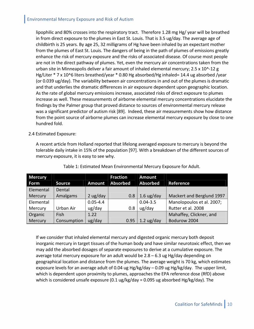
A recent article from Holland reported that lifelong averaged exposure to mercury is beyond the tolerable daily intake in 15% of the population [97]. With a breakdown of the different sources of mercury exposure, it is easy to see why.
Table 1: Estimated Mean Environmental Mercury Exposure for Adult.
Mercury Form Source Amount
Fraction Absorbed
Amount Absorbed Reference
Elemental Mercury
Dental Amalgams 2 ug/day 0.8 1.6 ug/day Mackert and Berglund 1997
Elemental Mercury Urban Air
0.05-4.4 ug/day 0.8
0.04-3.5 ug/day
Manolopoulos et al. 2007; Rutter et al. 2008
Organic Mercury
Fish Consumption
1.22 ug/day 0.95 1.2 ug/day
Mahaffey, Clickner, and Bodurow 2004
If we consider that inhaled elemental mercury and digested organic mercury both deposit inorganic mercury in target tissues of the human body and have similar neurotoxic effect, then we may add the absorbed dosages of separate exposures to derive at a cumulative exposure. The average total mercury exposure for an adult would be 2.8 – 6.3 ug Hg/day depending on geographical location and distance from the plumes. The average weight is 70 kg, which estimates exposure levels for an average adult of 0.04 ug Hg/kg/day – 0.09 ug Hg/kg/day. The upper limit, which is dependent upon proximity to plumes, approaches the EPA reference dose (RfD) above which is considered unsafe exposure (0.1 ug/kg/day = 0.095 ug absorbed Hg/kg/day). The

Environmental Mercury Exposure and Risk of Autism
Coalition for SafeMinds 11
previously reported exposure levels were only average levels and do not describe the elevated levels found in the most at risk subpopulations that eat more fish and have more dental amalgams. For the fifty percent of people above the median exposure, the chronic exposure levels may easily stretch beyond the RfD.
Although infants don’t have dental amalgams and may not eat fish, their mothers do, and they pass on their exposures to the child through breast milk [6,54,75]. This exposure to organic mercury through breast milk may be the dominant route of exposure for the infant if one does not consider thimerosal containing vaccines [6]. One study estimated that breast milk accounted for 96-99% of the total infant environmental mercury exposure, excluding vaccines [27]. Thus, cumulative exposure of the child may mirror that of the parent, yet the child weighs less and has not developed the biliary excretion or the intestinal microflora necessary to excrete the mercury load.
Table 2: Estimated Mean Environmental Mercury Exposure for Neonatal Infant.
Mercury Form
Source
Amount
Fraction Absorbed
Amount Absorbed
Reference
Organic Mercury
Breast Milk 0.16 ug/kg/day
0.95 0.15 ug/kg/day Amin-Zaki et al. 1981; Marques, Dorea, Fonseca et al. 2007
Elemental Mercury Urban Air
0.00023-0.023 ug/kg/day 0.8
0.00019-0.019 ug/kg/day
Manolopoulos et al. 2007; Rutter et al. 2008
In an estimation based on world data of human milk mercury concentrations, the world, median mean mercury exposure from breast milk was estimated at 0.266 ug Hg/kg/day [76]. Another study on Iraqi suckling infants estimated that sixty percent of the mercury found in breast milk was organic mercury (MeHg) and the other forty percent is mostly poorly absorbed ionic, inorganic mercury [6]. If we combine these estimates, 0.266 ug Hg/kg/day * 0.60 = 0.16 ug MeHg/kg/day. The absorbed dose would be 0.16 ug MeHg/kg/day * 0.95 (fraction absorbed) = 0.15 ug MeHg/kg/day. This amount of organic mercury is already above the EPA RfD of 0.1 ug/kg/day without taking into account the amount of prenatal mercury exposure, elemental mercury inhaled by the infant from the air, or the amount of mercury absorbed from thimerosal containing vaccines.
A neonate breathes 8 cc/kg/breath. 8 cc/kg/breath * 3 kg (average weight) * 24 breaths/minute * 60 minutes/hr * 24 hrs/day = 830 liters/day. In a region close to the plumes of a point source, the amount breathed in would be 830 * 253 x10^-12 gHg/Liter = 0.21 ug Hg/day= 0.07 ug Hg/day. The amount absorbed would be 0.07 ug Hg/day * 0.80 (fraction absorbed) = 0.056 ug Hg/day or divided by three Kg (average weight) = 0.019 ug Hg/kg/day. As a neonate does not breathe a considerable volume of air, even in the worst scenario, close to the plumes of a point source, the amount of inhaled mercury is below the EPA’s RfD (0.1 ug/kg/day). In regular urban air, the amount breathed in by a neonate would be 0.00019 ug Hg/kg/day which is almost insignificant when compared to
Environmental Mercury Exposure and Risk of Autism
Coalition for SafeMinds 12
the average amount of mercury contributed by breast feeding. If we sum the environmental mercury exposures, an urban infant is at risk from 0.15 ug Hg/kg/day– 0.17 ug Hg/kg/day depending on the distance from the point source plumes . In both cases, the breast milk contributes the vast majority of mercury. However, mother’s milk mercury concentration may be dependent on distance from point source plumes. Although this estimation leaves out the considerable dose of organic mercury delivered by Thimerosal preservative in vaccines, the high amounts of environmental mercury exposure from other sources define a preexisting risk of mercury exposure that would suggest added mercury exposure from thimerosal will aggravate and almost double an already elevated risk of mercury associated disease.
There is considerable variability around the world mean value of infant exposure. In a cohort from Saudis Arabia, mean breast milk mercury concentration was sixty three percent above the world median mean value [5]. In a cohort of fish dependent mothers in the Faroe islands, breast feeding was associated with less benefits on neurobehavioral development than in previously published studies [65]. The implication is that elevated mercury concentrations in breast milk reduce the beneficial effects of breast feeding on an infant’s neurobehavioral development.
For adults, the data suggests that cumulative exposure to environmental mercury results in a substantial subpopulation that is at risk for associated diseases. The risk posed by environmental mercury exposure is related to the distance from point sources, as our calculations and other studies have shown [89,127]. Neonates are particularly susceptible as their ability to excrete mercury is not developed and they inherit their mother’s mercury body burden primarily through breast milk.
2.5 Recommended Exposure:
A dose response relationship analysis of methyl mercury and cases of poisoning reveals a toxic threshold in blood mercury levels that triggers disease response in acute exposure [9]. With rising baseline levels of chronic mercury exposure, the toxic threshold for poisoning may decrease. The Swedish Expert Group (1971) calculated the average long-term daily intake of methyl mercury associated with adverse health effects at 4.3 ug/kg/day or a steady state blood level of 200ug/L. The EPA’s RfD (recommended safe dose) for ingested CH3
Hg is 0.1 ug/kg/day which was reduced from 0.5 ug.kg/day due to increased concerns over adverse health risks [33]. The RfD estimation assumed a maternal blood to cord blood ratio of 1 however many studies have found variability in this ratio with ratios closer to 2:1, cord blood: maternal blood [107]. This discrepancy would change the estimated safe dose. Other scientists have argued that there “does not appear to be a threshold for adverse neuropsychological effects based on available data” [102]. Rice argues that there is evidence even moderate levels of mercury exposure may result in delayed neurotoxicity years or decades later. This argument leads to the conclusion that there may be no safe or tolerable levels of mercury exposure.
Environmental Mercury Exposure and Risk of Autism
Coalition for SafeMinds 13
2.6 Biotransformation:
Under normal conditions, the GI tract that initially absorbs digested, organic mercury, ultimately excretes the vast majority (95%) of organic mercury. In the body, absorbed methyl mercury travels from the blood to the liver and through the bile duct in return to the GI tract. Demethylation of Methyl Mercury to water soluble, mercuric ions (Hg++) is a necessary step for excretion from the gastrointestinal tract (GI) and kidney [2]. In human tissue and organs, this demethylation process, crucial to excretion, also results in the accumulation of inorganic mercury in the brain [43]. In the brain, inorganic mercury deposits play an important role in mercury neurotoxicity. While organic mercury has a half life in the brain of approximately one month, inorganic mercury deposits have a half life on the order of years [116,117]. Animal studies on monkeys reveal that methylmercury is demethylated into inorganic mercury in the brain and other tissues upon long term, chronic exposure [101]. As a result of inorganic mercury deposition, the percentage of inorganic mercury relative to total mercury concentration in the brain increases after long term exposure to organic mercury [117]. Because of tissue demethylation of CH3
Scientific studies provide evidence that organic forms of mercury and even dimethylmercury may be produced in the human GI tract by sulfate reducing bacteria (SRB) and methanogenic bacteria. The sulfate reducing bacteria, desulfovibrio, which actively methylates mercury, has been found both in the human colon and in the periodontal pocket of the human mouth in 58% of study subjects [119,126]. Desulfovibrio are ecologically the most significant group of sulfate reducing bacteria (SRB) in the human colon. These flora exist as 66% of SRB in the healthy gut and 92% in persons with ulcerative colitis. SRB may methylate mercurous ions into the more readily absorbed organic form, CH
Hg and resultant I-Hg deposition in the brain, Rice et al. recommend that “health effects of methylmercury should focus on long-term exposure”[101] . Thus, chronic, environmental mercury exposure and resultant inorganic mercury deposition should be a major focus of research into the toxicological effect of mercury. It may be reasonable to assume that blood inorganic mercury is a reflection of this demethylation process [116]. In which case, blood inorganic mercury should be used as a biomarker of chronic mercury exposure. Instead, most studies to date use organic mercury, a marker for recent exposure, as the biomarker of chronic mercury exposure.
3Hg. SRB have been shown to methylate mercury in dental waste water [134]. In the same manner, they may methylate inorganic mercury in the human gut. One scientist estimates that 9 micrograms of organic mercury may be formed per day in the gut of humans [2]. GI flora populations are variable and change with diet and the environment. SRB are in a competitive relationship with methanogenic flora populations. The availability of dietary sulfates selects for an increase in SRB flora populations. Sulfates are generally released into the environment through industrial pollution or as chemical byproducts. Methyltransferases involved in methionine synthesis may be involved in Hg methylation by the sulfate reducing bacteria, desulfovibrio. The enzyme responsible for methylating Hg in microbes, was found to transfer methyl groups from methyltetrahydrofolate to thiols such as homocysteine. This finding supports the hypothesis that the methylation process in humans may mistakenly methylate mercury rather than of homocysteine.
Environmental Mercury Exposure and Risk of Autism
Coalition for SafeMinds 14
GI flora develop soon after birth but their constituent populations don’t resemble the relatively stable populations of adult flora until age 2-3 years [58]. In suckling animals, GI flora demethylation of organic mercury does not facilitate excretion as it does in the adult; whether this is true in human infants is unknown [28]. During the development of the infant GI microflora, chronic exposure to mercury, or sulfates may select certain types of flora populations to develop. Microbial selection may predispose an infant to increased mercury absorption, impaired excretion, or increased biotransformation of mercury. Increased susceptibility to mercury exposure may occur after the administration of antibiotics as subsequent exposure to mercury or sulfates may select for certain flora populations and determine the rate of mercury excretion. In fact, antibiotic use was correlated with autism subjects who had elevated mercury levels in their baby teeth [4]. Baby teeth are formed in utero and so mercury levels represent cumulative exposure during the critical period of early development. The teeth of children with autism had a 2 fold higher mean levels of mercury as compared to controls (healthy children). While seafood consumption and dental amalgam use was similar between the two groups, the autistic children had significantly higher levels of antibiotic use as compared to controls. The author reasons that antibiotics impair the excretion of mercury from the body by reducing normal gut flora which demethylate organic mercury and increasing E. coli which may methylate inorganic mercury. Thus, by this reasoning, it is the excretion rate of mercury which defines a susceptible subpopulation to mercury exposure.
2.7 Enterohepatic Circulation:
Much of the methylmercury excreted from the liver in bile is reabsorbed in the gut, producing entero-hepatic circulation of methyl mercury. Mercury exposure disturbs GI function [13]. If demethylation rates in the GI tract are reduced, excretion of organic mercury may be impaired and methylmercury may be effectively trapped in enterohepatic circulation and retained by the body in a GI absorption/retention loop. CH3Hg circulating through the body may create an elevated risk of biotransformation to dimethyl mercury, absorption by the brain, and resultant neurodegeneration. In addition, any increase in the half-life of mercury would result in a greater probability of mercury deposition in the brain. Thus the toxic dose of mercury (LD50) may vary within the human population due to variable microflora involved in biotransformation, and excretion of organic mercury. Available levels of cysteine and the rate of glutathione conjugation of mercury in the bile, may directly affect the rates of enterohepatic circulation. Changes in infant cysteine levels may thereby influence the absorption rates of mercury due to enterohepatic circulation. Treatment for overexposure to organic mercury involves interrupting enterohepatic circulation with surgical drainage of the gallbladder or oral administration of a non absorbable thiol resin which binds mercury and enhances intestinal excretion.
Environmental Mercury Exposure and Risk of Autism
Coalition for SafeMinds 15
3.
3.1 Cellular Toxicity:
Toxicology
Studies across many species provide conclusive evidence of chronic organic mercury exposure’s neurotoxic effects on developing organisms; to disrupt proliferation, migration, and differentiation of brain cells [17,90]. In the adult brain, CH3
Acute exposure to mercury triggers a toxic response upon reaching a critical concentration, or threshold. This toxic threshold may vary within populations as baseline mercury concentrations, deposition, and rates of excretion vary within populations. The toxicological effects of mercury on cellular function are widespread and numerous. Mercury has particular affinity to sulfhydryl groups and thiol bonding. As a result, soluble mercury complexes with cysteine, glutathione, and a variety of enzyme systems produce nonspecific cell injury or death. Every protein and amino acid chain in the body is a potential target of mercury.
Hg damage is focal, yet in the developing brain, the damage is more diffuse [33]. In utero mercury exposure may disrupt microtubule assembly and impair cellular migration during brain development. This is consistent with the findings that abnormal regulation of brain growth in autism results in unusual brain growth patterns [34].
Methyl mercury (CH3
Brain cells exposed to organic mercury respond with neurotransmission disruption, disruption of high affinity dopamine uptake, and cell-surface recognition disruption [33,132]. Chronic methylmercury exposure results in decreased ion currents in membrane channels of cultured cells [106]. Glutamate and acetylcholine receptors are impaired by chronic mercury exposure [22,33,132]. On a study of rat brain cells, mercury was shown to inhibit neurite outgrowth in PC 12 cells by disrupting nerve growth factor stimulated tyrosine kinase receptor (TrkA) activity [64,90]. Inside the cell, mercury disrupts nuclear DNA and RNA synthesis, producing chromosome aberrations, and mitotic arrest. Similarly, mercury affects mitochondrial DNA synthesis. Studies have found a 50% decrease in protein synthesis in methyl mercury exposed rat brain [13,133].
Hg) affects many areas of the cell. On the cell surface, mercury binds irreversibly and inhibits voltage sensitive calcium channels [110,112]. Thimerosal, or ethylmercury, has been shown to cause calcium related cytotoxicity and cell death [23]. This process was linked with oxidation of the cell membrane. Chronic mercury exposure has been shown to inhibit neurite extension by perturbation of calcium regulation and impairment of microtubule assembly [106]. Recent studies provide evidence for a role of calcium signaling in autism [69]. The disruption of calcium regulation induced by mercury exposure affects neurodevelopment by impairing calcium dependent pathways in the central nervous system.
Environmental Mercury Exposure and Risk of Autism
Coalition for SafeMinds 16
3.2 Oxidative Stress:
A dynamic equilibrium exists in the cell to regulate the pro-oxidant elements and the antioxidant capacity. If an imbalance occurs in this system, due to a decrease in anti-oxidants such as glutathione and an increase in pro-oxidants such as mercury, the generation of free radicals leads to mitochondrial damage, oxidation of lipids, protein, and DNA, and widespread cellular damage that is known as oxidative stress. The brain is vulnerable to oxidative stress and there is evidence that in autism, oxidative stress leads to lipid abnormalities, mitochondrial impairment, inflammation, and immune dysregulation [24]. Autistic patients have elevated lipid peroxidation in their plasma as compared to normal children. Mercury is a likely candidate as a causal factor in the development of oxidative stress in the brains of autistic patients. “Mercury is a potent pro-oxidant that targets the developing nervous system”[24]. In a study on guinea-pig brain slices, both inorganic and organic mercury “progressively depressed oxygen uptake and carbon dioxide production with increasing concentration” [42]. Decreased mitochondrial respiration was demonstrated in methylmercury exposed rats with neurological symptoms [133]. In this study, decreased protein synthesis was shown to precede the onset of neurological symptoms and it was concluded that the “inhibition of protein synthesis may have a direct bearing on the poisoning”. Another study on rat brain cells found that impaired oxygen metabolism may be due to inhibition of cytochrome c oxidase by methylmercury and disruption of the mitochondrial membrane [121]. Mitochondrial disorders are frequently associated with autism [94]. Mercury produces changes in cell and lipid bilayer permeability by affecting protein induction of lipid peroxidation. As mercury binds to tubulin, it prompts disassembly of microtubules and results in the disruption of the neuronal cytoskeleton. Markers for lipoxidation are higher in autism cases [79,83]. There is mounting evidence that autism is correlated with oxidative stress [62,63,82,105]. Mercury is a potent oxidant. While this correlation does not prove causality, it does provide a mechanism of action leading from mercury exposure to oxidative stress, cellular disruption, and neurodevelopmental impairment.
4.
4.1 Health Risks:
Mechanism for Disease
Health risks associated with the neurotoxic effects of methylmercury were studied in several large human populations subject to acute and severe, chronic exposure. In 1953, inorganic mercury in the effluent of a vinyl chloride factory was discharged into Minamata Bay, Japan. Contaminated fish and shellfish caused an epidemic of medical disorders, termed “Minamata Disease”, caused by the ingestion of methylmercury [111]. The clinical features of chronic mercury exposure were numbness, speech impairment, deafness, impaired vision, tremor, mental confusion, involuntary movement disorders, rise in gamma globulin levels in cerebrospinal fluid, and incontinence of urine and feces. Upon autopsy, the pathology of chronic mehylmercury exposure cases revealed conspicuous signs of neurodegeneration. The main targets for mercury deposition in acute and subacute cases were the liver, kidney and brain. The same neurological symptoms were observed in local bird and fish populations as well as in experimental studies on rats. Another methylmercury
Environmental Mercury Exposure and Risk of Autism
Coalition for SafeMinds 17
poisoning outbreak occurred in rural Iraq in 1971-2 due to the consumption of bread made with seeds that had been treated with organic mercury fungicides [9]. Scientific studies of this disaster confirmed there was a latent period between exposure and disease symptoms, wide variation in disease response between individuals, and symptoms including paresthesia, dysarthria, ataxia, visual impairments, and fatalities resulting from central nervous system failure. In addition, these outbreaks demonstrated that prenatal methylmercury exposure, transferred from the mother’s body burden, produces subtle neurodevelopmental disability and effects later neurobehavioural performance [9,33,38].
Along with temporary symptoms, permanent effects on the brain from mercury exposure are well documented. The main pathologic features of acute, organic mercury exposure include degeneration and necrosis of neurons in focal areas of the cerebral cortex [22]. In severe mercury poisoning, as seen during the Minamata and Iraqi episodes, infants exposed in utero were born with severe mental retardation, seizure disorders, cerebral palsy, blindness, and deafness [2]. Mercury vapor inhalation results in tremors, spasm, erethrism (excitability), loss of memory, depression, delirium and hallucination . Hair mercury levels are associated with detectable alterations in performance tests of fine motor speed and dexterity, verbal learning, and memory in a dose dependent manner [100]. Chronic exposure to mercury vapor can also produce fatigue, anorexia, GI disturbances, muscle tremors and shaking [13]. Chronic organic mercury exposure may produce glomerulonephritis in the anti-basement of the kidneys progressing to interstitial immune complex nephritis [57,91].
Neurodegeneration is characterized by abnormal protein dynamics, oxidative stress, mitochondrial dysfunction, and a process of neuroinflammation [64]. As this review will illustrate, chronic mercury exposure results in the targeted deposition of inorganic mercury that produces focal impairments in the brain, liver, kidney, adrenal, endocrine and immune systems. Chronic mercury exposure is associated with both the neurotoxic and immune responses that classify neurodegenerative disease. There is strong evidence that focal inorganic mercury deposits play a crucial role in organic mercury neurotoxicity [44,117]. Certain target organs act as a sink for mercury deposition and associated neurodegeneration. The liver, adrenal, and pituitary, trap and accumulate inorganic mercury despite the body’s reaction to expel the foreign agent. As a result of mercury deposition and accumulation in the adrenal gland and pituitary gland, chronic exposure to mercury may lead directly to endocrine impairment and an autoimmune response leading to neurodegeneration.
4.2 Inflammation and Neurogenesis:
Autism is marked by increased neuroglial activation and neuroinflammation in the brain [120]. Mercury can lead to inflammation through a direct effect on acetylcholine receptors and immune cells, and indirectly by affecting the immune and endocrine systems, primarily by disrupting the pituitary and adrenal glands. Autism is characterized by immune dysregulation involving pro-inflammatory cytokines [30]. In fact, the brains of autistic patients are marked by elevations in both pro-inflammatory and anti-inflammatory cytokines [120]. Several seminal studies have shown that
Environmental Mercury Exposure and Risk of Autism
Coalition for SafeMinds 18
inflammation and the cytokines released in response to inflammation, including cytokines that are elvated in the autistic brain, inhibit neurogenesis, the creation of neurons [40,84,118]. With this knowledge there is a direct mechanism that explains how mercury exposure leads to the development of autism. A disruption of the pituitary, adrenal gland, and acetylcholine receptors, incurred by focal inorganic mercury deposition, may result in a cascade of events leading from oxidative stress and cell impairment to an unbalanced neuro-immune response, inflammation, a decrease in neurogenesis, impaired migration of neural progenitors, and a resultant malformed cytoarchitecture of the developing brain which characterizes autistic patients.
4.3 Acetylcholine:
“The cholinergic anti-inflammatory pathway is a neural pathway that utilizes A7 receptors to control cytokine synthesis.” Gallowitsch-Puerta M et al., 2005 [45]. The structure of the A7 acetylcholine receptor is similar to the MerR (mercury chelating) receptors of certain bacteria. A highly conserved disulfide bridge in the N-terminal domain region consisting of 2 cysteine residues is available for mercury binding [45]. In the brain, inorganic mercury inhibits ligand binding to acetylcholine receptors that are involved in memory [11]. In autism, patients are characterized by cortical acetylcholine deficiency [14]. Acetylcholine receptor activity is lower in the brains of autistic patients [92]. This may be due to oxidative stress caused by mercury exposure [79].
The “cholinergic anti inflammatory pathway” includes alpha 7 (Ach7) acetylcholine receptors. Activation of Ach7 receptors prevents cytokine release and inflammation. Acetylcholine is the principal neurotransmitter of the vagus nerve (10th accessory nerve). Acetylcholine transmission is associated with a T-2, anti-inflammatory, immune response that inhibits cytokine release [114]. Acetylcholine receptors expressed on lymphocytes and Ach secreting neurons of the parasympathetic nervous system suppress the acute inflammatory response, a function of the Vagus Nerve (10th
4.4 Adrenal:
accessory nerve)[130]. An impaired acetylcholinergic system due to the binding of mercury would lead to activation of the inflammatory response.
Adrenal secretion produces a shift from a T-1 immune response(inflammatory) to a T-2 immune response (anti-inflammatory) [130]. A balance between the T-1 immune response (inflammation) and the T-2 immune response (anti-inflammatory) characterizes a functioning immune system. An imbalance in the immune response may lead to a cycle of exposure, inflammation and disease [91].
The pituitary produces ACTH (adrenocorticotropic hormone) that stimulates the adrenal gland to produce corticosteroid hormones, such as glucocorticoids, involved in inflammation and immune responses. Glucocorticoids released by the adrenal gland play a role in transcription activation of Ach7 receptors. Glucocorticoids released from the adrenal cortex have multiple effects on metabolism and also anti-inflammatory and immuosuppressive effects [130]. Glucocorticoids block cell proliferation and neurogenesis, the creation of new neurons [19]. Lymphocytes and
Environmental Mercury Exposure and Risk of Autism
Coalition for SafeMinds 19
other cells of the immune system also express adrenoceptors. In a study of the adrenal glands and chronic mercury exposure stress (100-200ug/7-180 days), Hg deposits were found primarily in the zona glomerulosa and elsewhere dependent on dose and method of injection or digestion [99]. Necrotic cells were localized in cortical areas in both epinephric and norepinephric cells, in cortical lysosomes and in both the lysosomes and secretory granules of chromaffin cells. Thus, chronic mercury exposure may impair the adrenal, T-2, anti-inflammatory response, and promote an immune response shifting balance towards inflammation.
4.5 Pituitary:
The effect of mercury accumulation in the pituitary and resultant endocrine disruption provides a mechanism to explain the progression from chronic mercury exposure to neurodegenerative disease and autism. A study of infant monkeys exposed to ethyl and methyl mercury, by Burbacher et al., found that ethyl mercury left a higher proportion of inorganic mercury in the brain than methyl mercury (34% vs. 7%)[18]. This is significant because inorganic mercury remained in the brain for longer than a year (540 days in this study) while the organic form had a half life of 34 days. An increase of microglia and decrease in astrocytes was associated with the persistent inorganic mercury in the brain, 6 months after exposure had ended.
In another clinical study on Macaca Fascicularis monkeys, mercury concentration in different brain sites was measured following subclinical, chronic organic mercury exposure [117]. The one test monkey that died from mercury exposure, presumably from liver disease (another target organ), recorded an unusually high percentage of inorganic mercury in its pituitary, 81% as compared to the mean of 17% for other brain areas. In fact, after clinical administration of chronic methyl mercury exposure, the brain site with the highest population mean concentration of inorganic mercury was the pituitary, 200 - 300% higher than other brain sites. The inorganic form of mercury was found to deposit in the brain for almost two years, whereas the organic form had a half life in the brain of only one month.
These results show that the pituitary acts as a sink for inorganic mercury deposits and accumulation. This is explained physiologically as the pituitary is the one area in the brain that has no blood brain barrier and is therefore prone to absorbing molecules from the adjacent, main arterial supply to the head. The tissue of the pituitary is heavily vascularized and contains a large amount of fatty tissue. The other paraventricular organs around the 3rd
The pituitary hormone prolactin induces the expression of IL-2 receptors (inflammatory cytokines) on the surface of lymphocytes and is associated with autoimmune disease [37]. The pituitary produces TSH (thyroid stimulating hormone) to stimulate the thyroid gland. In the parafollicular cells of the thyroid gland, this triggers a release of calcitonin which in turn regulates
ventricle are at similar risk of mercury deposition (amygdala, hippocampus, nucleus basalis of meynert). A recent prospective study has shown that autistic patients have higher androgen levels and reduced levels of follicle stimulating hormone, a hormone released by the pituitary [48]. This study is indirect evidence of an impaired pituitary manifest in autism.
Environmental Mercury Exposure and Risk of Autism
Coalition for SafeMinds 20
Calcium (Ca++) concentrations in the body. Calcium signaling may play a crucial role in autism [69]. Luteinizing Hormone (LH) is a gonadotropin secreted by the anterior pituitary that is involved in gonadotroph stimulation, mitogenisis, and immune regulation [21]. LH receptors are found on immune cells where they are associated with neuroprotection and a role in the pro-inflammatory signaling process in the brain [10]. An impaired pituitary would therefore have a profound effect on the immune system and inflammatory response.
4.6 Immune Response:
There is evidence for immunogenetic susceptibility to both mercury exposure and autism [3,35,59,61,86,103].
Mercury exposure may create an immune imbalance by a twofold effect; initiation of a T-1 immune response (inflammatory reaction) and suppression of a T-2 immune response (anti inflammatory response, acetylcholinergic receptors, adrenal gland). The immune system is a main target for mercury deposition and toxic effect. Inorganic mercury deposits are associated with neurotoxic and immune pathways implicated in neurodegeneration [33].
The brain can have widespread effects on the immune system [125]. Interactions between endocrine outflow (CRH, ACTH) and the production of lymphocytes work in concert with direct hormonal binding to lymphocyte receptors. In addition, the CNS can affect the immune system through sympathetic innervation of lymphoid organs [130].
There is ample evidence for immunogenetic susceptibility to mercury exposure [13,61,86]. A direct interaction between the immune system and mercury exposure leads to the suppression of white blood cell activation[44]. Low levels of ethylmercury can disrupt dendritic cells of the immune system directly [52]. Even at sub acute, chronic mercury exposure levels, in vitro experiments demonstrate the immunomodulatory effects of mercury exposure [56]. The modulatory cytokine TGF-beta1 is consistently elevated in autistic brains [30]. TGF-beta1 strongly inhibits neurogenesis [16,122]. Elevations of both pro-inflammatory cytokine levels and anti-inflammatory cytokine levels in the autistic brain are evidence of a chronic state of specific cytokine activation in autism [120]. As a result of chronic inflammation, perhaps the result of impaired endocrine and immune systems, there is direct inhibition on the creation of neurons and thus a mechanism to explain the loss of cognitive function exhibited in autistic patients.
4.7 Autoimmune Response:
An autoimmune response to mercury exposure may exacerbate the inflammation cascade. Patients with autism exhibit elevated antibody levels that suggest a process of autoimmunity [30] . There is evidence of immunogenetic susceptibililty to autoimmune disorders among patients with autism [124]. A study from Johns Hopkins concluded that there is familial clustering of autoimmune disorders associated with autistic patients [31]. This study concludes that an “increased number of autoimmune disorders suggests that in some families with autism, immune dysfunction could interact with various environmental factors to play a role in autism pathogenesis”. Mercury exposure provides the environmental factor and the mechanism to
Environmental Mercury Exposure and Risk of Autism
Coalition for SafeMinds 21
explain this relationship between autism and autoimmune disorders. In studies on genetically susceptible strains of rodents, subtoxic organic mercury exposure results in systemic autoimmunity and enhanced allergic inflammation [55]. Chronic administration of subtoxic doses of mercury induce systemic autoimmune disease in mice, rat and rabbits [3,61,86].
In fact, as mercury targets the endocrine system, it thereby increases the likelihood of an autoimmune response. “Disturbances at any level of the hypothalamic-pituitary-adrenal axis or glucocorticoid action lead to an imbalance of this system and enhanced susceptibility to infection and inflammatory or autoimmune disease," The National Institute of Mental Health (NIMH), review article (Webster JI et al., 2002)( [125].
A fact that underlies the connection between autism and a genetic basis for immune response comes from a study that showed the frequency of autoimmune disorders was significantly higher in families with pervasive developmental disorders [109]. There are a host of immune system abnormalities in patients with autism [14]. This evidence suggests that a susceptible subpopulation may exist with genetic proclivity for an autoimmune reaction to mercury exposure.
4.8 Susceptible Subpopulation:
Because of an autoimmune response present in both mercury exposure and autism, the possibility of a genetic subpopulation that is most susceptible to autoimmune disease in response to mercury exposure is biologically plausible. There is known to be wide variation in population response to mercury exposure[2]. Acrodynia or “Pink’s Disease” was caused by the mercury in calomel, a teething powder. Only a susceptible subpopulation of children exposed to calomel developed the disease. This idiosyncratic response to mercury may be similar to a susceptible subpopulation that develops autism after exposure to mercury in thimerosal containing vaccines or environmental mercury.
There is evidence of a genetic predisposition to an autoimmune response leading to autism. The major histocompatability complex, on the short arm of chromosome 6, comprises a number of genes that control the function and regulation of the immune system. One of these genes, the CB4 gene, encodes a product that is involved in eliminating pathogens, viruses and bacteria, from the body. Studies have confirmed the finding of an increased frequency of the CB4 null allele in autism [123,124]. A specific link between autism and the major histocompatability complex (MHC) has been made in other studies where “an association of autism with the major histocompatability complex has been reported with an increased frequency of the extended haplotype B44-SC30-DR4 in autistic subjects, their mothers, or both (40%) as compared to controls (2%) [35]. MHC genes exhibit “extremely high” levels of polymorphisms “relating to its role in presenting antigens” [67]. This high rate of polymorphism may be related to a variable disease response to mercury exposure within populations.
Environmental Mercury Exposure and Risk of Autism
Coalition for SafeMinds 22
The defining characteristics of a high risk subpopulation may not rely entirely on genetic predisposition. A susceptible subpopulation may be defined by having an elevated baseline of chronic mercury exposure. Higher levels of baseline chronic mercury exposure may reduce the toxic dose necessary to surpass a threshold for neurotoxic effect. Another high risk subpopulation may be defined by liver function, GI motility, GI flora populations and the overall rate of methylmercury excretion. As one study has shows, exposure to antibiotics may disrupt microflora and their role in mercury elimination. Subpopulations with impaired excretion of methylmercury will suffer from body burdens with longer half-lives, and a resultant increased proportion of demethylation and deposition. Variability among populations with regards to genetic response, baseline exposure levels, and rate of excretion may all contribute to define subpopulations that are high risk to exposure and disease response. In addition, susceptibility to mercury exposure may appear to be hereditary in that previous, maternal exposure is passed down to the child during gestation and breast feeding. Geographic clustering and variability may also play a role in exposure as atmospheric deposition is related to local plume sources. In this manner, geography may dictate a pattern of exposure that may characterize the incidence of disease.
4.9 Liver:
The Vagus nerve connects the hypothalamus with the liver to suppress cytokine release and inflammation response in the liver and kidney [45,114]. Impaired cholinergic pathways affect bile secretion, portal blood flow and liver regeneration. In addition, endocrine disruption may decrease bilary flow via the vagal nerve or hormonal interactions. Hormones released by the hypothalmus, pituitary, and adrenal glands modulate hepatic function. Mercury deposits in the liver and kidney are associated with necrosis. Cumulative liver damage and a reduced rate of bile excretion may lead to an increased rate of mercury absorption through enterohepatic circulation. Thus, in an autocatalytic process, mercury exposure may lead to a rising rate of absorption, organ deposition, and disease risk.
4.10 Autocatalytic Origin of Disease:
Autism, like many neurodegenerative diseases, may be caused by an autocatalytic cycle wherein each exposure to mercury increases the rate and effect of future exposure. A functional endocrine reaction should induce the excretion of mercury from the body but an impaired endocrine-immune response may be ineffective to expel the body’s mercury load. Chronic mercury exposure may further impair future mercury excretion through necrosis of the liver, disrupted biliary secretion, GI and GI flora disturbances, and necrosis of the kidney. Thus, mercury exposure may be a process involving increasing rates of absorption. Crucial early, developmental exposure may increase the risk of disease by increasing the rate of absorption and deposition. In an autocatalytic manner, each mercury exposure may decrease the rate of mercury excretion and thereby increase the rate of mercury deposition in the brain.
The transport of mercury from blood into brain occurs along hormonal-immune complexes that involve the flow of cytokines, hormones, and neuropeptides. Mercury binds selectively to cell
Environmental Mercury Exposure and Risk of Autism
Coalition for SafeMinds 23
surface receptors of the endocrine-immune complex, the acetylcholine neurotransmission pathway that regulates the brain’s immune response to general infection. Circulating cytokines enter the brain by carrier-mediated transport mechanisms, and through areas with poorly developed blood brain barrier [130]. The circumventricular organs (CVO) include the pineal gland, the subfornical organ, the median emminence, the neural lobe of the pituitary, the area postrema, the subcommisural organ, and the organum vasculosum of the lamina terminalis. In CVO, the blood brain barrier is scarce and a probable route for entry of both cytokines and toxins. Cytokines help recruit lymphocytes into the brain. They induce changes in endothelial cells via adhesion-signaling molecules, modulated to reduce adhesion and induce microglia to release more IL-1 cytokines in a positive feedback loop required for recruitment of lymphocytes into the central nervous system. Specific binding to acetylcholine receptors may enable inorganic mercury to travel into the brain along this neuro-immunological pathway and result in targeted, focal deposition of mercury [45]. Chronic mercury deposition may accumulate, surpass a neurotoxic threshold of mercury concentration, and trigger an inflammation response leading to brain cell death. In an autocatalytic manner, inflammation and the response of cytokines facilitate the transport of mercury into the brain which produces more inflammation which in turn increases the transport of more mercury into the brain and so on.
5.
5.1 Policy Review:
Policy
U.S. and global policy regarding chronic mercury exposure suffers from political and scientific uncertainty. On March 15th
The government policy surrounding the health risks posed by mercury exposure is defined by the many industries that profit from its sale, application, and emissions. The NRDC, founded in 1970, claims to be the first public interest law firm to work on national environmental issues. To gain access to the “energy task force”, the NRDC sued the Department of Energy( D.O.E.) and pursued litigation until some “energy task force” papers were released. Even then the D.O.E. violated the freedom of information act, illegally withheld documents with no legal justification and censored public documents in order to conceal details of the new energy plan. From those released documents, details revealed how energy companies had authored their own regulations and proposed revisions on previous EPA rulings.
, 2005, the Environmental Protection Agency (E.P.A.) approved the deceptively titled, “Clear Skies” initiative. The “Clear Skies” Act, regarding the regulation of mercury emissions from power plants, was first put to Congress in July 29, 2002 by Republican Senator Robert Smith, from New Hampshire. This bill was designed to revise the more stringent regulatory plans of the “Clean Air” Act put in place by the Clinton administration. Analysis of this policy to deregulate mercury emissions reveals a repeated trend: conflict of interest, corporate politics, and negligence in regards to safeguarding public health.
Industries that profit from the manufacture and sale of the mercury commodity exerted immense power and influence on rewriting the policy towards deregulation. A thorough and
Environmental Mercury Exposure and Risk of Autism
Coalition for SafeMinds 24
documented conflict of interest between industry and government policy remains at the source of poor mercury regulations. The NRDC notes that the Bush administration deceptively claims that the pollution trading rule makes the United States “the first country in the world to regulate mercury emissions from utilities.” As noted above, the rule does not directly reduce mercury pollution until 2018. This rhetorical argument diverts attention away from the fact that the administration threw out the Clean Air Act’s requirement that power plants make deep cuts in their mercury emissions over the next three years, substituting it with a scheme that delays any mercury reductions for at least 13 years. Indeed, for the United States to be first in any meaningful sense, other countries must refrain from regulating the toxin for the next 13 years. For now, the NRDC is still considering what future actions to take. From the D.O.E. documents attained by the NRDC, there is ample evidence to support a case of a conflict of interest between industry, policy and the public health. According to the NRDC, the new EPA rule was made behind closed doors with industry while the conservation community had no seat at the table.
EPA and Bush administration officials stressed that they could not require tighter controls on mercury pollution because the cost to industry was much higher than the benefits to public health. They did not disclose that an EPA-funded, peer-reviewed Harvard University study concluded just the opposite. The Harvard study estimated health benefits 100 times as great as the EPA did, and, according to the Washington Post, top EPA officials deleted any mention of the analysis from public documents. The Harvard analysis and a recent study by the Mount Sinai School of Medicine both show that more stringent controls on power plant mercury pollution are necessary to protect public health. (See http://www.washingtonpost.com/wp-dyn/articles/A55268-2005Mar21.html.)
The threat of rising mercury levels was clearly reviewed and outlined by government scientists and made available to regulatory officials. In 2000, the National Research Council published “The Toxicological Effects of Methylmercury”[2]. This comprehensive report on mercury hazards clearly detailed the growing health threat from utility emissions of mercury. The Bush administration edited this scientific document to downplay the health risks of mercury exposure.
“This is a pattern of undermining and disregarding science on political considerations,” said Senator Hillary Rodham Clinton, citing a letter by the Union of Concerned Scientists, signed by 60 scientists, including 20 Nobel laureates, which criticized the Bush administration’s handling of science issues (New York Times, April 7, 2004). In July 2001, one third of congress wrote a letter to the President, urging him not to revise the original EPA plans for immediate regulation of mercury by “maximum achievable control technology.”
When EPA originally proposed the D.O.E., “energy task force” rules to deregulate industry in December 2003, the proposal contained whole paragraphs taken directly from memos provided to the agency by Latham & Watkins, a law and lobbying firm that represents large coal-fired utilities. An enormous public outcry followed release of the proposal. Forty-five U.S. senators sent a letter to then-EPA Administrator Mike Leavitt, urging him “to take prompt and effective action to clean up mercury pollution from power plants,” and noted that EPA’s “current proposals … fall far short
Environmental Mercury Exposure and Risk of Autism
Coalition for SafeMinds 25
of what the law requires, and … fail to protect the health of our children and our environment.” One-hundred-eighty U.S. representatives also publicly opposed the proposal. The attorneys general of New Jersey, California, Connecticut, Maine, Massachusetts, New Hampshire, New York, Vermont and Wisconsin, the chief counsel of the Pennsylvania Department of Environmental Protection, and the New Mexico environment secretary condemned the rules. The association of state and local air protection officials and NESCAUM likewise denounced the proposal.
In spring, 2004, attorney generals from ten states and 45 senators asked the E.P.A. to scrap the new “Clear Skies” proposal, saying it was not strict enough. But instead, the Bush administration went ahead and set forth the new proposal to delay any mercury restrictions until 2018. The ruling on March 15, 2005 that ratified the Bush proposal effectively revised the scientific assessment of the serious health risks posed by mercury exposure. The new proposal that passed contained an act to revise previous EPA regulatory findings that it was “appropriate and necessary” to regulate mercury emissions. Now, apparently, it is not. This revision was originally suggested to the energy task force by a Southern Company lobbyist (source: NRDC).
In 1999, concern was expressed over the safety of thimerosal containing vaccines by the American Academy of Pediatrics and the U.S. Public Health Service [28]. Within 18 months, mercury preservative was purportedly removed from vaccines destined for use in the U.S.. In reality, thimerosal containing vaccines continued to be routinely administered to children under 5 years of age until at least 2003 per FDA letter to Congress. Furthermore, over 90% of influenza vaccines contain thimerosal and other thimerosal containing vaccines are still routinely administered to children over 5 years of age. This policy restriction did not last and was never put into full effect. In fact, the World Health Organization (WHO) “continues to recommend the use of vaccines containing thiomersal for global immunization programs since the benefits of using such products far outweigh any theoretical risk of toxicity” [68].
At a global level, the Bush administration in 2005 blocked international efforts to limit mercury pollution and trade at a United Nations Environmental Program (UNEP) conference in Nairobi (www.nrdc.org/media/pressreleases/050225a.asp.). While world mercury production is rising and chronic mercury exposure may be affecting the health of everyone on the planet, government agencies regulate the many sources of mercury with ambivalence and contradictions. On one hand, the National Research Council published a report on the growing risks of mercury exposure. On the other hand, the Energy Task Force dismantles the regulatory actions scheduled by the Clean Air act. The FDA and CDC phased out thimerosal from routinely recommended vaccines in 1999-2003, but then the thimerosal containing influenza vaccine has since become routinely recommended with a result that thimerosal exposures are about 50% of the level in 1999. In addition, the World Health Organization (WHO) claims that the benefits outweigh the risks for thimerosal containing vaccines in developing countries. On one hand, the EPA has lowered the acceptable level of mercury exposure and advises pregnant mothers against eating more than three fish a month because of high mercury levels. On the other hand, background levels of mercury are rising and human exposure from the medical establishment is still largely unrestricted regarding vaccines and dental
Environmental Mercury Exposure and Risk of Autism
Coalition for SafeMinds 26
amalgams. Only in 2008, in response to a lawsuit, has the FDA finally, officially stated that the mercury in dental amalgams pose a real health risk to pregnant women.
5.2 Recommendations:
It is imperative that further research is undertaken to elucidate the mechanisms of mercury deposition and toxicity, define the role of microflora in setting the rate of mercury excretion, investigate the role of the endocrine system in the progression of autism, and characterize the connection between autism and chronic mercury exposure. A screen should be developed to identify the subpopulation most vulnerable to mercury toxicity. Blood inorganic mercury should be investigated and employed as a more suitable biomarker for chronic mercury exposure. Some chelating compounds are available that have shown effectiveness at reducing the body burden of mercury [51]. Methods should be developed to effectively reduce body and brain mercury burdens.
In light of the fact that mercury exposure is rising over time, it is logical to assume that the risks of associated diseases are rising as well. Public policy needs to be implemented to reduce exposure to mercury. There should be immediate removal of mercury from health related products including pediatric vaccines in developing countries, influenza vaccines in the U.S., diphtheria and tetanus vaccines, and immunoglobulins given to Rh negative women [15]. In addition, the FDA should regularly test and label human food and animal feed for mercury content. Dental amalgams should be banned. Emissions from power plants should be regulated immediately with proper filter technology to restrict mercury emissions. Such technology should be promoted for use in China and in other industrialized countries.
6.
There is ample historical and toxicological evidence that mercury is a potent neurotoxin with strong links to neurodegenerative disease. There exist enough associations between mercury exposure and autism for autism to be considered a definite health risk of early mercury exposure. Infants are particularly susceptible to mercury exposure as the elimination pathway is not yet developed. The risk of autism in response to mercury exposure can be explained by a mechanism of focal endocrine deposition, oxidative stress, disruption of the neuro-immune complex, inflammation, decreased neurogenesis, impaired neural progenitor migration, and resultant malformed neuronal networks. A subpopulation most susceptible to mercury exposure may be defined by previous cumulative mercury exposure, a variable rate of mercury excretion, and/or a genetic predisposition towards a disease response.
Conclusions
The rate of atmospheric mercury deposition and worldwide emissions of mercury are rising. Evidence from around the world supports the logical assumption that chronic environmental mercury exposure within the human population is rising over time as well. Chronic environmental mercury exposure estimates place children around the world at high risk for neurodevelopmental deficits associated with mercury exposure. All steps should be taken by government and public officials to reduce levels of environmental mercury exposure and the associated risks of autism.
Environmental Mercury Exposure and Risk of Autism
Coalition for SafeMinds 27
7.
[1] The Madison Declaration on Mercury Pollution, International Conference on Mercury as a Global Pollutant, Ambio, 2007.
References
[2] Toxicological Effects of Methylmercury, Editoin Edition, National Academy Press, 2000. [3] M. Abedi-Valugerdi, G. Moller, Contribution of H-2 and non-H-2 genes in the control of mercury-
induced autoimmunity, Int Immunol 12 (2000) 1425-30. [4] J.B. Adams, J. Romdalvik, V.M. Ramanujam, M.S. Legator, Mercury, lead, and zinc in baby teeth of
children with autism versus controls, J Toxicol Environ Health A 70 (2007) 1046-51. [5] I. Al-Saleh, N. Shinwari, A. Mashhour, Heavy metal concentrations in the breast milk of Saudi
women, Biol Trace Elem Res 96 (2003) 21-37. [6] L. Amin-Zaki, M.A. Majeed, M.R. Greenwood, S.B. Elhassani, T.W. Clarkson, R.A. Doherty,
Methylmercury poisoning in the Iraqi suckling infant: a longitudinal study over five years, J Appl Toxicol 1 (1981) 210-4.
[7] K. Ask, A. Akesson, M. Berglund, M. Vahter, Inorganic mercury and methylmercury in placentas of Swedish women, Environ Health Perspect 110 (2002) 523-6.
[8] D.A. Axelrad, D.C. Bellinger, L.M. Ryan, T.J. Woodruff, Dose-response relationship of prenatal mercury exposure and IQ: an integrative analysis of epidemiologic data, Environ Health Perspect 115 (2007) 609-15.
[9] F. Bakir, S.F. Damluji, L. Amin-Zaki, M. Murtadha, A. Khalidi, N.Y. al-Rawi, S. Tikriti, H.I. Dahahir, T.W. Clarkson, J.C. Smith and others, Methylmercury poisoning in Iraq, Science 181 (1973) 230-41.
[10] A.M. Barron, S.J. Fuller, G. Verdile, R.N. Martins, Reproductive hormones modulate oxidative stress in Alzheimer's disease, Antioxid Redox Signal 8 (2006) 2047-59.
[11] N. Basu, C.J. Stamler, K.M. Loua, H.M. Chan, An interspecies comparison of mercury inhibition on muscarinic acetylcholine receptor binding in the cerebral cortex and cerebellum, Toxicol Appl Pharmacol 205 (2005) 71-6.
[12] M.N. Bates, J. Fawcett, N. Garrett, T. Cutress, T. Kjellstrom, Health effects of dental amalgam exposure: a retrospective cohort study, Int J Epidemiol 33 (2004) 894-902.
[13] M. Berlin, G. Nordberg, Handbook on the Toxicology of Metals, Editoin Edition, Elsevier, 1986. [14] S. Bernard, A. Enayati, L. Redwood, H. Roger, T. Binstock, Autism: a novel form of mercury
poisoning, Med Hypotheses 56 (2001) 462-71. [15] S. Bernard, A. Enayati, H. Roger, T. Binstock, L. Redwood, The role of mercury in the pathogenesis
of autism, Mol Psychiatry 7 Suppl 2 (2002) S42-3. [16] M.S. Buckwalter, M. Yamane, B.S. Coleman, B.K. Ormerod, J.T. Chin, T. Palmer, T. Wyss-Coray,
Chronically increased transforming growth factor-beta1 strongly inhibits hippocampal neurogenesis in aged mice, Am J Pathol 169 (2006) 154-64.
[17] T.M. Burbacher, P.M. Rodier, B. Weiss, Methylmercury developmental neurotoxicity: a comparison of effects in humans and animals, Neurotoxicol Teratol 12 (1990) 191-202.
Environmental Mercury Exposure and Risk of Autism
Coalition for SafeMinds 28
[18] T.M. Burbacher, D.D. Shen, N. Liberato, K.S. Grant, E. Cernichiari, T. Clarkson, Comparison of blood and brain mercury levels in infant monkeys exposed to methylmercury or vaccines containing thimerosal, Environ Health Perspect 113 (2005) 1015-21.
[19] H.A. Cameron, E. Gould, Adult neurogenesis is regulated by adrenal steroids in the dentate gyrus, Neuroscience 61 (1994) 203-9.
[20] M.A. Capo, C.E. Alonso, M.B. Sevil, M.T. Frejo, "In vitro" effects of methyl-mercury on the nervous system: a neurotoxicologic study, J Environ Pathol Toxicol Oncol 13 (1994) 117-23.
[21] G. Casadesus, C.S. Atwood, X. Zhu, A.W. Hartzler, K.M. Webber, G. Perry, R.L. Bowen, M.A. Smith, Evidence for the role of gonadotropin hormones in the development of Alzheimer disease, Cell Mol Life Sci 62 (2005) 293-8.
[22] L. Casarett, J. Doull, Toxicology, 6th edition, Editoin Edition, McGraw HIll Companies, 2000. [23] H.T. Chang, C.S. Liu, C.T. Chou, C.H. Hsieh, C.H. Chang, W.C. Chen, S.I. Liu, S.S. Hsu, J.S. Chen, B.P.
Jiann and others, Thimerosal-induced cytosolic Ca2+ elevation and subsequent cell death in human osteosarcoma cells, Pharmacol Res 52 (2005) 328-33.
[24] A. Chauhan, V. Chauhan, Oxidative stress in autism, Pathophysiology 13 (2006) 171-81. [25] C.Y. Chen, C.Y. Liu, W.C. Su, S.L. Huang, K.M. Lin, Factors associated with the diagnosis of
neurodevelopmental disorders: a population-based longitudinal study, Pediatrics 119 (2007) e435-43.
[26] J. Cheng, T. Yuan, W. Wang, J. Jia, X. Lin, L. Qu, Z. Ding, Mercury pollution in two typical areas in Guizhou province, China and its neurotoxic effects in the brains of rats fed with local polluted rice, Environ Geochem Health 28 (2006) 499-507.
[27] L.C. Chien, B.C. Han, C.S. Hsu, C.B. Jiang, H.J. You, M.J. Shieh, C.Y. Yeh, Analysis of the health risk of exposure to breast milk mercury in infants in Taiwan, Chemosphere 64 (2006) 79-85.
[28] T.W. Clarkson, The three modern faces of mercury, Environ Health Perspect 110 Suppl 1 (2002) 11-23.
[29] T.W. Clarkson, L. Magos, G.J. Myers, The toxicology of mercury--current exposures and clinical manifestations, N Engl J Med 349 (2003) 1731-7.
[30] H.H. Cohly, A. Panja, Immunological findings in autism, Int Rev Neurobiol 71 (2005) 317-41. [31] A.M. Comi, A.W. Zimmerman, V.H. Frye, P.A. Law, J.N. Peeden, Familial clustering of autoimmune
disorders and evaluation of medical risk factors in autism, J Child Neurol 14 (1999) 388-94. [32] S.J. Corbett, C.C. Poon, Toxic levels of mercury in Chinese infants eating fish congee, Med J Aust
188 (2008) 59-60. [33] S.A. Counter, L.H. Buchanan, Mercury exposure in children: a review, Toxicol Appl Pharmacol 198
(2004) 209-30. [34] E. Courchesne, C.M. Karns, H.R. Davis, R. Ziccardi, R.A. Carper, Z.D. Tigue, H.J. Chisum, P. Moses,
K. Pierce, C. Lord and others, Unusual brain growth patterns in early life in patients with autistic disorder: an MRI study, Neurology 57 (2001) 245-54.
[35] W.W. Daniels, R.P. Warren, J.D. Odell, A. Maciulis, R.A. Burger, W.L. Warren, A.R. Torres, Increased frequency of the extended or ancestral haplotype B44-SC30-DR4 in autism, Neuropsychobiology 32 (1995) 120-3.
Environmental Mercury Exposure and Risk of Autism
Coalition for SafeMinds 29
[36] P.W. Davidson, G.J. Myers, B. Weiss, C.F. Shamlaye, C. Cox, Prenatal methyl mercury exposure from fish consumption and child development: a review of evidence and perspectives from the Seychelles Child Development Study, Neurotoxicology 27 (2006) 1106-9.
[37] A. De Bellis, A. Bizzarro, R. Pivonello, G. Lombardi, A. Bellastella, Prolactin and autoimmunity, Pituitary 8 (2005) 25-30.
[38] F. Debes, E. Budtz-Jorgensen, P. Weihe, R.F. White, P. Grandjean, Impact of prenatal methylmercury exposure on neurobehavioral function at age 14 years, Neurotoxicol Teratol 28 (2006) 363-75.
[39] M.C. Desoto, R.T. Hitlan, Blood levels of mercury are related to diagnosis of autism: a reanalysis of an important data set, J Child Neurol 22 (2007) 1308-11.
[40] C.T. Ekdahl, J.H. Claasen, S. Bonde, Z. Kokaia, O. Lindvall, Inflammation is detrimental for neurogenesis in adult brain, Proc Natl Acad Sci U S A 100 (2003) 13632-7.
[41] L. Farzin, M. Amiri, H. Shams, M.A. Ahmadi Faghih, M.E. Moassesi, Blood Levels of Lead, Cadmium, and Mercury in Residents of Tehran, Biol Trace Elem Res (2008).
[42] J.H. Fox, K. Patel-Mandlik, M.M. Cohen, Comparative effects of organic and inorganic mercury on brain slice respiration and metabolism, J Neurochem 24 (1975) 757-62.
[43] L. Friberg, N.K. Mottet, Accumulation of methylmercury and inorganic mercury in the brain, Biol Trace Elem Res 21 (1989) 201-6.
[44] J.D. Gallagher, R.J. Noelle, F.V. McCann, Mercury suppression of a potassium current in human B lymphocytes, Cell Signal 7 (1995) 31-8.
[45] M. Gallowitsch-Puerta, K.J. Tracey, Immunologic role of the cholinergic anti-inflammatory pathway and the nicotinic acetylcholine alpha 7 receptor, Ann N Y Acad Sci 1062 (2005) 209-19.
[46] Y. Gao, C.H. Yan, Y. Tian, Y. Wang, H.F. Xie, X. Zhou, X.D. Yu, X.G. Yu, S. Tong, Q.X. Zhou and others, Prenatal exposure to mercury and neurobehavioral development of neonates in Zhoushan City, China, Environ Res 105 (2007) 390-9.
[47] D.A. Geier, M.R. Geier, A case series of children with apparent mercury toxic encephalopathies manifesting with clinical symptoms of regressive autistic disorders, J Toxicol Environ Health A 70 (2007) 837-51.
[48] D.A. Geier, M.R. Geier, A prospective assessment of androgen levels in patients with autistic spectrum disorders: biochemical underpinnings and suggested therapies, Neuro Endocrinol Lett 28 (2007) 565-73.
[49] D.A. Geier, M.R. Geier, A prospective assessment of porphyrins in autistic disorders: a potential marker for heavy metal exposure, Neurotox Res 10 (2006) 57-64.
[50] D.A. Geier, M.R. Geier, A prospective study of mercury toxicity biomarkers in autistic spectrum disorders, J Toxicol Environ Health A 70 (2007) 1723-30.
[51] D. Gonzalez-Ramirez, M. Zuniga-Charles, A. Narro-Juarez, Y. Molina-Recio, K.M. Hurlbut, R.C. Dart, H.V. Aposhian, DMPS (2,3-dimercaptopropane-1-sulfonate, dimaval) decreases the body burden of mercury in humans exposed to mercurous chloride, J Pharmacol Exp Ther 287 (1998) 8-12.
Environmental Mercury Exposure and Risk of Autism
Coalition for SafeMinds 30
[52] S.R. Goth, R.A. Chu, J.P. Gregg, G. Cherednichenko, I.N. Pessah, Uncoupling of ATP-mediated calcium signaling and dysregulated interleukin-6 secretion in dendritic cells by nanomolar thimerosal, Environ Health Perspect 114 (2006) 1083-91.
[53] P. Grandjean, E. Budtz-Jorgensen, R.F. White, P.J. Jorgensen, P. Weihe, F. Debes, N. Keiding, Methylmercury exposure biomarkers as indicators of neurotoxicity in children aged 7 years, Am J Epidemiol 150 (1999) 301-5.
[54] P. Grandjean, P.J. Jorgensen, P. Weihe, Human Milk as a Source of Methylmercury Exposure in Infants, Environ Health Perspect 102 (1994) 74-7.
[55] S. Havarinasab, E. Bjorn, J. Ekstrand, P. Hultman, Dose and Hg species determine the T-helper cell activation in murine autoimmunity, Toxicology 229 (2007) 23-32.
[56] N.Y. Hemdan, I. Lehmann, G. Wichmann, J. Lehmann, F. Emmrich, U. Sack, Immunomodulation by mercuric chloride in vitro: application of different cell activation pathways, Clin Exp Immunol 148 (2007) 325-37.
[57] G.A. Henry, B.M. Jarnot, M.M. Steinhoff, P.E. Bigazzi, Mercury-induced renal autoimmunity in the MAXX rat, Clin Immunol Immunopathol 49 (1988) 187-203.
[58] J.A. Hoogkamp-Korstanje, J.G. Lindner, J.H. Marcelis, H. den Daas-Slagt, N.M. de Vos, Composition and ecology of the human intestinal flora, Antonie Van Leeuwenhoek 45 (1979) 35-40.
[59] M. Hornig, D. Chian, W.I. Lipkin, Neurotoxic effects of postnatal thimerosal are mouse strain dependent, Mol Psychiatry 9 (2004) 833-45.
[60] C.S. Hsu, P.L. Liu, L.C. Chien, S.Y. Chou, B.C. Han, Mercury concentration and fish consumption in Taiwanese pregnant women, BJOG 114 (2007) 81-5.
[61] P. Hultman, H. Hansson-Georgiadis, Methyl mercury-induced autoimmunity in mice, Toxicol Appl Pharmacol 154 (1999) 203-11.
[62] R.W. Irwin, J. Yao, R. Hamilton, E. Cadenas, R.D. Brinton, J. Nilsen, Progesterone and Estrogen Regulate Oxidative Metabolism in Brain Mitochondria, Endocrinology (2008).
[63] S.J. James, P. Cutler, S. Melnyk, S. Jernigan, L. Janak, D.W. Gaylor, J.A. Neubrander, Metabolic biomarkers of increased oxidative stress and impaired methylation capacity in children with autism, Am J Clin Nutr 80 (2004) 1611-7.
[64] K.A. Jellinger, General aspects of neurodegeneration, J Neural Transm Suppl (2003) 101-44. [65] T.K. Jensen, P. Grandjean, E.B. Jorgensen, R.F. White, F. Debes, P. Weihe, Effects of breast
feeding on neuropsychological development in a community with methylmercury exposure from seafood, J Expo Anal Environ Epidemiol 15 (2005) 423-30.
[66] F. Keller, A.M. Persico, The neurobiological context of autism, Mol Neurobiol 28 (2003) 1-22. [67] J. Kelley, B. de Bono, J. Trowsdale, IRIS: a database surveying known human immune system
genes, Genomics 85 (2005) 503-11. [68] I. Knezevic, E. Griffiths, F. Reigel, R. Dobbelaer, Thiomersal in vaccines: a regulatory perspective
WHO Consultation, Geneva, 15-16 April 2002, Vaccine 22 (2004) 1836-41. [69] J.F. Krey, R.E. Dolmetsch, Molecular mechanisms of autism: a possible role for Ca2+ signaling,
Curr Opin Neurobiol 17 (2007) 112-9.
Environmental Mercury Exposure and Risk of Autism
Coalition for SafeMinds 31
[70] V. Lyons, M. Fitzgerald, Asperger (1906-1980) and Kanner (1894-1981), the two pioneers of autism, J Autism Dev Disord 37 (2007) 2022-3.
[71] J.R. Mackert, Jr., A. Berglund, Mercury exposure from dental amalgam fillings: absorbed dose and the potential for adverse health effects, Crit Rev Oral Biol Med 8 (1997) 410-36.
[72] L. Magos, A.W. Brown, S. Sparrow, E. Bailey, R.T. Snowden, W.R. Skipp, The comparative toxicology of ethyl- and methylmercury, Arch Toxicol 57 (1985) 260-7.
[73] K.R. Mahaffey, R.P. Clickner, C.C. Bodurow, Blood organic mercury and dietary mercury intake: National Health and Nutrition Examination Survey, 1999 and 2000, Environ Health Perspect 112 (2004) 562-70.
[74] H. Manolopoulos, D.C. Snyder, J.J. Schauer, J.S. Hill, J.R. Turner, M.L. Olson, D.P. Krabbenhoft, Sources of speciated atmospheric mercury at a residential neighborhood impacted by industrial sources, Environ Sci Technol 41 (2007) 5626-33.
[75] R.C. Marques, J.G. Dorea, W.R. Bastos, O. Malm, Changes in children hair-Hg concentrations during the first 5 years: maternal, environmental and iatrogenic modifying factors, Regul Toxicol Pharmacol 49 (2007) 17-24.
[76] R.C. Marques, J.G. Dorea, M.F. Fonseca, W.R. Bastos, O. Malm, Hair mercury in breast-fed infants exposed to thimerosal-preserved vaccines, Eur J Pediatr 166 (2007) 935-41.
[77] N.N. Maserejian, F.L. Trachtenberg, S.F. Assmann, L. Barregard, Dental amalgam exposure and urinary mercury levels in children: the New England Children's Amalgam Trial, Environ Health Perspect 116 (2008) 256-62.
[78] M.A. McDowell, C.F. Dillon, J. Osterloh, P.M. Bolger, E. Pellizzari, R. Fernando, R. Montes de Oca, S.E. Schober, T. Sinks, R.L. Jones and others, Hair mercury levels in U.S. children and women of childbearing age: reference range data from NHANES 1999-2000, Environ Health Perspect 112 (2004) 1165-71.
[79] W.R. McGinnis, Oxidative stress in autism, Altern Ther Health Med 10 (2004) 22-36; quiz 37, 92. [80] W. McKelvey, R.C. Gwynn, N. Jeffery, D. Kass, L.E. Thorpe, R.K. Garg, C.D. Palmer, P.J. Parsons, A
biomonitoring study of lead, cadmium, and mercury in the blood of New York city adults, Environ Health Perspect 115 (2007) 1435-41.
[81] M. Milstein, Errors understate mercury emissions, The Oregonian, Portland, 2006. [82] X. Ming, e. al., Evidence of Oxidative Stress in Autism Derived from Animal Models, American
Journal of Biochemistry and Biotechnology 4 (2008) 8. [83] X. Ming, T.P. Stein, M. Brimacombe, W.G. Johnson, G.H. Lambert, G.C. Wagner, Increased
excretion of a lipid peroxidation biomarker in autism, Prostaglandins Leukot Essent Fatty Acids 73 (2005) 379-84.
[84] M.L. Monje, H. Toda, T.D. Palmer, Inflammatory blockade restores adult hippocampal neurogenesis, Science 302 (2003) 1760-5.
[85] R. Nataf, C. Skorupka, L. Amet, A. Lam, A. Springbett, R. Lathe, Porphyrinuria in childhood autistic disorder: implications for environmental toxicity, Toxicol Appl Pharmacol 214 (2006) 99-108.
[86] J.B. Nielsen, P. Hultman, Mercury-induced autoimmunity in mice, Environ Health Perspect 110 Suppl 5 (2002) 877-81.
Environmental Mercury Exposure and Risk of Autism
Coalition for SafeMinds 32
[87] E. Oken, R.O. Wright, K.P. Kleinman, D. Bellinger, C.J. Amarasiriwardena, H. Hu, J.W. Rich-Edwards, M.W. Gillman, Maternal fish consumption, hair mercury, and infant cognition in a U.S. Cohort, Environ Health Perspect 113 (2005) 1376-80.
[88] R.F. Palmer, S. Blanchard, Z. Stein, D. Mandell, C. Miller, Environmental mercury release, special education rates, and autism disorder: an ecological study of Texas, Health Place 12 (2006) 203-9.
[89] R.F. Palmer, S. Blanchard, R. Wood, Proximity to point sources of environmental mercury release as a predictor of autism prevalence, Health Place (2008).
[90] D.K. Parran, S. Barone, Jr., W.R. Mundy, Methylmercury decreases NGF-induced TrkA autophosphorylation and neurite outgrowth in PC12 cells, Brain Res Dev Brain Res 141 (2003) 71-81.
[91] L. Pelletier, P. Druet, Immunotoxicology of Metals, Editoin Edition, Springer-Verlag, 1995. [92] E.K. Perry, M.L. Lee, C.M. Martin-Ruiz, J.A. Court, S.G. Volsen, J. Merrit, E. Folly, P.E. Iversen, M.L.
Bauman, R.H. Perry and others, Cholinergic activity in autism: abnormalities in the cerebral cortex and basal forebrain, Am J Psychiatry 158 (2001) 1058-66.
[93] S.A. Peterson, J. Van Sickle, A.T. Herlihy, R.M. Hughes, Mercury concentration in fish from streams and rivers throughout the western United States, Environ Sci Technol 41 (2007) 58-65.
[94] J.S. Poling, R.E. Frye, J. Shoffner, A.W. Zimmerman, Developmental regression and mitochondrial dysfunction in a child with autism, J Child Neurol 21 (2006) 170-2.
[95] M. Pottinger, Invisible Export - A Hidden Cost of China's Growth: Mercury Migration, The Wall Street Journal, The Dow Jones & Company, 2004.
[96] G. Qiu, X. Feng, P. Li, S. Wang, G. Li, L. Shang, X. Fu, Methylmercury Accumulation in Rice (Oryza sativa L.) Grown at Abandoned Mercury Mines in Guizhou, China, J Agric Food Chem (2008).
[97] A.M. Ragas, F.P. Brouwer, F.L. Buchner, H.W. Hendriks, M.A. Huijbregts, Separation of uncertainty and interindividual variability in human exposure modeling, J Expo Sci Environ Epidemiol (2008).
[98] R. Ramon, M. Murcia, F. Ballester, M. Rebagliato, M. Lacasana, J. Vioque, S. Llop, A. Amurrio, X. Aguinagalde, A. Marco and others, Prenatal exposure to mercury in a prospective mother-infant cohort study in a Mediterranean area, Valencia, Spain, Sci Total Environ 392 (2008) 69-78.
[99] B.L. Rasmussen, O. Thorlacius-Ussing, Ultrastructural localization of mercury in adrenals from rats exposed to methyl mercury, Virchows Arch B Cell Pathol Incl Mol Pathol 52 (1987) 529-38.
[100] H.E. Ratcliffe, G.M. Swanson, L.J. Fischer, Human exposure to mercury: a critical assessment of the evidence of adverse health effects, J Toxicol Environ Health 49 (1996) 221-70.
[101] D.C. Rice, Blood mercury concentrations following methyl mercury exposure in adult and infant monkeys, Environ Res 49 (1989) 115-26.
[102] D.C. Rice, The US EPA reference dose for methylmercury: sources of uncertainty, Environ Res 95 (2004) 406-13.
[103] N. Risch, D. Spiker, L. Lotspeich, N. Nouri, D. Hinds, J. Hallmayer, L. Kalaydjieva, P. McCague, S. Dimiceli, T. Pitts and others, A genomic screen of autism: evidence for a multilocus etiology, Am J Hum Genet 65 (1999) 493-507.
Environmental Mercury Exposure and Risk of Autism
Coalition for SafeMinds 33
[104] A.P. Rutter, J.J. Schauer, G.C. Lough, D.C. Snyder, C.J. Kolb, S. Von Klooster, T. Rudolf, H. Manolopoulos, M.L. Olson, A comparison of speciated atmospheric mercury at an urban center and an upwind rural location, J Environ Monit 10 (2008) 102-8.
[105] E.e.a. Sajdel-Sulkowska, Oxidative Stress in Autism: Elevated Cerebellar 3-Nitrotyrosine Levels, American Journal of Biochemistry and Biotechnology 4 (2008) 12.
[106] T.J. Shafer, C.A. Meacham, S. Barone, Jr., Effects of prolonged exposure to nanomolar concentrations of methylmercury on voltage-sensitive sodium and calcium currents in PC12 cells, Brain Res Dev Brain Res 136 (2002) 151-64.
[107] A.H. Stern, A.E. Smith, An assessment of the cord blood:maternal blood methylmercury ratio: implications for risk assessment, Environ Health Perspect 111 (2003) 1465-70.
[108] E.M. Sunderland, Mercury exposure from domestic and imported estuarine and marine fish in the U.S. seafood market, Environ Health Perspect 115 (2007) 235-42.
[109] T.L. Sweeten, S.L. Bowyer, D.J. Posey, G.M. Halberstadt, C.J. McDougle, Increased prevalence of familial autoimmunity in probands with pervasive developmental disorders, Pediatrics 112 (2003) e420.
[110] A. Szucs, C. Angiello, J. Salanki, D.O. Carpenter, Effects of inorganic mercury and methylmercury on the ionic currents of cultured rat hippocampal neurons, Cell Mol Neurobiol 17 (1997) 273-88.
[111] T. Takeuchi, T. Kambara, N. Morikawa, H. Matsumoto, Y. Shiraishi, H. Ito, Pathologic observations of the Minamata disease, Acta Pathol Jpn 9(Suppl) (1959) 769-83.
[112] B. Tarabova, M. Kurejova, Z. Sulova, M. Drabova, L. Lacinova, Inorganic mercury and methylmercury inhibit the Cav3.1 channel expressed in human embryonic kidney 293 cells by different mechanisms, J Pharmacol Exp Ther 317 (2006) 418-27.
[113] L. Trasande, P.J. Landrigan, C. Schechter, Public health and economic consequences of methyl mercury toxicity to the developing brain, Environ Health Perspect 113 (2005) 590-6.
[114] N. Uyama, A. Geerts, H. Reynaert, Neural connections between the hypothalamus and the liver, Anat Rec A Discov Mol Cell Evol Biol 280 (2004) 808-20.
[115] M. Vahter, A. Akesson, B. Lind, U. Bjors, A. Schutz, M. Berglund, Longitudinal study of methylmercury and inorganic mercury in blood and urine of pregnant and lactating women, as well as in umbilical cord blood, Environ Res 84 (2000) 186-94.
[116] M. Vahter, N.K. Mottet, L. Friberg, B. Lind, D.D. Shen, T. Burbacher, Speciation of mercury in the primate blood and brain following long-term exposure to methyl mercury, Toxicol Appl Pharmacol 124 (1994) 221-9.
[117] M.E. Vahter, N.K. Mottet, L.T. Friberg, S.B. Lind, J.S. Charleston, T.M. Burbacher, Demethylation of methyl mercury in different brain sites of Macaca fascicularis monkeys during long-term subclinical methyl mercury exposure, Toxicol Appl Pharmacol 134 (1995) 273-84.
[118] L. Vallieres, I.L. Campbell, F.H. Gage, P.E. Sawchenko, Reduced hippocampal neurogenesis in adult transgenic mice with chronic astrocytic production of interleukin-6, J Neurosci 22 (2002) 486-92.
[119] J.S. van der Hoeven, C.W. van den Kieboom, M.J. Schaeken, Sulfate-reducing bacteria in the periodontal pocket, Oral Microbiol Immunol 10 (1995) 288-90.
Environmental Mercury Exposure and Risk of Autism
Coalition for SafeMinds 34
[120] D.L. Vargas, C. Nascimbene, C. Krishnan, A.W. Zimmerman, C.A. Pardo, Neuroglial activation and neuroinflammation in the brain of patients with autism, Ann Neurol 57 (2005) 67-81.
[121] M.A. Verity, W.J. Brown, M. Cheung, Organic mercurial encephalopathy: in vivo and in vitro effects of methyl mercury on synaptosomal respiration, J Neurochem 25 (1975) 759-66.
[122] F.P. Wachs, B. Winner, S. Couillard-Despres, T. Schiller, R. Aigner, J. Winkler, U. Bogdahn, L. Aigner, Transforming growth factor-beta1 is a negative modulator of adult neurogenesis, J Neuropathol Exp Neurol 65 (2006) 358-70.
[123] R.P. Warren, J.D. Odell, W.L. Warren, R.A. Burger, A. Maciulis, W.W. Daniels, A.R. Torres, Strong association of the third hypervariable region of HLA-DR beta 1 with autism, J Neuroimmunol 67 (1996) 97-102.
[124] R.P. Warren, V.K. Singh, R.E. Averett, J.D. Odell, A. Maciulis, R.A. Burger, W.W. Daniels, W.L. Warren, Immunogenetic studies in autism and related disorders, Mol Chem Neuropathol 28 (1996) 77-81.
[125] J.I. Webster, L. Tonelli, E.M. Sternberg, Neuroendocrine regulation of immunity, Annu Rev Immunol 20 (2002) 125-63.
[126] C.L. Willis, J.H. Cummings, G. Neale, G.R. Gibson, Nutritional aspects of dissimilatory sulfate reduction in the human large intestine, Curr Microbiol 35 (1997) 294-8.
[127] G.C. Windham, L. Zhang, R. Gunier, L.A. Croen, J.K. Grether, Autism spectrum disorders in relation to distribution of hazardous air pollutants in the san francisco bay area, Environ Health Perspect 114 (2006) 1438-44.
[128] J.M. Wood, F.S. Kennedy, C.G. Rosen, Synthesis of methyl-mercury compounds by extracts of a methanogenic bacterium, Nature 220 (1968) 173-4.
[129] J.S. Woods, M.D. Martin, B.G. Leroux, T.A. DeRouen, J.G. Leitao, M.F. Bernardo, H.S. Luis, P.L. Simmonds, J.V. Kushleika, Y. Huang, The contribution of dental amalgam to urinary mercury excretion in children, Environ Health Perspect 115 (2007) 1527-31.
[130] D. Wrona, Neural-immune interactions: an integrative view of the bidirectional relationship between the brain and immune systems, J Neuroimmunol 172 (2006) 38-58.
[131] Y. Wu, S. Wang, D.G. Streets, J. Hao, M. Chan, J. Jiang, Trends in anthropogenic mercury emissions in China from 1995 to 2003, Environ Sci Technol 40 (2006) 5312-8.
[132] E.M. Yokoo, J.G. Valente, L. Grattan, S.L. Schmidt, I. Platt, E.K. Silbergeld, Low level methylmercury exposure affects neuropsychological function in adults, Environ Health 2 (2003) 8.
[133] Y. Yoshino, T. Mozai, K. Nakao, Biochemical changes in the brain in rats poisoned with an alkymercury compound, with special reference to the inhibition of protein synthesis in brain cortex slices, J Neurochem 13 (1966) 1223-30.
[134] X. Zhao, K.J. Rockne, J.L. Drummond, R.K. Hurley, C.W. Shade, R.J. Hudson, Characterization of methyl mercury in dental wastewater and correlation with sulfate-reducing bacterial DNA, Environ Sci Technol 42 (2008) 2780-6.


![Environmental Mercury Exposure and the Risk of Autism edita...autism is reiterated in a study of baby teeth [4]. In this study, mercury in baby’s teeth represented cumulative exposure](https://img.pdfslide.net/doc/110x75/5eddf203ad6a402d6669304b/environmental-mercury-exposure-and-the-risk-of-autism-edita-autism-is-reiterated.jpg)