Embed Size (px)
Citation preview
of July 27, 2018.This information is current as
Injury and FibrosisDistinct Roles in Bleomycin-Induced Lung Eosinophils and T Lymphocytes Possess
Ullenbruch, Zhou Xing and Sem H. PhanFrancois Huaux, Tianju Liu, Bridget McGarry, Matt
http://www.jimmunol.org/content/171/10/5470doi: 10.4049/jimmunol.171.10.5470
2003; 171:5470-5481; ;J Immunol
Referenceshttp://www.jimmunol.org/content/171/10/5470.full#ref-list-1
, 9 of which you can access for free at: cites 39 articlesThis article
average*
4 weeks from acceptance to publicationFast Publication! •
Every submission reviewed by practicing scientistsNo Triage! •
from submission to initial decisionRapid Reviews! 30 days* •
Submit online. ?The JIWhy
Subscriptionhttp://jimmunol.org/subscription
is online at: The Journal of ImmunologyInformation about subscribing to
Permissionshttp://www.aai.org/About/Publications/JI/copyright.htmlSubmit copyright permission requests at:
Email Alertshttp://jimmunol.org/alertsReceive free email-alerts when new articles cite this article. Sign up at:
Print ISSN: 0022-1767 Online ISSN: 1550-6606. Immunologists All rights reserved.Copyright © 2003 by The American Association of1451 Rockville Pike, Suite 650, Rockville, MD 20852The American Association of Immunologists, Inc.,
is published twice each month byThe Journal of Immunology
by guest on July 27, 2018http://w
ww
.jimm
unol.org/D
ownloaded from
by guest on July 27, 2018
http://ww
w.jim
munol.org/
Dow
nloaded from
Eosinophils and T Lymphocytes Possess Distinct Roles inBleomycin-Induced Lung Injury and Fibrosis1
Francois Huaux,*† Tianju Liu,* Bridget McGarry,* Matt Ullenbruch,* Zhou Xing, ‡ andSem H. Phan2*
Leukocyte infiltration is characteristic of lung injury and fibrosis, and its role during tissue repair and fibrosis is incompletelyunderstood. We found that overexpression of IL-5 in transgenic mice (IL-5TG) or by adenoviral gene transfer increased bleomycin(blm)-induced lung injury, fibrosis, and eosinophilia. Surprisingly, blm-treated IL-5-deficient (IL-5 �/�) mice also developed pro-nounced pulmonary fibrosis but characterized by marked T lymphocyte infiltration and absence of eosinophilia. In both murinestrains however, induction of lung TGF-� expression was evident. Purified lung eosinophils from blm-treated IL-5TG mice stim-ulated �-smooth muscle actin and collagen expression in mouse lung fibroblasts, without affecting proliferation. Furthermoreinstillation of purified eosinophils into murine lungs resulted in extension of blm-induced lung fibrosis, thus confirming a role foreosinophils. However, lung T lymphocytes from blm-treated IL-5�/� mice were able to stimulate fibroblast proliferation but not�-smooth muscle actin or collagen expression. Blocking T cell influx by anti-CD3 Abs abrogated lung fibrosis, thus also implicatingT lymphocytes as a key participant in fibrosis. Pulmonary fibrosis in IL-5TG mice was preferentially associated with type 2cytokines (IL-4 and IL-13), whereas fibrotic lesions in IL-5�/� animals were accompanied by proinflammatory cytokine (TNF-�,IL-1 �, and IFN-�) expression. We suggest that eosinophils and T cells contribute distinctly to the development of blm-induced lungfibrosis potentially via their production of different cytokine components, which ultimately induce TGF-� expression that isintimately involved with the fibrosis. The Journal of Immunology, 2003, 171: 5470–5481.
T he development of fibrotic lesions can be initiated by avariety of factors such as drug and alcohol abuse, radia-tion, and toxic agents, but fibrotic lesions are also impor-
tant pathological manifestations of a number of chronic and de-bilitating illnesses such as several autoimmune, allergic, andinfectious diseases. Unfortunately, there is still no effective treat-ment to directly ameliorate the outcome of such diseases (1, 2).
Lung fibrosis can be defined as uncontrolled or abnormal woundhealing characterized by a vigorous replication of mesenchymalcells, accumulation of myofibroblasts, and exuberant deposition ofextracellular matrices (3). Leukocytic infiltration is also a charac-teristic of lung fibrosis and is believed to contribute, at least in part,to the fibrotic response (1). Among these recruited cells, macro-phages and their profibrotic activity have been the focus of intenseresearch over the past 20 years. It is now clear that macrophagesgenerate and modulate inflammation and fibrosis extension as wellas alterations in the cytokine milieu (4). However, the exact role ofother cells, such as lymphocytes, neutrophils, and eosinophils alsorecruited during the establishment of many forms of fibrosis, is stillnot fully delineated and remains a controversial topic in the pub-
lished literature (5–8). Interestingly, all of these immune cells are,as the macrophages, potent sources of mediators capable of acti-vating fibroblast functions, such as cytokines; hence, these im-mune cell populations have the potential of playing comparableroles.
The importance of cytokines produced during fibrosis is welldocumented, demonstrating that these mediators have the potentialof orchestrating and amplifying inflammation and fibrotic re-sponses (9). For instance, several studies have demonstrated thekey role of proinflammatory cytokines such as TNF-� and IL-1�(10, 11) as well as growth factors such as TGF-� and platelet-derived growth factor (PDGF)3 (12, 13). Exaggerated lung re-sponses to these cytokines may represent key events in the devel-opment of pulmonary fibrosis (14). Recently, human andexperimental studies have highlighted the potential profibroticroles of additional cytokines, including IL-4, IL-10, and IL-13, allclassified as belonging to the class of Th2 type cytokines (15–17).
Recent reports have identified the importance of another Th2cytokine, IL-5, in eosinophil recruitment to the lung in bleomycin(blm)-induced lung inflammation and fibrosis (7, 18). To clarifythe role of IL-5/eosinophils, further in vivo experiments were per-formed with transgenic mice overexpressing IL-5 (IL-5TG), withIL-5 expressing adenoviral construct (AdIL-5) and with mice de-ficient in IL-5 (IL-5�/�). Analysis of the amplitude of fibrosisdemonstrated the presence of marked blm-induced lung fibrosis inIL-5TG and in AdIL-5 mice but paradoxically also in blm-treatedIL-5�/� mice. Pulmonary fibrosis was accompanied in IL-5 over-expression models by a massive influx of eosinophils, whereas thelung response of knockout mice was characterized by specific Tcell accumulation. To define the exact role in fibrosis of these two
*Department of Pathology, University of Michigan, Ann Arbor, MI 48109-0602;†Unit of Industrial Toxicology and Occupational Medicine, Universite Catholique deLouvain, Brussels, Belgium; and ‡Department of Pathology, McMaster University,Hamilton, Ontario, Canada
Received for publication April 24, 2003. Accepted for publication September 4, 2003.
The costs of publication of this article were defrayed in part by the payment of pagecharges. This article must therefore be hereby marked advertisement in accordancewith 18 U.S.C. Section 1734 solely to indicate this fact.1 This work was supported by Grants HL28737, HL31963, and HL52285 from theNational Institutes of Health. During this study F.H. was a Research Fellow inthe Department of Pathology, University of Michigan, and a research assistant withthe Fonds National de la Recherche Scientifique, Belgium.2 Address correspondence and reprint requests to Dr. Sem H. Phan, Department ofPathology, University of Michigan Medical School, Ann Arbor, MI 48109-0602.E-mail address: [email protected]
3 Abbreviations used in this paper: PDGF, platelet-derived growth factor; blm,bleomycin; BAL, bronchoalveolar lavage; BALF, BAL fluid; MCP, monocytechemoattractant protein; Ad, adenovirus construct; TG, transgenic; WT, wild-type; SMA, smooth muscle actin.
The Journal of Immunology
Copyright © 2003 by The American Association of Immunologists, Inc. 0022-1767/03/$02.00
by guest on July 27, 2018http://w
ww
.jimm
unol.org/D
ownloaded from
recruited immune cell populations, we dissected their separate bi-ological activity both in vitro and in vivo as well as their specificcytokine production profiles.
Materials and MethodsMouse fibrosis model
C57BL/6 (IL-5�/�) and IL-5�/� mice on C57BL/6 background (19) wereobtained from The Jackson Laboratory (Bar Harbor, ME). IL-5TG micewere obtained from Glaxo Wellcome (Stevenage, U.K.) (20). Their con-trols, CBA/Ca (wild-type IL-5) were purchased from Harlan Olac (Biches-ter, U.K.). Blm (Blenoxane, Mead Johnson, NJ) was suspended in sterilePBS at 1 U/ml. Either 0.02 or 0.05 U was administered by transoral in-stillation (1 or 2.5 �l/g of mouse, respectively).
Bronchoalveolar lavage (BAL) and whole lung homogenates
BAL was performed with 1 ml of sterile 0.9% NaCl. The BAL fluid(BALF) was centrifuged (1200 rpm, 10 min, 4°C) and the cell-free super-natant used for albumin measurements. BAL was then repeated twice with2 ml of sterile 0.9% saline. Cells from all the lavage fractions were com-bined and cell differential counts determined (Diff-Quik staining; DadeBehring, Deerfield, IL). Separately, nonlavaged whole lungs were excisedand homogenized on ice in 2 ml of cold 0.9% NaCl. After centrifugationat 4°C (10,000 rpm, 15 min), the supernatants were kept frozen at �80°Cuntil use.
Collagen assays
Collagen deposition was estimated by measuring the hydroxyproline andsoluble collagen contents of whole lung homogenates. Hydroxyproline wasassessed by colorimetric analysis as previously described (15). Solublecollagen levels were estimated by Sircol collagen assay following the man-ufacturer’s protocols (Biocolor, Westbury, NY).
Cytokine assays and ELISA
TNF-�, IL-1�, IFN-�, IL-4, and IL-13 concentrations were measured byELISA kits (R&D Systems, Minneapolis, MN) following the manufactur-er’s protocols. TGF-� was measured by an assay using mink lung epithelialcells stably transfected with a plasminogen activator inhibitor-1 promoter-luciferase construct (generous gift of Dr. D. B. Rifkin, New York Univer-sity, New York, NY) as previously described (21). The relative mRNAexpression of 20 cytokines was analyzed in freshly purified eosinophils andT cells (see below) with chemiluminescent GEArray gene arrays (Super-Array, Bethesda, MD) according to the manufacturer’s protocol. The geneexpression of the following cytokines or cytokine receptors were analyzed:IFN-�, IL-12p40, IL-12p35, IL-18, TNF-�, IL-1�, IL-4, IL-5, IL-9, IL-10,IL-13, IL-16, IL-3, eotaxin, eotaxin-2, RANTES, monocyte chemoattrac-tant protein (MCP)-1, MCP-3, TGF-�1, -�2, and -�3, CCR2, CCR3.
Albumin levels were measured in BALF by ELISA quantification kitprovided by Bethyl Laboratories (Montgomery, TX). Fibronectin, type Icollagen and �-smooth muscle actin (�-SMA) were measured using a stan-dardized ELISA already detailed in a previous study (15).
Isolation and culture conditions of pulmonary eosinophils and Tlymphocytes
Lungs from mice were digested enzymatically to obtain cell suspension asalready detailed elsewhere (15). For eosinophil purification, the digestionsolution also contained 5 ng/ml IL-5 and GM-CSF (R&D Systems). Lym-phocytes and granulocytes were isolated by density centrifugation in 40%Percoll. Eosinophils and T cells were further isolated using immunomagneticbeads and the magnetic cell separation system (MACS; Miltenyi Biotec, Au-burn, CA). Eosinophils were purified by negative selection using rat IgG Absdirected against cells such as: erythrocytes (anti-Ter 119; PharMingen, BDBiosciences, Bedford, MA), lymphocytes (anti-CD3, anti-CD4, anti-CD19;Caltag Laboratories, Burlingame, CA; anti-CD8a, anti-CD90 Thy1.2, NK1.1;PharMingen, BD Biosciences), and neutrophils (anti-GR-1; PharMingen, BDBiosciences). Goat anti-rat IgG microbeads were used to recognize primary ratIgG Ab. The purity of eosinophil preparations was �95% (Diff-Quik staining,Dade Behring). T lymphocytes were isolated by positive selection with anti-CD90 (Thy-1.2) magnetic beads. The resulting lymphocyte purity was �90%.Purified eosinophils and T cells were resuspended at 2 � 106/ml in completeRPMI medium supplemented with 10% FBS and antibiotics then plated at 0.2ml/well in 96-well plates with calcium ionophore (A23187, Sigma-Aldrich, St.Louis, MO) and anti-CD3 Ab (BD Biosciences), respectively, as stimulators.After 24 (eosinophil cultures) and 48 h (lymphocyte cultures), supernatants ofcell cultures were collected and analyzed by ELISA for cytokine secretion.
Adenoviral constructs
AdIL-5-expressing and control adenoviral (AdCTL) constructs were pre-pared as previously described (22). A dose of 0.5 � 108 PFU of AdIL-5 orAdCTL vector was diluted in 50 �l of PBS and delivered in mice bytransoral instillation 4 days after blm treatment (0.02 U/mouse). This ap-proach allowed us to maintain high lung expression of IL-5 and eosino-philia, until at least day 14 after blm treatment. For example, on day 14, thelung IL-5 levels in mice transfected with AdIL-5 were 648 � 69 pg/lungcompared with 7 � 3 pg IL-5/lung in mice transfected with AdCTL. Thecorresponding lung eosinophil counts at the same time point were 2.5 �0.3 � 106/lung and 0.1 � 0.04 � 106/lung, respectively, for AdIL-5 andAdCTL transfected mice.
In vivo injection of eosinophils and anti-CD3 treatment
Eosinophils from blm-treated IL-5TG mice were purified as previously de-scribed. Naive and blm-treated (0.01 U/mouse) wild-type mice were ad-ministrated by transoral instillation with 5 � 106 eosinophils at days 7 and14 after blm treatment. Intensity of pulmonary fibrotic lesions was ana-lyzed at day 21.
To deplete T cells, hamster anti-CD3 mAb (clone 145-2C11) and con-trol hamster IgG anti-TNP-PE (clone A19-3) were purchased from PharM-ingen (BD Biosciences). Groups of 5–10 mice were injected i.p. with atotal of 150 �g of anti-CD3 or hamster IgG as control (3 � 50 �g, every7 days and starting 1 day before blm instillation). This dosing regimen isknown to cause T lymphocyte depletion for up to 2 wk (5). Lung fibrosiswas assessed 21 days after blm administration.
Mouse lung fibroblast culture
Mouse lung fibroblasts were isolated from lung tissue by mincing andenzymatic digestion as previously described (15). Cells were cultured incomplete medium composed of DMEM (Life Technologies, Grand Island,NY) supplemented with 10% plasma-derived serum (Cocalico Biologicals,Reamstown, PA), human recombinant PDGF-BB (5 ng/ml, R&D Sys-tems), recombinant human epidermal growth factor (10 ng/ml, R&D Sys-tems), insulin, transferring and selenium liquid media supplement (1:100,Sigma-Aldrich), and antibiotics. Fibroblasts used after the first passagewere seeded into 24-well or 96-well plates at 40 or 10 � 103 cells/wells,respectively. Subconfluent cell monolayers were treated for 24 h with var-ious concentrations of recombinant cytokines (R&D Systems) or with eo-sinophils and T cells suspended in medium supplemented with 0.5% plas-ma-derived serum. Cocultures were performed with 2 � 106/ml of purifiedlung eosinophils or T cells. Fibroblast proliferation was estimated by[3H]thymidine incorporation in 96 wells. Type I collagen and �-SMA weremeasured by ELISA after sonication of the lung fibroblasts cultivated in24-well plates.
Flow cytometry
The following rat anti-mouse mAbs were used: anti-CD3, anti-CD4, anti-CD19 (Caltag Laboratories), anti-CD8a, anti-CD90 (Thy1.2), anti-NK1.1,and anti-CD62L (PharMingen, BD Biosciences). Cell-fixed Abs were rec-ognized by a secondary R-PE-conjugated goat anti-rat Ig polyclonal Ab(PharMingen, BD Biosciences). R-PE-conjugated hamster anti-mouse ��-and �-chain T cell receptor (PharMingen, BD Biosciences) were also used.After staining, cells were fixed in paraformaldehyde (1.25%) and 104 cells/sample were analyzed on a FACScan apparatus (BD Biosciences). Anal-ysis of the lymphocyte population was undertaken with appropriate gatingaccording to side and forward light scatter to exclude granulocytes, mac-rophages, and dead cells.
Histology
Animals were euthanized and perfused via the right ventricle with saline.Lungs were inflated with 1 ml 10% neutral-buffered formalin and fixedovernight. After dehydration in 70% ethanol, the lungs were then processedusing standard procedures and embedded in paraffin. Sections were cut,mounted on slides, and stained with H&E or Masson Trichrome stain.
Statistics
Treatment-related differences were evaluated using Student’s t test or one-way ANOVA, followed by pairwise comparisons using the Student-New-man-Keuls test, as appropriate. For flow cytometry data, statistical analyseswere performed by Mann-Whitney U test for unpaired values using Instatsoftware (GraphPad Software, San Diego, CA). Statistical significance wasconsidered at p � 0.05.
5471The Journal of Immunology
by guest on July 27, 2018http://w
ww
.jimm
unol.org/D
ownloaded from
ResultsPulmonary expression of IL-5 after blm treatment
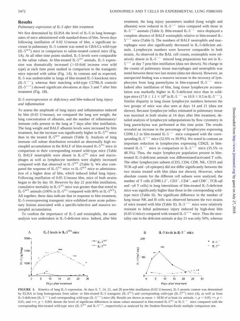
We first determined by ELISA the level of IL-5 in lung homoge-nates of mice administered with standard doses of blm. Seven daysfollowing instillation of 0.02 U/mouse of blm, a significant in-crease in pulmonary IL-5 content was noted in CBA/Ca wild-type(IL-5WT) mice in comparison to saline-treated control mice (Fig.1A). At all other time points studied, IL-5 levels were comparableto the saline values. In blm-treated IL-5TG animals, IL-5 expres-sion was dramatically increased (�10-fold increase over wildtype) at each time point studied in comparison to the transgenicmice injected with saline (Fig. 1A). In contrast and as expected,IL-5 was undetectable in lungs of blm-treated IL-5 knockout mice(IL-5�/�), whereas their matching wild-type C57BL/6 controls(IL-5�/�) showed significant elevations at days 3 and 7 after blmtreatment (Fig. 1B).
IL-5 overexpression or deficiency and blm-induced lung injuryand inflammation
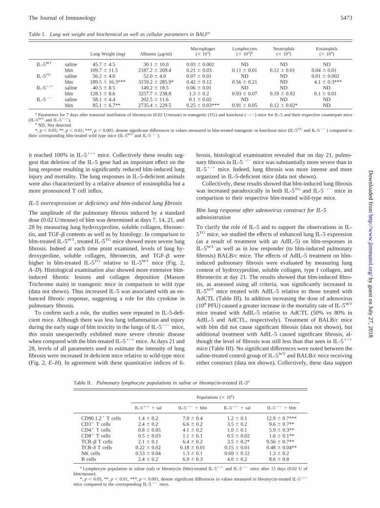
To assess the amplitude of lung injury and inflammation inducedby blm (0.02 U/mouse), we compared the lung wet weight, thelung concentration of albumin, and the number of inflammatory/immune cells present in the lung at 7 days after blm instillation.The lung weight and BALF albumin levels were increased by blmtreatment, but the increase was significantly higher in IL-5TG micethan in the treated IL-5WT animals (Table I). Analysis of lungimmune cell subset distribution revealed an abnormally high eo-sinophil accumulation in the BALF of blm-treated IL-5TG mice incomparison to their corresponding treated wild-type mice (TableI). BALF neutrophils were absent in IL-5TG mice and macro-phages as well as lymphocyte numbers were slightly increasedcompared with that observed in IL-5WT (Table I). We also com-pared the response of IL-5TG mice vs IL-5WT mice to administra-tion of a higher dose of blm, which induced lethal lung injury.Following instillation of 0.05 U/mouse blm, mice of both strainsbegan to die by day 10. However by day 21 post-blm instillation,cumulative mortality in IL-5TG mice was greater than that noted inIL-5WT animals (100% in IL-5TG compared with 80% in IL-5WT).All together, these data indicate that in response to blm treatment,IL-5 overexpressing transgenic mice exhibited more acute pulmo-nary lesions associated with a specific/selective and massive eo-sinophil accumulation.
To confirm the importance of IL-5 and eosinophils, the sameanalysis was undertaken in IL-5-deficient mice. Indeed, after blm
treatment, the lung injury parameters studied (lung weight andalbumin) were reduced in IL-5�/� mice compared with those inIL-5�/� animals (Table I). Blm-treated IL-5�/� mice displayed acomplete absence of BALF eosinophils relative to blm-treated IL-5�/� mice (Table I). The numbers of BALF neutrophils and mac-rophages were also significantly decreased in IL-5-deficient ani-mals. Lymphocyte numbers were however comparable in bothstrains. As observed in the BAL cell counts, eosinophils were rel-atively absent in IL-5�/� minced lung preparations but not in IL-5�/� at day 7 post-blm instillation (data not shown). No change inthe counts of pulmonary tissue macrophages and neutrophils wasnoted between these two last strains (data not shown). However, anunexpected finding was a massive increase in the recovery of lym-phocytes from lung parenchyma in blm-exposed IL-5�/� mice.Indeed after instillation of blm, lung tissue lymphocyte accumu-lation was markedly higher in IL-5-deficient mice than in wild-type mice (17.0 � 1.1 � 106 in IL-5�/� vs 10.9 � 0.5 in IL-5�/�).Similar disparity in lung tissue lymphocyte numbers between thetwo groups of mice was also seen at days 14 and 21 (data notshown). Because lymphocyte influx estimated in pulmonary tissuewas maximal in both strains at 14 days after blm treatment, de-tailed analysis of lymphocyte subpopulations by flow cytometry inlung parenchyma was performed at this time point. The resultsrevealed an increase in the percentage of lymphocytes expressingCD90.1.2 in blm-treated IL-5�/� mice compared with the corre-sponding IL-5�/� mice (52.6% vs 39.9%). We noted in contrast animportant reduction in lymphocytes expressing CD62L in blm-treated IL-5�/� mice in comparison to IL-5�/� mice (35.5% vs48.3%). Thus, the major lymphocyte population present in blm-treated IL-5-deficient animals was differentiated/activated T cells.The other lymphocytes subsets (CD3, CD4, CD8, NK, CD19 andTCR-�� and -�� epitopes) did not differ significantly between thetwo strains treated with blm (data not shown). However, whenabsolute counts for the different cell subsets were analyzed, thenumber of T cells (CD90.1.2�, CD3�, CD4�, and CD8�, TCR-��
and -�� T cells) in lung interstitium of blm-treated IL-5-deficientmice was significantly higher than those in the corresponding wild-type mice (Table II). No significant difference in the number oflung tissue NK and B cells was observed between the two strainsof mice treated with blm (Table II). IL-5�/� mice were relativelyresistant to lethal pulmonary injury induced by high-dose blm(0.05 U/mice) compared with treated IL-5�/� mice. Thus the mor-tality rate in the deficient animals at day 21 was only 50%, whereas
FIGURE 1. Kinetics of lung IL-5 expression. At days 0, 7, 14, 21, and 28 post-blm instillation (0.02 U/mouse), IL-5 protein content was determinedby ELISA in lung homogenates from saline- or blm-treated IL-5 transgenic (IL-5TG) and corresponding wild-type (IL-5WT) mice (A), as well as fromIL-5-deficient (IL-5�/�) and corresponding wild-type (IL-5�/�) mice (B). Results are shown as mean � SEM of at least six animals. �, p � 0.05; ��, p �0.01; and ���, p � 0.001 denote the level of significant differences in mean values measured in blm-treated IL-5TG or IL-5�/� mice compared with thecorresponding blm-treated wild-type mice (IL-5WT and IL-5�/�, respectively) as analyzed by the Student-Newman-Keuls multiple comparison test.
5472 EOSINOPHILS AND T CELLS IN EXPERIMENTAL LUNG FIBROSIS
by guest on July 27, 2018http://w
ww
.jimm
unol.org/D
ownloaded from
it reached 100% in IL-5�/� mice. Collectively these results sug-gest that deletion of the IL-5 gene had an important effect on thelung response resulting in significantly reduced blm-induced lunginjury and mortality. The lung responses in IL-5-deficient animalswere also characterized by a relative absence of eosinophilia but amore pronounced T cell influx.
IL-5 overexpression or deficiency and blm-induced lung fibrosis
The amplitude of the pulmonary fibrosis induced by a standarddose (0.02 U/mouse) of blm was determined at days 7, 14, 21, and28 by measuring lung hydroxyproline, soluble collagen, fibronec-tin, and TGF-� contents as well as by histology. In comparison toblm-treated IL-5WT, treated IL-5TG mice showed more severe lungfibrosis. Indeed at each time point examined, levels of lung hy-droxyproline, soluble collagen, fibronectin, and TGF-� werehigher in blm-treated IL-5TG relative to IL-5WT mice (Fig. 2,A–D). Histological examination also showed more extensive blm-induced fibrotic lesions and collagen deposition (MassonTrichrome stain) in transgenic mice in comparison to wild type(data not shown). Thus increased IL-5 was associated with an en-hanced fibrotic response, suggesting a role for this cytokine inpulmonary fibrosis.
To confirm such a role, the studies were repeated in IL-5-defi-cient mice. Although there was less lung inflammation and injuryduring the early stage of blm toxicity in the lungs of IL-5�/� mice,this strain unexpectedly exhibited more severe chronic diseasewhen compared with the blm-treated IL-5�/� mice. At days 21 and28, levels of all parameters used to estimate the intensity of lungfibrosis were increased in deficient mice relative to wild-type mice(Fig. 2, E–H). In agreement with these quantitative indices of fi-
brosis, histological examination revealed that on day 21, pulmo-nary fibrosis in IL-5�/� mice was substantially more severe than inIL-5�/� mice. Indeed, lung fibrosis was more intense and moreorganized in IL-5-deficient mice (data not shown).
Collectively, these results showed that blm-induced lung fibrosiswas increased paradoxically in both IL-5TG and IL-5�/� mice incomparison to their respective blm-treated wild-type mice.
Blm lung response after adenovirus construct for IL-5administration
To clarify the role of IL-5 and to support the observations in IL-5TG mice, we studied the effects of enhanced lung IL-5 expression(as a result of treatment with an AdIL-5) on blm-responses inIL-5WT as well as in low responder (to blm-induced pulmonaryfibrosis) BALB/c mice. The effects of AdIL-5 treatment on blm-induced pulmonary fibrosis were evaluated by measuring lungcontent of hydroxyproline, soluble collagen, type I collagen, andfibronectin at day 21. The results showed that blm-induced fibro-sis, as assessed using all criteria, was significantly increased inIL-5WT mice treated with AdIL-5 relative to those treated withAdCTL (Table III). In addition increasing the dose of adenovirus(108 PFU) caused a greater increase in the mortality rate of IL-5WT
mice treated with AdIL-5 relative to AdCTL (50% vs 80% inAdIL-5 and AdCTL, respectively). Treatment of BALB/c micewith blm did not cause significant fibrosis (data not shown), butadditional treatment with AdIL-5 caused significant fibrosis, al-though the level of fibrosis was still less than that seen in IL-5�/�
mice (Table III). No significant differences were noted between thesaline-treated control group of IL-5WT and BALB/c mice receivingeither construct (data not shown). Collectively, these data support
Table I. Lung wet weight and biochemical as well as cellular parameters in BALFa
Lung Weight (mg) Albumin (�g/ml)Macrophages
(� 103)Lymphocytes
(� 103)bNeutrophils
(� 103)Eosinophils
(� 103)
IL-5WT saline 45.7 � 4.5 30.1 � 10.0 0.03 � 0.002 ND ND NDblm 109.7 � 11.5 2187.2 � 269.4 0.21 � 0.03 0.11 � 0.01 0.12 � 0.01 0.04 � 0.01
IL-5TG saline 56.2 � 4.0 52.0 � 4.0 0.07 � 0.01 ND ND 0.01 � 0.002blm 189.5 � 16.3*** 3159.2 � 285.9* 0.42 � 0.12 0.56 � 0.21 ND 4.1 � 0.3***
IL-5�/� saline 40.5 � 8.5 149.2 � 18.5 0.06 � 0.01 ND ND NDblm 128.1 � 8.6 3257.7 � 238.8 1.3 � 0.2 0.93 � 0.07 0.19 � 0.02 0.1 � 0.01
IL-5�/� saline 58.1 � 4.4 202.5 � 11.6 0.1 � 0.02 ND ND NDblm 85.1 � 6.7** 2735.4 � 229.5 0.25 � 0.03*** 0.91 � 0.05 0.12 � 0.02* ND
a Parameters for 7 days after transoral instillation of bleomycin (0.02 U/mouse) in transgenic (TG) and knockout (�/�) mice for IL-5 and their respective counterpart mice(IL-5WT and IL-5�/�).
b ND, Not detected.*, p � 0.05; **, p � 0.01; ***, p � 0.001; denote significant differences in values measured in blm-treated transgenic or knockout mice (IL-5TG and IL-5�/�) compared to
their corresponding blm-treated wild type mice (IL-5WT and IL-5�/�).
Table II. Pulmonary lymphocyte populations in saline or bleomycin-treated IL-5a
Populations (� 106)
IL-5�/� � sal IL-5�/� � blm IL-5�/� � sal IL-5�/� � blm
CD90.1.2� T cells 1.4 � 0.2 7.0 � 0.4 1.2 � 0.1 12.9 � 0.7***CD3� T cells 2.4 � 0.2 6.6 � 0.2 3.5 � 0.2 9.6 � 0.7**CD4� T cells 0.8 � 0.05 4.1 � 0.2 1.0 � 0.1 5.9 � 0.3**CD8� T cells 0.5 � 0.03 1.1 � 0.1 0.5 � 0.02 1.6 � 0.1**TCR-� T cells 2.1 � 0.1 6.4 � 0.2 3.5 � 0.2* 9.56 � 0.7**TCR-� T cells 0.22 � 0.02 0.18 � 0.01 0.15 � 0.01 0.48 � 0.04**NK cells 0.53 � 0.04 1.3 � 0.1 0.69 � 0.12 1.3 � 0.2B cells 2.4 � 0.2 6.9 � 0.3 4.0 � 0.2 8.6 � 0.8
a Lymphocyte population in saline (sal) or bleomycin (blm)-treated IL-5�/� and IL-5�/� mice after 15 days (0.02 U ofblm/mouse).
*, p � 0.05, **, p � 0.01, ***, p � 0.001, denote significant differences in values measured in bleomycin-treated IL-5�/�
mice compared to the corresponding IL-5�/� mice.
5473The Journal of Immunology
by guest on July 27, 2018http://w
ww
.jimm
unol.org/D
ownloaded from
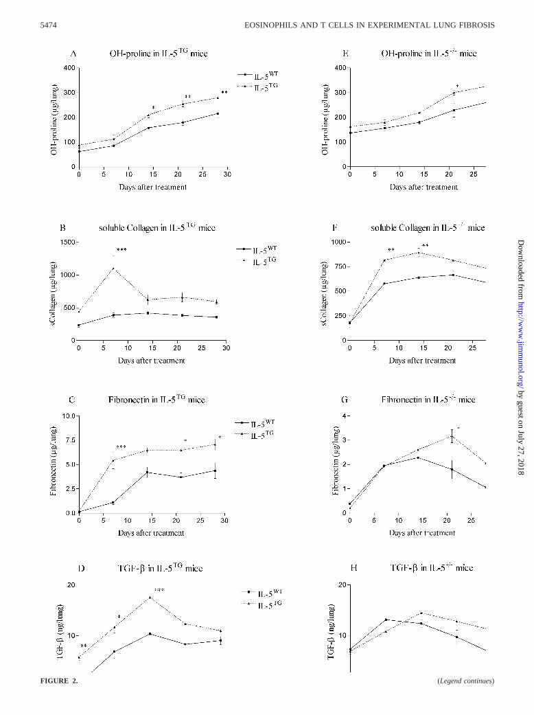
FIGURE 2. (Legend continues)
5474 EOSINOPHILS AND T CELLS IN EXPERIMENTAL LUNG FIBROSIS
by guest on July 27, 2018http://w
ww
.jimm
unol.org/D
ownloaded from
our observations obtained in blm-treated IL-5TG mice and suggesta key role for IL-5 in the extension of blm-induced pulmonaryfibrosis. However these results appear to contradict the findingsusing the IL-5-deficient mice, wherein reduced eosinophils due toIL-5 deficiency but enhanced lung T lymphocyte influx are asso-ciated with enhanced pulmonary fibrosis.
Characterization of pulmonary eosinophils and T cells
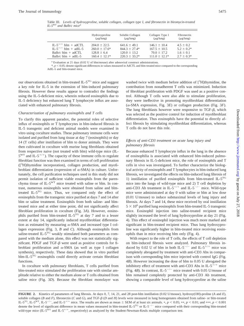
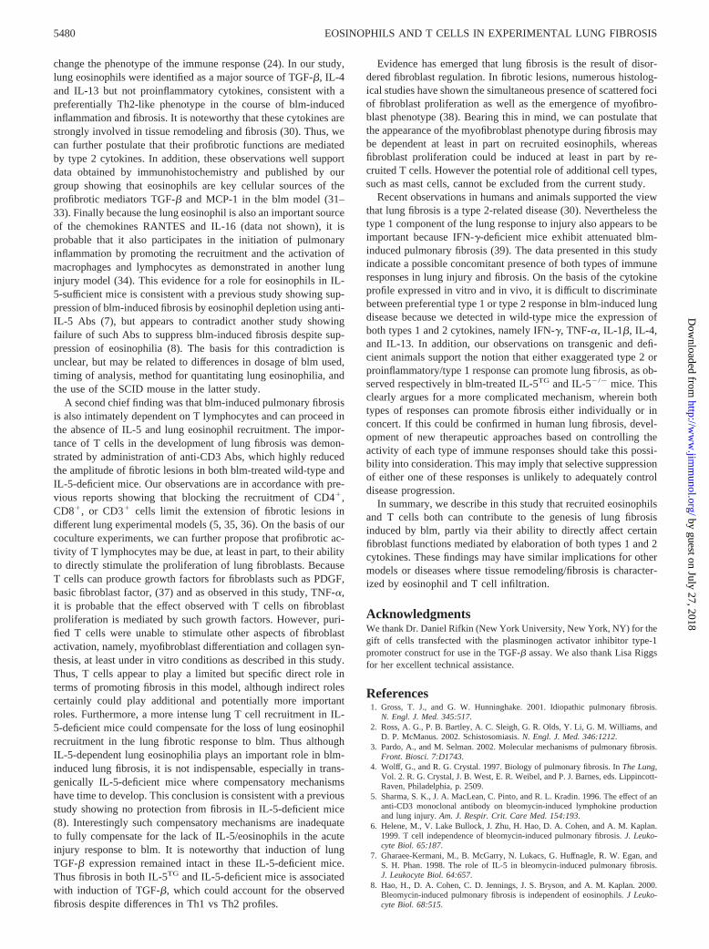
To clarify this apparent paradox, the potential roles of selectiveinflux of eosinophils or T lymphocytes in blm-induced fibrosis inIL-5 transgenic and deficient animal models were examined invitro using coculture studies. These pulmonary immune cells wereisolated and purified from lung tissue at day 7 (eosinophils) or day14 (T cells) after instillation of blm to donor animals. They werethen cultivated in coculture with murine lung fibroblasts obtainedfrom respective naive (not treated with blm) wild-type mice (IL-5WT and IL-5�/�). The capacity of these immune cells to regulatefibroblast function was then examined in terms of cell proliferation([3H]thymidine incorporation), collagen production, and myofi-broblast differentiation (expression of �-SMA) in culture. Unfor-tunately, the cell purification techniques used in this study did notpermit isolation of sufficient viable eosinophils from the paren-chyma tissue of IL-5WT mice treated with saline or blm. In con-trast, numerous eosinophils were obtained from saline and blm-treated IL-5TG mice. Thus, we compared only the effect ofeosinophils from the transgenic mice purified at days 7 and 14 afterblm or saline treatment. Eosinophils from both saline- and blm-treated mice and at either time point, did not significantly affectfibroblast proliferation in coculture (Fig. 3A). However, eosino-phils purified from blm-treated IL-5TG at day 7 and to a lesserextent at day 14, significantly induced myofibroblast differentia-tion as estimated by measuring �-SMA and increased type I col-lagen expression (Fig. 3, B and C). Although eosinophils fromsaline-treated IL-5TG weakly stimulated both parameters as com-pared with the medium alone, this effect was not statistically sig-nificant. PDGF and TGF-� were used as positive controls for fi-broblast proliferation and �-SMA (as well as type I collagensynthesis), respectively. These data showed that in vitro purifiedblm-IL-5TG eosinophils could directly activate certain fibroblastfunctions.
In coculture with pulmonary fibroblasts, T cells purified fromblm-treated mice stimulated the proliferation rate with similar am-plitude relative to either the medium alone or T cells obtained fromsaline mice (Fig. 3D). Because the fibroblast monolayer was
washed twice with medium before addition of [3H]thymidine, thecontribution from nonadherent T cells was minimized. Inductionof fibroblast proliferation with PDGF was used as a positive con-trol. Although T cells were also able to stimulate proliferation,they were ineffective in promoting myofibroblast differentiation(�-SMA expression, Fig. 3E) or collagen production (Fig. 3F).The lung fibroblasts however were responsive to TGF-�, whichwas selected as the positive control for induction of myofibroblastdifferentiation. Thus eosinophils have the potential to directly af-fect fibrosis by stimulating myofibroblast differentiation, whereasT cells do not have this role.
Effects of anti-CD3 treatment on acute lung injury andpulmonary fibrosis
Because enhanced T lymphocyte influx in the lung in the absenceof eosinophilia is associated with enhanced blm-induced pulmo-nary fibrosis in IL-5-deficient mice, the role of eosinophils and Tcells in vivo was investigated. To further characterize the biolog-ical activity of eosinophils and T lymphocytes in blm-induced lungfibrosis, we investigated the effects on blm-induced lung fibrosis of1) instillation of purified eosinophils from blm-treated IL-5TG
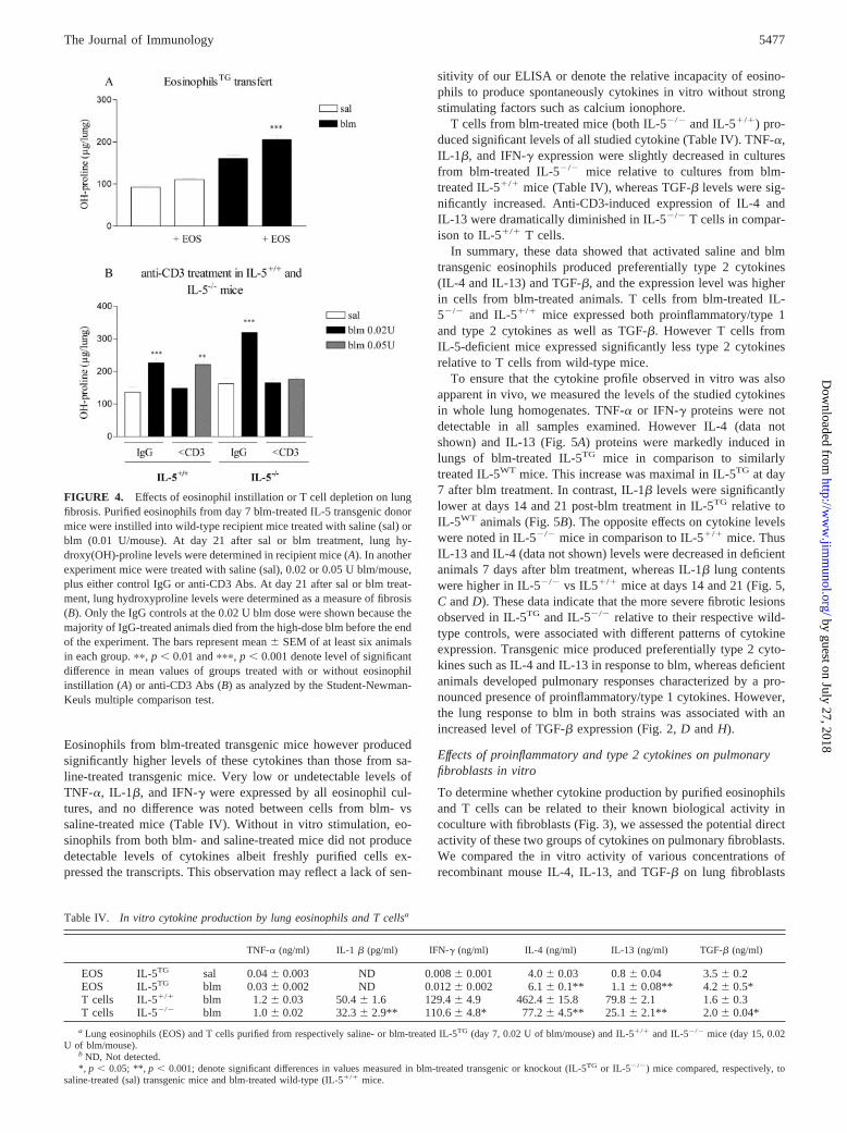
mice into the lungs of wild-type mice and 2) T cell depletion byanti-CD3 Ab treatment in IL-5�/� and IL-5�/� mice. Wild-typemice were administrated at day 0 with saline or blm at low dose(0.01 U/mouse) to induce submaximal levels of lung injury andfibrosis. At days 7 and 14, these mice received by oral instillation5 � 106 purified lung eosinophils from blm-treated IL-5 transgenicmice. Eosinophil injection into saline-treated recipient miceslightly increased the level of lung hydroxyproline at day 21 (Fig.4). This effect of eosinophil injection was much more marked andsignificant in blm-treated recipient mice. Thus lung hydroxypro-line was significantly higher in blm-treated mice receiving eosin-ophils than in mice receiving blm only (Fig. 4).
With respect to the role of T cells, the effects of T cell depletionon blm-induced fibrosis were analyzed. Pulmonary fibrosis in-duced by 0.02 U of blm in both IL-5�/� and IL-5�/� mice wascompletely abrogated by treatment with anti-CD3 Abs in compar-ison with corresponding blm mice injected with control IgG (Fig.4B). However increasing the dose of blm to 0.05 U abrogated theinhibitory effect of treatment with anti-CD3 Abs in IL-5�/� mice(Fig. 4B). In contrast, IL-5�/� mice treated with 0.05 U/mouse ofblm remained completely protected by anti-CD3 Ab treatment,showing a comparable level of lung hydroxyproline as the saline
FIGURE 2. Kinetics of parameters of lung fibrosis. At days 0, 7, 14, 21, and 28 post-blm instillation (0.02 U/mouse), hydroxy(OH)-proline (A and E),soluble collagen (B and F), fibronectin (C and G), and TGF-� (D and H) levels were measured in lung homogenates obtained from saline- or blm-treatedIL-5TG, IL-5WT, IL-5�/�, and IL-5�/� mice. The results are shown as mean � SEM of at least six animals. �, p � 0.05; ��, p � 0.01; and ���, p � 0.001denote the level of significant difference in mean values measured in blm-treated IL-5TG or IL-5�/� mice compared with their corresponding blm-treatedwild-type mice (IL-5WT and IL-5�/�, respectively) as analyzed by the Student-Newman-Keuls multiple comparison test.
Table III. Levels of hydroxyproline, soluble collagen, collagen type I, and fibronectin in bleomycin-treatedIL-5WT and Balb/c micea
Hydroxyproline(�g/lung)
Soluble Collagen(�g/lung)
Collagen Type I(�g/lung)
Fibronectin(�g/lung)
IL-5�/� blm � adCTL 204.0 � 22.5 641.6 � 49.1 146.1 � 10.4 4.5 � 0.2IL-5�/� blm � adIL-5 260.0 � 17.6* 844.3 � 27.4* 167.5 � 10.5 5.2 � 0.2*Balb/c blm � adCTL 128.8 � 6.4 120.0 � 13.2 70.0 � 17.2 1.6 � 0.1Balb/c blm � adIL-5 160.4 � 12.1* 220.3 � 33.2* 111.0 � 12.1* 2.7 � 0.3*
a Evaluation at 21 days (0.02 U of blm/mouse) after adenoviral construct administrations.*, p � 0.05; denote significant differences in values measured in AdCTL and blm-treated mice compared to the corresponding
AdIL-5 and blm-treated mice.
5475The Journal of Immunology
by guest on July 27, 2018http://w
ww
.jimm
unol.org/D
ownloaded from
control group (Fig. 4B). These results demonstrated that the pul-monary fibrosis observed in IL-5 transgenic and deficient animalswere at least in part associated with heightened recruitment ofeosinophils and T lymphocytes, respectively. They also show thatthe fibrosis in IL-5-deficient mice is totally dependent on T cells,even at a high dose of blm, but independent of T cells in IL-5-sufficient mice treated with high-dose blm.
Cytokine profile of purified eosinophils and T cells
To determine the mechanism by which eosinophils and T cellsparticipate to the extension of fibrosis, we then explored their ex-pression of cytokines implicated in the pathogenesis of fibrosis.GEArrays gene arrays allowed us to profile the expression of 20preselected genes known to activate fibroblast functions and to beproduced specifically by both these immune cell types. Transcriptlevels of type 2 cytokines such as IL-4, IL-13, and IL-10 were
significantly increased in eosinophils purified from IL-5TG miceadministrated with blm in comparison to eosinophils from saline-treated IL-5TG animals. Similarly higher expression of TGF-�1and -�3 was found in eosinophils from blm- vs saline-treated mice(data not shown). Analysis of T cells using a similar array revealedthat blm-treated IL-5�/� and IL-5�/� mice significantly expressedTNF-�, IL-1�, and IFN-�, although expression was higher in de-ficient cells. Similarly, TGF-�1 was more highly induced in T cellsfrom IL-5�/� vs IL-5�/� mice (data not shown).
On the basis of these array analyses, we further focused ourwork on selected cytokines. Isolated and purified eosinophils andT lymphocytes were analyzed for TNF-�, IL-1�, IFN-�, IL-4, IL-13, and TGF-� production in the presence of calcium ionophore(eosinophils) or anti-CD3 Abs (T cells). Consistent with the arraydata, purified and activated eosinophils from saline and blm-treated IL-5TG mice expressed IL-4, IL-13, and TGF-� (Table IV).
FIGURE 3. Effects of lung eosinophil or T cell coculture with lung fibroblasts. Lung fibroblasts isolated and purified from naive (untreated) IL-5WT orIL-5�/� mice were cocultured with either purified lung eosinophils or T lymphocytes obtained from IL-5TG and IL-5�/� animals on day 7 or day 14 aftersaline or blm (0.02 U/mouse) instillation. Cell proliferation was measured by [3H]thymidine incorporation (A and D), and myofibroblast differentiation wasdetermined as �-SMA (B and E) protein expression. Fibrogenic effect was assessed by type I collagen expression (C and F). Bars represent means � SEMwith n � 4–6. �, p � 0.05; ��, p � 0.01; and ���, p � 0.001 denote level of significant difference in mean values measured in culture with purifiedleukocytes compared with medium alone as analyzed by the Student-Newman-Keuls multiple comparison test. PDGF (10 ng/ml) (A and D) and TGF-�(10 ng/ml) (B, C, E, and F) were used as positive controls for proliferation and �-SMA, respectively, as well as type I collagen parameters.
5476 EOSINOPHILS AND T CELLS IN EXPERIMENTAL LUNG FIBROSIS
by guest on July 27, 2018http://w
ww
.jimm
unol.org/D
ownloaded from
Eosinophils from blm-treated transgenic mice however producedsignificantly higher levels of these cytokines than those from sa-line-treated transgenic mice. Very low or undetectable levels ofTNF-�, IL-1�, and IFN-� were expressed by all eosinophil cul-tures, and no difference was noted between cells from blm- vssaline-treated mice (Table IV). Without in vitro stimulation, eo-sinophils from both blm- and saline-treated mice did not producedetectable levels of cytokines albeit freshly purified cells ex-pressed the transcripts. This observation may reflect a lack of sen-
sitivity of our ELISA or denote the relative incapacity of eosino-phils to produce spontaneously cytokines in vitro without strongstimulating factors such as calcium ionophore.
T cells from blm-treated mice (both IL-5�/� and IL-5�/�) pro-duced significant levels of all studied cytokine (Table IV). TNF-�,IL-1�, and IFN-� expression were slightly decreased in culturesfrom blm-treated IL-5�/� mice relative to cultures from blm-treated IL-5�/� mice (Table IV), whereas TGF-� levels were sig-nificantly increased. Anti-CD3-induced expression of IL-4 andIL-13 were dramatically diminished in IL-5�/� T cells in compar-ison to IL-5�/� T cells.
In summary, these data showed that activated saline and blmtransgenic eosinophils produced preferentially type 2 cytokines(IL-4 and IL-13) and TGF-�, and the expression level was higherin cells from blm-treated animals. T cells from blm-treated IL-5�/� and IL-5�/� mice expressed both proinflammatory/type 1and type 2 cytokines as well as TGF-�. However T cells fromIL-5-deficient mice expressed significantly less type 2 cytokinesrelative to T cells from wild-type mice.
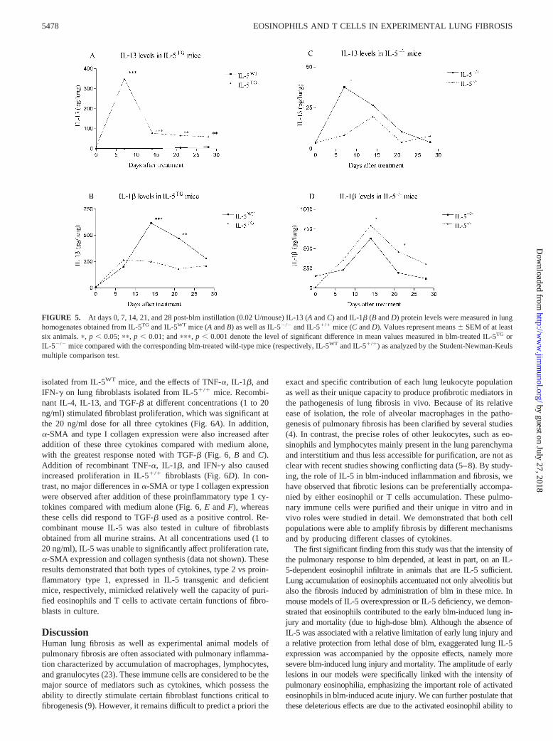
To ensure that the cytokine profile observed in vitro was alsoapparent in vivo, we measured the levels of the studied cytokinesin whole lung homogenates. TNF-� or IFN-� proteins were notdetectable in all samples examined. However IL-4 (data notshown) and IL-13 (Fig. 5A) proteins were markedly induced inlungs of blm-treated IL-5TG mice in comparison to similarlytreated IL-5WT mice. This increase was maximal in IL-5TG at day7 after blm treatment. In contrast, IL-1� levels were significantlylower at days 14 and 21 post-blm treatment in IL-5TG relative toIL-5WT animals (Fig. 5B). The opposite effects on cytokine levelswere noted in IL-5�/� mice in comparison to IL-5�/� mice. ThusIL-13 and IL-4 (data not shown) levels were decreased in deficientanimals 7 days after blm treatment, whereas IL-1� lung contentswere higher in IL-5�/� vs IL5�/� mice at days 14 and 21 (Fig. 5,C and D). These data indicate that the more severe fibrotic lesionsobserved in IL-5TG and IL-5�/� relative to their respective wild-type controls, were associated with different patterns of cytokineexpression. Transgenic mice produced preferentially type 2 cyto-kines such as IL-4 and IL-13 in response to blm, whereas deficientanimals developed pulmonary responses characterized by a pro-nounced presence of proinflammatory/type 1 cytokines. However,the lung response to blm in both strains was associated with anincreased level of TGF-� expression (Fig. 2, D and H).
Effects of proinflammatory and type 2 cytokines on pulmonaryfibroblasts in vitro
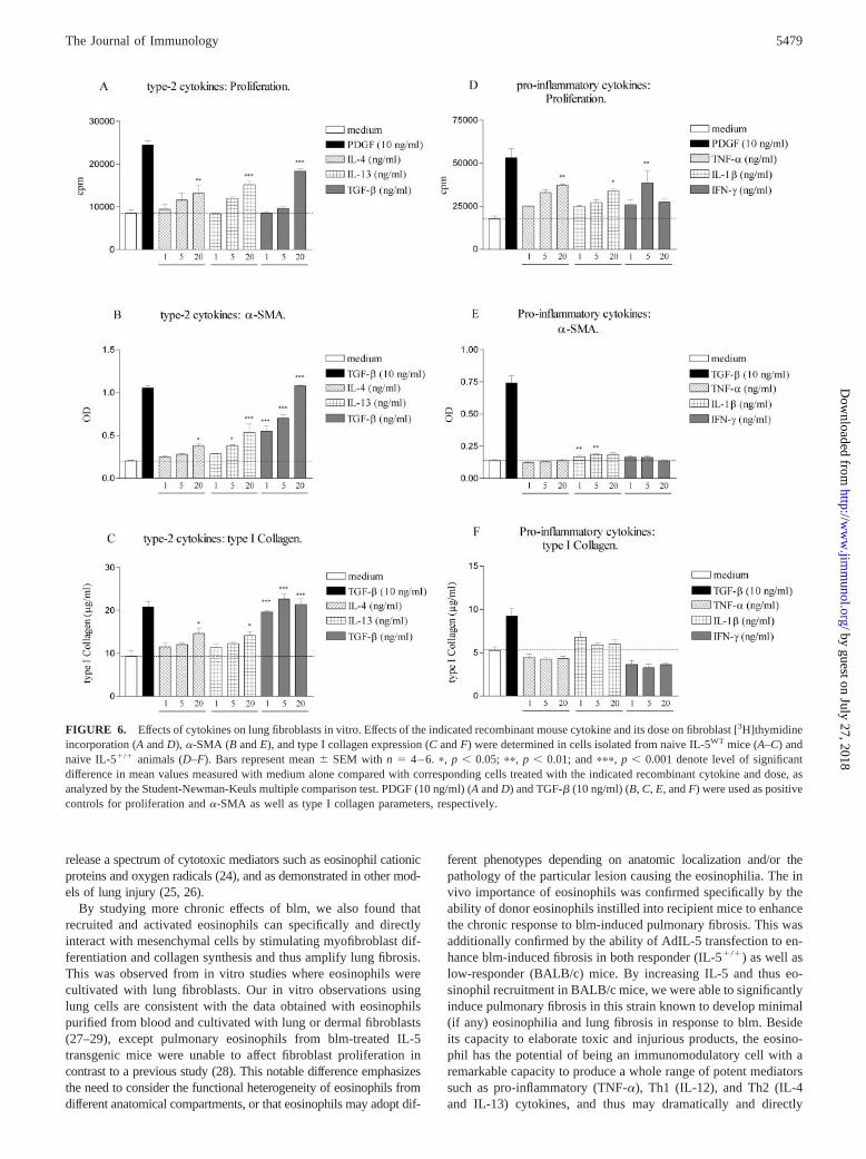
To determine whether cytokine production by purified eosinophilsand T cells can be related to their known biological activity incoculture with fibroblasts (Fig. 3), we assessed the potential directactivity of these two groups of cytokines on pulmonary fibroblasts.We compared the in vitro activity of various concentrations ofrecombinant mouse IL-4, IL-13, and TGF-� on lung fibroblasts
FIGURE 4. Effects of eosinophil instillation or T cell depletion on lungfibrosis. Purified eosinophils from day 7 blm-treated IL-5 transgenic donormice were instilled into wild-type recipient mice treated with saline (sal) orblm (0.01 U/mouse). At day 21 after sal or blm treatment, lung hy-droxy(OH)-proline levels were determined in recipient mice (A). In anotherexperiment mice were treated with saline (sal), 0.02 or 0.05 U blm/mouse,plus either control IgG or anti-CD3 Abs. At day 21 after sal or blm treat-ment, lung hydroxyproline levels were determined as a measure of fibrosis(B). Only the IgG controls at the 0.02 U blm dose were shown because themajority of IgG-treated animals died from the high-dose blm before the endof the experiment. The bars represent mean � SEM of at least six animalsin each group. ��, p � 0.01 and ���, p � 0.001 denote level of significantdifference in mean values of groups treated with or without eosinophilinstillation (A) or anti-CD3 Abs (B) as analyzed by the Student-Newman-Keuls multiple comparison test.
Table IV. In vitro cytokine production by lung eosinophils and T cellsa
TNF-� (ng/ml) IL-1 � (pg/ml) IFN-� (ng/ml) IL-4 (ng/ml) IL-13 (ng/ml) TGF-� (ng/ml)
EOS IL-5TG sal 0.04 � 0.003 ND 0.008 � 0.001 4.0 � 0.03 0.8 � 0.04 3.5 � 0.2EOS IL-5TG blm 0.03 � 0.002 ND 0.012 � 0.002 6.1 � 0.1** 1.1 � 0.08** 4.2 � 0.5*T cells IL-5�/� blm 1.2 � 0.03 50.4 � 1.6 129.4 � 4.9 462.4 � 15.8 79.8 � 2.1 1.6 � 0.3T cells IL-5�/� blm 1.0 � 0.02 32.3 � 2.9** 110.6 � 4.8* 77.2 � 4.5** 25.1 � 2.1** 2.0 � 0.04*
a Lung eosinophils (EOS) and T cells purified from respectively saline- or blm-treated IL-5TG (day 7, 0.02 U of blm/mouse) and IL-5�/� and IL-5�/� mice (day 15, 0.02U of blm/mouse).
b ND, Not detected.*, p � 0.05; **, p � 0.001; denote significant differences in values measured in blm-treated transgenic or knockout (IL-5TG or IL-5�/�) mice compared, respectively, to
saline-treated (sal) transgenic mice and blm-treated wild-type (IL-5�/� mice.
5477The Journal of Immunology
by guest on July 27, 2018http://w
ww
.jimm
unol.org/D
ownloaded from
isolated from IL-5WT mice, and the effects of TNF-�, IL-1�, andIFN-� on lung fibroblasts isolated from IL-5�/� mice. Recombi-nant IL-4, IL-13, and TGF-� at different concentrations (1 to 20ng/ml) stimulated fibroblast proliferation, which was significant atthe 20 ng/ml dose for all three cytokines (Fig. 6A). In addition,�-SMA and type I collagen expression were also increased afteraddition of these three cytokines compared with medium alone,with the greatest response noted with TGF-� (Fig. 6, B and C).Addition of recombinant TNF-�, IL-1�, and IFN-� also causedincreased proliferation in IL-5�/� fibroblasts (Fig. 6D). In con-trast, no major differences in �-SMA or type I collagen expressionwere observed after addition of these proinflammatory type 1 cy-tokines compared with medium alone (Fig. 6, E and F), whereasthese cells did respond to TGF-� used as a positive control. Re-combinant mouse IL-5 was also tested in culture of fibroblastsobtained from all murine strains. At all concentrations used (1 to20 ng/ml), IL-5 was unable to significantly affect proliferation rate,�-SMA expression and collagen synthesis (data not shown). Theseresults demonstrated that both types of cytokines, type 2 vs proin-flammatory type 1, expressed in IL-5 transgenic and deficientmice, respectively, mimicked relatively well the capacity of puri-fied eosinophils and T cells to activate certain functions of fibro-blasts in culture.
DiscussionHuman lung fibrosis as well as experimental animal models ofpulmonary fibrosis are often associated with pulmonary inflamma-tion characterized by accumulation of macrophages, lymphocytes,and granulocytes (23). These immune cells are considered to be themajor source of mediators such as cytokines, which possess theability to directly stimulate certain fibroblast functions critical tofibrogenesis (9). However, it remains difficult to predict a priori the
exact and specific contribution of each lung leukocyte populationas well as their unique capacity to produce profibrotic mediators inthe pathogenesis of lung fibrosis in vivo. Because of its relativeease of isolation, the role of alveolar macrophages in the patho-genesis of pulmonary fibrosis has been clarified by several studies(4). In contrast, the precise roles of other leukocytes, such as eo-sinophils and lymphocytes mainly present in the lung parenchymaand interstitium and thus less accessible for purification, are not asclear with recent studies showing conflicting data (5–8). By study-ing, the role of IL-5 in blm-induced inflammation and fibrosis, wehave observed that fibrotic lesions can be preferentially accompa-nied by either eosinophil or T cells accumulation. These pulmo-nary immune cells were purified and their unique in vitro and invivo roles were studied in detail. We demonstrated that both cellpopulations were able to amplify fibrosis by different mechanismsand by producing different classes of cytokines.
The first significant finding from this study was that the intensity ofthe pulmonary response to blm depended, at least in part, on an IL-5-dependent eosinophil infiltrate in animals that are IL-5 sufficient.Lung accumulation of eosinophils accentuated not only alveolitis butalso the fibrosis induced by administration of blm in these mice. Inmouse models of IL-5 overexpression or IL-5 deficiency, we demon-strated that eosinophils contributed to the early blm-induced lung in-jury and mortality (due to high-dose blm). Although the absence ofIL-5 was associated with a relative limitation of early lung injury anda relative protection from lethal dose of blm, exaggerated lung IL-5expression was accompanied by the opposite effects, namely moresevere blm-induced lung injury and mortality. The amplitude of earlylesions in our models were specifically linked with the intensity ofpulmonary eosinophilia, emphasizing the important role of activatedeosinophils in blm-induced acute injury. We can further postulate thatthese deleterious effects are due to the activated eosinophil ability to
FIGURE 5. At days 0, 7, 14, 21, and 28 post-blm instillation (0.02 U/mouse) IL-13 (A and C) and IL-1� (B and D) protein levels were measured in lunghomogenates obtained from IL-5TG and IL-5WT mice (A and B) as well as IL-5�/� and IL-5�/� mice (C and D). Values represent means � SEM of at leastsix animals. �, p � 0.05; ��, p � 0.01; and ���, p � 0.001 denote the level of significant difference in mean values measured in blm-treated IL-5TG orIL-5�/� mice compared with the corresponding blm-treated wild-type mice (respectively, IL-5WT and IL-5�/�) as analyzed by the Student-Newman-Keulsmultiple comparison test.
5478 EOSINOPHILS AND T CELLS IN EXPERIMENTAL LUNG FIBROSIS
by guest on July 27, 2018http://w
ww
.jimm
unol.org/D
ownloaded from
release a spectrum of cytotoxic mediators such as eosinophil cationicproteins and oxygen radicals (24), and as demonstrated in other mod-els of lung injury (25, 26).
By studying more chronic effects of blm, we also found thatrecruited and activated eosinophils can specifically and directlyinteract with mesenchymal cells by stimulating myofibroblast dif-ferentiation and collagen synthesis and thus amplify lung fibrosis.This was observed from in vitro studies where eosinophils werecultivated with lung fibroblasts. Our in vitro observations usinglung cells are consistent with the data obtained with eosinophilspurified from blood and cultivated with lung or dermal fibroblasts(27–29), except pulmonary eosinophils from blm-treated IL-5transgenic mice were unable to affect fibroblast proliferation incontrast to a previous study (28). This notable difference emphasizesthe need to consider the functional heterogeneity of eosinophils fromdifferent anatomical compartments, or that eosinophils may adopt dif-
ferent phenotypes depending on anatomic localization and/or thepathology of the particular lesion causing the eosinophilia. The invivo importance of eosinophils was confirmed specifically by theability of donor eosinophils instilled into recipient mice to enhancethe chronic response to blm-induced pulmonary fibrosis. This wasadditionally confirmed by the ability of AdIL-5 transfection to en-hance blm-induced fibrosis in both responder (IL-5�/�) as well aslow-responder (BALB/c) mice. By increasing IL-5 and thus eo-sinophil recruitment in BALB/c mice, we were able to significantlyinduce pulmonary fibrosis in this strain known to develop minimal(if any) eosinophilia and lung fibrosis in response to blm. Besideits capacity to elaborate toxic and injurious products, the eosino-phil has the potential of being an immunomodulatory cell with aremarkable capacity to produce a whole range of potent mediatorssuch as pro-inflammatory (TNF-�), Th1 (IL-12), and Th2 (IL-4and IL-13) cytokines, and thus may dramatically and directly
FIGURE 6. Effects of cytokines on lung fibroblasts in vitro. Effects of the indicated recombinant mouse cytokine and its dose on fibroblast [3H]thymidineincorporation (A and D), �-SMA (B and E), and type I collagen expression (C and F) were determined in cells isolated from naive IL-5WT mice (A–C) andnaive IL-5�/� animals (D–F). Bars represent mean � SEM with n � 4–6. �, p � 0.05; ��, p � 0.01; and ���, p � 0.001 denote level of significantdifference in mean values measured with medium alone compared with corresponding cells treated with the indicated recombinant cytokine and dose, asanalyzed by the Student-Newman-Keuls multiple comparison test. PDGF (10 ng/ml) (A and D) and TGF-� (10 ng/ml) (B, C, E, and F) were used as positivecontrols for proliferation and �-SMA as well as type I collagen parameters, respectively.
5479The Journal of Immunology
by guest on July 27, 2018http://w
ww
.jimm
unol.org/D
ownloaded from
change the phenotype of the immune response (24). In our study,lung eosinophils were identified as a major source of TGF-�, IL-4and IL-13 but not proinflammatory cytokines, consistent with apreferentially Th2-like phenotype in the course of blm-inducedinflammation and fibrosis. It is noteworthy that these cytokines arestrongly involved in tissue remodeling and fibrosis (30). Thus, wecan further postulate that their profibrotic functions are mediatedby type 2 cytokines. In addition, these observations well supportdata obtained by immunohistochemistry and published by ourgroup showing that eosinophils are key cellular sources of theprofibrotic mediators TGF-� and MCP-1 in the blm model (31–33). Finally because the lung eosinophil is also an important sourceof the chemokines RANTES and IL-16 (data not shown), it isprobable that it also participates in the initiation of pulmonaryinflammation by promoting the recruitment and the activation ofmacrophages and lymphocytes as demonstrated in another lunginjury model (34). This evidence for a role for eosinophils in IL-5-sufficient mice is consistent with a previous study showing sup-pression of blm-induced fibrosis by eosinophil depletion using anti-IL-5 Abs (7), but appears to contradict another study showingfailure of such Abs to suppress blm-induced fibrosis despite sup-pression of eosinophilia (8). The basis for this contradiction isunclear, but may be related to differences in dosage of blm used,timing of analysis, method for quantitating lung eosinophilia, andthe use of the SCID mouse in the latter study.
A second chief finding was that blm-induced pulmonary fibrosisis also intimately dependent on T lymphocytes and can proceed inthe absence of IL-5 and lung eosinophil recruitment. The impor-tance of T cells in the development of lung fibrosis was demon-strated by administration of anti-CD3 Abs, which highly reducedthe amplitude of fibrotic lesions in both blm-treated wild-type andIL-5-deficient mice. Our observations are in accordance with pre-vious reports showing that blocking the recruitment of CD4�,CD8�, or CD3� cells limit the extension of fibrotic lesions indifferent lung experimental models (5, 35, 36). On the basis of ourcoculture experiments, we can further propose that profibrotic ac-tivity of T lymphocytes may be due, at least in part, to their abilityto directly stimulate the proliferation of lung fibroblasts. BecauseT cells can produce growth factors for fibroblasts such as PDGF,basic fibroblast factor, (37) and as observed in this study, TNF-�,it is probable that the effect observed with T cells on fibroblastproliferation is mediated by such growth factors. However, puri-fied T cells were unable to stimulate other aspects of fibroblastactivation, namely, myofibroblast differentiation and collagen syn-thesis, at least under in vitro conditions as described in this study.Thus, T cells appear to play a limited but specific direct role interms of promoting fibrosis in this model, although indirect rolescertainly could play additional and potentially more importantroles. Furthermore, a more intense lung T cell recruitment in IL-5-deficient mice could compensate for the loss of lung eosinophilrecruitment in the lung fibrotic response to blm. Thus althoughIL-5-dependent lung eosinophilia plays an important role in blm-induced lung fibrosis, it is not indispensable, especially in trans-genically IL-5-deficient mice where compensatory mechanismshave time to develop. This conclusion is consistent with a previousstudy showing no protection from fibrosis in IL-5-deficient mice(8). Interestingly such compensatory mechanisms are inadequateto fully compensate for the lack of IL-5/eosinophils in the acuteinjury response to blm. It is noteworthy that induction of lungTGF-� expression remained intact in these IL-5-deficient mice.Thus fibrosis in both IL-5TG and IL-5-deficient mice is associatedwith induction of TGF-�, which could account for the observedfibrosis despite differences in Th1 vs Th2 profiles.
Evidence has emerged that lung fibrosis is the result of disor-dered fibroblast regulation. In fibrotic lesions, numerous histolog-ical studies have shown the simultaneous presence of scattered fociof fibroblast proliferation as well as the emergence of myofibro-blast phenotype (38). Bearing this in mind, we can postulate thatthe appearance of the myofibroblast phenotype during fibrosis maybe dependent at least in part on recruited eosinophils, whereasfibroblast proliferation could be induced at least in part by re-cruited T cells. However the potential role of additional cell types,such as mast cells, cannot be excluded from the current study.
Recent observations in humans and animals supported the viewthat lung fibrosis is a type 2-related disease (30). Nevertheless thetype 1 component of the lung response to injury also appears to beimportant because IFN-�-deficient mice exhibit attenuated blm-induced pulmonary fibrosis (39). The data presented in this studyindicate a possible concomitant presence of both types of immuneresponses in lung injury and fibrosis. On the basis of the cytokineprofile expressed in vitro and in vivo, it is difficult to discriminatebetween preferential type 1 or type 2 response in blm-induced lungdisease because we detected in wild-type mice the expression ofboth types 1 and 2 cytokines, namely IFN-�, TNF-�, IL-1�, IL-4,and IL-13. In addition, our observations on transgenic and defi-cient animals support the notion that either exaggerated type 2 orproinflammatory/type 1 response can promote lung fibrosis, as ob-served respectively in blm-treated IL-5TG and IL-5�/� mice. Thisclearly argues for a more complicated mechanism, wherein bothtypes of responses can promote fibrosis either individually or inconcert. If this could be confirmed in human lung fibrosis, devel-opment of new therapeutic approaches based on controlling theactivity of each type of immune responses should take this possi-bility into consideration. This may imply that selective suppressionof either one of these responses is unlikely to adequately controldisease progression.
In summary, we describe in this study that recruited eosinophilsand T cells both can contribute to the genesis of lung fibrosisinduced by blm, partly via their ability to directly affect certainfibroblast functions mediated by elaboration of both types 1 and 2cytokines. These findings may have similar implications for othermodels or diseases where tissue remodeling/fibrosis is character-ized by eosinophil and T cell infiltration.
AcknowledgmentsWe thank Dr. Daniel Rifkin (New York University, New York, NY) for thegift of cells transfected with the plasminogen activator inhibitor type-1promoter construct for use in the TGF-� assay. We also thank Lisa Riggsfor her excellent technical assistance.
References1. Gross, T. J., and G. W. Hunninghake. 2001. Idiopathic pulmonary fibrosis.
N. Engl. J. Med. 345:517.2. Ross, A. G., P. B. Bartley, A. C. Sleigh, G. R. Olds, Y. Li, G. M. Williams, and
D. P. McManus. 2002. Schistosomiasis. N. Engl. J. Med. 346:1212.3. Pardo, A., and M. Selman. 2002. Molecular mechanisms of pulmonary fibrosis.
Front. Biosci. 7:D1743.4. Wolff, G., and R. G. Crystal. 1997. Biology of pulmonary fibrosis. In The Lung,
Vol. 2. R. G. Crystal, J. B. West, E. R. Weibel, and P. J. Barnes, eds. Lippincott-Raven, Philadelphia, p. 2509.
5. Sharma, S. K., J. A. MacLean, C. Pinto, and R. L. Kradin. 1996. The effect of ananti-CD3 monoclonal antibody on bleomycin-induced lymphokine productionand lung injury. Am. J. Respir. Crit. Care Med. 154:193.
6. Helene, M., V. Lake Bullock, J. Zhu, H. Hao, D. A. Cohen, and A. M. Kaplan.1999. T cell independence of bleomycin-induced pulmonary fibrosis. J. Leuko-cyte Biol. 65:187.
7. Gharaee-Kermani, M., B. McGarry, N. Lukacs, G. Huffnagle, R. W. Egan, andS. H. Phan. 1998. The role of IL-5 in bleomycin-induced pulmonary fibrosis.J. Leukocyte Biol. 64:657.
8. Hao, H., D. A. Cohen, C. D. Jennings, J. S. Bryson, and A. M. Kaplan. 2000.Bleomycin-induced pulmonary fibrosis is independent of eosinophils. J Leuko-cyte Biol. 68:515.
5480 EOSINOPHILS AND T CELLS IN EXPERIMENTAL LUNG FIBROSIS
by guest on July 27, 2018http://w
ww
.jimm
unol.org/D
ownloaded from
9. Gharaee-Kermani, M., and S. H. Phan. 2001. Role of cytokines and cytokinetherapy in wound healing and fibrotic diseases. Curr. Pharm. Des. 7:1083.
10. Sime, P. J., R. A. Marr, D. Gauldie, Z. Xing, B. R. Hewlett, F. L. Graham, andJ. Gauldie. 1998. Transfer of tumor necrosis factor-� to rat lung induces severepulmonary inflammation and patchy interstitial fibrogenesis with induction oftransforming growth factor-�1 and myofibroblasts. Am. J. Pathol. 153:825.
11. Kolb, M., P. J. Margetts, D. C. Anthony, F. Pitossi, and J. Gauldie. 2001. Tran-sient expression of IL-1� induces acute lung injury and chronic repair leading topulmonary fibrosis. J. Clin. Invest. 107:1529.
12. Sime, P. J., Z. Xing, F. L. Graham, K. G. Csaky, and J. Gauldie. 1997. Ad-enovector-mediated gene transfer of active transforming growth factor-�1 in-duces prolonged severe fibrosis in rat lung. J. Clin. Invest. 100:768.
13. Hoyle, G. W., J. Li, J. B. Finkelstein, T. Eisenberg, J. Y. Liu, J. A. Lasky,G. Athas, G. F. Morris, and A. R. Brody. 1999. Emphysematous lesions, inflam-mation, and fibrosis in the lungs of transgenic mice overexpressing platelet-de-rived growth factor. Am. J. Pathol. 154:1763.
14. Piguet, P. F., M. A. Collart, G. E. Grau, A. P. Sappino, and P. Vassalli. 1990.Requirement of tumour necrosis factor for development of silica-induced pulmo-nary fibrosis. Nature 344:245.
15. Huaux, F., T. Liu, B. McGarry, M. Ullenbruch, and S. H. Phan. 2003. Dual Rolesof IL-4 in lung injury and fibrosis. J. Immunol. 170:2083.
16. Lee, C. G., R. J. Homer, L. Cohn, H. Link, S. Jung, J. E. Craft, B. S. Graham,T. R. Johnson, and J. A. Elias. 2002. Transgenic overexpression of IL-10 in thelung causes mucus metaplasia, tissue inflammation and airways remodeling viaIL-13-dependent and -independent pathways. J. Biol. Chem. 277:35466.
17. Wallace, W. A., E. A. Ramage, D. Lamb, and S. E. Howie. 1995. A type 2(Th2-like) pattern of immune response predominates in the pulmonary intersti-tium of patients with cryptogenic fibrosing alveolitis (CFA). Clin. Exp. Immunol.101:436.
18. Gharaee-Kermani, M., and S. H. Phan. 1997. Lung interleukin-5 expression inmurine bleomycin-induced pulmonary fibrosis. Am. J. Respir. Cell Mol. Biol.16:438.
19. Kopf, M., F. Brombacher, P. D. Hodgkin, A. J. Ramsay, E. A. Milbourne,W. J. Dai, K. S. Ovington, C. A. Behm, G. Kohler, I. G. Young, andK. I. Matthaei. 1996. IL-5-deficient mice have a developmental defect in CD5�
B-1 cells and lack eosinophilia but have normal antibody and cytotoxic T cellresponses. Immunity 4:15.
20. Dent, L. A., M. Strath, A. L. Mellor, and C. J. Sanderson. 1990. Eosinophilia intransgenic mice expressing interleukin 5. J. Exp. Med. 172:1425.
21. Abe, M., J. G. Harpel, C. N. Metz, I. Nunes, D. J. Loskutoff, and D. B. Rifkin.1994. An assay for transforming growth factor-� using cells transfected with aplasminogen activator inhibitor-1 promoter-luciferase construct. Anal. Biochem.216:276.
22. Wang, J., K. Palmer, J. Lotvall, S. Milan, X. F. Lei, K. I. Matthaei, J. Gauldie,M. D. Inman, M. Jordana, and Z. Xing. 1998. Circulating, but not local lung, IL-5is required for the development of antigen-induced airways eosinophilia. J. Clin.Invest. 102:1132.
23. Crystal, R. G., P. B. Bitterman, B. Mossman, M. I. Schwarz, D. Sheppard,L. Almasy, H. A. Chapman, S. L. Friedman, T. E. King, Jr., L. A. Leinwand, etal. 2002. Future research directions in idiopathic pulmonary fibrosis: summary of
a National Heart, Lung, and Blood Institute working group. Am. J. Respir. Crit.Care Med. 166:236.
24. Dombrowicz, D., and M. Capron. 2001. Eosinophils, allergy and parasites. Curr.Opin. Immunol. 13:716.
25. Iijima, H., A. Duguet, S. Y. Eum, Q. Hamid, and D. H. Eidelman. 2001. Nitricoxide and protein nitration are eosinophil dependent in allergen-challenged mice.Am. J. Respir. Crit. Care Med. 163:1233.
26. Wu, W., M. K. Samoszuk, S. A. Comhair, M. J. Thomassen, C. F. Farver,R. A. Dweik, M. S. Kavuru, S. C. Erzurum, and S. L. Hazen. 2000. Eosinophilsgenerate brominating oxidants in allergen-induced asthma. J. Clin. Invest105:1455.
27. Rochester, C. L., S. J. Ackerman, T. Zheng, and J. A. Elias. 1996. Eosinophil-fibroblast interactions: granule major basic protein interacts with IL-1 and trans-forming growth factor-� in the stimulation of lung fibroblast IL-6-type cytokineproduction. J. Immunol. 156:4449.
28. Levi-Schaffer, F., E. Garbuzenko, A. Rubin, R. Reich, D. Pickholz, P. Gillery,H. Emonard, A. Nagler, and F. A. Maquart. 1999. Human eosinophils regulatehuman lung- and skin-derived fibroblast properties in vitro: a role for transform-ing growth factor � (TGF-�). Proc. Natl. Acad. Sci. USA 96:9660.
29. Phipps, S., S. Ying, A. Wangoo, Y. E. Ong, F. Levi-Schaffer, and A. B. Kay.2002. The relationship between allergen-induced tissue eosinophilia and markersof repair and remodeling in human atopic skin. J. Immunol. 169:4604.
30. Lukacs, N. W., C. Hogaboam, S. W. Chensue, K. Blease, and S. L. Kunkel. 2001.Type 1/type 2 cytokine paradigm and the progression of pulmonary fibrosis.Chest 120:5S.
31. Zhang, K., M. Gharaee-Kermani, B. McGarry, D. Remick, and S. H. Phan. 1997.TNF-�-mediated lung cytokine networking and eosinophil recruitment in pulmo-nary fibrosis. J. Immunol. 158:954.
32. Zhang, K., M. Gharaee-Kermani, M. L. Jones, J. S. Warren, and S. H. Phan. 1994.Lung monocyte chemoattractant protein-1 gene expression in bleomycin-inducedpulmonary fibrosis. J. Immunol. 153:4733.
33. Zhang, K., K. C. Flanders, and S. H. Phan. 1995. Cellular localization of trans-forming growth factor-� expression in bleomycin-induced pulmonary fibrosis.Am. J. Pathol. 147:352.
34. Shi, H. Z., A. Humbles, C. Gerard, Z. Jin, and P. F. Weller. 2000. Lymph nodetrafficking and antigen presentation by endobronchial eosinophils. J. Clin. Invest.105:945.
35. Piguet, P. F., M. A. Collart, G. E. Grau, Y. Kapanci, and P. Vassalli. 1989. Tumornecrosis factor/cachectin plays a key role in bleomycin-induced pneumopathyand fibrosis. J. Exp. Med. 170:655.
36. Westermann, W., R. Schobl, E. P. Rieber, and K. H. Frank. 1999. Th2 cells aseffectors in postirradiation pulmonary damage preceding fibrosis in the rat. Int.J. Radiat. Biol. 75:629.
37. Yamamoto, T., I. Katayama, and K. Nishioka. 1999. Fibroblast proliferation bybleomycin stimulated peripheral blood mononuclear cell factors. J. Rheumatol.26:609.
38. Phan, S. H. 2002. The myofibroblast in pulmonary fibrosis. Chest 122:286S.39. Chen, E. S., B. M. Greenlee, M. Wills-Karp, and D. R. Moller. 2001. Attenuation
of lung inflammation and fibrosis in interferon-�-deficient mice after intratrachealbleomycin. Am. J. Respir. Cell Mol. Biol. 24:545.
5481The Journal of Immunology
by guest on July 27, 2018http://w
ww
.jimm
unol.org/D
ownloaded from