Embed Size (px)
Citation preview
Epidemiology and Etiology of Leukemiaand Lymphoma
Jordan A. Baeker Bispo,1 Paulo S. Pinheiro,2 and Erin K. Kobetz3
1Department of Public Health Sciences, Miller School of Medicine, University of Miami, Miami,Florida 33136, USA
2Sylvester Comprehensive Cancer Center and Department of Public Health Sciences, Miller Schoolof Medicine, University of Miami, Miami, Florida 33136, USA
3Sylvester Comprehensive Cancer Center and Department of Medicine, Miller School of Medicine,University of Miami, Miami, Florida 33136, USA
Correspondence: [email protected]
Available evidence suggests that the incidence of leukemia and lymphoma tends to be higherin highly developed regions of the world and among Whites in the United States. Temporaltrends in incidence are dynamic and multifactorial; for instance, the incidence of non-Hodgkin’s lymphoma increased around the turn of the century, in part because of the ac-quired immune deficiency syndrome (AIDS) epidemic. Most leukemias and lymphomas aresporadic and the specific etiology remains elusive. Still, research shows that these malignan-cies often develop in the context of genetic abnormalities, immunosuppression, and exposureto risk factors like ionizing radiation, carcinogenic chemicals, and oncogenic viruses. Theprognosis varies by subtype, with poorer survival outcomes for acute leukemias amongadults, and more favorable outcomes for Hodgkin’s lymphoma. At a time when specificprevention efforts targeting these malignancies are nonexistent, there is a great need toensure equitable access to diagnostic services and treatments worldwide.
Hematopoiesis is the highly regulated pro-cess by which stem cells differentiate and
mature into erythrocytes, megakaryocytes, andimmune cells of myeloid, lymphoid, or mono-cytic lineage in bone marrow or lymphatictissues. Genetic errors, such as reciprocal chro-mosomal translocations, chromosomal deletions,point mutations, and epigenetic alterations, canarrest the maturation of stem cells across differ-ent stages of hematopoiesis, giving rise to theuncontrolled proliferation of immature, leuke-mic immune cells. Leukemia refers to the clonal
expansion of leukemic cells in the bone marrow,classically resulting in elevated numbers of cellsof the affected lineage in circulating blood and,with certain lymphoid malignancies, abnormalcellular proliferation in lymphatic tissue. Lym-phomas are neoplasms of well-differentiated Band T lymphocytes, which typically present asmalignant masses in lymphatic tissue. Leuke-mias are generally classified into subtypes de-fined by cell lineage (lymphocytic or myeloid)and stage of maturation arrest (acute or chron-ic). Mature lymphoid neoplasms are classified
Editors: Michael G. Kharas, Ross L. Levine, and Ari M. MelnickAdditional Perspectives on Leukemia and Lymphoma: Molecular and Therapeutic Insights available atwww.perspectivesinmedicine.org
Copyright © 2020 Cold Spring Harbor Laboratory Press; all rights reserved; doi: 10.1101/cshperspect.a034819Cite this article as Cold Spring Harb Perspect Med 2020;10:a034819
1
ww
w.p
ersp
ecti
vesi
nm
edic
ine.
org
on January 31, 2022 - Published by Cold Spring Harbor Laboratory Presshttp://perspectivesinmedicine.cshlp.org/Downloaded from
broadly into three groups: mature B-cell neo-plasms, mature T- or natural killer (NK)-cellneoplasms, and Hodgkin’s lymphoma (HL).Collectively, B- and T/NK-cell neoplasms com-prise non-Hodgkin’s lymphomas (NHLs), a het-erogeneous group of more than 60 subtypes. HLarises from B cells, but is distinguished fromNHL morphologically by the presence ofReed–Sternberg clonal tumor cells, which arelarge and often multinucleated.
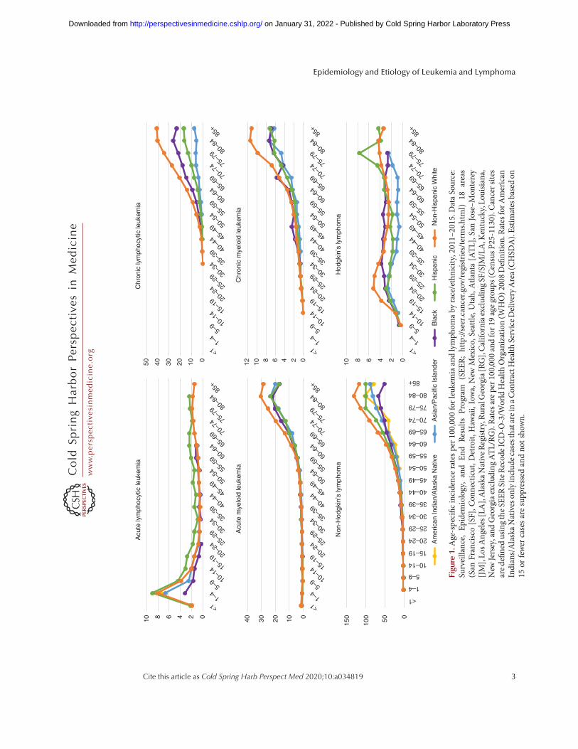
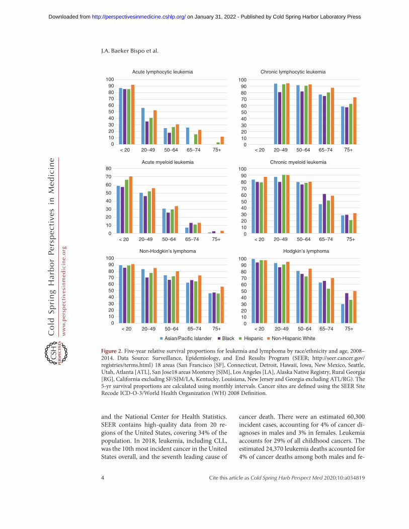
In this review, we discuss the epidemiologyand etiology of leukemia (overall and by sub-type), NHL, andHL.Our discussion emphasizesracial/ethnic and age-related disparities in dis-ease burden, as illustrated in part by Figure 1(patterns of disease incidence) and Figure 2(patterns of 5-yr relative survival). It shouldbe noted that the World Health Organization(WHO) classification for hematopoietic neo-plasms is based on cell of origin, and some formsof leukemia and lymphomas are thus considereddifferent presentations of the same disease. Ex-amples include chronic lymphocytic leukemiaand small cell lymphoma (a form of matureB-cell lymphoma; CLL/SLL), and acute lympho-cytic leukemia and acute precursor B- and T-celllymphoblastic leukemia/lymphoma (ALL/LBL).Clinically, CLL/SLL is considered a lymphoma.However, population-based cancer surveillanceprograms typically count CLL/SLL cases as ei-ther leukemias or lymphomas depending onwhether the cancer cells were found in thebone marrow, blood, or lymph nodes. Here wepresent CLL/SLL and ALL/LBL as primarilyleukemias, recognizing the overlapping classifi-cations.
GLOBAL EPIDEMIOLOGY OF LEUKEMIA
The World Health Organization’s InternationalAgency for Research on Cancer (IARC) produc-es global estimates for all cancers contained inthe GLOBOCAN database, using data frommostly high-quality population-based cancerregistries worldwide. The Institute for HealthMetrics and Evaluation’s Global Burden of Dis-ease (GBD) study also produces estimatesthrough modeling techniques that incorporatedata from numerous additional sources, includ-
ing lower-quality cancer registries in countriesthat are disproportionately low and middleincome.
According to GLOBOCAN, leukemia wasthe 15th most commonly diagnosed cancer and11th leading cause of cancer mortality world-wide in 2018, accounting for 437,033 incidentcancer cases and 309,006 cancer deaths. Global-ly, the leukemia disease burden is higher amongmales than females. In 2018, the age-standard-ized incidence rate formales was 6.1 per 100,000compared to 4.3 per 100,000 for females. Mor-tality was also higher in males (4.2 per 100,000)than females (2.8 per 100,000) (Bray et al. 2018).The age distribution of chronic leukemia is gen-erally unimodal, with incidence rates that tend toincrease with age. ALL and acute myeloid leuke-mia (AML), which are important diseases inchildhood, accordingly have bimodal age distri-butions. By GBD estimates, the total number ofleukemia cases globally increased by 26% from2005 to 2015, and population growth and agingaccounted for all but 3% of this (Fitzmauriceet al. 2017).
The geographic distribution of leukemiaburden is patterned by country-level develop-ment, with age-standardized incidence, andmor-tality higher in more developed countries. TheIARC classifies 185 countries in the GLO-BOCAN database according to the human de-velopment index (HDI), a composite measure oflife expectancy, education, and standard of liv-ing. In 2018, incidence in high/very high HDIcountries substantially exceeded that of low/me-dium HDI countries (7.5 vs. 4.0 per 100,000 formales; 5.3 vs. 3.0 per 100,000 for females). Thesame was true for mortality in high/very highHDI countries and low/medium countries (4.5vs. 3.2 per 100,000 for males; 2.9 vs. 2.4 per100,000 for females) (Bray et al. 2018).
EPIDEMIOLOGY OF LEUKEMIA IN THEUNITED STATES
In the United States, national trends in leukemiaincidence and mortality are monitored usingdata from state population–based cancer regis-tries, namely through the Surveillance, Epide-miology, and End Results program (SEER),
J.A. Baeker Bispo et al.
2 Cite this article as Cold Spring Harb Perspect Med 2020;10:a034819
ww
w.p
ersp
ecti
vesi
nm
edic
ine.
org
on January 31, 2022 - Published by Cold Spring Harbor Laboratory Presshttp://perspectivesinmedicine.cshlp.org/Downloaded from
10 8 6 4 2 0 40 30 20 10 0
150
100 50 0
Acu
te ly
mph
ocyt
ic le
ukem
ia
Acu
te m
yelo
id le
ukem
ia
Non
-Hod
gkin
’s ly
mph
oma
50 40 30 20 10 0 12 10 8 6 4 2 0 10 8 6 4 2 0
Chr
onic
lym
phoc
ytic
leuk
emia
Chr
onic
mye
loid
leuk
emia
Hod
gkin
’s ly
mph
oma
<11–
45–
9 10–1
4 15–1
9 20–2
4 25–2
9 30–3
4 35–3
9 40–4
4 45–4
9 50–5
4 55–5
9 60–6
4 65–6
9 70–7
4 75–7
9 80–8
485
+
<11–
45–
9 10–1
4 15–1
9 20–2
4 25–2
9 30–3
4 35–3
9 40–4
4 45–4
9 50–5
4 55–5
9 60–6
4 65–6
9 70–7
4 75–7
9 80–8
485
+
<11–
45–
9 10–1
4 15–1
9 20–2
4 25–2
9 30–3
4 35–3
9 40–4
4 45–4
9 50–5
4 55–5
9 60–6
4 65–6
9 70–7
4 75–7
9 80–8
485
+
<11–
45–
9 10–1
4 15–1
9 20–2
4 25–2
9 30–3
4 35–3
9 40–4
4 45–4
9 50–5
4 55–5
9 60–6
4 65–6
9 70–7
4 75–7
9 80–8
485
+
<11–
45–
9 10–1
4 15–1
9 20–2
4 25–2
9 30–3
4 35–3
9 40–4
4 45–4
9 50–5
4 55–5
9 60–6
4 65–6
9 70–7
4 75–7
9 80–8
485
+
<1
1–4
5–9
10–14
15–19
20–24
25–29
30–34
35–39
40–44
45–49
50–54
55–59
60–64
65–69
70–74
75–79
80–84
85+
Am
eric
an In
dian
/Ala
ska
Nat
ive
Asi
an/P
acifi
c Is
land
erH
ispa
nic
Bla
ckN
on-H
ispa
nic
Whi
te
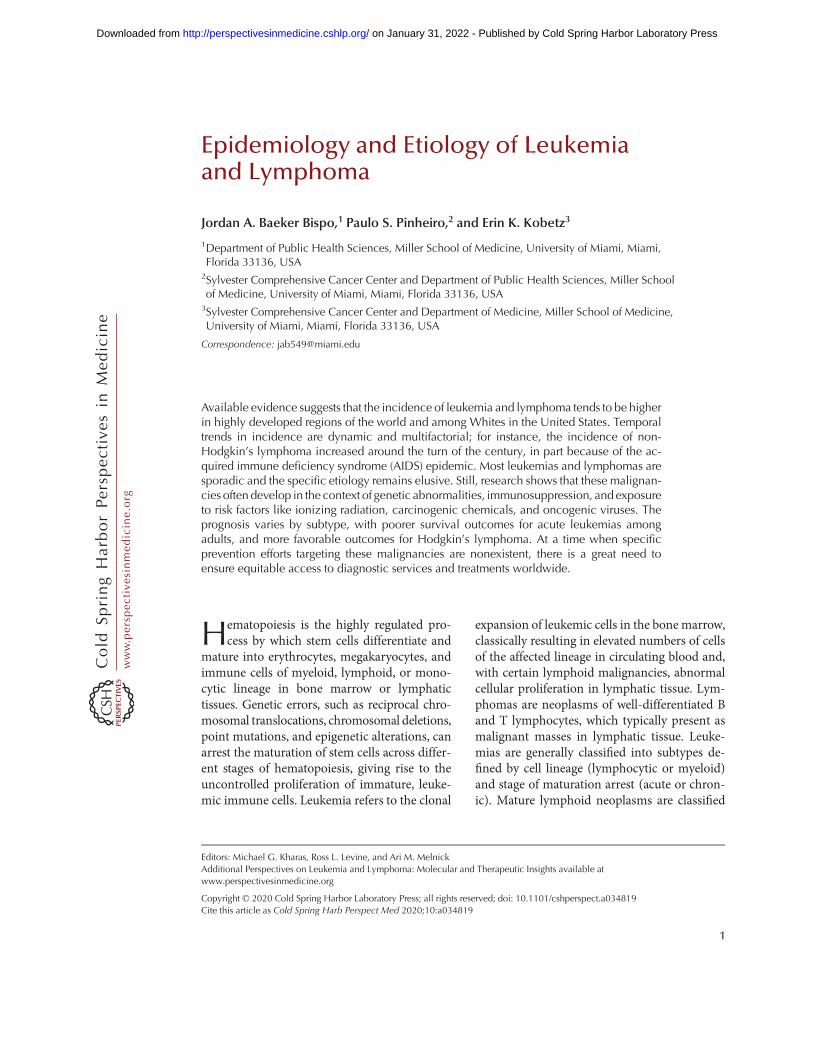
Figu
re1.Age-specificincidenceratesper100,000forleukem
iaandlymph
omaby
race/ethnicity,2011–2015.D
ataSource:
Surveillance,
Epidemiology,and
End
Results
Program
(SEER;http://seer.cancer.gov/registries/terms.html)
18areas
(San
Francisco[SF],C
onnecticut,D
etroit,H
awaii,Iowa,New
Mexico,
Seattle,U
tah,
Atlanta[ATL],San
Jose–M
onterey
[JM],Lo
sAngeles[LA],AlaskaNativeRegistry,RuralGeorgia[RG],Califo
rniaexclud
ingSF/SJM
/LA,K
entucky,Lo
uisiana,
New
Jersey,and
Georgiaexclud
ingATL/RG).Ratesareper100,000andfor19
agegrou
ps(CensusP25-1130).C
ancersites
aredefinedusingtheSE
ERSiteRecod
eICD-O
-3/W
orldHealth
Organization(W
HO)2
008Definition
.RatesforA
merican
Indians/AlaskaNativeson
lyinclud
ecasesthatareinaCon
tractH
ealth
ServiceDeliveryArea(CHSD
A).Estim
atesbasedon
15or
fewer
casesaresupp
ressed
andno
tshow
n.
Epidemiology and Etiology of Leukemia and Lymphoma
Cite this article as Cold Spring Harb Perspect Med 2020;10:a034819 3
ww
w.p
ersp
ecti
vesi
nm
edic
ine.
org
on January 31, 2022 - Published by Cold Spring Harbor Laboratory Presshttp://perspectivesinmedicine.cshlp.org/Downloaded from
and the National Center for Health Statistics.SEER contains high-quality data from 20 re-gions of the United States, covering 34% of thepopulation. In 2018, leukemia, including CLL,was the 10th most incident cancer in the UnitedStates overall, and the seventh leading cause of
cancer death. There were an estimated 60,300incident cases, accounting for 4% of cancer di-agnoses in males and 3% in females. Leukemiaaccounts for 29% of all childhood cancers. Theestimated 24,370 leukemia deaths accounted for4% of cancer deaths among both males and fe-
Acute lymphocytic leukemia Chronic lymphocytic leukemia
100 10090 9080 8070 7060 6050 5040 4030 3020 2010 100 0
< 20 20–49 50–64 65–74 75+ < 20 20–49 50–64 65–74 75+
Acute myeloid leukemia Chronic myeloid leukemia80 100
70 90
60 8070
50 6040 50
30 40
20 3020
10 100 0
< 20 20–49 50–64 65–74 75+ < 20 20–49 50–64 65–74 75+
Non-Hodgkin′s lymphoma Hodgkin′s lymphoma100 10090 9080 8070 7060 6050 5040 4030 3020 2010 100 0
< 20 20–49 50–64 65–74 75+ < 20 20–49 50–64 65–74 75+
Asian/Pacific Islander Black Hispanic Non-Hispanic White
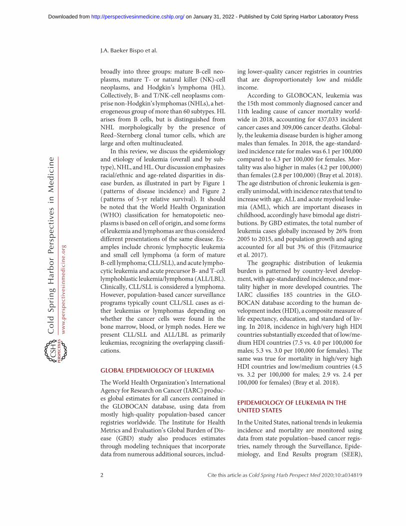
Figure 2. Five-year relative survival proportions for leukemia and lymphoma by race/ethnicity and age, 2008–2014. Data Source: Surveillance, Epidemiology, and End Results Program (SEER; http://seer.cancer.gov/registries/terms.html) 18 areas (San Francisco [SF], Connecticut, Detroit, Hawaii, Iowa, New Mexico, Seattle,Utah, Atlanta [ATL], San Jose18 areas Monterey [SJM], Los Angeles [LA], Alaska Native Registry, Rural Georgia[RG], California excluding SF/SJM/LA, Kentucky, Louisiana, New Jersey and Georgia excluding ATL/RG). The5-yr survival proportions are calculated using monthly intervals. Cancer sites are defined using the SEER SiteRecode ICD-O-3/World Health Organization (WH) 2008 Definition.
J.A. Baeker Bispo et al.
4 Cite this article as Cold Spring Harb Perspect Med 2020;10:a034819
ww
w.p
ersp
ecti
vesi
nm
edic
ine.
org
on January 31, 2022 - Published by Cold Spring Harbor Laboratory Presshttp://perspectivesinmedicine.cshlp.org/Downloaded from
males. Since 2006, the incidence of leukemia hasincreased by an average of 0.6% per year, where-as mortality has decreased by an annual averageof 1.5% (Siegel et al. 2018).
Overall age-adjusted leukemia incidence inthe United States tends to be highest in Whites(15 per 100,000), followed by Blacks (11 per100,000), and Hispanics (10.6 per 100,000). In-cidence among Asian/Pacific Islanders (API; 7.8per 100,000) and American Indian/Alaskan Na-tives (AIAN; 8.3 per 100,000) is lower by com-parison. Similar racial and ethnic patterns holdfor age-adjusted mortality rates, which are alsohigher for Whites (7 per 100,000), Blacks (5.6per 100,000), and Hispanics (4.8 per 100,000)than API (3.8 per 100,000) and AIAN (3.3 per100,000). Although incidence and mortalityrates are highest amongWhites, survival is poor-est for Black patients across age strata. Five-yearrelative survival for patients of <65 yr of age atdiagnosis is 73% forWhites and 63% for Blacks.For ages 65 and older, 5-yr relative survival is50% forWhites and 43% for Blacks (Noone et al.2017). Importantly, racial/ethnic patterns in leu-kemia incidence, mortality, and survival varywidely by subtype, as discussed in the sectionsthat follow.
OVERVIEW OF LEUKEMIA RISK FACTORS
Among those exposures most consistently iden-tified as risk factors for leukemia are radiation(therapeutic, occupational, and wartime-relat-ed), chemotherapy, family history, genetic syn-dromes and abnormalities, chemical exposures(e.g., residential and occupational), and lifestylefactors like smoking. Although some exposureshave been associatedwith specific leukemias, themost notable risk factors have an impact on sev-eral subtypes. For example, high doses of ioniz-ing atomic bomb radiation among residents ofJapan has been associated with increased mor-tality from all non-CLL leukemias independent-ly (ALL, AML, and CML) (Preston et al. 1994;Richardson et al. 2009; Hsu et al. 2013). Risk ofany non-CLL leukemia has been attributed toionizing radiation exposure in nuclear workersand radiologists prior to 1950 (Mohan et al.2003; Yoshinaga et al. 2004; Metz-Flamant
et al. 2012) and to therapeutic radiation expo-sure in patients with primary pelvic cancers(Boice et al. 1987; Wright et al. 2010) or benigndisorders like cervical polyps and endometrialhyperplasia (Sakata et al. 2012). Occupationalexposure to formaldehyde, a chemical used inmany building materials, household products,and industrial disinfectants, has demonstratedparticularly strong associations with myeloidleukemias (Beane Freeman et al. 2009; Zhanget al. 2009). The mechanisms by which risk fac-tors shared across leukemias promote oncogenicprocesses likely exhibit commonalities, althoughdetailed discussion of such mechanisms is be-yond the scope of this review.
PEDIATRIC LEUKEMIAS
Almost all leukemias in the pediatric populationare acute types. ALL is the most commonly di-agnosed childhood cancer worldwide. It ac-counts for ∼75% of leukemia cases in childrenof <15 yr of age, with a peak incidence rate of 7.8per 100,000 among children 2–4 yr of age in theUnited States (Noone et al. 2017). Unlike otherleukemias, which are more highly incidentamong Whites, the incidence of pediatric ALLis higher for Hispanics than for other racial andethnic groups. For Hispanic children of <20 yrof age, ALL incidence was 4.3 per 100,000 in theUnited States from 2001 to 2014, compared to3.4 forWhite, 3.0 for AIAN, 3.2 for API, and 1.9for Black children (Siegel et al. 2017). These rateshave largely remained stable since 2008. AML isthe second most common hematologic malig-nancy in children, with peak incidence in thepediatric population occurring in the first yearof life.
The causes of leukemia in pediatric patientsremain elusive. Several genetic syndromes andimmune disorders are associated with both ALLand AML risk, although most cases are not fa-milial. These include Down syndrome (DS), Li–Fraumeni syndrome, neurofibromatosis, DNArepair deficiency syndromes like Fanconi ane-mia and Bloom syndrome, and rare inheritedbone marrow failure syndromes like Kostmannsyndrome, Diamond–Blackfan anemia, dysker-atosis congenita, and Schwachman–Diamond
Epidemiology and Etiology of Leukemia and Lymphoma
Cite this article as Cold Spring Harb Perspect Med 2020;10:a034819 5
ww
w.p
ersp
ecti
vesi
nm
edic
ine.
org
on January 31, 2022 - Published by Cold Spring Harbor Laboratory Presshttp://perspectivesinmedicine.cshlp.org/Downloaded from
syndrome (Alter 2007; Owen et al. 2008). Ap-proximately 2.1% of individuals with DS de-velop leukemia by the age of 5, and 2.7% byage 30. Incidence of both ALL and AML amongindividuals with DS is more than 20-fold thatexpected in the general population. The magni-tude of DS-associated risk for AML is muchhigher than that for ALL in children of <5 yrof age (short interest ratio (SIR) = 154 vs. SIR= 41), but slightly higher for ALL compared toAML among children and young adults 5–30 yrof age (SIR = 12 vs. SIR = 10) (Hasle et al. 2000).
Pediatric leukemia is also associated withcertain therapeutic exposures, such as chemo-therapy with alkylating agents and topoisomer-ase II inhibitors for primary cancers (Hijiya et al.2009); diagnostic exposures such as ionizing ra-diation from CT scans (Pearce et al. 2012); andorgan transplantation, which may be related toiatrogenic immunosuppression and exposure tooncogenic viruses (Yanik et al. 2017). Severalrecent studies indicate that environmental expo-sure to benzene (e.g., via proximity to automo-bile traffic and factories), elevates AML risk inchildren (Honoré et al. 2015; Janitz et al. 2017).
Some studies indicate that early immunestimulation (e.g., via exposure to infection)may be protective against ALL (Rudant et al.2015; Hwee et al. 2018). Using data pooledacross 11 studies from the Childhood LeukemiaInternational Consortium, Rudant et al. (2015)reported a 23% reduction in risk of ALL amongchildren who attended day care (a proxy for im-mune stimulation) in the first year of life, with atrend for lower risks at younger start dates. Sev-eral important hypotheses articulate an infec-tious etiology of ALL. Greaves’ delayed-infectionhypothesis, first outlined in 1988, posits that de-layed (e.g., postinfancy) exposure to commoninfectious agents, for which the immune systemis unprepared, elicits an abnormal immune re-sponse that triggers ALL (Greaves 1988, 2006).In his model, an initiating genetic event in uterocreates a covert preleukemic clone that, upon asecond postnatal genetic “hit,” progresses toovert leukemia (Greaves 2006). By Kinlen’s pop-ulation-mixing hypothesis, also outlined in 1988,childhood leukemia is a rare response to a com-mon, unidentified infection that occurs when an
infected population “mixes” with a susceptible,nonimmune population (e.g., urban–rural mix-ing) (Kinlen 1988, 2012). The adrenal hypothe-sis, more recently proposed by Schmiegelow etal. (2008) postulates that early infection inducesthe hypothalamus–pituitary–adrenal axis toincrease plasma cortisol levels, which mayeliminate leukemic and preleukemic cells, andreduces risk of leukemogenesis by suppressingproinflammatory responses. Importantly, not allstudies support an infectious etiology of ALL.Designing epidemiologic studies to evaluatethese hypotheses is challenging—for example,prospective studies are unfeasible due to therarity of ALL, and factors like recall bias andtemporal ambiguity threaten the validity of ret-rospective studies (Hwee et al. 2018).
With improvements in recent decades torisk classification and treatments like combina-tion chemotherapies and targeted drug thera-pies, overall relative survival from ALL is high.Between 1975–1979 and 2003–2009, 5-yr rela-tive survival from pediatric ALL increased dra-matically from 57% to a very favorable 90%(Ward et al. 2014). AML also saw major gainsduring this period (from 21% to 64%), althoughsurvival remains less favorable than that forALL. Numerous studies demonstrate that Blackand Hispanic children with ALL historicallyhave suffered worse outcomes than their Whiteand non-Hispanic counterparts (Bhatia et al.2002; Kadan-Lottick et al. 2003; Pui et al.2012; Tai et al. 2017). Although survival patternsbetween White and Black children have con-verged in recent decades (particularly in chil-dren of <15 yr of age), several studies havedocumented widening disparities for Hispanicchildren (Wang et al. 2015; Kahn et al. 2016).From 2000 to 2010, ALL mortality among His-panic children was nearly twice that of Whites(HR= 1.95). Survival disparities may reflectboth biologic (e.g., genetic variations associatedwith ancestry) and socioeconomic (e.g., accessto care and treatment adherence) pathways. Inone recent study, accounting for the effect ofneighborhood socioeconomic status (SES) onhazard of death from ALL reduced the observedracial disparity from HR= 1.43 to HR= 1.22 forBlacks and fromHR= 1.63 toHR= 1.40 for His-
J.A. Baeker Bispo et al.
6 Cite this article as Cold Spring Harb Perspect Med 2020;10:a034819
ww
w.p
ersp
ecti
vesi
nm
edic
ine.
org
on January 31, 2022 - Published by Cold Spring Harbor Laboratory Presshttp://perspectivesinmedicine.cshlp.org/Downloaded from
panics, respectively, relative to Whites (Kehmet al. 2018). Research on childhood AML hasalso attributed an elevated risk of death to neigh-borhood socioeconomic factors—specificallyeconomic and educational disadvantages, hous-ing instability, and immigration-related features(Knoble et al. 2016).
ADULT LEUKEMIAS AND LYMPHOMAS
Acute Myeloid Leukemia
Worldwide, AML occurs with greatest frequen-cy in highly developed regions; age-standard-ized incidence is highest for both males andfemales in Australia (2.8 and 2.0 per 100,000),Austria (2.7 and 2.2 per 100,000), and the Unit-ed Kingdom (2.7 and 2.0 per 100,000) (Miran-da-Filho et al. 2018). In the United States, anestimated 19,520 cases of AML were diagnosedin 2018. Beginning in young adulthood, the agedistribution of AML incidence is exponentiallyshaped. SEER data show an especially sharp in-crease in incidence after 75 yr of age, when therate nearly doubles that of adults aged 60–74(209 vs. 109 cases per 1,000,000 person-years)(Dores et al. 2012). AML incidence and mortal-ity are higher among Whites than other racialand ethnic groups. For example, age-adjustedincidence among White males in SEER is 5.4per 100,000, compared to 4.5 for Blacks and4.1 for Hispanics (Noone et al. 2017). Mortalityrates demonstrate a similar racial pattern (3.8,2.7, and 2.3 per 100,000 for Whites, Blacks, andHispanics, respectively), and SEER data indicatethat these racial and ethnic trends persist acrosssex and age groups (Zhao et al. 2018).
The causes of genetic mutations that giverise to most cases of AML are largely unknown.Prominent risk factors evaluated in the literatureinclude radiation therapy, chemotherapy, smok-ing, and other environmental exposures. Mor-ton et al. (2013) have reported that the numberof AML diagnoses among patients who receivedchemotherapy for a first primary cancer is 4.7times that expected in the general population.Therapy-related myelodysplasia (MDS)/AMLis diagnostically classified by treatment type—namely, alkylating agent/radiation regimens or
topoisomerase inhibitor-related regimens—andtypically occurs within 10 yr of initial therapy(Bhatia 2013). In an early case-control study ofAML in breast cancer patients, Curtis et al. re-ported relative risks of 2.4 for receipt of radiationtreatment alone, 10.0 for alkylating agents, and17.4 for combined radiation-alkylating agentregimens (Curtis et al. 1992). Saso et al. (2000)also found that the risk of MDS/AML in breastcancer patients treated with alkylating agentswas 10-fold that of the general population. Al-though AML is a relatively rare disease, theabsolute excess risk associated with therapy issizeable (ranging from 5 to 7 excess cases per100,000 person-years in studies by Howardand colleagues) (Howard et al. 2007, 2008). An-other important risk factor for AML in adult-hood is smoking. Multiple meta-analysis showelevated risk of AML in smokers. Using datafrom 23 studies, Fircanis et al. (2014) calculateda 40% and 25% increased risk of AML in currentand former smokers, respectively, compared tononsmokers. Pooling data from nine Japanesecohort studies, Ugai et al. also showed an in-creased risk of near 40% for current smokers,and a 66% increase for ever-smokers with a his-tory of more than 30 pack-years compared tononsmokers (Ugai et al. 2018). Although find-ings for environmental chemical exposures areless consistent, benzene is a well-established riskfactor and has been recognized by the IARC as acause of AML (International Agency for Re-search on Cancer 1987; Khalade et al. 2010; Car-los-Wallace et al. 2016; Raaschou-Nielsen et al.2018). Benzene is one of the most widely usedchemicals in theUnited States, and occupationalexposure is highest in industries related torubber, oil refining, shoe manufacturing, andgasoline.
Several genetic abnormalities are associatedwith AML risk. Although familial AML is rare,AML is considered part of the natural history ofrare inherited bone marrow failure syndromeslike Kostmann syndrome, Diamond–Blackfananemia, dyskeratosis congenita, and Schwach-man–Diamond syndrome, as well as DNA re-pair deficiency syndromes like Fanconi anemiaand Bloom syndrome (Alter 2007; Owen et al.2008).
Epidemiology and Etiology of Leukemia and Lymphoma
Cite this article as Cold Spring Harb Perspect Med 2020;10:a034819 7
ww
w.p
ersp
ecti
vesi
nm
edic
ine.
org
on January 31, 2022 - Published by Cold Spring Harbor Laboratory Presshttp://perspectivesinmedicine.cshlp.org/Downloaded from
Survival from AML varies substantially byage, with dramatic declines observed for olderpatients. For those diagnosed before age 65,overall 5-yr relative survival is 45.6%, comparedto 7.1% for those diagnosed at age 65 or older(Noone et al. 2017). Despite some studies dem-onstrating that White patients present with lessfavorable prognostic profiles than other racial/ethnic groups (namely, lower rates of t(8;21) andacute promyelocytic leukemia), survival out-comes tend to be worst for Black and Hispanicpatients (Pulte et al. 2013; Patel et al. 2015b). Thesurvival disparity between Black and WhiteAML patients is especially pronounced for thosediagnosed at younger ages (e.g., at <65 yr of age)(Noone et al. 2017) Even after controlling for ageand genetic factors, Black race has been associ-ated with increased risk of death relative toWhites (Patel et al. 2012, 2015b). Some of theracial survival disparity may reflect treatmentdifferences; in California, for example, Blackrace has been associated with lower odds of re-ceiving chemotherapy and transplant (Patelet al. 2015a). Finally, although AML survivalhas increased for all racial/ethnic groups since1991–1996, disparities between groups have alsoincreased as gains have favored Whites, partic-ularly at younger ages. For White patients 15–54 yr of age, survival significantly increased by12.8 percentage points through 2003–2008,whereas gains for other racial and ethnic groups(4.3 percentage points for AA andHispanics, 7.1points for API) did not reach statistical signifi-cance (Pulte et al. 2013).
Acute Lymphocytic Leukemia
Overall, an estimated 5960 total cases of ALLwere diagnosed in the United States in 2018(Siegel et al. 2018). Whereas the incidence ofother leukemias generally increases with age,ALL is distinctly bimodal, with pediatric inci-dence rates far exceeding those for older agegroups. Throughout adulthood, incidence is rel-atively stable around 1 per 100,000, with an in-crease to 1.9 per 100,000 among elderly adultsaged 80–84 (Noone et al. 2017). Like other leu-kemias, ALL demonstrates a slight male pre-dominance.
Global incidence patterns for ALL are alsounique; whereas other leukemias are patternedby HDI and concentrated in Europe, NorthernAmerica, and Australia, ALL incidence is high-est in South and Central American countries—namely, Ecuador (2.8 and 3.3 per 100,000 formales and females, respectively), Costa Rica(2.4 and 2.3 per 100,000), and Colombia (2.3and 2.1 per 100,000) (Miranda-Filho et al.2018). In the United States, incidence of ALLvaries substantially by ethnicity. It is the onlyleukemia in which incidence, in both pediatricand adult cases, is higher for Hispanics than anyother racial or ethnic group.
There are no known causes of ALL. ALLtypically arises from noninherited geneticabnormalities. The Philadelphia chromosome,or t(9;22), is the most common chromosomaltranslocation in adult ALL (present in 25% ofcases), and unlike in children it is characterizedby a highly aggressive clinical course (Gleissneret al. 2002; Pui et al. 2004; Thomas and Heiblig2016). Many risk factors for adult ALL are sim-ilar to those of pediatric ALL, including chemo-therapy (e.g., for primary cancers), ionizingradiation (e.g., therapy-related or atomic bombexposure), and chemical toxins like benzene.The specific mechanisms of risk for these expo-sures remain unclear.
Survival fromALL declines with age at diag-nosis; 5-yr relative survival is 35.8% for adults45–54 yr of age, 26.4% for adults 55–64 yr orage, and 16.5% for adults diagnosed at age 65or older (Noone et al. 2017). When stratified byrace and ethnicity, survival is higher for WhiteALL patients compared to other groups, partic-ularly Black ALL patients. SEER data from 2000to 2014 indicate that racial survival disparitiesare more pronounced at younger ages (namelyfor patients 15–39 yr of age) (Kirtane and Lee2017). Adult survival disparities have persisteddespite Black patients experiencing some ofthe largest recent improvements in 5-yr surviv-al (e.g., from 24.1% to 43.4% among patients15–44 yr of age between 1997–2002 and 2003–2008) (Pulte et al. 2013). Hispanic adults, forunclear reasons, also continue to face poorer5-yr relative survival than White adults andhave experienced smaller gains in survival com-
J.A. Baeker Bispo et al.
8 Cite this article as Cold Spring Harb Perspect Med 2020;10:a034819
ww
w.p
ersp
ecti
vesi
nm
edic
ine.
org
on January 31, 2022 - Published by Cold Spring Harbor Laboratory Presshttp://perspectivesinmedicine.cshlp.org/Downloaded from
pared to other racial/ethnic groups (Kirtane andLee 2017).
Chronic Myeloid Leukemia
CML is cytogenetically characterized by thePhiladelphia chromosome––a truncation ofchromosome 22 resulting from the reciprocaltranslocation t(9;22)(q32;q11). CML accountsfor ∼15% of leukemia diagnoses in the UnitedStates, or an estimated 8430 new cases in 2018(Siegel et al. 2018). Incidence increases steadilywith age, peaking at 10.3 cases per 100,000among individuals 80–84 yr of age, althoughthe most frequent age of diagnosis is between65 and 74 yr of age (comprising 21% of CMLdiagnoses). Racial and ethnic patterns in CMLincidence are more disparate than for other leu-kemias. In U.S. males, incidence is highest inWhites (2.4 per 100,000) and AI/AN (2.3 per100,000), whereas for females incidence is high-est in Whites and Blacks (1.4 per 100,000) (No-one et al. 2017). Worldwide, there is somevariability in CML incidence rates by country,but no clear patterning by HDI. Rates are high-est in Australia (1.8 and 1.0 per 100,000 inmalesand females, respectively), Lithuania (1.6 and0.9 per 100,000), France (1.7 per 100,000males),and Uruguay (1.1 per 100,000 females) (Miran-da-Filho et al. 2018).
Apart from increasing age, the only knownrisk factor for CML is exposure to ionizing ra-diation, which has been described in literatureon leukemia among atomic bomb survivors(Heyssel et al. 1960).
CML has a moderate prognosis, with 5-yrrelative survival at 68.7% (Noone et al. 2017).Survival from CML underwent drastic improve-ments after the introduction of the first tyrosinekinase inhibitor (TKI), imatinib mesylate (Glee-vec), in 2001. Prior to this, the prognosis waspoor, with overall relative survival <50% for pa-tients within 3 yr of diagnosis (Mandal et al.2013). Although survival has improved acrossracial and ethnic groups, Mandal et al. foundthat 3-yr relative survival in the post-imatiniberawas nevertheless significantly lower for Blackfemales (80.5%) than White females (90.3%),and that survival gains favored younger patients
(<50 yr) over older patients. The reason for ra-cial and ethnic disparities in CML survival havebeen difficult to identify. Wiggins et al. did notfind TKI treatment disparities in the UnitedStates by race/ethnicity, SES, urban/rural resi-dence, comorbidity, or insurance status aftercontrolling for age (Wiggins et al. 2010). How-ever, age disparities in survival improvementpartially reflected less frequent administrationof imatinib regimens in elderly patients.
Chronic Lymphocytic Leukemia/Small CellLymphoma
Worldwide, CLL/SLL incidence is highest incountries of very high HDI—namely, Canadaand France, where annual rates exceed fournew cases per 100,000 males. Incidence is espe-cially low in Asian countries, particularly Japan(0.1 per 100,000), Malaysia (0.1 per 100,000),and the Philippines (0.2 per 100,000). Sex-spe-cific differences are stronger for CLL/SLL thanfor other leukemias, with incidence amongmales nearly double that of females, both glob-ally and in the United States (Miranda-Filhoet al. 2018).
CLL/SLL is themost common leukemia sub-type in the United States, with an estimated20,940 new cases diagnosed in 2018 (Siegelet al. 2018). Incidence in theUnited States varieswidely by race. The most elevated incidencerates are observed for Whites (5.1 per 100,000)and Blacks (3.6 per 100,000). Incidence is mark-edly lower, roughly a quarter that of Whites,among the API and AIAN population. Risk ofCLL/SLL is strongly age-dependent, with 67% ofdiagnosesmade to individuals older than age 65.Among individuals aged 65 and older, incidenceis 26.4 per 100,000; for the oldest age strata (85and older), incidence is 35.8 per 100,000 (Nooneet al. 2017).
Family history of hematologic malignancy isthe strongest and most consistent risk factor forCLL/SLL (even if the absolute risk among first-degree relatives is low), implicating commoninherited genetic pathways in CLL/SLL patho-genesis. Large-scale studies using data from theSwedish Cancer Registry have demonstratedthat relatives of CLL/SLL cases have a 7.5- to
Epidemiology and Etiology of Leukemia and Lymphoma
Cite this article as Cold Spring Harb Perspect Med 2020;10:a034819 9
ww
w.p
ersp
ecti
vesi
nm
edic
ine.
org
on January 31, 2022 - Published by Cold Spring Harbor Laboratory Presshttp://perspectivesinmedicine.cshlp.org/Downloaded from
8.5-fold risk of developing CLL/SLL over rela-tives of controls (Goldin et al. 2004b, 2009). Inone of the largest epidemiologic studies on CLL/SLL risk, which pooled data across 13 case-con-trol studies in Europe, North America, and Aus-tralia as part of the International LymphomaEpidemiology Consortium (InterLymph), his-tory of any hematological malignancy amongfirst-degree relatives was associated with a great-er than twofold odds of CLL/SLL odds ratio (OR= 2.17) (Slager et al. 2014).
No single germline mutation has been iden-tified as a causal precursor to CLL/SLL (Goldinand Caporaso 2007). Monoclonal B-cell lym-phocytosis (MBL), which is more frequent inhigh-risk CLL/SLL families than the generalpopulation, may be an early genetic factor indic-ative of inherited predisposition (Rawstron et al.2002; Goldin et al. 2013). Some studies suggestthat geographic and racial variability in CLL/SLL incidence and prognosis reflect underlyingdifferences in genetic risk factors betweengroups. For example, Coombs et al. found thatthe risk allele frequency of most single nucleo-tide polymorphisms known to confer risk ofCLL/SLL in Whites is not associated with riskamong Black CLL/SLL patients (Coombs et al.2012b). The rarity of CLL/SLL among Asians,both in Asia and abroad, also supports the no-tion of a strong genetic component to diseaserisk. Several studies have failed to show differ-ences in rates of CLL/SLL between Asian mi-grants to the United States (foreign-born) andtheir U.S.-born descendants, suggesting a limit-ed role for the impact of environmental andlifestyle-related exposures on CLL/SLL risk atthe population level (Herrinton et al. 1996;Gale et al. 2000; Pan et al. 2002).
Other exposures evaluated as risk factors forCLL/SLL include medical history, biometriccharacteristics, lifestyle-related factors, and var-ious environmental, occupational, and chemicalexposures. Pooled analyses across InterLymphstudies have demonstrated elevated odds ofCLL/SLL associated with increasing height(OR= 1.09 per 10 cm), hepatitis C seropositivity(OR= 1.99), residential or occupational historyon a farm (OR= 1.20), and occupational historyas a hairdresser (OR= 1.77) (Slager et al. 2014).
Although studies on chemical exposures arelargely inconsistent, the National Institute ofMedicine concluded in 2003 that Agent Orange,an dioxin-containing herbicide used in Viet-nam, is associated with CLL/SLL in veterans(Institute of Medicine 2009). Protective factorsfrom pooled InterLymph data include history ofatopic disorder (OR= 0.85), blood transfusion(OR= 0.79), cigarette smoking (OR= 0.91),and sun exposure (OR= 0.71 for highest quartilecompared to lowest) (Slager et al. 2014).
Unlike other leukemias, CLL/SLL generallyis considered nonradiogenic. Some researcherschallenge this conclusion, particularly in light ofevidence on CLL/SLL in Czech uranium minersand those exposed to radiation following theChernobyl nuclear power plant accident (Řeři-cha et al. 2006; Hamblin 2008). Results aroundoccupational and medical radiation exposureare inconsistent overall; however, Silver et al.noted that CLL/SLL risk estimates for irradiatedpatients inmedical cohort studies with >15 yr offollow-up were almost uniformly elevated, albeitnonsignificantly (Silver et al. 2007). The de-cades-long latency period of CLL/SLL, lowcase-fatality rate, lack of diagnostic specificity,historical underreporting bias, and difficulty inachieving an adequate sample size are all majorchallenges in observational studies that examineradiation (and environmental exposures gener-ally) as a risk factor of CLL/SLL (Richardsonet al. 2005).
Relative 5-yr survival in the United Statesoverall is high, exceeding 84% (Noone et al.2017). Both institutional and population-basedstudies indicate that Black patients have poorerprognostic profiles at diagnosis, and worse sur-vival, than other racial and ethnic groups (She-noy et al. 2011; Coombs et al. 2012a; Falchi et al.2013). Pooled data from MD Anderson andDuke University demonstrated that comparedto other races combined, Black patients hadworse biological and genetic characteristics atdiagnosis, including lower hemoglobin andhigher β-microglobulin; more frequently pre-sented with unmutated IGHV gene, ZAP70 ex-pression, and chromosome 17p or 11q deletion;more frequently required first-line therapy; andhad shorter overall and event-free survival after
J.A. Baeker Bispo et al.
10 Cite this article as Cold Spring Harb Perspect Med 2020;10:a034819
ww
w.p
ersp
ecti
vesi
nm
edic
ine.
org
on January 31, 2022 - Published by Cold Spring Harbor Laboratory Presshttp://perspectivesinmedicine.cshlp.org/Downloaded from
controlling for prognostic factors (Falchi et al.2013). Using data from SEER, Shoney et al. alsoshowed that Black CLL/SLL patients are diag-nosed at younger ages and suffer worse survivalthan White patients after controlling for prog-nostic factors like disease stage, extra-nodal pri-mary site, and B symptoms (HR= 1.67) (Shenoyet al. 2011).
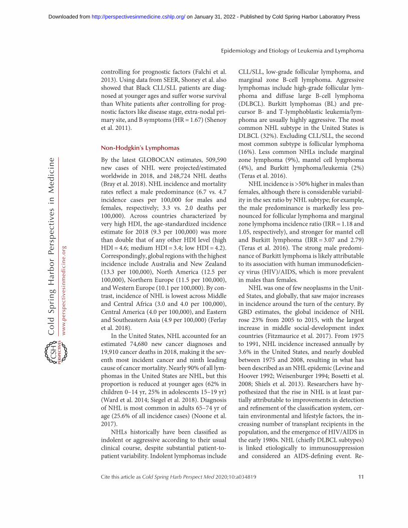
Non-Hodgkin’s Lymphomas
By the latest GLOBOCAN estimates, 509,590new cases of NHL were projected/estimatedworldwide in 2018, and 248,724 NHL deaths(Bray et al. 2018). NHL incidence and mortalityrates reflect a male predominance (6.7 vs. 4.7incidence cases per 100,000 for males andfemales, respectively; 3.3 vs. 2.0 deaths per100,000). Across countries characterized byvery high HDI, the age-standardized incidenceestimate for 2018 (9.3 per 100,000) was morethan double that of any other HDI level (highHDI = 4.6; medium HDI = 3.4; low HDI = 4.2).Correspondingly, global regionswith the highestincidence include Australia and New Zealand(13.3 per 100,000), North America (12.5 per100,000), Northern Europe (11.5 per 100,000),andWestern Europe (10.1 per 100,000). By con-trast, incidence of NHL is lowest across Middleand Central Africa (3.0 and 4.0 per 100,000),Central America (4.0 per 100,000), and Easternand Southeastern Asia (4.9 per 100,000) (Ferlayet al. 2018).
In the United States, NHL accounted for anestimated 74,680 new cancer diagnoses and19,910 cancer deaths in 2018, making it the sev-enth most incident cancer and ninth leadingcause of cancermortality. Nearly 90% of all lym-phomas in the United States are NHL, but thisproportion is reduced at younger ages (62% inchildren 0–14 yr, 25% in adolescents 15–19 yr)(Ward et al. 2014; Siegel et al. 2018). Diagnosisof NHL is most common in adults 65–74 yr ofage (25.6% of all incidence cases) (Noone et al.2017).
NHLs historically have been classified asindolent or aggressive according to their usualclinical course, despite substantial patient-to-patient variability. Indolent lymphomas include
CLL/SLL, low-grade follicular lymphoma, andmarginal zone B-cell lymphoma. Aggressivelymphomas include high-grade follicular lym-phoma and diffuse large B-cell lymphoma(DLBCL). Burkitt lymphomas (BL) and pre-cursor B- and T-lymphoblastic leukemia/lym-phoma are usually highly aggressive. The mostcommon NHL subtype in the United States isDLBCL (32%). Excluding CLL/SLL, the secondmost common subtype is follicular lymphoma(16%). Less common NHLs include marginalzone lymphoma (9%), mantel cell lymphoma(4%), and Burkitt lymphoma/leukemia (2%)(Teras et al. 2016).
NHL incidence is >50% higher inmales thanfemales, although there is considerable variabil-ity in the sex ratio by NHL subtype; for example,the male predominance is markedly less pro-nounced for follicular lymphoma and marginalzone lymphoma incidence ratio (IRR= 1.18 and1.05, respectively), and stronger for mantel celland Burkitt lymphoma (IRR= 3.07 and 2.79)(Teras et al. 2016). The strong male predomi-nance of Burkitt lymphoma is likely attributableto its association with human immunodeficien-cy virus (HIV)/AIDS, which is more prevalentin males than females.
NHL was one of few neoplasms in the Unit-ed States, and globally, that saw major increasesin incidence around the turn of the century. ByGBD estimates, the global incidence of NHLrose 23% from 2005 to 2015, with the largestincrease in middle social-development indexcountries (Fitzmaurice et al. 2017). From 1975to 1991, NHL incidence increased annually by3.6% in the United States, and nearly doubledbetween 1975 and 2008, resulting in what hasbeen described as anNHL epidemic (Levine andHoover 1992; Weisenburger 1994; Bosetti et al.2008; Shiels et al. 2013). Researchers have hy-pothesized that the rise in NHL is at least par-tially attributable to improvements in detectionand refinement of the classification system, cer-tain environmental and lifestyle factors, the in-creasing number of transplant recipients in thepopulation, and the emergence of HIV/AIDS inthe early 1980s. NHL (chiefly DLBCL subtypes)is linked etiologically to immunosuppressionand considered an AIDS-defining event. Re-
Epidemiology and Etiology of Leukemia and Lymphoma
Cite this article as Cold Spring Harb Perspect Med 2020;10:a034819 11
ww
w.p
ersp
ecti
vesi
nm
edic
ine.
org
on January 31, 2022 - Published by Cold Spring Harbor Laboratory Presshttp://perspectivesinmedicine.cshlp.org/Downloaded from

cently, NHL incidence has plateaued, with anannual 0.8% decrease in incidence since 2007(Noone et al. 2017). Shiels et al. (2013) havesuggested that the recent stabilization of NHLrates in the general population is independent ofHIV. Among NHL subtypes, decreases in inci-dence have been most pronounced for CLL/SLL(APC=−2.7% and −2.9% for males and fe-males, respectively, since 2005), and follicularlymphoma (APC=−2.2 for males since 2004,and −3.6% for females since 2007). Rates ofDLBCL have remained stable since 2001 (Teraset al. 2016).
NHL incidence and mortality tend to behigher in Whites than other racial/ethnicgroups, with some variability in racial and eth-nic incidence patterns by subtype. Incidence ofB-cell NHL is 7%, 18%, and 72% higher amongWhites compared to Blacks, Hispanics, andAPI,respectively. On the other hand, the incidence ofT/NK-cell NHL is 49% higher in Blacks thanWhites (Teras et al. 2016)
Lymphomas typically are characterizedby chromosomal translocations that are notheritable, and research on NHL etiology mostconsistently supports a role for infection (pre-dominantly viral) and immunosuppression.Specific pathogens that have been linked to riskof NHL include human herpes virus 8 (Oksen-hendler et al. 2002; Deloose et al. 2005), hepatitisC (HCV) (Matsuo et al. 2004; Morton et al.2014), humanT-cell lymphotropic virus (Mannset al. 1993; Cleghorn et al. 1995), and Helico-bacter pylorus (Parsonnet et al. 1994; Sagaertet al. 2010), among others. In onemeta-analysis,the pooled relative risk of NHL associated withHCV infection was 2.5, and the authors estimat-ed that upward of 10%ofNHL in areas with highHCVprevalence could be attributed to the infec-tion (Dal Maso and Franceschi 2006). In 2009,the IARC Working Group classified Epstein–Barr virus (EBV), amemberof the herpes family,a class 1 carcinogen that causes severalNHL sub-types, including Burkitt lymphoma, sinonasalangiocentric T-cell lymphoma, and immuno-suppression-related NHL (IARC 2012).
The mechanisms of carcinogenesis for viralexposures vary, but EBV has been the focus ofmost research. Possible oncogenic properties of
EBV include the immortalization of B cells andthe encoding of gene products that cause geno-mic instability, induce cell proliferation, andblock apoptosis. Worldwide, EBV infection isubiquitous; primary infection typically occursin childhood or adolescence, after which the vi-rus establishes lifelong latency in lymphocytes,with the possibility of reactivation. The carcino-genic potential of EBV frequently arises in thecontext of sustained immunosuppression or in-fectious cofactors. For example, there is strikingcorrespondence in the geographic distributionof endemic BL and holoendemic malaria. Intropical zones of subequatorial Africa, endemicBL accounts for 20% of childhoodmalignancies,and EBV is present in nearly 100% of cases(Orem et al. 2007; Silver et al. 2007). It is gener-ally understood that early coinfection withmalaria alters EBV persistence and deterioratesimmunoregulatory control of EBV, resulting ina malignant B-cell clone (Rochford et al. 2005;Orem et al. 2007; Chene et al. 2009; Moormannet al. 2011). Several studies support a synergisticeffect of malaria and EBV coinfection on risk ofchildhood BL, with the odds of elevated anti-body titers for both EBV and malaria in casesbetween five to 13 times that of controls (Car-penter et al. 2008; Mutalima et al. 2008).
In individuals withHIVorAIDS, NHL is thesecond most common malignancy after Kapo-si’s sarcoma. From 1996 to 2010, incidence ofNHL among individuals registered with HIV orAIDS was 193.7 per 100,000 person years, abouthalf of which were DLBCL (Gibson et al. 2014).In one review of population-based studies acrossthe United States, Australia, and Italy, the risk ofNHL in individuals with AIDS compared to thegeneral population ranged from 15-fold for low-gradeNHL to 400-fold for high-gradeNHL (DalMaso and Franceschi 2003). Incidence of NHLin people with AIDS declined dramatically fol-lowing the introduction of highly active antire-troviral therapy in 1996. In theUnited States, therelative risk of NHL in people with AIDS com-pared to the general population reduced fromSIR = 53.2 to SIR = 22.6 between 1990–1995and 1996–2002 (Engels et al. 2006). In a reportby Hernández-Ramírez et al., NHL risks since1996 have declined significantly for DLBCL and
J.A. Baeker Bispo et al.
12 Cite this article as Cold Spring Harb Perspect Med 2020;10:a034819
ww
w.p
ersp
ecti
vesi
nm
edic
ine.
org
on January 31, 2022 - Published by Cold Spring Harbor Laboratory Presshttp://perspectivesinmedicine.cshlp.org/Downloaded from

central nervous system NHL, but not BL (Her-nández-Ramírez et al. 2017). Nevertheless, inci-dence of NHL remains elevated for individualswith HIV or AIDS. Analogous to models of ma-laria as a cofactor in EBV-driven BL pathogen-esis, current opinion holds that HIV infectioncontributes to NHL pathogenesis indirectlythrough sustained B-cell activation and im-paired immunoregulatory control of oncogenicviruses like EBV. Emerging evidence suggeststhat HIV may also contribute directly throughHIV-encoded proteins that enhance B-cell clo-nogenicity and increase propensity for chromo-somal translocations (Dolcetti et al. 2016).
Populations treated with immunosuppres-sive drugs, including organ transplant recipientsand individuals with autoimmune diseases, alsoexperience elevated risk of NHL. In a large co-hort study linking 20 years of data from theUnited States Scientific Registry of Transplantswith state and regional cancer registries, Engelset al. reported a more than sevenfold increase inrisk of NHL in transplant recipients comparedto the general population. Among transplantrecipients, an excess of 168.3 cases of NHL per100,000 person-years could be attributed totransplant (Engels et al. 2011). Relative risksare strongest at younger ages. In a recent regis-try-linked study on pediatric recipients, 71% ofposttransplant malignancies were NHL; risk ofNHL in recipients was 212 times that of thegeneral population, and exceeded 300 for recip-ients of <5 yr of age (Yanik et al. 2017). Datafrom this study also indicate EBV as a cofactorin pathogenesis. Patients who were seronegativefor EBV prior to transplantation had 2.7 timesthe risk of NHL compared to those already sero-positive. Presumably, drug-induced immuno-suppression impairs the control of newlyacquired EBV infection posttransplant, result-ing in heightened risk of the oncogenic effectsassociated with EBV.
Individuals with autoimmune disorders alsoexperience increased risk of NHL. In their meta-analysis of 20 cohort studies, Zintzaras reportedthat NHL risk was greatest for patients withSjögren’s syndrome (SIR = 18.8), followed bysystemic lupus (SIR = 7.4) and rheumatoidarthritis (SIR = 3.9) (Zintzaras et al. 2005).
A pooled analysis of 12 InterLymph case-controlstudies showed more modest effect estimates;Sjögren’s syndrome and systemic lupus were as-sociated with 6.5-fold and 2.7-fold odds ofNHL,respectively, and both conditions demonstratedsignificant subtype-specific risk for DLBCL(Ekström Smedby et al. 2008). A large Swedishregistry study found that diagnosis with any of33 autoimmune diseases corresponded to a 60%increase is risk of NHL (Fallah et al. 2014a). Therisk of NHL in this population may reflect theeffects of sustained inflammatory activity anddisease severity over treatment related immu-nosuppression or EBV infection (Smedby et al.2006).
Familial aggregation of NHL has been wide-ly reported in the literature. Several large popu-lation-based studies in Europe have reportedrelative risks of NHL near 1.8 for first-degreerelatives of individuals with NHL (Altieri et al.2005; Goldin et al. 2005). It appears that relativesof NHL patients are at highest risk for subtype-concordant malignancy; for example, Goldinet al. (2009) found a 10-fold increase in risk ofDLBCL in relatives of DLBCL patients, and afourfold risk of follicular lymphoma in relativesof follicular lymphoma patients. Despite theseassociations, the absolute risk attributed to fa-milial predisposition is very modest and doesnot warrant clinical surveillance of first-degreerelatives (Cerhan and Slager 2015).
Research on NHL risks linked to environ-mental exposures and lifestyle-related factors islargely inconclusive. Although studies generallyshow no or only modest associations betweensmoking and NHL overall, pooled data fromnine InterLymph case-control studies suggestthat smoking is positively associated with follic-ular lymphoma, specifically; the odds of follicu-lar lymphoma were 31% higher in currentsmokers than nonsmokers, and 45% higher incurrent heavy smokers (with a history of morethan 36 pack-years) (Morton et al. 2005). Re-searchers have hypothesized that exposure toultraviolet radiation (UV) may confer NHLrisk, but results are inconsistent. Thirty yearsof prospective data from the Nurses’ Healthstudy showed that residing in an area with high-er versus lower ambient UV radiation was asso-
Epidemiology and Etiology of Leukemia and Lymphoma
Cite this article as Cold Spring Harb Perspect Med 2020;10:a034819 13
ww
w.p
ersp
ecti
vesi
nm
edic
ine.
org
on January 31, 2022 - Published by Cold Spring Harbor Laboratory Presshttp://perspectivesinmedicine.cshlp.org/Downloaded from
ciated with a 21% increased risk of NHL (Ber-trand et al. 2011). Conversely, several studieshave found that UV exposure provides a protec-tive effect. In an analysis with data from theCalifornia Teachers Study, exposure to residen-tial UV in the highest versus lowest quartile wasassociated with a 42% decrease in risk of NHLoverall, with an even stronger protective effectagainst DLBCL (relative risk [RR] = 0.36)(Chang et al. 2011). Pooled data from Inter-Lymph studies have also shown a protective ef-fect of recreational sun exposure (Morton et al.2014). InterLymph analyses do not support alink between BMI as a young adult and NHLrisk (Morton et al. 2014); however, a recentmeta-analysis of 22 studies by Hidayat and col-leagues showed a significant 11% increase inNHL risk per 5 kg/m2 increase in BMI duringearly adulthood, and a 21% increase in risk per10 kg increase in weight (Hidayat et al. 2018).Other important risk factors described inpooled analyses of InterLymph studies includeoccupational history as a farm worker and ex-posure to hair dye prior to 1980; additional pro-tective factors include history of atopic diseases,blood transfusion, and alcohol consumption ofat least one drink per month (Zhang et al. 2008;Morton et al. 2014).
From 2008 to 2014, the overall 5-yr relativesurvival in patients diagnosed with NHL was74.1%, but there is considerable variability bysubtype, and survival is typically worse for BlackNHL patients than those of other racial and eth-nic groups (Noone et al. 2017). Five-year sur-vival tends to be higher for follicular lymphoma(86% and 81% for White and Black males, re-spectively) andmarginal zone lymphomas (89%and 83%), and lower for Burkitt (63% and 47%)and DLBCL (62% and 55%) (Teras et al. 2016).
Hodgkin’s Lymphoma
In 2018, HL accounted for an estimated 79,990incident cancer diagnoses and 26,167 cancerdeaths worldwide (Bray et al. 2018). Like NHL,incidence of HL in very high HDI countries (2.1per 100,000) ismore than double that of all otherHDI levels. Regions with the most elevated HLincidence rates include Southern, Northern, and
Western Europe (2.8, 2.6, and 2.5 per 100,000),Australia and New Zealand (2.5 per 100,000),and North America (2.4 per 100,000). Incidenceis lowest across Eastern and Southern Asia, andsub-Saharan Africa (Ferlay et al. 2018). By GBDestimates, the number of new HL diagnosesglobally declined by 6.1% after accounting forpopulation growth and changing age structure(Fitzmaurice et al. 2017).
HL comprises roughly 10% of all lympho-mas in the United States. In 2018, HL accountedfor an estimated 8500 new cancer diagnoses and1050 cancer deaths (Siegel et al. 2018). Incidenceis higher among Whites than other racial andethnic groups. The age distribution of HL is dis-tinctly bimodal, with peak incidence occurringfirst among young adults 20–24 yr of age andagain among elderly adults 75–79 yr of age (No-one et al. 2017). Most patients diagnosed withHL are of <40 yr of age. In the pediatric context,HL is the most common malignancy in adoles-cents 15–19 yr of age, accounting for 15% ofcancer diagnoses in this age group (Ward et al.2014). Of note, one large SEER study using 15years of data observed distinct racial patterns inthe age distribution of HL; for Black males, HLincidence remained relatively stable after peak-ing in early adulthood, and for Hispanics, inci-dence rose in an exponential form after age 40,with only a small uptick in incidence duringearly adulthood (Evens et al. 2012).
Clinical features of HL also vary by race andethnicity in the United States. Several studieshave shown that Blacks and Hispanics are morelikely than Whites to be diagnosed with HL ofmixed cellularity and less likely to be diagnosedwith nodular sclerosis HL (the predominantsubtype of classic HL in the United States) (Gla-ser et al. 2008, 2014;Evens et al. 2012;Grubbet al.2016). Incidence of nodular sclerosis HL reflectsa socioeconomic gradient, whereby higher SES isassociated with elevated risk (Clarke et al. 2005).EBV-positive HL, which tends to be of mixedcellularity, is more common in Hispanics thanWhites, particularly among foreign-born His-panics, and those diagnosed at younger andolder ages (Glaser et al. 2008, 2014).
Family studies indicate that risk of HL, likeNHL, shows a familial predisposition. Large
J.A. Baeker Bispo et al.
14 Cite this article as Cold Spring Harb Perspect Med 2020;10:a034819
ww
w.p
ersp
ecti
vesi
nm
edic
ine.
org
on January 31, 2022 - Published by Cold Spring Harbor Laboratory Presshttp://perspectivesinmedicine.cshlp.org/Downloaded from
population-based studies from Sweden andDenmark have reported relative risks rangingfrom 3.1-fold to 8.8-fold for first-degree relativesof HL patients (Goldin et al. 2004a; Crump et al.2012). The high concordance of HL in monozy-gotic, but not dizygotic, twins indicates thatgenetic factors play an etiologic role in the devel-opment of some cases of HL (Mack et al. 1995).
HL pathogenesis likely involves a complexinterplay between genetic susceptibility, im-mune impairment, and environmental expo-sures. Abnormal immune response to infectiousagents may trigger oncogenic processes thatcause HL. By the delayed-exposure model ofHL etiology, increased exposure to infection ata young age may protect against HL by promot-ing early maturation of cellular immunity.Markers of early exposure to common child-hood pathogens, like day care attendance(Chang et al. 2004b; Rudant et al. 2011) andhaving older siblings (Chang et al. 2004a; Altieriet al. 2006), typically show a protective effectagainst HL. Data linking EBV to HL risk alsosupports an infectious etiology for some HL.EBV infection in adolescence manifests clinical-ly as infectious mononucleosis (IM). Whereashistory of infection with chicken pox, measles,mumps, pertussis, and rubella have shown aprotective effect against HL, history of IM is as-sociated with increased HL risk in young adults(Alexander et al. 2000; Hjalgrim et al. 2000).Furthermore, IM-related risk appears specificto EBV-positive HL (Hjalgrim et al. 2003).
Immunosuppression is associated with riskof HL, although typically to a smallermagnitudethan that for NHL. In a recent study that evalu-ated cancer outcomes in 448,258 HIV-infectedindividuals using linked population-based reg-istries, risk of HLwas 4.6-fold and 9.4-fold high-er for individuals with HIV only and AIDS,respectively (Hernández-Ramírez et al. 2017).An earlier meta-analysis of cohort studiesreported 11-fold and fourfold risks of HL inindividuals with HIV/AIDS and transplant re-cipients, respectively. In AIDS patients, nearly100% of classical HL is EBV-positive and ofmixed cellularity histology (Bibas and Antinori2009). Recently, in a large analysis of linked can-cer and transplant registry data in the United
States, incidence of HL in pediatric transplantsrecipients was 19 times that of the general pop-ulation (Yanik et al. 2017). The risk associatedwith transplant appears somewhat attenuated(SIR = 3.6) when not restricting to the pediatricpopulation (Engels et al. 2011). Autoimmunediseases characterized by chronic inflammation,like rheumatoid arthritis and systemic lupus,have also been linked to HL, with stronger asso-ciations noted for EBV-positive malignancy(Fallah et al. 2014b; Hollander et al. 2015).
Some evidence suggests modest associationsbetween HL and lifestyle factors like smokingand exposure to UV radiation. In a pooled anal-ysis of 12 case-control studies, overall odds ofHL in ever smokers were 1.1 times that of neversmokers, with higher odds for mixed cellularityHL (OR= 1.6) and EBV-positive HL (OR= 1.8).Risks associated with UV exposure also appearspecific to EBV-positive HL, but in the oppositedirection; in a pooled analysis of four case-con-trol studies, individuals in the highest categoryof UV exposure had a 44% reduction in odds ofEBV-positive HL compared to those in the low-est category (Monnereau et al. 2013).
Overall, HL has a favorable prognosis; the 5-yr relative survival rate exceeded 88% from 2008to 2014, and was even higher for individualsyounger than 45 yr of age at diagnosis (94%)and pediatric cases (97%) (Ward et al. 2014;Noone et al. 2017). Still, SEER and state-specificregistry analyses tend to show poorer survivalpatterns among Black and Hispanic patientscompared to White patients (Keegan et al.2009, 2016; Grubb et al. 2016). In Grubb’s anal-ysis of SEER data, overall survival disparitiespersisted through 25 years of follow-up aftercontrolling for demographics, stage, histology,and treatment; however, the magnitude of dif-ferences was less pronounced for Hispanics, andnot significant for Blacks, when considering dis-ease-specific survival versus overall survival(Grubb et al. 2016).
CONCLUDING REMARKS
Leukemias and lymphomas comprise a hetero-geneous group of malignancies characterized bythe uncontrolled proliferation of cells from pre-
Epidemiology and Etiology of Leukemia and Lymphoma
Cite this article as Cold Spring Harb Perspect Med 2020;10:a034819 15
ww
w.p
ersp
ecti
vesi
nm
edic
ine.
org
on January 31, 2022 - Published by Cold Spring Harbor Laboratory Presshttp://perspectivesinmedicine.cshlp.org/Downloaded from
dominantly myeloid and lymphoid lineages inhematopoietic and lymphoid tissues. The vastmajority of these malignancies are sporadic,and specific etiologic mechanisms remain elu-sive. Leukemias and lymphomas arise in thecontext of various host and environmental fac-tors. Host factors include genetic abnormalities(most often chromosomal translocations), rareinherited disorders, and iatrogenic or disease-related immunosuppression. Key environmen-tal factors related to leukemia include ionizingradiation, chemotherapy, and carcinogenicchemicals like benzene. Associations betweenlymphomas and EBV are suggestive of an infec-tious etiology for certain subtypes such as Bur-kitt lymphoma and DLBCL.
Leukemia and lymphoma incidence appearto be highest in highly developed regions acrossEurope, Northern America, and Australia, andamong U.S. Whites. An exception to this isALL, for which incidence is highest in Southand Central American countries, and amongHispanics in the United States. Global compar-isons in leukemia and lymphoma incidence aremade difficult by limited diagnostic infrastruc-ture in less developed countries. Incidence andmortality rates in part reflect a population’s ac-cess to the formal health-care system, as well asthe availability of medical facilities that supportmorphological, immunohistochemical, and cy-togenetic profiling involved in leukemia diag-nosis. These factors likely affect overall globalcounts of leukemia and lymphoma. Further-more, they may exaggerate differences in overallincidence between more and less developed re-gions of the world and particularly limit con-clusions about observed global patterns that aresubtype-specific.
Survival from leukemia and lymphoma var-ies widely by subtype, ranging from 27.4% 5-yrrelative survival from AML to 84.2% for CLL/SLL and 86.6% for HL. Clearly, new treatmentsare necessary for poor prognosis malignanciessuch as ALL and AML among adults both withvery low survival. Chemotherapy is the mostcommon treatment modality; additionally,treatment recommendations include radiother-apy, immunotherapy, surgery, and bone mar-row or stem cell transplantation depending on
the specific disease. Treatment disparities maypartially explain poorer survival outcomesamong Blacks and Hispanics when comparedto theirWhite counterparts. In this regard, equalaccessibility to novel treatments, like chimericantigen receptor T-cell therapy currently beingused to treat certain forms of ALL, CLL, and B-cell lymphomas, and precision medicine gener-ally, may be an important focus in the proactivefight against disparities.
Temporal trends in hematologic malignan-cies are dynamic and sometimes unpredictable,as demonstrated by large global increases inNHL incidence around the turn of the century,because of the HIV epidemic and the increasingnumber of transplants performed in the devel-oped countries. Understanding and monitoringepidemiologic trends of these malignancies inthe context of emerging infections is an impor-tant area of research. With the increasingdiversity of the United States population, high-quality data from population-based cancer reg-istries, and new data from studies on geneticsusceptibility, there is great potential for futureresearch to unlock important discoveries on leu-kemia and lymphoma etiology and distinguishthe relative effects of genetic and environmentalrisk factors.
ACKNOWLEDGMENTS
The authors thank the Sylvester ComprehensiveCancer Center at theUniversity ofMiami,MillerSchool of Medicine for generously supportingthe graduate research assistantship of J.A.B.B.
REFERENCES
Alexander FE, Jarrett RF, Lawrence D, Armstrong AA, Free-land J, Gokhale DA, Kane E, Taylor GM, Wright DH,Cartwright RA. 2000. Risk factors for Hodgkin’s diseaseby Epstein–Barr virus (EBV) status: Prior infection byEBV and other agents. Br J Cancer 82: 1117–1121.doi:10.1054/bjoc.1999.1049
Alter BP. 2007. Diagnosis, genetics, and management ofinherited bone marrow failure syndromes. HematologyAm Soc Hematol Educ Program 2007: 29–39.
Altieri A, Bermejo JL, Hemminki K. 2005. Familial risk fornon-Hodgkin lymphoma and other lymphoproliferativemalignancies by histopathologic subtype: The Swedish
J.A. Baeker Bispo et al.
16 Cite this article as Cold Spring Harb Perspect Med 2020;10:a034819
ww
w.p
ersp
ecti
vesi
nm
edic
ine.
org
on January 31, 2022 - Published by Cold Spring Harbor Laboratory Presshttp://perspectivesinmedicine.cshlp.org/Downloaded from
Family-Cancer Database. Blood 106: 668–672. doi:10.1182/blood-2005-01-0140
Altieri A, Castro F, Bermejo JL, Hemminki K. 2006. Numberof siblings and the risk of lymphoma, leukemia, and my-eloma by histopathology. Cancer Epidemiol BiomarkersPrev 15: 1281–1286. doi:10.1158/1055-9965.EPI-06-0087
Beane Freeman LE, Blair A, Lubin JH, Stewart PA,Hayes RB,Hoover RN, Hauptmann M. 2009. Mortality from lym-phohematopoietic malignancies among workers in form-aldehyde industries: The National Cancer Institute co-hort. J Natl Cancer Inst 101: 751–761. doi:10.1093/jnci/djp096
Bertrand KA, Chang ET, Abel GA, Zhang SM, SpiegelmanD,Qureshi AA, Laden F. 2011. Sunlight exposure, vitaminD, and risk of non-Hodgkin lymphoma in the nurses’health study. Cancer Causes Control 22: 1731–1741.doi:10.1007/s10552-011-9849-x
Bhatia S. 2013. Therapy-related myelodysplasia and acutemyeloid leukemia. Semin Oncol 40: 666–675. doi:10.1053/j.seminoncol.2013.09.013
Bhatia S, Sather HN, Heerema NA, Trigg ME, Gaynon PS,Robison LL. 2002. Racial and ethnic differences in sur-vival of children with acute lymphoblastic leukemia.Blood 100: 1957–1964. doi:10.1182/blood-2002-02-0395
Bibas M, Antinori A. 2009. EBV and HIV-related lympho-ma. Mediterr J Hematol Infect Dis 1: e2009032.
Boice JD Jr, BlettnerM, Kleinerman RA, Stovall M,MoloneyWC, Engholm G, Austin DF, Bosch A, Cookfair DL, Kre-mentz ET, et al. 1987. Radiation dose and leukemia risk inpatients treated for cancer of the cervix. J Natl Cancer Inst79: 1295–1311.
Bosetti C, Levi F, Ferlay J, Lucchini F, Negri E, La Vecchia C.2008. Incidence and mortality from non-Hodgkin lym-phoma in Europe: The end of an epidemic? Int J Cancer123: 1917–1923. doi:10.1002/ijc.23722
Bray F, Ferlay J, Soerjomataram I, Siegel RL, Torre LA, JemalA. 2018. Global cancer statistics 2018: GLOBOCAN esti-mates of incidence and mortality worldwide for 36 can-cers in 185 countries. CA Cancer J Clin 68: 394–424.doi:10.3322/caac.21492
Carlos-Wallace FM, Zhang L, Smith MT, Rader G, Stein-maus C. 2016. Parental, in utero, and early-life exposureto benzene and the risk of childhood leukemia: A meta-analysis. Am J Epidemiol 183: 1–14. doi:10.1093/aje/kwv120
Carpenter LM, Newton R, Casabonne D, Ziegler J, Mbulai-teye S, Mbidde E, Wabinga H, Jaffe H, Beral V. 2008.Antibodies against malaria and Epstein–Barr virus inchildhood Burkitt lymphoma: A case-control study inUganda. Int J Cancer 122: 1319–1323. doi:10.1002/ijc.23254
Cerhan JR, Slager SL. 2015. Familial predisposition and ge-netic risk factors for lymphoma. Blood 126: 2265–2273.doi:10.1182/blood-2015-04-537498
Chang ET,Montgomery SM, Richiardi L, Ehlin A, EkbomA,Lambe M. 2004a. Number of siblings and risk of Hodg-kin’s lymphoma. Cancer Epidemiol Biomarkers Prev 13:1236–1243.
Chang ET, Zheng T, Weir EG, Borowitz M, Mann RB, Spie-gelman D, Mueller NE. 2004b. Childhood social environ-ment and Hodgkin’s lymphoma: New findings from a
population-based case-control study. Cancer EpidemiolBiomarkers Prev 13: 1361–1370.
Chang ET, Canchola AJ, Cockburn M, Lu Y, Wang SS,Bernstein L, Clarke CA, Horn-Ross PL. 2011. Adulthoodresidential ultraviolet radiation, sun sensitivity, dietaryvitamin D, and risk of lymphoid malignancies in the Cal-ifornia teachers study. Blood 118: 1591–1599. doi:10.1182/blood-2011-02-336065
Chene A, Donati D, Orem J, Björkman A, Mbidde ER, Kir-onde F, Wahlgren M, Bejarano MT. 2009. Endemic Bur-kitt’s lymphoma as a polymicrobial disease: New insightson the interaction between Plasmodium falciparum andEpstein–Barr virus. Semin Cancer Biol 19: 411–420.doi:10.1016/j.semcancer.2009.10.002
Clarke CA, Glaser SL, Keegan THM, Stroup A. 2005. Neigh-borhood socioeconomic status andHodgkin’s lymphomaincidence in California. Cancer Epidemiol BiomarkersPrev 14: 1441–1447. doi:10.1158/1055-9965.EPI-04-0567
Cleghorn FR, Manns A, Falk R, Hartge P, Hanchard B, JackN, Williams E, Jaffe E, White F, Bartholomew C, et al.1995. Effect of human T-lymphotropic virus type I infec-tion on non-Hodgkin’s lymphoma incidence. J Natl Can-cer Inst 87: 1009–1014. doi:10.1093/jnci/87.13.1009
Coombs CC, Falchi L, Weinberg JB, Ferrajoli A, Lanasa MC.2012a. Chronic lymphocytic leukemia in African Amer-icans. Leuk Lymphoma 53: 2326–2329. doi:10.3109/10428194.2012.698276
Coombs CC, Rassenti LZ, Falchi L, Slager SL, Strom SS,Ferrajoli A, Weinberg JB, Kipps TJ, Lanasa MC. 2012b.Single nucleotide polymorphisms and inherited risk ofchronic lymphocytic leukemia among African Ameri-cans. Blood 120: 1687–1690. doi:10.1182/blood-2012-02-408799
Crump C, Sundquist K, SiehW,Winkleby MA, Sundquist J.2012. Perinatal and family risk factors for Hodgkin’s lym-phoma in childhood through young adulthood. Am JEpidemiol 176: 1147–1158. doi:10.1093/aje/kws212
Curtis RE, Boice JD Jr, Stovall M, Bernstein L, Greenberg RS,Flannery JT, Schwartz AG, Weyer P, Moloney WC, Hoo-ver RN. 1992. Risk of leukemia after chemotherapy andradiation treatment for breast cancer. N Engl J Med 326:1745–1751. doi:10.1056/NEJM199206253262605
DalMaso L, Franceschi S. 2003. Epidemiology of non-Hodg-kin lymphomas and other haemolymphopoietic neo-plasms in people with AIDS. Lancet Oncol 4: 110–119.doi:10.1016/S1470-2045(03)00983-5
Dal Maso L, Franceschi S. 2006. Hepatitis C virus and risk oflymphoma and other lymphoid neoplasms: A meta-analysis of epidemiologic studies. Cancer Epidemiol Bio-markers Prev 15: 2078–2085. doi:10.1158/1055-9965.EPI-06-0308
Deloose ST, Smit LA, Pals FT, Kersten MJ, van Noesel CJ,Pals ST. 2005. High incidence of Kaposi sarcoma–associ-ated herpesvirus infection in HIV-related solid immuno-blastic/plasmablastic diffuse large B-cell lymphoma.Leukemia 19: 851–855. doi:10.1038/sj.leu.2403709
Dolcetti R, Gloghini A, Caruso A, Carbone A. 2016. A lym-phomagenic role for HIV beyond immune suppression?Blood 127: 1403–1409. doi:10.1182/blood-2015-11-681411
Dores GM, Devesa SS, Curtis RE, Linet MS, Morton LM.2012. Acute leukemia incidence and patient survival
Epidemiology and Etiology of Leukemia and Lymphoma
Cite this article as Cold Spring Harb Perspect Med 2020;10:a034819 17
ww
w.p
ersp
ecti
vesi
nm
edic
ine.
org
on January 31, 2022 - Published by Cold Spring Harbor Laboratory Presshttp://perspectivesinmedicine.cshlp.org/Downloaded from
among children and adults in the United States, 2001–2007. Blood 119: 34–43. doi:10.1182/blood-2011-04-347872
Ekström Smedby K, Vajdic CM, Falster M, Engels EA, Mar-tínez-Maza O, Turner J, Hjalgrim H, Vineis P, SenioriCostantini A, Bracci PM, et al. 2008. Autoimmune disor-ders and risk of non-Hodgkin lymphoma subtypes: Apooled analysis within the InterLymph Consortium.Blood 111: 4029–4038. doi:10.1182/blood-2007-10-119974
Engels EA, Pfeiffer RM, Goedert JJ, Virgo P, McNeel TS,Scoppa SM, Biggar RJ. 2006. Trends in cancer risk amongpeople with AIDS in the United States 1980–2002. AIDS20: 1645–1654. doi:10.1097/01.aids.0000238411.75324.59
Engels EA, Pfeiffer RM, Fraumeni JF Jr, Kasiske BL, IsraniAK, Snyder JJ, Wolfe RA, Goodrich NP, Bayakly AR,Clarke CA, et al. 2011. Spectrum of cancer risk amongUS solid organ transplant recipients. J AmMedAssoc 306:1891–1901. doi:10.1001/jama.2011.1592
Evens AM, Antillón M, Aschebrook-Kilfoy B, Chiu BC.2012. Racial disparities in Hodgkin’s lymphoma: A com-prehensive population-based analysis. Ann Oncol 23:2128–2137. doi:10.1093/annonc/mdr578
Falchi L, Keating MJ, Wang X, Coombs CC, Lanasa MC,Strom S, Wierda WG, Ferrajoli A. 2013. Clinical charac-teristics, response to therapy, and survival of AfricanAmerican patients diagnosed with chronic lymphocyticleukemia: Joint experience of the MD Anderson CancerCenter and Duke University Medical Center. Cancer 119:3177–3185. doi:10.1002/cncr.28030
Fallah M, Liu X, Ji J, Försti A, Sundquist K, Hemminki K.2014a. Autoimmune diseases associated with non-Hodg-kin lymphoma: A nationwide cohort study. Ann Oncol25: 2025–2030. doi:10.1093/annonc/mdu365
Fallah M, Liu X, Ji J, Försti A, Sundquist K, Hemminki K.2014b. Hodgkin lymphoma after autoimmune diseasesby age at diagnosis and histological subtype. Ann Oncol25: 1397–1404. doi:10.1093/annonc/mdu144
Ferlay J, Ervik M, Lam F, Colombet M, Mery L, Piñeros M,Znaor A, Soerjomataram I, Bray F. 2018. Global cancerobservatory: Cancer today. International Agency for Re-search on Cancer, Lyon, France. Available from https://gco.iarc.fr/today, accessed January 10, 2019.
Fircanis S,MerriamP, KhanN, Castillo JJ. 2014. The relationbetween cigarette smoking and risk of acute myeloid leu-kemia: an updatedmeta-analysis of epidemiological stud-ies. Am J Hematol 89: E125–E132. doi:10.1002/ajh.23744
Fitzmaurice C, Allen C, Barber RM, Barregard L, Bhutta ZA,Brenner H, Dicker DJ, Chimed-Orchir O, Dandona R,Dandona L, et al. 2017. Global, regional, and nationalcancer incidence, mortality, years of life lost, years livedwith disability, and disability-adjusted life-years for 32cancer groups, 1990 to 2015: A systematic analysis forthe global burden of disease study. JAMA Oncol 3: 524–548. doi:10.1001/jamaoncol.2017.1747
Gale RP, Cozen W, Goodman MT, Wang FF, Bernstein L.2000. Decreased chronic lymphocytic leukemia incidencein Asians in Los Angeles county. Leuk Res 24: 665–669.doi:10.1016/S0145-2126(00)00038-2
Gibson TM, Morton LM, Shiels MS, Clarke CA, Engels EA.2014. Risk of non-Hodgkin lymphoma subtypes in HIV-
infected people during the HAART era: A population-based study. AIDS 28: 2313–2318. doi:10.1097/QAD.0000000000000428
Glaser SL, Gulley ML, Clarke CA, Keegan TH, Chang ET,Shema SJ, Craig FE, Digiuseppe JA, Dorfman RF, MannRB, et al. 2008. Racial/ethnic variation in EBV-positiveclassical Hodgkin lymphoma in California populations.Int J Cancer 123: 1499–1507. doi:10.1002/ijc.23741
Glaser SL, Clarke CA, Chang ET, Yang J, Gomez SL, KeeganTH. 2014. Hodgkin lymphoma incidence in CaliforniaHispanics: Influence of nativity and tumor Epstein–Barrvirus. Cancer Causes Control 25: 709–725. doi:10.1007/s10552-014-0374-6
Gleissner B, Gokbuget N, Bartram CR, Janssen B, Rieder H,Janssen JW, Fonatsch C, Heyll A, Voliotis D, Beck J, et al.2002. Leading prognostic relevance of the BCR-ABLtranslocation in adult acute B-lineage lymphoblastic leu-kemia: A prospective study of the German MulticenterTrial Group and confirmed polymerase chain reactionanalysis. Blood 99: 1536–1543. doi:10.1182/blood.V99.5.1536
Goldin LR, Caporaso NE. 2007. Family studies in chroniclymphocytic leukaemia and other lymphoproliferative tu-mours. Br J Haematol 139: 774–779. doi:10.1111/j.1365-2141.2007.06810.x
Goldin LR, Pfeiffer RM, Gridley G, Gail MH, Li X,Mellemk-jaer L, Olsen JH, Hemminki K, Linet MS. 2004a. Familialaggregation of Hodgkin lymphoma and related tumors.Cancer 100: 1902–1908. doi:10.1002/cncr.20189
Goldin LR, Pfeiffer RM, Li X, Hemminki K. 2004b. Familialrisk of lymphoproliferative tumors in families of patientswith chronic lymphocytic leukemia: Results from theSwedish family-cancer database. Blood 104: 1850–1854.doi:10.1182/blood-2004-01-0341
Goldin LR, Landgren O, McMaster ML, Gridley G, Hem-minki K, Li X, Mellemkjaer L, Olsen JH, Linet MS. 2005.Familial aggregation and heterogeneity of non-Hodgkinlymphoma in population-based samples. Cancer Epide-miol Biomarkers Prev 14: 2402–2406. doi:10.1158/1055-9965.EPI-05-0346
Goldin LR, Björkholm M, Kristinsson SY, Turesson I,Landgren O. 2009. Elevated risk of chronic lymphocyticleukemia and other indolent non-Hodgkin’s lymphomasamong relatives of patients with chronic lymphocytic leu-kemia. Haematologica 94: 647–653. doi:10.3324/haematol.2008.003632
Goldin LR,McMasterML, CaporasoNE. 2013. Precursors tolymphoproliferative malignancies. Cancer Epidemiol Bio-markers Prev 22: 533–539. doi:10.1158/1055-9965.EPI-12-1348
Greaves MF. 1988. Speculations on the cause of childhoodacute lymphoblastic leukemia. Leukemia 2: 120–125.
Greaves M. 2006. Infection, immune responses and the ae-tiology of childhood leukaemia. Nat Rev Cancer 6: 193–203. doi:10.1038/nrc1816
Grubb WR, Neboori HJ, Diaz AD, Li H, Kwon D, Panoff J.2016. Racial and ethnic disparities in the pediatric Hodg-kin lymphoma population. Pediatr Blood Cancer 63: 428–435. doi:10.1002/pbc.25802
Hamblin TJ. 2008. Have we been wrong about ionizing ra-diation and chronic lymphocytic leukemia? Leuk Res 32:523–525. doi:10.1016/j.leukres.2007.08.015
J.A. Baeker Bispo et al.
18 Cite this article as Cold Spring Harb Perspect Med 2020;10:a034819
ww
w.p
ersp
ecti
vesi
nm
edic
ine.
org
on January 31, 2022 - Published by Cold Spring Harbor Laboratory Presshttp://perspectivesinmedicine.cshlp.org/Downloaded from
Hasle H, Clemmensen IH, Mikkelsen M. 2000. Risks of leu-kaemia and solid tumours in individuals with Down’ssyndrome. Lancet 355: 165–169. doi:10.1016/S0140-6736(99)05264-2
Hernández-Ramírez RU, Shiels MS, Dubrow R, Engels EA.2017. Cancer risk in HIV-infected people in the USAfrom 1996 to 2012: A population-based, registry-linkagestudy. Lancet HIV 4: e495–e504. doi:10.1016/S2352-3018(17)30125-X
Herrinton LJ, GoldoftM, Schwartz SM,Weiss NS. 1996. Theincidence of non-Hodgkin’s lymphoma and its histologicsubtypes in Asian migrants to the United States and theirdescendants. Cancer Causes Control 7: 224–230. doi:10.1007/BF00051298
Heyssel R, Brill AB, Woodbury LA, Nishimura ET, Ghose T,Hoshino T, Yamasaki M. 1960. Leukemia in Hiroshimaatomic bomb survivors. Blood 15: 313–331.
Hidayat K, Li HJ, Shi BM. 2018. Anthropometric factors andnon-Hodgkin’s lymphoma risk: Systematic review andmeta-analysis of prospective studies.Crit Rev Oncol Hem-atol 129: 113–123. doi:10.1016/j.critrevonc.2018.05.018
Hijiya N, Ness KK, Ribeiro RC, Hudson MM. 2009. Acuteleukemia as a secondary malignancy in children and ad-olescents: Current findings and issues. Cancer 115: 23–35. doi:10.1002/cncr.23988
Hjalgrim H, Askling J, Sorensen P, Madsen M, Rosdahl N,Storm HH, Hamilton-Dutoit S, Eriksen LS, Frisch M,EkbomA, et al. 2000. Risk of Hodgkin’s disease and othercancers after infectiousmononucleosis. J Natl Cancer Inst92: 1522–1528. doi:10.1093/jnci/92.18.1522
Hjalgrim H, Askling J, Rostgaard K, Hamilton-Dutoit S,Frisch M, Zhang JS, Madsen M, Rosdahl N, KonradsenHB, Storm HH, et al. 2003. Characteristics of Hodgkin’slymphoma after infectious mononucleosis. N Engl J Med349: 1324–1332. doi:10.1056/NEJMoa023141
Hollander P, Rostgaard K, Smedby KE, Chang ET, AminiRM, de Nully Brown P, Glimelius B, Adami HO, MelbyeM, Glimelius I, et al. 2015. Autoimmune and atopic dis-orders and risk of classical Hodgkin’s lymphoma. Am JEpidemiol 182: 624–632. doi:10.1093/aje/kwv081
Honoré C, Hémon D, Marquant F, Houot J, Faure L, RothMH, Goujon S, Clavel J. 2015. Residential proximity toheavy-traffic roads, benzene exposure, and childhood leu-kemia—The GEOCAP Study, 2002–2007. Am J Epide-miol 182: 685–693. doi:10.1093/aje/kwv111
Howard RA, Gilbert ES, Chen BE, Hall P, Storm H, PukkalaE, Langmark F, KaijserM, AnderssonM, JoensuuH, et al.2007. Leukemia following breast cancer: An internationalpopulation-based study of 376,825 women. Breast CancerRes Treat 105: 359–368. doi:10.1007/s10549-006-9460-0
Howard R, Gilbert E, Lynch CF, Hall P, Storm H, HolowatyE, Pukkala E, Langmark F, Kaijser M, AnderssonM, et al.2008. Risk of leukemia among survivors of testicular can-cer: A population-based study of 42,722 patients. AnnEpidemiol 18: 416–421. doi:10.1016/j.annepidem.2008.01.003
Hsu WL, Preston DL, Soda M, Sugiyama H, Funamoto S,Kodama K, Kimura A, Kamada N, Dohy H, TomonagaM, et al. 2013. The incidence of leukemia, lymphoma andmultiple myeloma among atomic bomb survivors: 1950–2001. Radiat Res 179: 361–382. doi:10.1667/RR2892.1
Hwee J, Tait C, Sung L, Kwong JC, Sutradhar R, Pole JD.2018. A systematic review and meta-analysis of the asso-ciation between childhood infections and the risk ofchildhood acute lymphoblastic leukaemia. Br J Cancer118: 127–137. doi:10.1038/bjc.2017.360
IARC. 2012. IARCmonographs on the evaluation of carcino-genic risks to humans, No. 100B. Epstein–Barr Virus,Lyon, France
Institute of Medicine. 2009. Veterans and Agent Orange:Update 2008. National Academies Press (US). NationalAcademy of Sciences, Washington, D.C.
International Agency for Research on Cancer. 1987. Ben-zene: IARC monographs—100f. International Agencyfor Research on Cancer, Lyon, France.
Janitz AE, Campbell JE, Magzamen S, Pate A, Stoner JA,Peck JD. 2017. Benzene and childhood acute leukemiain Oklahoma. Environ Res 158: 167–173. doi:10.1016/j.envres.2017.06.015
Kadan-Lottick NS, Ness KK, Bhatia S, Gurney JG. 2003.Survival variability by race and ethnicity in childhoodacute lymphoblastic leukemia. J Am Med Assoc 290:2008–2014. doi:10.1001/jama.290.15.2008
Kahn JM, Keegan THM, Tao L, Abrahão R, Bleyer A, VinyAD. 2016. Racial disparities in the survival of Americanchildren, adolescents, and young adults with acute lym-phoblastic leukemia, acute myelogenous leukemia, andHodgkin lymphoma. Cancer 122: 2723–2730. doi:10.1002/cncr.30089
Keegan TH, Clarke CA, Chang ET, Shema SJ, Glaser SL.2009. Disparities in survival after Hodgkin lymphoma:A population-based study. Cancer Causes Control 20:1881–1892. doi:10.1007/s10552-009-9382-3
Keegan TH, DeRouen MC, Parsons HM, Clarke CA, Gold-berg D, Flowers CR, Glaser SL. 2016. Impact of treatmentand insurance on socioeconomic disparities in survivalafter adolescent and young adult Hodgkin lymphoma: Apopulation-based study. Cancer Epidemiol BiomarkersPrev 25: 264–273. doi:10.1158/1055-9965.EPI-15-0756
Kehm RD, Spector LG, Poynter JN, Vock DM, Altekruse SF,Osypuk TL. 2018. Does socioeconomic status account forracial and ethnic disparities in childhood cancer survival?Cancer 124: 4090–4097. doi:10.1002/cncr.31560
Khalade A, Jaakkola MS, Pukkala E, Jaakkola JJ. 2010. Ex-posure to benzene at work and the risk of leukemia: Asystematic review and meta-analysis. Environ Health 9:31. doi:10.1186/1476-069X-9-31
Kinlen L. 1988. Evidence for an infective cause of childhoodleukaemia: Comparison of a Scottish new town with nu-clear reprocessing sites in Britain. Lancet 332: 1323–1327.doi:10.1016/S0140-6736(88)90867-7
Kinlen LJ. 2012. An examination, with a meta-analysis, ofstudies of childhood leukaemia in relation to populationmixing. Br J Cancer 107: 1163–1168. doi:10.1038/bjc.2012.402
Kirtane K, Lee SJ. 2017. Racial and ethnic disparities inhematologic malignancies. Blood 130: 1699–1705.doi:10.1182/blood-2017-04-778225
Knoble NB, AlderferMA,HossainMJ. 2016. Socioeconomicstatus (SES) and childhood acute myeloid leukemia(AML) mortality risk: Analysis of SEER data. Cancer Epi-demiol 44: 101–108. doi:10.1016/j.canep.2016.07.007
Epidemiology and Etiology of Leukemia and Lymphoma
Cite this article as Cold Spring Harb Perspect Med 2020;10:a034819 19
ww
w.p
ersp
ecti
vesi
nm
edic
ine.
org
on January 31, 2022 - Published by Cold Spring Harbor Laboratory Presshttp://perspectivesinmedicine.cshlp.org/Downloaded from
Levine PH, Hoover R. 1992. The emerging epidemic of non-Hodgkin’s lymphoma: Current knowledge regarding eti-ological factors. Cancer Epidemiol Biomarkers Prev 1:515–517.
Mack TM, Cozen W, Shibata DK, Weiss LM, Nathwani BN,Hernandez AM, Taylor CR, Hamilton AS, Deapen DM,Rappaport EB. 1995. Concordance for Hodgkin’s diseasein identical twins suggesting genetic susceptibility to theyoung-adult form of the disease. N Engl J Med 332: 413–418. doi:10.1056/NEJM199502163320701
Mandal R, Bolt DM, Shah BK. 2013. Disparities in chronicmyeloid leukemia survival by age, gender, and ethnicity inpre- and post-imatinib eras in the US. Acta Oncol 52:837–841. doi:10.3109/0284186X.2012.707784
Manns A, Cleghorn FR, Falk RT, Hanchard B, Jaffe ES,Bartholomew C, Hartge P, Benichou J, Blattner WA.1993. Role of HTLV-I in development of non-Hodgkinlymphoma in Jamaica and Trinidad and Tobago. TheHTLV lymphoma study group. Lancet 342: 1447–1450.doi:10.1016/0140-6736(93)92931-I
Matsuo K, Kusano A, Sugumar A, Nakamura S, Tajima K,Mueller NE. 2004. Effect of hepatitis C virus infection onthe risk of non-Hodgkin’s lymphoma: A meta-analysis ofepidemiological studies. Cancer Sci 95: 745–752. doi:10.1111/j.1349-7006.2004.tb03256.x
Metz-Flamant C, Samson E, Caër-Lorho S, Acker A, LaurierD. 2012. Leukemia risk associated with chronic externalexposure to ionizing radiation in a French cohort of nu-clear workers. Radiat Res 178: 489–498. doi:10.1667/RR2822.1
Miranda-Filho A, Piñeros M, Ferlay J, Soerjomataram I,Monnereau A, Bray F. 2018. Epidemiological patterns ofleukaemia in 184 countries: A population-based study.Lancet Haematol 5: e14–e24. doi:10.1016/S2352-3026(17)30232-6
Mohan AK, Hauptmann M, Freedman DM, Ron E, Mata-noski GM, Lubin JH, Alexander BH, Boice JD Jr, MorinDoody M, Linet MS. 2003. Cancer and other causes ofmortality among radiologic technologists in the UnitedStates. Int J Cancer 103: 259–267. doi:10.1002/ijc.10811
Monnereau A, Glaser SL, Schupp CW, Ekström Smedby K,de Sanjosé S, Kane E,MelbyeM, Forétova L,MaynadiéM,Staines A, et al. 2013. Exposure to UV radiation and riskof Hodgkin lymphoma: A pooled analysis. Blood 122:3492–3499. doi:10.1182/blood-2013-04-497586
Moormann AM, Snider CJ, Chelimo K. 2011. The companymalaria keeps: How co-infection with Epstein–Barr virusleads to endemic Burkitt lymphoma. Curr Opin Infect Dis24: 435–441. doi:10.1097/QCO.0b013e328349ac4f
Morton LM, Hartge P, Holford TR, Holly EA, Chiu BC,Vineis P, Stagnaro E,Willett EV, Franceschi S, La VecchiaC, et al. 2005. Cigarette smoking and risk of non-Hodgkinlymphoma: A pooled analysis from the international lym-phoma epidemiology consortium (interlymph). CancerEpidemiol Biomarkers Prev 14: 925–933. doi:10.1158/1055-9965.EPI-04-0693
Morton LM, Dores GM, Tucker MA, Kim CJ, Onel K, Gil-bert ES, Fraumeni JF Jr, Curtis RE. 2013. Evolving risk oftherapy-related acute myeloid leukemia following cancerchemotherapy among adults in the United States, 1975-2008. Blood 121: 2996–3004. doi:10.1182/blood-2012-08-448068
Morton LM, Slager SL, Cerhan JR, Wang SS, Vajdic CM,Skibola CF, Bracci PM, de Sanjose S, Smedby KE, ChiuBC, et al. 2014. Etiologic heterogeneity among non-Hodgkin lymphoma subtypes: The InterLymph non-Hodgkin lymphoma subtypes project. J Natl Cancer InstMonographs 2014: 130–144. doi:10.1093/jncimonographs/lgu013
Mutalima N, Molyneux E, Jaffe H, Kamiza S, Borgstein E,Mkandawire N, Liomba G, BatumbaM, Lagos D, GratrixF, et al. 2008. Associations between Burkitt lymphomaamong children in Malawi and infection with HIV,EBV and malaria: Results from a case-control study.PLoS ONE 3: e2505. doi:10.1371/journal.pone.0002505
Noone A, Howlader N, KrapchoM,Miller D, Brest A, YuM,Ruhl J, Tatalovich Z, Mariotto A, Lewis D, et al. 2017.SEER Cancer Statistics Review, 1975–2015. National Can-cer Institute, Bethesda, Maryland.
Oksenhendler E, Boulanger E, Galicier L, DuMQ, Dupin N,Diss TC, Hamoudi R, Daniel MT, Agbalika F, Boshoff C,et al. 2002. High incidence of Kaposi sarcoma-associatedherpesvirus-related non-Hodgkin’s lymphoma in pa-tients with HIV infection and multicentric Castlemandisease. Blood 99: 2331–2336. doi:10.1182/blood.V99.7.2331
Orem J, Mbidde EK, Lambert B, de Sanjose S, Weiderpass E.2007. Burkitt’s lymphoma in Africa, a review of the epi-demiology and etiology. Afr Health Sci 7: 166–175.
Owen C, Barnett M, Fitzgibbon J. 2008. Familial myelodys-plasia and acute myeloid leukaemia—A review. Br JHaematol 140: 123–132. doi:10.1111/j.1365-2141.2007.06909.x
Pan JW, Cook LS, Schwartz SM,Weis NS. 2002. Incidence ofleukemia in Asian migrants to the United States and theirdescendants. Cancer Causes Control 13: 791–795. doi:10.1023/A:1020608328969
Parsonnet J, Hansen S, Rodriguez L, Gelb AB, Warnke RA,Jellum E, Orentreich N, Vogelman JH, Friedman GD.1994. Helicobacter pylori infection and gastric lym-phoma. N Engl J Med 330: 1267–1271. doi:10.1056/NEJM199405053301803
Patel MI, Ma Y, Mitchell BS, Rhoads KF. 2012. Understand-ing disparities in leukemia: A national study. CancerCauses Control 23: 1831–1837. doi:10.1007/s10552-012-0062-3
Patel MI, Ma Y, Mitchell B, Rhoads KF. 2015a. How dodifferences in treatment impact racial and ethnic dispar-ities in acute myeloid leukemia? Cancer Epidemiol Bio-markers Prev 24: 344–349. doi:10.1158/1055-9965.EPI-14-0963
Patel MI, Ma Y, Mitchell BS, Rhoads KF. 2015b. Age andgenetics: How do prognostic factors at diagnosis explaindisparities in acute myeloid leukemia? Am J Clin Oncol38: 159–164. doi:10.1097/COC.0b013e31828d7536
Pearce MS, Salotti JA, Little MP, McHugh K, Lee C, KimKP,HoweNL, Ronckers CM, RajaramanP, Sir Craft AW, et al.2012. Radiation exposure from CT scans in childhoodand subsequent risk of leukaemia and brain tumours: Aretrospective cohort study. Lancet 380: 499–505. doi:10.1016/S0140-6736(12)60815-0
Preston DL, Kusumi S, Tomonaga M, Izumi S, Ron E, Kur-amoto A, Kamada N, Dohy H, Matsuo T, Matsui T, et al.1994. Cancer incidence in atomic bomb survivors. Part
J.A. Baeker Bispo et al.
20 Cite this article as Cold Spring Harb Perspect Med 2020;10:a034819
ww
w.p
ersp
ecti
vesi
nm
edic
ine.
org
on January 31, 2022 - Published by Cold Spring Harbor Laboratory Presshttp://perspectivesinmedicine.cshlp.org/Downloaded from
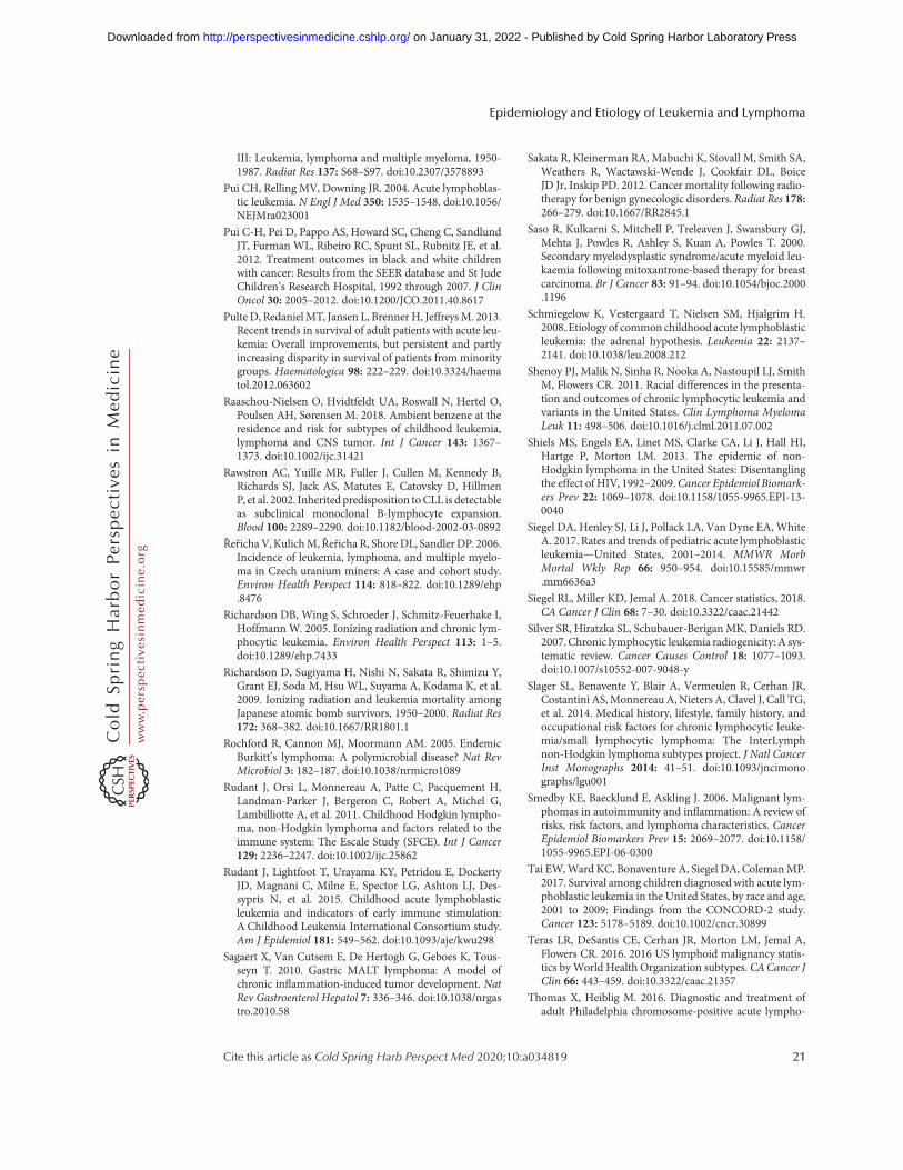
III: Leukemia, lymphoma and multiple myeloma, 1950-1987. Radiat Res 137: S68–S97. doi:10.2307/3578893
Pui CH, Relling MV, Downing JR. 2004. Acute lymphoblas-tic leukemia. N Engl J Med 350: 1535–1548. doi:10.1056/NEJMra023001
Pui C-H, Pei D, Pappo AS, Howard SC, Cheng C, SandlundJT, Furman WL, Ribeiro RC, Spunt SL, Rubnitz JE, et al.2012. Treatment outcomes in black and white childrenwith cancer: Results from the SEER database and St JudeChildren’s Research Hospital, 1992 through 2007. J ClinOncol 30: 2005–2012. doi:10.1200/JCO.2011.40.8617
Pulte D, RedanielMT, Jansen L, Brenner H, JeffreysM. 2013.Recent trends in survival of adult patients with acute leu-kemia: Overall improvements, but persistent and partlyincreasing disparity in survival of patients from minoritygroups. Haematologica 98: 222–229. doi:10.3324/haematol.2012.063602
Raaschou-Nielsen O, Hvidtfeldt UA, Roswall N, Hertel O,Poulsen AH, Sørensen M. 2018. Ambient benzene at theresidence and risk for subtypes of childhood leukemia,lymphoma and CNS tumor. Int J Cancer 143: 1367–1373. doi:10.1002/ijc.31421
Rawstron AC, Yuille MR, Fuller J, Cullen M, Kennedy B,Richards SJ, Jack AS, Matutes E, Catovsky D, HillmenP, et al. 2002. Inherited predisposition toCLL is detectableas subclinical monoclonal B-lymphocyte expansion.Blood 100: 2289–2290. doi:10.1182/blood-2002-03-0892
Řeřicha V, KulichM,Řeřicha R, ShoreDL, SandlerDP. 2006.Incidence of leukemia, lymphoma, and multiple myelo-ma in Czech uranium miners: A case and cohort study.Environ Health Perspect 114: 818–822. doi:10.1289/ehp.8476
Richardson DB, Wing S, Schroeder J, Schmitz-Feuerhake I,Hoffmann W. 2005. Ionizing radiation and chronic lym-phocytic leukemia. Environ Health Perspect 113: 1–5.doi:10.1289/ehp.7433
Richardson D, Sugiyama H, Nishi N, Sakata R, Shimizu Y,Grant EJ, Soda M, Hsu WL, Suyama A, Kodama K, et al.2009. Ionizing radiation and leukemia mortality amongJapanese atomic bomb survivors, 1950–2000. Radiat Res172: 368–382. doi:10.1667/RR1801.1
Rochford R, Cannon MJ, Moormann AM. 2005. EndemicBurkitt’s lymphoma: A polymicrobial disease? Nat RevMicrobiol 3: 182–187. doi:10.1038/nrmicro1089
Rudant J, Orsi L, Monnereau A, Patte C, Pacquement H,Landman-Parker J, Bergeron C, Robert A, Michel G,Lambilliotte A, et al. 2011. Childhood Hodgkin lympho-ma, non-Hodgkin lymphoma and factors related to theimmune system: The Escale Study (SFCE). Int J Cancer129: 2236–2247. doi:10.1002/ijc.25862
Rudant J, Lightfoot T, Urayama KY, Petridou E, DockertyJD, Magnani C, Milne E, Spector LG, Ashton LJ, Des-sypris N, et al. 2015. Childhood acute lymphoblasticleukemia and indicators of early immune stimulation:A Childhood Leukemia International Consortium study.Am J Epidemiol 181: 549–562. doi:10.1093/aje/kwu298
Sagaert X, Van Cutsem E, De Hertogh G, Geboes K, Tous-seyn T. 2010. Gastric MALT lymphoma: A model ofchronic inflammation-induced tumor development. NatRev Gastroenterol Hepatol 7: 336–346. doi:10.1038/nrgastro.2010.58
Sakata R, Kleinerman RA, Mabuchi K, Stovall M, Smith SA,Weathers R, Wactawski-Wende J, Cookfair DL, BoiceJD Jr, Inskip PD. 2012. Cancer mortality following radio-therapy for benign gynecologic disorders.Radiat Res 178:266–279. doi:10.1667/RR2845.1
Saso R, Kulkarni S, Mitchell P, Treleaven J, Swansbury GJ,Mehta J, Powles R, Ashley S, Kuan A, Powles T. 2000.Secondary myelodysplastic syndrome/acute myeloid leu-kaemia following mitoxantrone-based therapy for breastcarcinoma. Br J Cancer 83: 91–94. doi:10.1054/bjoc.2000.1196
Schmiegelow K, Vestergaard T, Nielsen SM, Hjalgrim H.2008. Etiologyof common childhood acute lymphoblasticleukemia: the adrenal hypothesis. Leukemia 22: 2137–2141. doi:10.1038/leu.2008.212
Shenoy PJ, Malik N, Sinha R, Nooka A, Nastoupil LJ, SmithM, Flowers CR. 2011. Racial differences in the presenta-tion and outcomes of chronic lymphocytic leukemia andvariants in the United States. Clin Lymphoma MyelomaLeuk 11: 498–506. doi:10.1016/j.clml.2011.07.002
Shiels MS, Engels EA, Linet MS, Clarke CA, Li J, Hall HI,Hartge P, Morton LM. 2013. The epidemic of non-Hodgkin lymphoma in the United States: Disentanglingthe effect of HIV, 1992–2009.Cancer Epidemiol Biomark-ers Prev 22: 1069–1078. doi:10.1158/1055-9965.EPI-13-0040
Siegel DA, Henley SJ, Li J, Pollack LA, Van Dyne EA, WhiteA. 2017. Rates and trends of pediatric acute lymphoblasticleukemia—United States, 2001–2014. MMWR MorbMortal Wkly Rep 66: 950–954. doi:10.15585/mmwr.mm6636a3
Siegel RL, Miller KD, Jemal A. 2018. Cancer statistics, 2018.CA Cancer J Clin 68: 7–30. doi:10.3322/caac.21442
Silver SR, Hiratzka SL, Schubauer-Berigan MK, Daniels RD.2007. Chronic lymphocytic leukemia radiogenicity: A sys-tematic review. Cancer Causes Control 18: 1077–1093.doi:10.1007/s10552-007-9048-y
Slager SL, Benavente Y, Blair A, Vermeulen R, Cerhan JR,Costantini AS,MonnereauA, Nieters A, Clavel J, Call TG,et al. 2014. Medical history, lifestyle, family history, andoccupational risk factors for chronic lymphocytic leuke-mia/small lymphocytic lymphoma: The InterLymphnon-Hodgkin lymphoma subtypes project. J Natl CancerInst Monographs 2014: 41–51. doi:10.1093/jncimonographs/lgu001
Smedby KE, Baecklund E, Askling J. 2006. Malignant lym-phomas in autoimmunity and inflammation: A review ofrisks, risk factors, and lymphoma characteristics. CancerEpidemiol Biomarkers Prev 15: 2069–2077. doi:10.1158/1055-9965.EPI-06-0300
Tai EW,Ward KC, Bonaventure A, Siegel DA, ColemanMP.2017. Survival among children diagnosed with acute lym-phoblastic leukemia in the United States, by race and age,2001 to 2009: Findings from the CONCORD-2 study.Cancer 123: 5178–5189. doi:10.1002/cncr.30899
Teras LR, DeSantis CE, Cerhan JR, Morton LM, Jemal A,Flowers CR. 2016. 2016 US lymphoid malignancy statis-tics byWorld Health Organization subtypes. CACancer JClin 66: 443–459. doi:10.3322/caac.21357
Thomas X, Heiblig M. 2016. Diagnostic and treatment ofadult Philadelphia chromosome-positive acute lympho-
Epidemiology and Etiology of Leukemia and Lymphoma
Cite this article as Cold Spring Harb Perspect Med 2020;10:a034819 21
ww
w.p
ersp
ecti
vesi
nm
edic
ine.
org
on January 31, 2022 - Published by Cold Spring Harbor Laboratory Presshttp://perspectivesinmedicine.cshlp.org/Downloaded from
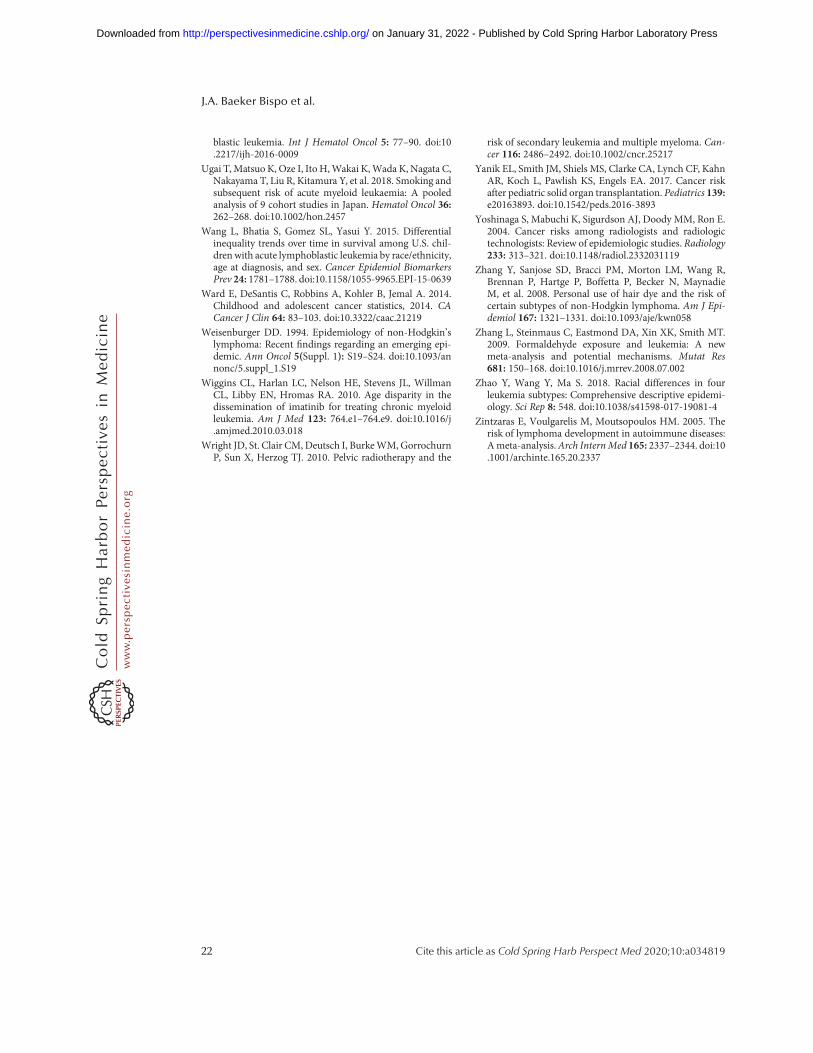
blastic leukemia. Int J Hematol Oncol 5: 77–90. doi:10.2217/ijh-2016-0009
Ugai T, Matsuo K, Oze I, Ito H,Wakai K,Wada K, Nagata C,Nakayama T, Liu R, Kitamura Y, et al. 2018. Smoking andsubsequent risk of acute myeloid leukaemia: A pooledanalysis of 9 cohort studies in Japan. Hematol Oncol 36:262–268. doi:10.1002/hon.2457
Wang L, Bhatia S, Gomez SL, Yasui Y. 2015. Differentialinequality trends over time in survival among U.S. chil-dren with acute lymphoblastic leukemia by race/ethnicity,age at diagnosis, and sex. Cancer Epidemiol BiomarkersPrev 24: 1781–1788. doi:10.1158/1055-9965.EPI-15-0639
Ward E, DeSantis C, Robbins A, Kohler B, Jemal A. 2014.Childhood and adolescent cancer statistics, 2014. CACancer J Clin 64: 83–103. doi:10.3322/caac.21219
Weisenburger DD. 1994. Epidemiology of non-Hodgkin’slymphoma: Recent findings regarding an emerging epi-demic. Ann Oncol 5(Suppl. 1): S19–S24. doi:10.1093/annonc/5.suppl_1.S19
Wiggins CL, Harlan LC, Nelson HE, Stevens JL, WillmanCL, Libby EN, Hromas RA. 2010. Age disparity in thedissemination of imatinib for treating chronic myeloidleukemia. Am J Med 123: 764.e1–764.e9. doi:10.1016/j.amjmed.2010.03.018
Wright JD, St. Clair CM, Deutsch I, BurkeWM, GorrochurnP, Sun X, Herzog TJ. 2010. Pelvic radiotherapy and the
risk of secondary leukemia and multiple myeloma. Can-cer 116: 2486–2492. doi:10.1002/cncr.25217
Yanik EL, Smith JM, Shiels MS, Clarke CA, Lynch CF, KahnAR, Koch L, Pawlish KS, Engels EA. 2017. Cancer riskafter pediatric solid organ transplantation. Pediatrics 139:e20163893. doi:10.1542/peds.2016-3893
Yoshinaga S, Mabuchi K, Sigurdson AJ, Doody MM, Ron E.2004. Cancer risks among radiologists and radiologictechnologists: Review of epidemiologic studies. Radiology233: 313–321. doi:10.1148/radiol.2332031119
Zhang Y, Sanjose SD, Bracci PM, Morton LM, Wang R,Brennan P, Hartge P, Boffetta P, Becker N, MaynadieM, et al. 2008. Personal use of hair dye and the risk ofcertain subtypes of non-Hodgkin lymphoma. Am J Epi-demiol 167: 1321–1331. doi:10.1093/aje/kwn058
Zhang L, Steinmaus C, Eastmond DA, Xin XK, Smith MT.2009. Formaldehyde exposure and leukemia: A newmeta-analysis and potential mechanisms. Mutat Res681: 150–168. doi:10.1016/j.mrrev.2008.07.002
Zhao Y, Wang Y, Ma S. 2018. Racial differences in fourleukemia subtypes: Comprehensive descriptive epidemi-ology. Sci Rep 8: 548. doi:10.1038/s41598-017-19081-4
Zintzaras E, Voulgarelis M, Moutsopoulos HM. 2005. Therisk of lymphoma development in autoimmune diseases:Ameta-analysis.Arch InternMed 165: 2337–2344. doi:10.1001/archinte.165.20.2337
J.A. Baeker Bispo et al.
22 Cite this article as Cold Spring Harb Perspect Med 2020;10:a034819
ww
w.p
ersp
ecti
vesi
nm
edic
ine.
org
on January 31, 2022 - Published by Cold Spring Harbor Laboratory Presshttp://perspectivesinmedicine.cshlp.org/Downloaded from
November 14, 20192020; doi: 10.1101/cshperspect.a034819 originally published onlineCold Spring Harb Perspect Med
Jordan A. Baeker Bispo, Paulo S. Pinheiro and Erin K. Kobetz Epidemiology and Etiology of Leukemia and Lymphoma
Subject Collection Leukemia and Lymphoma: Molecular and Therapeutic Insights
LymphomaTargeting of Metabolism in Leukemia and Oncogenic Mechanisms and Therapeutic
Andrew M. IntlekoferMaximilian Stahl, Zachary D. Epstein-Peterson and
Upcoming Challenges for Multiple Myelomaregarding the Recent Therapeutic Successes and Biological and Translational Considerations
Constantine S. Mitsiades
from Mature B CellsNon-Hodgkin Lymphomas: Malignancies Arising
Jennifer Shingleton, Jie Wang, Carolyn Baloh, et al.T-Cell LymphomasBiology and Molecular Pathogenesis of Mature
José R. Cortés and Teresa Palomero
Decade of Progress in the Molecular AgeMAP-Kinase-Driven Hematopoietic Neoplasms: A
Benjamin H. DurhamRikhia Chakraborty, Omar Abdel-Wahab and
Leukemia PathogenesisThe Role of Somatic Mutations in Acute Myeloid
Ashwin Kishtagari and Ross L. Levine
MalignanciesMouse Models in the Study of Mature B-Cell
Laura Pasqualucci and Ulf Klein
Chronic Lymphocytic Leukemia
RaiNicholas Chiorazzi, Shih-Shih Chen and Kanti R.
Mouse Models of Myeloid MalignanciesFaisal Basheer and George Vassiliou Self-Renewal and Regeneration
Normal Hematopoiesis Is a Balancing Act of
PasseguéOakley C. Olson, Yoon-A Kang and Emmanuelle
LymphomasEpigenetic Mechanisms in Leukemias and
Cihangir Duy, Wendy Béguelin and Ari MelnickLeukemias and LymphomasImpact of Genetics on Mature Lymphoid
Elenitoba-JohnsonNathanael G. Bailey and Kojo S.J.
LeukemiaThe Biology of B-Progenitor Acute Lymphoblastic
Kathryn G. Roberts and Charles G. MullighanLymphomaEpidemiology and Etiology of Leukemia and
K. KobetzJordan A. Baeker Bispo, Paulo S. Pinheiro and Erin
RNA Regulators in Leukemia and LymphomaCamila Prieto and Michael G. Kharas
Clonal Hematopoiesis and Premalignant Diseases
et al.Justin Kaner, Pinkal Desai, Nuria Mencia-Trinchant,
http://perspectivesinmedicine.cshlp.org/cgi/collection/ For additional articles in this collection, see
Copyright © 2020 Cold Spring Harbor Laboratory Press; all rights reserved
on January 31, 2022 - Published by Cold Spring Harbor Laboratory Presshttp://perspectivesinmedicine.cshlp.org/Downloaded from