Embed Size (px)
Citation preview

13A. Tannapfel (ed.), Malignant Mesothelioma, Recent Results in Cancer Research 189, DOI: 10.1007/978-3-642-10862-4_2, © Springer-Verlag Berlin Heidelberg 2011
Epidemiology of Mesothelioma and Historical Background
J.E. Craighead
Abstract Mesothelioma is a “new” malignant disease strongly associated with exposure to amphibole asbestos exposure (amosite and cro-cidolite) environmentally and in the work place. Nonetheless, in recent years, we have learned that many cases of mesothelioma are idiopathic, while some are caused by therapeutic irradia-tion or chronic inflammation in body cavities. This paper reviews the key epidemiological fea-tures of the malignancy in the context of the biological and mineralogical factors that influ-ence mesothelioma development. These tumors challenge the diagnostic pathologist’s acumen, the epidemiologist’s skill in devising meaning-ful and definitive studies, the industrial hygien-ist’s knowledge of environmental hazards in diverse occupational settings, and the clini-cian’s skill in managing an intrepid and uni-formly fatal malignancy.
Many, if not most, of the major life-threatening diseases afflicting humankind were recognized well before the Christian era. In that context,
malignant mesothelioma is a “new” disease with its diagnostic features and natural history having been known to medical science for less than a century. It is my charge in this brief over-view to trace the development of our knowledge of mesotheliomas as clinical and pathological entities, relating the occurrence of this malig-nancy to exposure to a unique family of fibrous minerals that gives rise to the majority of cases of mesothelioma. In doing so, we now are obliged to recognize the occasional patient with idiopathic disease and as of yet unidentified genetic or environmental parameters of disease susceptibility as mesotheliomas are studied critically.
As I sat at the breakfast table this morning, the now inevitable television advertisement appeared announcing the availability of skilled litigants in nationally prominent law firms who will make themselves available to asbestos “vic-tims” whose suffering, they argue, deserves a substantial monetary award. Similarly, vivid advertisements soliciting the afflicted are plas-tered on the sides of municipal buses and in sub-ways in major cities in America. Clearly, the search for the rare unfortunate few who suffer from mesothelioma has become big business for lawyers in the USA. The ultimate outcome is ligation that has already bankrupted countless
2
J.E. Craighead Department of Pathology, University of Vermont, Burlington, VT 05405, USA e-mail: [email protected]
14
2
14 J.E. Craighead
American businesses as plaintiffs seek redress for the presumptive, subtle injury patients unknow-ingly suffered as a result of the supposed callous disregard of insensitive industrialists. Will adver-tisement focused on the general public bring to the attention of medical science “new” etiologies for these unique cancers? Or, will these cases redefine the epidemiological features of the dis-ease and its etiological relationship to low-dose asbestos exposure? Can subtle unrecognized exposures result in the malignant disease? Only time will tell. Unfortunately today’s juries, rather than scientists, are obliged to draw conclusions based on incomplete evidence presented by advo-cates in the courtroom.
It is difficult to be certain when mesothelioma became a recognizable clinical and pathological entity, given its rarity in the general popula-tion and the ability of these tumors to mimic common neoplasms involving the pleural and peritoneal cavities [54]. E. Wagner [79], a German pathologist, is generally accorded credit for the initial description of a tumor believed to be the prototype of the modern day mesothe-lioma. In the past, these malignant lesions often simulated the clinical picture of pleural tubercu-losis, a condition that was not uncommon centu-ries ago. Sensitive diagnostic tools, electron microscopy [22, 75], and immunocytochemistry [18], now make it possible for the pathologist to recognize these tumors with a high degree of certainty when, so often, skilled clinicians demure. It has only been during the last 3 decades that newer diagnostic tools have allowed the epidemiologist the luxury of carrying out analyses using dependable patient data.
Even the term mesothelioma has been a mat-ter of uncertainty for those who seek an orderly nomenclature. Thus, in the first few decades of the last century some 30 different names were used when referring to tumors having at least some of the morphological features of the malig-nant lesions now recognized as mesotheliomas, the most common of which was “endothelioma,” a convenient designation attesting to the vague
resemblance of the tumor cells to vascular endothelial cells. Finally, in the early 1930s, Klemperer and Rabin [41] proposed the desig-nation “mesothelioma” in describing a clinical/pathological entity that commonly exhibited both sarcomatous and carcinomatous histologi-cal features, either exclusively or as a random mixture of the two. But even as late 1957, an occasional “doubting Thomas” questioned the existence of such tumors. For example, in a case report published in the widely read New England Journal of Medicine, the renowned diagnostic pathologist and Harvard professor Benjamin Castleman announced to the medical commu-nity that a case under discussion in a clinical/pathological conference was the first mesothe-lioma he had been comfortable in diagnosing.
This was merely 2 years before Christopher Wagner (a pathologist) and his colleagues, the tuberculosis specialist Kit Sleggs and Paul Marchand [81], a chest physician, described in a landmark publication an epidemic of meso-thelioma consequent to environmental exposure to crocidolite asbestos. It was Sleggs who pro-phetically identified a cadre of unique patients believed to have tuberculous pleuritis but who failed to respond to the customarily effective management of tuberculosis at the time. It was Marchand [48] who helped recognize the com-mon occurrence of this disease among members of the indigenous population who were believed to have a most unusual form of lung cancer. However, at the time, senior South African pathologists, including Ian Webster [82], had little difficulty diagnosing the unique tumors which Wagner (at the time a junior level pathol-ogist) brought to their attention, for they were already aware of similar lesions occurring else-where in the amphibole asbestos mining dis-tricts of South Africa [80]. But who among the pathologists in the Northern Hemisphere paid much heed to an apparent epidemic of an unheard of malignancy occurring in the native population of an obscure corner of southern Africa, particularly when the mining industry
152 Epidemiology of Mesothelioma and Historical Background 15
was more than anxious to suppress knowledge of a suspect industry-associated cancer? At the time, everyone knew that, in general, cancer was a sporadically occurring condition, not one that manifested itself as an epidemic in both women and men, and on occasion, teenagers. To me, as a practicing pathologist in a major Boston teaching hospital in the early 1960s, mesothelioma was rarely a consideration in the differential diagnosis of a chest tumor.
Diagnostic uncertainty, nonetheless, contin-ued to plague the histopathologist for years thereafter when these rare entities came to their attention. Recognizing this conundrum in the mid-1960s, leaders in the world community of pathology established review panels in Europe and North America to evaluate pathological material from individual suspect cases [39]. These experts then tendered a specific diagnosis or arbitrarily expressed either uncertainty or frank disagreement as to the identity of the tumor among the members of the assembled panel. Clearly, clinical case surveys and epidemiologi-cal studies would have proven fruitless in the absence of a concrete diagnostic identification of the tumors. But, improvements in the tools available to the pathologist were forthcoming. As noted above, it was not until the 1970s that electron microscopy was introduced, imperfect as it was, and in the 1980s immunohistochemis-try came into vogue as a diagnostic crutch. To this date, new markers of malignant mesothelial cells continue to be introduced in an effort to confront the ambiguities of diagnostic pathol-ogy, allowing a more precise diagnosis. None-theless, an occasional case generates controversy even among experienced pathologists.
Prior to the 1960s, a case of mesothelioma was a “rare bird” perhaps coming to the atten-tion of the hospital pathologist once or twice in a professional lifetime. Often as a sporadic malignancy of childhood and adolescence, they were idiopathic curiosities too uncommon to warrant serious research (asbestos-related mesotheliomas have not been found to develop
in those younger than 35 years despite an occa-sional claim to the contrary) [33]. There is every reason to believe that many obscure thoracic neoplasms of unknown etiology in women were either classified in the past as breast cancer believed to have metastasized to the pleura, or ovarian cancer spreading unabated throughout the abdominal cavity, implanting on the perito-neal wall. And then there are the anatomic vari-ants, some simulating sarcomas or a complex obscure tumor such as a synovial sarcoma [40]. All too often, mesotheliomas mimicked adeno-carcinomas of bronchogenic origin developing at the periphery of the lung and invading the pleural cavity, the so-called pseudomesothe-liomatous adenocarcinoma.
Although asbestosis as a clinical and patho-logical entity among textile workers was recog-nized in the UK and the USA and was considered a potential cause of lung disease before 1900 [57], many millers died of asbestosis after a period of dust exposure of no longer than a decade. Accordingly, because of its relatively long latency period, it is the writer’s belief that mesotheliomas failed to appear before patients had died because of asbestosis or left the work force. It was not until after the First World War that public health authorities recognized what was believed to be an increase in lung cancer among tradesmen without clinical evidence of asbestosis, but a history of work in an industry where asbestos was liberally used [25, 57]. Most probably, some of these cases were meso-theliomas, but who would know in the absence of autopsies and a clear idea of the diverse path-ological features of these tumors? Who could imagine sarcomas developing in anatomic con-cert with malignant epithelial cells (the so-called biphasic tumors)? It was not until the Second World War that industry-related meso-theliomas were recognized to be occurring in Europe. Alas, these early cases were reported in the wartime German literature as “pleural can-cer” in publications [83, 84], out of the reach of most American and British physicians at the
16
2
16 J.E. Craighead
time (but apparently known and ignored by the Allied intelligence community).
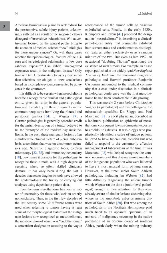
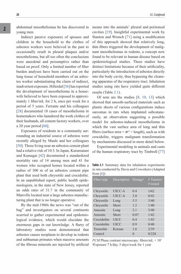
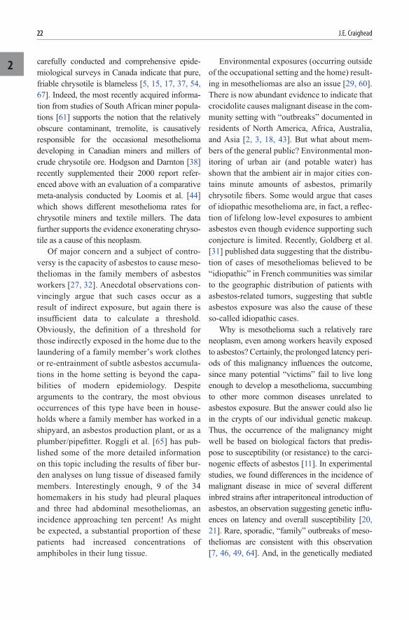
Prior to that time, more specifically in 1934, the passenger vessel SS Morro Castle was destroyed at sea by fire, a tragedy that prompted an inquiry by the US Congress into the apparent ineffectual fireproofing of American registered ships including naval vessels. It was already known that amosite asbestos was resistant to the degrading effects of sea water and could provide excellent insulation protection per unit of weight. Accordingly, by 1940 the US Navy specifica-tions for new ships and those undergoing reconditioning and repair dictated the routine insulation of a vessel’s interior with amosite and to a variable extent, chrysotile. Most commer-cial shippers (i.e., the merchant marine) soon abided by these regulatory criteria, precautions that no doubt saved ships and the lives of many sailors during the war, but has resulted in much suffering thereafter. With the mobilization for the Second World War, amosite was routinely incorporated into the insulation of some 3,000 newly launched merchant vessels and navy war-ships, resulting in the gross contamination of a vessel’s interior compartments, particularly the
engine rooms (Fig. 2.1). For example, a recent evaluation of a mothballed World War II Navy destroyer demonstrated roughly 25 t of asbestos insulation still intact in the bowels of the vessel.
It would be rank speculation to attempt to estimate the numbers of Navy personnel and merchant mariners who were heavily exposed aboard ship while serving their country, and to the best of the writer’s knowledge, no serious attempt has ever been made by governments in Europe or North America to estimate the expo-sures sustained by wartime servicemen and the outcome in the form of disease. Not surpris-ingly, shipyards were also heavily contaminated by friable asbestos and millions (because of a high turnover rate of shipyard workers in the Allied countries and occupied Europe) were heavily exposed to crocidolite and amosite as well as large amounts of chrysotile asbestos during the late 1930s and 1940s. Who knows how they fared.
Responsibly, the US Navy commissioned a study during the waning years of the Second World War to assess the possible adverse effects of asbestos on personnel, focusing on the disease asbestosis [30]. Unfortunately, the observation
for better
dispersionadd amosite
forfibre length-amosite
Amosite added to asbestos cementhas a dispersive action givinguniform fibre distribution leadingto greater strength andimproved surface texture.Full technical advisory serviceavailable to reinforced cement andinsulation material manufacturersfrom the world’s leadingproducers of amphibole fibres.
Amosite is naturally longer than othertypes of asbestos fibre. Length plusresilience makes Amosite the ideal fibrefor high temperature and acousticinsulations and for lightweight fireresistant products.Full technical advisory service availableto reinforced cement and insulationmaterial manufacturers from the world’sleading producers of amphibole fibres.
Cape Asbestos Fibres Limited114 Park Street London W1 · England · Telex 23759North American Asbestos Corporation200 South Michigan Ave · Chicago · Illinois 60604 · USATelephone: (312) 922-7435(Members of the Cape Asbestos Group of Companies)
Cape Asbestos Fibres Limited114 Park Street London W1 · England · Telex 23759North American Asbestos Corporation200 South Michigan Ave · Chicago · Illinois 60604 · USATelephone: (312) 922-7435(Members of the Cape Asbestos Group of Companies)
CAFCO CAFCO
TW2760TW2640
Fig. 2.1 Examples of promotional advertisements published in trade journals in the past
172 Epidemiology of Mesothelioma and Historical Background 17
period was much too short because the latency of asbestosis is variable but often a matter of decades, even with heavy exposure, and meso-thelioma rarely becomes evident before an elapsed period of some 20 years from the time of initial exposure. Drs. Fleisher and Drinker, who conducted the above study, may have been com-petent in their trade but they failed as historians. Either they ignored or were not aware of the European experience with asbestos malignan-cies. Importation of crocidolite and amosite into Germany and Britain began in the early 1900s. Clearly, mesotheliomas were erupting among industrial workers and naval personnel through-out the 1920s and 1930s. But, alas, at the time many mesotheliomas were believed to be tradi-tional lung cancers [67].
A recently completed, unpublished evalua-tion of case material in my laboratory strongly suggests that exposures in the 1940s during the war may give rise to mesotheliomas diagnosed some 40–60 years later (the duration of latency is thought by many authorities to be inversely related to the intensity of exposure). However, since the latency period of most mesotheliomas ranges from 20 to 40 years, it was not until the 1960s that mesotheliomas attributable to war-time exposure began to appear in large numbers in Great Britain [26, 34, 68, 74, 85] and Germany [9]. Soon, an increasingly large number of cases were diagnosed among American shipyard work-ers who were then engaged in other forms of employment [76]. But as noted above, it was not until 1960 that the first compelling report relat-ing environmental crocidolite exposure to meso-thelioma was published, and it was 1971 when amosite was also considered a likely cause, if not the major culprit, in industrialized societies by knowledgeable members of the public health community. In the USA, credit must be accorded Dr. Irving Selikoff, a chest physician, who rec-ognized the impending disaster as mesothe-liomas came to his attention among workers at the Union Asbestos and Rubber Company (UNARCO) in New Jersey where Unibestos
amosite insulation for newly constructed ships was manufactured. Interestingly enough, the ini-tial cases identified by Dr. Selikoff were peri-toneal mesotheliomas, attesting to the heavy exposures these workers had sustained.
It was then that the pathfinding physicians, Drs. Irving Selikoff and Christopher Wagner organized a landmark conference under the aus-pices of the New York Academy of Sciences to consider the accumulating scientific observa-tions associating asbestos exposure with malig-nant and nonmalignant diseases, including the common types of lung cancer and both perito-neal and pleural mesotheliomas.
At this juncture, a pause seems appropriate to summarize briefly what clinicians and epide-miologists have learned over the past half cen-tury regarding this fascinating malignancy and its relationship to asbestos exposure. As we all know, mesotheliomas usually develop unilater-ally in the pleural cavities, and to a more lim-ited extent in the abdomen. But they also develop on rare occasions in the pericardium, the spermatic cords, and both the male and female gonads. Because these highly malignant lesions are shrouded in body cavities, they gen-erally are widespread and incurable when clini-cians finally are obliged to search for the cause of subtle chest or abdominal discomfort accom-panied by a unilateral pleural effusion or ascites. Despite the current availability of potent chemotherapy (as discussed elsewhere in this symposium) and the increasingly com-mon extrapleural pneumonectomies (carried out by intrepid thoracic surgeons in an all too often futile attempt to eliminate or control the spread of the neoplasm) the prognosis is grim and most patients are dead within a period of 3 years from the time of diagnosis. As noted above, the vast majority of mesotheliomas develop in the chest cavities where they gradually invade the chest wall and mediastinum and not infrequently metastasize to the contralateral lung, the spinal vertebrae, and the peritoneal cavity. In the abdo-men they trigger the accumulation of massive
18
2
18 J.E. Craighead
ascites while spreading widely to implant on the surfaces of the peritoneal wall and major organs, only occasionally metastasizing to the chest.
The pathogenesis of mesotheliomas in a population of occupationally exposed men or women is largely dependent upon mineralogical type and the fiber dimension as well as the severity of exposure. On occasion, the incidence of abdominal tumors is as great as 20% of a heavily exposed worker population whereas in most situations it is lower. However, in Great Britain, Coggon et al. [16] discovered a greater than sixfold occurrence of peritoneal tumors in comparison to pleural malignant lesions among construction workers. Carpenters seem to be at exceptional risk for mesotheliomas in the UK, most probably because of the widespread use of composition asbestos boards in the past.
As noted above, the latency of these lesions from the time of first exposure until the onset of symptoms is unpredictable. Almost invariably, it is greater than 20 years but at times it can be as long as 50 or 60 years. Who knows what disease processes lurk in body cavities before the malig-nancy is sufficiently large to cause symptoms? Of interest has been the reported substantially shorter latency period among a few environmen-tally exposed patients in the crocidolite mining district of Western Australia [2, 3]. It is generally agreed that peritoneal mesotheliomas develop as a result of heavier and more prolonged expo-sures, but comparative quantitative thresholds have never been established for any asbestos type because of the profound difficulties of con-ducting comprehensive long-term studies on a rare disease sometimes caused by exceedingly low dosages of a toxic substance. But the lack of evidence is not evidence for a lack of a threshold since many members of the general population have asbestos particles in their lungs in the absence of disease [23]. The classical nonmalig-nant stigmata of exposure, that is, pleural plaques, bilaterally symmetrical pleural thickening, and asbestosis are surrogate measures of relatively heavy exposure to an amphibole. They occur
more frequently in those with peritoneal rather than pleural malignant disease, suggesting that a heavier exposure is required to initiate these lesions in the abdominal cavity. Too little epide-miological information on spermatic cord and gonadal lesions exists to allow conclusions regard-ing causation and latency since it is likely that many of these tumors are idiopathic and not caused by asbestos exposure. It has been the author’s experience that some peritoneal meso-theliomas present clinically for the first time as tumorous masses in the spermatic cord simulat-ing hernias. Anecdotally, it has been hypothe-sized that talc particles and asbestos accumulations on or around ovaries may play a causative role in the genesis of ovarian mesothelioma, a hypothe-sis that now dictates the nonuse of talc on surgi-cal gloves.
Are all mesotheliomas caused by exposure to asbestos? Of course not! According to the comprehensive studies of Spirtas et al. [70], overall the attributable risk for exposure to asbestos is 88% for men, but in only 58% of male cases could asbestos exposure be impli-cated in a patient’s abdominal tumor. In women, the attributable risk proved to be 23% for pleu-ral and peritoneal mesotheliomas combined. (Unfortunately, these epidemiologists were dealing with numbers and not detailed case information; thus, it is impossible to determine the validity of a claim of asbestos exposure, and the type(s) involved). But as William Blake has told us: “to generalize is to be an idiot!” Overstated? Yes, since all too often subtle, brief but heavy exposures to asbestos in a patient’s distant past can on occasion be linked causa-tively to the disease. The writer is aware of sev-eral cases of mesotheliomas in white collar, middle aged men whose only known exposure was summertime employment in industry while attending college.
To an extent, the information briefly summa-rized above represents events occurring in another time frame of history when preliminary infor-mation on environmental asbestos exposure was
192 Epidemiology of Mesothelioma and Historical Background 19
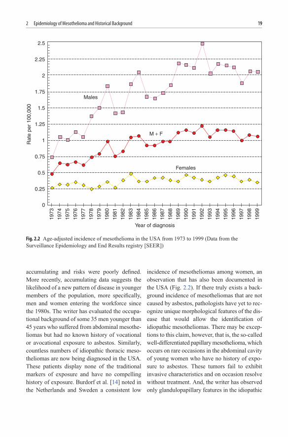
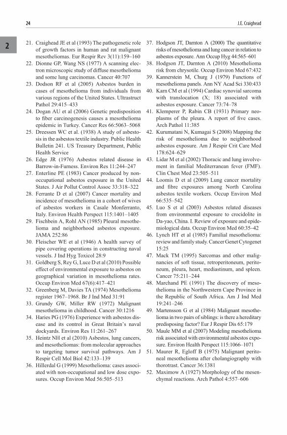
accumulating and risks were poorly defined. More recently, accumulating data suggests the likelihood of a new pattern of disease in younger members of the population, more specifically, men and women entering the workforce since the 1980s. The writer has evaluated the occupa-tional background of some 35 men younger than 45 years who suffered from abdominal mesothe-liomas but had no known history of vocational or avocational exposure to asbestos. Similarly, countless numbers of idiopathic thoracic meso-theliomas are now being diagnosed in the USA. These patients display none of the traditional markers of exposure and have no compelling history of exposure. Burdorf et al. [14] noted in the Netherlands and Sweden a consistent low
incidence of mesotheliomas among women, an observation that has also been documented in the USA (Fig. 2.2). If there truly exists a back-ground incidence of mesotheliomas that are not caused by asbestos, pathologists have yet to rec-ognize unique morphological features of the dis-ease that would allow the identification of idiopathic mesotheliomas. There may be excep-tions to this claim, however, that is, the so-called well-differentiated papillary mesothelioma, which occurs on rare occasions in the abdominal cavity of young women who have no history of expo-sure to asbestos. These tumors fail to exhibit invasive characteristics and on occasion resolve without treatment. And, the writer has observed only glandulopapillary features in the idiopathic
2.5
2.25
2
1.75Males
Females
M + F
1.5
1.25
Rat
e pe
r 10
0,00
0
Year of diagnosis
1
0.75
0.5
0.25
0
1973
1974
1975
1976
1977
1978
1979
1980
1981
1982
1983
1984
1985
1986
1987
1988
1989
1990
1991
1992
1993
1994
1995
1996
1997
1998
1999
Fig. 2.2 Age-adjusted incidence of mesothelioma in the USA from 1973 to 1999 (Data from the Surveillance Epidemiology and End Results registry [SEER])
20
2
20 J.E. Craighead
abdominal mesotheliomas he has discovered in young men.
Indirect passive exposures of spouses and children in the household to the clothes of asbestos workers were believed in the past to occasionally result in pleural plaques and/or mesothelioma, but all too often the conclusions were anecdotal and presumptive rather than based on proof. Only a limited number of fiber burden analyses have been carried out on the lung tissue of household members of an asbes-tos worker substantiating the claim of indirect, inadvertent exposure. Hillerdal [36] has reported the development of mesothelioma in a house-wife believed to have been exposed to approxi-mately 1 fiber/mL for 2 h, once per week for a period of 5 years. Ferrante and his colleagues [28] documented 18 cases of mesothelioma in homemakers who laundered the work clothes of their husbands, all cement factory workers, over a 20 year period [60].
Exposures of residents in a community sur-rounding an industrial source of asbestos were recently alleged by Maule and her colleagues [50]. Those living near an asbestos cement plant had a relative risk of 10.5. In Japan, Kurumatani and Kumagai [42] documented a standardized mortality rate of 14 among men and 41 for women who occupied homes located within a radius of 300 m of an asbestos cement pipe plant that used both chrysotile and crocidolite. In an unpublished report, public health epide-miologists, in the state of New Jersey, reported an odds ratio of 31.7 in the community of Manville located near a large asbestos manufac-turing plant that is no longer operative.
By the mid-1960s the news was “out of the bag” and investigators on several continents scurried to gather experimental and epidemio-logical evidence, which would elucidate the enormous gaps in our knowledge. A flurry of laboratory studies soon demonstrated that asbestos causes neoplasm to develop in rodents and subhuman primates when massive amounts of the fibrous minerals are injected by artificial
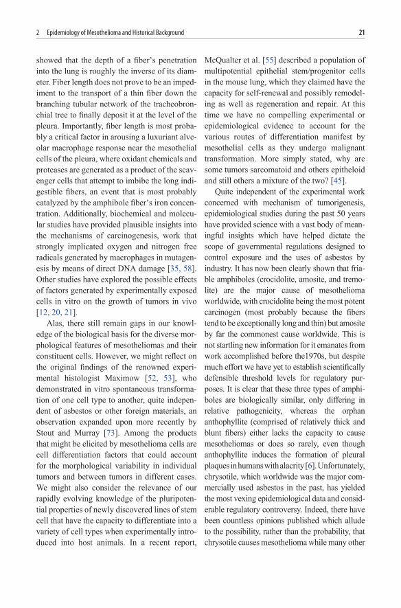
means into the animals’ pleural and peritoneal cavities [19]. Insightful experimental work by Stanton and Wrench [71] using a modification of this approach showed that relatively long, thin fibers triggered the development of malig-nant mesotheliomas in rodents, a concept now found to be relevant to human disease based on epidemiological studies. These studies have distinct limitations because of their artificiality, particularly the introduction of asbestos directly into the body cavity, thus bypassing the cleans-ing apparatus of the respiratory tract. Inhalation studies using rats have yielded quite different results (Table 2.1).
Of note are the studies [8, 10, 13] which showed that smooth-surfaced materials such as plastic sheets of various configurations induce sarcomas in rats when implanted subcutane-ously, an observation suggesting a possible model for asbestos-induced mesothelioma in which the vast surface area of long and thin fibers (surface area = pr2 × length), such as with crocidolite, triggers malignant transformation by mechanisms discussed in more detail below.
Experimental modeling in animals and casts of the human respiratory tract by Timbrell [77]
Fiber type Description Dosagea # Tumors/ # tested
Chrysotile UICC-A 0.4 1/42Chrysotile UICC-A 2.0 0/42Chrysotile Long 5.5 3/40Chrysotile Short 1.2 1/40Amosite Long 2.1 3/40Amosite Short 0.07 1/42Crocidolite UICC 0.4 1/43Crocidolite UICC 0.9 0/40Tremolite Korean 1.6 2/39Control 0 0/228
Table 2.1 Summary data for inhalation experiments in rats conducted by Davis and Coworkers (Adapted from [6])
PCM Phase contrast microscopy: fibers/mL × 103
aExposure 7 h/day, 5 days/week for 1 year
212 Epidemiology of Mesothelioma and Historical Background 21
showed that the depth of a fiber’s penetration into the lung is roughly the inverse of its diam-eter. Fiber length does not prove to be an imped-iment to the transport of a thin fiber down the branching tubular network of the tracheobron-chial tree to finally deposit it at the level of the pleura. Importantly, fiber length is most proba-bly a critical factor in arousing a luxuriant alve-olar macrophage response near the mesothelial cells of the pleura, where oxidant chemicals and proteases are generated as a product of the scav-enger cells that attempt to imbibe the long indi-gestible fibers, an event that is most probably catalyzed by the amphibole fiber’s iron concen-tration. Additionally, biochemical and molecu-lar studies have provided plausible insights into the mechanisms of carcinogenesis, work that strongly implicated oxygen and nitrogen free radicals generated by macrophages in mutagen-esis by means of direct DNA damage [35, 58]. Other studies have explored the possible effects of factors generated by experimentally exposed cells in vitro on the growth of tumors in vivo [12, 20, 21].
Alas, there still remain gaps in our knowl-edge of the biological basis for the diverse mor-phological features of mesotheliomas and their constituent cells. However, we might reflect on the original findings of the renowned experi-mental histologist Maximow [52, 53], who demonstrated in vitro spontaneous transforma-tion of one cell type to another, quite indepen-dent of asbestos or other foreign materials, an observation expanded upon more recently by Stout and Murray [73]. Among the products that might be elicited by mesothelioma cells are cell differentiation factors that could account for the morphological variability in individual tumors and between tumors in different cases. We might also consider the relevance of our rapidly evolving knowledge of the pluripoten-tial properties of newly discovered lines of stem cell that have the capacity to differentiate into a variety of cell types when experimentally intro-duced into host animals. In a recent report,
McQualter et al. [55] described a population of multipotential epithelial stem/progenitor cells in the mouse lung, which they claimed have the capacity for self-renewal and possibly remodel-ing as well as regeneration and repair. At this time we have no compelling experimental or epidemiological evidence to account for the various routes of differentiation manifest by mesothelial cells as they undergo malignant transformation. More simply stated, why are some tumors sarcomatoid and others epitheloid and still others a mixture of the two? [45].
Quite independent of the experimental work concerned with mechanism of tumorigenesis, epidemiological studies during the past 50 years have provided science with a vast body of mean-ingful insights which have helped dictate the scope of governmental regulations designed to control exposure and the uses of asbestos by industry. It has now been clearly shown that fria-ble amphiboles (crocidolite, amosite, and tremo-lite) are the major cause of mesothelioma worldwide, with crocidolite being the most potent carcinogen (most probably because the fibers tend to be exceptionally long and thin) but amosite by far the commonest cause worldwide. This is not startling new information for it emanates from work accomplished before the1970s, but despite much effort we have yet to establish scientifically defensible threshold levels for regulatory pur-poses. It is clear that these three types of amphi-boles are biologically similar, only differing in relative pathogenicity, whereas the orphan anthophyllite (comprised of relatively thick and blunt fibers) either lacks the capacity to cause mesotheliomas or does so rarely, even though anthophyllite induces the formation of pleural plaques in humans with alacrity [6]. Unfortunately, chrysotile, which worldwide was the major com-mercially used asbestos in the past, has yielded the most vexing epidemiological data and consid-erable regulatory controversy. Indeed, there have been countless opinions published which allude to the possibility, rather than the probability, that chrysotile causes mesothelioma while many other
22
2
22 J.E. Craighead
carefully conducted and comprehensive epide-miological surveys in Canada indicate that pure, friable chrysotile is blameless [5, 15, 17, 37, 54, 67]. Indeed, the most recently acquired informa-tion from studies of South African miner popula-tions [61] supports the notion that the relatively obscure contaminant, tremolite, is causatively responsible for the occasional mesothelioma developing in Canadian miners and millers of crude chrysotile ore. Hodgson and Darnton [38] recently supplemented their 2000 report refer-enced above with an evaluation of a comparative meta-analysis conducted by Loomis et al. [44] which shows different mesothelioma rates for chrysotile miners and textile millers. The data further supports the evidence exonerating chryso-tile as a cause of this neoplasm.
Of major concern and a subject of contro-versy is the capacity of asbestos to cause meso-theliomas in the family members of asbestos workers [27, 32]. Anecdotal observations con-vincingly argue that such cases occur as a result of indirect exposure, but again there is insufficient data to calculate a threshold. Obviously, the definition of a threshold for those indirectly exposed in the home due to the laundering of a family member’s work clothes or re-entrainment of subtle asbestos accumula-tions in the home setting is beyond the capa-bilities of modern epidemiology. Despite arguments to the contrary, the most obvious occurrences of this type have been in house-holds where a family member has worked in a shipyard, an asbestos production plant, or as a plumber/pipefitter. Roggli et al. [65] has pub-lished some of the more detailed information on this topic including the results of fiber bur-den analyses on lung tissue of diseased family members. Interestingly enough, 9 of the 34 homemakers in his study had pleural plaques and three had abdominal mesotheliomas, an incidence approaching ten percent! As might be expected, a substantial proportion of these patients had increased concentrations of amphiboles in their lung tissue.
Environmental exposures (occurring outside of the occupational setting and the home) result-ing in mesotheliomas are also an issue [29, 60]. There is now abundant evidence to indicate that crocidolite causes malignant disease in the com-munity setting with “outbreaks” documented in residents of North America, Africa, Australia, and Asia [2, 3, 18, 43]. But what about mem-bers of the general public? Environmental mon-itoring of urban air (and potable water) has shown that the ambient air in major cities con-tains minute amounts of asbestos, primarily chrysotile fibers. Some would argue that cases of idiopathic mesothelioma are, in fact, a reflec-tion of lifelong low-level exposures to ambient asbestos even though evidence supporting such conjecture is limited. Recently, Goldberg et al. [31] published data suggesting that the distribu-tion of cases of mesotheliomas believed to be “idiopathic” in French communities was similar to the geographic distribution of patients with asbestos-related tumors, suggesting that subtle asbestos exposure was also the cause of these so-called idiopathic cases.
Why is mesothelioma such a relatively rare neoplasm, even among workers heavily exposed to asbestos? Certainly, the prolonged latency peri-ods of this malignancy influences the outcome, since many potential “victims” fail to live long enough to develop a mesothelioma, succumbing to other more common diseases unrelated to asbestos exposure. But the answer could also lie in the crypts of our individual genetic makeup. Thus, the occurrence of the malignancy might well be based on biological factors that predis-pose to susceptibility (or resistance) to the carci-nogenic effects of asbestos [11]. In experimental studies, we found differences in the incidence of malignant disease in mice of several different inbred strains after intraperitoneal introduction of asbestos, an observation suggesting genetic influ-ences on latency and overall susceptibility [20, 21]. Rare, sporadic, “family” outbreaks of meso-theliomas are consistent with this observation [7, 46, 49, 64]. And, in the genetically mediated
232 Epidemiology of Mesothelioma and Historical Background 23
disease of humans known as Mediterranean Fever, the characteristic chronic serositis, which occurs in the body cavities of these patients, is associated with the sporadic, uncommon appearance of mesothelioma in mid-life [43, 63]. Perhaps this is a reflection of the apparent role of smoldering inflammation in the pathogenesis of mesothe-lioma, as has been proposed for the infrequent development of mesotheliomas in those afflicted with chronic tuberculosis [57, 66]. In Turkey, the relatively common appearance of mesotheliomas among members of isolated population groups who are exposed to erionite, a volcanic fibrous zeolite mineral, has again raised the possible role of genetic factors in carcinogenesis for consider-ation [4, 24]. Could inheritance be responsible for the development of mesothelioma in patients years after they received therapeutic irradiation for neoplastic disease [1, 51, 72]? Clearly, we are only now acquiring insights into possible predis-posing factors that might ultimately influence the development of this unique malignancy. The interplay between environmental and host fac-tors, to a large extent, remains to be defined [76].
References
1. Anderson KA et al (1985) Malignant pleural mesothelioma following radiotherapy in a 16-year-old boy. Cancer 56:273
2. Armstrong BK et al (1984) Epidemiology of malignant mesothelioma in Western Australia. Med J Aust 141:86
3. Armstrong BK et al (1988) Mortality in miners and millers of crocidolite in Western Australia. Br J Ind Med 45:5–13
4. Artvinli M, Baris YI (1979) Malignant meso-theliomas in a small village in the Anatolian region of Turkey: an epidemiologic study. J Natl Cancer Inst 63:17
5. Berman DW, Crump KS (2008) Technical sup-port document for a protocol to assess asbestos-related risk. US Environmental Protection Agency publication. US Environmental Pro-tection Agency, Washington, DC
6. Berman DW et al (1995) The sizes, shapes, and mineralogy of asbestos structures that induce lung tumors or mesothelioma in AF/HAN rats following inhalation. Risk Anal 15:181–195
7. Bianchi C et al (1993) Asbestos-related famil-ilal mesothelioma. Eur J Cancer Prevent 2: 247–250
8. Bischoff F, Bryson G (1964) Carcinogenesis through solid state surfaces. Prog Exp Tumor Res 5:65
9. Bohlig H et al (1970) Epidemiology of malignant mesothelioma in Hamburg. Environ Res 3:365
10. Bolen JW, Thorning D (1980) Mesotheliomas: a light and electronmicroscopical study con-cerning histogenetic relationships between the epithelial and the mesenchymal variants. Am J Surg Pathol 4:451
11. Brain JD (1989) The susceptible individual: an overview. In: Utell M (ed) Susceptibility to inhaled pollutants, ASTM Special Technical Publication. ASTM, Philadelphia
12. Brody AR, Overby LH (1989) Incorporation of tritiated thymidine by epithelial and interstitial cells in bronchiolar-alveolar regions of asbes-tos-exposed rats. Am J Pathol 134:133–140
13. Buoen LC et al (1975) Foreign body tumori-genesis: in vitro isolation and expansion of pre-neoplastaic clonal cell populations. J Natl Cancer Inst 55:721
14. Burdorf A et al (2007) Asbestos exposure and differences in occurrences of peritoneal meso-thelioma between men and women across coun-tries. Occup Environ Med 64:839–842
15. Chanhinian AP, Pass HI (2000) Malignant meso-thelioma. In: Holland JC, Frei E (eds) Cancer medicine, 5th edn. BC Decker, Hamilton
16. Coggon D et al (1995) Differences in occupa-tional mortality from pleural cancer, peritoneal cancer and asbestosis. Occup Environ Med 52:775–777
17. Craighead JE (1987) Current pathogenetic con-cepts of diffuse malignant mesothelioma. Hum Pathol 18:544–557
18. Craighead JE, Gibbs AR (2008) Asbestos and its diseases. Oxford University Press, New York
19. Craighead J et al (1987) Biologic characteris-tics of asbestos-induced malignant mesothe-lioma in rats. Chest 91S:12–13
20. Craighead JE et al (1993) Genetic factors influ-ence malignant mesothelioma development in mice. Eur Respir Rev 3:118–120
24
2
24 J.E. Craighead
21. Craighead JE et al (1993) The pathogenetic role of growth factors in human and rat malignant mesotheliomas. Eur Respir Rev 3(11):159–160
22. Dionne GP, Wang NS (1977) A scanning elec-tron microscopic study of diffuse mesothelioma and some lung carcinomas. Cancer 40:707
23. Dodson RF et al (2005) Asbestos burden in cases of mesothelioma from individuals from various regions of the United States. Ultrastruct Pathol 29:415–433
24. Dogan AU et al (2006) Genetic predisposition to fiber carcinogenesis causes a mesothelioma epidemic in Turkey. Cancer Res 66:5063–5068
25. Dreessen WC et al. (1938) A study of asbesto-sis in the asbestos textile industry. Public Health Bulletin 241. US Treasury Department, Public Health Service
26. Edge JR (1976) Asbestos related disease in Barrow-in-Furness. Environ Res 11:244–247
27. Enterline PE (1983) Cancer produced by non-occupational asbestos exposure in the United States. J Air Pollut Control Assoc 33:318–322
28. Ferrante D et al (2007) Cancer mortality and incidence of mesothelioma in a cohort of wives of asbestos workers in Casale Monferranto, Italy. Environ Health Perspect 115:1401–1405
29. Fischbein A, Rohl AN (1985) Pleural mesothe-lioma and neighborhood asbestos exposure. JAMA 252:86
30. Fleischer WE et al (1946) A health survey of pipe covering operations in constructing naval vessels. J Ind Hyg Toxicol 28:9
31. Goldberg S, Rey G, Luce D et al (2010) Possible effect of environmental exposure to asbestos on geographical variation in mesothelioma rates. Occup Environ Med 67(6):417–421
32. Greenberg M, Davies TA (1974) Mesothelioma register 1967–1968. Br J Ind Med 31:91
33. Grundy GW, Miller RW (1972) Malignant mesothelioma in childhood. Cancer 30:1216
34. Haries PG (1976) Experience with asbestos dis-ease and its control in Great Britain’s naval dockyards. Environ Res 11:261–267
35. Heintz NH et al (2010) Asbestos, lung cancers, and mesotheliomas: from molecular approaches to targeting tumor survival pathways. Am J Respir Cell Mol Biol 42:133–139
36. Hillerdal G (1999) Mesothelioma: cases associ-ated with non-occupational and low dose expo-sures. Occup Environ Med 56:505–513
37. Hodgson JT, Darnton A (2000) The quantitative risks of mesothelioma and lung cancer in relation to asbestos exposure. Ann Occup Hyg 44:565–601
38. Hodgson JT, Darnton A (2010) Mesothelioma risk from chrysotile. Occup Environ Med 67:432
39. Kannerstein M, Churg J (1979) Functions of mesothelioma panels. Ann NY Acad Sci 330:433
40. Karn CM et al (1994) Cardiac synovial sarcoma with translocation (X; 18) associated with asbestos exposure. Cancer 73:74–78
41. Klemperer P, Rabin CB (1931) Primary neo-plasms of the pleura. A report of five cases. Arch Pathol 11:385
42. Kurumatani N, Kumagai S (2008) Mapping the risk of mesothelioma due to neighborhood asbestos exposure. Am J Respir Crit Care Med 178:624–629
43. Lidar M et al (2002) Thoracic and lung involve-ment in familial Mediterranean fever (FMF). Clin Chest Med 23:505–511
44. Loomis D et al (2009) Lung cancer mortality and fibre exposures among North Carolina asbestos textile workers. Occup Environ Med 66:535–542
45. Luo S et al (2003) Asbestos related diseases from environmental exposure to crocidolite in Da-yao, China. I. Review of exposure and epide-miological data. Occup Environ Med 60:35–42
46. Lynch HT et al (1985) Familial mesothelioma: review and family study. Cancer Genet Cytogenet 15:25
47. Mack TM (1995) Sarcomas and other malig-nancies of soft tissue, retroperitoneum, perito-neum, pleura, heart, mediastinum, and spleen. Cancer 75:211–244
48. Marchand PE (1991) The discovery of meso-thelioma in the Northwestern Cape Province in the Republic of South Africa. Am J Ind Med 19:241–246
49. Martensson G et al (1984) Malignant mesothe-lioma in two pairs of siblings: is there a hereditary predisposing factor? Eur J Respir Dis 65:179
50. Maule MM et al (2007) Modeling mesothelioma risk associated with environmental asbestos expo-sure. Environ Health Perspect 115:1066–1071
51. Maurer R, Egloff B (1975) Malignant perito-neal mesothelioma after cholangiography with thorotrast. Cancer 36:1381
52. Maximow A (1927) Morphology of the mesen-chymal reactions. Arch Pathol 4:557–606
252 Epidemiology of Mesothelioma and Historical Background 25
53. Maximow A (1927) Uber das mesothel (deck-zellen der serosen haute) und die zellen der serosen exsudate. Untersuchungen an entzun-detem Gewebe und an Gewebskulturen. Arch Exp Zellforsch 4:1
54. McDonald JC et al (1999) Editorial: chrysotile, tremolite and fibrogenicity. Ann Occup Hyg 43(7):439–442
55. McQualter JL et al (2010) Evidence of an epithelial stem/progenitor cell hierarchy in the adult mouse lung. Proc Natl Acad Sci 107(4):1414–1419
56. Merewether ERA (1934) A memorandum on asbestosis. Tubercle 75:69–81, 109–118, 152–159
57. Merewether ERA, Price CW (1930) Report on the effects of asbestos dust on the lungs and dust suppression in the asbestos industry. Her Majesty’s Stationery Office, London
58. Mossman BT et al (1986) Alteration of super-oxide dismutase activity in tracheal epithelial cells by asbestos and inhibition of cytotoxicity by antioxidants. Lab Invest 54:204
59. Murphy RLH et al (1972) Low exposure to asbestos. Gas exchange in ship pipe coverers and controls. Arch Environ Health 25:253
60. Newhouse ML, Thompson H (1965) Epidemi-ology of mesothelial tumors in the London area. Ann NY Acad Sci 132:579–588
61. Rees D et al (2001) Asbestos lung fibre concen-trations in South African chrysotile mine work-ers. Ann Occup Hyg 45:473–477
62. Reid A et al (2007) Age and sex differences in malignant mesothelioma after residential exposure to blue asbestos (crocidolite). Chest 131:376–382
63. Riddell RH et al (1981) Peritoneal malignant mesothelioma in a patient with recurrent perito-nitis. Cancer 48:134
64. Risberg B et al (1980) Familial clustering of malignant mesothelioma. Cancer 45:2422
65. Roggli VL et al (1997) Malignant mesothe-lioma in women. Anat Pathol 2:147–163
66. Rovario GC et al (1982) The association of pleural mesothelioma and tuberculosis. Am Rev Respir Dis 126:569
67. Sebastien P, McDonald JC (1997) Mesothelioma in Quebec chrysotile miners and millers: epide-miology and aetiology. Ann Occup Hyg 41(6): 707–719
68. Sheers G (1980) Mesothelioma risks in a naval dockyard. Arch Environ Health 35:276–282
69. Sheers G, Templeton AR (1968) Effects of asbestos in dockyard workers. Br Med J 11: 574
70. Spirtas R et al (1994) Malignant mesothelioma: attributable risk of asbestos exposure. Occup Environ Med 51:804–811
71. Stanton MF, Wrench C (1978) Mechanisms of mesothelioma induction with asbestos and fibrous glass. J Natl Cancer Inst 48:797
72. Stock RJ et al (1979) Malignant peritoneal mesothelioma following radiotherapy for semi-noma of the testis. Cancer 44:914
73. Stout AP, Murray MR (1942) Localized pleural mesothelioma: investigation of its characteris-tics and histogenesis by the method of tissue culture. Arch Pathol 34:951
74. Stumphius J (1971) Epidemiology of mesothe-lioma on Waicheren Island. Br J Ind Med 28:59
75. Suzuki Y et al (1976) Ultrastructure of human malignant diffuse mesothelioma. Am J Pathol 85:241
76. Tagnon I et al (1980) Mesothelioma associated with the shipbuilding industry in coastal Virginia. Cancer Res 40:3875
77. Timbrell V (1965) The inhalation of fibrous dusts. Ann NY Acad Sci 132:255
78. Tossavainen A (2004) Global use of asbestos and the incidence of mesothelioma. Int J Occup Environ Health 10:22–25
79. Wagner E (1870) Das tuberkelahnliche lymph-adenom (der cytogene oder reticulierte Tuberkel). Arch Heilk 11:497
80. Wagner JC (1991) The discovery of the associ-ation between blue asbestos and mesotheliomas and the aftermath. Br J Ind Med 48:399–403
81. Wagner JC, Sleggs CA, Marchand P (1960) Diffuse pleural mesothelioma and asbestos exposure in the North Western Cape Province. Br J Ind Med 17:260
82. Webster I (1973) Asbestos and malignancy. S Afr Med J 47:165
83. Wedler HW (1943) Asbetsose und lungenkrebs bei asbestoste. Dtsch Arch Klin Med 191:189
84. Wedler HW (1943) Asbetsose und lungenkrebs. Dtsch Med Wochenschr 69:575
85. Whitwell F, Rawcliffe RM (1971) Diffuse malignant pleural mesothelioma and asbestos exposure. Thorax 26:6–22

http://www.springer.com/978-3-642-10861-7


![Mesothelioma lawyers ] mesothelioma attorneys](https://img.pdfslide.net/doc/110x75/5497f892ac795959288b5644/mesothelioma-lawyers-mesothelioma-attorneys.jpg)