Embed Size (px)
Citation preview
NATURE REVIEWS | RHEUMATOLOGY VOLUME 7 | MAY 2011 | 263
Chromatin and Disease Group, Cancer Epigenetics and Biology Program (PEBC), Bellvitge Biomedical Research Institute (IDIBELL), Avda. Gran Via 199–203, 08907 L’Hospitalet de Llobregat, Barcelona, Spain [email protected]
Epigenetic alterations in autoimmune rheumatic diseasesEsteban Ballestar
Abstract | The potential roles of epigenetic alterations in the pathogenesis of autoimmune rheumatic diseases are raising great expectations among clinicians and researchers. Epigenetic mechanisms regulate gene expression and are sensitive to external stimuli, bridging the gap between environmental and genetic factors. Considerable evidence of epigenetic changes, particularly altered patterns of DNA methylation, exists in diseases such as systemic lupus erythematosus (SLE) and rheumatoid arthritis. The importance of such changes in the pathology of rheumatic diseases has been demonstrated by examining the relationship between gene-specific methylation and SLE in monozygotic twins discordant for the disease, in whom genetic variability is excluded as a cause for discordance. Several studies have highlighted the importance of the tissue-specificity of DNA methylation changes, an aspect which—in contrast with genetic analysis—must be considered when designing epigenetic studies. Here I discuss the proposed mechanisms and implications of DNA methylation changes in the pathogenesis of autoimmune rheumatic diseases, the prospects for future epigenetic studies in rheumatology, the relevance of specific DNA methylation markers and the potential use of drugs with an epigenetic effect in the clinical management of these diseases.
Ballestar, E. Nat. Rev. Rheumatol. 7, 263–271 (2011); published online 22 February 2011; doi:10.1038/nrrheum.2011.16
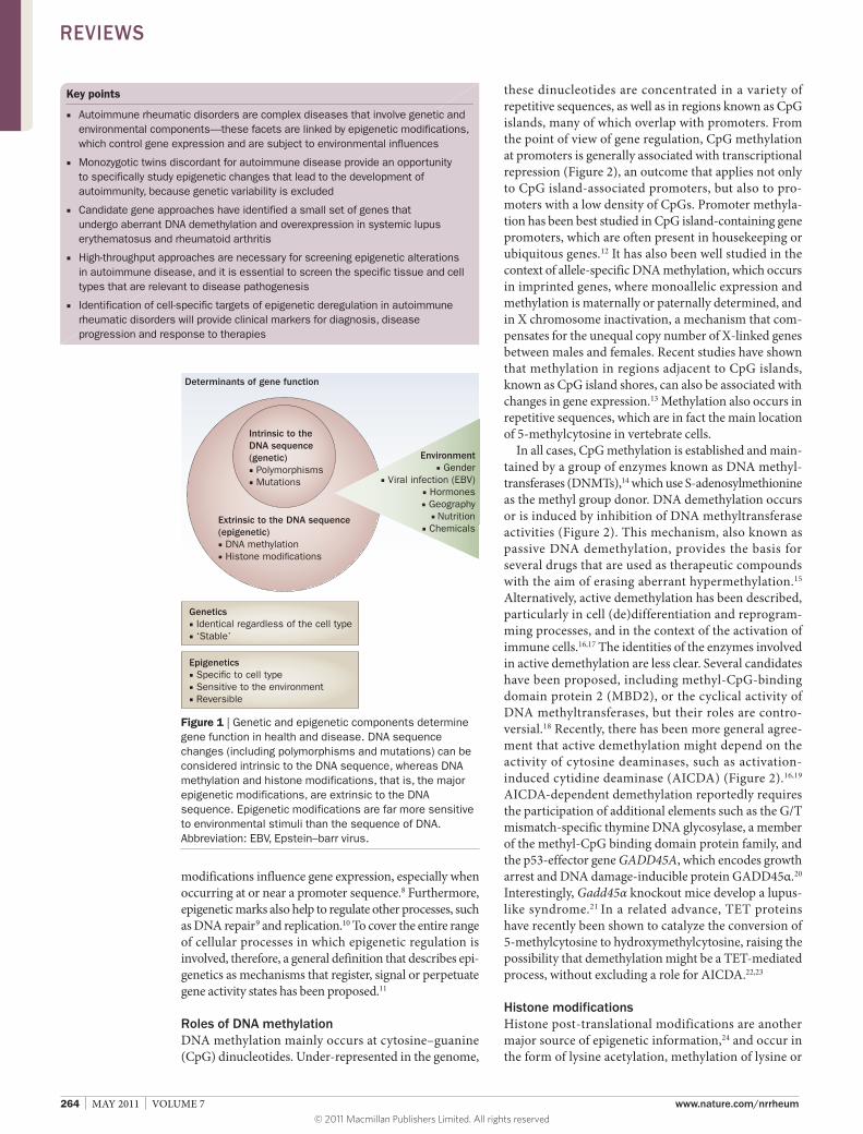
Introduction The complex etiopathology of autoimmune rheumatic disorders has been attributed to crosstalk between genetic predisposition and environmental factors. The first genetic studies in autoimmune disease revealed a strong association between genes within the major histo compatibility complex (MHC), particularly human leuko cyte antigen (HLA) genes, and several auto immune diseases.1 More recently, many other susceptibility genes have been uncovered, some of which, such as PTPN22, IRF5, and STAT4, are also associated with the development of several autoimmune rheumatic diseases,2 suggest ing overlap in their pathogenesis. Complexity arises from the fact that each of these susceptibility genes make small and interlinked contributions to the overall risk of disease. For example, a haplotype of IRF5 that is associated with risk for systemic lupus erythematosus (SLE) has been linked to increased production of interferon (IFN),3 and increased sensitivity to IFNα in patients with SLE is caused by a variant of STAT4 that is associated with autoimmune disease.4 Over and above the expression of basic genetic variability, the contribution of genetic factors to disease risk can be modulated by the environment (Figure 1). A number of internal and external environmental factors have been associated with the etiopathology of rheumatic disorders, including viral infection, nutrition, and exposure to chemicals and radiation.5,6 Such factors influence or modify the profile of epigenetic modifications, which, in turn, have a direct
relationship with the regulation of gene expression, and ultimately the function of the immune system.
Rapid progress in understanding epigenetic alterations in cancer has enabled us to determine the general mechanisms of epigenetic deregulation, identify clinical markers of epigenetic change, and embark on the development of novel therapeutic drugs.7 By contrast, advances in understanding epigenetic mechanisms in the context of rheumatic diseases, as well as in other disorders, have been much slower, and studies remain confined to a small number of laboratories. Nevertheless, evidence of important roles for these types of alterations in autoimmune diseases is increasing. Furthermore, novel technologies that facilitate gene identification and the systematic search for novel epigenetically deregulated genes support the investment of research in this area. This article summarizes and discusses the evidence for epigenetic mechanisms in autoimmune rheumatic diseases, with a focus on changes in DNA methylation, and outlines the future steps to be taken in the field.
Epigenetics and gene function Epigenetic gene regulation has an essential role in determining individual gene function and activity, and, at the genomic level, determines which sets of genes are functional in each specific cell type. Such regulation takes the form of small chemical group additions to DNA or to the protein complex around which DNA is wrapped, particularly core histones. In brief, the two major types of epigenetic modification are DNA methylation and histone posttranslational modifications. Both types of
Competing interestsThe author declares no competing interests.
REVIEWS
© 2011 Macmillan Publishers Limited. All rights reserved
264 | MAY 2011 | VOLUME 7 www.nature.com/nrrheum
modifications influence gene expression, especially when occurring at or near a promoter sequence.8 Furthermore, epigenetic marks also help to regulate other processes, such as DNA repair9 and replication.10 To cover the entire range of cellular processes in which epigenetic regulation is involved, therefore, a general definition that describes epigenetics as mechanisms that register, signal or perpetuate gene activity states has been proposed.11
Roles of DNA methylationDNA methylation mainly occurs at cytosine–guanine (CpG) dinucleotides. Underrepresented in the genome,
Key points
■ Autoimmune rheumatic disorders are complex diseases that involve genetic and environmental components—these facets are linked by epigenetic modifications, which control gene expression and are subject to environmental influences
■ Monozygotic twins discordant for autoimmune disease provide an opportunity to specifically study epigenetic changes that lead to the development of autoimmunity, because genetic variability is excluded
■ Candidate gene approaches have identified a small set of genes that undergo aberrant DNA demethylation and overexpression in systemic lupus erythematosus and rheumatoid arthritis
■ High-throughput approaches are necessary for screening epigenetic alterations in autoimmune disease, and it is essential to screen the specific tissue and cell types that are relevant to disease pathogenesis
■ Identification of cell-specific targets of epigenetic deregulation in autoimmune rheumatic disorders will provide clinical markers for diagnosis, disease progression and response to therapies
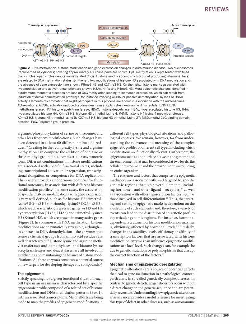
these dinucleotides are concentrated in a variety of repetitive sequences, as well as in regions known as CpG islands, many of which overlap with promoters. From the point of view of gene regulation, CpG methylation at promoters is generally associated with transcriptional repression (Figure 2), an outcome that applies not only to CpG islandassociated promoters, but also to promoters with a low density of CpGs. Promoter methylation has been best studied in CpG islandcontaining gene promoters, which are often present in housekeeping or ubiquitous genes.12 It has also been well studied in the context of allelespecific DNA methylation, which occurs in imprinted genes, where monoallelic expression and methylation is maternally or paternally determined, and in X chromosome inactivation, a mechanism that compensates for the unequal copy number of Xlinked genes between males and females. Recent studies have shown that methylation in regions adjacent to CpG islands, known as CpG island shores, can also be associated with changes in gene expression.13 Methylation also occurs in repetitive sequences, which are in fact the main location of 5methylcytosine in vertebrate cells.
In all cases, CpG methylation is established and maintained by a group of enzymes known as DNA methyltransferases (DNMTs),14 which use Sadenosylmethionine as the methyl group donor. DNA demethylation occurs or is induced by inhibition of DNA methyltransferase activities (Figure 2). This mechanism, also known as passive DNA demethylation, provides the basis for several drugs that are used as therapeutic compounds with the aim of erasing aberrant hyper methylation.15 Alternatively, active demethylation has been described, particularly in cell (de)differentiation and reprogramming processes, and in the context of the activation of immune cells.16,17 The identities of the enzymes involved in active demethylation are less clear. Several candidates have been proposed, including methylCpGbinding domain protein 2 (MBD2), or the cyclical activity of DNA methyltransferases, but their roles are controversial.18 Recently, there has been more general agreement that active demethylation might depend on the activity of cytosine deaminases, such as activationinduced cytidine deaminase (AICDA) (Figure 2).16,19 AICDAdependent demethylation reportedly requires the participation of additional elements such as the G/T mismatchspecific thymine DNA glycosylase, a member of the methylCpG binding domain protein family, and the p53effector gene GADD45A, which encodes growth arrest and DNA damageinducible protein GADD45α.20 Interestingly, Gadd45α knockout mice develop a lupuslike syndrome.21 In a related advance, TET proteins have recently been shown to catalyze the conversion of 5methylcytosine to hydroxymethylcytosine, raising the possibility that demethylation might be a TETmediated process, without excluding a role for AICDA.22,23
Histone modifications Histone posttranslational modifications are another major source of epigenetic information,24 and occur in the form of lysine acetylation, methylation of lysine or
Intrinsic to theDNA sequence(genetic)■ Polymorphisms■ Mutations
Genetics■ Identical regardless of the cell type■ ‘Stable’
Epigenetics■ Speci�c to cell type■ Sensitive to the environment■ Reversible
Extrinsic to the DNA sequence(epigenetic)■ DNA methylation■ Histone modi�cations
Determinants of gene function
Environment■ Gender
■ Viral infection (EBV)■ Hormones■ Geography
■ Nutrition■ Chemicals
Figure 1 | Genetic and epigenetic components determine gene function in health and disease. DNA sequence changes (including polymorphisms and mutations) can be considered intrinsic to the DNA sequence, whereas DNA methylation and histone modifications, that is, the major epigenetic modifications, are extrinsic to the DNA sequence. Epigenetic modifications are far more sensitive to environmental stimuli than the sequence of DNA. Abbreviation: EBV, Epstein–barr virus.
REVIEWS
© 2011 Macmillan Publishers Limited. All rights reserved
NATURE REVIEWS | RHEUMATOLOGY VOLUME 7 | MAY 2011 | 265
arginine, phosphorylation of serine or threonine, and other less frequent modifications. Such changes have been detected in at least 60 different amino acid residues.24 Creating further complexity, lysine and arginine methylation can comprise the addition of one, two or three methyl groups in a symmetric or asymmetric form. Different combinations of histone modifications are associated with specific functional states, including transcriptional activation or repression, transcriptional elong ation, or competence for DNA replication. This variety provides an enormous potential for functional outcomes, in association with different histone modification profiles.24 In some cases, the association of specific histone modifications with gene expression is very well defined, such as for histone H3 trimethyllysine9 (K9me3 H3) or trimethyllysine27 (K27me3 H3), which are charac teristic of repressed genes, or H3 and H4 hyper acetylation (H3Ac, H4Ac) and trimethyllysine4 H3 (K4me3 H3), which are present in many active genes (Figure 2). In common with DNA methylation, histone modifications are enzymatically reversible, although—in contrast to DNA demethylation—the enzymes that remove chemical groups from amino acid residues are well characterized.25 Histone lysine and arginine methyltransferases and demethylases, and histone lysine acetyltransferases and deacetylases, are all involved in establishing and maintaining the balance of histone modifications. All these enzymes constitute a potential source of new targets for developing therapeutic compounds.26
The epigenomeStrictly speaking, for a given functional situation, each cell type in an organism is characterized by a specific epigenomic profile composed of a related set of histone modifications and DNA methylation patterns (Box 1), with an associated transcriptome. Major efforts are being made to map the profiles of epigenetic modifications in
different cell types, physiological situations and pathological contexts. We remain, however, far from understanding the relevance and meaning of the complex epigenetic profiles of different cell types, including which modifications are functionally relevant. Furthermore, the epigenome acts as an interface between the genome and the environment that may be considered at two levels: the cellular environment and the environment surrounding an entire organism.
The enzymes and factors that comprise the epigenetic machinery are associated with, and targeted to, specific genomic regions through several elements, including hormone—and other ligand—receptors,27 as well as association with other transcription factors, such as those involved in cell differentiation.28 Thus, the targeting and setting of epigenetic marks is dependent on the availability of such elements, and, therefore, a variety of events can lead to the disruption of epigenetic profiles at particular genomic regions. For instance, hormonedependent recruitment of histone modification enzymes is, obviously, affected by hormonal levels.29 Similarly, changes in the stability, levels, efficiency or affinity of transcription factors that are associated with histone modification enzymes can influence epigenetic modifications at a local level. Such changes can, for example, be due to genetic mutations or polymorphisms that disrupt the correct function of the factors.30
Mechanisms of epigenetic deregulation Epigenetic alterations are a source of potential defects that lead to gene malfunction in a pathological context, particularly in socalled genetically complex diseases. In contrast to genetic defects, epigenetic errors occur without a direct change in the genetic sequence and are potentially reversible. Understanding how epigenetic alterations arise in cancer provides a useful reference for investigating this type of defect in other diseases, such as autoimmune
HDAC PcG
MBD
HAT
Potential targetsPotential targets
K27me3 H3 K9me3 H3K4me3 H3 H3Ac H4Ac
Transcription suppressed Active transcription
or
Active demethylation
Passive demethylation
K4MT
AICDA
Methylation
DNMTs
Nucleosome
Histone tail
DNA
Figure 2 | DNA methylation, histone modification and gene expression changes in autoimmune disease. Two nucleosomes (represented as cylinders) covering approximately 400 base pairs are shown. CpG methylation is represented with filled black circles, open circles denote unmethylated CpGs. Histone modifications, which occur at protruding N-terminal tails, are related to DNA methylation status. On the left, two modifications of histone H3 associated with DNA methylation and the absence of gene expression are shown: K9me3 H3 and K27me3 H3. On the right, histone marks associated with hypomethylation and active transcription are shown: H3Ac, H4Ac and K4me3 H3. Most epigenetic changes identified in autoimmune rheumatic diseases are loss of CpG methylation leading to increased expression, which can result from induction of active demethylation pathways, for instance involving AICDA, or passive demethylation, by loss of DNMT activity. Elements of chromatin that might participate in this process are shown in association with the nucleosomes. Abbreviations: AICDA, activation-induced cytidine deaminase; CpG, cytosine–guanine dinucleotide; DNMT, DNA methyltransferase; HAT, histone acetyltransferase; HDAC, histone deacetylase; H3Ac, hyperacetylated histone H3; H4Ac, hyperacetylated histone H4; K4me3 H3, histone H3 trimethyl lysine 4; K4MT, histone H4 lysine 4 methyltransferase; K9me3 H3, histone H3 trimethyl lysine 9; K27me3 H3, histone H3 trimethyl lysine 27; MBD, methyl-CpG binding domain proteins; PcG, Polycomb group proteins.
REVIEWS
© 2011 Macmillan Publishers Limited. All rights reserved
266 | MAY 2011 | VOLUME 7 www.nature.com/nrrheum
rheumatic disorders. Rapid progress in cancer epigenetics has also contributed to the development of novel strategies, platforms and approaches for investigating and screening for epigenetic alterations in human disease.31
The analysis of epigenetic alterations in cancer has revealed that DNA methylation is profoundly disrupted. For instance, cancer cells display hypermethylation at the promoter CpG islands of many genes including, but not limited to, tumor suppressor and other cancer relevant genes.7 In parallel, cancer cells undergo a global decrease in the content of 5methylcytosine as a consequence of hypomethylation at repetitive elements.32 Hypomethylation of mobile elements such as retrotransposons is of particular interest as it can play a major part in genomic instability.33
To explain how aberrant promoter hyper methylation in cancer is targeted to specific sites, it has been proposed that DNA methyltransferases associate with factors that are involved in repressing genes in a tissuespecific manner, as is the case with the Polycomb group proteins, which are normally involved in cell differentiation.34,35 The tissuetypespecific association of Polycomb group proteins enables the generation of hypermethylation profiles that are specific to cancer type.35 In parallel, several cellular mechanisms result in the generation of aberrant profiles of histone modifications, although their tumortype specificity has been less well studied. For example, in leukemia, many fusion proteins—resulting from chromo somal translocations—contain a histone modification enzyme fused to another factor that binds DNA in a sequencespecific manner.36 Also, aberrant CpG island hypermethylation leads to the recruitment of methylCpG binding domain proteins, which are integral subunits of histone modification complexes.37
Epigenetic changes, which are well studied in the case of cancer, have been less widely investigated in other diseases. In contrast to the genome, which is almost identical in all cell types of an organism, profiles of epigenetic modifications, both normal and pathological, are tissuespecific and can be differentially influenced by external factors. When investigating epigenetic alterations, therefore, it is essential to focus on a pure or enriched cell type. Such specificity is relatively easy to achieve in the case of cancer, because the clonal nature of tumors allows relatively pure populations of highly proliferating cells, and cell model systems, to be analyzed. When investigating other diseases, such as autoimmune rheumatic diseases, it is important to analyze cell types relevant to the pathogenesis, although the capacity to do so is usually limited by the number of cells that can be obtained.
Epigenetics in rheumatic diseases One of the most striking pieces of evidence of a role for environmental effects in the development of auto immune rheumatic diseases is the high discordance rate observed in monozygotic twins, who share their genetic information and therefore possess identical genetic suscep tibility variants. For instance, rheumatoid arthritis (RA) shows 12–35% concordance between twins, while the figure for SLE is 20–40%.38,39 These rates, which are considerably higher than for nonidentical siblings but which fall far short of 100% concordance, indicate the dual genetic and environmental origin of rheumatic disorders. Recently, environmental influences have been shown to be more directly associated with epigenetic deregulation than it was previously possible to demonstrate, due to the great progress made in the study of epigenetics in the past decade, and in the light of several studies that have addressed the relevance of epigenetic changes against a background of phenotypic divergence (or similarity) between twins,40,41 and in cloned animal models.42,43 Furthermore, the recognition that the environment can indeed directly affect DNA methylation and histone modification patterns has strengthened the notion of environmental influence.44,45
During the past twenty years, epigenetic studies in rheumatology have been restricted to a few diseases, and most of the data are limited to SLE and RA. The first suggestions of a potential role for DNA methylation in autoimmune disease came from studies in which small compounds that result in decreased DNA methylation, such as 5azacytidine, hydralazine or procainamide, induced symptoms that are associated with auto immune disease. For example, these drugs induce autoreactivity in CD4+ T cells, or antinuclear factors in both human and mouse models.46–48 Azacytidinetreated cells were later found to cause SLElike disease when transferred to mice.49,50 Furthermore, direct administration of 5azacytidine and procainamide induced a lupuslike syndrome in mice,49,51 and 5azacytidinetreated T cells were shown to be phenotypically and functionally similar to T cell subsets from patients with active SLE.52 All these data point to the role of epigenetically altered T cells in causing autoimmunity.
Box 1 | Associations between epigenetic marks
Epigenetic modifications at a given genomic sequence are not fully independent, and various mechanisms couple the establishment and maintenance of different epigenetic marks. Indeed, histone modifications are associated with DNA methylation via several routes. For example, members of the polycomb group (PcG) protein family of transcription factors, responsible for the repression of key genes involved in cell differentiation and development, form part of complexes containing histone modification enzymes and are strongly associated with genomic regions of methylated DNA.34,35,87 The exact mechanism by which this connection occurs is not yet understood, but it denotes a context-dependent collaboration between PcG proteins and DNA methylation. Similarly, methyl-CpG binding domain (MBD) proteins interact directly with methylated DNA, and are associated with several histone modification and remodeling complexes.88,89 Many transcription factors are also associated with such complexes. As well as DNA methylation having an influence on histone modifications, the inverse relationship can occur; it has been shown that de novo methyltransferases bind to chromatin that contains unmethylated histone H3 lysine 4.90
As well as the connections between histone and DNA modifications, different histone modifications are also linked. Multiple feedback loops exist, in which enzymes that are responsible for the establishment of a particular histone modification specifically interact with chromatin characterized by another, or the same, histone modification. These interactions occur because the modifications provide specific binding sites for protein domains. Histone modification and chromatin remodeling enzymes are among the factors that contain such domains, including the bromodomain, which binds acetyl-lysine, and the chromodomain, which binds methyl-lysine.91
REVIEWS
© 2011 Macmillan Publishers Limited. All rights reserved
NATURE REVIEWS | RHEUMATOLOGY VOLUME 7 | MAY 2011 | 267
Moving beyond pharmacological induction of demethyla tion, we have known for some time that T cells from patients with SLE or RA, as well as synovial fibroblasts from individuals with RA, have a lower content of 5methylcytosine than their healthy equivalents.53,54 One common approach to identify sequences that undergo DNA methylation changes is the analysis of genes that are known to be relevant for a particular cell type. The socalled candidate sequence DNA methylation analysis has identified genes and other types of sequences that are hypomethylated in SLE and RA. Several pieces of evidence support the notion that hypomethylation could be relevant in other cell types, including B cells in SLE,55 and the specific targets, pathways and cell types involved are discussed in further detail in the following sections.
Global and highthroughput assessments of the DNA methylation profiles of white blood cells from twins discordant for SLE have recently revealed hypomethylation in the twins with SLE in comparison with their healthy siblings.56 A number of specific sites of hypomethylation were identified in this first highthroughput analysis; indeed, 49 genes were hypomethylated in the twins with SLE. Furthermore, a decrease in the DNA methylation status of the 18S and 28S sequences of ribosomal RNA was found. Although the functional relevance of hypomethylation in these elements has yet to be determined, these findings reinforce the notion that SLE, and perhaps other auto immune rheumatic diseases, are associated with global and sequencespecific decreases in methylation, which cause overexpression of affected genes.
Mechanisms of loss of DNA methylation Several mechanisms are thought to participate in the hypomethylation of cells in SLE, which are the best studied example of this change in rheumatic disease. A potential source of epigenetic deregulation comes from the effects of aging on DNA methylation patterns, which can explain variations and dynamics of epigenetic profiles within and between tissues.57,58 Early studies showed that DNA methylation decreases during aging in several tissue types,59 and cells cultured to senescence undergo a progressive loss of methylation.60 It is thought that loss of DNA methylation during aging results from passive demethylation, as a result of decreased efficacy of DNMTs. An alternative mechanism proposed to explain both global and sequencespecific hypomethylation is based on the finding that levels of DNMTs are decreased in T cells in individuals with SLE (Figure 2),61 perhaps due to decreased extracellular signalregulated kinase (ERK) pathway signaling.62 In this sense, decreased levels of DNMTs might result in failure to maintain DNA methylation patterns throughout mitosis and therefore contribute to SLE pathogenesis. Another proposed mechanism that might explain decreased levels of DNMTs in SLE involves microRNAs (miRNAs), a class of small noncoding RNAs that regulate gene expression by pairing with their target mRNAs and that are often deregulated in cancer and other human diseases.63 Specifically, two miRNAs, miR21 and miR148a, have recently been identified that are overexpressed in CD4+
T cells of both patients with SLE and lupusprone MRL/lpr mice. Data from this mouse model show that the miRNAs promote hypomethylation by repressing the expression of DNMT1.64
On the other hand, it has also been suggested that active mechanisms of DNA demethylation might be involved (Figure 2). Although the identities of bona fide DNA demethylases remain elusive, several factors that have been reported to participate in active demethylation are altered, in terms of activity and/or expression, in the context of autoimmune diseases. Such factors might include GADD45α, which occurs at high levels in CD4+ T cells of patients with SLE.65 Interestingly, recent data indicate that suppression of AICDA, which is currently one of the best candidate enzymes for promoting active demethylation, results in a significant decrease of auto antibody production in an SLE mouse model.66
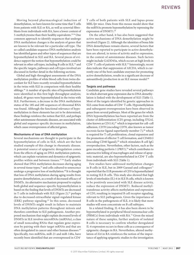
Targets and pathways Candidate gene studies have revealed several pathways in which aberrant gene expression due to DNA demethylation is linked with the development of SLE (Table 1). Most of the targets identified by genetic approaches in SLE come from studies of CD4+ T cells. Hypomethylation and subsequent overexpression have been observed in genes from several pathways. Most of the genes for which DNA hypomethylation has been reported are from the cluster of differentiation (CD) group, including ITGAL (also known as CD11A),67 which is important for cell–cell adhesion, CD70 (encoding CD70, also known as tumor necrosis factor ligand superfamily member 7),68 which is required for T cell proliferation, clonal expansion and the promotion of effector T cell formation, and CD40LG (encoding CD40 ligand),69 which stimulates B cell IgG overproduction. Nevertheless, other factors, such as the gene encoding perforin 1 (PRF1),70 which contributes to autoreactive killing of macrophages and release of apoptotic material, are also hypomethylated in CD4+ T cells from individuals with SLE (Table 1).
Few studies have addressed methylation changes in B cells in SLE, but in 2009 Garaud and colleagues71 reported that the E1B promoter of CD5 is hypo methylated in resting SLE B cells. This study also showed that high levels of interleukin (IL)6 in SLE B cells, which is known to be positively associated with SLE disease activity, reduce the expression of DNMT1. Reduced methyltransferase activity affects methylation and expression of CD5, resulting in impaired B cell receptor signaling, relevant to SLE pathogenesis. Given the importance of B cells in the pathogenesis of SLE, it is likely that more studies will soon concentrate on B cell subtypes.
In a related finding, IL6 has also been shown to be hypomethylated in peripheral blood mononuclear cells (PBMCs) from individuals with RA.72 Given the mixed nature of these samples, further analysis of isolated B cells is necessary to confirm whether deregulated IL6 expression occurs in these cells as a consequence of epigenetic changes in RA. Nevertheless, altered methylation of IL6 in RA reinforces the notion of the importance of applying epigenetic studies to the investigation
REVIEWS
© 2011 Macmillan Publishers Limited. All rights reserved
268 | MAY 2011 | VOLUME 7 www.nature.com/nrrheum
of pathways that are affected in rheumatic diseases. So far, few data are available on epigenetic changes in RA. As the disease is characterized by the progressive destruction of joints by invasive synovial fibroblasts, most of the studies that do exist have focused on this cell type. Remarkably, increased expression of retrotransposable L1 elements, one of the major classes of repetitive elements that are interspersed in the genome, has been found to occur in association with global hypo methylation in synovial tissue.73 Hypomethylation of specific CpG sites upstream of an L1 openreading frame has since been reported to play an essential part in the irreversible phenotypic changes that occur in synovial fibroblasts.74 More recently, DNA demethylation associated deregulation of one miRNA has also been associated with the deregulation of target genes that seem to have a key role in RA pathogenesis (Table 1).75
Many susceptibility genes have been identified for several autoimmune rheumatic diseases, but it is not yet known whether epigenetic changes affect some of these genes, or other genes for which no disease links have been established, or both. It seems unlikely that genes for which certain haplotypes have been found to confer disease risk will be the same as those that undergo altered epigenetic regulation, because the mechanisms and processes that generate genetic and epigenetic variation are entirely different. Highthroughput DNA methylation and histone modification analyses will surely contribute to systematic identification of the relationships between genetic and epigenetic alterations in pathological pathways. In this sense, arraybased analysis of twins dis cordant for
SLE56 has revealed new pathways and groups of factors that might be relevant to epi genetic deregulation in autoimmune disease (Table 1). These potential culprits include an interleukin receptor gene (IFNGR2), a matrix metalloproteinase (MMP), and a molecule involved in cellular transport (encoded by LCN2).56 Although twins discordant for disease provide a unique model for studying epigenetic involvement in pathogenesis, given that genetic variability is excluded as a cause for discordance, genomewide analyses of epigenetic changes are becoming easier and cheaper to perform, and will increasingly be carried out with larger collections of individuals with rheumatic disease. Such analyses are of clinical interest for two reasons: firstly, they might provide key information to enable classification of patients with respect to their clinical phenotype or their response to different biologic therapeutic agents, and secondly, they might reveal novel targets for potential epigenetic therapeutic treatment.
The therapeutic potential of compounds directed against epigenetic changes is obviously a primary line of research. In other pathological contexts, such as in hematological malignancies, several inhibitors of DNA methylation and histone deacetylation are now used for the clinical treatment of patients.15,76 In autoimmune rheumatic disease, inhibiting DNA methylation would not be appropriate to revert DNA methylation changes (as the changes identified to date are hypomethylation, not hypermethylation, Table 1), and agents should be designed to specifically increase methylation (no such agent yet exists). This need highlights the importance of the identification of mechanisms that promote
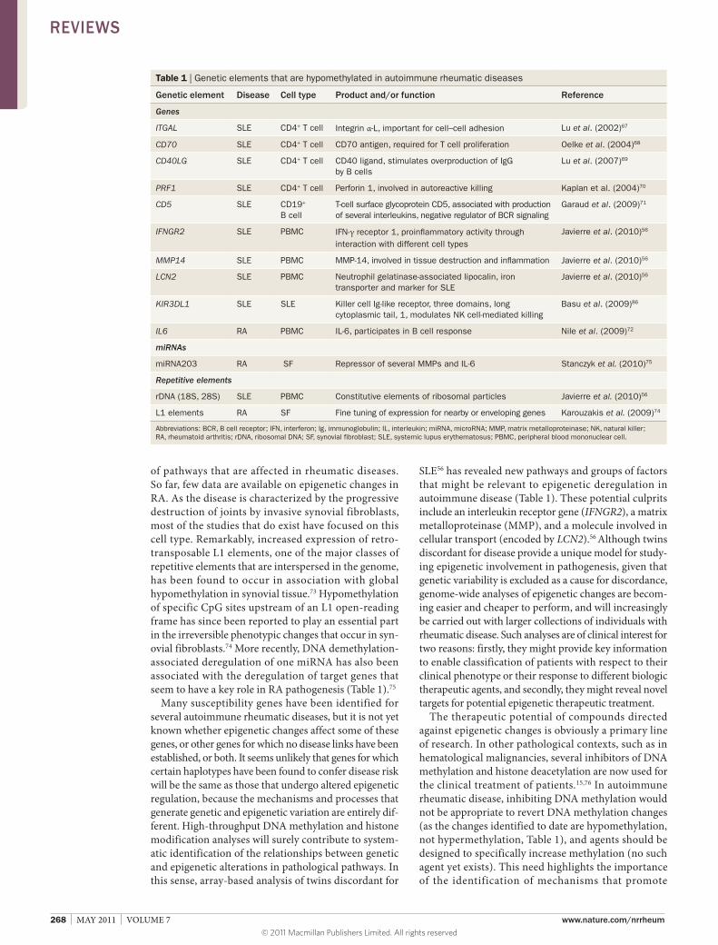
Table 1 | Genetic elements that are hypomethylated in autoimmune rheumatic diseases
Genetic element Disease Cell type Product and/or function Reference
Genes
ITGAL SLE CD4+ T cell Integrin α-L, important for cell–cell adhesion Lu et al. (2002)67
CD70 SLE CD4+ T cell CD70 antigen, required for T cell proliferation Oelke et al. (2004)68
CD40LG SLE CD4+ T cell CD40 ligand, stimulates overproduction of IgG by B cells
Lu et al. (2007)69
PRF1 SLE CD4+ T cell Perforin 1, involved in autoreactive killing Kaplan et al. (2004)70
CD5 SLE CD19+ B cell
T-cell surface glycoprotein CD5, associated with production of several interleukins, negative regulator of BCR signaling
Garaud et al. (2009)71
IFNGR2 SLE PBMC IFN-γ receptor 1, proinflammatory activity through interaction with different cell types
Javierre et al. (2010)56
MMP14 SLE PBMC MMP-14, involved in tissue destruction and inflammation Javierre et al. (2010)56
LCN2 SLE PBMC Neutrophil gelatinase-associated lipocalin, iron transporter and marker for SLE
Javierre et al. (2010)56
KIR3DL1 SLE SLE Killer cell Ig-like receptor, three domains, long cytoplasmic tail, 1, modulates NK cell-mediated killing
Basu et al. (2009)86
IL6 RA PBMC IL-6, participates in B cell response Nile et al. (2009)72
miRNAs
miRNA203 RA SF Repressor of several MMPs and IL-6 Stanczyk et al. (2010)75
Repetitive elements
rDNA (18S, 28S) SLE PBMC Constitutive elements of ribosomal particles Javierre et al. (2010)56
L1 elements RA SF Fine tuning of expression for nearby or enveloping genes Karouzakis et al. (2009)74
Abbreviations: BCR, B cell receptor; IFN, interferon; Ig, immunoglobulin; IL, interleukin; miRNA, microRNA; MMP, matrix metalloproteinase; NK, natural killer; RA, rheumatoid arthritis; rDNA, ribosomal DNA; SF, synovial fibroblast; SLE, systemic lupus erythematosus; PBMC, peripheral blood mononuclear cell.
REVIEWS
© 2011 Macmillan Publishers Limited. All rights reserved
NATURE REVIEWS | RHEUMATOLOGY VOLUME 7 | MAY 2011 | 269
both global and sequencespecific hypomethylation. Inhibition of histone deacetylases, which has been shown to alleviate renal disease in a mouse model of SLE, does have potential in the treatment of autoimmune rheumatic diseases.77,78 In line with this, it has been recently reported that histone deacetylase is efficient in the treatment of juvenile idiopathic arthritis.79 Identification of novel epigenetic targets, a better understanding of the epi genetic mechanisms and development of novel compounds directed against them will surely open novel therapeutic approaches in rheumatic disease.
Investigating the correct cell or tissue type As mentioned previously, the study of epigenetic alterations requires analysis of the appropriate cell or tissue types. The reasons for this are threefold: firstly, each cell type is characterized by a particular epigenetic profile, or epigenome, that is exquisitely associated with a specific gene expression profile; secondly, different cell types can undergo epigenetic alterations independently (because they are exposed to the environment in distinct manners, have different underlying metabolic and signaling pathways, and are thus altered by divergent pathways); and finally, different cell types have distinct roles in the pathogenesis of each disease.
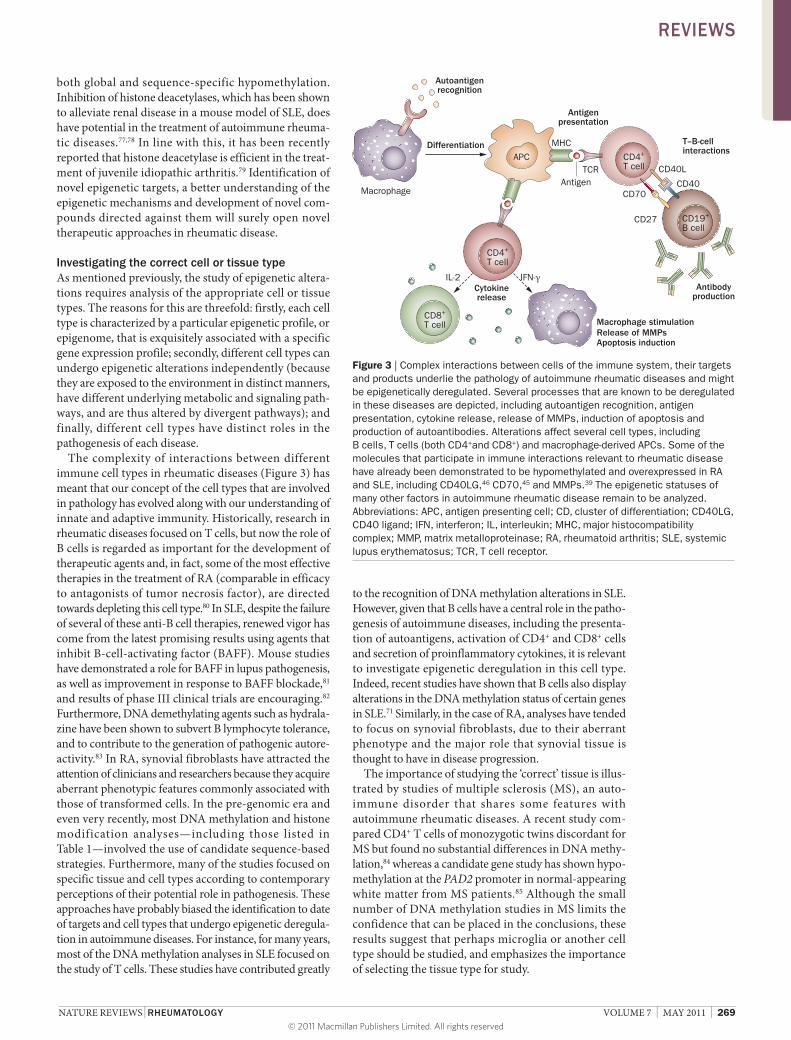
The complexity of interactions between different immune cell types in rheumatic diseases (Figure 3) has meant that our concept of the cell types that are involved in pathology has evolved along with our understanding of innate and adaptive immunity. Historically, research in rheumatic diseases focused on T cells, but now the role of B cells is regarded as important for the develop ment of therapeutic agents and, in fact, some of the most effective therapies in the treatment of RA (comparable in efficacy to antagonists of tumor necrosis factor), are directed towards depleting this cell type.80 In SLE, despite the failure of several of these antiB cell therapies, renewed vigor has come from the latest promising results using agents that inhibit Bcellactivating factor (BAFF). Mouse studies have demonstrated a role for BAFF in lupus pathogenesis, as well as improvement in response to BAFF blockade,81 and results of phase III clinical trials are encouraging.82 Furthermore, DNA demethylat ing agents such as hydralazine have been shown to subvert B lympho cyte tolerance, and to contribute to the genera tion of pathogenic autoreactivity.83 In RA, synovial fibroblasts have attracted the attention of clinicians and researchers because they acquire aberrant phenotypic features commonly associated with those of transformed cells. In the pregenomic era and even very recently, most DNA methylation and histone modification analyses—including those listed in Table 1—involved the use of candidate sequencebased strategies. Furthermore, many of the studies focused on specific tissue and cell types according to contemporary perceptions of their potential role in pathogenesis. These approaches have probably biased the identification to date of targets and cell types that undergo epigenetic deregulation in autoimmune diseases. For instance, for many years, most of the DNA methylation analyses in SLE focused on the study of T cells. These studies have contributed greatly
to the recognition of DNA methylation alterations in SLE. However, given that B cells have a central role in the pathogenesis of autoimmune diseases, including the presentation of autoantigens, activation of CD4+ and CD8+ cells and secretion of proinflammatory cytokines, it is relevant to investigate epigenetic deregulation in this cell type. Indeed, recent studies have shown that B cells also display alterations in the DNA methylation status of certain genes in SLE.71 Similarly, in the case of RA, analyses have tended to focus on synovial fibroblasts, due to their aberrant pheno type and the major role that synovial tissue is thought to have in disease progression.
The importance of studying the ‘correct’ tissue is illustrated by studies of multiple sclerosis (MS), an autoimmune disorder that shares some features with auto immune rheumatic diseases. A recent study compared CD4+ T cells of monozygotic twins discordant for MS but found no substantial differences in DNA methylation,84 whereas a candidate gene study has shown hypomethylation at the PAD2 promoter in normal appearing white matter from MS patients.85 Although the small number of DNA methylation studies in MS limits the confidence that can be placed in the conclusions, these results suggest that perhaps microglia or another cell type should be studied, and emphasizes the importance of selecting the tissue type for study.
Autoantigenrecognition
Antigenpresentation
Cytokinerelease
IL-2 IFN-γ
Release of MMPs
T–B-cellinteractions
Antibodyproduction
Macrophage stimulation
Apoptosis induction
CD4+
T cell
CD4+
T cell
CD19+
B cell
CD8+
T cell
MHC
TCR CD40L
CD40CD70
Differentiation
Macrophage
APC
CD27
Antigen
Figure 3 | Complex interactions between cells of the immune system, their targets and products underlie the pathology of autoimmune rheumatic diseases and might be epigenetically deregulated. Several processes that are known to be deregulated in these diseases are depicted, including autoantigen recognition, antigen presentation, cytokine release, release of MMPs, induction of apoptosis and production of autoantibodies. Alterations affect several cell types, including B cells, T cells (both CD4+and CD8+) and macrophage-derived APCs. Some of the molecules that participate in immune interactions relevant to rheumatic disease have already been demonstrated to be hypomethylated and overexpressed in RA and SLE, including CD40LG,46 CD70,45 and MMPs.39 The epigenetic statuses of many other factors in autoimmune rheumatic disease remain to be analyzed. Abbreviations: APC, antigen presenting cell; CD, cluster of differentiation; CD40LG, CD40 ligand; IFN, interferon; IL, interleukin; MHC, major histocompatibility complex; MMP, matrix metalloproteinase; RA, rheumatoid arthritis; SLE, systemic lupus erythematosus; TCR, T cell receptor.
REVIEWS
© 2011 Macmillan Publishers Limited. All rights reserved
270 | MAY 2011 | VOLUME 7 www.nature.com/nrrheum
ConclusionsGreater efforts are needed to understand the molecular mechanisms that contribute to the pathogenesis of autoimmune rheumatic diseases. Disorders of this type share many features, and genetic studies have revealed the existence of several common susceptibility genes. However, genetic variation represents only half of the story. Gene function depends not only on DNA sequence, but also on epigenetic modifications, including both DNA methyla tion and histone posttranslational modifications. These modifications are influenced by environmental factors and are known to contribute to the patho genesis of several auto immune diseases. Most importantly at present, epigenetic alterations can be used as clinical markers of disease progression or response to therapy. Furthermore, epi genetic alterations can be pharmacologically reverted. This potential to tackle aberrant changes opens the possibility of developing novel therapies, some of which are already used for the clinical treatment of hemato logical malignancies. Most efforts to identify the epigenetic altera tions that occur in autoimmune rheumatic disease have focused on SLE and RA, and have served to identify both global and sequencespecific hypomethylation and overexpression
of key genes in immune function. Studies have made use of candidategene methylation analysis and have concentrated their attention on a few cell types. We now face several challenges: to make use of highthroughput approaches, to systematically analyze all potential specific cell types relevant to disease patho genesis, to perform prospective studies to better understand the extent and role of these alterations in these diseases, and to find the best way of using this information in a clinical setting.
Review criteria
The studies included in this Review were identified by searching PubMed using the phrases “autoimmune AND epigenetics”, “lupus AND epigenetics”, “lupus AND methylation”, “arthritis AND epigenetics”, or “arthritis AND methylation”. The searches were restricted to full-text papers in the English language, without limitation of publication date, and were completed on January 17th, 2011. In addition, a few selected key chromatin and epigenetics Reviews, as well as well as a few seminal papers on epigenetic regulation have been included. Papers cited in this Review were selected based on the author’s view of their direct relevance to the concepts illustrated.
1. Lie, B. A. & Thorsby, E. Several genes in the extended human MHC contribute to predisposition to autoimmune diseases. Curr. Opin. Immunol. 17, 526–531 (2005).
2. Delgado-Vega, A., Sánchez, E., Löfgren, S., Castillejo-López, C. & Alarcón-Riquelme, M. E. Recent findings on genetics of systemic autoimmune diseases. Curr. Opin. Immunol. 22, 698–705 (2010).
3. Niewold, T. B. et al. Association of the IRF5 risk haplotype with high serum interferon-α activity in systemic lupus erythematosus patients. Arthritis Rheum. 58, 2481–2487 (2008).
4. Kariuki, S. N. et al. Cutting edge: autoimmune disease risk variant of STAT4 confers increased sensitivity to IFN-α in lupus patients in vivo. J. Immunol. 182, 34–38 (2009).
5. Gourley, M. & Miller, F. W. Mechanisms of disease: Environmental factors in the pathogenesis of rheumatic disease. Nat. Clin. Pract. Rheumatol. 3, 172–180 (2007).
6. Klareskog, L., Padyukov, L., Lorentzen, J. & Alfredsson, L. Mechanisms of disease: genetic susceptibility and environmental triggers in the development of rheumatoid arthritis. Nat. Clin. Pract. Rheumatol. 2, 425–433 (2006).
7. Esteller, M. Epigenetics in cancer. N. Engl. J. Med. 358, 1148–1159 (2008).
8. Allis, C. D., Jenuwein, T. & Reinberg, D. in Epigenetics Ch. 3: Overview and Concepts (eds Allis, C. D., Jenuwein, T. & Reinberg, D.) 23–62 (Cold Spring Harbor Laboratory Press, Cold Spring Harbor, NY, 2007).
9. Huertas, D., Sendra, R. & Muñoz, P. Chromatin dynamics coupled to DNA repair. Epigenetics 4, 31–42 (2009).
10. Corpet, A. & Almouzni, G. Making copies of chromatin: the challenge of nucleosomal organization and epigenetic information. Trends Cell Biol. 19, 29–41 (2009).
11. Bird, A. Perceptions of epigenetics. Nature 447, 396–398 (2007).
12. Sandelin, A. et al. Mammalian RNA polymerase II core promoters: insights from genome-wide studies. Nat. Rev. Genet. 8, 424–436 (2007).
13. Irizarry, R. A. et al. The human colon cancer methylome shows similar hypo- and hypermethylation at conserved tissue-specific CpG island shores. Nat. Genet. 41, 178–186 (2009).
14. Goll, M. G. & Bestor, T. H. Eukaryotic cytosine methyltransferases. Annu. Rev. Biochem. 74, 481–514 (2005).
15. Garcia-Manero, G. Demethylating agents in myeloid malignancies. Curr. Opin. Oncol. 20, 705–710 (2008).
16. Bhutani, N. et al. Reprogramming towards pluripotency requires AID-dependent DNA demethylation. Nature 463, 1042–1047 (2010).
17. Bruniquel, D. & Schwartz, R. H. Selective, stable demethylation of the interleukin-2 gene enhances transcription by an active process. Nat. Immunol. 4, 235–240 (2003).
18. Ooi, S. K. & Bestor, T. H. The colorful history of active DNA demethylation. Cell 133, 1145–1148 (2008).
19. Fritz, E. L. & Papavasiliou, F. N. Cytidine deaminases: AIDing DNA demethylation? Genes Dev. 24, 2107–2114 (2010).
20. Rai, K. et al. DNA demethylation in zebrafish involves the coupling of a deaminase, a glycosylase, and gadd45. Cell 135, 1201–1212 (2008).
21. Salvador, J. M. et al. Mice lacking the p53-effector gene Gadd45a develop a lupus-like syndrome. Immunity 16, 499–508 (2002).
22. Tahiliani, M. et al. Conversion of 5-methylcytosine to 5-hydroxymethylcytosine in mammalian DNA by MLL partner TET1. Science 324, 930–935 (2009).
23. Ito, S. et al. Role of Tet proteins in 5mC to 5hmC conversion, ES-cell self-renewal and inner cell mass specification. Nature 466, 1129–1133 (2010).
24. Kouzarides, T. Chromatin modifications and their function. Cell 128, 693–705 (2007).
25. Allis, C. D. et al. New nomenclature for chromatin-modifying enzymes. Cell 131, 633–636 (2007).
26. Biancotto, C., Frigè, G. & Minucci, S. Histone modification therapy of cancer. Adv. Genet. 70, 341–386 (2010).
27. Kraus, W. L. & Wong, J. Nuclear receptor-dependent transcription with chromatin. Is it all about enzymes? Eur. J. Biochem. 269, 2275–2283 (2002).
28. Schwartz, Y. B. et al. Alternative epigenetic chromatin states of polycomb target genes. PLoS Genet. 6, e1000805 (2010).
29. Bell, C. G. & Beck, S. The epigenomic interface between genome and environment in common complex diseases. Brief. Funct. Genomics 9, 477–485 (2010).
30. Richly, H., Lange. M., Simboeck. E. & Di Croce, L. Setting and resetting of epigenetic marks in malignant transformation and development. Bioessays 32, 669–679 (2010).
31. Esteller, M. Cancer epigenomics: DNA methylomes and histone-modification maps. Nat. Rev. Genet. 8, 286–298 (2007).
32. Ehrlich, M. DNA hypomethylation in cancer cells. Epigenomics 1, 239–259 (2009).
33. Konkel, M. K. & Batzer, M. A. A mobile threat to genome stability: The impact of non-LTR retrotransposons upon the human genome. Semin. Cancer Biol. 20, 211–221 (2010).
34. Schlesinger, Y. et al. Polycomb-mediated methylation on Lys27 of histone H3 pre-marks genes for de novo methylation in cancer. Nat. Genet. 39, 232–236 (2007).
35. Widschwendter, M. et al. Epigenetic stem cell signature in cancer. Nat. Genet. 39, 157–158 (2007).
36. Di Croce, L. Chromatin modifying activity of leukaemia associated fusion proteins. Hum. Mol. Genet. 14 (Suppl. 1), R77–R84 (2005).
37. Ballestar, E. et al. Methyl-CpG binding proteins identify novel sites of epigenetic inactivation in human cancer. EMBO J. 22, 6335–6345 (2003).
38. Cooper, G. S., Miller, F. W. & Pandey, J. P. The role of genetic factors in autoimmune disease: implications for environmental research. Environ. Health Perspect. 107 (Suppl. 5), 693–700 (1999).
REVIEWS
© 2011 Macmillan Publishers Limited. All rights reserved
NATURE REVIEWS | RHEUMATOLOGY VOLUME 7 | MAY 2011 | 271
39. Salvetti, M., Ristori, G., Bomprezzi, R., Pozzilli, P. & Leslie, R. D. Twins: mirrors of the immune system. Immunol. Today 21, 342–347 (2000).
40. Fraga, M. F. et al. Epigenetic differences arise during the lifetime of monozygotic twins. Proc. Natl Acad. Sci. USA 102, 10604–10609 (2005).
41. Kaminsky, Z. A. et al. DNA methylation profiles in monozygotic and dizygotic twins. Nat. Genet. 41, 240–245 (2009).
42. Cezar, G. G. et al. Genome-wide epigenetic alterations in cloned bovine fetuses. Biol. Reprod. 68, 1009–1014 (2003).
43. Young, L. E. et al. Conservation of IGF2-H19 and IGF2R imprinting in sheep: effects of somatic cell nuclear transfer. Mech. Dev. 120, 1433–1442 (2003).
44. Heijmans, B. T. et al. Persistent epigenetic differences associated with prenatal exposure to famine in humans. Proc. Natl Acad. Sci. USA 105, 17046–17049 (2008).
45. Sinclair, K. D. et al. DNA methylation, insulin resistance, and blood pressure in offspring determined by maternal periconceptional B vitamin and methionine status. Proc. Natl Acad. Sci. USA 104, 19351–19356 (2007).
46. Cannat, A. & Seligmann, M. Induction by isoniazid and hydrallazine of antinuclear factors in mice. Clin. Exp. Immunol. 3, 99–105 (1968).
47. Richardson, B. Effect of an inhibitor of DNA methylation on T cells. II. 5-Azacytidine induces self-reactivity in antigen-specific T4+ cells. Hum. Immunol. 17, 456–470 (1986).
48. Cornacchia, E. et al. Hydralazine and procainamide inhibit T cell DNA methylation and induce autoreactivity. J. Immunol. 140, 2197–2200 (1988).
49. Quddus, J. et al. Treating activated CD4+ T cells with either of two distinct DNA methyltransferase inhibitors, 5-azacytidine or procainamide, is sufficient to cause a lupus-like disease in syngeneic mice. J. Clin. Invest. 92, 38–53 (1993).
50. Yung, R. L., Quddus, J., Chrisp, C. E., Johnson, K. J. & Richardson, B. C. Mechanism of drug-induced lupus. I. Cloned Th2 cells modified with DNA methylation inhibitors in vitro cause autoimmunity in vivo. J. Immunol. 154, 3025–3035 (1995).
51. Blomgren, S. E., Condemi, J. J. & Vaughan, J. H. Procainamide-induced lupus erythematosus. Clinical and laboratory observations. Am. J. Med. 52, 338–348 (1972).
52. Richardson, B. C. et al. Phenotypic and functional similarities between 5-azacytidine- treated T cells and a T cell subset in patients with active systemic lupus erythematosus. Arthritis Rheum. 35, 647–662 (1992).
53. Richardson, B. et al. Evidence for impaired T cell DNA methylation in systemic lupus erythematosus and rheumatoid arthritis. Arthritis Rheum. 33, 1665–1673 (1990).
54. Corvetta, A., Della Bitta, R., Luchetti, M. M. & Pomponio, G. 5-Methylcytosine content of DNA in blood, synovial mononuclear cells and synovial tissue from patients affected by autoimmune rheumatic diseases. J. Chromatogr. 566, 481–491 (1991).
55. Huck, S. & Zouali, M. DNA methylation: a potential pathway to abnormal autoreactive lupus B cells. Clin. Immunol. Immunopathol. 80, 1–8 (1996).
56. Javierre, B. M. et al. Changes in the pattern of DNA methylation associated with twin
discordance in systemic lupus erythematosus. Genome Res. 20, 170–179 (2010).
57. Fraga, M. F. & Esteller, M. Epigenetics and aging: the targets and the marks. Trends Genet. 23, 413–418 (2007).
58. Maegawa, S. et al. Widespread and tissue specific age-related DNA methylation changes in mice. Genome Res. 20, 332–340 (2010).
59. Wilson, V. L. & Jones, P. A. DNA methylation decreases in aging but not in immortal cells. Science 220, 1055–1057 (1983).
60. Zhang, W. et al. Comparison of global DNA methylation profiles in replicative versus premature senescence. Life Sci. 83, 475–480 (2008).
61. Deng, C. et al. Decreased Ras-mitogen- activated protein kinase signaling may cause DNA hypomethylation in T lymphocytes from lupus patients. Arthritis Rheum. 44, 397–407 (2001).
62. Sawalha, A. H. et al. Defective T-cell ERK signaling induces interferon-regulated gene expression and overexpression of methylation-sensitive genes similar to lupus patients. Genes Immun. 9, 368–378 (2008).
63. Tili, E., Michaille, J. J., Costinean, S., Croce, C. M. MicroRNAs, the immune system and rheumatic disease. Nat. Clin. Pract. Rheumatol. 4, 534–541 (2008).
64. Pan, W. et al. MicroRNA-21 and microRNA-148a contribute to DNA hypomethylation in lupus CD4+ T cells by directly and indirectly targeting DNA methyltransferase 1. J. Immunol. 184, 6773–6781 (2010).
65. Li, Y. et al. Overexpression of the growth arrest and DNA damage-induced 45α gene contributes to autoimmunity by promoting DNA demethylation in lupus T cells. Arthritis Rheum. 62, 1438–1447 (2010).
66. Hsu, H.-C. et al. Inhibition of the catalytic function of activation-induced cytidine deaminase (AICDA) promotes apoptosis of germinal center B cells. Arthritis Rheum. doi:10.1002/art.30257.
67. Lu, Q. et al. Demethylation of ITGAL (CD11a) regulatory sequences in systemic lupus erythematosus. Arthritis Rheum. 46, 1282–1291 (2002).
68. Oelke, K. et al. Overexpression of CD70 and overstimulation of IgG synthesis by lupus T cells and T cells treated with DNA methylation inhibitors. Arthritis Rheum. 50, 1850–1860 (2004).
69. Lu, Q. et al. Demethylation of CD40LG on the inactive X in T cells from women with lupus. J. Immunol. 179, 6352–6358 (2007).
70. Kaplan, M. J., Lu, Q., Wu, A., Attwood, J. & Richardson, B. Demethylation of promoter regulatory elements contributes to perforin overexpression in CD4+ lupus T cells. J. Immunol. 172, 3652–3661 (2004).
71. Garaud, S. et al. IL-6 modulates CD5 expression in B cells from patients with lupus by regulating DNA methylation. J. Immunol. 182, 5623–5632 (2009).
72. Nile, C. J., Read, R. C., Akil, M., Duff, G. W. & Wilson, A. G. Methylation status of a single CpG site in the IL-6 promoter is related to IL-6 messenger RNA levels and rheumatoid arthritis. Arthritis Rheum. 58, 2686–2693 (2008).
73. Neidhart, M. et al. Retrotransposable L1 elements expressed in rheumatoid arthritis
synovial tissue: association with genomic DNA hypomethylation and influence on gene expression. Arthritis Rheum. 43, 2634–2647 (2000).
74. Karouzakis, E., Gay, R. E., Michel, B. A., Gay, S. & Neidhart, M. DNA hypomethylation in rheumatoid arthritis synovial fibroblasts. Arthritis Rheum. 60, 3613–3622 (2009).
75. Stanczyk, J. et al. Altered expression of miR-203 in rheumatoid arthritis synovial fibroblasts and its role in fibroblast activation. Arthritis Rheum. 63, 373–381 (2011).
76. Lane, A. A. & Chabner, B. A. Histone deacetylase inhibitors in cancer therapy. J. Clin. Oncol. 27, 5459–5468 (2009).
77. Mishra, N. et al. Histone deacetylase inhibitors modulate renal disease in the MRL-lpr/lpr mouse. J. Clin. Invest. 111, 539–552 (2003).
78. Reilly, C. M. et al. Modulation of renal disease in MRL/lpr mice by suberoylanilide hydroxamic acid. J. Immunol. 173, 4171–4178 (2004).
79. Vojinovic, J. et al. Safety and efficacy of an oral histone deacetylase inhibitor in systemic onset juvenile idiopathic arthritis. Arthritis Rheum. doi:10.1002/art.30238.
80. Dörner, T., Radbruch, A. & Burmester, G. R. B-cell-directed therapies for autoimmune disease. Nat. Rev. Rheumatol. 5, 433–441 (2009).
81. Petri, M. et al., Association of plasma B lymphocyte stimulator levels and disease activity in systemic lupus erythematosus. Arthritis Rheum. 58, 2453–2459 (2008).
82. Sanz, I. & Lee, F. E. B cells as therapeutic targets in SLE. Nat. Rev. Rheumatol. 6, 326–337 (2010).
83. Mazari, L., Ouarzane, M. & Zouali, M. Subversion of B lymphocyte tolerance by hydralazine, a potential mechanism for drug-induced lupus. Proc. Natl Acad. Sci. USA 104, 6317–6322 (2007).
84. Baranzini, S. E. et al. Genome, epigenome and RNA sequences of monozygotic twins discordant for multiple sclerosis. Nature 464, 1351–1356 (2010).
85. Mastronardi, F. G., Noor, A., Wood, D. D., Paton, T. & Moscarello, M. A. Peptidyl argininedeiminase 2 CpG island in multiple sclerosis white matter is hypomethylated. J. Neurosci. Res. 85, 2006–2016 (2007).
86. Basu, D. et al. Stimulatory and inhibitory killer Ig-like receptor molecules are expressed and functional on lupus T cells. J. Immunol. 183, 3481–3487 (2009).
87. Mikkelsen, T. S. et al. Genome-wide maps of chromatin state in pluripotent and lineage-committed cells. Nature 448, 553–560 (2007).
88. Ballestar, E. & Wolffe, A. P. Methyl-CpG-binding proteins. Targeting specific gene repression. Eur. J. Biochem. 268, 1–6 (2001).
89. Dhasarathy, A. & Wade, P. A. The MBD protein family—reading an epigenetic mark? Mutat. Res. 647, 39–43 (2008).
90. Ooi, S. K. et al. DNMT3L connects unmethylated lysine 4 of histone H3 to de novo methylation of DNA. Nature 448, 714–717 (2007).
91. de la Cruz, X., Lois, S., Sánchez-Molina, S. & Martínez-Balbás, M. A. Do protein motifs read the histone code? Bioessays 27, 164–175 (2005).
AcknowledgmentsE. Ballestar is supported by PI081346 (FIS) grant from the Spanish Ministry of Science and Innovation (MICINN) and 2009SGR184 grant from AGAUR (Catalan Government).
REVIEWS
© 2011 Macmillan Publishers Limited. All rights reserved