Embed Size (px)
Citation preview
GENES, CHROMOSOMES & CANCER 49:526–538 (2010)
Epigenetic Alterations of the SERPINE1 Gene in OralSquamous Cell Carcinomas and Normal Oral Mucosa
Shan Gao,1* Boye Schnack Nielsen,2 Annelise Krogdahl,3 Jens Ahm Sørensen,4 Jan Tagesen,5 Sally Dabelsteen,6
Erik Dabelsteen,7 and Peter A. Andreasen1
1Departmentof Molecular Biology, Danish-Chinese Centre for Proteases and Cancer,Universityof Aarhus,8000 Aarhus C,Denmark2The Finsen Laboratory, Copenhagen Biocentre,Ole Maal�esVej, 2200 Copenhagen N,Denmark3Departmentof Pathology, Odense University Hospital, 5000 Odense,Denmark4Departmentof Plastic Surgery, Odense University Hospital, 5000 Odense,Denmark5Departmentof Oral Pathology and Maxillofacial Surgery,School of Dentistry, Universityof Aarhus,8000 Aarhus C,Denmark6Departmentof Oral Medicine,Clinical Oral Physiology,Oral Pathologyand Oral Anatomy,School of Dentistry, UniversityofCopenhagen, 2200 Copenhagen N,Denmark7Departmentof Oral Diagnostics, School of Dentistry,Universityof Copenhagen, 2200,Copenhagen N,Denmark
A high level of plasminogen activator inhibitor-1 (PAI-1 or SERPINE1) in tumor extracts is a marker of a poor prognosis in
human cancers, including oral carcinomas. However, the mechanisms responsible for the upregulation of PAI-1 in cancers
remain unclear. Investigating specific PAI-1 expressing cells in oral carcinomas by immunohistochemistry, we found that
PAI-1 was expressed in 18 of the 20 patients, mainly by cancer cells. Two showed PAI-1 positive stromal cells surrounding
the tumor areas and five showed PAI-1 positive cells in tumor-adjacent normal epithelium. By real-time RT-PCR analysis,
17 of 20 patients with oral carcinoma were found to have between 2.5- and 50-fold increased tumor PAI-1 mRNA level, as
compared with the matched tumor-adjacent normal tissues. The PAI-1 mRNA level in connective tissues from 15 healthy
volunteers was similar to the level in tumor-adjacent normal tissues, but the level in epithelium was 5- to 10-fold lower.
Analyzing DNA methylation of 25 CpG sites within 960 bp around the transcription initiation site of the SERPINE1 gene
by bisulfite sequencing, we did the surprising observation that both tumors and tumor-adjacent normal tissue had a signifi-
cant level of methylation, whereas there was very little methylation in tissue from healthy volunteers, suggesting that tu-
mor-adjacent normal tissue already contains transformation-associated epigenetic changes. However, there was no general
inverse correlation between PAI-1 mRNA levels and SERPINE1 gene methylation in all tissues, showing that CpG methyla-
tion is not the main determinant of the PAI-1 expression level in oral tissue. VVC 2010 Wiley-Liss, Inc.
INTRODUCTION
Oral carcinogenesis is believed to represent a
multistep process driven by the accumulation of
carcinogen-induced genetic and epigenetic
changes. It is generally accepted that many of the
cancer-associated gene changes stem from the
gain, loss, or mutation of genetic information, as
well as DNA methylation (Ha and Califano,
2006; Shaw, 2006). However, not only alterations
of proto-oncogenes and tumor suppressor genes
but also many other regulatory factors related to
cell growth, migration, and proliferation are
involved in tumorigenesis.
It has been demonstrated that the urokinase-
type plasminogen activator (uPA) system plays an
important role in tumor metastasis and invasion.
The uPA system is a serine protease system with
a complex pericellular organization. The main
biochemical function of the system is the uPA-
catalyzed proteolytic conversion of the inactive
zymogen plasminogen to the active, nonspecific
protease plasmin, which can catalyze degradation
of many extracellular matrix proteins. The activ-
ity of uPA is inhibited by plasminogen activator
inhibitor-1 (PAI-1). A high level of uPA in tumors
is correlated with a poor prognosis in many cancer
types, including oral cancer. Furthermore, it has
been shown that high tumor PAI-1 levels, simi-
larly to high uPA levels, predict a poor prognosis
(Andreasen et al., 2000; Durand et al., 2004).
PAI-1 is expressed in oral squamous cell carcino-
mas but not in normal oral mucosa; furthermore,
PAI-1 is mainly expressed in cancer cells in oral
Supported by: The Danish Cancer Society, The DanishResearch Agency, The Novo-Nordisk Foundation, GrossererAlfred Nielsen and Hustrus Fond, The Danish Cancer ResearchFoundation, and The Danish National Research Foundation.
*Correspondence to: Shan Gao, Department of Molecular Biol-ogy, University of Aarhus, C. F. Møllers Alle, Building 1130, 8000Aarhus C, Denmark. E-mail: [email protected]
Received 23 December 2009; Accepted 2 February 2010
DOI 10.1002/gcc.20762
Published online 10 March 2010 inWiley InterScience (www.interscience.wiley.com).
VVC 2010 Wiley-Liss, Inc.
carcinoma (Clayman et al., 1993; Nozaki et al.,
1998; Curino et al., 2004; Lindberg et al., 2006). In
this respect, oral carcinomas are different from other
cancer types, such as breast, prostate, and colon
cancers, in which PAI-1 is predominantly found in
stromal myofibroblasts and not in the cancer cells
themselves (Offersen et al., 2003; Illemann et al.,
2004; Usher et al., 2005). Although it has been
well-demonstrated that PAI-1 is overexpressed in
human cancers, the mechanism of the altered level
of PAI-1 expression in tumors is unclear.
It has become increasingly clear that epigenetic
events, i.e., inheritable changes in gene function
that cannot be explained by changes in DNA
sequence, play an important role in development
of cancer by modifying gene expression levels
(Herman, 1999; Esteller and Herman, 2002). In
this way, aberrant DNA methylation is important
in inactivation of tumor suppressor genes and other
cancer-related genes in most types of tumors,
including oral carcinomas (McGregor et al., 2002).
Concerning the effect of CpG methylation of the
SERPINE1 gene on regulation of transcriptional
activity, we have previously found that methylation
of 25 CpG sites within approximately 960 bp
around the transcriptional initiation site is inversely
correlated with the PAI-1 expression in five human
tumor cell lines (Gao et al., 2005).
In the present study, we characterized 20
patients with oral squamous cell carcinomas and
15 healthy volunteers with respect to PAI-1
expressing cell types by immunohistochemical
staining, PAI-1 mRNA levels by RT-PCR, and
SERPINE1 gene methylation status to study
mechanisms behind the increased expression in
oral cancers.
MATERIALS AND METHODS
Sample Preparation
Frozen surgical specimens of oral squamous cell
carcinoma from 20 patients were obtained from
Odense University Hospital, Denmark. The me-
dian age of the 20 patients was 63 years (range,
48–94 years); there were six women and 14 men.
The location of the carcinomas was six at the
tongue (presented as T1–T6), nine at the floor of
the mouth (presented as F1–F9), and five at the
alveolar ridge (presented as A1–A5). For all
patients, a biopsy from clinically normal mucosa
close to the tumor was included as well. Hematox-
ylin-eosin staining of the sections was performed
and the ratio between cancer and normal tissues
were examined on histological sections. In 14
cases, in which the cancer cells contributed 80% or
more, the tissue blocks were used directly for
RNA preparation. In six cases, a laser microdissec-
tion system (P.A.L.M.) was used to separate can-
cer cells from adjacent normal tissue, as well as to
separate histologically normal epithelium from the
connective tissue. Fifteen samples from normal
buccal mucosa obtained from healthy volunteers
served as control tissue, too. There were five
males and 10 females, the median age was 36
years (range, 25 to 62 years), and one smoker.
Before the following procedures for DNA/RNA
preparation, epithelium in the normal tissue was
separated from the stroma by incubation in 2 mM
EDTA buffer (Mackenzie et al., 1979).
For all tissues, informed consent and approval
by the Ethics Committee were obtained accord-
ing to Danish legislation.
DNA was extracted by routine procedures
using the DNeasy Kit (Qiagen, Copenhagen,
Denmark). Total RNA was extracted by routine
procedures using the RNeasy Mini Kit (Qiagen,
Copenhagen, Denmark). On-column DNase
digestion by RNA-free DNase I (Qiagen, Copen-
hagen, Denmark) was performed during the RNA
purification procedure.
PAI-1 mRNA Expression Analyzed by
Quantitative Real-Time RT-PCR
Reverse transcription reaction was performed
with 2 lg DNA-free RNA, using First-strand
cDNA Synthesis Kit (Amersham, Copenhagen,
Denmark). Quantitative real-time RT PCR was
performed as described elsewhere (Gao et al.,
2005). Amplification reactions were carried out in
a final volume of 25 ll containing 2� Brilliant
Multiplex Master Mix (Stratagene, Copenhagen,
Denmark). The comparative CT (threshold cycle)
and copy numbers described in the manufac-
turer’s protocol was used to quantitate the rela-
tive PAI-1 mRNA expression level, comparing
tumor samples to corresponding normal controls.
Thermal conditions were as follows: 95�C for 10
min to activate the DNA polymerase, followed by
40 cycles of 95�C for 30 sec and 62�C for 30 sec,
and 72�C for 30 sec. Amplification was performed
on a Mx4000VR
Multiplex Quantitative PCR Sys-
tem (Stratagene, Copenhagen, Denmark). The
glyceraldehyde 3-phosphate dehydrogenase
(GAPDH) gene mRNA was amplified as an inter-
nal control to normalize the data of the PAI-1
mRNA level. All PAI-1 mRNA levels were
expressed relative to the PAI-1 mRNA in the cell
line HT-1080, which has a high PAI-1 expression
PAI-1 IN ORAL CARCINOMAS 527
Genes, Chromosomes & Cancer DOI 10.1002/gcc
and was run as a positive control in each reaction
(Gao et al., 2005, 2008).
Immunohistochemical Staining for PAI-1 Antigen
To investigate the histological distribution of
PAI-1 antigen in oral carcinomas, immunohisto-
chemical staining was performed. Affinity-purified
rabbit polyclonal antibodies as well as control rab-
bit anti-PAI-1 polyclonal antibodies depleted for
anti-PAI-1 IgG have been described previously
(Offersen et al., 2003).
Four micrometer frozen sections were pre-
pared. The primary antibodies were diluted in 50
mM Tris, 150 mM NaCl, pH 7.6 (TBS), contain-
ing 0.25% BSA, and incubated on the sections
overnight in Shandon racks (Thermo Shandon,
Pittsburgh, PA) at the following concentrations:
PAI-1 polyclonal antibodies (0.5 lg/ml). After 30
min at room temperature, sections were washed
in 50 mM Tris, 150 mM NaCl, pH 7.6, containing
0.5% Triton X-100 (TBS-T). The primary anti-
bodies were detected with envision reagents and
anti-rabbit IgG horseradish peroxidase-conjugated
polymers (DakoCytomation, Copenhagen, Den-
mark). Each incubation step was followed by
washing with 6 ml of TBS-T. Endogenous perox-
idase was blocked by incubation in 1% H2O2 for
15 min. Sections were developed with 3, 30-dia-minobenzidine chromogenic substrate (DAB,
DakoCytomation, Copenhagen, Denmark) for 15
min and finally counterstained with hematoxylin.
Methylation Analysis
Genomic DNA was treated with sodium bisul-
phite as described previously (Clark et al., 1994).
Bisulfite sequencing was performed as described
elsewhere (Gao et al., 2005). PCR amplification
was performed in 25 ll of reaction mixture con-
taining 100 ng bisulfite-treated DNA, 10 pmol of
each primer, 0.2 mM each dNTP, 1 unit of Hot-
StarTaq DNA polymerase (Qiagen, Copenhagen,
Denmark), and 1� PCR buffer. Three pairs of
primers were used for amplifying the 958 bp
sequence from �805 to þ152, one covering CpG
#1 to #7, which will be referred as the Exon1
(SeqE), one covering CpG #8 to #15, which will
be referred as the proximal sequence (SeqP) and
the other covering CpG #16 to #25, which will
referred as the distal sequence (SeqD). The
sequences of the primers were as follows: PAI-
1SeqE-F, AGTTGTGT TTGGTTGTAGGGT-
TAAG; PAI-1SeqE-R, CT TTTCTCCTACC-
TAAAATTCTCAAAAA (4 to 152, 151 bp); PAI-
1SeqP-F, TTTGATAATTT TATAGT-
GATTTGGTT; PAI-1SeqP-R CACC CACT-
CACTAACTCTAAAAATC (�433 to �73, 361
bp); PAI-1SeqD-F, TTTTTATTATGGT
AATTTTTGGTTT; PAI-1SeqD-R, AAATTA
TCAAAAATAACCTCCA TCAA (�805 to �422,
384 bp). The reaction for PAI-1SeqE was started
with initial denaturation at 95�C for 10 min, fol-
lowed by 35 cycles at 95�C for 30 sec, 55�C for 30
sec, and 72�C for 30 sec. Reactions for PAI-1SeqP
and PAI-1SeqD were started with initial denatura-
tion at 95�C for 10 min, followed by 35 cycles at
95�C for 1 min, 57�C for 1 min, and 72�C for 2
min. Amplified DNA was subcloned into vector
pCR2.1 by using a TOPO TA cloning kit (Invitro-
gen, Copenhagen, Denmark) and sequenced.
Eight or 10 positive clones were analyzed for each
sample. The total frequency of total methylated
CpGs was calculated as (number of methylated
CpGs/number of totally analyzed CpGs) � 100%.
Statistical Analysis
Paired t test was performed for comparison of
the PAI-1 mRNA expression level and FTM
between tumor samples and corresponding nor-
mal tissues. Fisher’s exact probability test was
used for analysis the correlation between PAI-1
mRNA expression and immunohistochemical
staining intensity, as well as for analysis the cor-
relation between methylation frequency and PAI-
1 mRNA levels in carcinomas. Wilcoxon rank
sum test was performed for comparison the PAI-1
expression in patients with and without lymph
node metastasis. The v2 test was performed for
comparison between 4G/5G polymorphism in tu-
mor and adjacent histologically normal samples.
Student’s t test was performed for comparison of
methylation frequency between 4G and 5G al-
leles in both tumor and tumor-adjacent, histologi-
cally normal tissue samples.
RESULTS
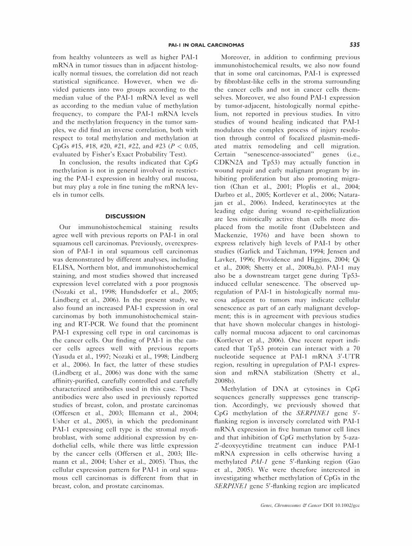
Immunohistochemical Staining of PAI-1 Antigen
in Oral Carcinomas Specimens
All of the 20 investigated tumors were squa-
mous cell carcinomas. In eight cases, minor parts
of histologically normal mucosa were presented
adjacent to the tumor sample.
Immunohistochemical staining for PAI-1 was
performed in the 20 tumor samples. Two tumors
were negative and in three tumors only a few posi-
tive areas were found (Fig. 1A); a correspondingly
528 GAO ET AL.
Genes, Chromosomes & Cancer DOI 10.1002/gcc
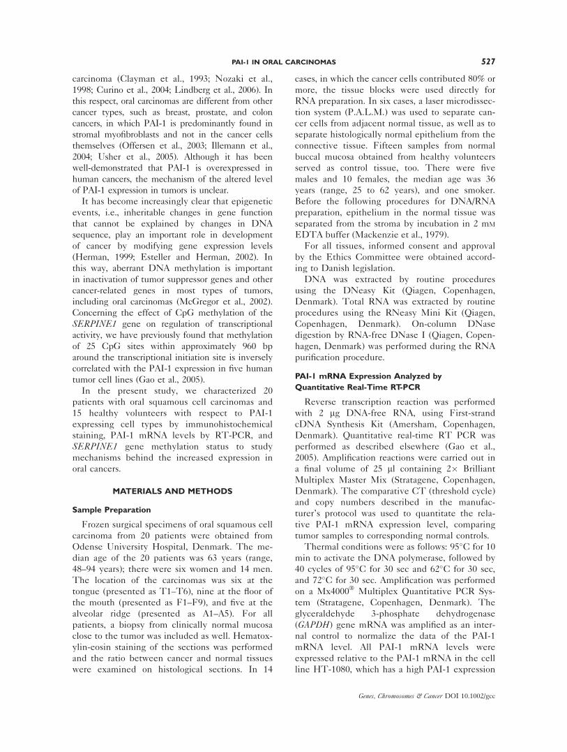
Figure 1. Immunohistochemical staining of PAI-1 expressing celltypes. (A) A few cells were positive for PAI-1, but most are negative;(B) a heterogeneous distribution of PAI-1 positive carcinoma cellswas demonstrated; (C, D) the positive PAI-1 staining was mainlylocated at the invasive front of the tumor island; (E) the staining reac-tion was negative in the cancer cells themselves but positive in thesurrounding stroma; (F) PAI-1 expression was also seen in endothelial
cells of small vessels surrounding the malignantly invasive strands(arrow) or clusters (star); (G) the positivity of PAI-1 staining waslocated in the subepithelial connective tissue in histologically normalepithelium, but it was negative in cancer cells with a clear borderline(the picture is from the same sample of E); (H) the positivity of PAI-1staining was also seen in basal cells in tumor-adjacent, histologicallynormal epithelium.
PAI-1 IN ORAL CARCINOMAS 529
Genes, Chromosomes & Cancer DOI 10.1002/gcc
low level of PAI-1 mRNA was found in these cases.
In 17 patients, PAI-1 was demonstrated in the carci-
noma cells. The distribution was heterogeneous,
with some parts of the tumor negative and other
parts positive (Fig. 1B). The positive PAI-1 staining
was mainly located at the basal outer cell layer of
the tumor island (Figs. 1C and1D). In two patients,
PAI-1 positive cells were found in tumor stroma. In
one of these cases, the staining reaction was nega-
tive in the cancer cells themselves (Fig. 1E). In five
cases, PAI-1 expression was also seen in endothelial
cells of small vessels surrounding the malignantly
invasive strands or clusters (Fig. 1F). Normal mu-
cosa adjacent to the tumor showed positive staining
in five cases. In one of these, the positivity was
located in the subepithelial connective tissue (Fig.
1G), in the rest of the cases in basal epithelial cells
(Fig. 1H).
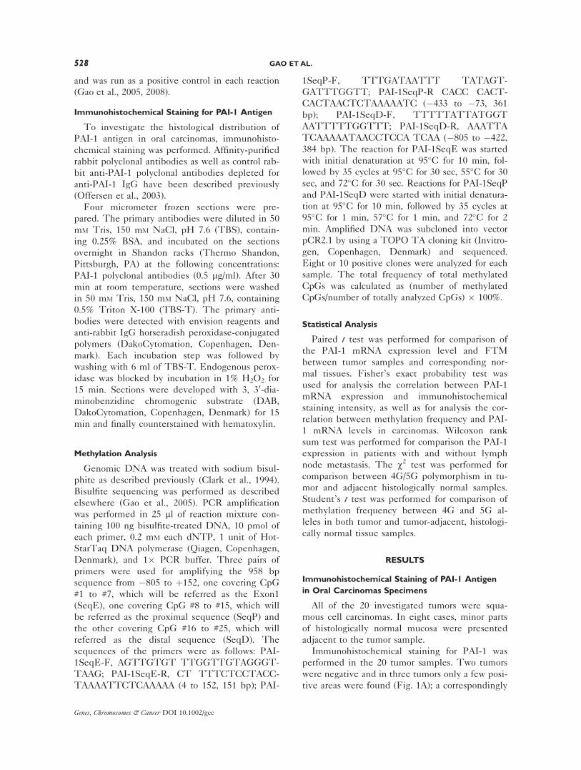
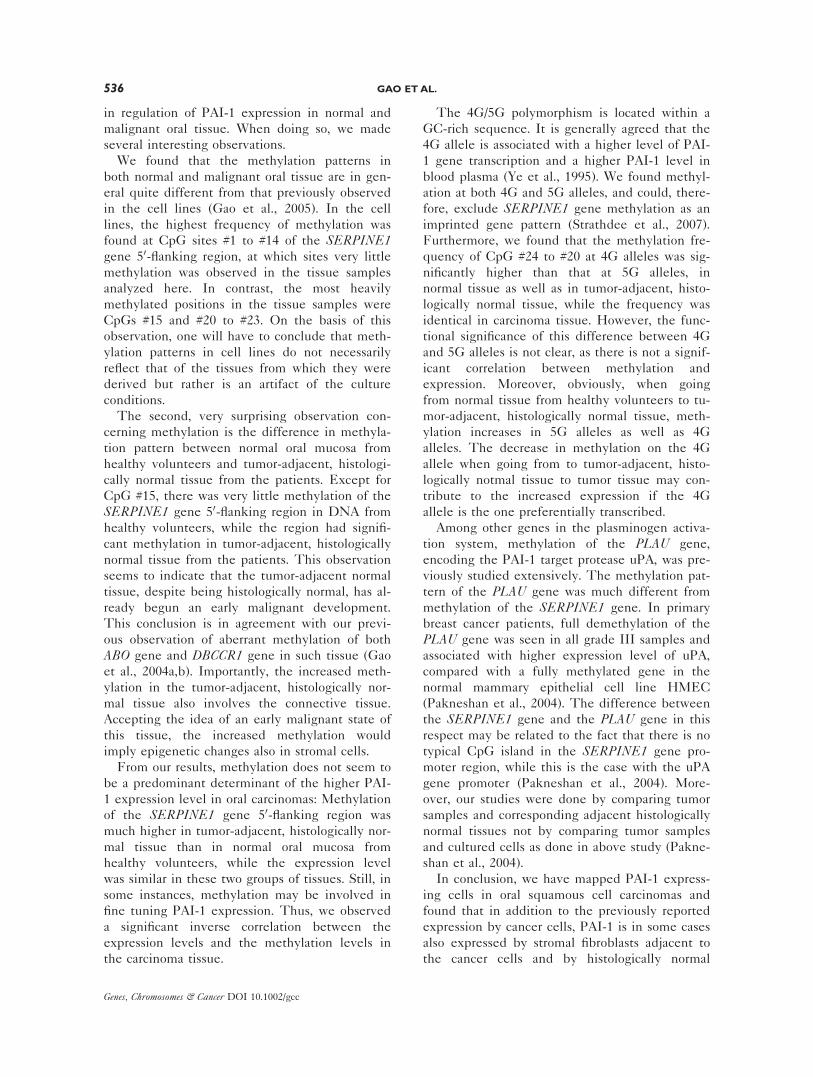
PAI-1 mRNA Levels, as Evaluated by Quantitative
Real-Time RT-PCR
By quantitative real-time RT-PCR analysis,
oral carcinomas from 17 of 20 patients were found
to have a higher PAI-1 mRNA level than control
tissue, which was tumor-adjacent, histologically
normal oral tissue from the same patient. The
fold increase of the PAI-1 mRNA level in the
tumors, as compared with the tumor-adjacent,
histologically normal tissue, was from 2.5- to 50-
fold (Fig. 2). The average PAI-1 mRNA level rel-
ative to the positive control (HT-1080 cells) in
the 20 tumors was 27% � 24%, which is signifi-
cantly higher than the level of 4.5% � 3.7% in
the corresponding adjacent, histologically normal
tissue from the same patients (P < 0.001, paired ttest). There was no significant difference
between patients with or without lymph nodes
metastasis (P > 0.05, by Wilcoxon rank sum test).
To evaluate the correlation between PAI-1
mRNA level in tumors and the immunohisto-
chemical staining results, patients were divided
into two groups according to the median value of
the PAI-1 mRNA level as well as according to
the intensity and the positive area in immunohis-
tochemical stainings for PAI-1 in the tumors.
These two assays demonstrated a statistically sig-
nificant correlation as analyzed by Fisher’s Exact
Probability Test (P ¼ 0.0013).
The PAI-1 mRNA levels in epithelium and
connective tissues from 15 healthy volunteers
were also measured. The average PAI-1 mRNA
level in epithelium was 0.38% � 0.52%. The av-
erage PAI-1 mRNA level in connective tissue
was 2.3% � 1.2%. The two values are signifi-
cantly different (P < 0.001, paired t test; Fig. 3).The PAI-1 mRNA level in tumor-adjacent, histo-
logically normal tissues from cancer patients was
not different from the level in connective tissues
from healthy volunteers, but 5- to 10-fold higher
than the level in epithelium from healthy volun-
teers (P < 0.01). Although we did not have
enough tissue to do RT-PCR analysis on RNA
from epithelium and connective tissue from
tumor-adjacent, histologically normal tissue
Figure 2. PAI-1 mRNA expression level in 20 oral carcinomas andmatched tumor-adjacent, histologically normal tissue. PAI-1 mRNAlevels were determined by Q-real time RT-PCR and normalized againstthe GAPDH mRNA level. The PAI-1 level was given as a percentageof the level in HT-1080 cells. The figure shows means 6 SD for threeindependent determinations. The level in tumors (black bars) was sig-nificantly elevated compared with the level in the corresponding sam-ples of histologically normal tissue from the same patient (gray bars; P< 0.001, as evaluated by a paired t test). The patients are organizedinto two subgroups in the figure, according to lymph node involve-ment. The locations of the carcinomas are indicated: tongue (T 1–6),the floor of the mouth (F 1–9), and alveolar ridge (A1–5).
Figure 3. PAI-1 mRNA expression level in epithelium and connec-tive tissue in samples of tissue from healthy volunteers. PAI-1 mRNAlevels in epithelium and connective tissue from 15 healthy volunteers(H1–H15) were determined by Q-real time RT PCR and normalizedagainst the GAPDH mRNA level. The PAI-1 level was given as a per-centage of the level in HT-1080 cells. The figure shows means 6 SDfor three independent determinations. The level in the connective tis-sues (black bars) was higher than the level in corresponding epithe-lium from the same healthy volunteers (gray bars; P < 0.01, asevaluated by a paired t test).
530 GAO ET AL.
Genes, Chromosomes & Cancer DOI 10.1002/gcc
separately, this result indicated that epithelium
from tumor-adjacent, histologically normal epithe-
lium has a higher PAI-1 mRNA level than epi-
thelium from healthy volunteers.
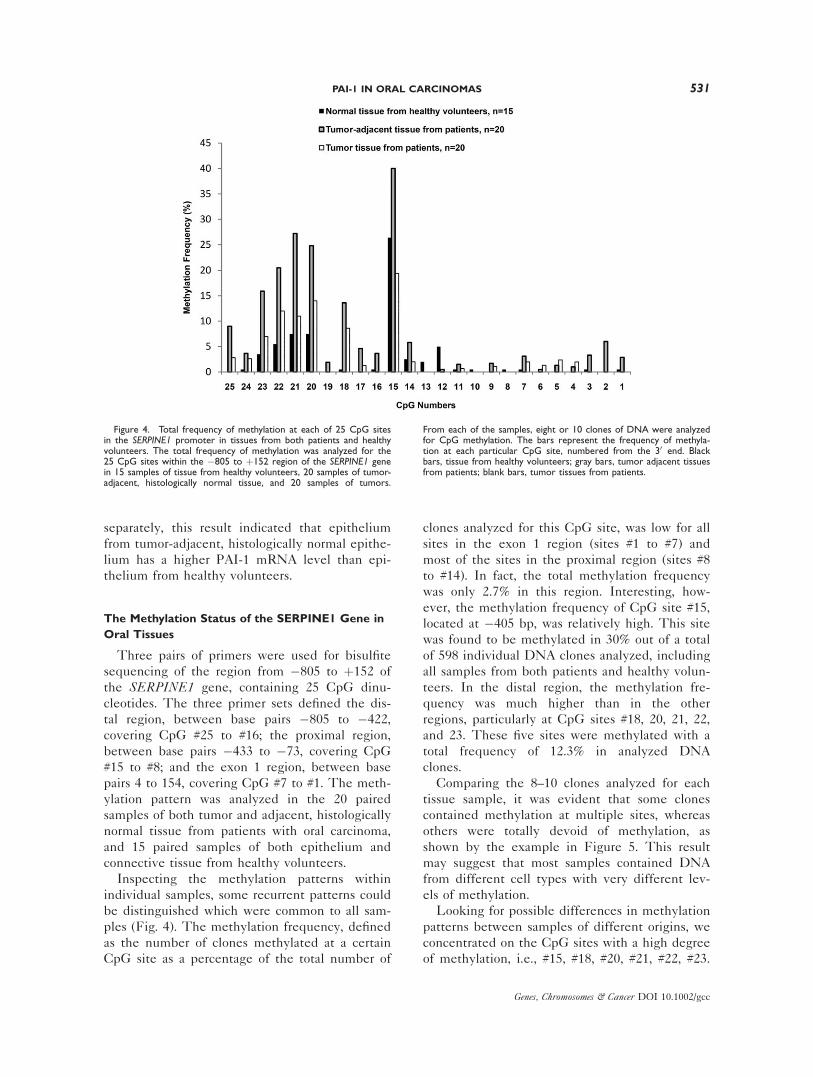
The Methylation Status of the SERPINE1 Gene in
Oral Tissues
Three pairs of primers were used for bisulfite
sequencing of the region from �805 to þ152 of
the SERPINE1 gene, containing 25 CpG dinu-
cleotides. The three primer sets defined the dis-
tal region, between base pairs �805 to �422,
covering CpG #25 to #16; the proximal region,
between base pairs �433 to �73, covering CpG
#15 to #8; and the exon 1 region, between base
pairs 4 to 154, covering CpG #7 to #1. The meth-
ylation pattern was analyzed in the 20 paired
samples of both tumor and adjacent, histologically
normal tissue from patients with oral carcinoma,
and 15 paired samples of both epithelium and
connective tissue from healthy volunteers.
Inspecting the methylation patterns within
individual samples, some recurrent patterns could
be distinguished which were common to all sam-
ples (Fig. 4). The methylation frequency, defined
as the number of clones methylated at a certain
CpG site as a percentage of the total number of
clones analyzed for this CpG site, was low for all
sites in the exon 1 region (sites #1 to #7) and
most of the sites in the proximal region (sites #8
to #14). In fact, the total methylation frequency
was only 2.7% in this region. Interesting, how-
ever, the methylation frequency of CpG site #15,
located at �405 bp, was relatively high. This site
was found to be methylated in 30% out of a total
of 598 individual DNA clones analyzed, including
all samples from both patients and healthy volun-
teers. In the distal region, the methylation fre-
quency was much higher than in the other
regions, particularly at CpG sites #18, 20, 21, 22,
and 23. These five sites were methylated with a
total frequency of 12.3% in analyzed DNA
clones.
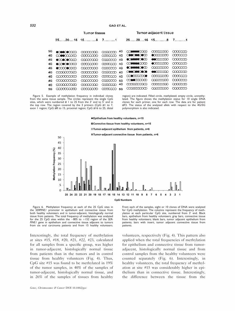
Comparing the 8–10 clones analyzed for each
tissue sample, it was evident that some clones
contained methylation at multiple sites, whereas
others were totally devoid of methylation, as
shown by the example in Figure 5. This result
may suggest that most samples contained DNA
from different cell types with very different lev-
els of methylation.
Looking for possible differences in methylation
patterns between samples of different origins, we
concentrated on the CpG sites with a high degree
of methylation, i.e., #15, #18, #20, #21, #22, #23.
Figure 4. Total frequency of methylation at each of 25 CpG sitesin the SERPINE1 promoter in tissues from both patients and healthyvolunteers. The total frequency of methylation was analyzed for the25 CpG sites within the �805 to þ152 region of the SERPINE1 genein 15 samples of tissue from healthy volunteers, 20 samples of tumor-adjacent, histologically normal tissue, and 20 samples of tumors.
From each of the samples, eight or 10 clones of DNA were analyzedfor CpG methylation. The bars represent the frequency of methyla-tion at each particular CpG site, numbered from the 30 end. Blackbars, tissue from healthy volunteers; gray bars, tumor adjacent tissuesfrom patients; blank bars, tumor tissues from patients.
PAI-1 IN ORAL CARCINOMAS 531
Genes, Chromosomes & Cancer DOI 10.1002/gcc
Interestingly, the total frequency of methylation
at sites #15, #18, #20, #21, #22, #23, calculated
for all samples from a specific group, was higher
in tumor-adjacent, histologically normal tissue
from patients than in the tumors and in control
tissue from healthy volunteers (Fig. 4). Thus,
CpG site #15 was found to be methylated in 19%
of the tumor samples, in 40% of the samples of
tumor-adjacent, histologically normal tissue, and
in 26% of the samples of tissues from healthy
volunteers, respectively (Fig. 4). This pattern also
applied when the total frequencies of methylation
for epithelium and connective tissue from tumor-
adjacent, histologically normal tissue and from
control samples from the healthy volunteers were
counted separately (Fig. 6). Interestingly, in
healthy volunteers, the total frequency of methyl-
ation at site #15 was considerably higher in epi-
thelium than in connective tissue. Interestingly,
the difference between the tissue from the
Figure 6. Methylation frequency at each of the 25 CpG sites inthe SERPINE1 promoter in epithelium and connective tissue fromboth healthy volunteers and in tumor-adjacent, histologically normaltissue from patients. The total frequency of methylation was analyzedfor the 25 CpG sites within the �805 to þ152 region of the SER-PINE1 gene in epithelium and connective tissue adjacent to tumorsfrom six oral carcinoma patients and from 15 healthy volunteers.
From each of the samples, eight or 10 clones of DNA were analyzedfor CpG methylation. The columns represent the frequency of meth-ylation at each particular CpG site, numbered from 30 end. Blackbars, epithelium from healthy volunteers; gray bars, connective tissuefrom healthy volunteers; blank bars, tumor adjacent epithelium frompatients; bars with insert, tumor adjacent connective tissue frompatients.
Figure 5. Example of methylation frequency in individual clonesfrom the same tissue sample. The circles represent the single CpGsites, which were numbered # 1 to 25 from the 30 end to 50 end inthe top row. The region covered by the 3 primers (CpG #1 to 7,exon 1 region; CpG #8 to 15, proximal region; CpG #16 to 25, distal
region) are indicated. Filled circle, methylated; empty circle, unmethy-lated. The figure shows the methylation status for 10 single DNAclones for each primer, one for each row. The data are for patient#F3. The status of the analyzed allels with respect to the 4G/5Gpolymorphism is also indicated.
532 GAO ET AL.
Genes, Chromosomes & Cancer DOI 10.1002/gcc
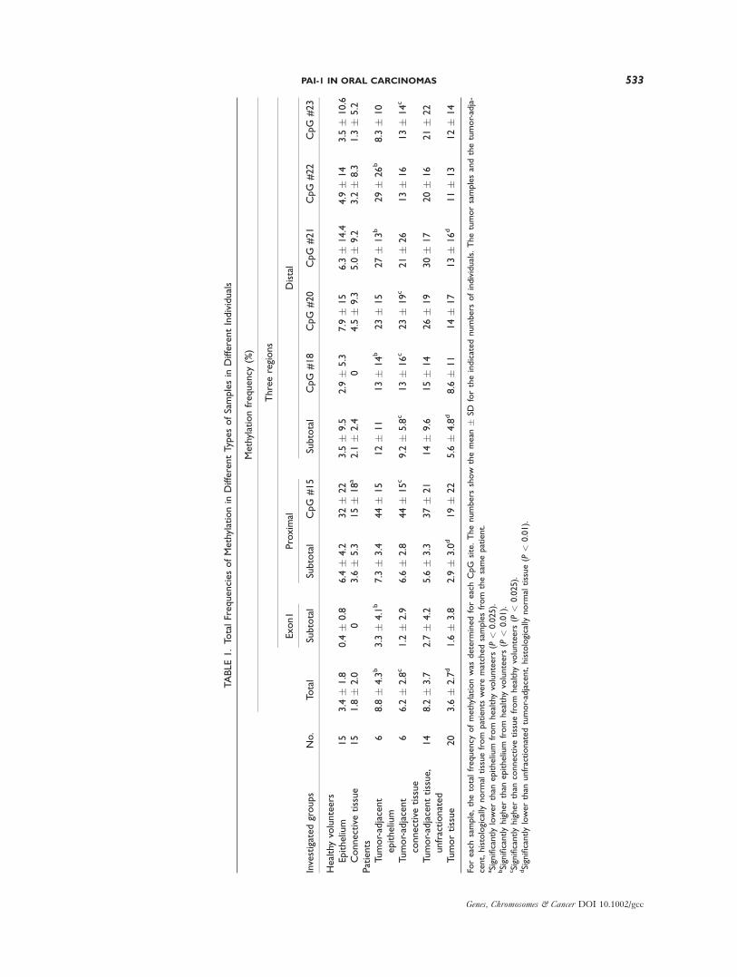
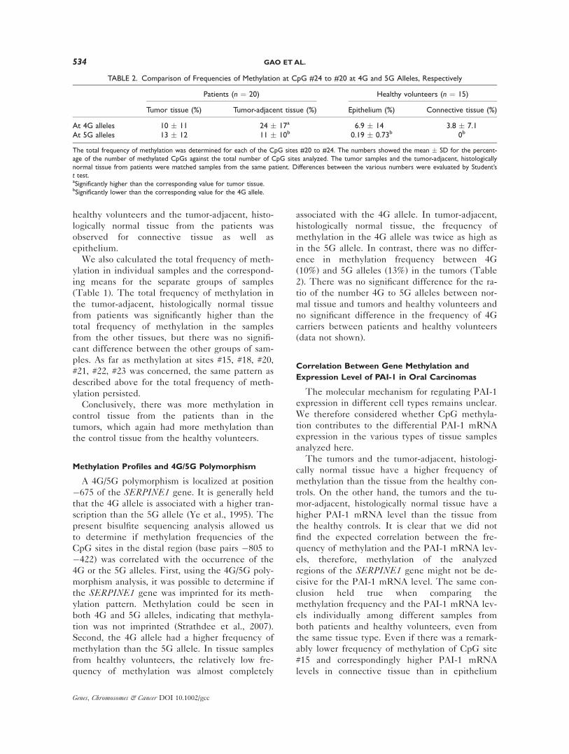
TABLE1.To
talFrequenciesofMethylationin
DifferentTypesofSamplesin
DifferentIndividuals
Investigatedgroups
No.
Methylationfrequency
(%)
Total
Threeregions
Exon1
Proximal
Distal
Subtotal
Subtotal
CpG
#15
Subtotal
CpG
#18
CpG
#20
CpG
#21
CpG
#22
CpG
#23
Healthyvolunteers
Epithelium
15
3.4
�1.8
0.4
�0.8
6.4
�4.2
32�
22
3.5
�9.5
2.9
�5.3
7.9
�15
6.3
�14.4
4.9
�14
3.5
�10.6
Connectivetissue
15
1.8
�2.0
03.6
�5.3
15�
18a
2.1
�2.4
04.5
�9.3
5.0
�9.2
3.2
�8.3
1.3
�5.2
Patients
Tumor-adjacent
epithelium
68.8
�4.3b
3.3
�4.1b
7.3
�3.4
44�
15
12�
11
13�
14b
23�
15
27�
13b
29�
26b
8.3
�10
Tumor-adjacent
connectivetissue
66.2
�2.8c
1.2
�2.9
6.6
�2.8
44�
15c
9.2
�5.8c
13�
16c
23�
19c
21�
26
13�
16
13�
14c
Tumor-adjacenttissue,
unfractionated
14
8.2
�3.7
2.7
�4.2
5.6
�3.3
37�
21
14�
9.6
15�
14
26�
19
30�
17
20�
16
21�
22
Tumortissue
20
3.6
�2.7d
1.6
�3.8
2.9
�3.0d
19�
22
5.6
�4.8d
8.6
�11
14�
17
13�
16d
11�
13
12�
14
Foreachsample,thetotalfrequency
ofmethylationwas
determ
inedforeachCpG
site.Thenumbers
show
themean
�SD
fortheindicatednumbers
ofindividuals.Thetumorsamplesandthetumor-adja-
cent,histologically
norm
altissuefrom
patients
were
matchedsamplesfrom
thesamepatient.
aSignificantlylowerthan
epithelium
from
healthyvolunteers
(P<
0.025).
bSignificantlyhigherthan
epithelium
from
healthyvolunteers
(P<
0.01).
cSignificantlyhigherthan
connectivetissuefrom
healthyvolunteers
(P<
0.025).
dSignificantlylowerthan
unfractionatedtumor-adjacent,histologically
norm
altissue(P
<0.01).
PAI-1 IN ORAL CARCINOMAS 533
Genes, Chromosomes & Cancer DOI 10.1002/gcc
healthy volunteers and the tumor-adjacent, histo-
logically normal tissue from the patients was
observed for connective tissue as well as
epithelium.
We also calculated the total frequency of meth-
ylation in individual samples and the correspond-
ing means for the separate groups of samples
(Table 1). The total frequency of methylation in
the tumor-adjacent, histologically normal tissue
from patients was significantly higher than the
total frequency of methylation in the samples
from the other tissues, but there was no signifi-
cant difference between the other groups of sam-
ples. As far as methylation at sites #15, #18, #20,
#21, #22, #23 was concerned, the same pattern as
described above for the total frequency of meth-
ylation persisted.
Conclusively, there was more methylation in
control tissue from the patients than in the
tumors, which again had more methylation than
the control tissue from the healthy volunteers.
Methylation Profiles and 4G/5G Polymorphism
A 4G/5G polymorphism is localized at position
�675 of the SERPINE1 gene. It is generally held
that the 4G allele is associated with a higher tran-
scription than the 5G allele (Ye et al., 1995). The
present bisulfite sequencing analysis allowed us
to determine if methylation frequencies of the
CpG sites in the distal region (base pairs �805 to
�422) was correlated with the occurrence of the
4G or the 5G alleles. First, using the 4G/5G poly-
morphism analysis, it was possible to determine if
the SERPINE1 gene was imprinted for its meth-
ylation pattern. Methylation could be seen in
both 4G and 5G alleles, indicating that methyla-
tion was not imprinted (Strathdee et al., 2007).
Second, the 4G allele had a higher frequency of
methylation than the 5G allele. In tissue samples
from healthy volunteers, the relatively low fre-
quency of methylation was almost completely
associated with the 4G allele. In tumor-adjacent,
histologically normal tissue, the frequency of
methylation in the 4G allele was twice as high as
in the 5G allele. In contrast, there was no differ-
ence in methylation frequency between 4G
(10%) and 5G alleles (13%) in the tumors (Table
2). There was no significant difference for the ra-
tio of the number 4G to 5G alleles between nor-
mal tissue and tumors and healthy volunteers and
no significant difference in the frequency of 4G
carriers between patients and healthy volunteers
(data not shown).
Correlation Between Gene Methylation and
Expression Level of PAI-1 in Oral Carcinomas
The molecular mechanism for regulating PAI-1
expression in different cell types remains unclear.
We therefore considered whether CpG methyla-
tion contributes to the differential PAI-1 mRNA
expression in the various types of tissue samples
analyzed here.
The tumors and the tumor-adjacent, histologi-
cally normal tissue have a higher frequency of
methylation than the tissue from the healthy con-
trols. On the other hand, the tumors and the tu-
mor-adjacent, histologically normal tissue have a
higher PAI-1 mRNA level than the tissue from
the healthy controls. It is clear that we did not
find the expected correlation between the fre-
quency of methylation and the PAI-1 mRNA lev-
els, therefore, methylation of the analyzed
regions of the SERPINE1 gene might not be de-
cisive for the PAI-1 mRNA level. The same con-
clusion held true when comparing the
methylation frequency and the PAI-1 mRNA lev-
els individually among different samples from
both patients and healthy volunteers, even from
the same tissue type. Even if there was a remark-
ably lower frequency of methylation of CpG site
#15 and correspondingly higher PAI-1 mRNA
levels in connective tissue than in epithelium
TABLE 2. Comparison of Frequencies of Methylation at CpG #24 to #20 at 4G and 5G Alleles, Respectively
Patients (n ¼ 20) Healthy volunteers (n ¼ 15)
Tumor tissue (%) Tumor-adjacent tissue (%) Epithelium (%) Connective tissue (%)
At 4G alleles 10 � 11 24 � 17a 6.9 � 14 3.8 � 7.1At 5G alleles 13 � 12 11 � 10b 0.19 � 0.73b 0b
The total frequency of methylation was determined for each of the CpG sites #20 to #24. The numbers showed the mean � SD for the percent-
age of the number of methylated CpGs against the total number of CpG sites analyzed. The tumor samples and the tumor-adjacent, histologically
normal tissue from patients were matched samples from the same patient. Differences between the various numbers were evaluated by Student’s
t test.aSignificantly higher than the corresponding value for tumor tissue.bSignificantly lower than the corresponding value for the 4G allele.
534 GAO ET AL.
Genes, Chromosomes & Cancer DOI 10.1002/gcc
from healthy volunteers as well as higher PAI-1
mRNA in tumor tissues than in adjacent histolog-
ically normal tissues, the correlation did not reach
statistical significance. However, when we di-
vided patients into two groups according to the
median value of the PAI-1 mRNA level as well
as according to the median value of methylation
frequency, to compare the PAI-1 mRNA levels
and the methylation frequency in the tumor sam-
ples, we did find an inverse correlation, both with
respect to total methylation and methylation at
CpGs #15, #18, #20, #21, #22, and #23 (P < 0.05,
evaluated by Fisher’s Exact Probability Test).
In conclusion, the results indicated that CpG
methylation is not in general involved in restrict-
ing the PAI-1 expression in healthy oral mucosa,
but may play a role in fine tuning the mRNA lev-
els in tumor cells.
DISCUSSION
Our immunohistochemical staining results
agree well with previous reports on PAI-1 in oral
squamous cell carcinomas. Previously, overexpres-
sion of PAI-1 in oral squamous cell carcinomas
was demonstrated by different analyses, including
ELISA, Northern blot, and immunohistochemical
staining, and most studies showed that increased
expression level correlated with a poor prognosis
(Nozaki et al., 1998; Hundsdorfer et al., 2005;
Lindberg et al., 2006). In the present study, we
also found an increased PAI-1 expression in oral
carcinomas by both immunohistochemical stain-
ing and RT-PCR. We found that the prominent
PAI-1 expressing cell type in oral carcinomas is
the cancer cells. Our finding of PAI-1 in the can-
cer cells agrees well with previous reports
(Yasuda et al., 1997; Nozaki et al., 1998; Lindberg
et al., 2006). In fact, the latter of these studies
(Lindberg et al., 2006) was done with the same
affinity-purified, carefully controlled and carefully
characterized antibodies used in this case. These
antibodies were also used in previously reported
studies of breast, colon, and prostate carcinomas
(Offersen et al., 2003; Illemann et al., 2004;
Usher et al., 2005), in which the predominant
PAI-1 expressing cell type is the stromal myofi-
broblast, with some additional expression by en-
dothelial cells, while there was little expression
by the cancer cells (Offersen et al., 2003; Ille-
mann et al., 2004; Usher et al., 2005). Thus, the
cellular expression pattern for PAI-1 in oral squa-
mous cell carcinomas is different from that in
breast, colon, and prostate carcinomas.
Moreover, in addition to confirming previous
immunohistochemical results, we also now found
that in some oral carcinomas, PAI-1 is expressed
by fibroblast-like cells in the stroma surrounding
the cancer cells and not in cancer cells them-
selves. Moreover, we also found PAI-1 expression
by tumor-adjacent, histologically normal epithe-
lium, not reported in previous studies. In vitro
studies of wound healing indicated that PAI-1
modulates the complex process of injury resolu-
tion through control of focalized plasmin-medi-
ated matrix remodeling and cell migration.
Certain ‘‘senescence-associated’’ genes (i.e.,
CDKN2A and Tp53) may actually function in
wound repair and early malignant program by in-
hibiting proliferation but also promoting migra-
tion (Chan et al., 2001; Ploplis et al., 2004;
Darbro et al., 2005; Kortlever et al., 2006; Natara-
jan et al., 2006). Indeed, keratinocytes at the
leading edge during wound re-epithelialization
are less mitotically active than cells more dis-
placed from the motile front (Dabelsteen and
Mackenzie, 1976) and have been shown to
express relatively high levels of PAI-1 by other
studies (Garlick and Taichman, 1994; Jensen and
Lavker, 1996; Providence and Higgins, 2004; Qi
et al., 2008; Shetty et al., 2008a,b). PAI-1 may
also be a downstream target gene during Tp53-
induced cellular senescence. The observed up-
regulation of PAI-1 in histologically normal mu-
cosa adjacent to tumors may indicate cellular
senescence as part of an early malignant develop-
ment; this is in agreement with previous studies
that have shown molecular changes in histologi-
cally normal mucosa adjacent to oral carcinomas
(Kortlever et al., 2006). One recent report indi-
cated that Tp53 protein can interact with a 70
nucleotide sequence at PAI-1 mRNA 30-UTR
region, resulting in upregulation of PAI-1 expres-
sion and mRNA stabilization (Shetty et al.,
2008b).
Methylation of DNA at cytosines in CpG
sequences generally suppresses gene transcrip-
tion. Accordingly, we previously showed that
CpG methylation of the SERPINE1 gene 50-flanking region is inversely correlated with PAI-1
mRNA expression in five human tumor cell lines
and that inhibition of CpG methylation by 5-aza-
20-deoxycytidine treatment can induce PAI-1
mRNA expression in cells otherwise having a
methylated PAI-1 gene 50-flanking region (Gao
et al., 2005). We were therefore interested in
investigating whether methylation of CpGs in the
SERPINE1 gene 50-flanking region are implicated
PAI-1 IN ORAL CARCINOMAS 535
Genes, Chromosomes & Cancer DOI 10.1002/gcc
in regulation of PAI-1 expression in normal and
malignant oral tissue. When doing so, we made
several interesting observations.
We found that the methylation patterns in
both normal and malignant oral tissue are in gen-
eral quite different from that previously observed
in the cell lines (Gao et al., 2005). In the cell
lines, the highest frequency of methylation was
found at CpG sites #1 to #14 of the SERPINE1gene 50-flanking region, at which sites very little
methylation was observed in the tissue samples
analyzed here. In contrast, the most heavily
methylated positions in the tissue samples were
CpGs #15 and #20 to #23. On the basis of this
observation, one will have to conclude that meth-
ylation patterns in cell lines do not necessarily
reflect that of the tissues from which they were
derived but rather is an artifact of the culture
conditions.
The second, very surprising observation con-
cerning methylation is the difference in methyla-
tion pattern between normal oral mucosa from
healthy volunteers and tumor-adjacent, histologi-
cally normal tissue from the patients. Except for
CpG #15, there was very little methylation of the
SERPINE1 gene 50-flanking region in DNA from
healthy volunteers, while the region had signifi-
cant methylation in tumor-adjacent, histologically
normal tissue from the patients. This observation
seems to indicate that the tumor-adjacent normal
tissue, despite being histologically normal, has al-
ready begun an early malignant development.
This conclusion is in agreement with our previ-
ous observation of aberrant methylation of both
ABO gene and DBCCR1 gene in such tissue (Gao
et al., 2004a,b). Importantly, the increased meth-
ylation in the tumor-adjacent, histologically nor-
mal tissue also involves the connective tissue.
Accepting the idea of an early malignant state of
this tissue, the increased methylation would
imply epigenetic changes also in stromal cells.
From our results, methylation does not seem to
be a predominant determinant of the higher PAI-
1 expression level in oral carcinomas: Methylation
of the SERPINE1 gene 50-flanking region was
much higher in tumor-adjacent, histologically nor-
mal tissue than in normal oral mucosa from
healthy volunteers, while the expression level
was similar in these two groups of tissues. Still, in
some instances, methylation may be involved in
fine tuning PAI-1 expression. Thus, we observed
a significant inverse correlation between the
expression levels and the methylation levels in
the carcinoma tissue.
The 4G/5G polymorphism is located within a
GC-rich sequence. It is generally agreed that the
4G allele is associated with a higher level of PAI-
1 gene transcription and a higher PAI-1 level in
blood plasma (Ye et al., 1995). We found methyl-
ation at both 4G and 5G alleles, and could, there-
fore, exclude SERPINE1 gene methylation as an
imprinted gene pattern (Strathdee et al., 2007).
Furthermore, we found that the methylation fre-
quency of CpG #24 to #20 at 4G alleles was sig-
nificantly higher than that at 5G alleles, in
normal tissue as well as in tumor-adjacent, histo-
logically normal tissue, while the frequency was
identical in carcinoma tissue. However, the func-
tional significance of this difference between 4G
and 5G alleles is not clear, as there is not a signif-
icant correlation between methylation and
expression. Moreover, obviously, when going
from normal tissue from healthy volunteers to tu-
mor-adjacent, histologically normal tissue, meth-
ylation increases in 5G alleles as well as 4G
alleles. The decrease in methylation on the 4G
allele when going from to tumor-adjacent, histo-
logically notmal tissue to tumor tissue may con-
tribute to the increased expression if the 4G
allele is the one preferentially transcribed.
Among other genes in the plasminogen activa-
tion system, methylation of the PLAU gene,
encoding the PAI-1 target protease uPA, was pre-
viously studied extensively. The methylation pat-
tern of the PLAU gene was much different from
methylation of the SERPINE1 gene. In primary
breast cancer patients, full demethylation of the
PLAU gene was seen in all grade III samples and
associated with higher expression level of uPA,
compared with a fully methylated gene in the
normal mammary epithelial cell line HMEC
(Pakneshan et al., 2004). The difference between
the SERPINE1 gene and the PLAU gene in this
respect may be related to the fact that there is no
typical CpG island in the SERPINE1 gene pro-
moter region, while this is the case with the uPA
gene promoter (Pakneshan et al., 2004). More-
over, our studies were done by comparing tumor
samples and corresponding adjacent histologically
normal tissues not by comparing tumor samples
and cultured cells as done in above study (Pakne-
shan et al., 2004).
In conclusion, we have mapped PAI-1 express-
ing cells in oral squamous cell carcinomas and
found that in addition to the previously reported
expression by cancer cells, PAI-1 is in some cases
also expressed by stromal fibroblasts adjacent to
the cancer cells and by histologically normal
536 GAO ET AL.
Genes, Chromosomes & Cancer DOI 10.1002/gcc
epithelium adjacent to the tumors. We made the
surprising observation that the SERPINE1 gene
is most highly methylated in tumor-adjacent, his-
tologically normal tissue and completely different
from the tissues from healthy volunteers, indicat-
ing that these cells are probably already in early
malignant development. Although CpG methyla-
tion may be implicated in fine tuning PAI-1
expression, it is not the main determinant of the
high PAI-1 expression in oral squamous cell carci-
noma cells. Other regulatory mechanisms rather
than methylation need to be investigated in dif-
ferent cell types in tumors in the future.
ACKNOWLEDGMENTS
Anni Christensen is thanked for excellent tech-
nical assistance.
REFERENCES
Andreasen PA, Egelund R, Petersen HH. 2000. The plasminogenactivation system in tumor growth, invasion, and metastasis.Cell Mol Life Sci 57:25–40.
Chan JC, Duszczyszyn DA, Castellino FJ, Ploplis VA. 2001. Accel-erated skin wound healing in plasminogen activator inhibitor-1-deficient mice. Am J Pathol 159:1681–1688.
Clark SJ, Harrison J, Paul CL, Frommer M. 1994. High sensitivitymapping of methylated cytosines. Nucleic Acids Res 22:2990–2997.
Clayman G, Wang SW, Nicolson GL, el Naggar A, Mazar A, Hen-kin J, Blasi F, Goepfert H, Boyd DD. 1993. Regulation of uro-kinase-type plasminogen activator expression in squamous-cellcarcinoma of the oral cavity. Int J Cancer 54:73–80.
Curino A, Patel V, Nielsen BS, Iskander AJ, Ensley JF, Yoo GH,Holsinger FC, Myers JN, El Nagaar A, Kellman RM, ShillitoeEJ, Molinolo AA, Gutkind JS, Bugge TH. 2004. Detection ofplasminogen activators in oral cancer by laser capture microdis-section combined with zymography. Oral Oncol 40:1026–1032.
Dabelsteen E, Mackenzie I. 1976. Selective loss of blood groupantigens during wound healing. Acta Pathol Microbiol Scand A84:445–450.
Darbro BW, Schneider GB, Klingelhutz AJ. 2005. Co-regulation ofp16INK4A and migratory genes in culture conditions that leadto premature senescence in human keratinocytes. J InvestigDermatol 125:499–509.
Durand MK, Bodker JS, Christensen A, Dupont DM, Hansen M,Jensen JK, Kjelgaard S, Mathiasen L, Pedersen KE, Skeldal S,Wind T, Andreasen PA. 2004. Plasminogen activator inhibitor-Iand tumour growth, invasion, and metastasis. Thromb Haemost91:438–449.
Esteller M, Herman JG. 2002. Cancer as an epigenetic disease:DNA methylation and chromatin alterations in human tumours.J Pathol 196:1–7.
Gao S, Worm J, Guldberg P, Eiberg H, Krogdahl A, Liu CJ, Rei-bel J, Dabelsteen E. 2004a. Genetic and epigenetic alterationsof the blood group ABO gene in oral squamous cell carcinoma.Int J Cancer 109:230–237.
Gao S, Worm J, Guldberg P, Eiberg H, Krogdahl A, Sorensen JA,Liu CJ, Reibel J, Dabelsteen E. 2004b. Loss of heterozygosityat 9q33 and hypermethylation of the DBCCR1 gene in oralsquamous cell carcinoma. Br J Cancer 91:760–764.
Gao S, Skeldal S, Krogdahl A, Sorensen JA, Andreasen PA. 2005.CpG methylation of the PAI-1 gene 50-flanking region is inver-sely correlated with PAI-1 mRNA levels in human cell lines.Thromb Haemost 94:651–660.
Gao S, Krogdahl A, Sorensen JA, Kousted TM, Dabelsteen E,Andreasen PA. 2008. Overexpression of protease nexin-1mRNA and protein in oral squamous cell carcinomas. OralOncol 44:309–313.
Garlick JA, Taichman LB. 1994. Effect of TGF-b 1 on re-epithe-lialization of human keratinocytes in vitro: An organotypicmodel. J Investig Dermatol 103:554–559.
Ha PK, Califano JA. 2006. Promoter methylation and inactivationof tumour-suppressor genes in oral squamous-cell carcinoma.Lancet Oncol 7:77–82.
Herman JG. 1999. Hypermethylation of tumor suppressor genesin cancer. Semin Cancer Biol 9:359–367.
Hundsdorfer B, Zeilhofer HF, Bock KP, Dettmar P, Schmitt M,Kolk A, Pautke C, Horch HH. 2005. Tumour-associated uroki-nase-type plasminogen activator (uPA) and its inhibitor PAI-1in normal and neoplastic tissues of patients with squamous cellcancer of the oral cavity—clinical relevance and prognosticvalue. J Craniomaxillofac Surg 33:191–196.
Illemann M, Hansen U, Nielsen HJ, Andreasen PA, Hoyer-Han-sen G, Lund LR, Dano K, Nielsen BS. 2004. Leading-edgemyofibroblasts in human colon cancer express plasminogen acti-vator inhibitor-1. Am J Clin Pathol 122:256–265.
Jensen PJ, Lavker RM. 1996. Modulation of the plasminogen acti-vator cascade during enhanced epidermal proliferation in vivo.Cell Growth Differ 7:1793–1804.
Kortlever RM, Higgins PJ, Bernards R. 2006. Plasminogen ac-tivator inhibitor-1 is a critical downstream target of p53 inthe induction of replicative senescence. Nat Cell Biol 8:877–884.
Lindberg P, Larsson A, Nielsen BS. 2006. Expression of plasmino-gen activator inhibitor-1, urokinase receptor and laminin c-2chain is an early coordinated event in incipient oral squamouscell carcinoma. Int J Cancer 118:2948–2956.
Mackenzie IC, Dabelsteen E, Roed-Petersen B. 1979. A methodfor studying epithelial-mesenchymal interactions in human oralmucosal lesions. Scand J Dent Res 87:234–243.
McGregor F, Muntoni A, Fleming J, Brown J, Felix DH, Mac-Donald DG, Parkinson EK, Harrison PR. 2002. Molecularchanges associated with oral dysplasia progression and acquisi-tion of immortality: Potential for its reversal by 5-azacytidine.Cancer Res 62:4757–4766.
Natarajan E, Omobono JD, Guo Z, Hopkinson S, Lazar AJ, BrennT, Jones JC, Rheinwald JG. 2006. A keratinocyte hypermotility/growth-arrest response involving laminin 5 and p16INK4A acti-vated in wound healing and senescence. Am J Pathol 168:1821–1837.
Nozaki S, Endo Y, Kawashiri S, Nakagawa K, Yamamoto E, Yone-mura Y, Sasaki T. 1998. Immunohistochemical localization of aurokinase-type plasminogen activator system in squamous cellcarcinoma of the oral cavity: Association with mode of invasionand lymph node metastasis. Oral Oncol 34:58–62.
Offersen BV, Nielsen BS, Hoyer-Hansen G, Rank F, Hamilton-Dutoit S, Overgaard J, Andreasen PA. 2003. The myofibroblastis the predominant plasminogen activator inhibitor-1-expressingcell type in human breast carcinomas. Am J Pathol 163:1887–1899.
Pakneshan P, Tetu B, Rabbani SA. 2004. Demethylation of uroki-nase promoter as a prognostic marker in patients with breastcarcinoma. Clin Cancer Res 10:3035–3041.
Ploplis VA, Balsara R, Sandoval-Cooper MJ, Yin ZJ, Batten J,Modi N, Gadoua D, Donahue D, Martin JA, Castellino FJ.2004. Enhanced in vitro proliferation of aortic endothelial cellsfrom plasminogen activator inhibitor-1-deficient mice. J BiolChem 279:6143–6151.
Providence KM, Higgins PJ. 2004. PAI-1 expression is requiredfor epithelial cell migration in two distinct phases of in vitrowound repair. J Cell Physiol 200:297–308.
Qi L, Higgins SP, Lu Q, Samarakoon R, Wilkins-Port CE, Ye Q,Higgins CE, Staiano-Coico L, Higgins PJ. 2008. SERPINE1(PAI-1) is a prominent member of the early G0 ! G1 transition‘‘wound repair’’ transcriptome in p53 mutant human keratino-cytes. J Invest Dermatol 128:749–753.
Shaw R. 2006. The epigenetics of oral cancer. Int J Oral Maxillo-fac Surg 35:101–108.
Shetty P, Velusamy T, Bhandary YP, Shetty RS, Liu MC, ShettyS. 2008a. Urokinase expression by tumor suppressor proteinp53: A novel role in mRNA turnover. Am J Respir Cell MolBiol 39:364–372.
Shetty S, Shetty P, Idell S, Velusamy T, Bhandary YP, Shetty RS.2008b. Regulation of plasminogen activator inhibitor-1 expres-sion by tumor suppressor protein p53. J Biol Chem 283:19570–19580.
Strathdee G, Sim A, Soutar R, Holyoake TL, Brown R. 2007.HOXA5 is targeted by cell type specific CpG island
PAI-1 IN ORAL CARCINOMAS 537
Genes, Chromosomes & Cancer DOI 10.1002/gcc

methylation in normal cells and during the development ofacute myeloid leukaemia. Carcinogenesis 28:299–309.
Usher PA, Thomsen OF, Iversen P, Johnsen M, Brunner N,Hoyer-Hansen G, Andreasen P, Dano K, Nielsen BS. 2005.Expression of urokinase plasminogen activator, its receptor andtype-1 inhibitor in malignant and benign prostate tissue. Int JCancer 113:870–880.
Yasuda T, Sakata Y, Kitamura K, Morita M, Ishida T. 1997.Localization of plasminogen activators and their inhibitor in
squamous cell carcinomas of the head and neck. Head Neck19:611–616.
Ye S, Green FR, Scarabin PY, Nicaud V, Bara L, Dawson SJ,Humphries SE, Evans A, Luc G, Cambou JP. 1995. The 4G/5G genetic polymorphism in the promoter of the plasminogenactivator inhibitor-1 (PAI-1) gene is associated with differencesin plasma PAI-1 activity but not with risk of myocardial infarc-tion in the ECTIM study. Etude CasTemoins de I’nfarctus duMycocarde. Thromb Haemost 74:837–841.
538 GAO ET AL.
Genes, Chromosomes & Cancer DOI 10.1002/gcc