Embed Size (px)
Citation preview


April | 2017 ISSN 1660-3656Epileptologie | 34. Jahrgang
Epilepsie-LigaSeefeldstrasse 84CH-8008 Zürich
Redaktionskommission
Reinhard E. Ganz | ZürichMartinus Hauf | Tschugg Christian M. Korff | GenèveGünter Krämer | Zürich (Vorsitz)Oliver Maier | St. GallenJan Novy | LausanneFabienne Picard | GenèveStephan Rüegg | BaselSerge Vulliémoz | GenèveFred Zubler | Bern
Beirat
Pamela Agazzi | LuganoAlexandre Datta | BaselThomas Grunwald | ZürichChristian W. Hess | BernAnna Marie Hew-Winzeler | ZürichGünter Krämer | ZürichTheodor Landis | GenèveMalin Maeder | LavignyKlaus Meyer | TschuggAndrea O. Rossetti | Lausanne Stephan Rüegg | BaselKaspar Schindler | BernMarkus Schmutz | BaselMargitta Seeck | Genève Urs Sennhauser | HettlingenFranco Vassella | BremgartenElmar Zwahlen | Tschugg
Inhalt
Editorial 1 - 3
Vagusnervstimulation in der Epilepsie – PatientenselektionMartinus Hauf, Eirini Tsampikakiund Klaus Meyer 4 - 9
Transcranial Direct-Current Stimulation as Treatment in EpilepsyMarkus Gschwind 10 - 18
Deep Brain Stimulation for Epilepsy: an Update Claudio Pollo and Kaspar Schindler 19 - 24
Neurofeedback in der Epilepsie Ute Strehl 25 - 30
The Effects of Transcranial Stimulation on Language Related Large Scale Brain Networks Jochen Kindler, Martinus Haufand Daniela Hubl 31 - 37
Epilepsie-Liga-Mitteilungen 38 - 46
Kongresskalender 47 - 48
Schweizerische Epilepsie-LigaLigue Suisse contre l’Epilepsie Lega Svizzera contro l’EpilessiaSwiss League Against Epilepsy
- Zusammenfassung, Résumé und englischer Ab-stract (mit Titel der Arbeit): Ohne Literaturzitate und Akronyme sowie unübliche Abkürzungen ( je maximal 250 Wörter).
- Text: Dabei bei Originalarbeiten Gliederung in Ein-leitung, Methode (inkl. Untersuchungsmaterial, Pa-tienten, Versuchstiere etc., ggf. auch Angabe über Einwilligung bzw. Einhaltung der Deklaration von Helsinki inkl. Votum einer Ethikkommission), Ergeb-nisse und Diskussion. Abkürzungen sind bei ihrem ersten Erscheinen im Text voll auszuschreiben.
- Literaturverzeichnis: Am Ende der Arbeit werden die Literaturstellen in der im Text zitierten Reihen-folge aufgeführt und nach untenstehendem Muster zitiert. Persönliche Mitteilungen, unveröffentlichte Befunde oder zur Publikation eingereichte Manu-skripte werden nicht aufgenommen, sondern ent-sprechend im Text vermerkt. Zitierungen „im Druck“ bzw. „in press“ beziehen sich nur auf von einer Zeit-schrift bereits angenommene Arbeiten (mit Angabe von Zeitschrift und – soweit bekannt – Band und Erscheinungsjahr. Das Zitieren von Arbeiten als „in Vorbereitung“ oder „in preparation“ ist nicht zuläs-sig. Kongressmitteilungen können nur als zitierbare Abstracts oder Beitrag in Proceedings-Bänden be-rücksichtigt werden.
- Tabellen: Jede Tabelle steht auf einer neuen Seite und hat eine kurze erklärende Überschrift. Abkür-zungen oder Zeichen sind in einer Fussnote zu erklä-ren.
- Abbildungslegenden: Die Legende für jede Abbil-dung steht auf einer neuen Seite; alle Abkürzungen oder Zeichen sind darin zu erklären.
- Abbildungen: Strichzeichnungen, schattierte Zeich-nungen oder Fotografien (SW oder Farbe).
- Zitierweise: Zeitschriftenartikel: Daoud AS, Bati-eha A, Abu-Ekteish F et al. Iron status: a possible risk factor for the first febrile seizure. Epilepsia 2002; 43: 740-743 (bei bis zu vier Autoren werden alle genannt; Abkürzungen der Zeitschriften nach der „List of Journals indexed in Index Medicus“); Bücher: Shorvon S. Status Epilepticus. Its Clinical Features and Treatment in Children and Adults. Cambridge: Cambridge University Press, 1994; Buchkapitel: Holthausen H, Tuxhorn I, Pieper T et al. Hemispherectomy in the treatment of neuronal migrational disorders. In: Kotagal P, Lüders HO (eds): The Epilepsies. Etiologies and Prevention. San Diego, London, Boston et al.: Academic Press, 1999: 93-102
Was ist an die Redaktion einzureichen?
Alle Manuskripte sind inklusive Abbildungen und Tabellen in dreifacher Ausführung einzureichen. Bevor-zugt wird eine elektronische Manuskripteinreichung per e-mail (Textverarbeitung: MS Word), alternativ die Zusendung von drei Ausdrucken und einer CD (für Abb. und Tab. ist das verwendete Programm anzugeben).
Richtlinien für die Autoren
Allgemeines
Epileptologie veröffentlicht sowohl angeforderte als auch unaufgefordert eingereichte Manuskripte über al-le Themen der Epileptologie. Es werden in der Regel nur bislang unveröffentlichte Arbeiten angenommen. Die Manuskripte oder wesentliche Teile daraus dürfen auch nicht gleichzeitig anderen Zeitschriften angeboten wer-den oder anderweitig bereits zur Publikation angenom-men worden sein. Alle Manuskripte werden zweifach begutachtet. Von den Beiträgen werden keine Sonder-drucke erstellt, sie werden jedoch als pdf-Datei zusätz-lich auf der Liga-Homepage (www.epi.ch) veröffentlicht und können von dort heruntergeladen werden.
Redaktionsanschrift
Unaufgefordert eingereichte Manuskripte (inkl. Briefe an die Herausgeber) sind zu richten an: Frau M. Becker, Redaktion Epileptologie, Schwei-zerische Liga gegen Epilepsie, Seefeldstr. 84, 8008 Zürich. Tel. 043 477 01 39, Fax 043 488 67 78, e-mail: [email protected].
Hinweise zur Manuskripterstellung
Manuskripte werden nur akzeptiert, wenn sie den folgenden Kriterien entsprechen. Nicht entsprechend abgefasste Manuskripte werden vor der Begutachtung zurückgesandt.1. Sprache: Neben deutsch auch englisch und franzö-
sisch möglich.2. Schreibweise (deutsch): Als Schreibweise gilt die
deutsche Form mit „z“ und „k“ (also z. B. Karzinom), lateinische Fachtermini behalten aber ihre Schreib-weise (also z. B. Arteria carotis).
3. Form: Der gesamte Text, einschliesslich Literaturver-zeichnis, Tabellen und Abbildungslegenden, ist fol-gendermassen zu formatieren:
- DIN-A4-Papier, einseitig (1 1/2- oder 2-zeilig mit max. 30 Zeilen je Seite).
- Literaturverweise werden gemäss der Reihenfolge, in der sie im Text vorkommen, arabisch nummeriert; im Text erscheinen die Verweiszahlen in eckigen Klammern.
- Tabellen und Abbildungen haben eine jeweils fort-laufende arabische Nummerierung.
4. Reihenfolge: 1. Titelblatt (ggf. inkl. Danksagung, För-derung durch Hilfe anderer oder Drittmittelfinanzie-rung), 2. Zusammenfassung in Deutsch, Résumé in Französisch und Summary in Englisch sowie je drei bis fünf Schlüsselwörter, 3. Text, 4. Literatur, 5. Ta-bellen, 6. Abbildungslegenden und 7. Abbildungen:
- Das Titelblatt enthält den vollen Titel der Arbeit (deutsch und englisch), Namen und Titel der Auto-ren, die Kliniken bzw. Institutionen, an denen alle Autoren arbeiten, sowie die vollständige Adresse des federführenden Autors mit Telefon- und Fax-nummer sowie e-mail.

1Epileptologie 2017; 34
Liebe Leserin, lieber Leser
Die Hirnstimulation ist eine Methode, die durch Anwendung von elektrischem Strom die Hirnaktivität modifiziert. Die Idee ist bei weitem nicht neu, schon Luigi Galvani und Alessandro Volta haben im späten 18. Jahrhundert Patienten mit Strom behandelt. In den letzten Jahren ist Hirnstimulation aber zu einem ech-ten Headliner geworden, und gerade in der Neurologie ist die tiefe Hirnstimulation (THS) aus der Behandlung des idiopathischen Parkinsonsyndroms nicht mehr wegzudenken. In der Epilepsie sieht es ähnlich aus, ne-ben der schon seit ca. 20 Jahren angewandten Behand-lung mittels Vagusnervstimulation (VNS) kommt zu-nehmend auch die THS zur Anwendung. In den ersten zwei Artikeln wird der aktuelle Wissensstand zu diesen etablierten Behandlungsmethoden zusammengefasst. Im Artikel zur VNS liegt der Schwerpunkt auf den Kri-terien der Patientenselektion. Die Autoren diskutieren, ob enzephalographische oder bildgebende Methoden hilfreich sein können, den individuellen Therapieer-folg vorauszusagen. Den Einsatz der THS in der Epilep-sie diskutiert Prof. Claudio Pollo und fokussiert auf die Vorstellung und Erfahrungen zu den unterschiedlichen Zielpunkten der Elektrodenlage. In dem sich schnell entwickelnden Gebiet der Hirnstimulation ist das, was heute noch experimentell ist, morgen bereits eine eta-blierte Therapie. Transkranielle Gleichstromstimulation (tDCS) ist dafür ein spannender Kandidat. Dr. Markus Gschwind stellt in einem lesenswerten Artikel diese einfach anzuwendende Methode der nicht-invasiven Neuromodulation und die Möglichkeiten in der Epilep-sie vor. Eine häufig etwas vergessene Möglichkeit, die Anfallssituation der Patienten mit Epilepsie zu verbes-sern, ist das Neurofeedback. Ich bin sehr froh über das Update zu dieser Technik der endogenen Hirnstimula-tion von Frau PD Dr. Ute Strehl, Universität Tübingen.
Der abrundende Schluss dieses Heftes ist ein Artikel zur transkraniellen Magnetstimulation (TMS) von PD Dr. Jochen Kindler. Die vorgestellten Daten der neurobiolo-gischen Korrelate zur TMS-Therapie bei psychiatrischen Patienten erinnern immer wieder daran, dass nicht nur die Epilepsien zerebrale Netzwerkerkrankungen sind, und wir von den wissenschaftlichen Erkenntnissen in-terdisziplinär profitieren können.
Ich wünsche Ihnen eine gute Lektüre.
Martinus Hauf
Neurostimulation
PD Dr. med. Martinus Hauf

2 Epileptologie 2017; 34
N
Chère lectrice, cher lecteur,
La stimulation cérébrale est une méthode qui mo-difie l’activité du cerveau par application d’un courant électrique. L’idée est loin d’être nouvelle, Luigi Galvani et Alessandro Volta, à la fin du 18e siècle, avaient déjà traité des patients par l’électricité. Ces dernières an-nées, la stimulation cérébrale a cependant pris de l’im-portance et occupe aujourd’hui véritablement le devant de la scène. En neurologie, justement, la stimulation cérébrale profonde (SCP) est désormais incontournable dans le traitement du syndrome parkinsonien idiopa-thique. Il en va un peu de même dans l’épilepsie : outre le traitement par stimulation du nerf vague (SNV) ap-pliqué déjà depuis 20 ans environ, la SCP est également de plus en plus utilisée. Les deux premiers articles ré-sument l’état actuel des connaissances concernant ces deux méthodes thérapeutiques établies. L’article sur la SNV se concentre quant à lui sur les critères de sélec-tion des patients. Les auteurs se demandent si les mé-thodes encéphalographiques ou d’imagerie pourraient aider à prévoir la réussite individuelle d’un traitement. Prof. Claudio Pollo s’intéresse à l’utilisation de la SCP et plus particulièrement aux premières données à long terme sur cette technique. Le domaine de la stimulation cérébrale évolue rapidement : ce qui est aujourd’hui en phase d’expérimentation sera demain déjà un traite-ment établi. La stimulation transcrânienne à courant direct (tDCS) est ici un candidat prometteur. Dans un article à lire absolument, Dr Markus Gschwind présente la neuromodulation non invasive, une méthode facile à utiliser, et les possibilités qu’elle offre dans l’épilep-sie. Le neurofeedback est une technique thérapeutique trop souvent oubliée qui est utilisée pour améliorer la situation de crise des patients. Je me réjouis donc beau-coup de l’article de PD Dr Ute Strehl, de l’université de Tübingen, qui propose une actualisation des informa-
tions sur cette technique de stimulation cérébrale en-dogène. Enfin, cette édition se termine par un article de PD Dr Jochen Kindler sur la stimulation magnétique transcrânienne (TMS). Les données de corrélations neu-robiologiques exposées sur le traitement par TMS chez les patients psychiatriques nous rappellent que les épi-lepsies ne sont pas les seules maladies cérébrales dites „de réseaux“. Les résultats présentés de la recherche in-terdisciplinaire offrent des perspectives nouvelles pour épilepsie également.
Je vous souhaite une bonne lecture.
Martinus Hauf
PD Dr méd. Martinus Hauf
Neurostimulation
Dear Reader,
Brain stimulation is a method, which modifies brain activity by using electric current. The idea is far from new, Luigi Galvani and Alessandro Volta had already treated patients with electric current in the late 18th century. However, in recent years brain stimulation has really been making the headlines, and it is precisely in neurology that deep brain stimulation (DBS) has now become indispensable as treatment option of idi-opathic parkinsonian syndrome. It is similar in epilepsy; in addition to treatment with vagus nerve stimulation (VNS), which has already been used for about 20 years, DBS is also being used increasingly. The first two arti-cles summarise the current level of knowledge about these established methods of treatment. The article on VNS concentrates on the criteria of patient selection. The authors discuss whether encephalographic or im-aging methods can be helpful for predicting individual treatment outcomes. Prof. Claudio Pollo discusses the use of DBS in epilepsy and focuses on the first long-term data on this technique. The field of brain stimu-lation is advancing rapidly: what is still experimental today, is already an established treatment tomorrow. Transcranial direct current stimulation (tDCS) is a prom-ising candidate in this respect. In an article, which is well worth reading, Dr. Markus Gschwind presents this simple-to-use method of non-invasive neuromodula-tion and the possibilities it offers in epilepsy. A fre-quently forgotten option for improving the crisis situ-ation of patients with epilepsy is neurofeedback. I am delighted with the update on this endogenous brain stimulation technique from PD Dr. Ute Strehl from the University of Tübingen. Finally, this issue concludes with an article on transcranial magnetic stimulation (TMS) by PD Dr. Jochen Kindler. The data presented on the neurobiological correlates of treatment with TMS
in psychiatric patients provide us with a constant re-minder that epilepsies are not the only diseases of the cerebral network and that the presented findings from interdisciplinary research may provide new insights in epilepsies.
I wish you an enjoyable read.
Martinus Hauf
PD Dr. med. Martinus Hauf
Neurostimulation
3Epileptologie 2016; 33
4 Epileptologie 2017; 34 Vagusnervstimulation in der Epilepsie – Patientenselektion | M. Hauf, E. Tsampikaki, K. Meyer
Vagusnervstimulation in der Epilepsie – Patientenselektion
Zusammenfassung
Die Vagusnervstimulation (VNS) ist eine wirksame Behandlungsmethode für pharmakoresistente Epilepsi-en, die dann in Erwägung gezogen werden kann, wenn ein resektiver epilepsiechirurgischer Eingriff nicht mög-lich ist oder mit hohen Risiken einhergehen würde. In diesem Artikel werden wir die Wirkungen der VNS-The-rapie diskutieren und darlegen, welche klinischen Si-tuationen als besonders geeignet für eine VNS-Thera-pie zu beurteilen sind. Im zweiten Teil werden wir die vorhandenen Erfahrungen zusammenstellen, wie mit elektrophysiologischen oder bildgebenden Methoden eine Prädiktion der VNS-Wirkung möglich ist.
Epileptologie 2017; 34: 4 – 9
Schlüsselwörter: Vagusnervstimulation, Wirkungsme-chanismen, Prädiktion, EEG, funktionelle MRT
Vagus Nerve Stimulation in Epilepsy – Patient Selection
Vagus nerve stimulation (VNS) is an adjunctive treatment for medically refractory epilepsy, which may be considered if a resective epilepsy surgery is not pos-sible or associated to a significant risk of post interven-tional deficits. In this article, we will review the neuro-biological VNS effects and discuss parameters of clini-cal patient selection for VNS. In the second part elec-trophysiological and imaging techniques are presented which allow to identify and monitor in vivo VNS effects.
Key words: Vagus nerve stimulation, mode of action, prediction, EEG, functional MRI
La stimulation cérébrale du nerf vague chez l’épilepsie
La stimulation cérébrale du nerf vague (SNV) est un traitement efficace pour les épilepsies réfractaires, qui devrait être envisagé lors qu’une intervention chirur-
Martinus Hauf1,2, Eirini Tsampikaki1 und Klaus Meyer1
1 Klinik Bethesda Tschugg, Tschugg2 Universitätsinstitut für Diagnostische und Interventionelle Neuroradiologie, Inselspital, Universität Bern
gicale de résection n’est pas envisageable ou comporte de grands risques. Nous allons discuter les effets neu-robiologiques d’une SNV et les situations cliniques qui sont favorables pour une SNV. Dans la deuxième partie nous allons détailler les données éléctrographiques et de l’imagerie cérébrale qui peuvent mener dans le future à une présélection de patients pour une SNV plus précise.
Mots clés : Stimulation du nerf vague, effets neurolo-giques, prédiction, EEG, IRM fonctionelle
Einleitung
Die Vagusnervstimulation (VNS) ist eine wirksame Behandlungsmethode für pharmakoresistente Epilepsi-en, die dann in Erwägung gezogen werden kann, wenn ein resektiver epilepsiechirurgischer Eingriff nicht mög-lich ist oder mit hohen Risiken einhergehen würde. Das Therapieziel mit VNS ist in der Regel eine Anfallsreduk-tion und primär nicht Anfallsfreiheit. Daher gehört die VNS auch zu den so genannten adjuvanten oder palli-ativen epilepsiechirurgischen Therapien. Basierend auf mehreren grossen Langzeitstudien ist eine signifikante Reduktion der Anfallsfrequenz bei ca. 50 % der Patien-ten zu erwarten [1]. Im Umkehrschluss bedeuten diese Studienresultate aber auch, dass bis zu 50 % der behan-delten Epilepsie-Patienten in Bezug auf die Anfallsfre-quenz keine signifikante Verbesserung ihrer Situation erleben. Die Tatsachen, dass a) für VNS ein operativer Eingriff nötig ist und Fremdmaterial im Körper verbleibt und b) die Verbesserung in Bezug auf die Anfallsfreiheit nur bei jedem zweiten Patient eintritt, haben dazu ge-führt, dass das Potenzial der VNS nicht ausgeschöpft wird. Diese Entwicklung ist insofern unbefriedigend, da die VNS-Therapie mit vertretbar geringen Risiken und Nebenwirkungen verbunden ist.
In diesem Artikel werden wir die Wirkungen der VNS-Therapie diskutieren und darlegen, welche klini-schen Situationen als besonders geeignet für eine VNS- Therapie zu beurteilen sind. Im zweiten Teil werden wir die vorhandenen Erfahrungen zusammenstellen, wie mit elektrophysiologischen oder bildgebenden Metho-den eine Prädiktion der VNS-Wirkung möglich ist.

5Epileptologie 2017; 34Vagusnervstimulation in der Epilepsie – Patientenselektion | M. Hauf, E. Tsampikaki, K. Meyer
VNS – zerebrale Wirkungen
Die VNS wirkt als Neuromodulation und verändert die Gehirnaktivität kurzfristig, verursacht aber auch längerfristige zerebrale Anpassungsvorgänge. Die zu-grundeliegenden Wirkungsweisen einer VNS-Therapie werden weiterhin nur ansatzweise verstanden, und un-ten beschriebene Effekte treten zum Teil erst nach eini-gen Monaten der Anwendung auf.
Die elektrische Stimulation des Nervus vagus im Be-reich des Halses wird in der Basiseinstellung der VNS in regelmässigen Abständen (alle 5 Minuten für 30 Sekun-den) durchgeführt. Dieser Stimulationszyklus wieder-holt sich über 24 Stunden am Tag. Die Stimulation führt zu Depolarisationen im peripheren Nerv, die sich insbe-sondere nach kranial ausbreiten. Der Grossteil der va-galen Nervenfasern projizieren sich in die Kerngebiete im Zentralnervensystem, insbesondere in den Nucleus tractus solitarius (NTS). Dort werden exzitatorische oder erregende Neurotransmitter, insbesondere Gluta-mat und Aspertat, aber auch inhibitorische Neurotrans-mitter, insbesondere GABA, ausgeschüttet. Der NTS hat ausgedehnte efferente Verbindungen im Zentralner-vensystem, insbesondere zum parabrachialen Nukleus, zur retikulären Formation, zum basalen Vorderhirn, zu Amygdala, Hippokampus und Hypothalamus, dorsaler Raphe, Zerebellum und Rückenmark [2]. NTC-Projekti-on zu Hirnstammkernen, insbesondere zum Locus coe-ruleus und den dorsalen Raphekernen modulieren die Serotonin- und Nordrenalin-Ausschüttung im gesam-ten Gehirn. Diese Modulation der Hirnaktivität führt zu Veränderungen von physiologischen Netzwerken, insbesondere messbar durch Veränderungen des zereb-ralen Blutflusses und des zerebralen Glutamatgehaltes [3, 4].
Bildgebende Methoden zeigen hämodynamische Korrelate der akuten VNS im Bereich des Nucleus coe-ruleus wie auch supratentoriell in einer Verteilung, die dem primären Ruhenetzwerk, dem „default network“ (DMN) ähneln [5, 6]. Dieses Ruhenetzwerk nimmt eine wichtige Stellung in der Regulation der globalen und regionalen Hirnaktivität wahr, wenn auch seine Funk-tionen noch nicht komplett verstanden sind. Die akute Stimulation des Vagusnerves führt zu Aktivierung und Deaktivierung in vielen Gehirnregionen mit einer von der Stimulationsstärke abhängigen überwiegend aber aktivierenden Wirkung [6, 7]. Die chronische VNS wirkt in den subkortikalen Regionen insbesondere Thalamus und Kleinhirn vergleichbar zur akuten VNS, die kortika-len Durchblutungsveränderungen atenuieren oder sind teilweise nicht mehr nachweisbar [8].
Klinische VNS-Wirkungen
Wirkung der akuten VNS
Eine VN-Stimulation kann eingesetzt werden, um ein klinisches Anfallsereignis zu unterbrechen und die Dauer der klinischen Defizite der postiktalen Phase zu verkürzen. Diese Therapieoption ist für die implantier-ten VNS-Generatoren gegeben, dadurch dass ein exter-ner Magnet über den Generator im linken Thorakalbe-reich gestrichen wird, und darüber eine akute Stimula-tion ausgelöst werden kann. Diese Akut-Stimulation ist im Regelfall doppelt so lang und von der Stromstärke um 0.25-0.5 mAmp höher als die Stimulation im Rah-men der chronischen VNS. Diese akute Stimulation kann vom Patient selber während der Prodromalphase des Anfalls oder während einer Aura ausgelöst werden, alternativ kann auch von Betreuern oder Angehörigen in der frühen Phase eines klinischen Anfalls die akute VNS therapeutisch eingesetzt werden. Im Durchschnitt können 45 % der Patienten durch diese Intervention von dem anfallsunterbrechenden Effekt profitieren [9]. Die Erfassung und Validierung dieses Effektes ist aber eine Herausforderung und die Wirkungshäufigkeit ist in den Studien sehr variabel und reicht von 0 % bis 89 % [9]. Als neue Option, die akute VNS zu nutzen, ist ein neuar-tiger Generatortyp der VNS verfügbar (Aspire SR 106 R, Livanova). Dieser Generator detektiert prä- oder frühik-tale Tachykardien und löst selbstständig im Sinne eines „closed-loop system“ einen akuten VNS-Impuls aus. Die Detektionsrate von anfallsassoziierten Tachykardien ist gut, belastbare Daten zu einer klinischen Verbesserung für diese Patienten liegen jedoch noch nicht vor [10]. Neben den medizinischen Wirkungen auf die Anfälle vermittelt die Möglichkeit der VNS-Selbststimulation in vielen Fällen das Gefühl der Selbstkontrolle, stärkt da-mit Selbstvertrauen und ermöglicht eine bessere sozi-ale Teilhabe.
Neu verfügbar ist ein tragbares VNS-Gerät, das transkutan im Bereich des Halses appliziert wird und den Vagusnerven an vergleichbarer Stelle wie die im-plantierte VNS stimuliert (Gamma Core R, Electro co-re). Technisch wäre dieses Gerät bei Patienten ohne implantierte VNS im Anfall anwendbar. Die Sicherheit und Wirksamkeit in der Epilepsie ist jedoch noch nicht untersucht.
Wirkungen der chronischen VNS auf Anfälle
In den Zulassungs-Studien der 90er Jahre wurde im kurzfristigen Verlauf eine Anfallsreduktion um mehr als 50 % in 30 % der Patienten dokumentiert [11]. Die Erfahrungen der folgenden grossen und relativ lang-fristigen Therapiestudien berichten eine signifikante Anfallsreduktion um über 50 % in ca. 50 % der behan-
6 Epileptologie 2017; 34 Vagusnervstimulation in der Epilepsie – Patientenselektion | M. Hauf, E. Tsampikaki, K. Meyer
delten Patienten [1]. In der Arbeit von Englot et al. 2011 waren nach 2 Jahren 4,6 % der Patienten anfallsfrei. Die Sturzanfälle zeigen eine Reduktion bis zu 70 %, und ge-neralisierte tonisch klonische Anfälle nahmen stärker als fokale Anfälle ab. In weiteren Langzeitbeobachtun-gen wurde auch eine signifikante Anfallsreduktion in über 70 % der Patienten publiziert [12]. Weniger gut untersucht, aber wiederholt in der Literatur beschrie-ben, sind Verkürzungen der postiktalen Phase durch die VNS mit einer schnelleren Reorientierung nach Anfäl-len. Darüber hinaus zeigen Beobachtungsstudien, und dies konkordant zu unserer persönlichen Erfahrung, dass eine Reduktion der anfallsunterdrückenden Medi-kation im chronischen Verlauf bei ca. 30 Patienten mit VNS möglich ist [12].
Wirkung der VNS auf Depression und Kognition
VNS hat – wie oben aufgeführt – Wirkungen auf diverse Neurotransmitter und führt zu transsynapti-schen Funktionsänderungen in weiten Teilen des Ge-hirns in kortikalen wie subkortikalen Arealen. Konseku-tiv ist eine Wirkung der VNS auf diverse zerebrale Funk-tionen zu erwarten. Am besten untersucht ist die stim-mungsstabilisierende und antidepressive Wirkung der VNS. Die implantierte VNS ist in einigen Ländern, u.a. den USA, als Therapie bei therapieresistenten Depressi-onen zugelassen. Auch bei Patienten mit Epilepsie und affektiven Störungen konnten wiederholt Verbesserun-gen der affektiven Störungen im Rahmen der VNS-The-rapie dokumentiert werden. Die aktuellen Richtlinien der amerikanischen neurologischen Gesellschaft für Neurologie dokumentieren eine gesicherte positive Wirkung der VNS auf eine Depression bei Patienten mit Epilepsie [13]. Ein positiver Effekt auf einzelne kogniti-ve Eigenschaften wurde wiederholt bei Patienten mit VNS beobachtet, jedoch die Resultate von dezidierten Studien wie auch Metastudien waren negativ in Be-zug auf klinisch relevante positive kognitive Effekte der VNS [14, 15]. Bei Patienten mit Epilepsie und Autismus wurde zumindest in einer grossen Untersuchung eine vergleichbare Verbesserung der Anfallssituation unter VNS zur Kontrollpopulation ohne Autismus gefunden. Die Lebensqualität der Patienten mit Autismus zeigte sogar in einigen Aspekten eine überproportionale Ver-besserung [16].
Nebenwirkungsprofil der VNS
Neben den perioperativen Komplikationen, hier sind als häufigste die Infektion und die Blutungen ins-besondere bei Kindern zu nennen, sind die Nebenwir-kungen der VNS abhängig von den Stimulationspara-metern und somit individuell anpassbar. Häufig ist die Veränderung der Stimme während der Stimulationspe-rioden. Es kann zu lokoregionalen Schmerzen kommen,
und Luftnot ist möglich. Es kann insbesondere zu einer Akzentuierung eines Schlafapnoesyndroms kommen. Selten sind bradykarde Herzrhythmusstörungen zum Teil mit Synkopen beschrieben [1, 17].
Prädiktion der VNS-Wirkung
Klinisch
Die längerfristige Anfallssituation verbessert sich signifikant in über 50 % der Patienten mit VNS und insbesondere bei schwerwiegenden Anfallstypen (ge-neralisierte tonisch klonische Anfälle und Anfälle mit Stürzen). Epilepsieformen, die spezifisch nicht auf VNS ansprechen, sind nicht dokumentiert. Primär zugelas-sen für strukturelle Epilepsien ist eine Anfallsreduktion durch VNS auch bei genetisch generalisierten Epilep-sien beobachtet worden [18]. Fontallappenanfälle re-agieren möglicherweise besser als Anfälle mit tempo-ralem Anfallsursprung [19, 20]. Als positive Kriterien für das Ansprechen auf VNS sind ein Alter unter 18 Jahren, eine kürzere Dauer der Epilepsieerkrankung, Patienten mit tuberöser Sklerose und strukturellen Epilepsien nach Schlaganfall und Trauma publiziert [21, 22]. Wich-tig erscheint den Autoren, das Vorhandensein einer Au-ra, die eine akute VNS erlaubt, eine präiktale Tachykar-die, eine lange postiktale Phase wie auch affektive Stö-rungen bei den Patienten bei der Therapieentscheidung zur VNS zu berücksichtigen, da diese Symptome unab-hängig von der Anfallsreduktion durch die VNS verbes-sert werden können. Die Datenlage erlaubt nicht, eine Verbesserung der kognitiven Leistungen durch die VNS zu erwarten. Patienten mit schweren Epilepsiesyndro-men, im Sinne von Lennox-Gastaut-Syndromen, oder Patienten mit Epilepsie und autistischen Symptomen profitieren mindestens in gleichem Umfang von der VNS wie die Vergleichspopulation ohne psychomotori-sche Beeinträchtigungen.
Prädiktion der VNS-Wirkung aufgrund von bild-gebenden und elektrophysiologischen Untersu-chungen
Aus der klinischen Patientenselektion für die VNS haben sich in den letzten 20 Jahren keine Kriterien er-geben, die eine Anfallsfrequenzreduktion beim einzel-nen Patienten vorhersagen lassen. Wie im ersten Teil des Artikels dargelegt, konnten mit der Entwicklung von quantitativen EEG-Analysen und der funktionellen Bildgebung in den letzten 20 Jahren VNS-Korrelate auf die Hirnfunktion direkt dargestellt werden. Ergänzend sind neuerdings VNS-Generatoren verfügbar, die trans-kutan, das heisst nicht invasiv und vor der eigentlichen Implantation des VNS-Generators, die akuten VNS-Ef-fekte auslösen können. Erste Arbeiten zeigen, dass die

7Epileptologie 2017; 34
transkutane und die implantierte VNS vergleichbare Hirndurchblutungsveränderungen auslösen [5]. Hier-mit ergibt sich die Möglichkeit, die VNS vor einer Im-plantation zu testen, und geeignete Prädiktoren für eine Anfallsreduktion zu definieren. Eine erfolgreiche Identifikation von Vorhersageparametern würde die frustranen Therapieversuche reduzieren.
In interessanten PET-Studien aus den 90er Jahren haben Henry et al. gezeigt, dass bei einer akuten VNS die Durchblutungssteigerung im Thalamus positiv kor-reliert ist mit einem Ansprechen auf die chronische
VNS-Therapie [23]. Abbildung 1 zeigt exemplarisch die Beteiligung der Basalganglien inklusive des Thalamus im epileptischen Netwerk eines Patienten mit generali-sierten Anfällen. Diese pathophysiologischen Erkennt-nisse unterstützen das Konzept, dass Änderungen der Hirnaktivität im Thalamus einen anfallsvermeidenden Effekt haben. Auch bei der Tiefenhirnstimulation in der Epilepsie ist der Thalamus das aktuell bevorzugte Ziel der Elektrodenplatzierung. Eine dezidierte Untersu-chungsreihe, die die Effekte der transkutanen VNS auf die Hirndurchblutung misst und Prädiktoren definiert, die das klinische Ansprechen voraussagen, ist noch nicht vorhanden. Hier zu erwähnen ist, dass seit einigen Jah-ren im MRT eine repetitive und quantifizierbare Hirn-durchblutungsmessung mit Arterial Spin Labeling (ASL)
etabliert ist und für diese Untersuchung keine Applika-tion von radioaktiven Substanzen wie im PET nötig ist.
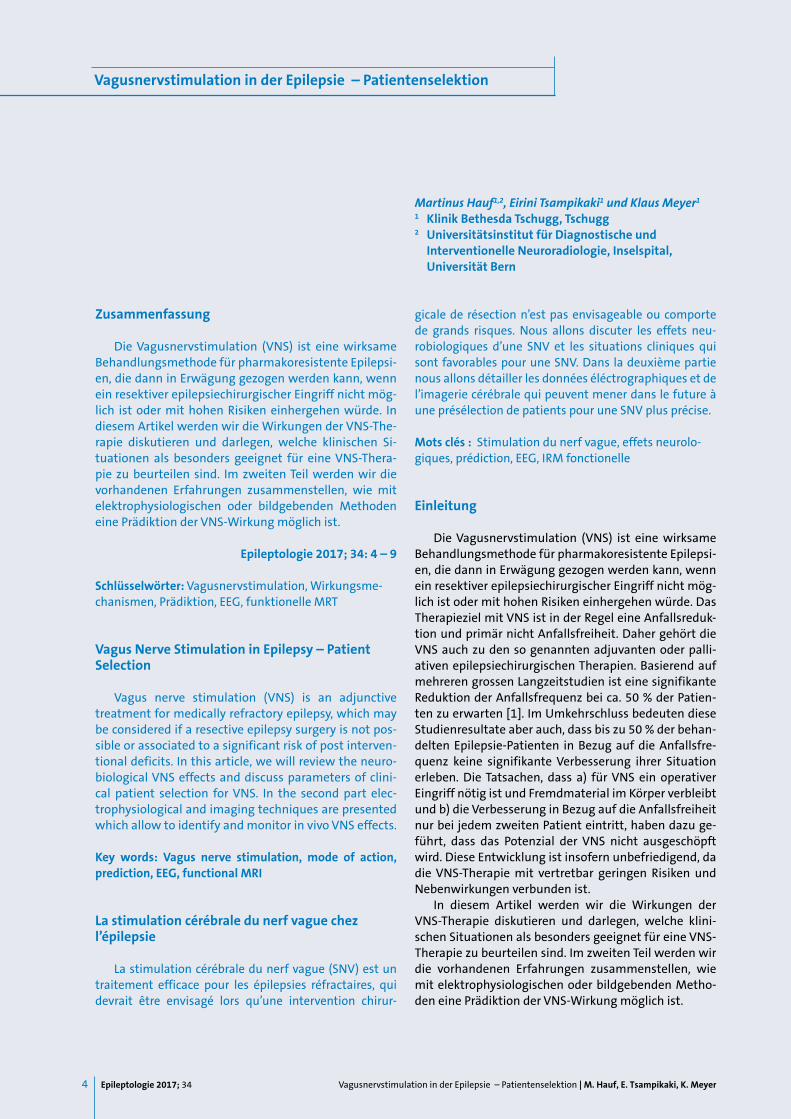
Im klinischen Kontext wäre ein EEG-basiertes Mo-nitoring der VNS-Wirkung wertvoll. Neben der hier dis-kutierten Prädiktion ist die Einstellung der optimalen VNS-Parameter komplex und basiert aktuell auf den individuellen Erfahrungen und den Beobachtungen beim einzelnen Patienten. Die Funktionsänderung des Gehirns durch die VNS kann auch im Oberflächen-EEG gemessen werden. EEG-basierte Arbeiten konnten VNS-induzierte Veränderungen in zerebralen Netz-werkaktivitäten durch die Anwendung von quantitati-ven mathematischen Analysen bei Patienten dokumen-tieren und Hinweise finden, dass eine Prädiktion des Ansprechens auf VNS möglich ist [24 - 26]. Eine darauf aufbauende Arbeit untersuchte einen methodologisch einfacheren Parameter der kortikalen Synchronisati-onsmessung („Phase lag index“) und zeigte eine sig-nifikante Assoziation von niedriger Synchronizität im Alpha- und Delta-Band des EEG und positivem Anspre-chen auf die VNS [27]. Eine Voraussetzung für einen EEG-basierten Parameter der VNS-Wirkung ist neben der Robustheit auch die Quantifizierbarkeit der Mas-se. Eine mögliche mathematische Methodik wurde mit Daten aus dem Oberflächen-EEG von einer Gruppe des Neurozentrums des Inselspitals etabliert [28]. Pilotda-ten zu einer Analyse mit diesen methodischen Ansät-zen zeigt Abbildung 2. Eine transkutane VNS wird wäh-rend eines Standard-EEG angewendet mit einem „on/off“-Design. Die Synchronisationswerte des EEG-Sig-nals zeigen deutliche Veränderungen während der ak-tiven Stimulation.
Zusammenfassung
Patienten mit pharmakoresistenten Epilepsien, die nicht resezierbar sind, können von einer VNS profitie-ren. Klinisch schwere Anfälle, insbesondere Sturzan-fälle, bessern sich stärker als fokal komplexe Anfallser-eignisse. Positive Kriterien für ein Ansprechen auf VNS sind junges Alter (< 18), kurze Epilepsiedauer, Tuberöse Sklerose und strukturelle Epilepsien nach Trauma und Schlaganfall. Positiv ist die Einsetzbarkeit der akuten VNS zu werten, die sowohl in der Aura durch den Pati-enten/Betreuenden, wie auch automatisiert bei präik-taler Tachykardie zur Verbesserung der Anfallssituation beitragen kann. Darüber hinaus wird die Möglichkeit der eigenständigen Anfallsunterbrechung als psycho-logisch stabilisierender Faktor erlebt. Begleitende af-fektive Störungen können sich durch die VNS bessern, und eine Reduktion der Medikation ist im chronischen Verlauf bei einem relevanten Anteil der Patienten mit VNS möglich. Die Lebensqualität wird in 80 % der Pati-enten mit VNS als gebessert geschildert. Das Erreichen einer Anfallsfreiheit ist durch eine VNS-Therapie nicht wahrscheinlich, und ca. 50 % der Patienten zeigen keine signifikante Anfallsreduktion. Hier sind Anstrengungen
Vagusnervstimulation in der Epilepsie – Patientenselektion | M. Hauf, E. Tsampikaki, K. Meyer
Abbildung 1. Axiale Schicht auf Höhe des Dienzephalons einer simultanen EEG/fMRT-Untersuchung. Daten eines Patienten mit generalisierten Anfällen und interiktalen bifrontal beton-ten Spike-Wave-Abläufen. Das Bild illustriert die hämodyna-mischen Korrelate der interiktalen epileptischen Aktivität in den Basalganglien und dem Thalamus. Zu Details siehe Text.
8 Epileptologie 2017; 34 Vagusnervstimulation in der Epilepsie – Patientenselektion | M. Hauf, E. Tsampikaki, K. Meyer
nötig, die neuen Methoden der EEG-Analyse, der zereb-ralen Bildgebung und der transkutanen VNS zu evaluie-ren, und robuste Parameter der Prädiktion der Anfalls-reduktion zu definieren.
The Copyright of the images stays with the authors.
Referenzen
1. Englot DJ, Chang EF, Auguste KI. Efficacy of vagus nerve stimulation for
epilepsy by patient age, epilepsy duration, and seizure type. Neurosurg
Clin N Amer 2011; 22: 443-448
2. Ansari S, Chaudhri K, Al Moutaery KA. Vagus nerve stimulation: indica-
tions and limitations. Acta Neurochir Suppl 2007; 97: 281-286
3. Cheyuo C, Jacob A, Wu R et al. The parasympathetic nervous system in
the quest for stroke therapeutics. J Cereb Blood Flow Metab 2011; 31:
1187-1195
4. Cai PY, Bodhit A, Derequito R et al. Vagus nerve stimulation in ischemic
stroke: old wine in a new bottle. Front Neurol 2014; 5: 107
5. Kraus T, Kiess O, Hosl K et al. CNS BOLD fMRI effects of sham-controlled
transcutaneous electrical nerve stimulation in the left outer auditory ca-
nal – a pilot study. Brain Stimul 2013; 6: 798-804
6. Henry TR, Bakay RA, Votaw JR et al. Brain blood flow alterations induced
by therapeutic vagus nerve stimulation in partial epilepsy: I. Acute ef-
fects at high and low levels of stimulation. Epilepsia 1998; 39: 983-990
7. Mu Q, Bohning DE, Nahas Z et al. Acute vagus nerve stimulation using
different pulse widths produces varying brain effects. Biol Psychiatry
2004; 55: 816-825
8. Henry TR, Bakay RA, Pennell PB et al. Brain blood-flow alterations
induced by therapeutic vagus nerve stimulation in partial epilepsy: II.
prolonged effects at high and low levels of stimulation. Epilepsia 2004;
45: 1064-1070
9. Fisher RS, Eggleston KS, Wright CW. Vagus nerve stimulation magnet ac-
tivation for seizures: a critical review. Acta Neurol Scand 2015; 131: 1-8
10. Fisher RS, Afra P, Macken M et al. Automatic vagus nerve stimulation
triggered by ictal tachycardia: Clinical outcomes and device performance
– The U.S. E-37 Trial. Neuromodulation 2016; 19: 188-195
11. Ben-Menachem E, Manon-Espaillat R, Ristanovic R et al. Vagus nerve sti-
mulation for treatment of partial seizures: 1. A controlled study of effect
on seizures. First International Vagus Nerve Stimulation Study Group.
Epilepsia 1994; 35: 616-626
12. Wasade VS, Schultz L, Mohanarangan K et al. Long-term seizure and psy-
chosocial outcomes of vagus nerve stimulation for intractable epilepsy.
Epilepsy Behav 2015; 53: 31-36
13. Morris GL 3rd, Gloss D, Buchhalter J et al. Evidence-based guideline up-
date: vagus nerve stimulation for the treatment of epilepsy: report of
the Guideline Development Subcommittee of the American Academy of
Neurology. Neurology 2013; 81: 1453-1459
14. Vonck K, Raedt R, Naulaerts J et al. Vagus nerve stimulation...25 years
later! What do we know about the effects on cognition? Neurosci Biobe-
hav Rev 2014; 45: 63-71
15. Ryvlin P, Gilliam FG, Nguyen DK et al. The long-term effect of vagus nerve
stimulation on quality of life in patients with pharmacoresistant focal
epilepsy: the PuLsE (Open Prospective Randomized Long-term Effective-
ness) trial. Epilepsia 2014; 55: 893-900
16. Levy ML, Levy KM, Hoff D et al. Vagus nerve stimulation therapy in pa-
tients with autism spectrum disorder and intractable epilepsy: results
from the vagus nerve stimulation therapy patient outcome registry. J
Neurosurg Pediatr 2010; 5: 595-602
17. Panebianco M, Rigby A, Weston J, Marson AG. Vagus nerve stimulation
for partial seizures. Cochrane Database Syst Rev 2015: CD002896
18. Ng M, Devinsky O. Vagus nerve stimulation for refractory idiopathic ge-
neralised epilepsy. Seizure 2004; 13: 176-178
19. Burakgazi AZ, Burakgazi-Dalkilic E, Caputy AJ, Potolicchio SJ. The correla-
tion between vagus nerve stimulation efficacy and partial onset epilep-
sies. J Clin Neurophysiol 2011; 28: 380-383
20. Englot DJ, Rolston JD, Wright CW et al. Rates and predictors of seizure
freedom with vagus nerve stimulation for intractable epilepsy. Neuro-
surgery 2016; 79: 345-353
21. Englot DJ, Rolston JD, Wang DD et al. Efficacy of vagus nerve stimulation
in posttraumatic versus nontraumatic epilepsy. J Neurosurg 2012; 117:
970-977
22. Colicchio G, Montano N, Fuggetta F et al. Vagus nerve stimulation in
drug-resistant epilepsies. Analysis of potential prognostic factors in a
cohort of patients with long-term follow-up. Acta Neurochir 2012; 154:
2237-2240
23. Henry TR, Votaw JR, Pennell PB et al. Acute blood flow changes and effi-
cacy of vagus nerve stimulation in partial epilepsy. Neurology 1999; 52:
1166-1173
24. Bartolomei F, Bonini F, Vidal E et al. How does vagal nerve stimulation
(VNS) change EEG brain functional connectivity? Epilepsy Res 2016; 126:
141-146
Abbildung 2. Quantitative EEG-Analyse bei Applikation einer transkutanen VNS im „on/off“-Design. Die normalisierten Eigenwerte als Mass der Synchronisation des EEG-Signals zeigen deutliche Veränderungen während der aktiven Va-gusnervstimulation (c/o Dr. F. Zubler, Neurologie, Inselspital Bern, Universität Bern).

9Epileptologie 2017; 34Vagusnervstimulation in der Epilepsie – Patientenselektion | M. Hauf, E. Tsampikaki, K. Meyer
25. Fraschini M, Demuru M, Puligheddu M et al. The re-organization of func-
tional brain networks in pharmaco-resistant epileptic patients who re-
spond to VNS. Neurosci Lett 2014; 580: 153-157
26. Fraschini M, Puligheddu M, Demuru M et al. VNS induced desynchroniza-
tion in gamma bands correlates with positive clinical outcome in tempo-
ral lobe pharmacoresistant epilepsy. Neurosci Lett 2013; 536: 14-18
27. Bodin C, Aubert S, Daquin G et al. Responders to vagus nerve stimulation
(VNS) in refractory epilepsy have reduced interictal cortical synchronicity
on scalp EEG. Epilepsy Res 2015; 113: 98-103
28. Zubler F, Koenig C, Steimer A et al. Prognostic and diagnostic value of EEG
signal coupling measures in coma. Clin Neurophysiol 2016; 127: 2942-
2952
Address for correspondence:PD Dr. med. Martinus HaufKlinik Bethesda Tschugg CH 3233 TschuggTel. 0041 32 338 41 [email protected]

10 Epileptologie 2017; 34 Transcranial Direct-Current Stimulation as Treatment in Epilepsy | M. Gschwind
Transcranial Direct-Current Stimulation as Treatment in Epilepsy
Summary
Transcranial direct-current stimulation (tDCS) is a widely explored and easy to use technique of non-inva-sive neuromodulation, which has shown both excitato-ry and inhibitory effects, depending on the direction of the current flow. Cathodal stimulation has an inhibito-ry effect on multiple cortical levels, which is of particu-lar interest for the use as treatment of epilepsy. Here, we review the recent literature, especially of 2016, and discuss the important aspects of tDCS, including pa-tient selection, stimulation localization, and evaluation of treatment success, as well as safety, and regulatory aspects.
Epileptologie 2017; 34: 10 – 18
Keywords: Epilepsy, transcranial direct-current stimula-tion, tDCS, outcome, drug therapy
Transkranielle Gleichstromstimulation als Behandlungsoption bei Epilepsie
Transkranielle Gleichstromstimulation (tDCS) ist ei-ne weitverbreitete und einfach zu bedienende Metho-de der nicht-invasiven Neuromodulation. Je nach Rich-tung des Stromflusses hat sie exzitatorische oder hem-mende Effekte gezeigt. Kathodale Stimulation hat eine kortikal hemmende Wirkung auf mehreren Ebenen, was von besonderem Interesse ist für die Verwendung bei der Behandlung von Epilepsie. Hier besprechen wir die neuere Literatur, insbesondere des Jahres 2016, und diskutieren die wichtigsten Aspekte von tDCS, einschliesslich Patientenauswahl, Lokalisierung der Stimulation und die Evaluation des Behandlungs- erfolges, sowie sicherheitstechnische und regulatori-sche Aspekte.
Schlüsselwörter: Epilepsie, transkranielle Gleichstrom-stimulation, tDCS, Behandlungserfolg, medikamentöse Behandlung
Markus GschwindDivision of Neurology, Department of Clinical Neurosciences, University Hospitals of Geneva
La stimulation transcrânienne à courant continu comme traitement d’épilepsie
La stimulation transcrânienne à courant continu (tDCS) est une technique de neuromodulation non in-vasive, largement explorée et facile à utiliser. En fonc-tion de la direction du courant, elle a montré à la fois des effets excitateurs et inhibiteurs. La stimulation cathodique a un effet inhibiteur cortical à de multiples niveaux, ce qui est particulièrement intéressant pour l’utilisation dans le traitement de l’épilepsie. Ici, nous passons en revue la littérature récente, en particulier de 2016, et discutons les aspects importants autour de la tDCS, y compris la sélection des patients et la loca-lisation de la stimulation, l’évaluation de l’efficacité du traitement, ainsi que les aspects de sécurité et de régle-mentation.
Mots clés : Epilepsie, stimulation transcrânienne à cou-rant continu, tDCS, efficacité du traitement, traitement médicamenteux
Introduction
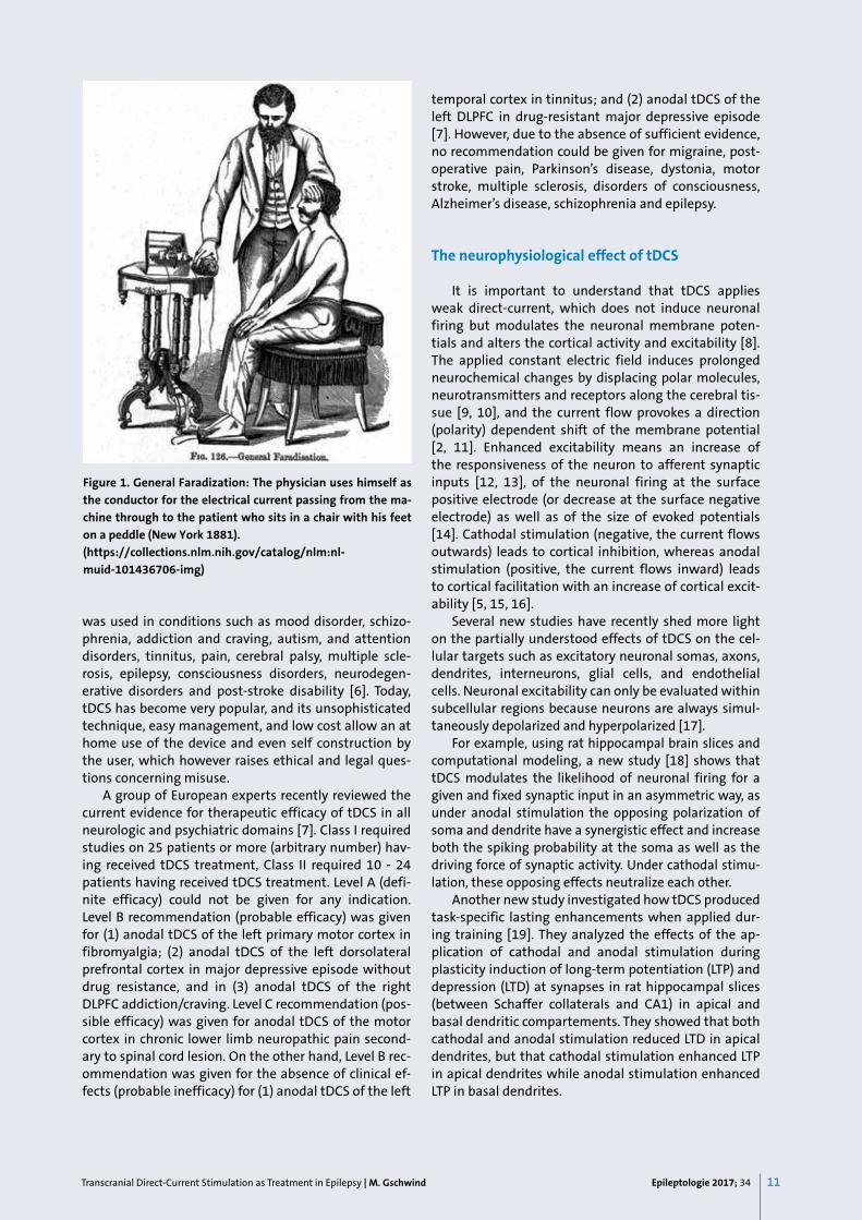
After the initial experiments of Luigi Galvani and Allessandro Volta in the 1790ies, it was discovered that weak galvanic current flowing through different parts of the body could have interesting “physiological ef-fects”, for example relief of musculoskeletal and noci- ceptive pain (nowadays called Transcutaneous Electri-cal Nerve Stimulation, TENS), or relief of mental dis-orders [1]. During the following centuries electrical stimulation of the brain was part of the psychiatrist’s armamentarium, but was largely overshadowed by electroconvulsive therapy and the advent of psycho-pharmacologic drugs.
Only in the last two decades, this technique, now called transcranial Direct Current Stimulation (tDCS) was given new scientific basis by fundamental work that promoted growing interest in almost all neuro-logical and psychiatric domains [2 - 5]. In 2016, a re-view listed 340 published studies (not counting single case reports) on clinical effect of tDCS in patients. TDCS
11Epileptologie 2017; 34Transcranial Direct-Current Stimulation as Treatment in Epilepsy | M. Gschwind
was used in conditions such as mood disorder, schizo-phrenia, addiction and craving, autism, and attention disorders, tinnitus, pain, cerebral palsy, multiple scle-rosis, epilepsy, consciousness disorders, neurodegen-erative disorders and post-stroke disability [6]. Today, tDCS has become very popular, and its unsophisticated technique, easy management, and low cost allow an at home use of the device and even self construction by the user, which however raises ethical and legal ques-tions concerning misuse.
A group of European experts recently reviewed the current evidence for therapeutic efficacy of tDCS in all neurologic and psychiatric domains [7]. Class I required studies on 25 patients or more (arbitrary number) hav-ing received tDCS treatment, Class II required 10 - 24 patients having received tDCS treatment. Level A (defi-nite efficacy) could not be given for any indication. Level B recommendation (probable efficacy) was given for (1) anodal tDCS of the left primary motor cortex in fibromyalgia; (2) anodal tDCS of the left dorsolateral prefrontal cortex in major depressive episode without drug resistance, and in (3) anodal tDCS of the right DLPFC addiction/craving. Level C recommendation (pos-sible efficacy) was given for anodal tDCS of the motor cortex in chronic lower limb neuropathic pain second-ary to spinal cord lesion. On the other hand, Level B rec-ommendation was given for the absence of clinical ef-fects (probable inefficacy) for (1) anodal tDCS of the left
temporal cortex in tinnitus; and (2) anodal tDCS of the left DLPFC in drug-resistant major depressive episode [7]. However, due to the absence of sufficient evidence, no recommendation could be given for migraine, post-operative pain, Parkinson’s disease, dystonia, motor stroke, multiple sclerosis, disorders of consciousness, Alzheimer’s disease, schizophrenia and epilepsy.
The neurophysiological effect of tDCS
It is important to understand that tDCS applies weak direct-current, which does not induce neuronal firing but modulates the neuronal membrane poten-tials and alters the cortical activity and excitability [8]. The applied constant electric field induces prolonged neurochemical changes by displacing polar molecules, neurotransmitters and receptors along the cerebral tis-sue [9, 10], and the current flow provokes a direction (polarity) dependent shift of the membrane potential [2, 11]. Enhanced excitability means an increase of the responsiveness of the neuron to afferent synaptic inputs [12, 13], of the neuronal firing at the surface positive electrode (or decrease at the surface negative electrode) as well as of the size of evoked potentials [14]. Cathodal stimulation (negative, the current flows outwards) leads to cortical inhibition, whereas anodal stimulation (positive, the current flows inward) leads to cortical facilitation with an increase of cortical excit-ability [5, 15, 16].
Several new studies have recently shed more light on the partially understood effects of tDCS on the cel-lular targets such as excitatory neuronal somas, axons, dendrites, interneurons, glial cells, and endothelial cells. Neuronal excitability can only be evaluated within subcellular regions because neurons are always simul-taneously depolarized and hyperpolarized [17].
For example, using rat hippocampal brain slices and computational modeling, a new study [18] shows that tDCS modulates the likelihood of neuronal firing for a given and fixed synaptic input in an asymmetric way, as under anodal stimulation the opposing polarization of soma and dendrite have a synergistic effect and increase both the spiking probability at the soma as well as the driving force of synaptic activity. Under cathodal stimu-lation, these opposing effects neutralize each other.
Another new study investigated how tDCS produced task-specific lasting enhancements when applied dur-ing training [19]. They analyzed the effects of the ap-plication of cathodal and anodal stimulation during plasticity induction of long-term potentiation (LTP) and depression (LTD) at synapses in rat hippocampal slices (between Schaffer collaterals and CA1) in apical and basal dendritic compartements. They showed that both cathodal and anodal stimulation reduced LTD in apical dendrites, but that cathodal stimulation enhanced LTP in apical dendrites while anodal stimulation enhanced LTP in basal dendrites.
Figure 1. General Faradization: The physician uses himself as the conductor for the electrical current passing from the ma-chine through to the patient who sits in a chair with his feet on a peddle (New York 1881).(https://collections.nlm.nih.gov/catalog/nlm:nl-muid-101436706-img)

12 Epileptologie 2017; 34 Transcranial Direct-Current Stimulation as Treatment in Epilepsy | M. Gschwind
In a third study, one-week lasting increases in hip-pocampal LTP, learning and memory was measured in mice after 20 min of tDCS [20]. These effects were as-sociated with enhancement of acetylation of Brain-Derived Neurotropic Factor (BDNF) at several levels and enhanced phosphorylation of the C-AMP Response Element-Binding (CREB) protein, suggesting that an-odal tDCS increases hippocampal LTP via chromatin remodeling of BDNF regulatory sequences that lead to increased expression of this gene. This gives support to the use of tDCS for the treatment of brain diseases with impaired neuroplasticity, such as Alzeimer’s dis-ease and dementia [21, 22], Huntington’s disease [22, 23], depression [24, 25], schizophrenia [26], obsessive-compulsive disorder [27], Rett syndrome [22, 28], or an-orexia nervosa [29], and it might also explain the effect of tDCS in patients with epilepsy [30, 31].
tDCS has promising antiepileptic effects
Five different approaches of brain stimulation are available for treatment of epilepsy, of which today only two are approved in Switzerland, namely Deep Brain Stimulation in the Anterior Thalamic Nuclei (DBS-ANT) and Vagal Nerve Stimulation (VNS). However, Transcra-nial Magnetic Stimulation (TMS), transcutaneous Vagal Stimulation (tVNS), and tDCS are still in experimental state. So far, the missing evidence of tDCS efficacy in treatment of epilepsy is most likely due to the hetero-geneity of the studies (i.e. different stimulation proto-cols and patient population). In our recent literature review (march 2016 [32]), we found 47 publications of which we counted six case reports [33 - 38] and three sham controlled studies [39 - 41]. In 2016, two new studies were published on patients with mesial-tem-poral lobe epilepsy and hippocampal sclerosis. One was a randomized placebo-controlled, double-blinded clini-cal trial in 28 adult patients [42], which compared 2mA cathodal tDCS over the epileptic focus during 30 min-utes in three consecutive days versus five consecutive days versus placebo stimulation. Seizure frequency and interictal epileptiform discharges (IEDs) were quanti-fied before and after treatment, as well as at 30 and 60 days follow-up. There was a significant reduction of seizure frequency at 30 (p = 0.001) and 60 days (p = 0.0001) compared to baseline (mean reduction -48%). The reduction was also greater in the five-days group compared to the three-days group. Also a significant short-term reduction of IED was found between base-line and immediately after interventions (p = 0.041) in all groups [42]. The other study compared in a cross over design the effect of modulated cathodal stimula-tion (2mA for 30 min on 3 consecutive days) to sham stimulation in 12 patients [43]. Sham was designed as a short 60 s stimulation that then decreased during 15 s, while the electrodes stayed for 30 minutes over the stimulation site. The mean seizure frequency decreased from 10.58 to 1.67 per month after cathodal tDCS ap-plication (p = 0.003), and ten patients (83.33%) had more than 50% decrease in their seizure frequency. Six patients (50%) were seizure-free one month after the cathodal tDCS session. However, two patients (16.67%) also showed positive sham effects [43].
Over all, we count now a total of 157 patients that were studied in clinical trials, most of which suffered from mesiotemporal lobe epilepsy (with or without hippocampal sclerosis, N = 89, 56%) [41 - 44], followed by dysplasia (N = 23, 20%) [45]. The other studies inves-tigated single patients with Rasmussen’s encephalitis [34, 35], continuous spikes and waves syndrome during slow sleep (CWSW) or Landau-Kleffner Syndrome [33, 36, 37], epilepsy from vascular lesions (N = 17).
tDCS also was evaluated in pediatric population. Although in some studies in patients with CSWS prom-ising results were reported [33, 37], a double-blinded and sham-controlled crossover study in five pediatric
Figure 2. Anodal tDCS (positive, the current flows inward) leads to cortical facilitation with an increase of cortical ex-citability. The mechanism is via a transient increase in intra-cellular calcium (Ca2+), which initiates molecular cascades that lead to persistent changes in chromatin structure of the Brain-Derived Neurotropic Factor (BDNF), including the phosphorylation of the C-AMP Response Element-Binding (CREB) protein and the recruitment of the CREB-Binding Pro-tein (CBP). As a result, long term potentiation (LTP), as well as learning and memory cause an increased transcription of BDNF [20].

13Epileptologie 2017; 34Transcranial Direct-Current Stimulation as Treatment in Epilepsy | M. Gschwind
patients with CSWS failed to show a decrease of epi-leptiform activity with cathodal tDCS [36]. However, suppression of interictal spikes for the duration of 48 hours, together with a small but significant decrease in seizure frequency (-4.8%) during the following four weeks was found in a randomized sham-controlled, unblinded study on 36 children with focal epilepsy (of undefined origin) [40]. Also in 22 children with Lennox-Gastaut syndrome, cathodal stimulation over the mo-tor cortex combined with pharmacologic treatment re-duced seizure frequency and IEDs [46].
Regulatory aspects of tDCS
In 2016, a group of experts proposed technical guidelines to ensure a proper and risk free use of tDCS protocols [47]. The safety profile of tDCS is extremely high, if the currently recommended stimulation proto-cols are respected (stimulation time under 40 min, cur-rent under 4 mA, electrical charge under 7.2 Coulombs) [48]. There are only very limited adverse effects, such as local sensory discomfort or mild headache [47 - 49]. Not any serious adverse effect has been reported in more than > 50’000 subjects described in the 340 publi-cations using tDCS in patients [6].
Data from relevant animal models indicate that brain injury by tDCS occurs at predicted brain current densities (6.3 - 13 A/m2) that are over one order of magnitude above those produced by tDCS respecting the guidelines on humans. Moreover, the large body of treated patients included all kinds of neurologically vul-nerable individuals, such as children, elderly, patients
suffering from stroke, mood disorders, and epilepsy [48].
In practice, a good contact between electrodes and skin across the whole electrode is important, provided by the use of a gel, cream, or appropriately large, wet electrode, in order to limit excessive current density (which depends on electrode size and shape) and local skin burns [50, 51].
From the regulatory point of view, because tDCS is not yet approved as a treatment for epilepsy, it falls under the requirements for clinical investigations with medical devices. In Switzerland, the requirements for such reseach carried out prospectively on patients is regarded as clinical trial with medical devices and de-scribed by the Human Research Act (HRA 810.30) [52], and Clinical Trials Ordinance (ClinO 810.305) [53], which are the integration of the European regulations into Swiss national laws.
In order to start such a clinical study, the following requirements must be met: Stimulation devices that are not certified for conformity with European standards (“CE marked”) must be considered as investigational devices, meaning that, apart from the aspects related to clinical evaluation, a proof of their compliancy with the essential requirements of the European Medical Device Directive (DIR 93/42/EEC) is requested. Current-ly, many of the tDCS devices available on the market are not approved for human use in Europe. Furthermore, the clinical study documentation must be prepared in compliance with the ISO 14155 standard for clinical in-vestigations (Good Clinical Practice) and needs to be ap-proved by both the competent authority (Swissmedic) and the competent Ethics Committee (Swissethics).
Figure 3. Example of a typical tDCS device comprising the stimulation unit, and 2 sponge electrodes of 5 x 7 cm, mounted on the head with a system of stripes. (http://soterixmedical.com/research/1x1).

14 Epileptologie 2017; 34 Transcranial Direct-Current Stimulation as Treatment in Epilepsy | M. Gschwind
Careful patient selection
In order to create a homogenous group of patients with the same epilepsy type and localization, patient selection warrants careful consideration. Drug studies have shown that 40 - 50% of patients with focal epilep-sy (restricted to one hemisphere only) and about 15% of patients with idiopathic generalized epilepsy (con-cerning both hemispheres) are refractory to pharmaco-logic treatment.
“Drug resistant epilepsy” is defined as the absence of complete seizure control despite regular drug in-take of 2 or more antiepileptic drugs (AEDs) up to suf-ficiently high dosages (serum drug level control). With the prescription of a 3rd AED the probability to obtain seizure control increases for not more than 2% [54]. Only in case of drug resistant epilepsy further treat-ment options need usually to be explored, i.e. epilepsy surgery or neuromodulation. TDCS will come into play only in patients with intractable epilepsy that are not eligible for epilepsy surgery or don’t opt for surgery for other reasons [55, 56]. A realistic patient selection will be biased on the drug resistant and inoperable cases. At the current state, neuromodulation techniques are con-sidered as second-line “palliative” treatment, providing additional seizure-control in < 10% of the cases [57].
Important role of ongoing antiepileptic medication
The ongoing antiepileptic medication appears to play a specific role in degree of antiepileptic effects of tDCS and should therefore be controlled during stimu-lation protocols, or, if not possible, at least reported. Although the enrollment of patients without drug ther-apy would be the best solution to avoid confounding effects due to the AEDs, this is hardly feasible for ethi-cal reasons, as a withdrawal of AEDs only for the study purpose could not be accepted.
An interesting research line investigated the behav-ior of neurotransmitters during tDCS, and in relation to this, the effect of concomitant antiepileptic drug treatment (AED). For example, the effects of both an-odal and cathodal tDCS were prevented by blocking of glutamate receptors, whereas blocking of sodium and calcium channels inhibited the effects of anodal stimu-lation only [4, 58]. Moreover, the modulation of GABA-ergic interneurons was associated to excitatory anodal tDCS effects: the excitatory anodal stimulation effect was decreased by the simultaneous use of tDCS togeth-er with lorazepam, resulting in a diminished intracorti-cal activation [59].
Reduced cortical GABA concentrations by decreased GABA synthesis could be demonstrated after the ex-citatory (anodal) tDCS, using MR spectroscopy, whereas inhibitory (cathodal) stimulation caused a reduction of excitatory glutamatergic neuronal activity [9].
Cathodal stimulation also prevented the normally occurring loss of GABAergic paired-pulse motor inhibi-tion that usually provokes seizures in the context of pen-tylene-tetrazol use. This shows a further antiepileptic mechanism of cathodal tDCS [60]. The same study also observed a down-shift of EEG frequency spectral power in favor of low EEG frequencies after cathodal tDCS.
Therefore, in order to increase the antiepileptic stimulation effect, it might be suggested to combine cathodal tDCS together with anti-GABAergic AEDs, such as benzodiazepines (or valproic acid, felbamate, topira-mate, and barbiturates). The sodium channel blocker carbamazepine selectively eliminated anodal effects by stabilizing the membrane potential voltage-depend-ently and inhibiting membrane depolarization [4, 58].
The role of localization of the epileptogenic focus
Because the direction of the electrical field influ-ences the tDCS stimulation effect, the exact electrode montage on the head is a primordial factor in the stimulation protocol. The electrical field depends on the polarity of the electrode, as well as on the proper-ties of the scalp and underlying tissue, and the possi-bility of current shunting by a transgyral or translobar short-cuts [61, 62]. Focality of tDCS is determined by the current density (mA/cm2), as well as by the distance between the active electrode (over the area of interest) and the reference, and by their sizes.
A more precise and focal control of the current flow might be reached by a multi-electrode approach [63], or even a geodesic guided stimulation localization [64, 65]. However, with an increased focality of tDCS also localization precision of the epileptic focus needs im-provement. This can be achieved when taking benefits from electrical high-resolution source localization (ESI) and allowing for in-depth target localization, especially when taking into account the patients individual anat-omy [66 - 68].
On the other hand, the advantage of focalization is limited by the sometimes rather diffuse description at hand of the epileptogenic region or even network. The greatest success of neuromodulation is probably deep brain stimulation of the subthalamic nucleus (STN) in Parkinson’s disease, which can be explained by the as-sociation of several factors: (1) the small size of the STN (< 1 cm), which is precisely localizable in MR scans; (2) the STN being a hub of the major cortico-basal loops; (3) the clear connectivity model of these loops ex-plaining precisely clinical presentation. None of these conditions are given in epilepsy, being a symptom of different heterogeneous conditions covering different brain regions. The different etiologies (cortical dyspla-sia or brain lesion of variable size, non-lesional epilepsy, variable foci) require different approaches. Although epileptic activity has been shown to not only represent a local process, but is also abnormal neuronal activity

15Epileptologie 2017; 34Transcranial Direct-Current Stimulation as Treatment in Epilepsy | M. Gschwind
in connected regions [69, 70], it is still very difficult to make use of these connections in order to stimulate the target area remotely. Moreover, the target areas are sometimes extending across several centimeters.
The evaluation of treatment success
Like for pharmacological studies, also in tDCS studies the evaluation of treatment success is based on seizure frequency and IEDs. To observe a significant decrease of seizure is a question of statistical power; for this result the patient needs to present a sufficient amount of sei-zures in a short time, and the observation period covered
by the study needs to be long enough. Three studies couldn’t find change of seizure frequency in the stimu-lation group compared to the sham group [45] or com-pared to baseline [36, 44]. Although, Liu et al. [44] had focused on patients with well-controlled TLE, in which no further improvement was actually to be expected.
However, San-Juan et al. used a 60-days observa-tion period on a very homogeneously selected patient group with TLE, in which the monthly seizure frequency at baseline was median of 6 (range 3 - 30). In the group with 3-days stimulation, they observed a > 50% re-duction of seizures in 50% of the patients, and in the group with 5-days stimulation even in 62.5% of the pa-tients, however, the same effect was also observed in
Figure 4. Simulation of the effects of high-definition tDCS (HD-tDCS), using a combination of dot shaped electrodes across the head, compared to conventional tDCS using a bipolar sponge montage. Top left: An individual anatomical head model is crea-ted using 4 - 8 tissue segments with different electrical properties. Boxed Right Panel: The distribution of anode and cathode in HD-tDCS (the anode, positioned over the motor region is surrounded by 4 cathodes), and conventional tDCS (anode centered over the motor region and cathode over the contralateral supraorbital region). A: HD-tDCS shows a restricted current flow com-pared to B: conventional tDCS where the current flow is largely diffuse (reprinted from [71] with permission of Elsevier).

16 Epileptologie 2017; 34 Transcranial Direct-Current Stimulation as Treatment in Epilepsy | M. Gschwind
References
1. Zago S, Ferrucci R, Fregni F et al. Bartholow, Sciamanna, Alberti: pioneers
in the electrical stimulation of the exposed human cerebral cortex. Neu-
roscientist 2008; 14: 521-528
2. Nitsche MA, Paulus W. Excitability changes induced in the human motor
cortex by weak transcranial direct current stimulation. J Physiol 2000;
527(Pt 3): 633-639
3. Nitsche MA, Paulus W. Sustained excitability elevations induced by
transcranial DC motor cortex stimulation in humans. Neurology 2001;
57: 1899-1901
4. Liebetanz D, Nitsche MA, Tergau F et al. Pharmacological approach to
the mechanisms of transcranial DC-stimulation-induced after-effects of
human motor cortex excitability. Brain 2002; 125: 2238-2247
5. Priori A, Berardelli A, Rona S et al. Polarization of the human motor cor-
tex through the scalp. Neuroreport 1998; 9: 2257-2260
6. Lefaucheur JP. A comprehensive database of published tDCS clinical trials
(2005-2016). Neurophysiol Clin 2016; 46: 319-398
7. Lefaucheur JP, Antal A, Ayache SS et al. Evidence-based guidelines on the
therapeutic use of transcranial direct current stimulation (tDCS). Clin
Neurophysiol 2017; 128: 56-92
8. Purpura DP, McMurtry JG. Intracellular activities and evoked potential
changes during polarization of motor cortex. J Neurophysiol 1965; 28:
166-185
9. Stagg CJ, Best JG, Stephenson MC et al. Polarity-sensitive modulation of
cortical neurotransmitters by transcranial stimulation. J Neurosci 2009;
29: 5202-5206
10. Cogiamanian F, Vergari M, Pulecchi F et al. Effect of spinal transcutane-
ous direct current stimulation on somatosensory evoked potentials in
humans. Clin Neurophysiol 2008; 119: 2636-2640
11. Nitsche MA, Cohen LG, Wassermann EM et al. Transcranial direct current
stimulation: State of the art 2008. Brain Stimul 2008; 1: 206-223
12. Jefferys JG. Influence of electric fields on the excitability of granule cells
in guinea-pig hippocampal slices. J Physiol 1981; 319: 143-152
13. Bikson M, Inoue M, Akiyama H et al. Effects of uniform extracellular DC
electric fields on excitability in rat hippocampal slices in vitro. J Physiol
2004; 557: 175-190
14. Bindman LJ, Lippold OC, Redfearn JW. The action of brief polarizing cur-
rents on the cerebral cortex of the rat (1) during current flow and (2) in
the production of long-lasting after-effects. J Physiol 1964; 172: 369-382
15. Creutzfeldt OD, Fromm GH, Kapp H. Influence of transcortical d-c cur-
rents on cortical neuronal activity. Exp Neurol 1962; 5: 436-452
16. Bindman LJ, Lippold OC, Redfearn JW. The action of brief polarizing cur-
rents on the cerebral cortex of the rat (1) during current flow and (2) in
the production of long-lasting after-effects. J Physiol 1964; 172: 369-382
17. Jackson MP, Rahman A, Lafon B et al. Animal models of transcranial di-
rect current stimulation: Methods and mechanisms. Clin Neurophysiol
2016; 127: 3425-3454
18. Lafon B, Rahman A, Bikson M et al. Direct current stimulation alters neu-
ronal input/output function. Brain Stimul 2017; 10: 36-45
19. Kronberg G, Bridi M, Abel T et al. Direct current stimulation modulates
LTP and LTD: Activity dependence and dendritic effects. Brain Stimulation
2017; 10: 51-58
20. Podda MV, Cocco S, Mastrodonato A et al. Anodal transcranial direct
current stimulation boosts synaptic plasticity and memory in mice via
epigenetic regulation of Bdnf expression. Sci Rep 2016; 6: 22180
21. Arancio O, Chao MV. Neurotrophins, synaptic plasticity and dementia.
Curr Opin Neurobiol 2007; 17: 325-330
25% of patients in the placebo group [42]. The study of Tektürk et al. found a similar result, i.e. a decrease of > 50% seizure frequency, in 10 out of 12 patients with TLE (83.33%), whereas 6 patients (50%) were even sei-zure-free during one month after stimulation. None-theless also here, a positive effect under the sham con-dition was found in two patients [43].
The effect of tDCS on IEDs is less clear. A significant reduction in IED was found only in one study, imme-diately after treatment, as well as persisting after 24 and after 48 h [40]. In another study, the focal decrease of 40 - 50%, was measured ipsilateral, but not on the contralateral side [37]. In the other studies it either did not reach significance [45], or no reduction of IEDs was found [36]. The most recent studies did not find any reduction, possibly due to infrequent IEDs at baseline [42, 44]. While interictal spikes are a marker of epilep-tic activity, their relationship to seizure frequency is not evident. On the other hand, the presence of IEDs in a routine EEG has been shown to be related to a two-fold higher risk of active epilepsy [72], suggesting therefore some prognostic information for clinical efficacy.
Conclusion
So far, tDCS has been investigated in a large spectrum of neurologic and psychiatric disorders in > 50.000 patients, with no reported complications at all. Concerning epilepsy a few studies have demon-strated seizure frequency reduction and IED decrease, however data are not sufficient to provide evidence of its antiepileptic efficacy. The currently most used stim-ulation protocol consists of a 20 - 40 minutes cathodal stimulation on three to five consecutive days. EEG is monitored before and after stimulation, as well as af-ter one and after two months for long-term effects. When further evaluating tDCS in an antiepileptic pur-pose, larger studies are warranted across homogenous groups of patients. Very importantly, the ongoing an-tiepileptic medication plays an important role in tDCS effects and should be taken into account. Several new approaches have been developed to increase stimula-tion focality (high-definition-tDCS) which parallel the most recent algorithms allowing better localization of the epileptogenic focus (ESI), based on inverse solu-tions, and the patient’s individual anatomy.
AcknowledgementsMy thank goes to Professor Margitta Seeck, Geneva, for inspiration and support, and to Dr Agustina Lascano, Geneva, for valuable feedback.

17Epileptologie 2017; 34Transcranial Direct-Current Stimulation as Treatment in Epilepsy | M. Gschwind
22. Zuccato C, Cattaneo E. Brain-derived neurotrophic factor in neurodege-
nerative diseases. Nat Rev Neurol 2009; 5: 311-322
23. Zajac MS, Pang TY, Wong N et al. Wheel running and environmental
enrichment differentially modify exon-specific BDNF expression in the
hippocampus of wild-type and pre-motor symptomatic male and female
Huntington‘s disease mice. Hippocampus 2010; 20: 621-636
24. Brunoni AR, Lopes M, Fregni F. A systematic review and meta-analysis of
clinical studies on major depression and BDNF levels: implications for the
role of neuroplasticity in depression. Int J Neuropsychopharmacol 2008;
11: 1169-1180
25. Dwivedi Y. Brain-derived neurotrophic factor: role in depression and sui-
cide. Neuropsychiatr Dis Treat 2009; 5: 433-449
26. Xiu MH, Hui L, Dang YF et al. Decreased serum BDNF levels in chronic
institutionalized schizophrenia on long-term treatment with typical and
atypical antipsychotics. Prog Neuropsychopharmacol Biol Psychiatry
2009; 33: 1508-1512
27. Maina G, Rosso G, Zanardini R et al. Serum levels of brain-derived neu-
rotrophic factor in drug-naive obsessive-compulsive patients: a case-
control study. J Affect Disord 2010; 122: 174-178
28. Zeev BB, Bebbington A, Ho G et al. The common BDNF polymorphism
may be a modifier of disease severity in Rett syndrome. Neurology 2009;
72: 1242-1247
29. Mercader JM, Fernandez-Aranda F, Gratacos M et al. Blood levels of
brain-derived neurotrophic factor correlate with several psychopatho-
logical symptoms in anorexia nervosa patients. Neuropsychobiology
2007; 56: 185-190
30. Gall C, Lauterborn J, Bundman M et al. Seizures and the regulation of
neurotrophic factor and neuropeptide gene expression in brain. Epilepsy
Res Suppl 1991; 4: 225-245
31. Tanaka T, Saito H, Matsuki N. Inhibition of GABAA synaptic responses by
brain-derived neurotrophic factor (BDNF) in rat hippocampus. J Neurosci
1997; 17: 2959-2966
32. Gschwind M, Seeck M. Transcranial direct-current stimulation as treat-
ment in epilepsy. Expert Rev Neurother 2016: 1-15
33. Yook SW, Park SH, Seo JH et al. Suppression of seizure by cathodal
transcranial direct current stimulation in an epileptic patient – a case
report. Ann Rehabil Med 2011; 35: 579-582
34. Tekturk P, Erdogan ET, Kurt A et al. Transcranial direct current stimulation
improves seizure control in patients with Rasmussen encephalitis. Epilep-
tic Disord 2016; 18: 58-66
35. San-Juan D, Calcaneo Jde D, Gonzalez-Aragon MF et al. Transcranial di-
rect current stimulation in adolescent and adult Rasmussen‘s encephali-
tis. Epilepsy Behav 2011; 20: 126-131
36. Varga ET, Terney D, Atkins MD et al. Transcranial direct current stimula-
tion in refractory continuous spikes and waves during slow sleep: a con-
trolled study. Epilepsy Res 2011; 97: 142-145
37. Faria P, Fregni F, Sebastiao F et al. Feasibility of focal transcranial DC po-
larization with simultaneous EEG recording: preliminary assessment in
healthy subjects and human epilepsy. Epilepsy Behav 2012; 25: 417-425
38. Assenza G, Campana C, Formica D et al. Efficacy of cathodal transcranial
direct current stimulation in drug-resistant epilepsy: a proof of principle.
Conf Proc IEEE Eng Med Biol Soc 2014; 2014: 530-533
39. Fregni F, Otachi PT, Do Valle A et al. A randomized clinical trial of repeti-
tive transcranial magnetic stimulation in patients with refractory epilep-
sy. Ann Neurol 2006; 60: 447-455
40. Auvichayapat N, Rotenberg A, Gersner R et al. Transcranial direct current
stimulation for treatment of refractory childhood focal epilepsy. Brain
Stimul 2013; 6: 696-700
41. Del Felice A, Magalini A, Masiero S. Slow-oscillatory transcranial direct
current stimulation modulates memory in temporal lobe epilepsy by
altering sleep spindle generators: A possible rehabilitation tool. Brain
Stimul 2015; 8: 567-573
42. San-Juan D, Espinoza Lopez DA, Vazquez Gregorio R et al. Transcranial
direct current stimulation in mesial temporal lobe epilepsy and hippo-
campal sclerosis. Brain Stimul 2017; 10: 28-35
43. Tekturk P, Erdogan ET, Kurt A et al. The effect of transcranial direct cur-
rent stimulation on seizure frequency of patients with mesial temporal
lobe epilepsy with hippocampal sclerosis. Clin Neurol Neurosurg 2016;
149: 27-32
44. Liu A, Bryant A, Jefferson A et al. Exploring the efficacy of a 5-day course
of transcranial direct current stimulation (TDCS) on depression and me-
mory function in patients with well-controlled temporal lobe epilepsy.
Epilepsy Behav 2016; 55: 11-20
45. Fregni F, Thome-Souza S, Nitsche MA et al. A controlled clinical trial of
cathodal DC polarization in patients with refractory epilepsy. Epilepsia
2006; 47: 335-342
46. Auvichayapat N, Sinsupan K, Tunkamnerdthai O et al. Transcranial di-
rect current stimulation for treatment of childhood pharmacoresistant
Lennox-Gastaut Syndrome: A pilot study. Front Neurol 2016; 7: 66
47. Woods AJ, Antal A, Bikson M et al. A technical guide to tDCS, and rela-
ted non-invasive brain stimulation tools. Clin Neurophysiol 2016; 127:
1031-1048
48. Bikson M, Grossman P, Thomas C et al. Safety of transcranial direct cur-
rent stimulation: Evidence based update 2016. Brain Stimul 2016; 9:
641-661
49. Brunoni AR, Amadera J, Berbel B et al. A systematic review on reporting
and assessment of adverse effects associated with transcranial direct
current stimulation. Int J Neuropsychopharmacol 2011; 14: 1133-1145
50. Loo CK, Martin DM, Alonzo A et al. Avoiding skin burns with transcranial
direct current stimulation: preliminary considerations. Int J Neuropsy-
chopharmacol 2011; 14: 425-426
51. Palm U, Feichtner KB, Hasan A et al. The role of contact media at the skin-
electrode interface during transcranial direct current stimulation (tDCS).
Brain Stimul 2014; 7: 762-764
52. https://www.admin.ch/opc/en/classified-compilation/20061313/in-
dex.html.
53. https://www.admin.ch/opc/en/classified-compilation/20121176/in-
dex.html.
54. Brodie MJ, Barry SJ, Bamagous GA et al. Patterns of treatment response
in newly diagnosed epilepsy. Neurology 2012; 78: 1548-1554
55. Hakimi AS, Spanaki MV, Schuh LA et al. A survey of neurologists‘ views on
epilepsy surgery and medically refractory epilepsy. Epilepsy Behav 2008;
13: 96-101
56. Uijl SG, Leijten FS, Moons KG et al. Epilepsy surgery can help many more
adult patients with intractable seizures. Epilepsy Res 2012; 101: 210-216
57. Fisher RS, Velasco AL. Electrical brain stimulation for epilepsy. Nat Rev
Neurol 2014; 10: 261-270
58. Nitsche MA, Fricke K, Henschke U et al. Pharmacological modulation of
cortical excitability shifts induced by transcranial direct current stimula-
tion in humans. J Physiol 2003; 553: 293-301

18 Epileptologie 2017; 34 Transcranial Direct-Current Stimulation as Treatment in Epilepsy | M. Gschwind
59. Nitsche MA, Liebetanz D, Schlitterlau A et al. GABAergic modulation of
DC stimulation-induced motor cortex excitability shifts in humans. Eur J
Neurosci 2004; 19: 2720-2726
60. Dhamne SC, Ekstein D, Zhuo Z et al. Acute seizure suppression by tran-
scranial direct current stimulation in rats. Ann Clin Transl Neurol 2015;
2: 843-856
61. Miranda PC, Lomarev M, Hallett M. Modeling the current distribution
during transcranial direct current stimulation. Clin Neurophysiol 2006;
117: 1623-1629
62. Wagner T, Fregni F, Fecteau S et al. Transcranial direct current stimulati-
on: a computer-based human model study. Neuroimage 2007; 35: 1113-
1124
63. Shin YI, Foerster A, Nitsche MA. Transcranial direct current stimulation
(tDCS) – Application in neuropsychology. Neuropsychologia 2015; 69:
154-175
64. Turovets S, Volkov V, Zherdetsky A et al. A 3D finite-difference BiCG itera-
tive solver with the Fourier-Jacobi preconditioner for the anisotropic EIT/
EEG forward problem. Comput Math Methods Med 2014; 2014: 426902
65. Dannhauer M, Brooks D, Tucker D et al. A Pipeline for the Simulation of
Transcranial Direct Current Stimulation for Realistic Human Head Mo-
dels Using SCIRun/BioMesh3D. San Diego, California USA: 34th Annual
International Conference of the IEEE EMBS; 28 August - 1 September,
2012
66. Birot G, Spinelli L, Vulliemoz S et al. Head model and electrical source
imaging: A study of 38 epileptic patients. NeuroImage Clinical 2014; 5:
77-83
67. Lascano AM, Perneger T, Vulliemoz S et al. Yield of MRI, high-density
electric source imaging (HD-ESI), SPECT and PET in epilepsy surgery can-
didates. Clin Neurophysiol 2016; 127: 150-155
68. Brodbeck V, Spinelli L, Lascano AM et al. Electroencephalographic source
imaging: a prospective study of 152 operated epileptic patients. Brain
2011; 134: 2887-2897
69. Engel J, Thompson PM, Stern JM et al. Connectomics and epilepsy. Curr
Opin Neurol 2013; 26: 186-194
70. Vollmar C, O‘Muircheartaigh J, Symms MR et al. Altered microstructural
connectivity in juvenile myoclonic epilepsy: the missing link. Neurology
2012; 78: 1555-1559
71. Borckardt JJ, Bikson M, Frohman H et al. A pilot study of the tolerabili-
ty and effects of high-definition transcranial direct current stimulation
(HD-tDCS) on pain perception. J Pain 2012; 13: 112-120
72. Marson A, Jacoby A, Johnson A et al. Immediate versus deferred antiepi-
leptic drug treatment for early epilepsy and single seizures: a randomi-
sed controlled trial. Lancet 2005; 365: 2007-2013
Address for correspondence:Dr. med. Markus GschwindDivision of NeurologyDepartment of Clinical NeurosciencesHôpitaux Universitaires de GenèveRue Gabrielle-Perret-Gentil 4CH 1211 GenèveTel. 0041 22 372 82 [email protected]

19Epileptologie 2017; 34Deep Brain Stimulation for Epilepsy: an Update | C. Pollo, K. Schindler
Deep Brain Stimulation for Epilepsy: an Update
Summary
Deep brain stimulation (DBS) has shown efficacy in achieving significant seizure reduction in patients with refractory epilepsy not suitable for resective surgery. Based on the identification of several cortico-subcor-tical networks involved in epilepsy, several anatomi-cal targets have been proposed for different epileptic syndromes. Among these targets, the most exten-sively investigated and also showing the most prom-ising results are the anterior nucleus (ANT) as well as the centro-median (CMT) nucleus of the thalamus, and the hippocampus (Hipp). This paper provides a review of the literature in DBS for epilepsy focused on these targets with available data on efficacy as well as side effects. Technological advancements like responsive stimulation (RNS) are also underlined.
Although promising results were reported for these different targets in specific epileptic syndromes, only ANT and RNS were tested on larger series of patients and could reach class I evidence level of efficacy. More-over, overall data suggest that seizure outcome im-proves over time.
We suggest that Hipp stimulation should be re-stricted to MTLE, whereas ANT stimulation could be properly performed also in multifocal seizures with predominant limbic involvement and CMT in general-ized epilepsy. RNS should be proposed in focal epilepsy especially from extratemporal origin, even if closed-loop technology could be applied to propagation nodes, such as ANT, Hipp or CMT, thus extending its use to multifocal or even generalized epilepsy.
The complex physiopathology of seizure onset and propagation justifies further electrophysiologic and clinical research for the better understanding of the precise correlations existing between clinical manifes-tations and the structural and functional networks in-volved in the control of epileptogenicity. Together with a deeper understanding of the mechanisms of action of DBS at cortico-subcortical, this will certainly be help-ful for the refinement of patient selection criteria, ana-tomical targets and stimulation parameters.
Epileptologie 2017; 34: 19 – 24
Claudio Pollo1 and Kaspar Schindler2
1 Neurosurgery and 2 Neurology Departments, University Hospital Bern, Inselspital, Bern
Keywords: Deep brain stimulation, epilepsy, anterior nu-cleus, centro-median nucleus, hippocampus, closed-loop stimulation
Tiefe Hirnstimulation bei Epilepsie: ein Update
Die Tiefe Hirnstimulation (THS) erzielt eine effek-tive Reduktion der Anfallshäufigkeit bei Patienten, die nicht für resezierende chirurgische Massnahmen geeig-net sind. Verschiedene anatomische Zielpunkte wurden aufgrund der Identifizierung von neuronalen kortikalen und subkortikalen Netzwerken als Grundlage für unter-schiedliche epileptische Syndrome vorgeschlagen. Unter diesen Zielpunkten wurden die anteriore Thalamuskern-gruppe (ANT), der Nucleus centromedianus (CMT) sowie der Hippokampus (Hipp) umfangreich untersucht und zeigen dabei zudem die vielversprechendsten Ergebnis-se. Diese Arbeit liefert einen Literaturüberblick über die THS zur Behandlung von Epilepsie mit Augenmerk auf diese drei Zielstrukturen unter Berücksichtigung von Daten über die Wirksamkeit als auch Nebenwirkungen. Innovative Technologien wie die responsive Neurostimu-lation (RNS) werden ebenfalls thematisiert.
Obwohl vielversprechende Ergebnisse für alle drei Zielpunkte für verschiedene spezifische Epilepsiesyndro-me berichtet wurden, gibt es lediglich zur Stimulation des ANT und für die RNS grössere Untersuchungen und eine Klasse-1-Evidenz der Wirksamkeit. Darüber hinaus deuten die Zahlen darauf hin, dass sich die Anfallsergeb-nisse über die Zeit verbessern.
Wir schlagen vor, dass eine Stimulation des Hipp auf Fälle einer mesialen Temporallappenepilepsie be-schränkt werden sollte, wohingegen die Stimulation des ANT im Falle einer multifokalen Epilepsie mit über-wiegender Beteiligung des limbischen Systems ange-wendet und die Stimulation des CMT bei generalisier-ter Epilepsie durchgeführt werden sollte. RNS sollte für fokale Epilepsieformen mit überwiegendem Anfalls- ursprung ausserhalb des Temporallappens vorgesehen werden, auch im Falle der Anwendung der adaptiven Stimulation im Bereich von Knotenpunkten der Anfalls- ausbreitung wie zum Beispiel der ANT, Hipp und CMT und damit einer möglichen Erweiterung der Anwen-dung im Bereich der generalisierten Epilepsie.
20 Epileptologie 2017; 34 Deep Brain Stimulation for Epilepsy: an Update | C. Pollo, K. Schindler
Introduction
Epilepsy is a potentially devastating neurological disorder affecting more than 50M people world-wide. People with uncontrolled epilepsy are subject to many consequences, such as falls and related injuries, driving restrictions, work limitations, lack of independence, the need of constant supervision, frequent medical con-sultations, hospital admissions and medically-related expenses. These result in a significant impact on the quality of life, leading to school absenteism, unemploy-ment, depression and other psychiatric conditions, such as generalized anxiety and phobic disorder, often lead-ing to social isolation and even suicide [1]. Antiepileptic drugs (AEDs) remain the first option for treating epi-lepsy. However, 20% to 40% of patients will suffer poor control from best medical treatment [2 - 4]. Standard of care for drug-resistant epilepsies in case of focal sei-zure onset is resective surgery [5]. Seizure freedom can be achieved in 60 - 90% of MTLE, glioneuronal tumors, vascular malformations and focal cortical dysplasias [6, 7]. The success of resective surgery is highly dependant on the ability to detect and resect a focal epileptogenic lesion or epileptogenic brain area as well as to interrupt an epileptogenic network. Thus, up to 30 - 40% of medi-cally resistant epileptic patients may not benefit from a resective surgical approach, due to multifocal epilep-sy, or high risks of neurological deficits associated with lesion or cortex resection in highly eloquent brain are-as. These patients may benefit from vagus nerve stimu-lation (VNS) in addition to continued medical therapy. VNS offers a mean seizure reduction of 28% for patients in whom it is employed with 23% of patients having a greater than 50% reduction in seizure frequency (Class 1 evidence) [8, 9].
Development of EEG signal analysis has improved our understanding in the neuronal pathways involved in seizure propagation, such as the limbic circuit of Papez [10 - 11] and the diffuse reticulo-thalamo-corti-cal pathway [12 - 14]. Consequently, the anterior tha-lamic nucleus (ANT), the hippocampus (Hipp) and the centromedian nucleus of the thalamus (CMT) have been proposed and investigated for Deep Brain Stimu-lation (DBS) in epilepsy. Other targets have been re-ported such as the subthalamic nucleus (STN), the cau-date nucleus, the mamillothalamic tract, the nucleus ventro-oralis posterior (Vop) of the thalamus, the glo-bus pallidus internus and the nucleus accumbens. De-spite increasing interest in DBS for epilepsy, the exact mechanism of DBS is still poorly understood with re-gard to one specific target or specific network involved in a given epileptic syndrome. The mechanism of DBS induced attenuation of seizure activity possibly mim-ics that of high frequency DBS for movement disorders. Specifically, neurons adjacent to stimulating electrodes appear to undergo long term inactivation following stimulation, leading to interruption of pathologic net-work activity [15, 16].
Schlüsselwörter: Tiefe Hirnstimulation, Epilepsie, Nu-kleus anterior, zentro-medianer Nukleus, Hippokam-pus, „closed-loop“-Stimulation
La stimulation cérébrale profonde: une revue de la littérature
La stimulation cérébrale profonde (DBS) a démon-tré son efficacité à réduire la fréquence des crises chez les patients souffrant d’épilepsie réfractaire et qui ne sont pas des candidats à la chirurgie ablative. Basé sur l’identification de différents circuits cortico-sous-corti-caux impliqués dans le contrôle de l’épilepsie, plusieurs cibles anatomiques ont été proposées pour différents syndromes épileptiques. Parmi elles, le noyau antérieur (ANT) et le noyau centro-médian (CMT) du thalamus, ainsi que l’hippocampe ont été investiguées de manière plus extensive et ont démontré les résultats les plus prometteurs. Ce travail propose une revue de la littéra-ture concernant la DBS appliquée à l’épilepsie, focalisée sur ces cibles anatomiques, d’après les données sur les résultats et les effets secondaires de cette thérapie dis-ponibles dans la littérature. Les avancées technologiques telles que la stimulation réactive (RNS) sont également soulignées. Bien que des résultats encourageants ont été rapportées pour ces différents cibles dans des syn-dromes épileptiques spécifiques, seules les stimulation du ANT et la RNS ont été testées à des séries de patients plus larges et ont pu ainsi atteindre un niveau d’évidence d’efficacité de classe I. De plus, l’ensemble des données suggère une augmentation de l’efficacité dans le temps.
Nous suggérons de réserver la stimulation hippo-campique aux patients souffrant d’épilepsie réfractaire d’origine mésio-temporale, alors que la stimulation du ANT peut être proposée lors d’épilepsies multifocales impliquant principalement le circuit limbique. Enfin la stimulation du CMT pour les épilepsies généralisées. La RNS devrait être proposée pour les épilepsies focales d’origine extratemporale, même si cette technologie pourrait être appliquée à différents nœuds de propaga-tion, tels que le ANT, le CMT ou même à l’hippocampe, étendant ainsi son indication à l’épilepsie multifocale ou voire même généralisée. La physiopathologie com-plexe de l’origine et de la propagation des crises justifie la continuation de recherches électrophysiologiques et cliniques pour permettre de mieux comprendre les cor-rélations existant entre les manifestations cliniques et les circuits structurels et fonctionnels impliqués dans le contrôle de l’épileptogénicité. De pair avec une meilleure compréhension des mécanismes d’action de la DBS au niveau cortico-sous-cortical, cette recherche sera certai-nement utile à la meilleure sélection des patients, des cibles anatomiques et des paramètres de stimulation.
Mots clés : La stimulation cérébrale profonde, épilepsie, noyau antérieur, noyau centro-médian, hippocampe, stimulation « closed-loop »

21Epileptologie 2017; 34Deep Brain Stimulation for Epilepsy: an Update | C. Pollo, K. Schindler
This paper provides an update in DBS for epilepsy, with special attention on the most extensively investi-gated targets and showing the most promising results, i.e the ANT, Hipp and CMT.
Targets
ANT stimulation
The ANT is an important hub within the circuit of Papez. It receives projections from the mammillary bodies via the mamillothalamic tract and projects to the cingulate, orbito-frontal and mesial prefrontal cor-tex [10, 17]. The role of ANT in seizure propagation was investigated in animal models of epilepsy [18]. Previous clinical studies have shown efficacy of high frequency bilateral ANT stimulation in achieving seizure reduction particularly in generalized tonic-clonic seizures (GTCS) as well as complex partial seizures (CPS) with or with-out secondarily generalized complex partial seizures (SGCPS) sustaining the hypothesis that ANT stimula-tion could interfere with ictal propagation rather than with seizure onset [19 - 23].
More recent studies conducted on larger series of patients have confirmed the efficacy of ANT stimula-tion in epilepsy [24 - 27]. The SANTE study group [26] investigated in a prospective double-blind trial the long-term effects of high frequency (145 Hz, 90 μs, al-ternating 1 minute on - 5 minutes off) bilateral ANT stimulation in a series of 110 refractory patients with partial seizures suggesting a focal onset, with or with-out secondary generalization (CPS in 92.7% and SGTCS in 77.3%), originating from different brain areas (TL in 60% and FL in 27.3%). The median seizure reduction was 49% at 1 year and 69% at 5 years follow-up. Accord-ingly, the responder rate (patients with > 50% seizure reduction) was increased in long-term follow-up (68% at 5 years vs. 43% at 1 year follow-up). These results confirmed that the ANT stimulation efficacy in drug-re-sistant epilepsy improves over time and that short-term responses may be predictive for long-term outcome. The study also suggested better outcomes in patients with TL (76%) and FL (68%) seizure onset compared to those with seizures originating in other lobes or multi-focal epilepsy. Correlation between involvement of the limbic system in the epilepsy genesis/propagation and better seizure control was suggested and confirmed by other groups [22].
Krishna et al. have recently shown that the most ef-ficacious site of stimulation appears to be in the ante-roventral part of the nucleus [28].
Hippocampal stimulation
Interest in performing DBS directly in the hippocam-pus is related to the fact that
(1) the hippocampus is a major component of the lim-bic system and the circuit of Papez [10, 11],
(2) MTLE associated with hippocampal sclerosis is the most common cause of therapy-resistant epilepsy,
(3) resection of the temporomesial structures is associ-ated with the most favorable seizure outcome, and
(4) resection of temporomesial structures have been reported to be associated with severe neuropsycho-logical deficits [29 - 30].
High-frequency Hipp stimulation has been shown to significantly decrease afterdischarges duration in stimulated rats versus controls [31].
Velasco et al. were the first to report the effects of high-frequency Hipp stimulation in a series of 10 pa-tients with refractory MTLE. Subacute stimulation (2 weeks) was delivered after invasive seizure recordings, through previously implanted recording electrodes, allowing the abolition of both CPS and SGTCS in 7/10 patients. The same group reported later the long-term effects (median follow-up: 18 months) of chronic Hf stimulation in 9 patients with refractory MTLE. The re-sponder rate was 55% with a mean seizure reduction of 80%. The authors observed better responses in patients with normal MRI (4/5 of them became seizure free) compared to those with hippocampal sclerosis. They suggested that neuronal pauperization and/or higher impedance in sclerotic hippocampi could explain less favourable outcomes in this subcategory of patients [32, 33].
Subsequently, the Ghent group [34 - 36] as well as the Lausanne-Geneva group [37 - 38] confirmed long-term effects of Hipp stimulation. In the former group, the responder rate was 70% with a mean seizure re-duction of 50% with an average follow-up period of 31 months and responder rate of 80% with mean seizure reduction of 70% after an average follow-up period of 96 months, also suggesting that the antiepileptic ef-fects of Hipp stimulation may improve with time.
The latter group reported a series of 8 MTLE, with a follow-up period of 12 - 24 months with similar re-sults on the seizure rate reduction. The subgroup of 2 patients with hippocampal sclerosis obtained a signifi-cant decrease (65 - 75%) in seizure rate only with higher voltage bipolar DBS (≥ 1 V) or with quadripolar stimula-tion compared to the group without hippocampal scle-rosis. They also suggested that better efficacy may be correlated with a biphasic pulse waveform compared to pseudomonophasic stimuli in reducing interictal epi-leptiform activity during acute stimulation.
The only closed label trial examining hippocampal stimulation, by Tellez-Zenteno et al., included only four patients but imposed a rigorous randomization pattern
22 Epileptologie 2017; 34 Deep Brain Stimulation for Epilepsy: an Update | C. Pollo, K. Schindler
for stimulation that continued throughout the dura-tion of seizure assessment [39]. Their results indicate a more modest improvement in seizure outcomes (over-all 15% reduction, than the open label trials.
The optimal site of stimulation within the hip-pocampal structure is still not known. Velasco et al. observed that better seizure suppressive effects were reported in patients with stimulating contacts located in the anterior hippocampus/parahippocampal gyrus, through a possible polysynaptic inhibition on the CA1-CA4 epileptogenic neurons [32]. More recently, Bon-dallaz et al. [40] reported more favorable antiepileptic effects (seizure reduction > 50%) in patients with the active contacts located close (< 3 mm) to the subicu-lum, supporting the concept that DBS may rather mod-ulate the epileptogenic network than playing a direct effect on the hippocampal neurons.
CMT stimulation
The rationale for CMT as a potential target for DBS in therapy-resistant epilepsy correlates with its role of important anatomical thalamic relay within the diffuse reticulo-thalamo-cortical activating system, with dif-fuse projections to the frontal cortex, which contribute to arousal and attention and participate to cortical ex-citability [41].
Fisher et al. [42] were the first to investigate the ef-fect of bilateral CMT stimulation in a series of 6 patients with intractable focal or generalized onset seizures in a double-blind, sham stimulation controlled design study. The authors reported an average seizure reduc-tion of 30% from baseline in on-stimulation condition (60Hz, 0,5- 10V, 90 μs, a 1 min on - 4 min off, 2 h/day) compared to 8% decrease when stimulation was turned off. The results did not achieved statistical significance. Nevertheless, in an open-label extension of the trial with stimulation conducted for 24 h/day, 3/6 patients (50%) experienced a ≥ 50% reduction of seizures (GTCS).
Velasco et al. [43] observed a significant seizure re-duction in patients suffering from Lennox-Gastaut syn-drome (80% median seizure reduction after a mean fol-low-up period of 46 months). Subsequently, Cukiert et al. [44] reported effectiveness of CMT stimulation also in idiopathic generalized epilepsy (and Lennox-like syn-drome with a 65 - 98% seizure reduction (mean 80%). Since all patients had received prior callosotomy, the authors were unable to clearly show the real impact of each procedure on seizure frequency.
Recently, Valentin et al. [45] investigated the effect of CMT stimulation in patients suffering from general-ized seizures compared with frontal onset CPS (mean follow-up 24 months). They observed an average sei-zure reduction of 82% (with a 83% responders rate) in generalized seizures compared to 49% (20% responders rate) in frontal CPS.
Specific side effects
Assuming that inhibition of neural pathways could be the underlying mechanism of DBS for better control-ling epileptic seizures, one could expect a deterioration of the specific function of the target/network modu-lated by the electrical current.
ANT stimulation
Oh et al. [27] studied the long-term neuropsycho-logical outcomes in a series of 9 patients who under-went bilateral ANT DBS. The authors did not report any substantial cognitive impairment after chronic ANT stimulation (mean follow-up 34.6 months). They actu-ally observed an improvement in word fluency and de-layed verbal memory, suggesting a possible collateral positive effect of ANT stimulation on the limbic mem-ory circuit.
The SANTE study group [26] recently reported sub-jective depression and memory impairment during the blinded phase in a minority of patients that were not accompanied by significant objective cognitive declines or worsening of depression scores, long-term neurobe-havioral worsening either through the blinded phase or in open-label at 7 years. Higher scores were observed at 7 years on measures of executive functions and atten-tion. The overall quality of life of stimulated patients was also improved. Nevertheless, they recommend monitoring and neuropsychological assessment of de-pression and memory from a theoretical standpoint and because more memory and depression adverse events occurred in the active stimulation than control group.
Hippocampal stimulation
Here also there is no evidence for a significant dete-rioration of the memory function, even in patients with bilateral implantation. Velasco et al. found a slight im-provement in verbal and nonverbal memory [33, 35, 46].
CMT stimulation
Cognitive impairment is usually associated with Lennox Gastaut syndrome as well as idiopathic gener-alized epilepsy. The assessment of the real stimulation-induced neuropsychological effects may be more diffi-cult. Interestingly, in this population, the seizure reduc-tion was associated with improved ability scale scores and patients with normal development before the onset of epilepsy tended towards a better recovery of their abilities, suggesting that an early approach could allow better psychomotor outcome [43].

23Epileptologie 2017; 34Deep Brain Stimulation for Epilepsy: an Update | C. Pollo, K. Schindler
Technological advancements
Closed loop stimulation (or responsive neurostimu-lation: RNS) is defined as the delivery of electrical cur-rent to a target, exclusively in response to a specific cue or command (seizure detection by a defined algorithm), compared to open-loop in which electrical current is delivered independently of seizure occurrence time. Af-ter a first pivotal study showing promising results [47] Bergey et al. [48] reported their long-term experience using NeuroPace RNS sytem (NeuroPace, Inc., Mountain View, CA, U.S.A.) on 256 patients. Approximately 1/3 had previous resective surgery and/or VNS. Mesial tem-poral onset was found in 111/256 of patients (bilateral in 72% of them) whereas neocortical in 126/256 cases (45% temporal and 38% frontal onset). Two epileptic fo-ci had been identified in 48% of cases. Compared to the pivotal trial (37.9% seizure reduction), results at 6-year follow-up showed increased efficacy (mean seizure re-duction of 66%).
The ability of reducing seizure rate was independ-ent from seizure onset (neocortex vs. mesial temporal lobe), from the number of epileptic foci as well as from previous invasive recordings or surgical treatments. RS was approved by the US Food and Drug Administration (FDA) in November 2013 for the treatment of refractory partial seizures.
Conclusion
DBS represents a promising treatment option for a subgroup of carefully selected patients suffering from intractable epilepsy who are not candidates for resec-tive surgery. So far, only ANT stimulation has achieved class I evidence of efficacy for partial onset seizures with or without secondary generalization. The small sample sizes reported prevents conclusive statements on efficacy of Hipp stimulation for MTLE and CMT stim-ulation for generalized epilepsy. Overall data suggest that seizure outcome improves over time.
As several distinct neuronal networks seem to be involved in the control of epileptogenicity, electrical stimulation delivered at different important nodes of these networks may, at least theoretically, show effi-cacy on seizure control. The complex physiopathology of seizure onset and propagation justifies further elec-trophysiologic and clinical research for the understand-ing of the precise correlations existing between clini-cal manifestations, epileptogenic brain pathologies, as well as structural and functional networks involved in the control of epileptogenicity. Furthermore, a deeper understanding of the mechanisms of action of DBS at cortico-subcortical levels will certainly be helpful for the refinement of patient selection criteria, anatomi-cal targets and stimulation parameters. In addition, closed-loop technology could be applied to propaga-
tion nodes, such as ANT, Hipp or CMT, thus extending its use to multifocal epilepsy.
References
1. Terra VC, D’Andrea-Meira I, Amorim R et al. Neuromodulation in refrac-
tory epilepsy: Brazilian specialists consensus. Arq Neuropsiquiatr 2016;
74: 1031-1034
2. Kwan P, Brodie MJ. Early identification of refractory epilepsy. New Engl J
Med 2000; 342: 314-319
3. Hodaie M, Wennberg RA, Dostrovsky JO, lozano AM. Chronic anterior
thalamus stimulation for intractable epilepsy. Epilepsia 2002; 43: 603-
608
4. Wiebe S, Blume WT, Girvin JP, Eliasziw M. A randomized, controlled trial
of surgery for temporal lobe epilepsy. N Engl J Med 2001; 345: 311-318
5. Engel J, Jr. Why is there still doubt to cut it out? Epilepsy Curr 2013; 13:
198-204
6. Morace R, DI Gennaro G, Quarato P et al. Deep brain stimulation for in-
tractabile epilepsy. J Neurosurg Sci 2016; 60: 189-198
7. Tellez-Zenteno F, Dhar R, Wiebe S. Long-term seizure outcomes following
epilepsy surgery: a systematic review and meta-analysis. Brain 2005;
128: 1188-1198
8. DeGiorgio CM, Schachter SC, Handforth A et al. Prospective long-term
study of vagus nerve stimulation for the treatment of refractory sei-
zures. Epilepsia 2000; 41: 1195-1200
9. Lega BC, Halpern CH, Jaggi JL, Baltuch GH. Deep brain stimulation in the
treatment of refractory epilepsy: update on current data and future di-
rections. Neurobiol Dis 2010; 38: 354-360
10. Papez JW. A proposed mechanism of emotion. Arch Neurol Psychiatry
1937; 38: 725-734
11. Oikawa H, Sasaki M, Tamakawa Y, Kamei A. The circuit of Papez in mesial
temporal sclerosis: MRI. Neuroradiology 2001; 43: 205-210
12. Guye M, Régis J, Tamura M et al. The role of corticothalamic coupling in
human temporal lobe epilepsy. Brain 2006;129: 1917-1928
13. Mueller SG, Laxer KD, Barakos J et al. Involvement of the thalamocorti-
cal network in TLE with and without mesiotemporal sclerosis. Epilepsia
2010; 51: 1436-1445
14. Detre JA, Alsop DC, Aguirre GK, Sperling MR. Coupling of cortical and tha-
lamic ictal activity in human partial epilepsy. Demonstration by functio-
nal magnetic resonance imaging. Epilepsia 1996; 37: 657- 661
15. Lee KH, Roberts DW, Kim U. Effect of high-frequency stimulation of the
subthalamic nucleus on subthalamic neurons: an intracellular study. Ste-
reotact Funct Neurosurg 2003; 80: 32-36
16. McIntyre CC, Savasta M, Kerkerian-Le Goff L, Vitek JL. Uncovering the
mechanism(s) of action of deep brain stimulation: activation, inhibition,
or both. Clin Neurophysiol 2004; 115: 1239-1248
17. Child ND, Benarroch EE. Anterior nucleus of the thalamus: functional or-
ganization and clinical implications. Neurology 2013; 81: 1869-1876
18. Mirski MA, Ferrendelli JA. Anterior thalamic mediation of generalized
pentylene-tetrazol seizures. Brain Res 1986; 399:212-223
19. Kerrigan Jf, Litt B, Fisher RS et al. Electrical stimulation of the anterior nu-
cleus of the thalamus for the treatment of intractable epilepsy. Epilepsia
2004; 45: 346-354
20. Upton AR, Cooper IS, Springman M, Amin I. Suppression of seizures and
psychosis of limbic system origin by chronic stimulation of anterior nu-
cleus of the thalamus. Int J Neurol 1985-1986; 19-20: 223-230

24 Epileptologie 2017; 34 Deep Brain Stimulation for Epilepsy: an Update | C. Pollo, K. Schindler
21. Lim SN, Lee ST, Tsai YT et al. Electrical stimulation of the anterior nucleus
of the thalamus for intractable epilepsy: a long-term follow-up study.
Epilepsia 2007; 48: 342-347
22. Osorio I, Overman J, Giftakis J, Wilkinson SB. High frequency thalamic
stimulation for inoperable mesial temporal epilepsy. Epilepsia 2007; 48:
1561-1571
23. Andrade DM, Hamani C, lozano AM, Wennberg RA. Dravet syndrome and
deep brain stimulation: seizure control after 10 years of treatment. Epi-
lepsia 2010; 51: 1314-1316
24. Lee KJ, Shon YM, Cho CB. Long-term outcome of anterior thalamic nucle-
us stimulation for intractable epilepsy. Stereotact Funct Neurosurg 2012;
90: 379-385
25. Lehtimäki K, Möttönen T, Järventausta K et al. Outcome based definition
of the anterior thalamic deep brain stimulation target in refractory epi-
lepsy. Brain Stimul 2016; 9: 268-275
26. Salanova V, Witt T, Worth R et al. Long-term efficacy and safety of thala-
mic stimulation for drug-resistant partial epilepsy. Neurology 2015; 84:
1017-1025
27. Oh YS, Kim HJ, Lee KJ et al. Cognitive improvement after long-term elec-
trical stimulation of bilateral anterior thalamic nucleus in refractory epi-
lepsy patients. Seizure 2012; 21: 183-187
28. Krishna V, King NK, Sammartino F et al. Anterior nucleus deep brain sti-
mulation for refractory epilepsy: Insights into patterns of seizure control
and efficacious target. Neurosurgery 2016; 78: 802-811
29. Dulay MF, Busch RM. Prediction of neuropsychological outcome after
resection of temporal and extratemporal seizure foci. Neurosurg Focus
2012; 32: E4 doi: 10.3171/2012.1.FOCUS11340
30. Sherman EM, Wiebe S, Fay-McClymont TB et al. Neuropsychological out-
comes after epilepsy surgery: systematic review and pooled estimates.
Epilepsia 2011; 52: 857-869
31. Wyckhuys T, De Smedt T, Claeys P et al. High frequency deep brainsti-
mulation in the hippocampus modifies seizure characteristics in kindled
rats. Epilepsia 2007; 48: 1543-1550
32. Velasco M, Velasco F, Velasco AL et al. Subacute electrical stimulation of
the hippocampus blocks intractable temporal lobe seizures and paroxys-
mal EEG activities. Epilepsia 2000; 41: 158-169
33. Velasco Al, Velasco F, Velasco M et al. Electrical stimulation of the hip-
pocampal epileptic foci for seizure control: a double-blind, long-term
follow-up study. Epilepsia 2007; 48: 1895-1903
34. Vonck K, Boon P, Achten E et al. Long-term amygdalohippocampal stimu-
lation for refractory temporal lobe epilepsy. Ann Neurol 2002; 52: 556-
565
35. Vonck K, Boon P, Claeys P et al. Long-term deep brain stimulation for re-
fractory temporal lobe epilepsy. Epilepsia 2005; 46: 98-99
36. Vonck K, Sprengers M, Carrette E et al. A decade of experience with
deep brain stimulation for patients with refractory medial tempo-
ral lobe epilepsy. Int J Neural Syst 2013; 23: 1250034. doi: 10.1142/
S0129065712500347
37. Boëx C, Seeck M, Vulliémoz S et al. Chronic deep brain stimulation in me-
sial temporal lobe epilepsy. Seizure 2011; 20: 485-490
38. Tyrand R, Seeck M, Spinelli l et al. Effects of amygdala-hippocampal sti-
mulation on interictal epileptic discharges. Epilepsy Res 2012; 99: 87-93
39. Tellez-Zenteno JF, McLachlan RS, Parrent A et al. Hippocampal electrical
stimulation in mesial temporal lobe epilepsy. Neurology 2006; 66: 1490-
1494
40. Bondallaz P, Boëx C, Rossetti AO et al. Electrode location and clinical
outcome in hippocampal electrical stimulation for mesial temporal lobe
epilepsy. Seizure 2013; 22: 390-395
41. Lüttjohann A, van Luijtelaar G. The dynamics of cortico-thalamo-cortical
interactions at the transition from pre-ictal to ictal LFPs in absence epi-
lepsy. Neurobiol Dis 2012; 47: 49-60
42. Fisher RS, Uematsu S, Krauss GL et al. Placebo-controlled pilot study of
centromedian thalamic stimulation in treatment of intractable seizures.
Epilepsia 1992; 33: 841-851
43. Velasco AL, Velasco F, Jiménez F et al. Neuromodulation of the centro-
median thalamic nuclei in the treatment of generalized seizures and the
improvement of the quality of life in patients with Lennox-Gastaut syn-
drome. Epilepsia 2006; 47: 1203-1212
44. Cukiert A, Burattini JA, Cukiert CM et al. Centro-median stimulati-
on yields additional seizure frequency and attention improvement in
patients previously submitted to callosotomy. Seizure 2009; 18: 588-592
45. Valentín A, García Navarrete E, Chelvarajah R et al. Deep brain stimula-
tion of the centromedian thalamic nucleus for the treatment of genera-
lized and frontal epilepsies. Epilepsia 2013; 54: 1823-1833
46. McLachlan RS, Pigott S, Tellez-Zenteno JF et al. Bilateral hippocampal sti-
mulation for intractable temporal lobe epilepsy: impact on seizures and
memory. Epilepsia 2010; 51: 304-307
47. Heck CN, King-Stephens D, Massey AD et al. Two-year seizure reduction
in adults with medically intractable partial onset epilepsy treated with
responsive neurostimulation: final results of the RNS System Pivotal trial.
Epilepsia 2014; 55: 432-441
48. Bergey GK, Morrell MJ, Mizrahi EM et al. Long-term treatment with
responsive brain stimulation in adults with refractory partial seizures.
Neurology 2015; 84: 810-817
Address for correspondence:Prof. Dr. med. Claudio PolloStv. ChefarztStereotactic, functional and epilepsy neurosurgeryDepartment of NeurosurgeryUniversity Hospital Bern InselspitalCH 3010 BernTel. 0041 31 632 08 [email protected]
25Epileptologie 2017; 34Neurofeedback in der Epilepsie | Ute Strehl
Neurofeedback in der Epilepsie
Zusammenfassung
Neurofeedback für Patienten mit einer Epilepsie beruht auf Studien in der ersten Hälfte des letzten Jahrhunderts, in denen gezeigt wurde, dass elektrophy-siologische Hirnaktivität willentlich gesteuert werden kann. Gut 30 Jahre später wurde diese Methode erst-mals erfolgreich in einer klinischen Population ange-wendet. Für das Feedback der langsamen kortikalen Po-tenziale wurde wiederholt und in einer kontrollierten Studie beobachtet, dass dies bei therapieresistenten Patienten mit fokalen Anfällen zu einer Kontrolle und Verringerung von Anfällen eingesetzt werden kann. Sowohl die Selbstregulation der langsamen Potenziale als auch der Rückgang der Anfälle erwies sich 10 Jahre nach Ende der Behandlung als stabil. Bildgebende Un-tersuchungen stützen die Annahme, dass mit Hilfe des Neurofeedbacks eine „endogene Stimulation“ ermög-licht wird, die zu einer Hemmung der sich vor einem Anfall ausbreitenden Erregung führt.
Epileptologie 2017; 34: 25 – 30
Schlüsselwörter: Neurofeedback, EEG, langsame kor-tikale Potenziale, Epilepsie, Therapieresistenz, fokale Anfälle
Neurofeedback en épilepsie
Le neurofeedback pour les patients épileptiques repose sur des études de la première moitié du siècle dernier qui ont montré que l’activité électrophysiolo-gique du cerveau peut être sciemment contrôlée. 30 ans plus tard, cette méthode a été appliquée pour la première fois avec succès auprès d’une population cli-nique. Concernant le feedback des potentiels corticaux lents, une étude contrôlée a permis d’observer une nou-velle fois que cette technique peut être utilisée chez les patients souffrant de crises focales et résistant au traitement pour contrôler et réduire les crises. Aussi bien l’autorégulation des potentiels lents que le recul des crises se sont avérés stables 10 ans après la fin du traitement. Les examens d’imagerie confirment l’hypo-
Ute Strehl, Institut für Medizinische Psychologie und Verhaltens-neurobiologie, Medizinische Fakultät; Eberhard Karls Universität Tübingen, Deutschland
thèse selon laquelle le neurofeedback permet une « stimulation endogène » entraînant une inhibition de l’excitation qui se propage avant une crise.
Mots clés : neurofeedback, ECG, potentiels corticaux lents, épilepsie, résistance au traitement, crises focales
Neurofeedback for Patients with Epilepsy
Already during the first half of the 20th century the possibility to self-regulate brain activity was reported. It took 30 more years to demonstrate that patients with epilepsy might benefit from this treatment. A suc-cessful study of feedback of slow cortical potentials (SCP) was replicated in a controlled design and yielded control of slow cortical potentials and reduction of fo-cal seizures in patients with refractory epilepsy. These results turned out to be stable 10 years after the end of treatment. An imaging study supports the assumption that SCP-neurofeedback works as “endogenous on-off stimulation” allowing inhibition of spreading excita-tion that normally would lead to a seizure.
Key words: Neurofeedback, EEG, slow cortical potentials, refractory epilepsy, focal seizures
In der Einleitung ihres Übersichtsartikels zu neuen Entwicklungen im Verständnis der Epilepsie und hie-raus abzuleitenden therapeutischen Strategien ver-weisen Lerche und Kollegen darauf, dass die bisherige Behandlung von Epilepsien abgesehen von der Epilep-siechirurgie rein symptomatisch erfolgt. Trotz Zulas-sung zahlreicher neuer Medikamente sei der Anteil von etwa 30 % therapieresistenter Menschen mit Epilepsie seit Jahren unverändert. Entsprechend fordern die Au-toren, therapeutische Strategien mit völlig neuen An-sätzen zu entwickeln [1]. Anders als die Autoren, die sich dabei auf verschiedene grundlegende Aspekte der Epileptogenese beziehen, soll im vorliegenden Artikel die Behandlung mit Hilfe von Neurofeedback als neu-er Ansatz vorgestellt werden. In der abschliessenden Diskussion wird dann die Frage aufgegriffen, ob und
26 Epileptologie 2017; 34 Neurofeedback in der Epilepsie | Ute Strehl
inwiefern sich Neurofeedback von symptomatischen Behandlungsansätzen unterscheidet.
Was ist Neurofeedback?
Neurofeedback bezieht sich auf die Rückmeldung neuronaler Vorgänge in Echtzeit. Das Ziel ist, dass der Patient diese Vorgänge selbst zu regulieren lernt und damit in die Lage versetzt wird, bestimmte Parameter seiner Hirnaktivität endogen zu stimulieren. Es handelt sich dabei um einen Lernprozess, der auf den Prinzipien der klassischen und operanten Konditionierung beruht. Die ersten Experimente zur klassischen Konditionie-rung von EEG-Aktivität wurden bereits 1935 [2] und 1936 [3] publiziert. Über die Konditionierung der Al-pha-Blockade wurde in den 40er Jahren berichtet [4, 5], es folgten die Arbeiten von Kamiya, der zunächst zeigte, dass Probanden das Auftreten von Alpha-Aktivität kor-rekt wahrnehmen, und dass sie diese Aktivität willent-lich beeinflussen können [6]. Erstmals bei einem Pati-enten mit Epilepsie verwendeten Sterman und Friar [7] ein Feedback des sensomotorischen Rhythmus (SMR), das zu einer deutlichen Reduktion der Anfallshäufigkeit führte. Seitdem wurde in weiteren Studien an Patien-ten die Wirksamkeit eines Feedbacks oszillatorischer Aktivität, aber auch die des Feedbacks der langsamen kortikalen Potenziale, das direkt das Erregungsniveau des Kortex zu regulieren vermag, untersucht. Auf die verschiedenen Vorgehensweisen und ihre pathophysio-logische Begründung wird im Detail weiter unten ein-gegangen.
An dieser Stelle ist anzumerken, dass „Neuro“- und „EEG“-Feedback zwar häufig synonym verwendet wer-den, es aber nicht sind. So ermöglicht ein Feedback mit Hilfe der funktionellen Magnetresonanztomographie (fMRT-Feedback) die lokale Veränderung des Sauer-stoffgehalts im Blut, der im engen Zusammenhang mit neuronaler Aktivität steht. Auch die Nahinfrarotspekt-roskopie (NIRS) erfasst hämodynamische Aspekte kor-tikaler Aktivität. Beide Verfahren kommen bislang nur in der Epilepsie-Diagnostik, nicht aber als Feedback-Me-thode zum Einsatz, was möglicherweise auf die eher eng umschriebenen Zielgebiete dieser Verfahren zu-rückzuführen ist.
Warum Neurofeedback?
Abgesehen von der eher unspezifischen Forderung, dass in Anbetracht des nicht unerheblichen Prozentsat-zes therapieresistenter Patienten weitere therapeuti-sche Optionen entwickelt und erprobt werden sollten, werden die Gründe für Neurofeedback spezifisch in Abweichungen oszillatorischer Aktivität und der Patho-physiologie einer Epilepsie bzw. eines Anfalls gesehen.
So hatten Sterman und Mitarbeiter zunächst in Tierstudien zeigen können, dass zunehmende Synchro-
nizität des sensomotorischen Rhythmus (SMR; 12 - 15 Hz, abgeleitet an der sensomotorischen Rinde) mit ver-mehrter thalamokortikaler Hemmung einhergeht. Pati-enten zeigten nach einem SMR-Training nicht nur we-niger Anfälle, sondern auch mehr Schlafspindeln und eine Konsolidierung des Schlafs sowie bessere kogniti-ve Leistungen [8]. Neuere Ansätze melden jene Oszilla-tionen zurück, die im Vergleich mit gesunden Kontroll-gruppen [9] über- oder unterrepräsentiert sind. Auch der Abgleich mit einer normativen Datenbasis und/oder die Verringerung von Kohärenzen zwischen den Hemisphären wird als Grundlage von Feedback-Proto-kollen gewählt [10]. Abgesehen davon, dass es für die-se Protokolle nur Fallberichte, aber keine kontrollierten Studien gibt, werden die an normativen Datenbanken ausgerichteten Ansätze wegen ihrer fraglichen Validi-tät kritisiert [11, 12].
Während Veränderungen in der oszillatorischen Ak-tivität allenfalls als Begleitphänomene oder Folgen epi-leptischer Aktivität anzusehen sind, sind die langsamen kortikalen Potenziale (LP) direkt am Anfallsgeschehen beteiligt. LP gehören zur Familie der ereigniskorrelier-ten Potenziale (EKP) und regulieren die Erregungsbe-reitschaft kortikaler Areale. Sie sind Phänomene im EEG, die zwischen mehreren hundert Millisekunden bis zu einigen Sekunden andauern können (und daher oft auch Gleichspannungspotenziale genannt werden). Neurophysiologisch repräsentieren sie den Depolari-sationsgrad der apikalen Dendriten kortikaler Pyrami-denzellen. Eine elektrisch negative Potenzialschwan-kung erleichtert aufmerksame, schnelle und zugleich korrekte motorische und kognitive Antworten. Eine elektrisch positive Potenzialschwankung „beendet“ die Erregungsbereitschaft und ist vermutlich auf soma-na-he Hemmprozesse zurückzuführen. Grundsätzlich wird eine Übererregung des Kortex durch negative Rückmel-deschleifen zwischen Kortex und Basalganglien ver-hindert [13]. Dass diese Hemmprozesse im Fall eines epileptischen Anfalls versagen, zeigt sich in negativen Potenzialen vor und während eines Anfalls [14 - 17]. Da nach einem Anfall positive Potenzialschwankungen beobachtet werden [17], sollen die Selbstregulation der LP und insbesondere die Veränderungen in Richtung ei-ner Positivierung dem Mangel an Hemmung entgegen-wirken.
Feedback der langsamen kortikalen Potenziale
Im Folgenden wird ausschliesslich das Feedback der LP vorgestellt. Wie dargestellt, gibt es hier eine patho-physiologische Begründung des Vorgehens und kont-rollierte Studien zur Wirksamkeit, auf die weiter unten eingegangen wird.
Für das Training werden als technische Komponen-ten ein Computer, ein Bildschirm für den Patienten sowie ein Verstärker benötigt. Der Verstärker muss die Daten mit einer Zeitkonstante von mindestens 10

27Epileptologie 2017; 34Neurofeedback in der Epilepsie | Ute Strehl
Sekunden aufzeichnen. Die Rückmeldung erfolgt in Echtzeit. Das EEG wird an Cz gegen eine inaktive Refe-renz abgeleitet. Da Augenbewegungen als Artefakte im EEG erkannt werden müssen, wird mit je zwei vertikal und horizontal angebrachten Elektroden ein Elektrook-ulogramm aufgezeichnet. Die Anbieter kommerzieller Systeme haben unterschiedliche Algorithmen entwi-ckelt, um eine online-Kontrolle zu ermöglichen. In der Regel bieten die Verstärker weitere Optionen zur poly-graphischen Ableitung etwa von Muskelaktivität und/oder der Atmung an.
Die Software für das Training der LP sieht N Durch-gänge à jeweils 8 Sekunden vor, in denen eine Verän-derung des Potenzials nach negativ oder positiv vorzu-nehmen ist. Eine einzelne Sitzung umfasst etwa 120 bis 140 Durchgänge. Die Baseline wird unmittelbar vor jedem einzelnen Durchgang neu bestimmt. Neben dem unmittelbaren Feedback, das durch beliebige Ani-mationen erfolgt, kann am Ende des Durchgangs eine Verstärkung (zum Beispiel eine Sonne oder ein Smiley) angeboten werden, wenn der Durchgang als Ganzer als erfolgreich eingestuft wurde (siehe Abbildung 1). Neben den beiden Aufgaben (Positivierung/Negati-vierung), die mit einfachen Symbolen angezeigt wer-den, unterscheidet man die Bedingungen Feedback und Transfer. In den Transfer-Durchgängen wird kein
Feedback gegeben, sondern nur am Ende die Verstär-kung gezeigt, wenn die Regulation erfolgreich war. Transfer-Durchgänge sollen den Patienten möglichst von Beginn an daran gewöhnen, die Regulation der Potenziale auch ohne Feedback anzuwenden. Die Fä-higkeit zur Selbstregulation ohne Feedback hat sich sowohl für Patienten mit einer Epilepsie [18] als auch bei Kindern mit ADHS [19, 20] als prädiktiv für den kli-nischen Erfolg erwiesen.
Die Anzahl notwendiger Sitzungen ist bislang nicht empirisch systematisch bestimmt, sondern leitet sich vor allem aus der Konvention ab. In kontrollierten Stu-dien werden zwischen 25 und 35 Sitzungen durchge-führt, wobei das Training in Blöcken von 10 bis 15 Sit-zungen während 2 bis 4 Wochen mit einer etwa 6-wö-chigen Pause zwischen den Blöcken stattfindet. In der Praxis wird das Training meist in geringerer Frequenz durchgeführt. Im Hinblick auf den (schnellen) Lernfort-schritt sollten zumindest für den Beginn mehrere Sit-zungen pro Woche vorgesehen werden.
Abbildung 1: Aufgaben, Bedingungen und zeitlicher Ablauf eines DurchgangsDie Abbildung zeigt den Verlauf eines Durchgangs. 5bedeutet, dass gegenüber der Baseline eine Negativierung hergestellt werden soll. Das Feedback-Objekt bewegt sich bei korrekter Regulierung in der oberen Hälfte des Bildschirms von links nach rechts. Ensprechend fordert 6zu einer positiven Potenzialverschiebung auf. Das Objekt soll sich in der unteren Hälfte von links nach rechts bewegen. Bei Transfer-Durchgängen wird das Objekt nicht gezeigt. War die Regulation erfolgreich, wird eine Sonne am Ende des Durchgangs gezeigt (oben rechts). War der Durchgang nicht erfolgreich, bleibt der Bildschirm leer (unten rechts). Bilder mit freundlicher Genehmigung der neuroCare group.
28 Epileptologie 2017; 34 Neurofeedback in der Epilepsie | Ute Strehl
Verhaltensmedizinisches Therapieprogramm
Mit Hilfe des Neurofeedback-Trainings können Pati-enten lernen, ihre neuronale Erregbarkeit zu regulieren. Ein verhaltensmedizinisch orientierter Rahmen vermit-telt darüber hinaus weitere Schritte zur Reduzierung der Anfallshäufigkeit. Mit Hilfe detaillierter Tagebü-cher werden mögliche externe und interne Auslöser von Anfällen erfasst, die Vermeidung oder Bewältigung dieser Auslöser erarbeitet sowie positive wie negative Konsequenzen von Anfällen aufgedeckt und, wo mög-lich und nötig, geändert. Im Mittelpunkt aber steht die Vermeidung des Ausbreitens paroxysmaler Aktivität durch die gezielte Herbeiführung elektrisch positiver Potenzialverschiebungen bzw. der Hemmung von ne-gativen Potenzialschwankungen. Die Möglichkeit der Selbstkontrolle von Anfällen wurde erstmals 1772 von einem englischen Landarzt beschrieben. Er beobachte-te bei einem Patienten, dass dieser von fokalen Anfäl-len ausgelöste Missempfindungen im Bein mit einer Ligatur „unterbinden“ und das Ausbreiten des Anfalls vermeiden konnte [21]. Berichte und Publikationen zu solchen „Gegenmassnahmen“ finden sich immer wie-der in der Literatur. So variierten Helmstaedter und Kollegen bei einer Patientin mit einem okzipitalen Fo-kus verschiedene Möglichkeiten zur Unterdrückung der paroxysmalen Aktivität. Im Ergebnis zeigte sich, dass Blickbewegungen und Vermeidung längerer Fixation eine signifikante Verringerung dieser Aktivität zur Fol-ge hatten [22]. Während es sich bei dieser sensorischen Kontrolle um Gegenmassnahmen handelt, die spezi-fisch auf die jeweils vom Fokus ausgehenden Ände-rungen in der Wahrnehmung und/oder dem Verhalten beziehen, steht mit der Selbstkontrolle der LP ein all-gemeines Mittel zur Verfügung, das dem Versagen von Mechanismen der Hemmung entgegen wirkt und ganz unabhängig von dem jeweiligen konkreten Erleben ist.
Wirksamkeit
Die Ergebnisse einer ersten Studie, mit der gezeigt wurde, dass ein Training der LP die Zahl der Anfälle re-duziert [23], wurde mit einem grösseren Patientenkol-lektiv in einem kontrollierten Design und im Rahmen einer multizentrischen Studie bestätigt [24]. Eine stär-kere Berücksichtigung der verhaltensmedizinischen Prinzipien sollte zudem einen besseren Transfer in den Alltag ermöglichen. Insgesamt 63 Patienten mit therapieresistenter fokaler Epilepsie und einer mittle-ren Krankheitsdauer von 23 Jahren konnten zwischen drei verschiedenen Gruppen wählen: eine Umstellung der Medikation im teil-stationären Setting (MED), ein Feedback von Parametern der Atmung im stationären Setting (RESP) oder ein LP-Feedback (LP) im ambulan-ten oder teil-stationären Rahmen. Die Medikation in RESP und LP blieb bis zum Ende des Follow-ups stabil. Während sich die Zahl der Anfälle pro Woche in der 12
Monate dauernden Follow-up-Phase in MED und LP ge-genüber einer 12-wöchigen Baseline signifikant verrin-gerte, zeigten sich keine Änderungen in RESP. In allen Gruppen verbesserte sich die psychosoziale Anpassung, und zwischen den Gruppen gab es keine Unterschie-de in der Zufriedenheit mit der Therapie. Ebenfalls unabhängig von der Intervention verlagerten sich die Kontrollüberzeugungen von externer zu interner Kontrolle und gingen die Depressionswerte zurück.
Nach 35 Sitzungen mit jeweils 140 Durchgängen Selbstregulation der LP hatten die Patienten gelernt, Potenzialverschiebungen gemäss den Aufgaben so-wohl mit als auch ohne Feedback vorzunehmen. Bei der Nachuntersuchung 6 Monate nach Ende des Trainings war diese Fähigkeit weiter konsolidiert. Hervorzuheben ist, dass zwischenzeitlich keine Auffrischungssitzungen stattgefunden hatten.
Nur in der LP-Gruppe zeigte sich eine hoch signifi-kante Verbesserung des IQ, die aber nicht im Zusam-menhang mit einer Veränderung der Anfallshäufigkeit stand. Eine Analyse der ereigniskorrelierten Poten-ziale im Verlauf der Trainingssitzungen ergab, dass die Verbesserung des IQ mit einer Veränderung des P2N2P3-Komplexes korreliert. Es wird angenommen, dass die Selbstregulation der LP Aufmerksamkeitsres-sourcen freisetzt, die dem Patienten erlauben, sein kognitives Potenzial besser zu nutzen, das zuvor durch die epileptogene Aktivität oder deren Folgen beein-trächtigt war [25].
Ergänzende Analysen untersuchten den Zusam-menhang zwischen Selbstkontrolle und Selbstwahr-nehmung. Anders als beim Feedback peripherer Para-meter, bei dem die (Schulung und Verbesserung der) Selbstwahrnehmung als wichtige Wirkvariable angese-hen wird, sind für die Wahrnehmung elektrophysiolo-gischer Aktivität im Gehirn keine Rezeptoren bekannt. Im Ergebnis zeigte sich, dass die Patienten im Lauf des Trainings immer besser in der Lage waren, ihre Leis-tung richtig einzuschätzen. Zunächst bildete sich die Selbstkontrolle und dann die Selbstwahrnehmung he-raus [26]. Hierzu passen auch Berichte von Patienten, ob, wann und wie es ihnen gelungen ist, mit Hilfe der Selbstregulation einen Anfall zu vermeiden.
Hinterberger und Kollegen zeigten mit Hilfe des fMRT, dass bei negativen und positiven Potenzialver-schiebungen unterschiedliche kortikale Areale aktiviert bzw. deaktiviert werden. So gehen Positivierungen mit einer Aktivierung primär inhibitorischer Strukturen der Basalganglien und einer Deaktivierung präfrontaler Areale einher [27]. Die gezielte Herstellung einer Posi-tivierung kann daher als konditionierte Hemmung von Aktivität in einem grossen neuronalen System verstan-den werden, die Anfälle in ihrer Entstehung und Aus-breitung unterdrücken kann.
Eine erneute Katamnese zehn Jahre nach Ende der Behandlung bestätigte die statistisch signifikante Re-duktion der Anfälle und die erhaltene Fähigkeit zur Re-gulation der LP [28].
29Epileptologie 2017; 34Neurofeedback in der Epilepsie | Ute Strehl
Fazit
Mit dem Feedback der langsamen kortikalen Poten-ziale steht eine Behandlungsoption zur Verfügung, die bei langjährig therapieresistenten Patienten mit einer Epilepsie zu einer statistisch signifikanten und anhal-tenden Verringerung von Anfällen führt. Da die Selbst-regulation physiologischer Parameter als Fertigkeit an-gesehen wird, die nach dem Prinzip des motorischen Lernens erworben wird, ist davon auszugehen, dass diese Fertigkeit sich mit der Zeit automatisiert und im impliziten Gedächtnis gespeichert ist. Der Abruf muss nicht mehr bewusst erfolgen, die Fertigkeit steht bei Bedarf zur Verfügung. Es kann sogar spekuliert werden, dass die im fMRT gezeigten Prozesse der aktiven Hem-mung vermutete Defizite in den negativen Rückmelde-schleifen zwischen Kortex und Basalganglien reduzie-ren. Damit unterscheidet sich Neurofeedback von rein symptomatischen Therapien, wie sie eingangs für den Bereich der fokalen Epilepsien als nach wie vor unzurei-chend bezeichnet wurden.
Eine kürzlich publizierte Studie liefert neue Hinwei-se, wonach die mit dem Neurofeedback vermittelte endogene Stimulation ein sinnvolles Vorgehen gerade bei fokalen Epilepsien ist. Lehnertz und Kollegen stell-ten im Rahmen ihrer Forschung zur Prognose eines An-falls fest, dass sich dieser auch bei therapieresistenten Patienten mit multiplen Herden bis zu vier Stunden im Voraus relativ gut vorhersagen lässt. Allerdings zeig-ten sich die Veränderungen nicht im Fokus, sondern im scheinbar gesunden Gewebe. Dies weise auf umfassen-de epileptogene Netzwerke hin, in die mit verschiede-nen Methoden, wie zum Beispiel mit elektrophysiolo-gischer oder am Verhalten orientierter Stimulation ein-gegriffen werden könne, um Anfälle zu vermeiden [29].
Trotz der positiven Ergebnisse einer ersten Pilotstu-die, der darauf folgenden kontrollierten Studie und den neurophysiologischen Zusammenhängen zwischen der Selbstregulation und einer verbesserten Anfallskontrol-le wird diese Therapieoption eher selten angeboten. So schreiben Birbaumer und Kollegen [30]: „Die Behand-lung und Rehabilitation neurologischer Erkrankungen mit verhaltensmedizinischen Methoden ist ein besonders erfolgreicher Abschnitt der Lernpsychologie. Allerdings mehr in wissenschaftlicher Hinsicht als in der Verbrei-tung der Anwendung, die häufig trotz nachgewiesener Effizienz an mangelnden Kenntnissen und Fertigkeiten der in Neurologie und Psychologie Tätigen scheitert.“ (Seite 45).
In der Tat stellt sich die Frage, warum Neuro- feedback für Patienten mit einer Epilepsie nach wie vor wenig verbreitet ist. So bietet keines der an den beiden Tübinger Studien beteiligten Epilepsiezentren noch Neurofeedback als Behandlungsoption an. Was wird benötigt, um ein solches Angebot zu realisieren? Im Rahmen einer Klinik und selbst einer neurologi-schen Praxis sollten die Kosten für die erforderliche Technik eher zu vernachlässigen sein. Auch mangelt
es sicher nicht – anders als im obigen Zitat behauptet – an Kenntnissen und Fertigkeiten. Diese sind, so nicht ohnehin vorhanden (zum Beispiel EEG), relativ einfach und schnell zu erwerben. Wichtiger scheinen die not-wendigen Veränderungen im Arbeitsablauf und in der Therapeut-Patienten-Interaktion zu sein, die das ver-haltensmedizinische Modell erfordert. Ohne ein spe-ziell für diese Aufgabe ausgebildetes therapeutisches Fachpersonal ist die Integration von Neurofeedback in das Angebot einer Klinik oder Praxis kaum vorstellbar. Die ebenfalls oben gescholtenen Psychologen böten sich hier eigentlich als Partner an, sei es aufgrund ihrer neuropsychologischen und/oder ihrer psychotherapeu-tischen Qualifikation. Möglicherweise können hier die Erfahrungen nützlich sein, die in den letzten Jahren mit der Behandlung der Aufmerksamkeitsdefizit-/Hyperak-tivitätsstörung (ADHS) gewonnen wurden. Psycholo-gen, Psychiater, Ergotherapeuten und Lerntherapeuten bieten selbstständig oder in Kooperation Neurofeed-back an. Für Deutschland wurde inzwischen erreicht, dass sowohl Ergotherapeuten als auch Psychothera-peuten mit der Fachkunde in Verhaltenstherapie Neu-rofeedback mit der gesetzlichen Krankenversicherung abrechnen können. Es bleibt abzuwarten, wie sich dies langfristig auf das Angebot auswirkt.
Referenzen
1. Lerche H,Vezzani A, Beck H, Blümcke I et al. Neue Entwicklungen der
Epileptogenese und therapeutische Perspektiven. Nervenarzt 2011; 82:
978-985
2. Durup G, Fessard A. L‘électrencéphalogramme de l‘homme. Observations
psycho-physiologiques relatives à l‘action des stimuli visuels et auditifs.
L‘année Psychologique 1935; 36: 1-32
3. Loomis AL, Harvey EN, Hobart G. Electrical potentials of the human
brain. J Exp Psychol 1936; 19: 249-279
4. Jasper H, Shagass C. Conditioning the occipital alpha rhythm in man. J
Exp Psychol 1941; 28: 373-387
5. Knott JR, Henry CE. The conditioning of the blocking of the alpha rhythm
of the human electroencephalogram. J Exp Psychol 1941; 28: 134-144
6. Kamiya J. Conscious control of brain waves. Psychol Today 1968; 1: 56-60
7. Sterman MB, Friar L. Suppression of seizures in an epileptic following sen-
sorimotor EEG biofeedback training. Electroencephalogr Clin Neurophy-
siol 1972; 33: 89-95
8. Sterman MB. Sensorimotor EEG feedback training in the study and treat-
ment of epilepsy. In: Mostofsky D, Løyning Y (eds): The Neurobehavioral
Treatment of Epilepsy. Hillsdale, NJ: Earlbaum 1993; 1-17
9. Diaz GF, Virues T, San Martin M et al. Generalized background qEEG ab-
normalities in localized symptomatic epilepsy. Electroencephalogr Clin
Neurophysiol 1998; 106: 501-507
10. Walker JE, Kozlowski GP. Neurofeedback treatment of epilepsy. Child
Adolesc Psychiatr Clin N Am 2005; 14: 163-176
11. Romano-Micha J. Databases or specific training protocols for neurothe-
rapy? A proposal for a „clinical approach to neurotherapy“. J Neurother
2003; 7: 69-85
12. Bauer H. Quantitative Elektroenzephalographie (QEEG) und Neurofeed-
back. In: Strehl U (Hrsg.): Neurofeedback. Stuttgart: Kohlhammer, 2013:
53-66

30 Epileptologie 2017; 34 Neurofeedback in der Epilepsie | Ute Strehl
13. Elbert T, Rockstroh B, Canavan A et al. Self-regulation of slow cortical
potentials and its role in epileptogenesis. In: Carlson J, Seifert AR (eds):
International Perspectives on Self-Regulation and Health. New York: Ple-
num Press, 1991; 65-94
14. Chatrian GE, Somasundaram M, Tassinari CA. DC changes recorded tran-
scranially during „typical“ 3/sec spike and wave discharges in men. Epi-
lepsia 1968; 9: 185-209
15. Cohn R. DC recordings of paroxysmal disorders in men. Electroencephalo-
gr Clin Neurophysiol 1964; 17: 17-24
16. Stodieck SR, Wieser HG. Epicortical DC changes in epileptic patients. In:
Wolf P, Dam M, Janz D, Dreifuss E (eds): Advances in Epileptology. Vol. 16.
New York: Raven Press 1987; 123-128
17. Ikeda A, Taki W, Kunieda T et al. Focal ictal direct current shifts in human
epilepsy as studied by subdural and scalp recording. Brain 1999; 122:
827-838
18. Rockstroh B, Elbert T, Birbaumer N et al. Cortical self-regulation in
patients with epilepsies. Epilepsy Res 1993; 14; 63-72
19. Strehl U, Leins U, Goth G et al. Self-regulation of slow cortical potentials:
A new treatment for children with attention-deficit/hyperactivity disor-
der. Pediatrics 2006; 118: e1530-1540
20. Drechsler R, Straub M, Doehnert M et al. Controlled evaluation of a neu-
rofeedback training of slow cortical potentials in children with Attention
Deficit/Hyperactivity Disorder (ADHD). Behav Brain Funct 2007; 3: 35
21. Lysons D. Practische Abhandlungen („Practical Essays upon Intermitting
Fevers“ aus dem Englischen übersetzt). Leipzig: Caspar Fritsch, 1774:
108-111
22. Helmstaedter C, Riedel R, Stefan H. Behandlung einer pharmakore-
sistenten fokalen Epilepsie mit Hilfe visueller Orientierungsaktivität. Klin
Neurophysiol 1988; 19: 92-95
23. Birbaumer N, Elbert T, Rockstroh B et al. Clinical-Psychological Treatment
of Epileptic Seizures: a Controlled Study. In: Ehlers A (ed): Perspectives
and Promises of Clinical Psychology. New York: Plenum Press, 1991; 81-
96
24. Kotchoubey B, Strehl U, Uhlmann C et al. Modification of slow cortical
potentials in patients with refractory epilepsy: a controlled outcome stu-
dy. Epilepsia 2001; 42: 406-416
25. Strehl U, Kotchoubey B, Martinetz S, Birbaumer N. Why do patients with
partial epilepsy improve their IQ after training to self-regulate slow cor-
tical potentials? J Neurotherapy 2011; 15: 200-213
26. Kotchoubey B, Kübler A, Strehl U et al. Can humans perceive their brain
states? Conscious Cogn 2002; 11: 98-113
27. Hinterberger T, Veit R, Strehl U et al. Brain areas activated in fMRI
during self-regulation of slow cortical potentials (SCPs). Exp Brain Res
2003; 152: 113-122
28. Strehl U, Birkle SM, Wörz S, Kotchoubey B. Sustained reduction of sei-
zures in patients with intractable epilepsy after self-regulation training
of slow cortical potentials – 10 years after. Front Hum Neurosci 2014; 8:
604
29. Klaus Lehnertz K, Dickten H, Porz S et al. Predictability of uncontrollable
multifocal seizures – towards new treatment options. Scientific Reports
2016; 6: 24584
30. Birbaumer N, Strehl U, Kübler K et al. Verhaltensmedizin neurologischer
Erkrankungen. In: Flor H, Hahlweg K, Birbaumer N (eds): Enzyklopädie
der Psychologie. Anwendungen der Verhaltensmedizin. Göttingen: Hog-
refe, 2001: 45-96
Korrespondenzadresse:PD Dr. Ute Strehl, M.sc. Institut für Medizinische Psychologie und Verhaltens-neurobiologieMedizinische Fakultät; Eberhard Karls Universität Tübin-gen Silcherstr.5D 72076 TübingenTel. 0049 7071 2973244Fax 0049 7071 [email protected]
Teile dieses Artikels beruhen auf Strehl U, Kotchoubey B, Birbaumer N. Epilepsien. In: Rief W, Birbaumer N (Hrsg.): Biofeedback. Stuttgart: Schattauer, 2011: 261-280

31Epileptologie 2017; 34The Effects of Transcranial Stimulation on Language Related Large Scale Brain Networks | J. Kindler, M. Hauf, D. Hubl
The Effects of Transcranial Stimulation on Language Related Large Scale Brain Networks
Summary
The potential of brain stimulation techniques, such as transcranial magnetic stimulation (TMS) and transcranial direct current stimulation (tDCS), for thera-peutic purposes in neurologic and psychiatric diseases is currently intensively investigated. The current article provides a framework about the nature of long term ef-fects of TMS and tDCS, as seen after therapeutic brain stimulation. Using auditory verbal hallucinations as a specific example of language network computation error, we demonstrate that brain stimulation changes language network activity, simultaneously with clini-cal improvements. Furthermore, we show that these stimulation effects are propagated from the directly stimulated region to structurally and functionally con-nected remote areas. Thus, we conclude hypothesizing that brain stimulation induces neuronal plasticity via disrupting pathological network connectivity. This prin-ciple of action needs to be further investigated in epi-lepsy: as a diagnostic tool for the detection of essential nods (hubs) within the epileptic large scale network in the workup for disconnective surgergy and as a poten-tial therapeutical option for modifying non-invasively the pathological activity within the epileptic network.
Epileptologie 2017; 34: 31 – 37
Key words: Connectivity, transcranial stimulation, large scale networks, language system, auditory hallucina-tions
Die Effekte von transkraniellen Stimulationsver-fahren auf Sprachnetzwerke
Das therapeutische Potenzial von Hirnstimulations-techniken wie der transkraniellen Magnetstimulation (TMS) und der transkraniellen Gleichstromstimulation
Jochen Kindler1, Martinus Hauf2,3 and Daniela Hubl4
1 University Hospital of Child and Adolescent Psychiatry and Psychotherapy, University of Bern2 Support Center for Advanced Neuroimaging (SCAN), Institute for Diagnostic and Interventional Neuroradiology, University of Bern3 Epilepsy Clinic, Bethesda, Tschugg4 Translational Research Center, University Hospital of Psychiatry, University of Bern
(tDCS) für psychiatrische und neurologische Erkrankun-gen wird aktuell intensiv untersucht. Der vorliegende Artikel stellt die Grundlagen der langandauernden Ef-fekte von TMS und tDCS auf das Gehirn dar, wie sie im Rahmen von therapeutischen Anwendungen der Hirn-stimulationen beschrieben wurden. Bei verbalen audi-torischen Halluzinationen, als spezifisches Beispiel ei-ner Verarbeitungsstörung im Sprachsystem, zeigen wir, dass TMS und tDCS Hirnaktivität im Sprachnetzwerk verändert und dies zu Verbesserungen der klinischen Symptome führt. Wir zeigen, dass sich die Stimulati-onseffekte vom eigentlichen Stimulationsort zu struk-turell und funktionell verbundenen Hirnregionen aus-breiten. Eine zusammenfassende Hypothese besagt, dass Hirnstimulation neuronale Plastizität durch die Unterbrechung pathologischer Netzwerk-Konnektivität verursacht. Dieses Wirkprinzip soll für die Epilepsie wei-ter untersucht werden: Als diagnostisches Mittel, um Hauptknoten (Hubs) des epileptischen Netzwerkes zu detektieren, zum Beispiel im Rahmen der Planung dis-konnektiver Operationen, und als potenzielle therpeu-tische Option im Sinne einer nicht-invasiven Therapie, die pathologische epileptische Hirnaktivität modifi-ziert.
Schlüsselwörter: Konnektivität, transkranielle Stimula-tion, „large scale“-Netzwerke, Sprachsystem, auditive Halluzinationen
Effets de la stimulation transcrânienne sur les ré-seaux cérébraux à grande échelle liés au langage
Le potentiel des techniques de stimulation céré-brale, telles que la stimulation magnétique transcrâ-nienne (TMS) et la stimulation transcrânienne à cou-rant direct (tDCS), dans le traitement des affections psychiatriques et neurologiques fait l’objet d’études approfondies. Le présent article expose les bases des ef-
32 Epileptologie 2017; 34 The Effects of Transcranial Stimulation on Language Related Large Scale Brain Networks | J. Kindler, M. Hauf, D. Hubl
fets à long terme de la TMS et de la tDCS sur le cerveau, tels qu’ils sont constatés dans le cadre des stimulations cérébrales thérapeutiques. Dans les hallucinations auditives verbales, exemple spécifique d’un trouble du traitement dans le système du langage, nous mon-trons que la stimulation du cerveau modifie l’activité cérébrale dans le réseau du langage tout en induisant une amélioration simultanée des symptômes cliniques. Nous montrons également que les effets de la stimu-lation se propagent de l’endroit d’origine aux régions cérébrales présentant une connexion structurelle et fonctionnelle. L’hypothèse émise pour résumer est que la stimulation cérébrale entraîne une plasticité provo-quée par l’interruption de la connectivité pathologique du réseau. Ce principe d’action doit faire l’objet d’autres recherches en épilepsie : comme moyen diagnostique, pour détecter les nœuds essentiels (hubs) du réseau épileptique, par exemple dans le cadre de la planifica-tion d’interventions chirurgicales de déconnexion, et comme option thérapeutique potentielle pour modifier l’activité épileptique pathologique dans le cerveau de manière non invasive.
Mots clés : Connectivité, stimulation transcrânienne, ré-seaux « large scale », système de langage, hallucinations auditives
Introduction
The present article aims to provide a survey of the effects of non invasive brain stimulation techniques such as transcranial magnetic and direct current stimu-lation (TMS and tDCS) on the human brain with a spe-cial focus on the language system. We will show that transcranial stimulation not only influences the directly stimulated area but also structurally and functionally connected large scale brain networks.
The article starts giving a definition and introduc-tion of brain networks and thereafter centers on lan-guage related brain networks. Subsequently, we will focus on aberrations and processing failures in lan-guage brain network using auditory verbal hallucina-tions (AVH) as one specific example of speech related network errors and will conclude describing their treat-ment with transcranial stimulation.
Brain networks and their structural and functio-nal connectivity
Since more than a century, anatomical connections between different brain areas via long- and short-range white matter fiber tracts have been matter of research. It is a well-known fact that structure and function of the brain are closely intertwined in mammalian brain. Recent methodological advances in non-invasive brain imaging such as diffusion tensor and functional brain
imaging in combination with novel statistical tools for network modeling have increased our understand-ing of these brain networks and their structural and functional basis. Spatially distant brain areas that are functionally and structurally connected are organized in networks that share and exchange information. One major goal of the brain’s network architecture is to fa-cilitate functional flexibility despite a relatively fixed structure [1]. Large scale brain networks are defined as widespread brain regions showing specific functional connections. The functional connections can be meas-ured with e.g. BOLD fMRI signal fluctuations and are defined as synchronized brain activity or, simply put, co-activation of different brain areas. Most important-ly, coordinated and orchestrated functional activity of different brain areas varies with cognitive functions. Furthermore, functional connectivity during cognitive activity has to be distinguished from functional con-nectivity without any cognitive task. Among the most influential concepts of the last 2 decades are the so-called resting state networks (RSN) [2, 3]. These RSN are defined by synchronized spontaneous oscillations between the cortical areas in the absence of any cog-nitive demands. Several specific RSN have been identi-fied in a bulk of studies, e.g. an executive control net-work, a salience network, a sensorimotor network, an auditory network, a visual network, a speech related network and a default mode network (DMN) [2, 3]. As mentioned above, these networks can also be meas-ured during task related activity. The DMN is a RSN that is highly active during rest and becomes deactivated during any goal-oriented activity [2]. It is therefore also called “task-negative network”. The deactivation dur-ing goal oriented activity distinguishes the DMN from the above mentioned other RSN, which show increased activity during cognitive tasks. The cerebral structures involved in the DMN comprise the posterior cingulate cortex, the precuneus, the inferior parietal cortex, the medial prefrontal cortex and the medial temporal lobe [2 - 6]. DMN activity is reflecting processes like internal mentation, the generation of self-referential sponta-neous thoughts and task-independent introspection. For an efficient interaction with the environment the translation between the self-referential resting state (default mode) und non-self-referential goal-directed processes is important [7].
Summing up, brain networks are spatially distant brain areas with functional and structural connections and the goal to integrate specific, especially cognitive, brain functions.
Brain networks of the language system
Language processing demands a functional interac-tion of an extended set of cortical regions to accomplish at least two major functions: language comprehension and language production [8]. Any incoming auditory

33Epileptologie 2017; 34The Effects of Transcranial Stimulation on Language Related Large Scale Brain Networks | J. Kindler, M. Hauf, D. Hubl
sensory information is collected via the primary audi-tory cortex (PAC; Brodmann area (BA) 41) located in the transverse temporal gyrus, also called Heschl’s gyrus. Traditionally, major language related cortical areas in-clude Broca’s area, consisting of the pars opercularis (BA 44) and the pars triangularis (BA 45) located in the infe-rior frontal gyrus, and Wernicke’s area (BA 22, 42), local-ized in the superior temporal gyrus, the middle temporal gyrus, the inferior parietal gyrus and the angular gyrus.
Here, we will describe the “dual stream model” of the functional organization of language processing as published by Hickok and Poeppel [9]. See Figure 1 B for a rough schematic overview. This model assumes two distinct computational networks to process incoming acoustic speech information:
First, early spectrotemporal analysis of the acoustic stimulus in the PAC, phonological processing is per-formed in the middle and posterior parts of the supe-rior temporal sulcus. Consecutively, the system diverg-es in the two subsystems, the dorsal and the ventral stream. The dorsal stream involves structures of the frontal lobe (Broca’s area) and the premotor cortex and relates acoustic speech signals to frontal lobe articula-tory networks. It is responsible for phonological pro-cessing and word production (sound to action).
The ventral stream consists of the posterior middle and inferior portions of the temporal lobes (lexical in-terface, partly Wernicke’s area), which links phonologi-cal and semantic information and a more anterior por-tion (combinatory network). Functionally, the ventral stream links acoustic speech signals with conceptual-semantic representations and is thus responsible for speech comprehension (sound to meaning).
One important functionally defined region in this language network is the area Spt (Sylvian parietotem-poral), located in the Sylvian fissure at the parieto-temporal boundary. It is considered to be a sensorimo-tor interface between the sensory and motor speech systems [9]. It is located strongly left dominant, and is activated equally by the perception and reproduc-tion of aurally or visually presented words (Figure 1).
These language areas are heavily connected via white matter fiber tracts. The most prominent fronto-temporal white matter bundles are the arcuate fasci-cle and the superior longitudinal fascicle connecting the superior temporal gyrus with the premotor cortex and the superior temporal gyrus with Broca (BA 44) via the dorsal pathway. Furthermore, on the ventral way, the extreme fiber capsule system connects the tempo-ral cortex with Broca (BA 45) and finally, the uncinate fascicle connects the frontal operculum of Broca’s area with the temporal gyrus and the superior temporal sul-cus. Even though dorsal and ventral stream are compu-tationally distinct, the core concept of the dual stream model recognizes a highly dynamic interaction of lan-guage areas, as the acoustic speech network must in-terface with a conceptual system and the motor/ar-ticulatory system to successfully accomplish language comprehension and production [9].
Most importantly, functional connectivity analysis using BOLD fMRI revealed significant functional con-nectivity between Broca’s area in the inferior frontal gyrus and Wernicke’s area in the temporal cortex [10]. Thus, the structurally related speech areas also show intensive functional connections.
Abberations of the language system
Hallucinations, found in various neuropsychiatric disorders, are sensory deceptions, commonly defined as conscious perceptions that occur in the absence of a corresponding adequate external sensory stimulus. Neurobiological data on AVH are mainly collected in schizophrenia patients, as here AVH found in above 70% of the patients and even count as a diagnostic symptom. Studies demonstrated that AVH are associat-ed with increased neuronal activity in cerebral areas re-sponsible for language production and perception [11]. These observations not only apply for schizophrenia patients, but have also been found in patients with par-tial epilepsy suffering from concomitant hallucinations, and thus are understood as being independent of the
Figure 1: (A) Individual localisation of language areas during BOLD fMRI using a speech production task in a single subject. (B) Area Spt (red circle) serves as a sensomotoric language interface and is functionally connected (yellow arrows) to Broca’s and Wernicke’s area.
34 Epileptologie 2017; 34 The Effects of Transcranial Stimulation on Language Related Large Scale Brain Networks | J. Kindler, M. Hauf, D. Hubl
underlying disorder [12, 13]. Importantly, in AVH, acti-vation of the PAC has been reported [11], interpreted as an integrative component of false perception that con-tributes to the physical quality of the hallucination and forming the subjective conviction that the perceived stimulus must come from any external side. Thus, PAC activation seems to be a constituting element of AVH generation, not only an epiphenomenon. Activation of Broca’s and Wernicke’s areas, or their contralateral ho-mologs, during AVHs, have also been reported. Further, deep brain structures and the limbic system are in-volved with the cortical network, probably contributing to the affective and mnestic features of AVH in psycho-sis. Finally, the anterior insula and the anterior cingu-late cortex show increased activity during AVH, both of them are critically involved in the discrimination of self versus external generated stimuli [14, 15]. Within the language network an increased functional connectivity between key players in the generation of AVH, namely the PAC, Wernicke’s and Broca’s area has been reported in resting state functional connectivity studies [16].
These functional alterations might be facilitated by structural changes in the brain fibers interconnecting the cortical language regions [17]. Thus, AVH might be caused by increased structural connections and patho-logically increased activity within the language net-work.
Transcranial stimulation modifies brain networks
Whereas the effects of repetitive (r)TMS are due to depolarization of neurons and consecutive neuronal long-term depression or potentiation, tDCS modulates the neuronal membrane potential and consequently it’s cortical excitability, potentially via shifts in Ca2+ and various neurotransmitters. High frequency TMS and anodal tDCS have excitatory effects, low frequency TMS and cathodal tDCS have inhibitory effects on cere-bral cortex [18]. Importantly, short-term effects, occur-ring during or immediately after stimulation, and long-term effects, measurable hours and days after brain stimulation, have to be differentiated. In the following section, we will refer to long-term effects and, thus, the induction of neuroplasticity as observable when using brain stimulation techniques for therapeutic purposes.
Even though pharmacological treatment is the common therapeutic approach for AVH in schizophre-nia, approximately 1/3 of the hallucinating patients are non-responders. In these patients, rTMS has been demonstrated as an effective therapy [19]. Recent stud-ies have also demonstrated beneficial effects on AVH using tDCS [14, 20]. Pursuing a translation approach, the pathological increased brain activity in language related brain regions involved in the generation of AVH is targeted with brain stimulation techniques. Impor-tantly, stimulation protocols with inhibitory effects are used to reduce and rebalance increased activity
with the aim to normalize psychopathology, or in other words to diminish or extinguish the voices.
Brain activity can be measured by magnetic reso-nance (MR) arterial spin labeling (ASL), providing a quantitative measure of cerebral perfusion that is re-lated to neuronal activity [21]. Compared with the es-timation of cerebral blood flow (CBF) by positron emis-sion tomography, no radiotracers have to be injected. ASL can, therefore, be applied in situations that require repeated examination and allows additional structural and functional MRIs to be measured within the same session. Robust changes to regional (r) CBF in motor and premotor areas have been demonstrated after rTMS to the motor cortex [18, 22]. However, a bulk of studies in-dicated that TMS not only affects the directly stimulat-ed regions but also remote areas that are functionally and structurally connected [18, 23]. Furthermore, the ASL technique has successfully measured differences in rCBF between healthy individuals and schizophrenia patients [7] and more specifically, while psychopatho-logical phenomena were present [24].
Hereafter, we describe an experiment where we in-vestigated the effects of inhibitory rTMS [25] on cortical neuronal activity to treat AVH, using MR-ASL to meas-ure rCBF before and after TMS [15]. Patients with phar-macoresistant AVH received a 10-day rTMS treatment to the left temporoparietal cortex. The stimulation lo-cation was individually defined using a language task during fMRI (Figure 1), which identified the sensorimo-tor language area Spt [9]. Navigation of the TMS coil was done with a neuronavigation system, targeting area Spt in each subject individually (Figure 2). Patients treated with rTMS showed positive clinical effects, which were indicated by a reduction in AVH scores [19]. rCBF significantly decreased in the PAC, Broca’s area, Wernicke’s area and the cingulate gyrus (Figure 3). Fur-thermore, the decrease in CBF in the PAC correlated with the decrease in AVH. Importantly, clinical response to rTMS therapy could be predicted by rCBF in the left superior temporal gyrus, identifying patients with the most impressive decrease of AVH after TMS, as having a significantly higher rCBF before TMS [26].
The main finding of the above mentioned study was a reduction of neuronal activity primarily within lan-guage related areas supporting AVH, as reflected by a CBF decrease after rTMS therapy, concurrently with a reduction of hallucinations. The CBF decreases are not explained by direct TMS effect, because they were re-mote from the directly stimulated temporoparietal ar-ea. Thus, our results strongly suggest a propagation of rTMS effects by functional and structural connections from the directly stimulated area Spt to Broca’s area and the PAC.
As commented earlier in this article, area Spt serves as a sensorimotor interface between the frontally and temporally distributed language network. In fact, the data suggest that area Spt serves as a gateway into the hallucination-generating cerebral network that can be
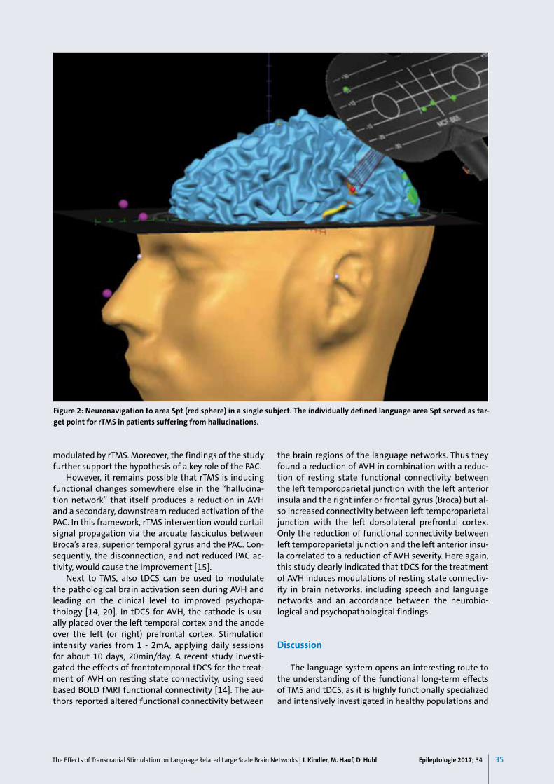
35Epileptologie 2017; 34The Effects of Transcranial Stimulation on Language Related Large Scale Brain Networks | J. Kindler, M. Hauf, D. Hubl
modulated by rTMS. Moreover, the findings of the study further support the hypothesis of a key role of the PAC.
However, it remains possible that rTMS is inducing functional changes somewhere else in the “hallucina-tion network” that itself produces a reduction in AVH and a secondary, downstream reduced activation of the PAC. In this framework, rTMS intervention would curtail signal propagation via the arcuate fasciculus between Broca’s area, superior temporal gyrus and the PAC. Con-sequently, the disconnection, and not reduced PAC ac-tivity, would cause the improvement [15].
Next to TMS, also tDCS can be used to modulate the pathological brain activation seen during AVH and leading on the clinical level to improved psychopa-thology [14, 20]. In tDCS for AVH, the cathode is usu-ally placed over the left temporal cortex and the anode over the left (or right) prefrontal cortex. Stimulation intensity varies from 1 - 2mA, applying daily sessions for about 10 days, 20min/day. A recent study investi-gated the effects of frontotemporal tDCS for the treat-ment of AVH on resting state connectivity, using seed based BOLD fMRI functional connectivity [14]. The au-thors reported altered functional connectivity between
the brain regions of the language networks. Thus they found a reduction of AVH in combination with a reduc-tion of resting state functional connectivity between the left temporoparietal junction with the left anterior insula and the right inferior frontal gyrus (Broca) but al-so increased connectivity between left temporoparietal junction with the left dorsolateral prefrontal cortex. Only the reduction of functional connectivity between left temporoparietal junction and the left anterior insu-la correlated to a reduction of AVH severity. Here again, this study clearly indicated that tDCS for the treatment of AVH induces modulations of resting state connectiv-ity in brain networks, including speech and language networks and an accordance between the neurobio-logical and psychopathological findings
Discussion
The language system opens an interesting route to the understanding of the functional long-term effects of TMS and tDCS, as it is highly functionally specialized and intensively investigated in healthy populations and
Figure 2: Neuronavigation to area Spt (red sphere) in a single subject. The individually defined language area Spt served as tar-get point for rTMS in patients suffering from hallucinations.

36 Epileptologie 2017; 34 The Effects of Transcranial Stimulation on Language Related Large Scale Brain Networks | J. Kindler, M. Hauf, D. Hubl
diseases states. Previous studies demonstrated that brain stimulation with TMS or tDCS does influence en-tire brain networks when used for therapeutic purpos-es, not only in AVH [14, 15] but also in aphasia, neglect and motor recovery after stroke [27]. In AVH, the stud-ies showed that pathologically increased language net-work activity and/or connectivity are reduced, concur-rently with symptomatic improvements. From a brain physiological and methodological point of view, study results strongly suggest a propagation of brain stimu-lation effects, spreading from the directly stimulated “gateway area” (here: area Spt) into the entire network (here: language network), most likely via structural and functional connections. Future studies will have to demonstrate, whether there is an optimal entry port into the network, yielding the best clinical results or whether the stimulation site is less important as ef-fects are further disseminated into the network. Based on the available data, we hypothesize that brain stimu-lation is disrupting pathological brain network activity, thereby forcing the network to form alternative con-nections and finally producing neuroplastic changes. When using brain stimulation for the treatment of AVH, studies indicate that clinical improvements last for up to 3 months. We believe that at the end of this 3-month period the network steadily relapses, more and more employing former pathological brain circuits.
In a case series, we reported a pathological brain network in acoustic hallucinations of epileptic origin that matches the spatial distribution of the psychotic AHV network as reviewed here [12]. This observation strengthens the concept of clinically brain function be-ing linked to specific anatomically defined networks. Pathological brain activity of different etiology and character would develop and propagate – at least in part – within these predefined networks.
Epilepsy has been conceptualized as a network dis-ease, representing a system of nods and hubs involved in seizure generation, propagation, maintenance and
termination [28]. With the emergence of new diagnos-tic tools in the clinical workup of epilepsies as quanti-tative EEG measures, electrical source imaging and si-multaneous EEG/fMRI recordings, large scale epileptic networks are detected with a precision that allows to specifically target hubs by TMS in individual patients.
Recently, a protocol combining EEG with TMS, which aims at assessing both the diagnostic accuracy and the response to antiepileptic treatment in Genetic General-ized Epilepsy (GGE), resulted in an accuracy of 0.84 for the classification of healthy controls versus GGE and 0.76 for the classification of responder versus non-re-sponder to antiepileptic medication [29]. Recent TMS work has shown that hyperexcitability of the motor cortex may predict resistance to pharmacological treat-ment in epileptic patients [30]. Currently, the thera-peutic potential of transcranial magnetic stimulation techniques for the diagnostic issues and treatment of epilepsy is intensively investigated. A Pubmed search in January 2017 using the terms “transcranial stimula-tion” and “epilepsy” yields 645 published studies. How-ever, the evidence for efficacy of rTMS for seizure reduc-tion is still in discussion [31].
Conclusion Summing up, studies suggest that transcranial
stimulation techniques cause clinical improvements by interrupting pathological network activity via small electromagnetical impulses, thereby changing network connectivity and ultimately inducing neuronal plastic-ity in the entire network.
TMS is a technique that allows safely, noninvasively to probe virtually any cortical areas and by these entry gateways, modulate large-scale networks in physiology and pathology. Applied with simultaneous quantified EEG-measures and functional imaging technics TMS is promising as diagnostic tool to precisely detect net-work connectivity by inducing remote effects through-out the brain. Results may allow targeting of invasive diagnostic or treatment options in psychiatry as well as in epilepsy.
The results from rTMS in drug-resistant AHV in psy-chosis represent a translational approach having dem-onstrated a clinically benefit for the individual patient. In epilepsy the potential of rTMS as a non-invasive treatment of pathological epileptic network activity needs to and will be explored further in the future.
Figure 3: Cerebral blood perfusion (CBF) before vs. after 10 sessions of rTMS treatment of auditory verbal hallucination, group effects of 12 subjects, whole brain voxel wise analysis. CBF measured with MR-ASL, paired t-test, p < 0.05 (uncorrec-ted), extended voxel treshold 250. A significant decrease of CBF has been detected in [A] left the auditory cortex (x/y/z=-46/ -22/10), the [B] motor speech area bilaterally (Broca, x/y/z=38/26/8 and x/y/z=-39/22/5), the [C] left temporal cortex (Wernicke x/y/z=-54/-59/7) and [D] the anterior cingulate cortex (x/y/z=2/34/7).

37Epileptologie 2017; 34The Effects of Transcranial Stimulation on Language Related Large Scale Brain Networks | J. Kindler, M. Hauf, D. Hubl
References
1. Park HJ, Friston K. Structural and functional brain networks: from con-
nections to cognition. Science 2013; 342: 1238411
2. Raichle ME, MacLeod AM, Snyder AZ et al. A default mode of brain func-
tion. Proc Natl Acad Sci U S A 2001; 98: 676-682
3. Greicius MD, Krasnow B, Reiss AL, Menon V. Functional connectivity in
the resting brain: a network analysis of the default mode hypothesis.
Proc Natl Acad Sci U S A 2003; 100: 253-258
4. Greicius MD, Menon V. Default-mode activity during a passive sensory
task: uncoupled from deactivation but impacting activation. J Cogn Neu-
rosci 2004; 16: 1484-1492
5. Jann K, Dierks T, Boesch C et al. BOLD correlates of EEG alpha phase-lo-
cking and the fMRI default mode network. Neuroimage 2009; 45: 903-
916
6. Jann K, Kottlow M, Dierks T et al. Topographic electrophysiological signa-
tures of FMRI resting state networks. PLoS One 2010; 5: e12945
7. Kindler J, Jann K, Homan P et al. Static and dynamic characteristics of
cerebral blood flow during the resting state in schizophrenia. Schizophr
Bull 2015; 41: 163-170
8. Friederici AD. The brain basis of language processing: from structure to
function. Physiol Rev 2011; 91: 1357-1392
9. Hickok G, Poeppel D. The cortical organization of speech processing. Nat
Rev Neurosci 2007; 8: 393-402
10. Lohmann G, Hoehl S, Brauer J et al. Setting the frame: the human brain
activates a basic low-frequency network for language processing. Cereb
Cortex 2010; 20: 1286-1292
11. Dierks T, Linden DE, Jandl M et al. Activation of Heschl’s gyrus during au-
ditory hallucinations. Neuron 1999; 22: 615-621
12. Hauf M, Wiest R, Schindler K et al. Common mechanisms of auditory
hallucinations-perfusion studies in epilepsy. Psychiatry Res 2013; 211:
268-270
13. Hubl D, Hauf M, van Swam C et al. Hearing dysphasic voices. Lancet
2007; 370: 538
14. Mondino M, Jardri R, Suaud-Chagny MF et al. Effects of fronto-temporal
transcranial direct current stimulation on auditory verbal hallucinations
and resting-state functional connectivity of the left temporo-parietal
junction in patients with schizophrenia. Schizophr Bull 2016; 42: 318-
326
15. Kindler J, Homan P, Jann K et al. Reduced neuronal activity in language-
related regions after transcranial magnetic stimulation therapy for au-
ditory verbal hallucinations. Biol Psychiatry 2013; 73: 518-524
16. Curcic-Blake B, Ford JM, Hubl D et al. Interaction of language, auditory
and memory brain networks in auditory verbal hallucinations. Prog Neu-
robiol 2017; 148: 1-20
17. Hubl D, Koenig T, Strik W et al. Pathways that make voices: white matter
changes in auditory hallucinations. Arch Gen Psychiatry 2004; 61: 658-
668
18. Ridding MC, Rothwell JC. Is there a future for therapeutic use of transcra-
nial magnetic stimulation? Nat Rev Neurosci 2007; 8: 559-567
19. Hoffman RE, Hawkins KA, Gueorguieva R et al. Transcranial magnetic
stimulation of left temporoparietal cortex and medication-resistant au-
ditory hallucinations. Arch Gen Psychiatry 2003; 60: 49-56
20. Homan P, Kindler J, Federspiel A et al. Muting the voice: a case of ar-
terial spin labeling-monitored transcranial direct current stimulation
treatment of auditory verbal hallucinations. Am J Psychiatry 2011; 168:
853-854
21. Homan P, Kindler J, Hubl D, Dierks T. Auditory verbal hallucinations: ima-
ging, analysis, and intervention. Eur Arch Psychiatry Clin Neurosci 2012;
262(Suppl 2): S91-95
22. Orosz A, Jann K, Wirth M et al. Theta burst TMS increases cerebral blood
flow in the primary motor cortex during motor performance as assessed
by arterial spin labeling (ASL). Neuroimage 2012; 61: 599-605
23. Hubl D, Nyffeler T, Wurtz P et al. Time course of blood oxygenation level-
dependent signal response after theta burst transcranial magnetic sti-
mulation of the frontal eye field. Neuroscience 2008; 151: 921-928
24. Homan P, Kindler J, Hauf M et al. Repeated measurements of cerebral
blood flow in the left superior temporal gyrus reveal tonic hyperactivity
in patients with auditory verbal hallucinations: a possible trait marker.
Front Hum Neurosci 2013; 7: 304
25. Kindler J, Homan P, Flury R et al. Theta burst transcranial magnetic sti-
mulation for the treatment of auditory verbal hallucinations: results of
a randomized controlled study. Psychiatry Res 2013; 209: 114-117
26. Homan P, Kindler J, Hauf M et al. Cerebral blood flow identifies respon-
ders to transcranial magnetic stimulation in auditory verbal hallucina-
tions. Transl Psychiatry 2012; 2: e189
27. Kindler J, Schumacher R, Cazzoli D et al. Theta burst stimulation over the
right Broca’s homologue induces improvement of naming in aphasic pa-
tients. Stroke 2012; 43: 2175-2179
28. Zubler F, Gast H, Abela E et al. Detecting functional hubs of ictogenic
networks. Brain Topogr 2015; 28: 305-317
29. Kimiskidis VK, Tsimpiris A, Ryvlin P et al. TMS combined with EEG in gene-
tic generalized epilepsy: A phase II diagnostic accuracy study. Clin Neuro-
physiol 2016; pii: S 1388-2457(16)30669-1 Epub ahead of print
30. Badawy RA, Vogrin SJ, Lai A, Cook MJ. Does the region of epileptogenicity
influence the pattern of change in cortical excitability? Clin Neurophysi-
ol 2015; 126: 249-256
31. Chen R, Spencer DC, Weston J, Nolan SJ. Transcranial magnetic stimula-
tion for the treatment of epilepsy. Cochrane Database Syst Rev 2016; 8:
CD011025
Address for correspondence:PD Dr. med. Jochen KindlerUniversity Hospital of Child and Adolescent Psychiatry and PsychotherapyUniversity of BernBolligenstrasse 111CH 3000 Bern 60Tel. 0041 31 932 85 [email protected]

38 Epileptologie 2017; 34
Epilepsie-Liga forscht - hilft - informiert
Werden Sie Mitglied der Epilepsie-Liga.Unterstützen Sie Menschen mit Epilepsie. Adhérez en tant que membre de la Ligue Suisse contre l’Epilepsie.Aidez les personnes atteintes d’épilepsie. Sostenete le persone affette da epilessia.Associandovi alla Lega contro l’Epilessia.

gsEpileptologie 2017; 34... | ...
Best
ellg
utsc
hein
Ist es Epilepsie oder nicht? Lassen sich Ursache und Epilepsieherd finden? Ist mit einem weiteren Anfall zu rechnen und wenn ja, wann? Beim Be-antworten dieser Fragen helfen EEG, MRI und – vielleicht – in Zukunft moderne Smartphone-Apps.
Drei erfahrene Spezialisten stellen auf unserer Fachveranstaltung „Epi-lepsie-Diagnostik 2017“ bewährte Methoden und aktuelle Trends vor, im Anschluss bleibt Zeit für Fragen und Diskussion. Mitveranstalter ist die Schweizerische Epilepsie-Klinik der Klinik Lengg, deren medizinischer Direktor Prof. Dr. Dr. med. Thomas Grunwald durch die Veranstaltung führt.
Fachveranstaltung der Schweizerischen Epilepsie-Liga, gratis
Donnerstag, 1. Juni 2017, 17.00 Uhr bis 18.40, anschl. ApéroSchweizerische Epilepsie-Klinik der Klinik Lengg, Gründerhaus, Park-SaalZürich, Bleulerstrasse 60
2 Credits SNG, SGAIM bzw. 16,5 FPH-Points
Vollständiges Programm und Anmeldung: www.epi.ch/fach
Epilepsie-Diagnostik in Zürich
Schweizerische Epilepsie-LigaLigue Suisse contre l’Epilepsie Lega Svizzera contro l’EpilessiaSwiss League Against Epilepsy
Foto
: ww
w.p
hoto
case
.de,
Urh
eber
rech
t: R
awpi
xel
D
F I
Send
en S
ie m
ir bi
tte:
n
n
__
____
_ Fl
yer „
Epile
psie
im A
lter“
n
n
n
____
___
Flye
r „M
ann
und
Epile
psie
“
n
n
n
____
___
Flye
r „W
as is
t Epi
leps
ie“
n
n
n
____
___
Flye
r „U
rsac
hen
von
Epile
psie
n“
n
n
n
____
___
Flye
r „M
erkm
ale
von
Anfä
llen“
n
n
n
____
___
Flye
r „H
äufig
e An
falls
form
en b
ei K
inde
rn“
n
n
n
____
___
Flye
r „M
edik
amen
töse
Beh
andl
ung“
n
n
n
____
___
Flye
r „Er
ste
Hilf
e be
i Epi
leps
ie“
n
n
n
____
___
Flye
r „Fr
au u
nd E
pile
psie
“
n
n
n
____
___
Flye
r „Ki
nder
wun
sch
und
Epile
psie
“
n
n
n
____
___
Flye
r „Re
isen
und
Epi
leps
ie“
n
n
__
____
_ Fl
yer „
Nic
htep
ilept
isch
e An
fälle
“
n
n
__
____
_ Pr
ogra
mm
heft
Ver
anst
altu
ngen
der
Epi
leps
ie-L
iga
n
n
n
____
___
Flye
r „Fü
hrer
sche
in u
nd E
pile
psie
“
n
n
__
____
_ Fl
yer „
Spor
t und
Epi
leps
ie”
n
n
__
____
_ Fl
yer „
Arbe
it un
d Ep
ileps
ie”
n
n
__
____
_ Fa
chze
itsch
rift „
Epile
ptol
ogie
“
n
n
__
____
_ Fl
yer „
Keto
gene
Diä
ten“
n
n
n
____
___
Einz
ahlu
ngss
chei
n(e)
zur U
nter
stüt
zung
der
Epi
leps
ie-L
iga
n
n
n
____
___
Ratg
eber
für L
egat
e
n
n
n
____
___
Ratg
eber
„Epi
leps
ie u
nd V
ersi
cher
unge
n” n
n
__
____
_ Fl
yer „
Vagu
sner
vstim
ulat
ion“
n
n
__
____
_Fl
yer „
Com
plia
nce“
DVD
s un
d üb
rige
Publ
ikat
ione
n si
ehe
ww
w.e
pi.ch
Ich
(wir)
möc
hte(
n):
n
Einz
elm
itglie
d de
r Epi
leps
ie-L
iga
wer
den
und
beza
hle
min
dest
ens
50
Fra
nken
jähr
lich.
n
Kolle
ktiv
mitg
lied
der E
pile
psie
-Lig
a w
erde
n un
d be
zahl
en m
inde
sten
s
100
Fran
ken
jähr
lich.
gt Epileptologie 2017; 34 ... | ...Epileptologie 2010; 27gt
Epileptologie 1 | 2017
Schweizerische Epilepsie-Liga Seefeldstrasse 84 CH 8008 Zürich
Bitte frankieren
Strasse | Nr.
Telefon
Nam
e | Vorname
PLZ | Ort
Absender/in
Ligue Suisse contre l’Epilepsie Schweizerische Epilepsie-LigaLega Svizzera contro l’EpilessiaSwiss League Against Epilepsy
Que ce soit dans les soins aigus ou de longue durée, le personnel soi-gnant devrait disposer des informations essentielles sur l’épilepsie chez la personne âgée. Il a en effet, plus souvent que les médecins, l’occasion d’être témoin de crises non convulsives.
Si les soignants possèdent la formation nécessaire dans ce domaine, les chances de diagnostic précoce d’une épilepsie ou de traitement adéquat d’un état de mal épileptique non convulsif chez la personne âgée aug-mentent.
C’est pourquoi la Ligue contre l’Epilepsie s’engage pour la formation dans ce domaine :Une nouvelle formation continue « L’épilepsie chez la personne âgée » pour personnel infirmier et aides-soignants aura lieu le mardi 21 novem-bre 2017, à Fribourg. Elle est organisée en collaboration avec la Haute école de santé Fribourg.
Veuillez attirer l’attention de la direction des soins de votre établissement hospitalier ou des soignants de votre entourage sur cette offre impor-tante !
Plus d’informations : www.epi.ch/vieillesse
L‘épilepsie au 3e âge
Foto
: ww
w.is
tock
phot
o.co
m/d
e, U
rheb
er: H
ighw
ayst
arz-
Phot
ogra
phy
39Epileptologie 2017; 34
Epilepsie-Liga-Mitteilungen
Forschungsförderungspreis
Förderung der wissenschaftlichen Forschung im Bereich der Epilepsie (vorwiegend Starthilfen) durch die Schweizerische Epilepsie-Liga
Die Epilepsie-Liga unterstützt wissenschaftliche Projekte im Bereich der Epileptologie im Gesamtbetrag von
CHF 25’000.—
pro Jahr. Insbesondere soll die Erforschung von Ur-sachen und Behandlungen der Epilepsie gefördert wer-den.
Stipendien für Aus- oder Weiterbildung oder Aus-landaufenthalte werden nicht ausgerichtet. Hingegen können Reise- und Aufenthaltskosten (ohne Salär) für Kurzaufenthalte (maximal einige Wochen) finanziert werden, sofern sie dem Erlernen von Methoden dienen, welche im Rahmen eines unterstützten Projektes in der Schweiz eingesetzt werden.
Falls der Antragsteller/die Antragstellerin bereits anderswo Anträge für Unterstützung gestellt hat, ist offen zu legen, bei wem und mit welchem Ergebnis. Termin für die Einreichung von Gesuchen: 31. Dezember 2017
Gesuche sind in elektronischer Form einzureichen an [email protected]
Siehe Richtlinien www.epi.ch/forschungsfoerderung
Schweizerische Epilepsie-LigaSeefeldstrasse 848008 ZürichTel. 043 488 67 77 | Fax 043 488 67 [email protected]
Vorschau Epileptologie 2 | 2017Aktuelles aus der Pharmakotherapie der Epilepsie
Initiierung, Implementierung, Abbruch – Die neue Taxonomie der Adhärenz und ihre Bedeutung im Alltag Isabelle Arnet und Kurt E. Hersberger | Basel
Die Bedeutung der galenischen Formulierung bei AntiepileptikaKurt E. Hersberger und Isabelle Arnet | Basel
Placebo – die Apotheke im KopfGeorg Schönbächler | Zürich
BrivaracetamGünter Krämer | Zürich
Zukünftige AntiepileptikaStephan Rüegg | Basel
Promotionspreis
Die Schweizerische Epilepsie-Liga vergibt alle 3 Jah-re einen Preis in Höhe von
CHF 1’000.—
für die beste Dissertation auf dem Gebiet der Epilep-tologie.
Bewerbungen sind aus allen Fachbereichen und Berufsgruppen möglich und erwünscht, sowohl aus Grundlagen- als auch klinischen Fächern. Eine Altersbe-schränkung erfolgt nicht.
Das Preisrichterkollegium setzt sich aus drei Vor-standsmitgliedern der Epilepsie-Liga zusammen, das bei Bedarf zusätzlich externe Gutachter hinzuziehen kann. Es trifft seine Entscheidung in geheimer Wahl.
Falls der Antragsteller/die Antragstellerin bereits anderswo Anträge für Unterstützung gestellt hat, ist offen zu legen, bei wem und mit welchem Ergebnis.
Die Preisverleihung erfolgt jeweils im da-rauf folgenden Jahr anlässlich der Jahrestagung oder Mitgliederversammlung der Epilepsie-Liga. Bewerbungen sind elektronisch oder in fünf Exem-plaren bis zum 31.12.2018
an die Geschäftsstelle der Epilepsie-Liga (Seefeldstras-se 84, 8008 Zürich, [email protected]) einzureichen und müssen beinhalten: die abgeschlossene und beim Dekanat eingereichte Dissertation und die Stellungnahme des Doktorvaters (dabei kann es sich auch um das entsprechende Gut-achten für die Dissertation handeln).
Bitte vormerken:Die nächste Mitgliederversammlung findet am 18. Mai 2017 um 13.15 Uhr, Hôtel de la Rose, rue de Morat 1 in Fribourg statt.
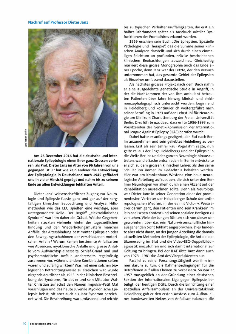
40 Epileptologie 2017; 34
Nachruf auf Professor Dieter Janz
Am 25.Dezember 2016 hat die deutsche und inter-nationale Epileptologie einen ihrer ganz Grossen verlo-ren, als Prof. Dieter Janz im Alter von 96 Jahren von uns gegangen ist. Er hat wie kein anderer die Entwicklung der Epileptologie in Deutschland nach 1945 gefördert und in vieler Hinsicht geprägt und nahm bis zu seinem Ende an allen Entwicklungen lebhaften Anteil.
Dieter Janz‘ wissenschaftlicher Zugang zur Neuro-logie und Epilepsie fusste ganz und gar auf der sorg-fältigen klinischen Beobachtung und Analyse. Hilfs-methoden wie das EEG spielten eine wichtige, aber untergeordnete Rolle. Der Begriff „elektroklinisches Syndrom“ war ihm daher ein Gräuel. Welche Gegeben-heiten steckten vielmehr hinter der tageszeitlichen Bindung und den Wiederholungsmustern mancher Anfälle, der Altersbindung bestimmter Epilepsien oder den Bewegungsschablonen der verschiedenen motori-schen Anfälle? Warum kamen bestimmte Anfallsarten wie Absencen, myoklonische Anfälle und grosse Anfäl-le vom Aufwachtyp einerseits, Schlaf-Grand mal und psychomotorische Anfälle andererseits regelmässig zusammen vor, während andere Kombinationen selten waren und zufällig wirkten? Was mit einer solchen bio-logischen Betrachtungsweise zu erreichen war, wurde nirgends deutlicher als 1953 in der klinischen Beschrei-bung des Syndroms, für das er und sein Mitautor Wal-ter Christian zunächst den Namen Impulsiv-Petit Mal vorschlugen und das heute Juvenile Myoklonische Epi-lepsie heisst, oft aber auch als Janz-Syndrom bezeich-net wird. Die Beschreibung war umfassend und reichte
bis zu typischen Verhaltensauffälligkeiten, die erst ein halbes Jahrhundert später als Ausdruck subtiler Dys-funktionen des Frontalhirns erkannt wurden.
1969 erschien sein Buch „Die Epilepsien. Spezielle Pathologie und Therapie“, das die Summe seiner klini-schen Analysen darstellt und sich durch einen einma-ligen Reichtum an profunden, präzise beschriebenen klinischen Beobachtungen auszeichnet. Gleichzeitig markiert diese grosse Monographie auch das Ende ei-ner Epoche, denn Janz war der Letzte, der den Versuch unternommen hat, das gesamte Gebiet der Epilepsien als Einzelner umfassend darzustellen.
Als nächstes grosses Projekt nach dem Buch nahm er eine ausgedehnte genetische Studie in Angriff, in der die Nachkommen der von ihm ambulant betreu-ten Patienten über Jahre hinweg klinisch und elekt-roenzephalographisch untersucht wurden, beginnend in Heidelberg und kontinuierlich weitergeführt nach seiner Berufung in 1973 auf den Lehrstuhl für Neurolo-gie am Klinikum Charlottenburg der Freien Universität Berlin. Dies führte u.a. dazu, dass er für 1986-1993 zum Vorsitzenden der Genetik-Kommission der Internatio-nal League Against Epilepsy (ILAE) berufen wurde.
Dabei hatte er anfangs gezögert, den Ruf nach Ber-lin anzunehmen und sein geliebtes Heidelberg zu ver-lassen. Erst als sein Lehrer Paul Vogel ihm sagte, nun gelte es, aus der Enge Heidelbergs und der Epilepsie in die Weite Berlins und der ganzen Neurologie hinauszu-treten, war die Sache entschieden. In Berlin entwickelte er sich zu dem grossen klinischen Lehrer, als den seine Schüler ihn immer im Gedächtnis behalten werden. Hier war am Krankenhaus Westend eine neue neuro-logische Abteilung aufzubauen, die sich unter den Ber-liner Neurologien vor allem durch einen Akzent auf der Rehabilitation auszeichnen sollte. Denn als Neurologe war Dieter Janz in seiner Generation einer der promi-nentesten Vertreter der Heidelberger Schule der anth-ropologischen Medizin, in der es mit Victor v. Weizsä-cker darum geht, den Patienten und sein Kranksein im leib-seelischen Kontext und seinen sozialen Bezügen zu verstehen. Viele der Jungen fühlten sich von dieser un-gewohnten, über das rein Naturwissenschaftliche hin-ausgehenden Sicht lebhaft angesprochen. Dies hinder-te aber nicht daran, an der jungen Abteilung die damals aktuellsten Methoden der Epileptologie, die Antiepilep-tikamessung im Blut und die Video-EEG-Doppelbilddi-agnostik einzuführen und sich damit international zur Geltung zu bringen. Bei der ILAE übte Janz dann auch von 1973 - 1981 das Amt des Vizepräsidenten aus.
Parallel zu seiner Forschungstätigkeit war ihm im-mer darum zu tun, die Rahmenbedingungen für die Betroffenen auf allen Ebenen zu verbessern. So war er 1957 massgeblich an der Gründung einer deutschen Sektion der Internationalen Liga gegen Epilepsie be-teiligt, der heutigen DGfE. Durch die Einrichtung einer speziellen Anfallsambulanz an der Universitätsklinik Heidelberg gab er den ersten Anstoss zum Aufbau ei-nes bundesweiten Netzes von Anfallsambulanzen, die
catli
nafil
m B
erlin

41Epileptologie 2017; 34
im Zusammenhang der Epilepsieliga zu einem wesent-lichen Instrument der Verbesserung der Versorgung der Epilepsiepatienten in Deutschland wurden.
Wichtige Unterstützung bei dieser Entwicklung gab die Stiftung Michael, die 1962 von Dr. Fritz Harzendorf, dem Vater eines seiner Patienten, gegründet worden war. Mit Dieter Janz als Mentor entwickelte sie sich bald zu einem Instrument, mit dem immer wieder wei-terführende Akzente gesetzt wurden: Stipendien für eine Spezialisierung in Epilepsie von Ärzten und ande-ren Berufsgruppen, ein Geräteprogramm zur adäqua-ten Ausstattung der Ambulanzen, Starthilfen für die Selbsthilfebewegung, Unterstützung von Fachtagun-gen zur Qualifizierung der Sozialarbeit für Epilepsie. Besonders am Herzen lag ihm die Entwicklung einer Reihe von Aufklärungsschriften über viele Aspekte der Epilepsie. Die Stiftung ergriff auf seine Anregung die Initiativen zur Gründung eines Informationszentrums Epilepsie und zur Einrichtung eines Epilepsiekuratori-ums, das die Lage der Epilepsie in Deutschland kritisch beobachtete und Berichte darüber veröffentlichte. Der von ihm angeregte Michael-Preis für die beste wissen-schaftliche Arbeit konzentrierte sich zunächst auf die deutschsprachige Epileptologie, hat sich aber seither zur wichtigsten internationalen Auszeichnung in unse-rem Fach gemausert.
Die Stiftung entwickelte sich zunehmend zu seinem Lieblingskind mit dem Filetstück der seit 1988 jährlich stattfindenden Praxis-Seminare für Epilepsie in Garg-nano am Gardasee, bei denen er nie fehlte, und die er auch noch in den letzten Jahren durch Abendvorlesun-gen voller Weisheit bereicherte.
Als Emeritus widmete Dieter Janz sich zunehmend der anthropologischen Medizin Victor v. Weizsäckers. Er war massgeblich an der Gründung der Weizsäcker-Ge-
sellschaft beteiligt und gehörte zu ihrem Gründungs-vorstand. Er war Mitherausgeber der Gesammelten Werke Weizsäckers und arbeitete in den letzten Jahren konzentriert an der Edition seines Schriftwechsels.
Im Laufe der Jahre wurde Prof. Janz vielfach geehrt. Hervorzuheben sind der „Ambassador for Epilepsy“ der internationalen Epilepsieorganisationen (1969), der Michaelpreis (1969), der Lifetime Achievement Award der internationalen Epilepsie-Organisationen (1999) und die Otfrid Foerster-Medaille der DGfE (2004). Er war Ehrenmitglied der DGfE, des Stiftungsrats Michael, der Victor v. Weizsäcker-Gesellschaft und des Wissen-schaftlichen Kuratoriums der Evangelischen Studienge-meinschaft sowie korrespondierendes und Ehrenmit-glied mehrerer internationaler Fachgesellschaften.
Sein lebenslanges Engagement für die junge Ge-neration kam vor einem Jahr noch einmal in ganz be-sonderer Weise zum Ausdruck: Als auf Anregung der Jungen Epileptologen in der DGfE ein Nachwuchsför-derungspreis eingerichtet wurde, war er gerne ein-verstanden, dass wir diesem seinen Namen gegeben haben. Der Dieter Janz-Preis zur Förderung des epilep-tologischen Nachwuchses wurde erstmals im Rahmen der Jahrestagung der DGfE in Jena am 4. Februar 2016 verliehen. Allen, die dabei waren, wird unvergesslich bleiben, dass der Namensgeber es sich nicht nehmen liess, eigens hierfür aus Berlin anzureisen und den Preis mit einer bewegenden Laudatio zu übergeben, in der er seiner eigenen Anfänge als Forscher gedachte.
Wir denken an Dieter Janz mit grosser Dankbarkeit und werden seine immer wieder überraschenden und befruchtenden Gedanken sehr vermissen.
Peter Wolf, Kopenhagen und Florianópolis
Zum Tod unseres Ehrenmitglieds Dieter Janz
Dieter Janz war einer der wenigen deutschsprachi-gen Neurologen, der die internationale Epileptologie über Jahrzehnte mit geprägt hat. Sein Menschenbild war durch den anthroposophischen Ansatz von Victor von Weizsäcker geprägt, seine klinische Arbeit beruhte in erster Linie auf Zuhören und akribischer Beobach-tung. Dies begann in seiner Heidelberger Zeit (1946-73) mit der systematischen Differenzierung „der Epilepsi-en“, u.a. in Abhängigkeit vom Schlaf-Wach-Rhythmus. Bekannt wurde er nicht nur wegen des Syndroms der besonders im deutschsprachigen Raum oft nach wie vor nach ihm benannten juvenilen myoklonischen Epi-lepsie (JME), sondern auch wegen vieler anderer Initia-tiven in seiner Berliner Zeit (1973-88) wie der Epilepsie-genetik, der Teratogenität von Antiepileptika oder der Beachtung eines Wirkungsverlusts oraler hormoneller Kontrazeptiva durch Antiepileptika. Zu seiner umfas-senden Würdigung sei auf den Nachruf seines langjäh-
rigen Mitarbeiters Peter Wolf verwiesen, der – in engli-scher Sprache – auch auf der Homepage der Internatio-nalen Liga gegen Epilepsie erschienen ist http://www.ilae.org/Visitors/Farewells/Janz.cfm.
Dieter Janz war ein begnadeter, manchmal etwas „pastoraler“ Redner. Er war Referent auf vielen Kon-gressen und Tagungen, u.a. auch bei meinem eigenen ersten Fortbildungssymposium an der „EPI“ im Früh-jahr 1994. 1998 hatte ich die Freude, gemeinsam mit Pierre Jallon (Marseille) und Peter Wolf (Bielefeld) eine unveränderte Neuauflage seines klassisches Lehrbuch „Die Epilepsien“ von 1969 zu ermöglichen. Obwohl ich formal nie Mitarbeiter von Dieter Janz war, verband mich mit ihm – und nach wie vor mit seiner Frau – eine jahrzehntelange freundschaftliche Beziehung, in den letzten Jahren u.a. mit regelmässigen Treffen anlässlich des von ihm inaugurierten jährlichen Epilepsie-Praxis-seminars in Gargnano am Gardasee.

42 Epileptologie 2017; 34
Weil vielen vielleicht weniger bekannt, sei abschlie-ssend nochmals die langjährige Verbundenheit von Dieter Janz mit der Schweiz anhand des Abdruckes eines Briefes von ihm von 1953 an den seinerzeitigen EPI-Chefarzt (Friedrich Braun) dokumentiert, den ich in einem antiquarisch erworbenen Buch gefunden
habe (er war seinerzeit im Rahmen seiner epileptolo-gischen Ausbildung eine Zeit lang in Zürich gewesen). Die Schweizer Epilepsie-Liga wählte ihn 1992 zu ihrem korrespondierenden und 2005 zu ihrem Ehrenmitglied. Günter Krämer
43Epileptologie 2017; 34
Prix d‘encouragement de la recherche
Promotion de la recherche scientifique dans le do-maine de l‘épilepsie (surtout sous forme d‘aide initiale) par la Ligue Suisse contre l‘Epilepsie (Ligue contre l‘Epi-lepsie)
La Ligue contre l‘Epilepsie soutient les projets scien-tifiques dans le domaine de l‘épileptologie par un mon-tant total de
CHF 25‘000.—
par an, la priorité étant accordée aux projets cher-chant à élucider les causes et à mettre au point des trai-tements de l‘épilepsie.
Aucune bourse ne sera octroyée pour la formation de base ou continue ou pour des séjours à l‘étranger. En revanche, la prise en charge de frais de voyage et de séjour (sans salaire) est possible pour les séjours de courte durée (quelques semaines au maximum) lorsque ces séjours servent à apprendre des méthodes appliquées dans le cadre d‘un projet bénéficiant de soutien en Suisse.
Si le requérant a déjà fait une demande de soutien ailleurs, il faut nous en informer en spécifiant où et avec quel résultat.
Délai de remise des demandes :
31 décembre 2017
Les demandes sont à adresser par voie électronique à [email protected].
Voir instructions : www.epi.ch/soutien_recherche
Ligue Suisse contre l‘EpilepsieSeefeldstrasse 848008 ZurichTél. 043 488 67 77Fax 043 488 67 [email protected]
Prix de la meilleure thèse
La Ligue Suisse contre l‘Epilepsie (Ligue contre l‘Epi-lepsie) décerne tous les 3 ans un prix d‘un montant de
CHF 1‘000.—
pour la meilleure dissertation dans le domaine de l‘épileptologie.
Tous les domaines spécialisés et tous les groupes professionnels couvrant les disciplines fondamentales ou cliniques sont invités à soumettre leur candidature. Aucune limite d‘âge n‘a été fixée.
Le jury décernant le prix se compose de trois membres du comité directeur de la Ligue contre l‘Epi-lepsie. Il peut être complété au besoin par des experts externes. La décision est prise par vote secret.
Si le requérant a déjà fait une demande de soutien ailleurs, il faut nous en informer en spécifiant où et avec quel résultat.
Le prix est toujours décerné l‘année suivante dans le cadre de l‘assemblée annuelle ou générale de la Ligue contre l‘Epilepsie.
Les dossiers de candidature doivent parvenir au Secrétariat général de la Ligue contre l’Epilepsie (Seefeldstrasse 84, 8008 Zurich, [email protected]), sous forme électronique ou en cinq exemplaires, jusqu’au
31 décembre 2018
et comporter les pièces suivantes :
- cinq exemplaires de la dissertation achevée et re-mise au décanat,
- cinq exemplaires d‘une prise de position du direc-teur de thèse (il peut par exemple s‘agir de l‘exper-tise concernant la dissertation).
Informations de la Ligue contre l’Epilepsie
A noter s.v.p. (en langue allemande)La prochaine assemblée générale aura lieu à Fribourg le 18 mai à 13h15, Hôtel de la Rose, rue de Morat 1

44 Epileptologie 2017; 34
Éloge funèbre du Professeur Dieter Janz
Le 25 décembre 2016, l’épileptologie allemande et internationale a perdu l’une de ses personnalités les plus éminentes lorsque le Prof. Dieter Janz nous a quit-tés à l’âge de 96 ans. Plus que tout autre, il a encou-ragé le développement de l’épileptologie en Allemagne après 1945, l’a façonnée à de nombreux égards, et a pris une part active à toutes les avancées jusqu’à la fin.
Son approche scientifique de la neurologie et de l’épi-lepsie se fondait entièrement sur de minutieuses obser-vations et analyses cliniques. Les méthodes auxiliaires telles que l’EEG jouaient un rôle important, mais subor-donné. La notion de « syndrome électro-clinique » lui faisait par conséquent horreur. Pour lui, il était beau-coup plus fondamental de déterminer les circon-stances derrière la survenance à une heure précise du jour, les modèles de récurrence de certaines crises, le lien avec l’âge de certaines épilepsies ou les schémas de mouvements lors des crises motrices. Pourquoi les absences, les crises myocloniques et l’épilepsie du ré-veil, d’une part, et les crises psychomotrices et crises tonico-cloniques généralisées du sommeil, d’autre part, sont-elles régulièrement associées, tandis que d’autres combinaisons sont rares et semblent plutôt dues au hasard ? L’utilité de telles observations biologiques n’a jamais été aussi évidente qu’en 1953, lors de la des-cription du syndrome clinique que Dieter Janz et son coauteur, Walter Christian, ont d’abord baptisé petit mal impulsif et que l’on connaît aujourd’hui sous le nom d’épilepsie myoclonique juvénile, mais aussi de syndrome de Janz. La description était très complète et
incluait même les anomalies du comportement carac-téristiques, qui ont été identifiées un demi-siècle plus tard comme l’expression de dysfonctionnements sub-tils du lobe frontal.
Paru en 1969, son ouvrage « Die Epilepsien. Spe-zielle Pathologie und Therapie » (Les épilepsies. Patho-logie et thérapie spéciales) constitue la somme de ses analyses cliniques et se distingue par ses observations approfondies et précisément décrites, d’une richesse unique. Dans le même temps, cette grande monogra-phie marque la fin d’une ère, car Janz a été le dernier à tenter individuellement de donner une représentation complète de tout le domaine des épilepsies.
Après son livre, son projet suivant a été une vaste étude génétique, dans le cadre de laquelle il a, sur plu-sieurs années, réalisé des examens cliniques et EEG sur les enfants de certains de ses patients en soins ambu-latoires. Ces recherches, commencées à Heidelberg, ont été systématiquement poursuivies après sa nomi-nation à la chaire de neurologie du centre hospitalier Charlottenburg de l’Université libre de Berlin. Cela a entre autres entraîné sa nomination au poste de pré-sident de la commission de génétique de l’International League Against Epilepsy (ILAE) de 1986 à 1993.
Pourtant, il avait au départ hésité à accepter le poste à Berlin et à quitter Heidelberg, qu’il adorait. Ce n’est que lorsque son professeur, Paul Vogel, lui a dit qu’il était temps de laisser derrière lui l’exiguïté de Hei-delberg et de l’épilepsie pour rejoindre le vaste monde de Berlin et de la neurologie tout entière que l’affaire a été entendue. À Berlin, il est devenu le grand profes-seur clinique dont ses étudiants conserveront toujours le souvenir. Là, il s’agissait de mettre sur pied à l’hôpi-tal Westend un nouveau service de neurologie censé se démarquer de ses homologues berlinois par l’accent mis sur la réadaptation. En effet, en tant que neuro-logue, Dieter Janz était l’un des représentants les plus éminents de sa génération de l’école heidelbergeoise d’anthropologie médicale qui se fixe pour objectif, avec Victor von Weizsäcker, de comprendre le patient et sa maladie dans son contexte physico-psychique et ses rapports sociaux. Beaucoup de jeunes se sont sentis très attirés par cette approche inhabituelle, sortant du cadre des sciences naturelles pures. Pour autant, cela n’a pas empêché le nouveau service d’introduire des méthodes dernier cri pour l’époque dans le domaine de l’épileptologie, comme la mesure des antiépileptiques dans le sang et le diagnostic par couplage vidéo et EEG, et d’acquérir ainsi un rayonnement international. Janz a également été vice-président de l’ILAE de 1973 à 1981.
En parallèle de ses activités de recherche, il s’effor-çait toujours d’améliorer à tous les niveaux les condi-tions-cadres pour les personnes atteintes d’épilepsie. C’est ainsi qu’en 1957, il a participé de manière déter-minante à la création d’une section allemande de la Ligue Internationale contre l’Epilepsie, l’actuelle DGfE. En mettant sur pied un service ambulatoire spécial pour les crises au centre hospitalier de Heidelberg, il a
catli
nafil
m B
erlin

45Epileptologie 2017; 34
donné la première impulsion en faveur de la création d’un réseau fédéral de services ambulatoires de crises qui, en lien avec la ligue allemande contre l’épilepsie, a été un instrument essentiel de l’amélioration de la prise en charge des patients atteints d’épilepsie en Alle-magne.
Ce développement a été largement soutenu par la Fondation Michael, créée en 1962 par Dr Fritz Harzen-dorf, le père d’un de ses patients. Sous la houlette de Dieter Janz, elle est rapidement devenue un outil per-mettant de poser sans cesse de nouveaux jalons : bourses pour une spécialisation en épilepsie de méde-cins et autres corps de métiers, gamme d’appareils pour l’équipement adéquat des services ambulatoires, aides de démarrage pour le mouvement d’entraide, soutien de colloques spécialisés pour la qualification du travail social en faveur de l’épilepsie. Il tenait tout spé-cialement à la mise au point d’une série de documents explicatifs traitant de nombreux aspects de l’épilepsie. Sur sa suggestion, la fondation a pris l’initiative de créer un centre d’information sur l’épilepsie et de constituer un comité consultatif, chargé d’observer de manière critique la situation de l’épilepsie en Allemagne et d’en rendre compte. Le prix Michael de la meilleure thèse scientifique créé sur sa proposition s’est d’abord concentré sur l’épileptologie germanophone, mais est entre-temps devenu l’une des récompenses internatio-nales les plus prestigieuses dans notre discipline.
Peu à peu, la fondation est devenue son enfant chéri, avec pour pièce maîtresse le séminaire pratique d’épilepsie à Gargnano, sur le lac de Garde, qui a lieu tous les ans depuis 1988 et qu’il ne manquait jamais. Ces dernières années encore, il l’enrichissait de cours du soir pleins de sagesse.
En tant que professeur émérite, Dieter Janz s’est toujours davantage consacré à l’anthropologie médi-
cale de Viktor von Weizsäcker. Il a joué un rôle clé dans la création de la Société Viktor von Weizsäcker et a fait partie de son comité fondateur. Il a coédité les œuvres complètes de Weizsäcker et a, ces dernières années, tra-vaillé avec application à faire éditer sa correspondance.
Au fil des ans, de nombreux hommages ont été ren-dus au Prof. Janz. Parmi ceux-ci, il convient en particu-lier de noter l’« Ambassador for Epilepsy » des organi-sations internationales contre l’épilepsie (1969), le prix Michael (1969), le Lifetime Achievement Award des organisations internationales contre l’épilepsie (1999) et la médaille Otfrid Foerster de la DGfE (2004). Il était membre d’honneur de la DGfE, du conseil de la fonda-tion Michael, de la Société Viktor von Weizsäcker et du comité scientifique de la communauté d’études pro-testante, ainsi que membre correspondant et membre d’honneur de plusieurs sociétés internationales de dis-cipline médicale.
Il y a un an, son engagement de toute une vie au service de la jeune génération a été une nouvelle fois mis en évidence, d’une façon très particulière : lorsqu’à l’initiative des jeunes épileptologues de la DGfE, un prix d’encouragement de la relève a été créé, il a volontiers accepté qu’il porte son nom. Le prix Dieter Janz d’encou-ragement de la relève en épileptologie a été décerné pour la première fois dans le cadre du congrès annuel de la DGfE à Iena, le 4 février 2016. Ceux qui étaient sur place n’oublieront pas que Janz avait tenu à se dépla-cer de Berlin et à remettre lui-même le prix, son pané-gyrique émouvant revenant sur ses propres débuts de chercheur.
Nous évoquons la mémoire de Dieter Janz avec une profonde gratitude. Ses idées toujours enrichissantes et souvent étonnantes vont nous manquer.
Peter Wolf, Copenhague et Florianópolis
Au sujet du décès de notre membre d’honneur, Dieter Janz
Dieter Janz a été l’un des rares neurologues germa-nophones à marquer l’épileptologie internationale de son empreinte durant des décennies. Sa conception de l’être humain s’appuyait sur l’approche anthroposo-phique de Victor von Weizsäcker, ses travaux cliniques reposaient en premier lieu sur l’écoute et l’observation minutieuse. Cela a commencé lors de sa période hei-delbergeoise (1946-73), avec la différenciation systé-matique « des épilepsies », entre autres en fonction du rythme sommeil/éveil. Il n’est pas seulement connu pour le syndrome de l’épilepsie myoclonique juvénile (EMJ), encore souvent appelé par son nom dans l’espace germanophone, mais aussi en raison de nombreuses autres initiatives datant de sa période berlinoise (1973-88), telle que la génétique de l’épilepsie, la tératogéni-cité des antiépileptiques ou l’observation d’une perte
d’efficacité des contraceptifs hormonaux oraux liée aux antiépileptiques. Parmi les nombreux hommages qui lui ont été rendus, celui de son collaborateur de longue date, Peter Wolf, qui a également été publié en anglais sur le site Internet de la Ligue Internationale contre l’Epilepsie (http://www.ilae.org/Visitors/Farewells/Janz.cfm) mérite l’attention.
Dieter Janz était un brillant orateur, parfois un peu « pastoral ». Il a donné des conférences lors de nom-breux congrès et colloques, y compris lors de mon pre-mier symposium de formation continue chez EPI au printemps 1994. En 1998, j’ai eu la joie de permettre, conjointement à Pierre Jallon (Marseille) et Peter Wolf (Bielefeld), la publication d’une nouvelle édition inchan-gée de son ouvrage d’enseignement classique « Die Epi-lepsien » de 1969. Même si je n’ai jamais formellement

46 Epileptologie 2017; 34
été le collaborateur de Dieter Janz, j’étais lié à lui – et je reste lié à son épouse – par une amitié de longue date et, ces dernières années, par des rencontres annuelles lors du séminaire pratique d’épilepsie à Gargnano, sur le lac de Garde, qu’il avait instauré.
Parce qu’il était sans doute moins connu de la plu-part des gens, je termine en évoquant l’attachement durable de Dieter Janz à la Suisse avec la reproduction d’une lettre qu’il avait écrite en 1953 au médecin-chef
d’EPI de l’époque (Friedrich Braun) et que j’ai trouvée dans un livre acheté auprès d’un antiquaire (dans le cadre de sa formation d’épileptologie, il avait séjourné un certain temps à Zurich). La Ligue Suisse contre l’Epi-lepsie l’avait élu membre correspondant en 1992 et membre d’honneur en 2005.
Günter Krämer
47Epileptologie 2017; 34
Kongresskalender
22.-27.4.2017 | Boston, USA69th Annual Meeting of the American Academy of Neurology (AAN)Information: AAN, 201 Chicago Avenue, Minneapolis, MN 55415, USA, Tel. 001 / 612 / 9286000, www.aan.com/conferences/2017-annual-meeting
27.4.2017 | Bern138. Epilepsie- und EEG-Symposium (SWEZ-Symposium)Information: Prof. K. Schindler,e-mail: [email protected]
27.-29.4.2017 | Leipzig, Deutschland61. Wissenschaftliche Jahrestagung der Deutschen (DGNR e.V.) und der Deutschen (DGNKN e.V)Information: Conventus Congressmanagement & Marketing GmbH, Nadia Al-Hamadi / Nora Caune, Carl-Pulfrich-Str. 1, 07745 Jena, Deutschland Tel. 0049 / 3641 / 31 / 16318, Fax . 0049 / 3641 / 31 / 16243, e-mail: [email protected], www.dgkn-kongress.de
3.-6.5.2017 | Wien, Österreich 10. Dreiländertagung der Österreichischen und Deutschen Gesellschaften für Epileptologie und der Schweizerischen Epilepsie-LigaInformation: Epilepsie-Liga,Seefeldstrasse 84, 8008 Zürich,Tel. 0041 / 43 / 4886777,Fax 0041 / 43 / 4886778,e-mail: [email protected],www.epi.ch
5.-7.5. 2017 | Dakar, Senegal3rd African Epilepsy CongressInformation: ILAE/IBE Congress Secretariat,7 Priory Office Park, Stillorgan Road,Blackrock, Co. Dublin A94 FN26, Ireland,Tel. 00353 / 1 / 2056720,Fax 00353 / 1 / 2056156
13.-16.5.2017 | Leuven, BelgienBrain Plasticity in EpilepsyInformation: www.epilepsy2017.com
18.5.2017 | FribourgJahrestagung der SGKN 2017 Information: www.sgkn.ch
24.-26.5.2017 | Faroer InselnFocal Epilepsy Conference 2017Information: www.mice.fo/focalepilepsyfaroeislands2017/fec2017.html
1.6.2017 | Zürich, Gründerhaus Klinik Lengg, 17 UhrFachveranstaltung der Epilepsie-LigaEpilepsie-Diagnostik 2017Information: Epilepsie-Liga, Seefeldstrasse 84, 8008 Zürich,Tel. 0041 / 43 / 4886777,Fax 0041 / 43 / 4886778,e-mail: [email protected], www.epi.ch
1.6.2017 | Zürich, Gründerhaus Klinik Lengg, 19.30 UhrPublikumsveranstaltung der Epilepsie-LigaEpilepsie-Diagnostik 2017Information: Epilepsie-Liga, Seefeldstrasse 84, 8008 Zürich,Tel. 0041 / 43 / 4886777,Fax 0041 / 43 / 4886778,e-mail: [email protected], www.epi.ch
15.-17.6.2017 | Miami Beach, Florida U.S.A.Intractable Focal EpilepsyState of the Art: Surgical and Medical ManagementInformation: www.epilepsycolloquium.com
24.-27.06.2017 | Amsterdam, Holland3rd Congress of the European Academy of Neurology (EAN)Information: ean Head Office, Breite Gasse 4/7, A 1070 Wien, Österreich, e-mail: [email protected], www.eaneurology.org/amsterdam2017
30.8.-2.9.2017 | Budapest, Ungarn16st European Congress of Clinical Neurophysiology (ECCN 2017)Information: Professor Dr. Anita Kamondi,President of HSCN, National Institute of Clinical Neurosciences, H-1145 Budapest, Amerikai ut 57. e-mail: [email protected],www.eccn2017.com
48 Epileptologie 2017; 34
Impressum
Herausgeber | Administration | SchlussredaktionSchweizerische Epilepsie-LigaMargret Becker, lic. phil. ISeefeldstrasse 84 CH-8008 ZürichTel. 0041 43 477 01 39Fax 0041 43 488 67 [email protected]
Konzeption | Gestaltung | Reinzeichnungscreenblue Büro für Design | Birgit DeppingGazellenkamp 99, D-22529 [email protected], www.screenblue.de
TitelbildAus dem Artikel: Markus Gschwind
Belichtung | Druck Bruns Druckwelt GmbH & Co. KGD-32423 Minden, www.bruns-druckwelt.de
Auflage1.100 Exemplare
VersandEingliederungs- und Dauerwerkstättedes Schweiz. Epilepsie-ZentrumsBleulerstrasse 72, 8008 Zürich
2.-6.9.2017 | Barcelona, Spanien32th International Epilepsy CongressInformation: ILAE/IBE Congress Secretariat, 7 Priory Office Park, Stillorgan Road,Blackrock, Co. Dublin A94 FN26, Ireland,Tel. 00353 / 1 / 2056720,Fax 00353 / 1 / 2056156,e-mail: [email protected]
16.-21.9.2017 | Kyoto, JapanXXIII World Congress of Neurology - WCN 2017Information: Kenes International, WCN 2017, Rue François-Versonnex 7, 1207 Genf, Tel. 0041 / 22 / 9080488, Fax 0041 / 22 / 9069140, e-mail: [email protected], www.2017.wcn-neurology.com
6.10.2017 | Zürich, Karl der Grosse, 19 UhrTag der EpilepsieTheater “Steile Welle”Information: Epilepsie-Liga, Seefeldstrasse 84, 8008 Zürich,Tel. 0041 / 43 / 4886777,Fax 0041 / 43 / 4886778,e-mail: [email protected], www.epi.ch
4.11.2017 | Zürich, Gründerhaus, Klinik Lengg, 10 UhrPatiententagEpilepsie – eine unsichtbare ErkrankungInformation: Epilepsie-Liga, Seefeldstrasse 84, 8008 Zürich,Tel. 0041 / 43 / 4886777,Fax 0041 / 43 / 4886778,e-mail: [email protected], www.epi.ch
23.11.2017 | Basel, Kantonsspital, 14.15 UhrFachveranstaltung der Epilepsie-LigaNeuigkeiten zur EpilepsiebehandlungInformation: : Epilepsie-Liga, Seefeldstrasse 84, 8008 Zürich,Tel. 0041 / 43 / 4886777,Fax 0041 / 43 / 4886778,e-mail: [email protected], www.epi.ch
23.11.2017 | Basel, Kantonsspital, 19 UhrPublikumsveranstaltung der Epilepsie-LigaNeuigkeiten zur EpilepsiebehandlungInformation: Epilepsie-Liga, Seefeldstrasse 84, 8008 Zürich,Tel. 0041 / 43 / 4886777,Fax 0041 / 43 / 4886778,e-mail: [email protected], www.epi.ch