Embed Size (px)
Citation preview
BRIEF COMMUNICATION
Epithelioid hemangioendothelioma of the
liver: A report of two cases
JE MORRIS, MD, DA M ALATJALIAN, MD, A BoDURTHRA, MD, M SMITH, MD, 0 KOLLER, MD, J BHAN, MD, L MAGTIBA Y, MD
JE MORRIS, DA MALATJALIAN, A BODURTHRA, et al. Epithelioid hemangioendothelioma of the liver: A report of two cases. Can J Gastroenterol 1993;7(7):530-534. Epithelioid hemangioendothelioma (EHE) isan uncommon tumour of endothelial origin having borderline malignant potential which frequently is initially misdiagnosed when occurring in the liver, often as cholangiocarcinoma, metastatic carcinoma or veno-occlusive disease. The authors report two cases of hepatic EHE. The fi rst case presented in a 33-year-old female and was originally misdiagnosed as cholangiocarcinoma. Only after re-evaluation of the case seven years later, because of an unexpectedly indolent clinical course, was the correct diagnosis made. The second case presented in a 26-year-old female having nonspecific gastrointestinal complaints. The diagnosis of hepatic EHE was made from a wedge biopsy of liver taken during an exploratory laparotomy. In both cases, immunohistochemical reactivity of the tumour cells for von Willebrand factor (FY1IIR:Ag) and Ulex europeaus I lectin was key to the final diagnosis. The first patient, to this time, remains asymptomatic. The second patient underwent liver transplantation and is being followed (now one year since the initial diagnosis). Hepatic EHE can present a diagnostic dilemma. The use of immunohistochemical techniques to demonstrate the vascular origin of this tumour is essential for its diagnosis. Better awareness of this lesion and the features differentiating it from other histologically similar lesions will prevent exposing affected patients to unnecessary investigations and treatments.
Key Words: Differential diagnosis, Epithelioid hemangioendothelioma, Primary liver tumour, Tumour markers
Hemangio--endotheliome epithelioi:de du foie: deux rapports de cas
RESUME: L'hemangio-endotheliome epithelioi:de est une tumeur rare d'origine endotheliale, dotee d'un potentiel de malignite limite et qui est souvent mal diagnostiquee au debut lorsqu'elle survient dans le foie, souvent prise a tort pour un cholangiocarcinome, w1 carcinome metastatique ou une maladie veno-occlu-
Deparmienrs of Pathology, Surgery and Medicine, Victoria General Hospital and Dalhousie University, Halifax, Nova Scotia; and Regional Labaratories and Department of Medicine, Dr Everett Chalmers Hospital, Fredericton, New Brunswick
Ccmespondence and reprints: Dr John Morris, Department of Pathology, Victoria General Hospital, Halifax, Nova Scotia B3H 2¥9
Received far publication November 20, 1992. Accepr.ed March 25, 1993
EPITHELIOID HEMANGIOENDOTHELIoma (EHE) is an uncommon tu
mour of endothelial cell origin which has been reported to occur as a primary lesion in soft tissues, bone, lungs and liver (1,2). Primary hepatic EHE appears to be rare, based on the number of cases reported in the medical literature. However, because of the diagnostic difficulties it can present - as has been demonstrated by the frequency of initial misdiagnosis - it may be a more common tumour than reported (2-12). The etiology is uncertain. However, predisposing risk factors for other primary hepatic malignancies have been postulated ( 13, 14). The clinical course of hepatic EHE is variable and unpredictable, and consensus regarding treatment has yet to be established (2,3,5, 7). However, for selected patients, liver transplantation is more frequently becoming a treatment option (4,5). The authors report two cases of hepatic EHE and briefly review the available literature.
CASE PRESENTATIONS Case 1: A 33-year-old female with an unremarkable past medical history, presented with an epigastric mass and recent 6 kg weight loss. She was admitted co hospital for investigation. Laboratory data on admission were: hemoglobin, 142 g/L; white blood cell count,
530 CAN] GASTROENTEROL VOL 7 No 7 SEM"EMBER/0crOBER 1993
sive. Les aureurs font etat de deux cas d'hemangio-endotheliomc epithelio'idc du foie: le premier concerne une patience de 33 ans chez qui l'on avait a crronement pose initialement un diagnostic de cho langiocarcmome. Ce n'est qu 'apres unc reevaluation de son cas, sept ans plus card, a cause d'unc evolution clinique inhabiruellemcnt lente, que le diagnostic juste a e te pose. Le deuxiemc cas s'est presence chez une paticnte de 26 ans qui sc pla1gnait de symptomes gastro-intestinaux non specifiques. Le diagnostic d'hcmangio-endo thcliome epithelio'ille hepatique a ere pose a partir d 'une biopsie du fo1e, pre levee lors d'une laparotomie exploratoire. Dans !es dcux cas, la reacrivite immuno-histo-chimique des cellulcs rumorales a l'cgard du facteur de von Willebrand et de la lectine Ulcx Europeaus I a etc la cle du diagnostic final. La premiere patiente, a l'heure actuelle, demeure asymptomatique; la seconde a subi une transplantation hepatique et est suivic (un an s'est ecoulc depuis le diagnost ic initial). L'hemangio-endorhclirnne epithclio'ide pcut representer un dilemme diagnostique. Le rccours a des techniques immuno-histo-chimiques afin de dcmontrer l'origine vasculaire de cette tumeur est essenciel pour son diagnostic. Une meilleure connaissance de cette lesion et de ses caractcrisciques differentielles par rapport a d'autres lesions hisrologiquernent similaires, empechera les patients affectes d'etre soumis a des investigations ou des traitements superflus.
,> ,. .._ , ... \ 1 ~ ' .. _, ··· . · ... ·, ·. ,, ... • • 19::,' ··~, 1' ~ ... ~ ~ • ... ' •
"'II ,.. ._ ' '\ii?~ I • • ,.
• ~ ' t .. I ~I\ .;..,... . . .... - , . ,, .. • "' .. . J' '
JI I It ... , :'" \-~; .... . •! Af ~ • •• ~ • '• :1 I ·, , ;" ~ J ._ I/ ' • • # J \ 'I 1 41 •
• • .., .. • \ •• # ' .,,• • ~ ,.:.,_ -
-~ , r • ~ .A •"' / .. ~ . - ,.. ~ - , • , \., • • , J ' I • J' .\ t ' ,' t • fJ f If" , • 4 J
\
... \I I . ' ,., I ' 'f • • • ., , ~ ' -~· ... . ' .• .. ....,... ti.. , . .., .. ... . .. ' ~ ' .... ,,. . ' -:- "') .... , ... . .,. .. , \ . . . -
· ~ I\' , -:· ' 1
'. • ,'1 ;ii) ~ ; • ~-:.~ ,•/ ,.. -~ . :\ ' \ \ ~l!. ~ "~ \.'.. • ' .. ?t' . ,t ' . ~ ' ..
"'
- ' · ; r • ,.\' ' - '?J\~ ·, - ·. . .. . -,,,. , . ' ' .. ' ,, ~,. .\ • .. t .. . ' .... . . ' ,.
• ·· ' "t. , "' '. \ .. . • '!(" ·~ • • , ., :· • ... p. ' . .,s ., , t• • , \ , - .1... • • - • •
> • I #'~ /, ~ .
~ . Ci
,· .. ~ ... .. ~,
••
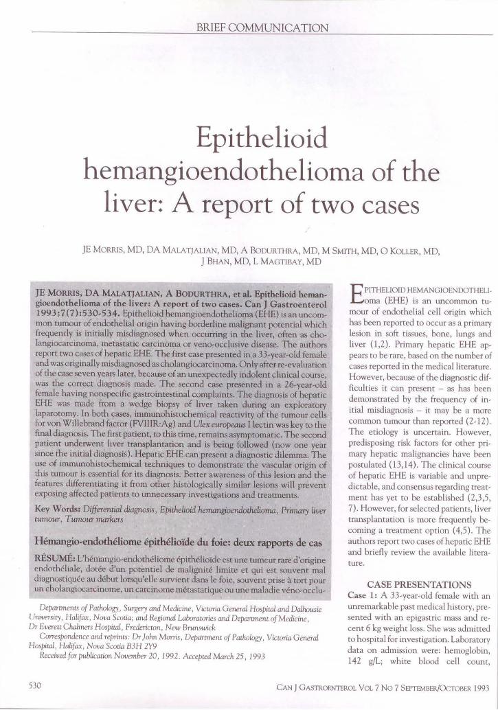
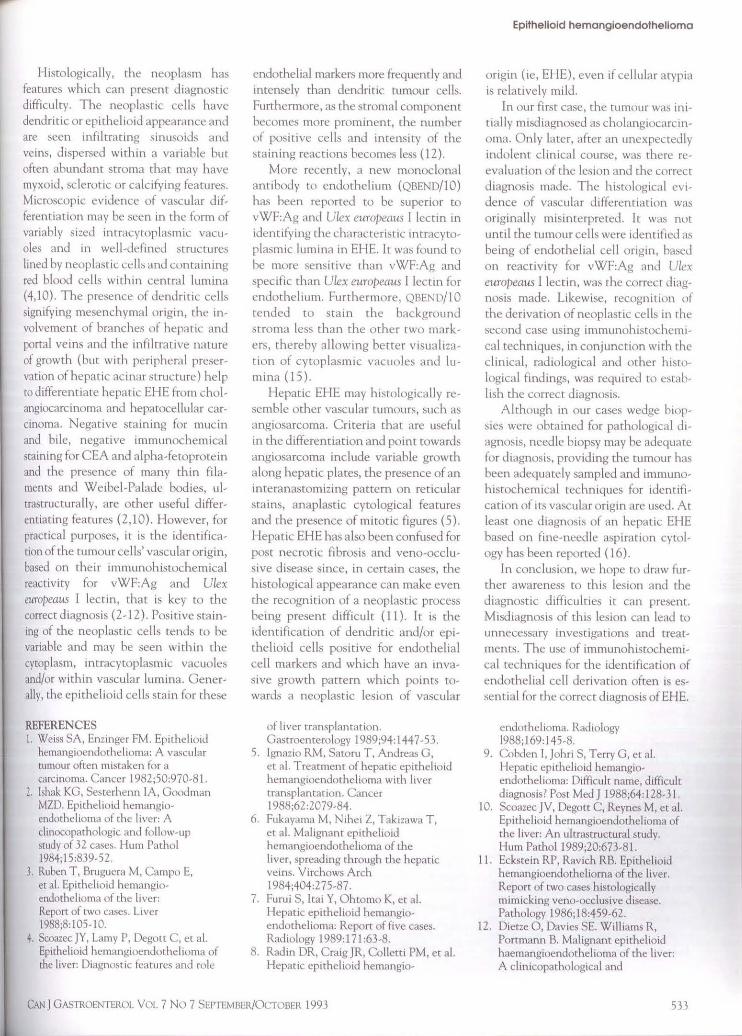
, . ' ' . .... . ... .. .. :., Figure 1) Strands and nem of tumour cells within a dense fibrouc .1troma. Tumour cells with cytoplasmic vacuoles, one wnh a red blood cell (Inset)
7.8xl09/L; erythrocyte sedimentation rate, 22 mm/h; total protein, 77 g/L; albumin, 42 g/L; total bilin,bin, 29 µmol/L, alanine aminotransferase, 76 U/L; a\panatc amino transferase, 14 7 U/L; and alkaline phosphatase, 984 U/L.
An abdominal ultrasound revealed a large, solid, echogenic mass replacing much of the left lobe of the liver. When the patient unJerwent a laparotomy a wedge biopsy of the left lobe ofli ver was taken. The biopsy specimen showeJ two no<lular, solid, grey-white lesions,
the larger measuring 2 cm in the greatest Jimension. H1stologically, the lesions showed prolifera tion of predominantly epithelio id cells which had abundant eosmophilic cytoplasm, large nuclei with coarse chromatin and prominent nucleoli. There were fewer spindle-shaped cells having similar nuclear features. Many of the cells showed large intracytoplasmtc vacuoles of various sizes and, in areas, neoplastic cells were arranged in a single layer forming a structure with a central lumen . ln
CA.NJ GASTROENTEROL Vrn 7 Nn 7 SEPTEMBER/0.'TOl"IER 1993
Epithelioid hemongioendotheliomo
some of these lumma were red blood cells. MitottL figures were infrequent. The neoplawc cells were d ispersed within a rather dense fibrotic strrnn.1. A t the periphery of the tumour fou, tumour cell involvement of adjacent sinusoids was seen wi th compression and mild disruption nf the hepatocyttL plates. Invasion of hepatic veins was prominent. These lesions were l11Scologically diagnosed as pnnrly di fferentiated cho langiocarcinoma (Figure l ). Further workup for metastases was negat ive.
T reatment was m itta ted with doxorubicin, 5-fluorouricil ( 5-FU ) and vincmcine. Multiple courses were given and after four years, repeated d iagnostic imaging showed the rumour had apparently regressed. I lowever, during this t ime, the patient had developed doxorubicin cardiotoxici ty (cardiomyopathy) and vincrist ine neurocoxicity requ iring discontinuation of these drugs. 5-FU was continued as a single agent for a short course afterward. The chemotherapy was stopped and not restarted for two years (\~hen an asymptomattc lung lesion was identified radiogrn phically ). Although a t issue d1agnos1s of this lesion was not obtained, 5-FU was resta rted for a presumed metastas is. The chemotherapy was di:scontinued after two course~ according to the patien t's wishes. further progression of the lung and hep,1tic lesions was not noted and the parient remained free from funher problems.
Because of the unexpectantly long, indo lent cl in ical cour:;e, the originrt l J iagnosis of c.ho lang1ocareinoma made seven years earlier wa~ reviewed. T he neoplaMic cells exh1b1reJ reactivity for the endothelial cell markers von Willebrand factor antigen ( vWF:Ag) and Ulex europeaus I lectin (Figure I ). Posit ive staining was seen within the cytoplasm, incracytoplasmic vacuoles ;ind vascular lumina as granu lar or homogeneous deposits. Approximately 50% of the neoplast ic cells were reactive for each marker. The h istological features were reinterpreted and a revised Jiagnosis of hepatic EHE was made. To this time, now 10 years smce first presenting to her physician, the patient remains asymp1omat1c.
MORRIS et al
. , ., .. " ., . • "ti • • ·r, - :,,,•
\ ... , ... ,.c.v ... ,• .. ,, . ,.,, ,. - . • --: 2 •• ,; • " • i, · .. I • , • '• .. .. "\ i ~ • • 1, l .. .. • • ,;., t. ·+ ..
,. \ .., ti • t " \ ,. t \ • ' I "* e • .. • • .. ,. , , 'l
I - ¥ •. • ; . 6 • - • • t • '8 • J!', I • • I'-
• le • I • ,. (I , ·~ " ' • t !. ' .. • • ~, - •" ~ ...
• f)• f #II • ~ t,- • • I e -. ~ "' ( '\ •. ._ "',. !! ' I ~.. I ,-: ;· 1' , • r, "'- .. ,
._, • ~ ,: '11 II 1' • • , .. ,Ill I
! \, . 1' . I .. ' l· ~ ~ • !-1, •. } .. • • . ' ~ , ~ " : .,, ,, 't. , ·""· , ,., ·.It. .~-. , • • • • r · .. , • f A • ' •'- 'N \ .. •• • ~· , '
• ...-; . • t 11> ·'· • ..,. ..... t .._ •• *). "' d • ,,. ,~ -· ~ • .. • • •
' •• ~ .. . II, . • • .,....~ .. i,4 t if~ '1 j '•1'-'• ,•• I -.~·,.f ,•, I .. "'t,• -. •.,•I Iii t, ~·'r. ""• .. •, • 4191 -,,, .• •t! ·~' . ,~ . " . . , • 9 ' • • • •I• ·•' • - '- • ti' - • • •& •. ~ • I . -"" . • t . .... .. ... . ·~-;, ~ •••• , tit~ :· '_. 'i . ~ ;, . ''. ~,.... ~ 't, ,.,- ;• ii ~,. • ·~,. ·' • 11 •·~ • l-,.,. ~, ~ ' •'
• •' J' •;' ~ , ,. • ~ I • I. -_ t f I • ,. f • • , f ~ 'C . 'I:• " ~ .... ., ~ • ', 't 'It' 'ii • ., : ' ..
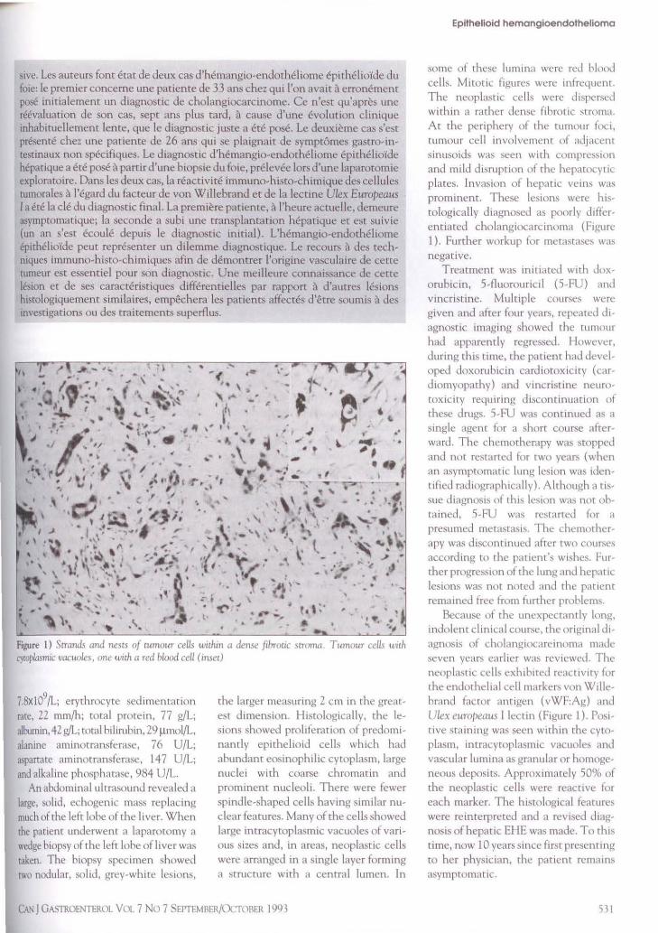
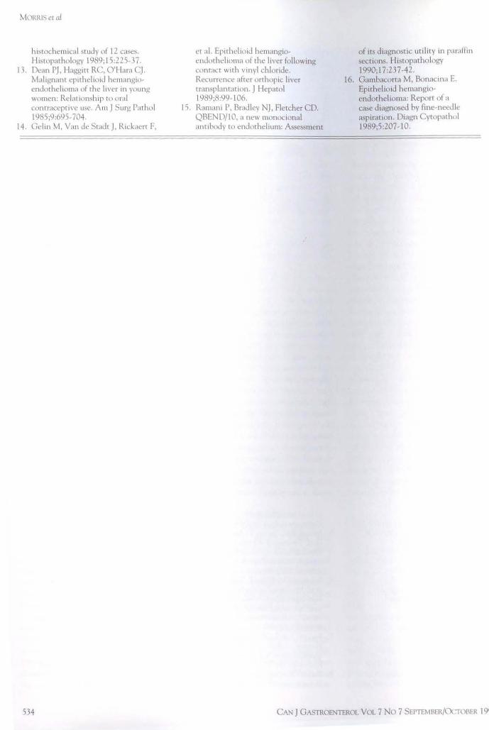
Figure 2) Tumou1· cells dispersed within fibrotic stroma, many with cytoplasmic vacuoles and occasional papillae formation. Individual tumour cells with positit1e staining of cytoplasmic vacuoles for van Willebrand factor ( inset)
Case 2: A 26-year-old female presented with a four-month history of dull, inter· mittent, right upper quadrant abdominal pain, nausea and 15 kg weight loss. She was admitted to hospital for investigation. There had been prior abuse of alcohol, and prescription and illegal drugs. She had been taking oral contraceptives for 10 years. Laboratory data on admission were: hemoglobin, 127 g/L; white blood cell count, 7xl 09{L, albumin, 38 g/dL; alkaline phosphatase, 435 U/L; lactate dehydrogenase, 138 U/L; total bilirubin, 6 µm ol/L; aspartate aminotransferase, 29 U/L; glutamylcransferase, 138 U/L;glucose, 5. l mmol/ L; and amylase, 50 U/L. A lpha-fctoprote in and carcinoembryonic antigen (CEA ) were not elevated. Antinuclear antigen was negative as were hepatitis B surface antigen and anti-hepatitis B core antigen. An abdominal ultrasound showed multiple hypoechoic masses of varying sizes within both lobes of the liver. Further investigations for extrahepatic lesions were negative.
The patient underwent an exploratory laparotomy during which a wedge biopsy of liver was taken. T he biopsy showed several grey-white, solid masses ( the largest measured 2 cm in greatest dimension). Histologically (Figure 2), the lesions showed features similar to
those in case 1 except that most cells were singly dispersed. There was intravascular and intrasinsoidal invasion and/or embolization and a mixed inflammatory infiltrate, including foreign body-type granulomas. Approximately 60% of the neoplastic cells exhibited reactivity of both vWF:Ag and Ulex europeaus I lectin. The diagnosis of hepatic EHE was made.
The patient underwent successful liver transplantation one year ago and currently is being followed. At this time, now two years since first presenting, there is no evidence of extrahepatic dissemination .
DISCUSSION EHE is an uncommon tumour of
endothelial cell oligin. Weiss and Enzinger ( 1) suggested the name after studying the soft t issue counterpart of the lesion, the intravascular bronchiolalveolar tumour originally described in the lung which was later shown co have a vascular histogenesis (1 ). Based on the epithelioid-appearing tumour cells, a pattern of solid growth and a clinical course intermediate between hemangioma and angiosarcoma, they suggested the new name, EHE. Other names have been proposed including sclerosing endothelial tumour, scleros-
ing angiogenic tumour and sclerosing epithelioid angiosarcoma (2) .
Fewer than 100 case of primary hepatic EHEs have been reported in the medical literature (2-14 ). The patients ranged in age from 12 co 86 years ( mean 41.5) with a female:malc ratio of 2:1. The most frequent signs and symptoms at presentation included right upper quadrant pain or discomfort, hepatomegaly, jaundice, fever, anorexia, weight loss and ascites. Laboratory data at presentation were rather nonspecific, but alkaline phosphatase was frequently elevated. Alpha-fecoprotein and CEA levels were normal. Most lesions were multiple and ranged in size from 0.5 to 12 cm. Approximately 40% of patients had, at some point, mediastatic extrahepatic tumour. The most common sites of metastases were lung, abdominal lymph nodes, peritoneum, spleen and medias tinum. Survival was extremely variable, ranging from months to 28 years from initial diagnosis. However, in many patients the rumour showed unusually slow progression, a feature of this lesion.
T o diagnose an hepatic EHE can be difficult. The clinical features are nonspecific and laboratory investigations are most useful in identifying general hepatic injury and in aiding the exclusion of some ocher lesions. Radiographically, the entity can present as nodular or diffuse lesions, the diffuse form probably resulting from coalescence of multiple nodules and representing a more advanced form of disease (7,8) . The radiographic appearance of nodular hepatic EHE is similar to that of some hepatic metastases, making differentiation difficult. However, certain combined radiological findings have been suggested as being useful in the identification of the diffuse form; these are moderately increased tumour vascularity spreading predominantly along the outside edge of both hepatic lobes, hypovascular areas within the tumour, intratumoural calcification , enlargement of the uninvolved hepatic parenchyma with resulting deformity of hepatic contour and absence of some intrahepatic portal branches shown angiographically (7).
532 CAN J GASTROENTEROL VOL 7 No 7 SEPTEMBER/OCTOBER 1993
Histologically, the neoplasm has features which can present <liagnostic difficulty. The neoplastic cells have denJritic or epirhel ioid appearance and are seen infiltrating sinusoi<ls and veins, dispersed within a variable but often abundant stroma that may have myxoid, sclerotic or calcifying features. Microscopic evidence of vascular Jifferentiation may be seen in the form of variably sized intracytoplasmic vacuoles and in well -defined structures lined by neoplastic cells an<l containing red blood cells within central lumina (4,10). The presence of den<lritic cells signifying mesenchymal origin, the involvement of branches of hepatic and portal veins and the infiltrative nature of growth (but with peripheral preservation of hepatic acinar structure) help to differentiate hepatic EHE from cholangiocarcinoma and hepatocellular carcinoma. Negative staining for mucin and bile, negative immunochemical staining for CEA and alpha-fetoprotein and the presence of many thin filaments and Weibel-Palade bodies, ultrastructurally, are other useful differentiating features (2, 10). However, for practical purposes, it is the identification of the tumour cells' vascular origin, based on their immunohistochemical reacttv1ty for vWF:Ag and Ulex europeaus I leccin, that is key to the correct diagnosis (2-12). Positive staining of the neoplastic cells tends to be variable and may be seen within the cytoplasm, intracytoplasmic vacuoles and/or within vascular lumina. Generally, the epithelioic.l cells stain for these
REFERENCES l. Weiss SA, Enzmger FM. Epithelioid
hemangioendothclioma: A vascular tumour often mistaken for a carcinoma. Cancer l 982;50:970-81.
2. Ishak KG, Sesterhenn IA, Goodman MZO. Epithelioid hemangioendothelioma of the liver: A clinocopathologic and follow-up study of 32 cases. Hum Pathol 1984;15:839-52.
3. Ruben T, Bruguera M, Campo E, et al. Epithelioid hemangioendothelioma of the liver: Report of two cases. Liver 1988;8: I 05-10.
4. Scoazec JY, Lamy P, Degou C, et al. Epithelioid hemangioendothelioma of the liver: Diagnostic features and role
endothelial markers more frequently an<l intensely than dendritic tumour cells. Furthermore, as the stromal component becomes more prominent, the number of positive cells and intensity of the staining reactions becomes less (12).
More recently, a new monoclonal antibody to endothelium (QBEND/10) has been reporce<l to be superior to vWF:Ag and Ulex europeaus I lectin in identifying the characteristic intracytoplasmic lumina in El IE. It was founJ to be more sensitive than vWF:Ag and specific than Ulex europeaus l lectin for endothelium. Furthermore, QBEND/10 tended to stain the background stroma less than the ocher two mark
ers, thereby a llowing better visualization of cytoplasmic vacuoles and lumina (15).
Hepatic EHE may histologically resemble other vascular tumours, such as angiosarcoma. Criteria that are useful in the d ifferentiation and point towards angiosarcoma include variable growth along hepatic plates, the presence of an interanastomizing pattern on reticular stains, anaplastic cytological features and the presence of mitotic figures (5). Hepatic EHE has also been confused for post necrotic fibrosis and veno-occlusive disease since, in certain cases, the histological appearance can make even the recognition of a neoplastic process being present difficult (11 ). It is the iJentification of dendritic anJ/or epithelioi<l cells positive for endothelial cell markers and which have an invasive growth pattern which points towards a neoplastic lesion of vascular
of liver transplantation. Gastrocn temlogy 1989 ;94: 144 7 -53.
5. Ignazio RM, Satoru T, Andreas G, ct al. Treatment of hepatic epithclioid hemangioendothelioma with liver transplantation. Cancer I 988;62:2079-84.
6. Fukayama M, Nihei Z, Takizawa T, et al. Malignant epithelioid hemangioendothelioma of the liver, spreading through the hepatic veins. Virchows Arch l 984;404:275-87.
7. Furui S, Ital Y, Ohtomo K, et al. Hepatic epithelioid hemangioendothelioma: Report of five cases. Radiology 1989:171:63-8.
8. Radin DR, Craig JR, Colletti PM, et al. Hepatic epithelioid hemangio-
CAN J GASTROENTEROI VOL 7 No 7 SEf'TEMBER/0CTOBER 1993
Epitheliold hemangloendothelloma
origin (ie, EHE), even if cel lular atypia is relatively mild.
ln our first case, the rumour was initially mis<liagnosed as cholangiocarcinoma. Only later, after an unexpecccJly in<lolent clinical course, was there reevaluation of the lesion and the correct Jiagnosis made. The histological cvi<lence of vascular c.liffcrentiation was originally misinterprete<l. It was not until the tumour cells were idenrificc.l as being of endothelial cell origin, based on reactivity for vWF:Ag and Ulex euro/)eaus I lectin, was the correct diagnosis made. Likewbe, recognition of the derivation of neoplastic cells in the second case using immunohiscochemical techniques, in conjunction with the clinical, radiological anJ other histological findings, was required to establish the correct diagnosis.
Although in our cases wedge biopsies were obtained for pathological diagnosis, needle biopsy may be a<lequate for diagnosis, providing the tumour has been a<lequatcly sampled an<l immunohistochemical techniques for iJentification of its vascular origin are used. At least one diagnosis of an hepatic EHE based on fine-needle aspiration cytology has been reported (16).
In conclusion, we hope to Jraw further awareness to this les ion anJ the Jiagnostic difficulties it can present. Misdiagnosis of this lesion can leac.l to unnecessary investigations and treatments. The use of immunohistochemical techniques for the i<lentification of endothelial cell Jerivation often is essential for the correct diagnosis of EHE.
endothelioma. Radiology 1988; L69:l 45-8.
9. Cobden I, Johri S, Terry G, et al. Hepatic epithelioid hemangioendothelioma: Difficult name, difficult diagnosis? Post Med J 1988;64: 128-3 l.
10. Scoazec JV, Degott C, Reynes M, et al. Epithelioid hemangioendothelioma of the liver: An ultrastructural srudy. Hum Pathol l 989;20:673-81.
LL . Eckstein RP, Ravich RB. Epithclioid hemangioendothelioma of the liver. Report of two cases hiscologically mimicking veno-occlusive disease. Pathology 1986; 18:459-62.
12. Dietze 0, Davies SE. Williams R, Portmann B. Malignant epithelioid haemangioendothclioma of the liver: A clinicopathological and
533
MORRIS l't al
histochemical stuJy of I Z cases. l l1sropachology 1989; I 5:22 5 37.
13 Dean PJ , H.1gg1tt RC, O'Hara CJ. M,1lignant ep1thelimll hcmang1<>emlothclioma of the li ver in young w,11m:n: Rclat 1onsh1p w oral contracepr1w use. Am J Surg Pathol l 985;9:695-704
14. Gclm M, Van Jc Stadt J, Rickaert F,
534
er ,11. l:.p1thcl1mJ hcmangioenJothcl1om.1 of the liver following contact with vinyl chloride. Rci:urrenc,· after orthop1c liver transplantation. J Hepatol 1989;8:99- l 06.
15 Raman1 P, Bradley NJ, Fletcher CD. QBEND/10, a new monnc1onal an11h,,Jy to enJothcl1um: Assessment
of m d1agnost1c utility m paraffm section,. Hi>topathology l 990;17:237-42.
16. Gambacorta M, Bonacina E. Ep1theltoid hcmangto· cndorhclioma: Report of a case diagnosed by fine-needle aspiration. Diagn Cyropathol l 989;5:207-1 o.
CAN J GA:-'TROENTEROL VOi. 7 No 7 SEPTEMHER/O<'TOllER 1993

Submit your manuscripts athttp://www.hindawi.com
Stem CellsInternational
Hindawi Publishing Corporationhttp://www.hindawi.com Volume 2014
Hindawi Publishing Corporationhttp://www.hindawi.com Volume 2014
MEDIATORSINFLAMMATION
of
Hindawi Publishing Corporationhttp://www.hindawi.com Volume 2014
Behavioural Neurology
EndocrinologyInternational Journal of
Hindawi Publishing Corporationhttp://www.hindawi.com Volume 2014
Hindawi Publishing Corporationhttp://www.hindawi.com Volume 2014
Disease Markers
Hindawi Publishing Corporationhttp://www.hindawi.com Volume 2014
BioMed Research International
OncologyJournal of
Hindawi Publishing Corporationhttp://www.hindawi.com Volume 2014
Hindawi Publishing Corporationhttp://www.hindawi.com Volume 2014
Oxidative Medicine and Cellular Longevity
Hindawi Publishing Corporationhttp://www.hindawi.com Volume 2014
PPAR Research
The Scientific World JournalHindawi Publishing Corporation http://www.hindawi.com Volume 2014
Immunology ResearchHindawi Publishing Corporationhttp://www.hindawi.com Volume 2014
Journal of
ObesityJournal of
Hindawi Publishing Corporationhttp://www.hindawi.com Volume 2014
Hindawi Publishing Corporationhttp://www.hindawi.com Volume 2014
Computational and Mathematical Methods in Medicine
OphthalmologyJournal of
Hindawi Publishing Corporationhttp://www.hindawi.com Volume 2014
Diabetes ResearchJournal of
Hindawi Publishing Corporationhttp://www.hindawi.com Volume 2014
Hindawi Publishing Corporationhttp://www.hindawi.com Volume 2014
Research and TreatmentAIDS
Hindawi Publishing Corporationhttp://www.hindawi.com Volume 2014
Gastroenterology Research and Practice
Hindawi Publishing Corporationhttp://www.hindawi.com Volume 2014
Parkinson’s Disease
Evidence-Based Complementary and Alternative Medicine
Volume 2014Hindawi Publishing Corporationhttp://www.hindawi.com