Embed Size (px)
Citation preview

Epitope Analysis of HLA-DQ Antigens: What Does theAntibody See?
Anat R. Tambur,1,5 Jimmy Rosati,1 Shirley Roitberg,1 Denis Glotz,2 John J. Friedewald,3
and Joseph R. Leventhal4
Background. Human leukocyte antigen (HLA)-DQ has emerged as the alloantibody most frequently associated withthe generation of de novo donor-specific antibody (DSA), antibody-mediated-rejection, and unfavorable transplan-tation outcome.Methods. The generation of HLA-DQ de novo DSA was interrogated in 40 transplant recipients who were immuno-logically naive before their failed transplantation. Eplet and epitope analyses were performed using HLAMatchmaker andCn3D software.Results. Ten DQA and thirteen DQB eplets or eplet combinations were identified. All but one revealed an epitopefootprint that includes both the DQ> and DQA chains. Four examples are illustrated in detail, representing a range ofdifferent epitope landscapes. A disparity between antigen density and mean fluorescence intensity values for somealleles within an eplet group was noted, with mean fluorescence intensity values of the lowest fluorescence bead beingone tenth of the highest fluorescence bead, despite the fact that the amount of antigen on these beads were not sig-nificantly different.Conclusion. Our data support the need for changing the manner in which HLA-DQ antigens and antibodies are eval-uated for organ transplantation. The current nomenclature system does not reflect the true nature of HLA-DQ poly-morphism. Moreover, epitope immunogenicity likely involves more than the mere presence of a specific eplet. Becauseour field contemplates the use of epitope matching as an approach to improve organ allocation and overall outcomes, itis imperative to have accurate characterization of the immunogenicity of each epitope. This will pave the way to iden-tifying acceptable mismatches and will allow risk stratification for generating de novo HLA-DSA after transplantation.
Keywords: HLA-DQ, Epitope, HLA-antibody, de novo DSA.
(Transplantation 2014;98: 157Y166)
The interactions between human leukocyte antigens (HLA)and HLA-antibodies are complex. Contrary to the previ-
ously accepted concept, it is now clear that HLA antibodies rec-ognize critical, polymorphic, short fragments of the HLA antigenrather than the complete antigen as one unit. These polymorphicsubunits of the antigen can be unique to one HLA-antigen or beshared by several antigens that have different serologic speci-ficities. Initial evidence for this phenomenon was observed inthe early days of histocompatibility testing and was referred toas cross-reactive group (CREG) responses (1Y4).
Several breakthroughs in understanding the HLA sys-tem allowed for better conceptualization of this phenomenon.
In the early 1990s, the ability to type for HLA-antigensby molecular techniques was acquired, demonstrating thatHLA-antigens are in fact ‘‘families’’ of many alleles, withminor sequence differences, which belong to a particularantigen group as recognized by serologic typing. Aroundthe same time, advances in crystallography techniques allowedto decipher the three-dimensional structures of HLA class Iand class II molecules (5Y7). It was then evident that manypolymorphic amino acid residues concentrate in particularlocations within the HLA molecule where they are exposedto antibody or T-cell receptor binding. Finally, in the early2000s, testing for HLA antibodies using solid phase singleantigen techniques improved the specificity and sensitivityof antibody identification, cumulatively providing us with
CLINICAL AND TRANSLATIONAL RESEARCH
Transplantation & Volume 98, Number 2, July 27, 2014 www.transplantjournal.com 157
The authors declare no funding or conflicts of interest.1 Transplant Immunology Laboratory, Comprehensive Transplant Center,
Feinberg School of Medicine, Northwestern University, Chicago, IL.2 Nephrology and Transplantation Service, Hopital Saint-Louis, Paris, France.3 Department of Internal Medicine, Comprehensive Transplant Center,
Feinberg School of Medicine, Northwestern University, Chicago, IL.4 Department of Surgery, Comprehensive Transplant Center, Feinberg
School of Medicine, Northwestern University, Chicago, IL.5 Address correspondence to: Anat R. Tambur, D.M.D., Ph.D., Transplant
Immunology Laboratory Comprehensive Transplant Center 303 E ChicagoAve, Tarry Bldg suite 11-711, Chicago IL 60611.
E-mail: [email protected]. participated in the research design, performance of the research, data
analysis, and writing of the article. J.R. and S.R. participated in the
performance of the research and data analysis. D.G. participated in dataanalysis and writing of the article. J.J.F. and J.R.L. participated in thewriting of the article.
Supplemental digital content (SDC) is available for this article. Direct URLcitations appear in the printed text, and links to the digital files areprovided in the HTML text of this article on the journal’s Web site(www.transplantjournal.com).
Received 8 October 2013. Revision requested 28 October 2013.Accepted 20 December 2013.Copyright * 2014 by Lippincott Williams & WilkinsISSN: 0041-1337/14/9802-157DOI: 10.1097/TP.0000000000000220
Copyright © 2014 Lippincott Williams & Wilkins. Unauthorized reproduction of this article is prohibited.

new tools to appreciate the complexity and ambiguities ofHLA antibody-antigen interactions. With the use of thesetools, new evidence was brought forth, documenting thatpatients who were exposed to particular HLA mismatcheshad developed antibodies to a much wider range of anti-gens (alleles) compared with the original mismatched an-tigen expressed by their donors (8Y11).
Two approaches were implemented in an attemptto identify the portion of the HLA molecule that is recognizedby the antibodyVthe epitope. Duquesnoy et al. (12Y16) in theearly 2000 developed a theoretical algorithm, HLAMatchmaker,in which each polymorphic subunit of the HLA molecule wasconsidered its own entity and termed originally triplets andlater eplets. A more hands-on approach, using actual absorp-tion or elution studies performed by the group of Terasakiet al. (17Y19) was proven comparable with the theoretical al-gorithm. Importantly, both investigators refer to sequences of1 to 3 amino acids as the critical difference between the anti-body producer and the immunizer. Yet, the antibody recog-nizes a greater area with a footprint of approximately 15 Aradius (20), which constitutes the actual epitope. Conse-quently, analyzing antibodies against HLA class I antigens,Duquesnoy et al. reported that epitopes defined by eplet pairsalways involved a nonYself-eplet and a self-eplet that is sharedbetween the immunizing antigen and the antibody producer(21Y23). In fact, the data suggest an autoreactive componentto the alloantibody response, which is in line with positive andnegative selection processes taking place during B-cell devel-opment and receptor editing (24Y26). The key concept of thistheory is the need for a significant similarity between theimmunizing HLA-antigen and the self-HLA-antigens expressedby the antibody producer, in the presence of a minor differ-ence between the two.
We have explored the nature of structural epitopes forHLA-DQ molecules. To this end, we have studied de novoHLA-DQ antibodies generated in naive patients followingfailed kidney transplantation. Using high-resolution typingof both donor and recipient and results from single anti-gen class II Luminex assays, we were able to define the rel-evant eplets that caused the de novo antibody formation,to analyze the epitope footprint, to investigate the role ofthe nonself or self model, and to speculate about the useof this approach in determining donor compatibility fororgan transplantation.
RESULTSForty recipients with de novo donor-specific HLA-DQ
antibody (DSA) were analyzed as described in the Materialsand Methods section. The complete list of epitopes identifiedin this study is presented in Table 1. These include 10 DQAeplets or eplet combinations and 13 DQB eplets (or com-binations). Examples of four representative cases are givenin detail in Figures 1 to 3. The first step of identifyingthe eplets is shown in Figure 1, where the information ofthe DQ-coated Luminex beads, the mean fluorescence in-tensity (MFI) values, the number of mismatched eplets, andtheir identity for DQ> or DQA chains are provided. Theeplet shared by all positive beads and the immunizer arehighlighted by bold and italicized letters. The Cn3D soft-ware was used to highlight the exact location of the eplet
within an HLA-DQ molecule (Fig. 2 left). With the useof the ‘‘select by distance’’ command to identify a radius of15 A from the eplet, the complete epitope (antibody foot-print) was highlighted in yellow as shown in Figure 2 center.This region, which is recognized by the antibody paratope,was then ‘‘detached’’ and reverted to the original colorscheme to reveal the specific contribution of the DQ> andthe DQA chains (Fig. 2 right). Polymorphic amino acidscomposing the epitope were then highlighted and catego-rized as being accessible to antibody or TCR recognition(located in areas on the surface of the DQ molecule) orburied below the surface. This information is provided inFigure 3, together with the MFI values of the positive beadsand along with the relative density of the antigen on each ofthese beads. The location of the polymorphic amino acidsfor each of the epitopes are illustrated in supplemental data(Figure 1, SDC, http://links.lww.com/TP/B15).
DQB77DR EpletFigure 1(A) presents the HLA-DQ antibody signature
of a previously nonsensitized patient, demonstrating theformation of de novo HLA antibodies to seven DQ alleles(beads). The number of mismatched eplets (mm Ep) rangedbetween 3 and 8. All seven DQ alleles share one eplet,DQB77DR, that likely explains the positive reactivity seenagainst nonimmunizer HLA-DQ antigens. Other eplets areshared by some, but not all, positive beads and thereforeare not likely to explain the antibody reactivity developedafter exposure to the immunizer. Amino acid DQB77 is lo-cated approximately midmolecule, on the surface of theDQA chain, close to the peptide-binding site (Fig. 2A left).Highlighting the antibody footprintVradius of 15 A fromamino acid DQB77 (Fig. 2A center) and then detaching thisarea and reverting to the original color code reveal that theepitope contains portions of both DQ> and DQA chains aswell as amino acids of peptide origin. To better understandthe epitope-paratope interactions, we identified the poly-morphic amino acids that are located on the surface on themolecule and therefore likely to come in contact with theantibody or the TCR (as defined by Cn3D), highlighted inbold and italicized fonts (Fig. 3A). The HLA-DQ specific-ities of the immunizer, the antibody producer, and positivebeads are listed with their associated MFI value. The relativedensity of HLA-DQ molecules coating the beads is alsoprovided, as the fluorescence emitted is dependent in parton the maximum amount of antigens attached to the bead.Interestingly, despite the fact that all positive beads share theDQB77DR epitope, the MFI values vary significantly, withthe strongest bead showing an MFI value of 19,823 and thelowest positive bead having the value of 3,114. Yet, thedensity of DQ antigens on the lowest MFI value bead is 74%of that of the highest MFI value bead.
To test the nonself or self theory in this example, wecompared the identity of the polymorphic amino acidsamong the antibody producer, the immunizer, and the DQalleles coating the positive beads. Note that in this example,both the DQ> and DQA chains contribute to the poly-morphic or critical amino acid residues of the epitope.Indeed, for the most part, at least one of the immunizeralleles shares the same amino acids in the critical positions
158 www.transplantjournal.com Transplantation & Volume 98, Number 2, July 27, 2014
Copyright © 2014 Lippincott Williams & Wilkins. Unauthorized reproduction of this article is prohibited.
with the antibody producer; however, in position DQB75,both immunizer alleles have Valine, whereas the antigenproducer has Leucine. All DQ alleles coating the positivebeads also have Valine in this position. This observationraises the question regarding the relative significance of eachpolymorphic residue within the epitope; do they all con-tribute in the same way? Similarly, is there an absolute needfor complete identity between the immunizer and antibodyproducer in critical positions?
DQB55-56PP EpletThis patient shows positive responses to 12 different
DQ-coated beads. The number of mismatched eplets rangesbetween 2 and 3. All positive beads express the 55 PPP and56 PPD eplets (Fig. 1B). The eplet is located at the peripheryof the DQA chain and, given a radius of 15 A, includes aminoacids from both DQ> and DQA chains and the peptide(Fig. 2B). Similar to the previous case, a significant spread ofMFI values for all positive beads is observed (range, 1,207Y16,661). The allele that shows the lowest fluorescence hasantigen density of 48% of the highest fluorescence allele(Fig. 3B). Remarkably, despite the fact that the antibodyfootprint covers both DQ> and DQA chains, ‘‘critical’’ resi-dues are located only on the DQA chain. In this example,consistent with the nonself or self theory, all polymorphicresidues other than the core eplet are shared between theimmunizer and the antibody producer.
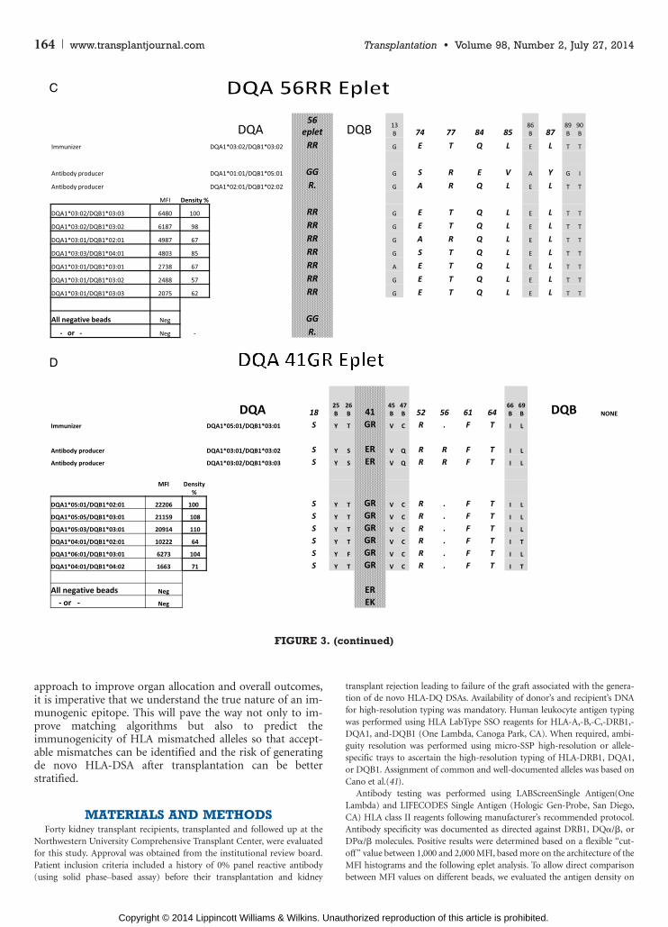
DQA56RR EpletThe third example is of an eplet located on the > chain
of the DQ molecule. Seven positive beads show fairly lowrange of MFI values with one to two eplet mismatches be-tween the antibody producer and immunizer and positivebeads (Fig. 1C). The DQA56RR eplet is located in the pe-riphery of the DQ> chain, close to the peptide-bindinggroove. Highlighting the 15-A radius area marking the epi-tope indicates that, similar to the other examples, all threestructures, DQ> and DQA chains as well as the peptide,
contribute to the antibody footprint (Fig. 2C). Interestingly,however, it is only the eplet itself DQA56RR that is locatedin the DQ> chain where the rest of the epitope footprint re-sides mostly within the DQA chain. Of the five polymorphicresidues, only three are identical between the antibody pro-ducer and immunizer, thus not fitting the nonself or selftheory or at least questioning the role and function of allamino acids within the epitope.
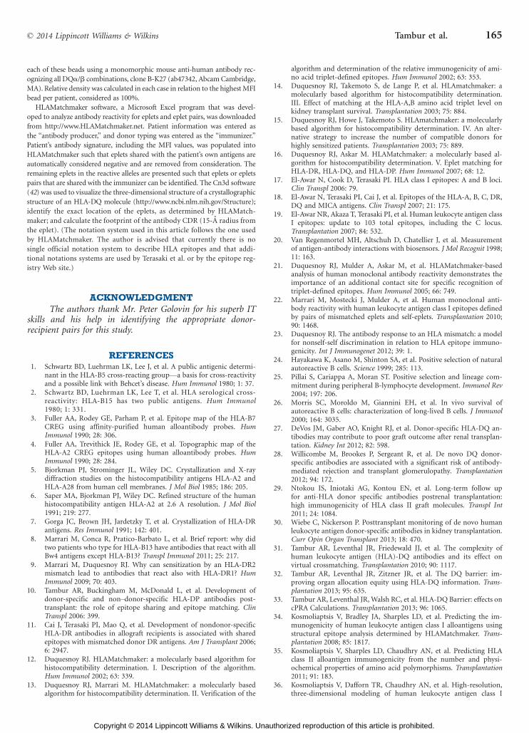
DQA41GR EpletThis is another example of an eplet located on the
DQ> chain showing six positive beads with two to fivemismatched eplets (Fig. 1D). The shared eplet, DQA41GR,is located furthest from the peptide-binding groove andis the only case in this study in which the 15-A radius areamarking the epitope showed no involvement of the DQA
chain (Fig. 2D). The density of DQ molecules on the lowestMFI bead is 71% of that of the highest MFI bead, withfluorescence range from 1,663 to 22,206 (Fig. 3). Of the fivepolymorphic amino acid residues within the epitope, fourare shared consistent with the nonself or self theory.
DISCUSSIONHLA-DQ antibodies have emerged as the antibodies
that are most frequently associated with the generation of denovo DSA, antibody-mediated rejection, and unfavorabletransplantation outcome (27Y30). Better understanding ofthe antigen-antibody interactions is required to minimizethe risk of developing de novo HLA-DQ antibodies. In thisstudy we analyzed DQ antibodies by interrogating de novoHLA-DQ DSA generated in immunologically naive trans-plant recipients, assessing their eplet and epitope specific-ities. Four examples of such analyses (AYD), representing arange of different epitope landscapes, are presented as fol-lows: (A) The eplet is located on the DQA chain, yet the an-tibody footprint covers both DQ> and DQA chains. (B) Theeplet is located on the DQA chain, and the antibody footprintcovers both DQ> and DQA chains; however, the ‘‘critical’’
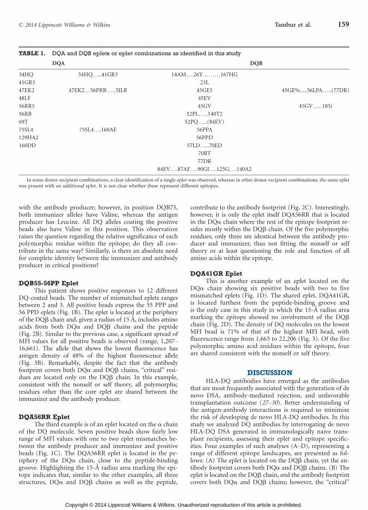
TABLE 1. DQA and DQB eplets or eplet combinations as identified in this study
DQA DQB
34HQ 34HQI..41GR3 14AMI.26YIII167HG
41GR3 23L
47EK2 47EK2I56PRBI..5ILR 45GE5 45GE%.....56LPAI..(77DR)
48LF 45EV
56RR5 45GV 45GVI. .185i
56RB 52PLI..140T2
69T 52PQI..(84EV)
75SL4 75SL4I.160AE 56PPA
129HA2 56PPD
160DD 57LDI..70ED
70RT
77DR
84EVI.87AFI.90GII.125GI.140A2
In some donor-recipient combinations, a clear identification of a single eplet was observed, whereas in other donor-recipient combinations, the same epletwas present with an additional eplet. It is not clear whether these represent different epitopes.
* 2014 Lippincott Williams & Wilkins Tambur et al. 159
Copyright © 2014 Lippincott Williams & Wilkins. Unauthorized reproduction of this article is prohibited.
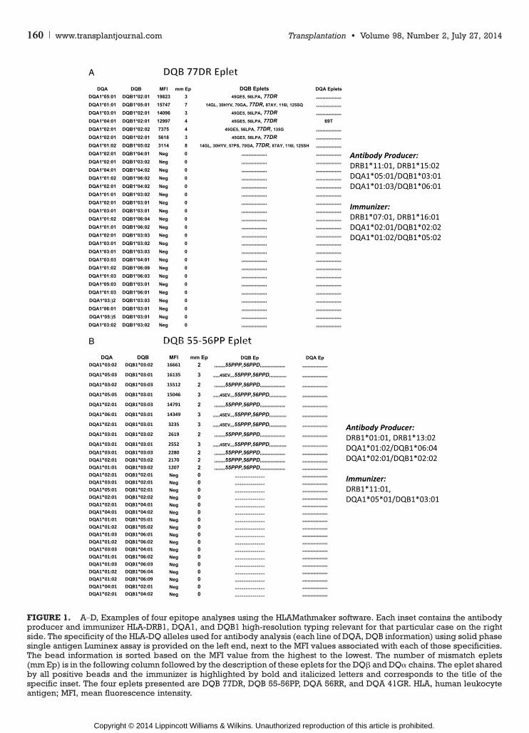
FIGURE 1. AYD, Examples of four epitope analyses using the HLAMathmaker software. Each inset contains the antibodyproducer and immunizer HLA-DRB1, DQA1, and DQB1 high-resolution typing relevant for that particular case on the rightside. The specificity of the HLA-DQ alleles used for antibody analysis (each line of DQA, DQB information) using solid phasesingle antigen Luminex assay is provided on the left end, next to the MFI values associated with each of those specificities.The bead information is sorted based on the MFI value from the highest to the lowest. The number of mismatch eplets(mmEp) is in the following column followed by the description of these eplets for the DQA and DQ> chains. The eplet sharedby all positive beads and the immunizer is highlighted by bold and italicized letters and corresponds to the title of thespecific inset. The four eplets presented are DQB 77DR, DQB 55-56PP, DQA 56RR, and DQA 41GR. HLA, human leukocyteantigen; MFI, mean fluorescence intensity.
160 www.transplantjournal.com Transplantation & Volume 98, Number 2, July 27, 2014
Copyright © 2014 Lippincott Williams & Wilkins. Unauthorized reproduction of this article is prohibited.

FIGURE 1. (continued)
* 2014 Lippincott Williams & Wilkins Tambur et al. 161
Copyright © 2014 Lippincott Williams & Wilkins. Unauthorized reproduction of this article is prohibited.

residues, considered to be more likely to contribute to anti-body recognition, concentrate only in the DQA chain. (C) Theeplet is located on the DQ> chain with an antibody footprintcovering both > and A chains. (D) A DQ> eplet has an anti-body footprint covering areas solely on the DQ> chain.
To the best of our knowledge, this is the first studyattempting to identify not only the eplet associated withHLA-DQ antigen-antibody recognition but also the com-plete antibody footprint. It is clear from these results thatthe antibody does not make a distinction between the twochains of the DQ molecule and can recognize either or bothin the same capacity. This is not a surprising observationbecause antibody analysis performed for class I moleculeshad shown similar data (23). In fact, in all five examplespresented in that study, the surface residues within 15 A ofthe eplet include amino acids of both the >1 and >2 do-mains of the class I molecule, corresponding to the >1 andA1 domains of a class II molecule. Yet, this is an importantobservation because current practices tend to look at HLA-DQ molecules as if they are composed of two independentcomponentsVDQ> and DQAVand in fact, the only way toenter data in UNOS is by dividing the patient or donortyping or the antibody specificities into these two categories.The results presented in this study and supported by pre-vious reports from our group (31Y33) indicate that thispractice is misleading and distorts the actual informationregarding the specificity of HLA-DQ antibodies.
A puzzling observation was the disparity between theMFI values (strength) and the amount or density of antigenbound to the bead among alleles sharing the same eplet
(Fig. 3AYD). With the potential exception of example 3 inwhich all the MFI values were rather low, the MFI values ofthe lowest fluorescence bead was between 7% and 12% ofthe highest fluorescence bead, despite the fact that theamount of antigen on these beads ranged between 48% and74%. For example, in Figure 3(A), two beadsVDQA1*03:01/DQB1*03:01 and DQA1*01:02/DQB1*05:02Vhave the samepercentage density of antigen (75% and 74%, respectively),both exhibiting the DQB77DR eplet; yet, the respective MFIvalues are 15,747 and 3,114. Artificial misalignment of HLAmolecules on solid phase microspheres could potentially ex-plain these observations. However, we think this hypothesis isnot applicable in our case because analyzing different donor-recipient pairs, we observed scenarios in which a higher MFIranking bead for one pair was among the lower ranking beadsfor another pair, despite the fact that the same eplet wasimplicated as the target of antibody binding. We thereforepropose that although the eplet is critical for the definitionof the immunogenic epitope, it is not sufficient to determinethe ‘‘strength’’ of the response. Indeed, Kosmoliaptsis et al.(34Y36), in a series of elegant experiments analyzing surfaceelectrostatic potentials, demonstrated that sequence informa-tion alone does not provide full insight into key determinantof epitope immunogenicity. In fact, even before sophisticatedmethodologies have been used, early serologic assays showedthat HLA-specific alloantibodies could show heterogenic pat-terns of binding to the same amino acid sequence motifs, ifthose motifs were expressed on the background of differentHLA-B alleles (37Y39). Additional studies such as the use ofsoluble HLA molecules, assessing antibody affinity and avidity,
FIGURE 2. Four insets follow the same sequence as in Figure 1, each corresponding to one epitope identified. The leftframe shows a three-dimensional structure of an HLA-DQ molecule in which the pink amino acids compose the DQ> chain,the blue amino acids compose the DQA chain, and the brown amino acids represent the peptide lodged within the peptide-binding groove. The specific eplet identified is highlighted in light blue. The middle frame highlights the complete epitopeby using the ‘‘select by distance’’ function of the 3nCd software to illustrate 15-Å radius from the eplet. The right frameisolates the eplet structure and presents it reverted into its original color scheme to identify the DQ molecule structures thatare part of the functional epitope. HLA, human leukocyte antigen.
162 www.transplantjournal.com Transplantation & Volume 98, Number 2, July 27, 2014
Copyright © 2014 Lippincott Williams & Wilkins. Unauthorized reproduction of this article is prohibited.

proteomics, or site-directed mutagenesis are required to betterunderstand this phenomenon.
The non-self/self theory as presented by Duquesnoyet al. is very appealing and supported by compelling data fromtheir studies (23, 40). Although our previous studies suggesteda high frequency of non-self/self DQ chains involved in anti-body development, our study of HLA-DQ epitope could notsubstantiate Duquesnoy’s theory in its current form. Giventhe heterogeneity in binding observed in this study and data
presented by others, it is likely that additional parametersshould be considered in refinement of the non-self/self theoryto fully explain its applicability in clinical use.
In summary, we have presented additional evidence tosupport the need for changing the manner in which HLA-DQantigens and antibodies are evaluated for organ transplanta-tion. The current nomenclature system does not reflect thetrue nature of HLA-DQ polymorphism. Moreover, becauseour field contemplates the use of epitope matching as an
FIGURE 3. The epitope analysis interrogating amino acids that contribute to the epitopeV15-Å radius from the epletVasdetermined by the Cn3d software. Only positive beads and their polymorphic positions are described. Amino acids that arenot exposed to themolecule’s surface are shaded gray. Those that are exposed to the paratope are in bold and italics. HLA-DRand HLA-DQ typing of the immunizer and antibody producer for each case are listed at the top portion of each panel, to theright of the amino acid sequences. Below them is the allelic identification of the positive SA beads. MFI values, as presentedin Figure 1, are listed to the right of the DQ alleles and adjacent to the % density of DQ molecule of each of these beads(assuming the highest MFI bead in each case represent 100% density). Given the significant difference between MFI valuesand % density as well as the known variability of the solid phase assays, those assays were repeated three times. No sig-nificant differences in the ratios were observed. Amino acids that are exposed to the surface, thus accessible to binding by anantibody or the TCR, are in bold and italics. Amino acids that are buried under the surface are indicated with the letter Bbelow the AA number. HLA, human leukocyte antigen; MFI, mean fluorescence intensity.
* 2014 Lippincott Williams & Wilkins Tambur et al. 163
Copyright © 2014 Lippincott Williams & Wilkins. Unauthorized reproduction of this article is prohibited.
approach to improve organ allocation and overall outcomes,it is imperative that we understand the true nature of an im-munogenic epitope. This will pave the way not only to im-prove matching algorithms but also to predict theimmunogenicity of HLA mismatched alleles so that accept-able mismatches can be identified and the risk of generatingde novo HLA-DSA after transplantation can be betterstratified.
MATERIALS AND METHODSForty kidney transplant recipients, transplanted and followed up at the
Northwestern University Comprehensive Transplant Center, were evaluated
for this study. Approval was obtained from the institutional review board.
Patient inclusion criteria included a history of 0% panel reactive antibody
(using solid phaseYbased assay) before their transplantation and kidney
transplant rejection leading to failure of the graft associated with the genera-
tion of de novo HLA-DQ DSAs. Availability of donor’s and recipient’s DNA
for high-resolution typing was mandatory. Human leukocyte antigen typing
was performed using HLA LabType SSO reagents for HLA-A,-B,-C,-DRB1,-
DQA1, and-DQB1 (One Lambda, Canoga Park, CA). When required, ambi-
guity resolution was performed using micro-SSP high-resolution or allele-
specific trays to ascertain the high-resolution typing of HLA-DRB1, DQA1,
or DQB1. Assignment of common and well-documented alleles was based on
Cano et al.(41).
Antibody testing was performed using LABScreenSingle Antigen(One
Lambda) and LIFECODES Single Antigen (Hologic Gen-Probe, San Diego,
CA) HLA class II reagents following manufacturer’s recommended protocol.
Antibody specificity was documented as directed against DRB1, DQ>/A, or
DP>/A molecules. Positive results were determined based on a flexible ‘‘cut-
off ’’ value between 1,000 and 2,000 MFI, based more on the architecture of the
MFI histograms and the following eplet analysis. To allow direct comparison
between MFI values on different beads, we evaluated the antigen density on
FIGURE 3. (continued)
164 www.transplantjournal.com Transplantation & Volume 98, Number 2, July 27, 2014
Copyright © 2014 Lippincott Williams & Wilkins. Unauthorized reproduction of this article is prohibited.
each of these beads using a monomorphic mouse anti-human antibody rec-
ognizing all DQ>/A combinations, clone B-K27 (ab47342, Abcam Cambridge,
MA). Relative density was calculated in each case in relation to the highest MFI
bead per patient, considered as 100%.
HLAMatchmaker software, a Microsoft Excel program that was devel-
oped to analyze antibody reactivity for eplets and eplet pairs, was downloaded
from http://www.HLAMatchmaker.net. Patient information was entered as
the ‘‘antibody producer,’’ and donor typing was entered as the ‘‘immunizer.’’
Patient’s antibody signature, including the MFI values, was populated into
HLAMatchmaker such that eplets shared with the patient’s own antigens are
automatically considered negative and are removed from consideration. The
remaining eplets in the reactive alleles are presented such that eplets or eplets
pairs that are shared with the immunizer can be identified. The Cn3d software
(42) was used to visualize the three-dimensional structure of a crystallographic
structure of an HLA-DQ molecule (http://www.ncbi.nlm.nih.gov/Structure);
identify the exact location of the eplets, as determined by HLAMatch-
maker; and calculate the footprint of the antibody CDR (15-A radius from
the eplet). (The notation system used in this article follows the one used
by HLAMatchmaker. The author is advised that currently there is no
single official notation system to describe HLA epitopes and that addi-
tional notations systems are used by Terasaki et al. or by the epitope reg-
istry Web site.)
ACKNOWLEDGMENTThe authors thank Mr. Peter Golovin for his superb IT
skills and his help in identifying the appropriate donor-recipient pairs for this study.
REFERENCES1. Schwartz BD, Luehrman LK, Lee J, et al. A public antigenic determi-
nant in the HLA-B5 cross-reacting groupVa basis for cross-reactivityand a possible link with Behcet’s disease. Hum Immunol 1980; 1: 37.
2. Schwartz BD, Luehrman LK, Lee T, et al. HLA serological cross-reactivity: HLA-B15 has two public antigens. Hum Immunol1980; 1: 331.
3. Fuller AA, Rodey GE, Parham P, et al. Epitope map of the HLA-B7CREG using affinity-purified human alloantibody probes. HumImmunol 1990; 28: 306.
4. Fuller AA, Trevithick JE, Rodey GE, et al. Topographic map of theHLA-A2 CREG epitopes using human alloantibody probes. HumImmunol 1990; 28: 284.
5. Bjorkman PJ, Strominger JL, Wiley DC. Crystallization and X-raydiffraction studies on the histocompatibility antigens HLA-A2 andHLA-A28 from human cell membranes. J Mol Biol 1985; 186: 205.
6. Saper MA, Bjorkman PJ, Wiley DC. Refined structure of the humanhistocompatibility antigen HLA-A2 at 2.6 A resolution. J Mol Biol1991; 219: 277.
7. Gorga JC, Brown JH, Jardetzky T, et al. Crystallization of HLA-DRantigens. Res Immunol 1991; 142: 401.
8. Marrari M, Conca R, Pratico-Barbato L, et al. Brief report: why didtwo patients who type for HLA-B13 have antibodies that react with allBw4 antigens except HLA-B13? Transpl Immunol 2011; 25: 217.
9. Marrari M, Duquesnoy RJ. Why can sensitization by an HLA-DR2mismatch lead to antibodies that react also with HLA-DR1? HumImmunol 2009; 70: 403.
10. Tambur AR, Buckingham M, McDonald L, et al. Development ofdonor-specific and nonYdonor-specific HLA-DP antibodies post-transplant: the role of epitope sharing and epitope matching. ClinTranspl 2006: 399.
11. Cai J, Terasaki PI, Mao Q, et al. Development of nondonor-specificHLA-DR antibodies in allograft recipients is associated with sharedepitopes with mismatched donor DR antigens. Am J Transplant 2006;6: 2947.
12. Duquesnoy RJ. HLAMatchmaker: a molecularly based algorithm forhistocompatibility determination. I. Description of the algorithm.Hum Immunol 2002; 63: 339.
13. Duquesnoy RJ, Marrari M. HLAMatchmaker: a molecularly basedalgorithm for histocompatibility determination. II. Verification of the
algorithm and determination of the relative immunogenicity of ami-no acid triplet-defined epitopes. Hum Immunol 2002; 63: 353.
14. Duquesnoy RJ, Takemoto S, de Lange P, et al. HLAmatchmaker: amolecularly based algorithm for histocompatibility determination.III. Effect of matching at the HLA-A,B amino acid triplet level onkidney transplant survival. Transplantation 2003; 75: 884.
15. Duquesnoy RJ, Howe J, Takemoto S. HLAmatchmaker: a molecularlybased algorithm for histocompatibility determination. IV. An alter-native strategy to increase the number of compatible donors forhighly sensitized patients. Transplantation 2003; 75: 889.
16. Duquesnoy RJ, Askar M. HLAMatchmaker: a molecularly based al-gorithm for histocompatibility determination. V. Eplet matching forHLA-DR, HLA-DQ, and HLA-DP. Hum Immunol 2007; 68: 12.
17. El-Awar N, Cook D, Terasaki PI. HLA class I epitopes: A and B loci.Clin Transpl 2006: 79.
18. El-Awar N, Terasaki PI, Cai J, et al. Epitopes of the HLA-A, B, C, DR,DQ and MICA antigens. Clin Transpl 2007; 21: 175.
19. El-Awar NR, Akaza T, Terasaki PI, et al. Human leukocyte antigen classI epitopes: update to 103 total epitopes, including the C locus.Transplantation 2007; 84: 532.
20. Van Regenmortel MH, Altschuh D, Chatellier J, et al. Measurementof antigen-antibody interactions with biosensors. J Mol Recognit 1998;11: 163.
21. Duquesnoy RJ, Mulder A, Askar M, et al. HLAMatchmaker-basedanalysis of human monoclonal antibody reactivity demonstrates theimportance of an additional contact site for specific recognition oftriplet-defined epitopes. Hum Immunol 2005; 66: 749.
22. Marrari M, Mostecki J, Mulder A, et al. Human monoclonal anti-body reactivity with human leukocyte antigen class I epitopes definedby pairs of mismatched eplets and self-eplets. Transplantation 2010;90: 1468.
23. Duquesnoy RJ. The antibody response to an HLA mismatch: a modelfor nonself-self discrimination in relation to HLA epitope immuno-genicity. Int J Immunogenet 2012; 39: 1.
24. Hayakawa K, Asano M, Shinton SA, et al. Positive selection of naturalautoreactive B cells. Science 1999; 285: 113.
25. Pillai S, Cariappa A, Moran ST. Positive selection and lineage com-mitment during peripheral B-lymphocyte development. Immunol Rev2004; 197: 206.
26. Morris SC, Moroldo M, Giannini EH, et al. In vivo survival ofautoreactive B cells: characterization of long-lived B cells. J Immunol2000; 164: 3035.
27. DeVos JM, Gaber AO, Knight RJ, et al. Donor-specific HLA-DQ an-tibodies may contribute to poor graft outcome after renal transplan-tation. Kidney Int 2012; 82: 598.
28. Willicombe M, Brookes P, Sergeant R, et al. De novo DQ donor-specific antibodies are associated with a significant risk of antibody-mediated rejection and transplant glomerulopathy. Transplantation2012; 94: 172.
29. Ntokou IS, Iniotaki AG, Kontou EN, et al. Long-term follow upfor anti-HLA donor specific antibodies postrenal transplantation:high immunogenicity of HLA class II graft molecules. Transpl Int2011; 24: 1084.
30. Wiebe C, Nickerson P. Posttransplant monitoring of de novo humanleukocyte antigen donor-specific antibodies in kidney transplantation.Curr Opin Organ Transplant 2013; 18: 470.
31. Tambur AR, Leventhal JR, Friedewald JJ, et al. The complexity ofhuman leukocyte antigen (HLA)-DQ antibodies and its effect onvirtual crossmatching. Transplantation 2010; 90: 1117.
32. Tambur AR, Leventhal JR, Zitzner JR, et al. The DQ barrier: im-proving organ allocation equity using HLA-DQ information. Trans-plantation 2013; 95: 635.
33. Tambur AR, Leventhal JR, Walsh RC, et al. HLA-DQ Barrier: effects oncPRA Calculations. Transplantation 2013; 96: 1065.
34. Kosmoliaptsis V, Bradley JA, Sharples LD, et al. Predicting the im-munogenicity of human leukocyte antigen class I alloantigens usingstructural epitope analysis determined by HLAMatchmaker. Trans-plantation 2008; 85: 1817.
35. Kosmoliaptsis V, Sharples LD, Chaudhry AN, et al. Predicting HLAclass II alloantigen immunogenicity from the number and physi-ochemical properties of amino acid polymorphisms. Transplantation2011; 91: 183.
36. Kosmoliaptsis V, Dafforn TR, Chaudhry AN, et al. High-resolution,three-dimensional modeling of human leukocyte antigen class I
* 2014 Lippincott Williams & Wilkins Tambur et al. 165
Copyright © 2014 Lippincott Williams & Wilkins. Unauthorized reproduction of this article is prohibited.
structure and surface electrostatic potential reveals the molecular basisfor alloantibody binding epitopes. Hum Immunol 2011; 72: 1049.
37. Lutz CT, Smith KD, Greazel NS, et al. Bw4-reactive and Bw6-reactiveantibodies recognize multiple distinct HLA structures that partiallyoverlap in the alpha-1 helix. J Immunol 1994; 153: 4099.
38. Arnett KL, Adams EJ, Gumperz JE, et al. Expression of an unusual Bw4epitope by a subtype of HLA-B8 [B*0802]. Tissue Antigens 1995; 46: 316.
39. Voorter CE, van der Vlies S, Kik M, et al. Unexpected Bw4 and Bw6reactivity patterns in new alleles. Tissue Antigens 2000; 56: 363.
40. Duquesnoy RJ, Marrari M, Mulder A, et al. Structural aspects of hu-man leukocyte antigen class I epitopes detected by human monoclo-nal antibodies. Hum Immunol 2012; 73: 267.
41. Cano P, Klitz W, Mack SJ, et al. Common and well-documentedHLA alleles: report of the Ad-Hoc committee of the american soci-ety for histocompatiblity and immunogenetics. Hum Immunol2007; 68: 392.
42. Hogue CW. Cn3D: a new generation of three-dimensional molecularstructure viewer. Trends Biochem Sci 1997; 22: 314.
166 www.transplantjournal.com Transplantation & Volume 98, Number 2, July 27, 2014
Copyright © 2014 Lippincott Williams & Wilkins. Unauthorized reproduction of this article is prohibited.


![Reduced Expression of HLA Class I and II Antigens in Colon Cancer1 · [CANCER RESEARCH 50, 8023-8027, December 15, 1990] Reduced Expression of HLA Class I and II Antigens in Colon](https://img.pdfslide.net/doc/110x75/5f5945785c4df2481d781bbc/reduced-expression-of-hla-class-i-and-ii-antigens-in-colon-cancer1-cancer-research.jpg)