Embed Size (px)
Citation preview
EPIZOOTIC LYMPHANGITIS
Equine blastomycosis, equine histoplasmosis, pseudoglanders or African glanders.
Definition
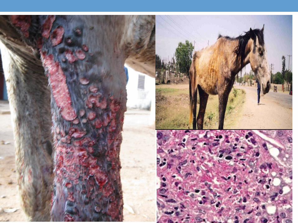
• It is a chronic pygranulomatous contagious disease of horse, caused by histoplasma farciminosum, characterized clinically by ulcerative, suppurative, spreading dermatitis and lymphangitisdermatitis and lymphangitis; however, other forms including pneumonia or ulcerative pneumonia or ulcerative conjunctivitis.conjunctivitis.
Etiology
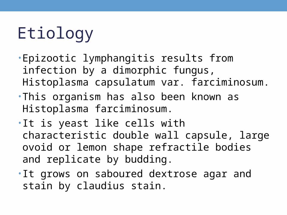
• Epizootic lymphangitis results from infection by a dimorphic fungus, Histoplasma capsulatum var. farciminosum.
• This organism has also been known as Histoplasma farciminosum.
• It is yeast like cells with characteristic double wall capsule, large ovoid or lemon shape refractile bodies and replicate by budding.
• It grows on saboured dextrose agar and stain by claudius stain.
Epidemiology• DistributionDistribution: The disease is endemic in some countries in the
Mediterranean region, and in parts of Africa and Asia and present in Egypt.
• Animal susceptibilityAnimal susceptibility: Epizootic lymphangitis mainly affects horses, donkeys and mules. H. capsulatum var. farciminosum has also been reported in camels, cattle and dogs.
• Mode of transmissionMode of transmission: • The source of the organisms The source of the organisms can be the skin lesions and nasal and skin lesions and nasal and
ocular exudates of infected animalsocular exudates of infected animals, or the soil.
• Biting flies in the genera Musca and StomoxysMusca and Stomoxys are thought to spread the conjunctival formconjunctival form. Flies may also transmit the skin form mechanically when they feed on lesions and exudates. Ticks might be involved in transmission. The pulmonary formpulmonary form, which is rare, probably develops when an animal inhalesinhales the organism
Pathogenesis• The fungus invade cutaneous abrasions or wounds result in
formation of S/C nodules which abscessed with discharging of thick creamy pus and then ulcerate and heal by scar.
• Spread of infection to adjacent lymphatic with formation of nodules along their course and adjacent lymph nodes are also abscessate.
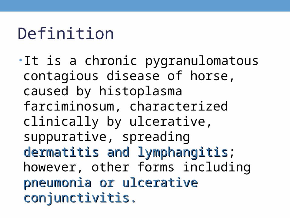
• Hematogenous spread (yeast cells are present intracellular or extracellular especially of macrophages) with visceral involvement may occurs which result in disturbance in general conditions of the animals.
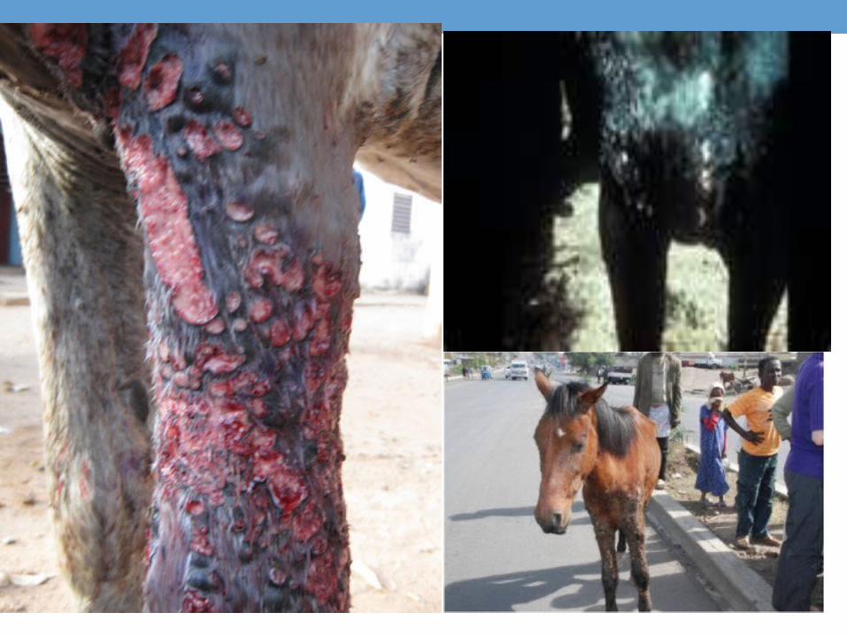
• Skin lesions are mainly present on head, neck, and limb (mainly hind one which is more exposed to abrasions than forelimb).
Clinical signs• The incubation period is usually several weeks to 2
months. Morbidity is high and mortality is 10-15%• The most common form of epizootic lymphangitis affects
the skin and lymphatics. It often occurs on the extremities, chest wall, face and neck, but can be seen wherever the organism is inoculated into a wound
• The first symptom is a painless, freely moveable S/C nodule, approximately 2 cm in diameter. This nodule enlarges at portal of entry which rupture discharging thick creamy pus with formation of indolent ulcer
• The skin over the nodules may be fixed to the underlying tissues.
Clinical signs• The surrounding skin is edematous at first, and later
becomes thickened, hard and variably painful• The regional lymph nodes can be enlarged, but fever is
uncommon. The infection also spreads along the lymphatics, causing cord–like thickening and further skin involvement
• The lesion develop mainly on limbs ecepcially hock but may present on the back, sides, vulva, scrotum, occasionally the lesion appear on nasal mucosa due to nibbling of the lesions on the limb and trunk but it lies just inside nostrils and do not inovlve nasal septum.
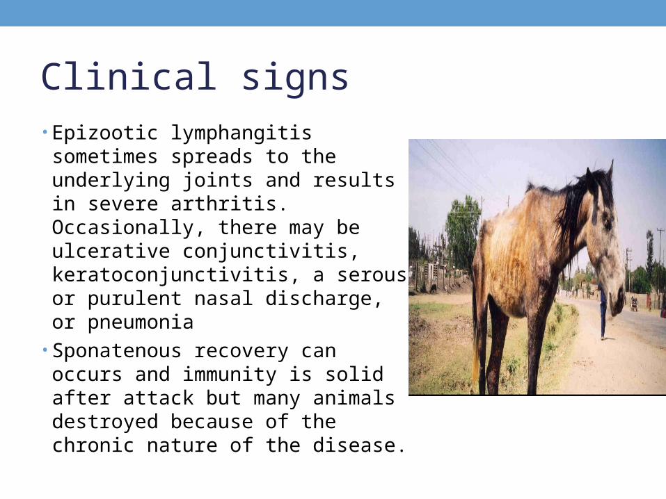
Clinical signs• Epizootic lymphangitis sometimes
spreads to the underlying joints and results in severe arthritis. Occasionally, there may be ulcerative conjunctivitis, keratoconjunctivitis, a serous or purulent nasal discharge, or pneumonia
• Sponatenous recovery can occurs and immunity is solid after attack but many animals destroyed because of the chronic nature of the disease.
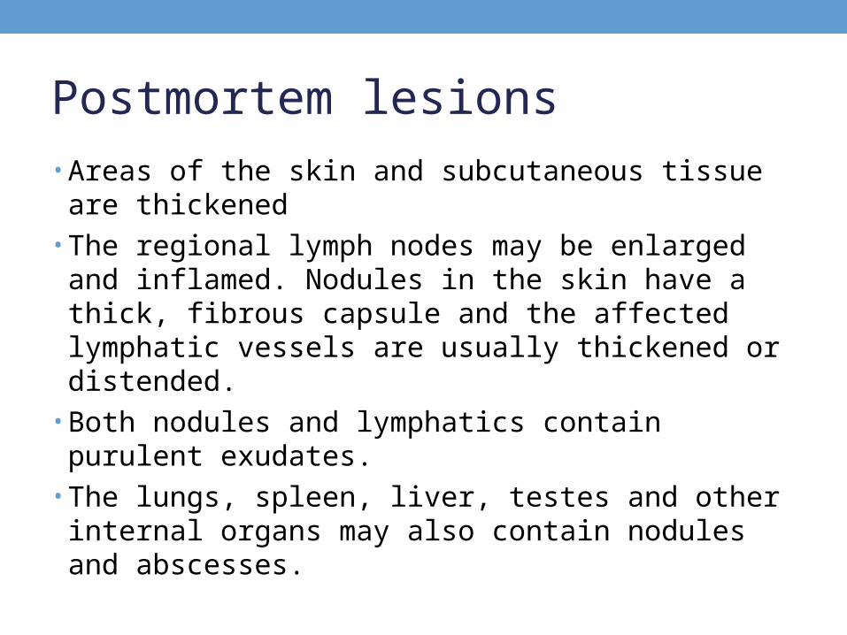
Postmortem lesions• Areas of the skin and subcutaneous tissue are thickened• The regional lymph nodes may be enlarged and inflamed.
Nodules in the skin have a thick, fibrous capsule and the affected lymphatic vessels are usually thickened or distended.
• Both nodules and lymphatics contain purulent exudates.• The lungs, spleen, liver, testes and other internal organs
may also contain nodules and abscesses.
Diagnosis• Field diagnosis: signs as cutaneous nodules,
lymphangitis and lymphadenitis with postmortem lesions.
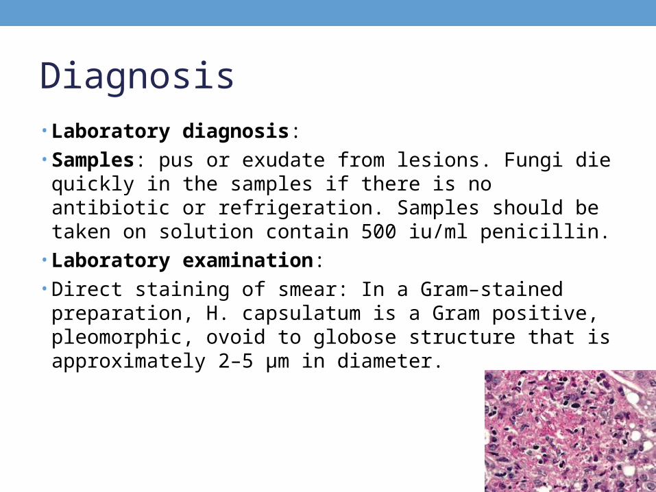
Diagnosis• Laboratory diagnosis:• Samples: pus or exudate from lesions. Fungi die quickly
in the samples if there is no antibiotic or refrigeration. Samples should be taken on solution contain 500 iu/ml penicillin.
• Laboratory examination:• Direct staining of smear: In a Gram–stained preparation,
H. capsulatum is a Gram positive, pleomorphic, ovoid to globose structure that is approximately 2–5 µm in diameter.
Diagnosis• Culture of the organism: H. capsulatum var
farciminosum can be cultured on a variety of fungal media as enriched Sabouraud’s dextrose agar with 2.5% glycerol This organism grows as a mycelium at cooler temperatures. These colonies grow slowly and develop in approximately 2 to 8 weeks at 26°C. They are dry, granular, wrinkled and grayish-white, becoming brown as they age. Aerial forms are rare.
• Serological tests include fluorescent antibody tests, enzyme–linked immunosorbent assays (ELISA) and passive hemagglutination.
Treatment• Early cases (less severe cases) are cured by extensive
excisions of affected parts followed by local application of iodine or silver nitrate with parental injection of iodines preparation. Affected animals are showed extensive lesions are destroyed and buried is advisable.
Control• Infected premises and equipment must be thoroughly
cleaned and disinfected (H. capsulatum can be inactivated by 1% sodium hypochlorite, glutaraldehyde, formaldehyde and phenolic disinfectants).
• Bedding should be burned. Organisms in the soil may survive for long periods.
• Early cases may be treated with sodium or potassium iodide, but the lesions may later recur.
• Vaccines are not widely available; however, live and inactivated vaccines have been used in some endemic regions. There is no vaccine in Egypt.


![Blastomycosis - A Northwoods Nuisance · Blastomycosis - A Northwoods Nuisance Lake Tides ... Photo courtesy of John Archer Photo courtesy of John Archer [Blastomycosis] is treatable,](https://img.pdfslide.net/doc/110x75/5b8fe87009d3f27a6d8d0c4a/blastomycosis-a-northwoods-nuisance-blastomycosis-a-northwoods-nuisance.jpg)