Embed Size (px)
Citation preview

EQUIPEQUIPTraining session 1Training session 1
Improving Improving polyp/adenoma polyp/adenoma
detectiondetection

BackgroundBackground
No prospective methods to increase No prospective methods to increase ADRADR
Detection of flat lesions not reportedDetection of flat lesions not reported

HypothesisHypothesis
Intensive training (detection & Intensive training (detection & classification)classification)
Increase in adenoma detection Increase in adenoma detection

Session I ObjectivesSession I Objectives
Importance Importance DefinitionDefinition PrevalencePrevalence HistopathologyHistopathology
Detection MethodsDetection Methods Subtle clues to flat polypsSubtle clues to flat polyps Colonoscopy TechniquesColonoscopy Techniques

Paris shape classificationParis shape classification
“Flat” polyps: Lesions with < 2.5mm elevation (width of snare catheter/bx cable)

DefinitionsDefinitions FlatFlat
Less than 2.5mm of elevationLess than 2.5mm of elevation
DepressedDepressed Base lower than normal mucosa heightBase lower than normal mucosa height
Well demarcated; round or star shapedWell demarcated; round or star shaped
Soetikno et al; JAMA 2008Soetikno et al; JAMA 2008
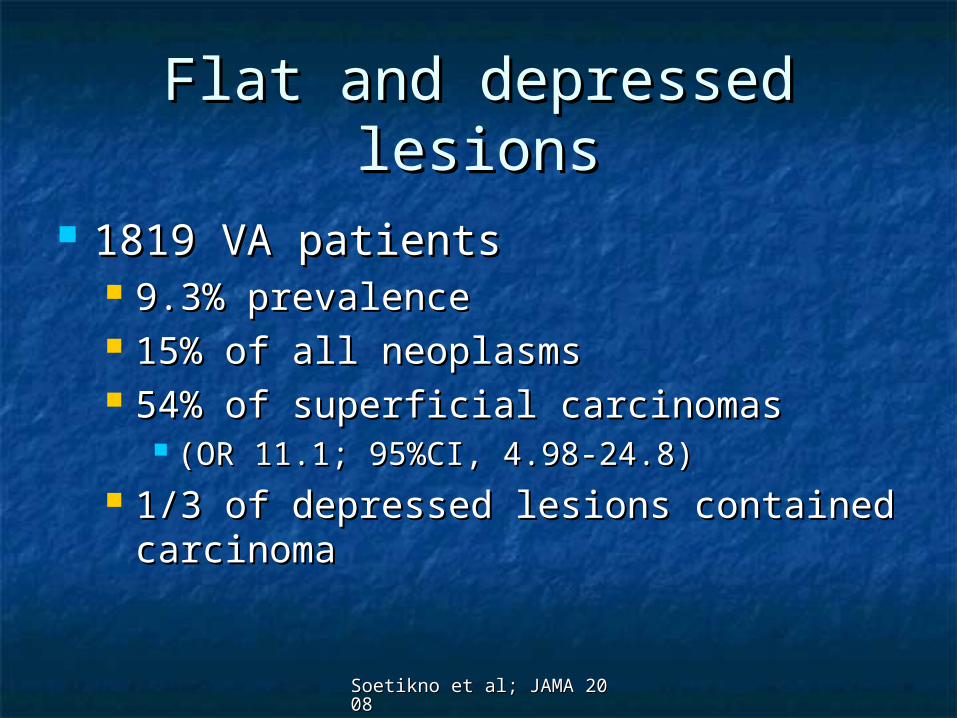
Flat and depressed lesionsFlat and depressed lesions
1819 VA patients 1819 VA patients 9.3% prevalence9.3% prevalence 15% of all neoplasms15% of all neoplasms 54% of superficial carcinomas 54% of superficial carcinomas
(OR 11.1; 95%CI, 4.98-24.8)(OR 11.1; 95%CI, 4.98-24.8) 1/3 of depressed lesions contained 1/3 of depressed lesions contained
carcinomacarcinoma

Prevalence of Flat PolypsPrevalence of Flat Polyps 27,400 colonoscopies27,400 colonoscopies
Flat adenomaFlat adenoma 5.3%5.3% Among all adenomasAmong all adenomas
PolypoidPolypoid 74%74% FlatFlat 26%26%
More likely in right colon (OR 2.92)More likely in right colon (OR 2.92) Risk of advanced histology similarRisk of advanced histology similar
Unless depressed (OR 10.56)Unless depressed (OR 10.56)
•Blanco et al. Endoscopy 2010;42:279
Soetikno et al; JAMA 2008Soetikno et al; JAMA 2008
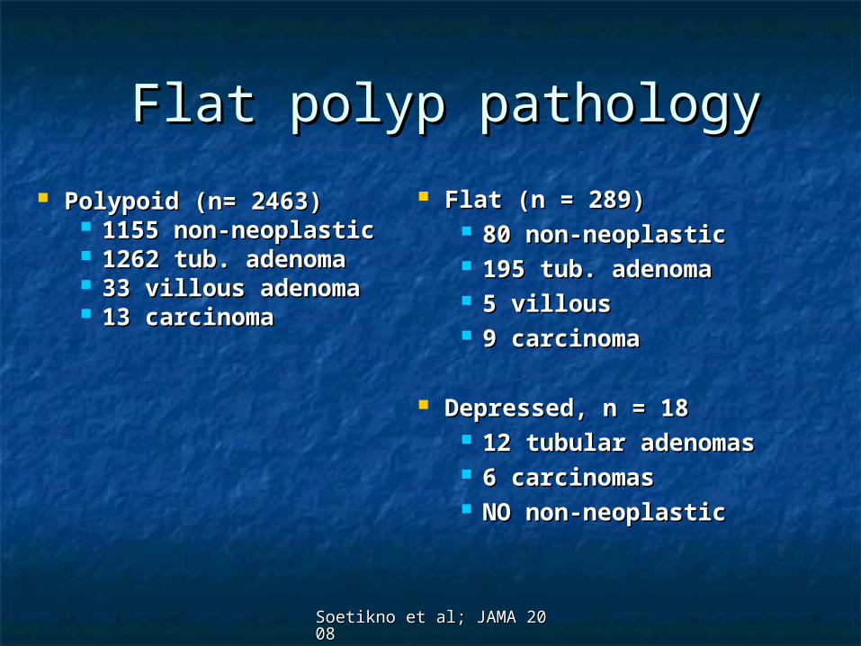
Flat polyp pathologyFlat polyp pathology
Polypoid (n= 2463)Polypoid (n= 2463) 1155 non-neoplastic1155 non-neoplastic 1262 tub. adenoma1262 tub. adenoma 33 villous adenoma33 villous adenoma 13 carcinoma13 carcinoma
Flat (n = 289) Flat (n = 289) 80 non-neoplastic80 non-neoplastic 195 tub. adenoma195 tub. adenoma 5 villous5 villous 9 carcinoma9 carcinoma
Depressed, n = 18Depressed, n = 18 12 tubular adenomas12 tubular adenomas 6 carcinomas6 carcinomas NO non-neoplasticNO non-neoplastic

Detection methodsDetection methods
Subtle clues to detectionSubtle clues to detection
Bowel preparationBowel preparation
Colonoscopy techniquesColonoscopy techniques Washing, Washing, working the foldsworking the folds WithdrawalWithdrawal Clear caps Clear caps Optical enhancement ?Optical enhancement ?

Subtle cluesSubtle clues
Subtle color differences (red or pale)Subtle color differences (red or pale)
Spontaneous hemorrhage/friabilitySpontaneous hemorrhage/friability
Deformity of colon wallDeformity of colon wall
Absence of vascular networkAbsence of vascular network
ASGE Learning Library: Diagnosis of Flat and Depressed Colorectal Neoplasms; 2006

Subtle clues: VideoSubtle clues: Video
ASGE Learning Library: Diagnosis of Flat and Depressed Colorectal Neoplasms; 2006


Detection methodsDetection methods
Colonoscopy techniqueColonoscopy technique Withdrawal time ?Withdrawal time ? WashingWashing
Bowel prep scoreBowel prep score ““Working” the foldsWorking” the folds Clear CapsClear Caps

Withdrawal TimeWithdrawal Time
Mandating longer WD time does Mandating longer WD time does NOT increase ADRNOT increase ADR
Compliance w/ mandate ADR after Mandate
Sawhaney Gastro 2008;135;1892

Rex D, GIE; 2000; Vol 51, No 1Rex D, GIE; 2000; Vol 51, No 1
Colonoscopy techniqueColonoscopy technique Criterion High adenoma detector Low adenoma detector p Value
Looking on the proximal sides of folds, valves, etc. 31.5 19.6 < 0.001
Adequacy of cleaning 33.1 21.9 < 0.001
Adequacy of distention 33.5 24.0 < 0.001
Adequacy of time spent viewing 32.4 21.0 < 0.001
*Scores are the means for all colonoscopies and for all 4 judges. The highest score possible is 35. ‡Colonoscopist
High detector vs. low detectorHigh detector vs. low detector Percentage of mucosa visualized (estimate)Percentage of mucosa visualized (estimate)
90.8% vs. 63.3%; p <0.00190.8% vs. 63.3%; p <0.001 Mean withdrawal timeMean withdrawal time
8 min 55 sec vs. 6 min 41 sec; p = 0.028 min 55 sec vs. 6 min 41 sec; p = 0.02 More retroflex exams (9 vs. 6)More retroflex exams (9 vs. 6) Re-examine prox. side rectal valves in all 9 (15 – 40 seconds)Re-examine prox. side rectal valves in all 9 (15 – 40 seconds)

Prep QualityPrep Quality Missed CRCMissed CRC
Retrospective data review; 5055 colonoscopiesRetrospective data review; 5055 colonoscopies 17/286 cancers missed by colonoscopy17/286 cancers missed by colonoscopy
6/17 (3.5%) incomplete due to “poor prep”6/17 (3.5%) incomplete due to “poor prep” 4/17 (2.4%) identified but not recognized as malignant4/17 (2.4%) identified but not recognized as malignant
Flat and depressed neoplasmsFlat and depressed neoplasms Detection lower with inadequate bowel prepDetection lower with inadequate bowel prep
Small adenoma detection Small adenoma detection Retrospective review; 93,000 Retrospective review; 93,000
Adequate prep (76.9%) more likely detectAdequate prep (76.9%) more likely detect ““Suspected neoplasia”Suspected neoplasia” Lesions < 9mmLesions < 9mm No difference in lesions >9 mmNo difference in lesions >9 mm

Boston Bowel Preparation Boston Bowel Preparation ScoreScore


Hidden flat lesionsHidden flat lesions
ASGE Learning Library: Diagnosis of Flat and Depressed Colorectal Neoplasms; 2006

““Working” the foldsWorking” the folds
Same day virtual and optical Same day virtual and optical colonoscopycolonoscopy
(1233 patients; 210 adenomas > 6mm) (1233 patients; 210 adenomas > 6mm)
21 adenomas > 6mm missed on OC 21 adenomas > 6mm missed on OC 7 = advanced lesions7 = advanced lesions 15 = non-rectal neoplasia (other 6 in rectum)15 = non-rectal neoplasia (other 6 in rectum) 14 located on folds (10 back, 4 front)14 located on folds (10 back, 4 front) 1 located inner aspect of a flexure1 located inner aspect of a flexure

Withdrawal techniqueWithdrawal technique

““Working” the foldsWorking” the folds

Clear capsClear caps
CapCap NBINBI
Procedure timeProcedure time 25m25m 21m 21m .04.04
Adenoma Adenoma detectiondetection
31%31% 5%5% <0.04<0.04
Horiuchi et al. CGH 2010;8:379
CapCap NBINBI
<5mm<5mm 2424 55
5-10mm5-10mm 99 00
FlatFlat 77 22
SessileSessile 2626 33
Retractable clear cap vs. NBI for 2nd colonoscopy in patients with known polyps
Interval increase in adenoma detection by size and shape

Clear capsClear caps

SummarySummary
Flat polyps existFlat polyps exist There are subtle clues to detect flat There are subtle clues to detect flat
polypspolyps Color, friability, wall deformity, vessel Color, friability, wall deformity, vessel
changeschanges Good colonoscopy technique is neededGood colonoscopy technique is needed
WashingWashing Clear capsClear caps Working the foldsWorking the folds