Embed Size (px)
Citation preview
FROM KAPTEIN W. WILHELMSEN OG FRUES BAKTERIOLOGISKE INSTITUT, 0s LO
ERYSIPELOTHRIX RHUSIOPATHIAE CAUSING A FATAL MENINGEAL INFECTION
IN A HUMAN BEING By Th. ThjBtta, M. D.
(Received for publication Dec. 6th 1942).
On the 23 of May 1942 we received a specimen of pus for bacteriological examination. The specimen was sent us by Dr. Arne Torkildsen who had evacuated the same from an abscess situated in the right temporal region between the dura mater and the arachnoidea of a patient with the fol- lowing history :
The patient was a veterinary physician, 53 years of age, who during the last years had suffered from attacks of what had been considered rheumatism. He had passed through such an attack during the months of February and March the same year. The consulting physician Dr. Leif Cordfsen, however, claims that this attack looked more like an attack of influenza with complications of the lungs. Having recovered from this disease he fehlt quite well till the 20th of April, when he had several slight attacks of paresis of the left side of the body. He had no nausea, no vomiting and no cramps during these attacks, and he was quite well in the intervals. During the following week the condition got worse, a strong headache came on and the patient lost his appetite. He was then sent to hospital in Oslo for treatment. Here he showed
598
a slight and irregular fever and his diagnosis was not clear. The clinicians held it most probable that he suffered from a tumour of the brain. His condition grew continually worse and on the 22nd of May he was transferred to a surgical hospital in comatous condition. The operation was performed on the next day, and the patient died the following night.
Examination of the pus. The pus was of a very thick and sticky consistency. There
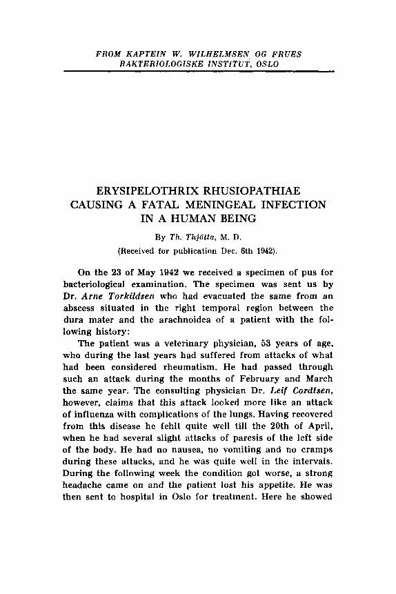
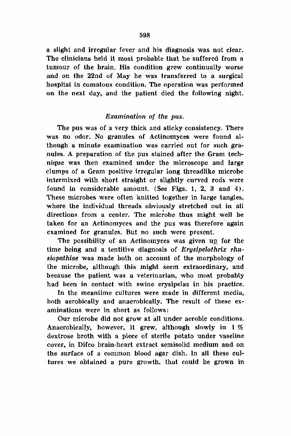
was no odor. No granules of Actinomyces were found al- though a minute examination was carried out for such gra- nules. A preparation of the pus stained after the Gram tech- nique was then examined under the microscope and large clumps of a Gram positive irregular long threadlike microbe intermixed with short straight or slightly curved rods were found in considerable amount. (See Figs. 1, 2, 3 and 4) . These microbes were often knitted together in large tangles, where the individual threads obviously stretched out in all directions from a center. The micfobe thus might well be taken for an Actinomyces and the pus was therefore again examined for granules. But no such were present.
The possibility of an Actinomyces was given up, for the time being and a tentitive diagnosis of Ergsipelothrix rhu- siopathiue was made both on account of the morphology of the microbe, although this might seem extraordinary, and because the patient was a veterinarian, who most probably had been in contact with swine erysipelas in his practice.
In the meantime cultures were made in different media, both aerobically and anaerobically. The result of these ex- aminations were in short as follows:
Our microbe did not grow at all under aerobic conditions. Anaerobicaljy, however, it grew, although slowly in 1 % dextrose broth with a piece of sterile potato under vaseline cover, in Difco brain-heart extract semisolid medium and on the surface of a common blood agar dish. In all these cul- tures we obtained a pure growth, that could be grown in
599
Figs. 4 , 2, 3, 4. Photos from different parts of pus from the meningeal absces.
x 1000.
600
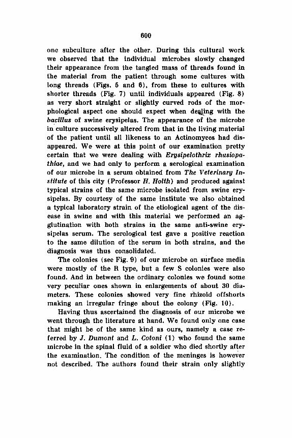
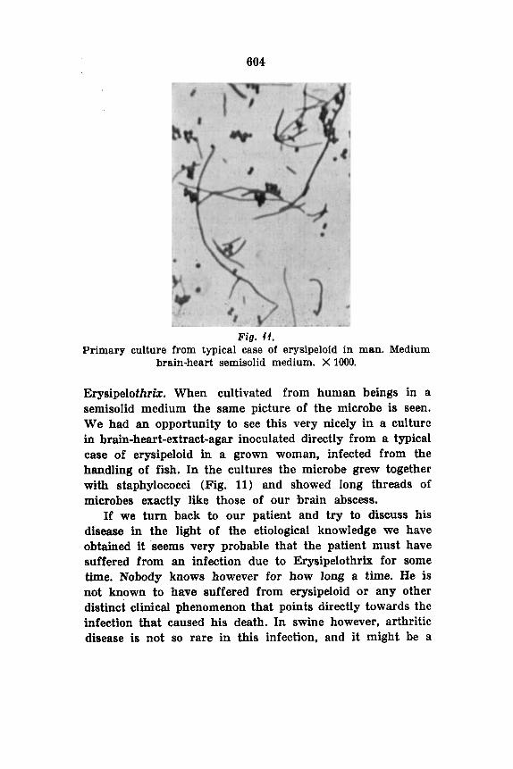
one subculture after the other. During this cultural work we observed that the individual microbes slowly changed their appearance from the tangled mass of threads found in the material from the patient through some cultures with long threads (Figs. 5 and 0), from these to cultures with shorter threads (Fig. 7) until individuals appeared (Fig. 8) as very short straight or slightly curved rods of the mor- phological aspect one should expect when deaing with the hacillus of swine erysipelas. The appearance of the microbe in' culture successively altered from that in the living material of the patient until all likeness to an Actinomyces had dis- appeared. We were at this point of our examination pretty certain that we were dealing with Erysipelothrk rhusiopa- thiae, and we had only to perform a serological examination of our microbe in a serum obtained from The Veterinary Zn- stitute of this city (Professor H. Holth) and produced against typical strains of the same microbe isolated from swine ery- sipelas. By courtesy of the same institute we also obtained a typical laboratory strain of the etiological agent of the dis- ease in swine and with this material we performed an ag- glutination with both strains in the same anti-swine ery- sipelas serum. The serological test gave a positive reaction to the same dilution of the serum in both strains, and the diagnosis was thus consolidated.
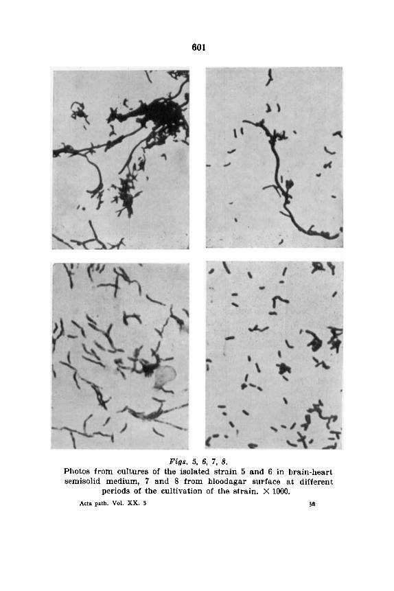
The colonies (see Fig. 9) of our microbe on surface media were mostly of the R type, but a few S colonies were also found. And in between the ordinary colonies we found some very peculiar ones shown in enlargements of about 30 dia- meters. These colonies showed very fine rhizoid offshorts making an irregular fringe about the colony (Fig. 10).
Having thus ascertained the diagnosis of our microbe we went through the literature at hand. We found o~ily one case that might be of the same kind as ours, namely a case re- ferred by J . Durnont and L. Cotoni (1) who found the same microbe in the spinal fluid of a soldier who died shortly after the examination. The condition of the meninges is however not described. The authors found their strain only slightly
601
Figs. 5, 6, 7, 8. Photos from cultures of the isolated strain 5 and 6 in brain-heart semisolid medium, 7 and 8 from bloodagar surface at different
periods of the cultivation of the strain. X 1000. 38 Acta path. Vol. XX. 3
602
Figs. 9, 40. Colonies of the strain on surface of bloodagar. 9: X 10, 10: X 30.
virulent for white mice, a point of interest in our case, since our strain did not show any virulence against mice.
The virulence against white mice has always been con- sidered a point of considerable interest in this microbe, since the passage through mice of some material containing the microbe in infection is claimed to be the most certain me- thod of obtaining this microbe in pure culture. This, however, seems to be quite uncertain, when one is dealing with ma- terial from a subacute or chronic type of infection. Thus W. Riebe (2,) found that strains cultivated from cases of endocarditis in swine are very often avirulent for mice. He adds that the virulence of this microbe may vary very con- siderably and that it is often very difficult to conserve the virulence in cultures.
Endocarditis due to Erysipelothrix is seen as a typical disease in swine. Also in the human infection this mani- festation has been seen, Thus c. Pracrsnitz (3) has gathered from the literature 100 cases of infection in man and amongst
these is one case of endocarditis, in which the microbe was grown from the blood. Likewise William 0. Russell and Marion E . Lamb (4) described a case of endocarditis in a lobsterfisher, who had injured himself while working with lobsters.
As f a r as we have found there is no case exactly like ours described in a human being. In swine, however, this microbe can produce lesions in the meninges, and P. Lang- rand ( 5 ) describes how the microbe, when growing on the surface of the meninges in animals, takes on the appearance of long, curved threads. And L. Panisset and J . Kolda ( 6 ) have described large nests of long, fine threads especially in the valvular lesions of endocarditis. And J. F. Rosenbach himself, who described the microbe ( 7 ) was in doubt whether it belonged to the bacteria or to the threadlike microbes and would have preferred the name Erysipelothrix from the very first. It was, however, named Bacillus erysipeloides or rhu- siopathiae and the name is still in use. The official name, however, is now Erysipelothrix, which is more suitable than Bacillus, as the microbe now is group in the family Acti- nomycetaceae.
There is consequently no atypical aspect in our strain when looked at in the light of the literature. Also the peculiar colonies of our strain have been described before. Hugo u. Preisz (8) says ,that the colonies may grow as very small spots without a distinct border. When seen at an enlarge- ment of 30-50 diameters these colonies will be seen as a dence center with a perifery that is dissolved into a confused network of irregular curved threads of different thickness.<
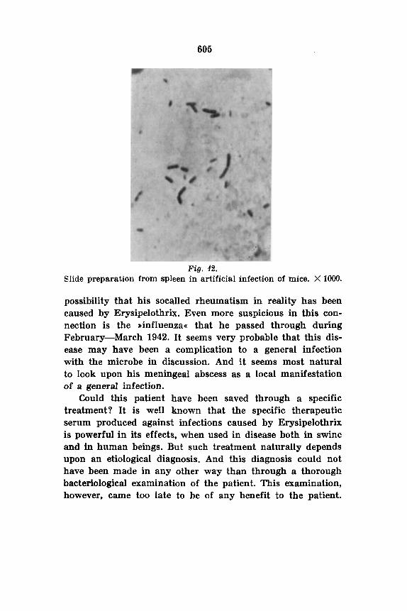
When the picture that met our eye when first seen in the slide preparation from the patient seemed so unfamiliar, the reason obviously was that the usual textbooks describe this microbe as a very small and delicate rod. This appearance may well be the ordinary in the acute swine erysipelas and in the spleen of mice infected with the microbe (Fig. 12). But in more chronic infections and obviously in the human being one must reckon with the threadlike microbe, the real
38.
to9
606
Fig. 62. Slide preparation from spleen in artificial infection of mice. X 1OOO.
possibility that his socalled rheumatism in reality has been caused by Erysipelothrix. Even more suspicious in this con- nection is the ,influenza< that he passed through during February-March 1942. It seems very probable that this dis- ease may have been a complication to a general infection with the microbe in discussion. And it seems most natural to look upon his meningeal abscess as a local manifestation of a general infection.
Could this patient have been saved through a specific treatment? It is well known that the specific therapeutic serum produced against infections caused by Erysipelothrix is powerful in its effects, when used in disease both in swine and in human beings. But such treatment naturally depends upon an etiological diagnosis. And this diagnosis could not have been made in any other way than through a thorough bacteriological examination of the patient. This examination, however, came too late to be of any benefit to the patient.

606
Conclusions. 1. The author deals with the history of a patient suffering
from an abscess of the meninges caused by Erysipelothrix rhusiopathiae.
2. The morphological and cultural characteristics of this microbe as found in pus from this abscess are described.
3. It is stressed that one should not always expect to find Erysipelothrix as a straight or slightly curved delicate rod, but rather expect to find this microbe as long threads twisted together in large bundles and clumps. This picture will be met with both in pus and in material from endocarditis and in semisolid media.
LITERATURE . 1. Dumont, J . et Cotoni, L.: Ann. Inst. Pasteur, 1921, 35, 625. 2. Riebe, W. : Arch. f. wiseenschaftl. u. prakt. Tierheilk. Bd. 37, cit.
fra Kolle, Kraus u. Uhlenhuth Handb. d. path. Mikroorg. 3de opl., Bd. VI, 1, 456.
3. Prausnitz , C.: Centralb. f. Bakt., 1921, 85, 362. 4. Russel, W i l l i a m 0. and Lamb, Marion E.: J. A. M. A., 1940, 444,
5. Langrand, P.: Hyg. d. la Viande et du lait, 1912, 361, cit. ,fra
6. Panisset , L . et Kolda, J.: C. rend. 8oc. biol., 1924, 343. 7 . Rosenbach, J . F.: Arch. f. klin. Chir., 1887, 36, 346. 8. Priesz, Hugo v.: Kolle, Kraus u. Uhlenhuth: Handb. d. Mikroorg.
1045.
Kolle, Kraus u. Uhlenhuth etc. 1929, VI, 1, 452.
1929, VI, 1.