Embed Size (px)
Citation preview

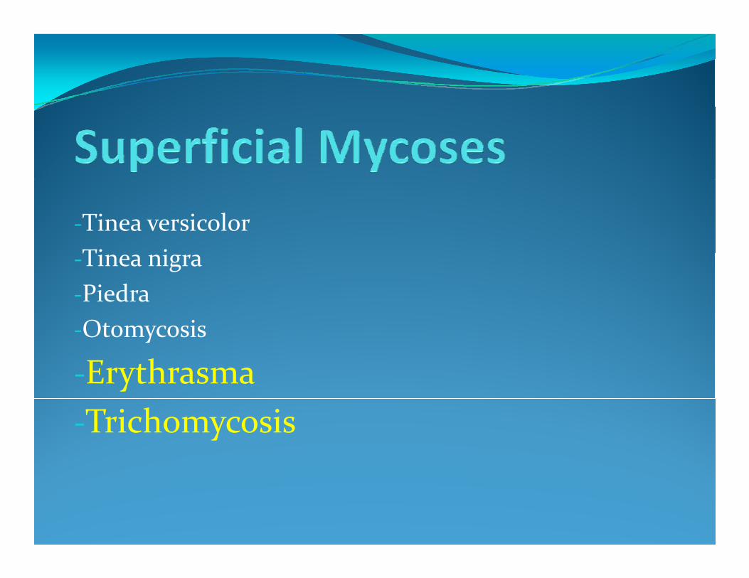
‐Tinea versicolorTi i‐Tinea nigra‐PiedraOtom cosis‐Otomycosis
‐Erythrasma‐Trichomycosis
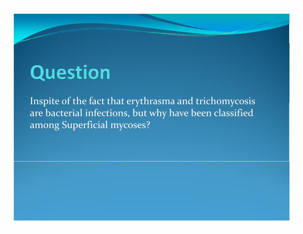
Inspite of the fact that erythrasma and trichomycosis Inspite of the fact that erythrasma and trichomycosis are bacterial infections, but why have been classified among Superficial mycoses?
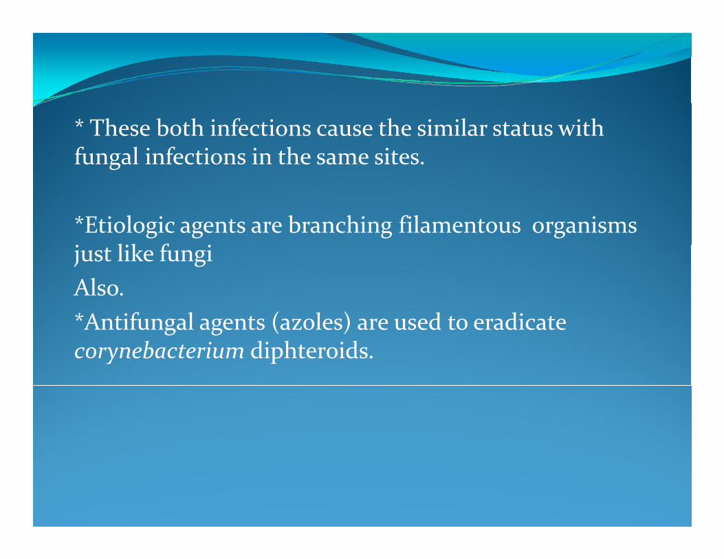
* These both infections cause the similar status with fungal infections in the same sites.
*Etiologic agents are branching filamentous organisms l k fjust like fungi
Also.*A if l ( l ) d di *Antifungal agents (azoles) are used to eradicate corynebacterium diphteroids.
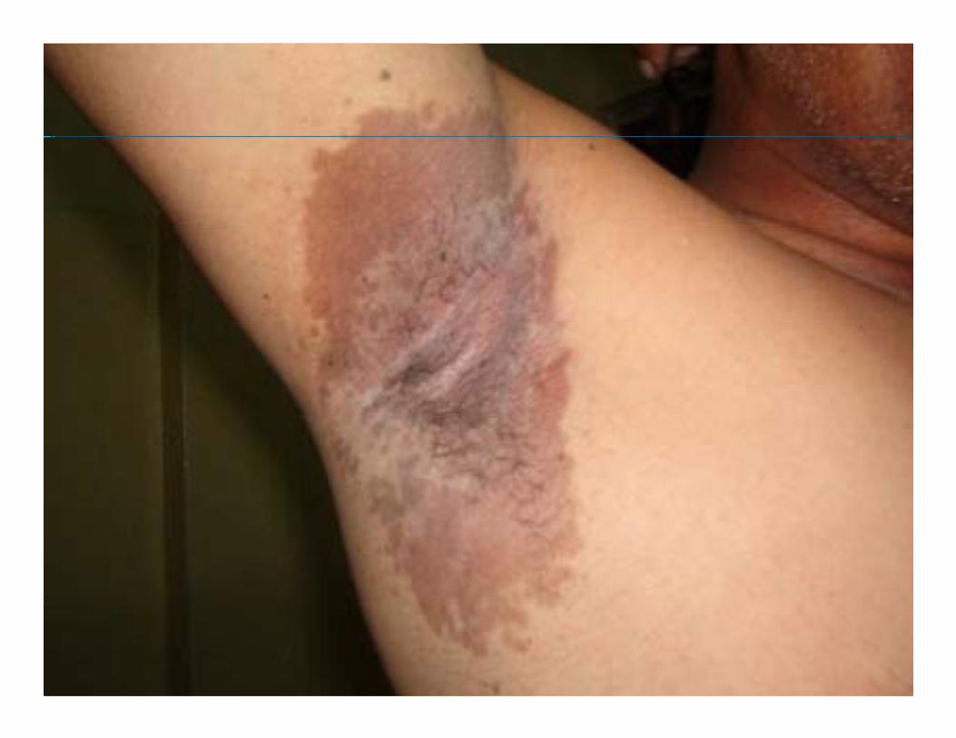
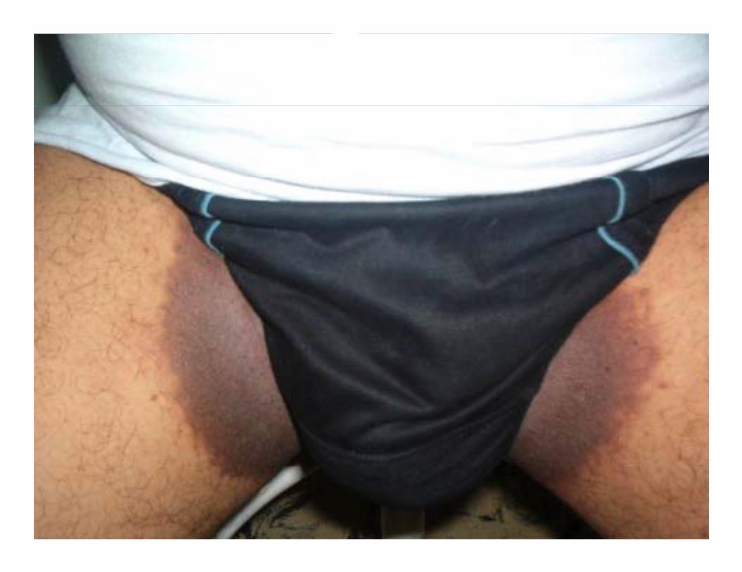
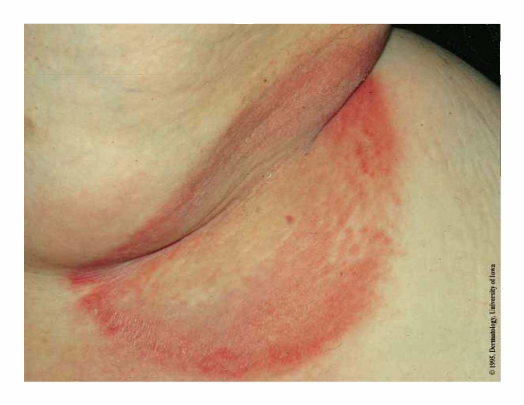
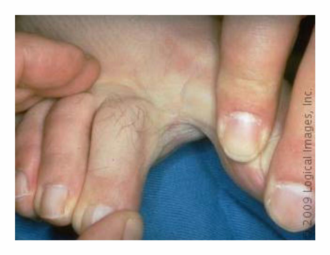
A chronic Superficial infection of The intertriginous of the Skin, Such as:‐Axillae‐Groin‐Inframammary‐Toe webs spaces (frequently fourth interdigital space)‐Scrotum‐Abdominal folds
‐Corynebacterium minutissimum, a lipophilic, Gram‐positive, non‐spore forming, aerobic and catalase‐p , p g,positive diphteroid‐Corynebacterium afermentase (1case in 1996)‐Up to 50% of the normal flora of the skin* The disease was considered as a superficial fungal i f i b i i d b i l di infection but, it was recognized as a bacterial disease by “Lagana” in 1960.
‐ Infection is observed all over the world.‐More common is in tropical and subtropical areas‐Both sexes are equally affected.q y‐Crural form is more common in men.‐Most common site of affection is toe‐clefts‐Inframammary, abdominal folds and perineum involvments are common in obese middle‐aged women.‐Rare in children.Bl k l hi h i k‐Black people are high risk
‐Excessive Sweating / hyperhydrosis‐Delicate Cutaneous barrier‐Obesity‐Diabetes melitus‐Warm climate‐Poor hygiene‐A dvanced age
h d‐Other immunocompromised states
‐Red –brownish, Slightly scaly macular patches with sharp borders in intertriginous areas of the skinsharp borders in intertriginous areas of the skin‐Patches may itch slightly‐Lichenification and hyperpigmentation are commonLichenification and hyperpigmentation are common‐The signs in toe web spaces as the most common interdigital infection of the foot are include:g•Fissuring •Scaling•Maceration
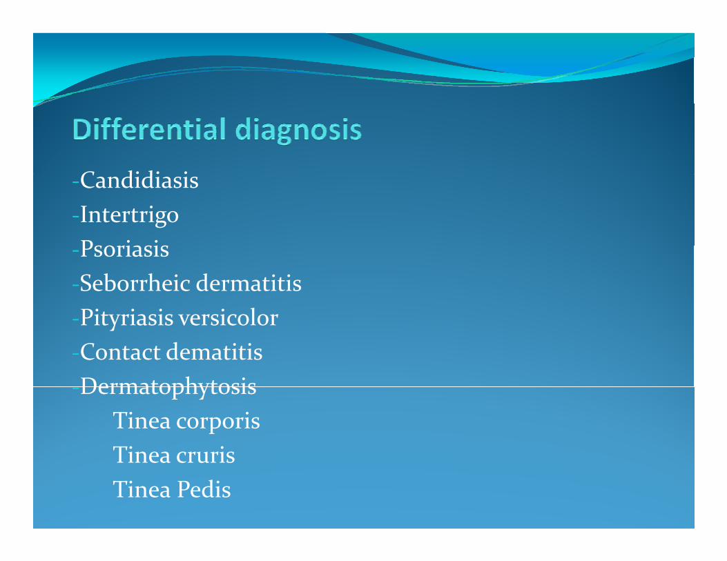
C didi i‐Candidiasis‐IntertrigoP i i‐Psoriasis‐Seborrheic dermatitisPityriasis versicolor‐Pityriasis versicolor‐Contact dematitisDermatophytosis‐Dermatophytosis
Tinea corporisTinea crurisTinea crurisTinea Pedis
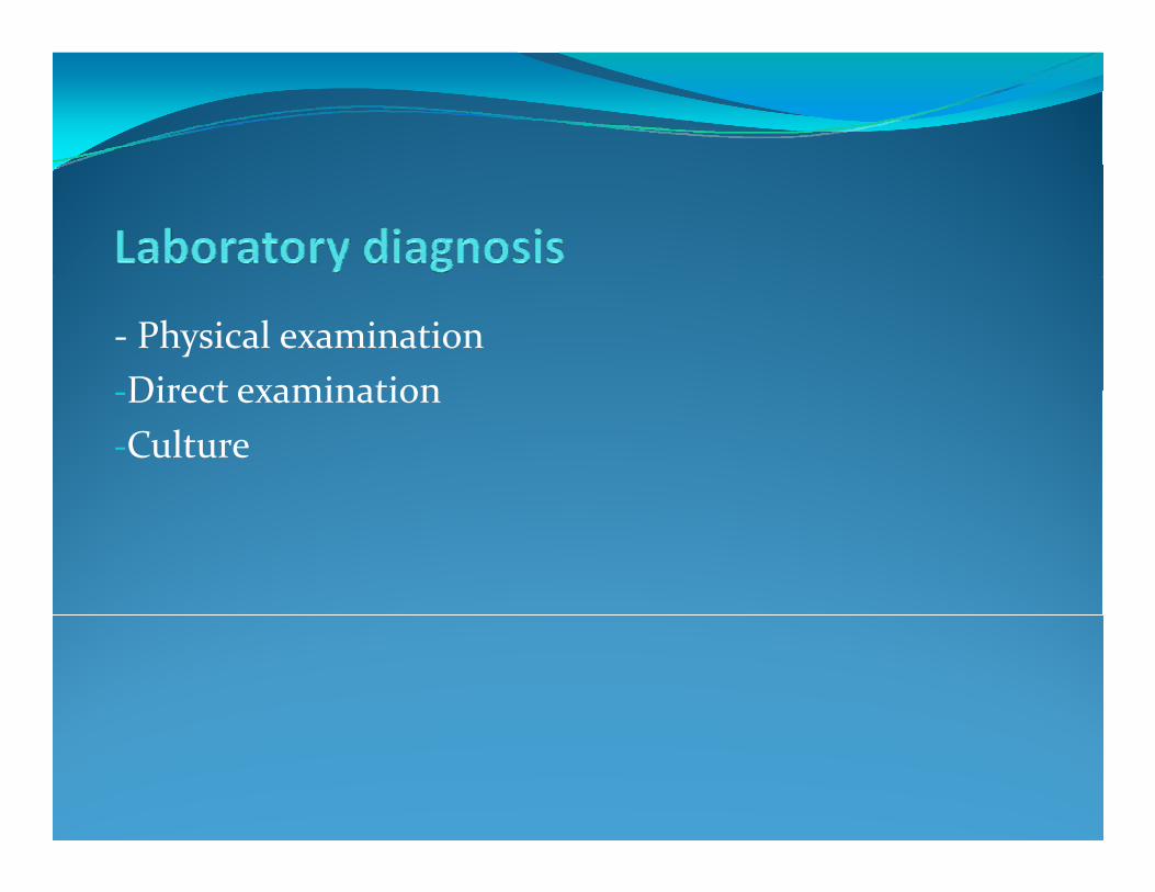
‐ Physical examinationDirect examination‐Direct examination‐Culture
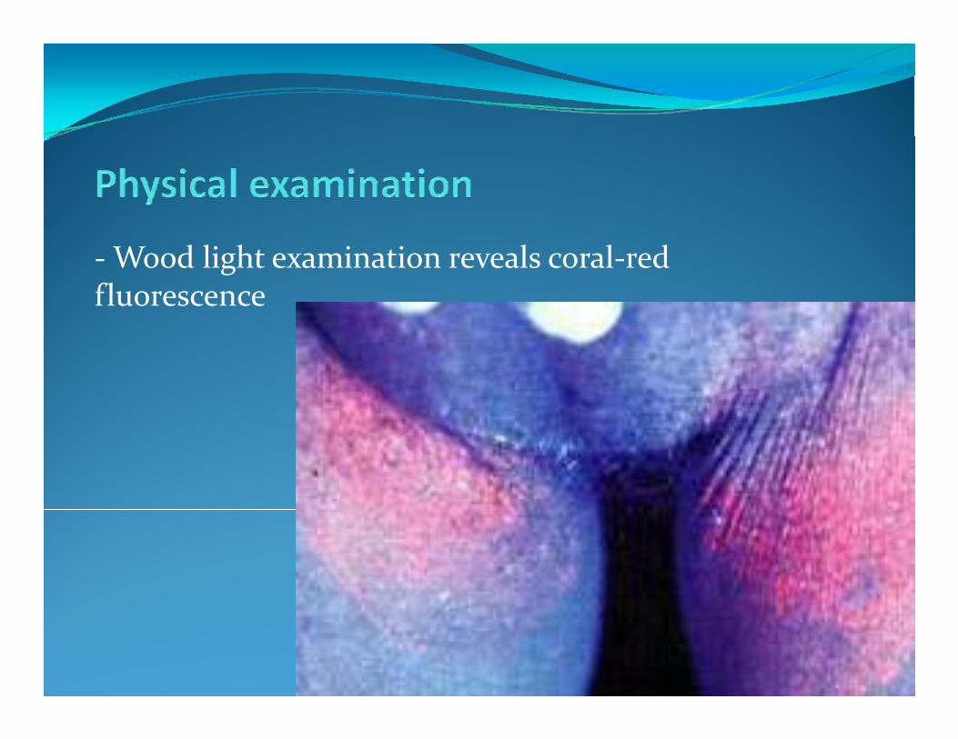
‐Wood light examination reveals coral‐red fluorescence
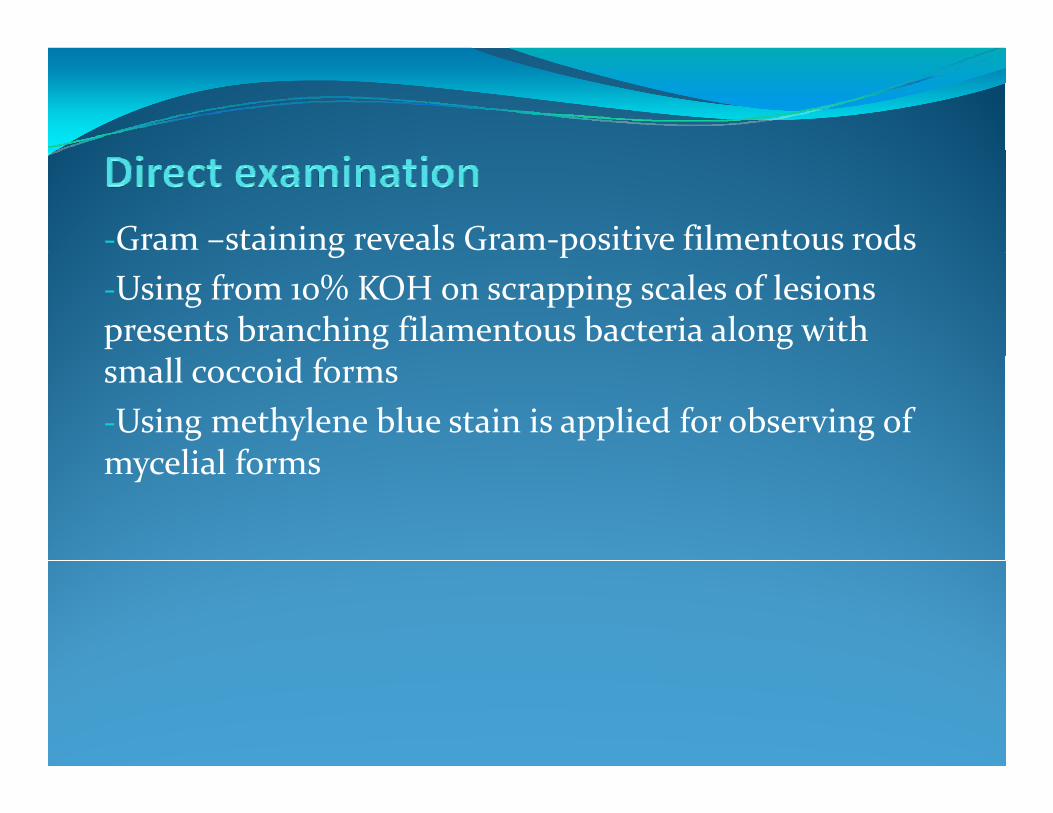
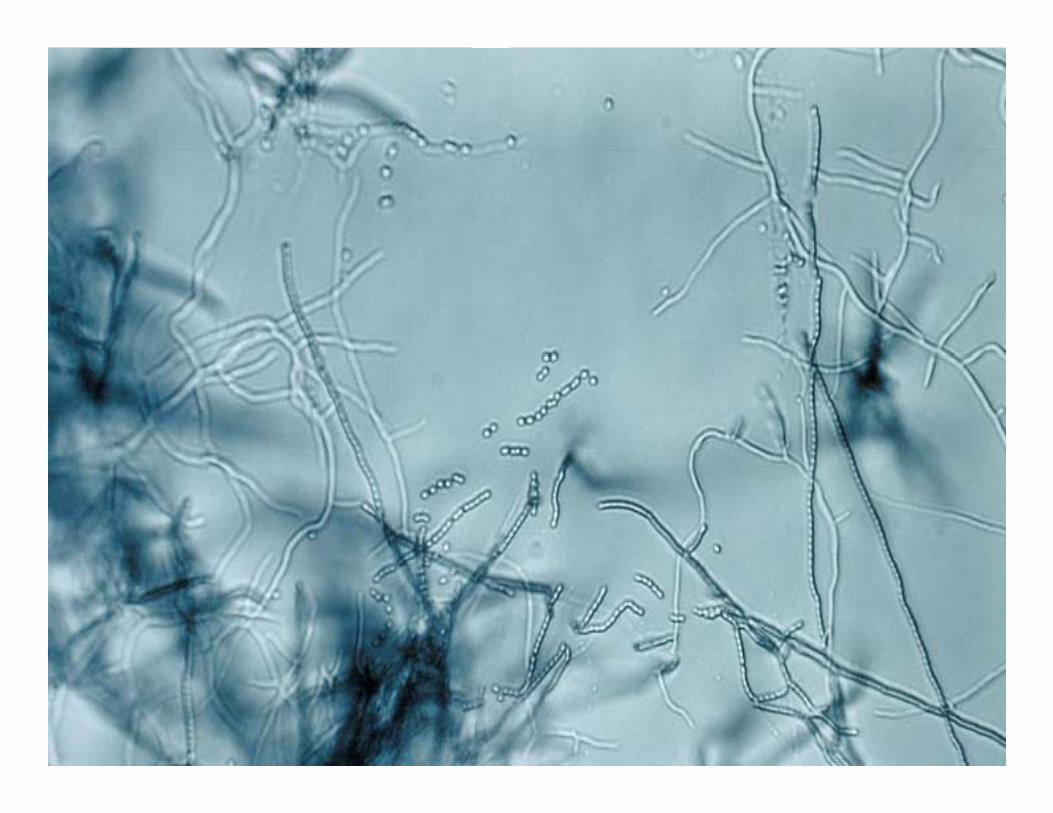
‐Gram –staining reveals Gram‐positive filmentous rodsg p‐Using from 10% KOH on scrapping scales of lesions presents branching filamentous bacteria along with small coccoid forms‐Using methylene blue stain is applied for observing of m celial formsmycelial forms
‐Cuture typically not recommended for diagnosis‐Isolation of organisms by growing in 20% fetal bovine serum 2% agar 78% tissue culture medium # 199 and serum, 2% agar, 78% tissue culture medium # 199 and 0.05% tris‐Mueller‐ Hinton agar is simple medium for pigment Mueller Hinton agar is simple medium for pigment production by erythrasma diphtheroids
‐Erythromycin 250 mg four times daily for 14 days (choise)Topical and systemic antibacterial and antifungal ‐Topical and systemic antibacterial and antifungal agents are also used‐Topical antimicrobial ointments are not ideal because Topical antimicrobial ointments are not ideal because of lack cosmetic elegance‐Generally, creams are not ideal for Using in skin folds y gand interdigital spaces‐Relapse may be occurred, so the areas should be k d l d dkeeped clean and dry
‐Maintaining good hygiene‐Keeping the skin dry‐Wearing clean, absorbent clothing‐Avoiding excessive heat or moisture‐Maintaining healthy body weight
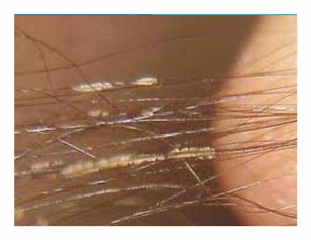
A superficial infection of the axillary or pubic hair, Ch t i d b th f ti f ll (fl ) d Characterized by the formation of yellow (flava), red (rubra) or black (nigra) nodules or Cylindrical sheaths around the hair shaft
‐Corynebacterium tenuisy‐Causative organism was thought to be a Nocordiaspecies ‐It is colonized on the hair shafts in sweat glandbearingareas such as the armpits and the pubic area
‐Widespread in the tropical and subtropical areas‐Incidence of the disease varies from 27% to 30% of adult malesPrevalence of trichomycosis axillaris is more than ‐Prevalence of trichomycosis axillaris is more than pubis‐The disease is not limited by race or sexThe disease is not limited by race or sex‐Age groups from puberty through adulthood* Risk factors including: Risk factors including:‐Hight humidity and warmth in tropacal areas ‐Poor hygiene
C l i t f di l d S t t i d l th‐Complaints of discolored, Sweat‐stained clothes‐It is characterized by yellow, black or red granular or Cylindrical nodules or concretions that stick to the hair Cylindrical nodules or concretions that stick to the hair shaft.‐Usually the condition is symptomless and there are y y pSweaty, smelly armpits‐Hair shafts expand apearing more noticeable after bathing
‐Pediculosis ‐Monilethrix (beaded hair)( )‐Trichorrhexis nodosa‐White piedrap‐Ringworm
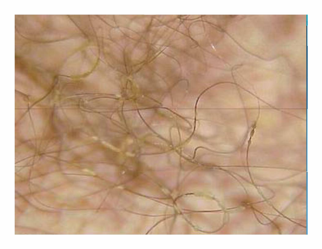
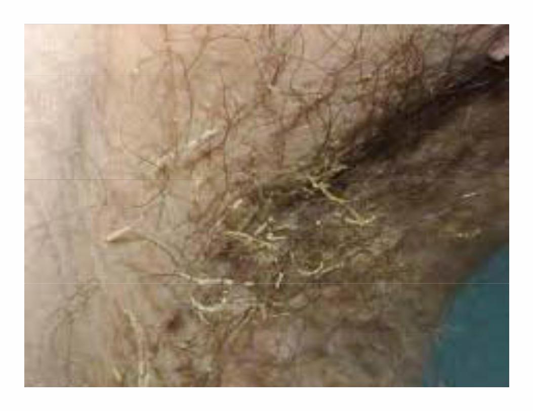
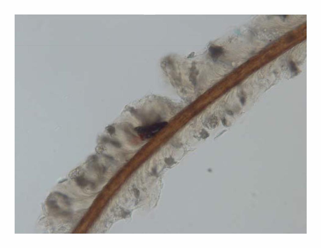
* Direct examination‐Observation of cylindrical or granular sheaths around the hair shafts (yellow, black or red) (magnification ×10)

C lt t i ll t d d f di i‐Culture typically not recommended for diagnosis‐Culture on sheep blood agar and brain heart infusion agar in 250 agar in 25 C
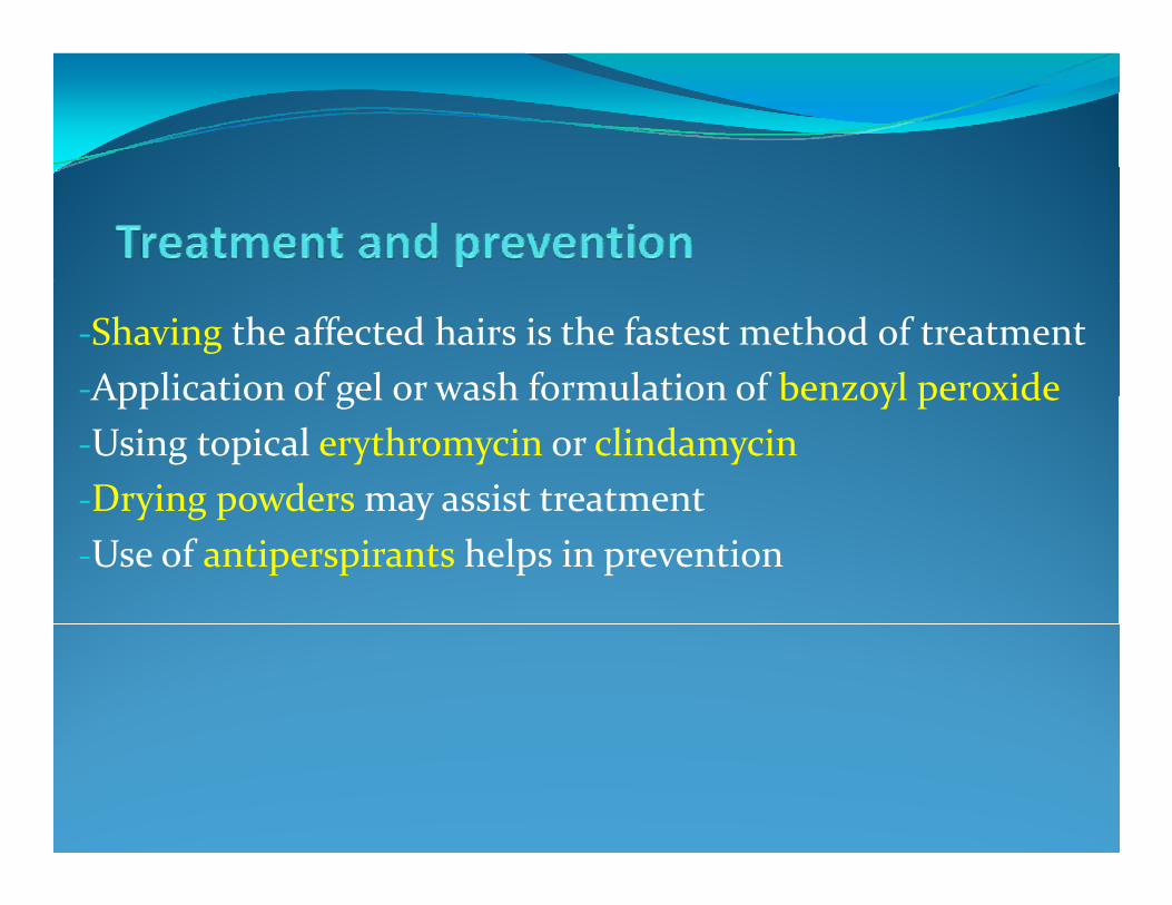
‐Shaving the affected hairs is the fastest method of treatment‐Application of gel or wash formulation of benzoyl peroxideApplication of gel or wash formulation of benzoyl peroxide‐Using topical erythromycin or clindamycin‐Drying powders may assist treatmentDrying powders may assist treatment‐Use of antiperspirants helps in prevention


![Open Journal of Clinical & Medical - jclinmedcasereports.comjclinmedcasereports.com/articles/OJCMCR-1022.pdf · [b] Erythrasma can be treated with topical agents such as clindamycin](https://img.pdfslide.net/doc/110x75/5ceb197888c993107a8c40d3/open-journal-of-clinical-medical-b-erythrasma-can-be-treated-with-topical.jpg)


![Dermatojuin09.ppt [Mode de compatibilité] - Psychaanalyse · (econazole..), Erythromycine locale (si erythrasma).. Intertrigo. Impetigo n Infection cutanés superficielle, souvent](https://img.pdfslide.net/doc/110x75/5ad803fa7f8b9ab8378cd600/mode-de-compatibilit-psychaanalyse-econazole-erythromycine-locale-si-erythrasma.jpg)