Embed Size (px)
Citation preview

Early View
Original article
Esophageal pressure as a surrogate of pleural
pressure in mechanically-ventilated patients
Antoine Tilmont, Benjamin Coiffard, Takeshi Yoshida, Florence Daviet, Karine Baumstarck, Geoffrey
Brioude, Sami Hraiech, Jean-Marie Forel, Antoine Roch, Laurent Brochard, Laurent Papazian,
Christophe Guervilly
Please cite this article as: Tilmont A, Coiffard B, Yoshida T, et al. Esophageal pressure as a
surrogate of pleural pressure in mechanically-ventilated patients. ERJ Open Res 2021; in
press (https://doi.org/10.1183/23120541.00646-2020).
This manuscript has recently been accepted for publication in the ERJ Open Research. It is published
here in its accepted form prior to copyediting and typesetting by our production team. After these
production processes are complete and the authors have approved the resulting proofs, the article will
move to the latest issue of the ERJOR online.
Copyright ©ERS 2021. This article is open access and distributed under the terms of the Creative Commons Attribution Non-Commercial Licence 4.0.

Esophageal pressure as a surrogate of pleural pressure in
mechanically-ventilated patients
Author Information:
Antoine Tilmont M.D.1, Benjamin Coiffard M.D., MsC
1, Takeshi Yoshida M.D., Ph.D.
2,7
Florence Daviet M.D.1, Karine Baumstarck M.D., Ph.D
3, Geoffrey Brioude M.D.
4, Sami
Hraiech M.D., Ph.D1, , Jean-Marie Forel, M.D., Ph.D
1, Antoine Roch M.D., Ph.D
1,6, Laurent
Brochard M.D., Ph.D.5,7
, Laurent Papazian M.D., Ph.D1,7
, Christophe Guervilly M.D. MsC1,7
1 Assistance Publique - Hôpitaux de Marseille, Hôpital Nord, Médecine Intensive Réanimation,
13015, Marseille, France; Aix-Marseille Université, Faculté de médecine, Centre d'Etudes et de
Recherches sur les Services de Santé et qualité de vie EA 3279, 13005, Marseille, France
2 Department of Anesthesiology and Intensive Care Medicine, Osaka University Graduate School of
Medicine, Suita, Japan
3 Aix-Marseille Univ, School of medicine - La Timone Medical Campus, EA 3279 CEReSS - Health
Service Research and Quality of Life Center, 27 bd Jean Moulin cedex 05, F-13385, Marseille, France
4 Department of Thoracic Surgery and Esophageal Diseases, Hôpital Nord, Assistance Publique-
Hôpitaux de Marseille, Aix-Marseille University, Marseille, France
5 Interdepartmental Division of Critical Care Medicine, University of Toronto, Keenan Research
Centre, Li Ka Shing Knowledge Institute, St. Michael's Hospital, 30 Bond St, Toronto, ON, M5B
1W8, Canada
6 Assistance Publique - Hôpitaux de Marseille, Hôpital Nord, Service des Urgences, 13015, Marseille,
France; Aix-Marseille Université, Faculté de médecine, Centre d'Etudes et de Recherches sur les
Services de Santé et qualité de vie EA 3279, 13005, Marseille, France
7 PLeUral pressure working Group (PLUG—Acute Respiratory Failure section of the European
Society of Intensive Care Medicine, Brussels, Belgium
Corresponding Author: Christophe Guervilly, Médecine Intensive Réanimation, Hôpital
Nord, Chemin des Bourrely, 13015 Marseille, France. Phone: +33 491 965 842 / Fax:
+33 491 965 837 / Email: [email protected]
Registered on June 7th
2017 in the clinical trial.gov database as NCT03179644, Principal
investigator: Christophe Guervilly M.D. A brief description of the protocol is available on
https://clinicaltrials.gov/ct2/show/NCT03179644
.
Word count: 2829 words;
Funding statement: A grant of 13650 Euros from Assistance Publique-Hôpitaux de
Marseille has covered the material costs of the study
Conflict of interest: Pr Laurent Brochard has declared financial relationships with Medtronic
Covidien, Fisher Paykel, and has received equipment from Air Liquide, Sentec and Philips.
Dr Christophe Guervilly has declared financial relationships with Xenios Fresenius Medical
Care. The other authors do not declare any competing interests.
Abstract
Background: Esophageal pressure (Pes) is used to approximate pleural pressure (PPL)
and therefore to estimate transpulmonary pressure (PL).
Objectives: We aimed to compare esophageal and regional pleural pressures and to
calculate transpulmonary pressures in a prospective physiological study on lung
transplant recipients during their stay in the intensive care unit of a tertiary university
hospital.
Methods: Lung transplant recipients receiving invasive mechanical ventilation and
monitored by esophageal manometry and dependent and non-dependent pleural catheters
were investigated during the post-operative period. We performed simultaneous short
time measurements and recordings of esophageal manometry and pleural pressures.
Expiratory and inspiratory PL were computed by subtracting regional PPL or Pes from
airway pressure; inspiratory PL was also calculated with the elastance ratio method.
Results: Sixteen patients were included. Among them, 14 were analyzed. Esophageal
pressures correlated with dependent and non-dependent pleural pressures during
expiration, respectively R2=0.71, p=0.005 and R
2=0.77, p=0.001 and during inspiration,
respectively, R2=0.66 for both (respectively p=0.01 and p=0.014). PL calculated using Pes
were close to those obtained from the dependent pleural catheter but higher than those
obtained from the non-dependent pleural catheter both during expiration and inspiration.
Conclusion: In ventilated lung transplant recipients, esophageal manometry is well
correlated to pleural pressure. Absolute value of Pes is higher than pleural pressure of
non-dependent lung regions and could therefore underestimate the highest level of lung
stress in these at high risk of overinflation.
Keywords : Pleural pressure; esophageal pressure; transpulmonary pressure; dependent
and non-dependent lung regions; lung transplant recipient.
Introduction
Mechanical ventilation for Acute Respiratory Distress Syndrome (ARDS) is still challenging.
Recent guidelines have established strong recommendations for using low tidal volumes (Vt)
(4-8 ml/kg predicted bodyweight) and limiting plateau pressure (Pplat) (1). Concerning the
level of positive end expiratory pressure (PEEP) to apply, there is no well-established
recommendation notably to use high level of PEEP for patients with the most severe ARDS.
Based on a previous pilot study (2), some experts recommend to set PEEP using esophageal
manometry by targeting the transpulmonary plateau pressure. Esophageal pressure (Pes) is
used since decades by physiologists as a surrogate of pleural pressure (PPL) measurement and
allows the calculation of the true lung distending pressure, the so-called transpulmonary
pressure, PL= P airway (Paw) minus Pes (3). However, there is controversies about using the
absolute value of Pes, and some authors recommend to consider the tidal variation of
esophageal pressure which allow the calculation of the ratio of the elastance of the chest wall
to the respiratory system (4).
Recently, in a ventilated lung-injured pig model and a human-cadaver ventilated model, Dr
Yoshida et al. have conciliated these two theories through comparisons of dependent and

non-dependent pleural pressures to esophageal pressure (3). The main result of this latter
study is that Pes accurately estimates the dependent pleural pressure both at inspiratory and
expiratory pressures and that elastance derived inspiratory transpulmonary pressure
accurately estimates the non-dependent inspiratory transpulmonary pressure.
Therefore, the objective of this study was to compare the Pes with dependent and non-
dependent pleural pressures in lung transplanted recipients receiving invasive mechanical
ventilation during the post-operative period. Our hypothesis is that transpulmonary pressure
calculated with the Pes could underestimate the regional PL of the non-dependent lung.
Methods
Study design, setting and participants
This study was registered in the clinical trial.gov database on June 7th
2017 as NCT03179644
and approved by the ethical committee (Comité de Protection des Personnes Sud
Méditerranée, as 2016-A00567-44). This study was conducted in the North University
Hospital medical ICU, Marseille, France. According to the French legislation, all patients
gave their written informed consent to participate.
Patient were included if they fulfilled the following inclusion criteria: age ≥ 18 years
admitted in the ICU after a double-lung transplantation and mechanically ventilated.
Exclusion criteria were: age < 18 years, pregnancy or breast feeding, lack of medical in
assurance, deprivation of liberty by a judicial or administrative decision, those hospitalized
without consent, single lung transplantation and contra-indication to placement of a
nasogastric tube (esophageal varices, esophageal cancer, surgery of the esophagus of less
than 1 year). Patients were not included in case of admission in the ICU with open chest after
surgery and/or high flow air leaks (> 10% of inspired volume) or if they had systemic
sclerosis with esophageal involvement.

Pleural Pressures Measurements
Before chest closure, the thoracic surgeon introduced the multi-holes pleural catheters
(Pleurocath, plastimed Inc, France) along the thoracic drains under direct view. The non-
dependent catheters were positioned at the surface of the anterior visceral pleura, dependent
catheters were positioned at the surface of the posterior visceral pleura (Supplementary
Figure 1). According to surgical considerations, two or four pleural catheters were positioned
on the right and/or left side, at least one to measure the dependent pleural pressure and one to
measure the non-dependent pleural pressure per patient. Before measurement, we verified
catheter emptiness with 5 ml of air. Chest tubes were then clamped during measurements.
Pleural catheters were thereafter connected to a pressure port of the Fluxmed monitor,
(MBMED Inc, Argentina). The good transmission of pleural pressure was assessed by an
occlusion test as shown in Figure 2 .We performed 3 to 5 minutes recordings for each pleural
tracings during the first 48 hours post-operative.
Esophageal Pressures Measurements
An esophageal balloon catheter (Nutrivent TM
, Sidam, Mirandola, Italy) was inserted and
inflated with a minimal, non-stress volume (2-3 ml) of air as recommended (4). The adequate
position of the balloon in the lower part of the esophagus was confirmed by presence of
cardiac artifacts on the esophageal curve and a positive occlusion test (expiratory hold on the
ventilator) in passive conditions with gentle chest compression (5). Esophageal pressure was
recorded by the same device used for pleural pressure recordings. The occlusion test was
considered as positive if the relationship between ∆PPL and ∆Paw should yield a slope of
1.0±0.2 cm H2O, as well as between ∆Pes and ∆Paw. In case of negative test, tracings and
measurements were not analyzed. Measurements were performed in static condition (zero
flow) during an end inspiratory occlusion pause of 2 sec allowing the measurement of

respectively Pplat and inspiratory Pes (Pes, insp) and following an end expiratory occlusion
pause of 5 sec allowing the measurement of respectively total PEEP (PEEPtot) and expiratory
Pes (Pes, exp).
Definitions and Calculations
The following formula were used for assessment of transpulmonary pressures (PL).
Inspiratory transpulmonary pressure (PL insp), using esophageal pressure as PL insp, es= Pplat -
Pes, insp, or using direct measurement of PPL in non-dependent lung, as PL, ND, insp=Pplat – PPL,
ND, insp and in dependent lung, as PL, D, insp=Pplat – PPL, D, insp.
Conversely, expiratory transpulmonary pressure (PL exp) were determined using esophageal
pressure as PL exp ,es= PEEPtot - Pes, exp, or using direct measurement of PPL in non-dependent
lung, as PL, ND, exp=PEEPtot – PPL, ND, exp and in dependent lung, as PL, D,exp=PEEPtot – PPL, D,exp.
Additionally, PL insp was also calculated from elastance ratio of chest wall to respiratory
system (6) , as PL insp, ER = Pplat – [Pplat x ELCW / ELRS]. Accordingly, respiratory system
elastance (ELRS) = (Pplat – PEEPtot) / Vt and, chest wall elastance (ELCW) = (Pes, insp - Pes, exp)
/ Vt. All pressures were expressed in cm of water (cmH2O).
Statistical analysis
As it is an exploratory physiological study, no statistical power calculation was anticipated.
However, the ethical committee allowed to enrol a maximum of 45 patients during a two
years period. All presented results are part of the primary analysis of the data. All statistics
were performed by two-tailed tests. Continuous variables were reported as the mean±sd or
median (inter-quartiles ranges) as appropriate. Comparisons were performed by Student’s t-
test or by Mann Whitney test as appropriate. Categorical variables were expressed as the
absolute value and percentage. Comparisons were performed by Chi-square test. Normality
of the distribution of variables were tested by the Kolmogorov-Smirnov and the Shapiro-Wilk

tests. Correlations were performed with Pearson correlation test with further Bland and
Altman analysis for each correlation. A two-way repeated-measures analysis of variance
(ANOVA) was performed to compare transpulmonary pressures at end expiration and end
inspiration according to the modality of calculation and to the level of applied PEEP. The
normality of the distribution of the residuals, the assumption of sphericity and the interaction
between transpulmonary pressures and PEEP were checked. Intra-group differences were
evaluated by post hoc Bonferroni pairwise multiple comparisons. A p value < 0.05 was
retained as significant. All statistics and figures were performed with the SPSS 20.0 package
(SPSS, Chicago, IL, USA).
Results
Patients and measurements
Twenty two lung transplant recipients gave their informed consent before surgery (see flow
chart as Figure 1). Six patients were secondary excluded. Sixteen lung transplant recipients
were recorded. Two additional patients were not analyzed because of negative occlusion test
(correlations between ∆Pes and ∆Paw and/or ∆PPL and ∆Paw <0.8). Main characteristics of
the fourteen remaining patients are displayed in Table 1. An illustrative tracing of pressures,
flow and volume during an occlusion test with chest compression is provided in Figure 2.
Fifty percent of patients were assisted by veno-venous extracorporeal membrane oxygenation
(vvECMO) at ICU admission. All measurements were performed while patients were
sedated and mechanically ventilated in volume assisted controlled mode with a range of
PEEP between 8 and 14 cmH2O without spontaneous breathing effort. Among the 14
patients, 4 had daily serial measurements totalizing 24 measurements.
Correlations between esophageal and pleural pressures
Occlusions tests yield 0.95±0.05 for ∆Pes/∆Paw and 0.94 ±0.06 for ∆PPL/∆Paw. Dependent
and non-dependent expiratory pleural pressures were significantly correlated with expiratory
esophageal pressure (respectively R2=0.71 and R
2=0.77, p<0.01 for both) (Figure 3, panel
A). Dependent and non-dependent inspiratory pleural pressures were significantly correlated
with inspiratory esophageal pressure, respectively R2=0.66 for each (p<0.05) (Figure 3,
panel B). Esophageal pressure was always found higher than non-dependent pleural pressure.
During expiration time, mean difference between esophageal pressure and dependent pleural
pressure was 0.48±2.87 cmH20 and 5.25± 2.51cmH2O between esophageal pressure and
nondependent pleural pressure (Figure 3, panel A). During inspiration time, mean difference
between esophageal pressure and dependent pleural pressure was 0.98±2.90 cmH20 and
6.09± 2.90 cmH2O between esophageal pressure and non-dependent pleural pressure. The
mean difference between dependent pleural pressure and non-dependent pleural pressure was
4.76± 2.94 cmH2O at expiratory time and 5.38± 2.11 cmH2O at inspiratory time.
Correlations between transpulmonary pressures
Correlations and Bland and Altman analysis between inspiratory transpulmonary pressures
according the four ways of calculation are presented in Figure 4 (panel A). Inspiratory PL
computed from esophageal pressure were better correlated with inspiratory PL calculated from
dependent and non-dependent pleural pressures than those calculated from the elastance ratio
method (6) (R2 =0.604, R
2 = 0.629 and R
2 = 0.45, p<0.05 for all, respectively). However, the
estimated bias was higher between PL, insp, es and PL, ND, insp than between PL, insp, es and PL, D, insp
(-6±3.94 and – 1.61±3.62 cm H2O respectively). Correlations and Bland and Altman analysis
between expiratory transpulmonary pressures according the three ways of calculation are
presented in Figure 4 (panel B). Expiratory PL computed from esophageal pressure were
modestly correlated with expiratory PL calculated from dependent and non-dependent pleural

pressures (R2 =0.479 and R
2 = 0.531, p<0.02, respectively). However, the agreement was
better between PL, exp, es and PL, D, exp than between PL, exp, es and PL, ND, exp (estimated bias -1.34 ±
3.32 and -5.55 ±3.36 cmH2O respectively).
Relationship between expiratory transpulmonary pressures at different PEEP levels
Expiratory transpulmonary pressures calculated using Pes were close to those obtained from
the dependent pleural catheter (Figure 5- panel A). Expiratory transpulmonary pressure
calculated with non-dependent pleural catheter (PL, ND, exp) were higher than those calculated
from both dependent catheter (PL, D, exp) and esophageal pressure (PL exp, es) whatever the PEEP
level. We also found a significant interaction between PEEP and PL exp (R2=0.301, p=0.02).
Relationship between inspiratory transpulmonary pressures at different PEEP levels
Inspiratory transpulmonary pressures calculated using Pes was close to those directly
measured by the dependent pleural catheter (Figure 5- panel B). Inspiratory transpulmonary
pressure calculated from the elastance ratio of chest wall to respiratory system (PL insp, ER) was
also close to those measured using the non-dependent pleural catheter (PL, ND, insp). In our
model, PL insp, es underestimates the true regional transpulmonary pressure of the non-
dependent lung region (PL, ND, insp). We did not find interaction between PEEP and PL insp
(R2=0.132, p=0.203).
Discussion
In this mechanically ventilated in vivo human model, Pes is close to the pleural pressures of
the dependent lung region. However, we found overestimation by Pes of the non-dependent
lung region pleural pressures. Therefore, the limitation of inspiratory lung stress using Pes
may lead to underestimate the lung stress in non-dependent lung regions. Rather, inspiratory
PL calculated with the elastance ratio (PL insp, ER) may reflect local lung stress in non-
dependent lung regions which are usually the overinflated lung regions.

From previous clinical and experimental studies, we know that 1/ because of the weight of
the heart and of the increase of the gravitational gradient of pleural pressure during ARDS,
Pes is higher in supine patient ventilated for ARDS than those of non-ventilated healthy
subject in upright position(2,7,8) 2/ from experimental study in dogs (9), and recently in man
(10), it was demonstrated that absolute pleural pressures are approximately 7 cmH2O lower
than Pes in the non-dependent regions and 5 cmH2O higher in the dependent regions at low
intrathoracic pressure. Therefore, some authors have proposed to apply a correction
subtraction between 2.5 to 5 cmH2O to the actual measured esophageal pressure to calculate
the transpulmonary pressure (8,9,11). However, the utility of a fixed correction of absolute
transpulmonary pressure is still debated (12,13).
An experimental previous study (3) has demonstrated that in anesthetized pigs and human
cadavers, 1/ Pes was midway between PPL in dependent region and PPL in non-dependent
region and 2/ elastance derived transpulmonary pressure matched the directly measured
transpulmonary pressure from non-dependent regions.
In addition, Terzi et al.(14) showed in a ventilated pig model that in supine position, mean
difference between Pes and PL, D was 2.2 cmH2O and 7.2 cmH2O between Pes and PL, ND at
10cm H2O of PEEP. Interestingly, whereas prone position did not modify gradient between
Pes and PL, D, the gradient between Pes and PL, ND decreased to 1.8 cmH2O.
Pasticci et al.(10) have recently investigated pleural pressures in human, through the chest
tube on the surgery side immediately after lung resection of the non-dependent lung region in
lateral and supine positions. The main finding of the study was that esophageal pressures was
7.3 ± 2.8 cmH2O higher than non-dependent pleural pressure pleural pressures in supine
position. But, because of change of pleural pressure induced an identical change in
esophageal pressure, the transpulmonary pressures calculated with the elastance ratio

methods were perfectly correlated.
Therefore, the principal strength of our study is to confirm and duplicate in a human in
vivo setting, results from previous experimental and clinical studies (3,10,14) with the
unique characteristic to investigate simultaneously dependent, non-dependent pleural
pressures and esophageal pressure.
Minimal discrepancies could be explained by some differences between the models. First,
anatomy of the esophagus of pig and human are different with a more posterior location in
pig. Second, different cardiac and vascular filling pressures may explain differences in
absolute value of esophageal pressure observed in lung transplant recipients and cadavers.
Third, the pleural pressure sensors were different.
Despite some differences between our model and previous experimental models (animal and
cadaver), they also share some common results. In the supine position, the dorsal-to-ventral
pleural gradient from dependent to non-dependent lung region was 5.0 IQR (2.7-6.4) cm H2O
at inspiration and 4.4 IQR (1.9-5.6) at expiration in our study which is very close from those
in measured in pigs (median 4.4 IQR (2.4-6.8) cmH2O) (14) but lower to those measured in
cadavers (n=3, 10.0±3.1 cmH2O) (3). In this latter experiment, despite the ―Thiel method‖ to
restore elasticity of the tissues, it is possible that the model affects chest wall recoil force as
compared with human.
The elastance derived method to assess transpulmonary pressure (PL, insp, ER) found very close
values than those directly measured by PL, ND, insp. These findings are concordant with
experimental results and therefore suggesting that PL, insp, ER could be a valuable target to
prevent regional stress and strain of the non-dependent lung regions (3).

There are several limitations to the present study. First, we used a very specific in vivo model
of mechanically ventilated patients with some of them presenting acute lung injury following
lung transplantation (primary graft dysfunction). Second, after open chest surgery, presence
of chest tubes, even clamped with no vacuum, may have created some artifacts in the pleural
pressure signal. Third, we used common pleural catheter to measure pleural pressure and not
specific flat balloon pleural sensors which has only been used only for animal studies so far.
However, this was the only device allowed by the French safety drug administration for the
study.
Finally, even if esophageal pressures were well correlated with pleural pressures, we found a
significant bias of agreement between esophageal pressures and non-dependent pleural
pressures of 5.25±2.51 cmH2O at expiration time and 6.09± 2.90 cmH2O at inspiration time.
Of note, a non-inferior bias of agreement of 7.2± 5.56 cmH2O was also reported in a pig
model under strict experimental conditions (14).
Although of potential clinical interest, esophageal manometry is still very underused in
clinical practice in ARDS patients (0.8% in the cohort of all ARDS patients in the LUNG
SAFE study and 1.2% for severe ARDS patients)(15). Recently, the largest trial (EPVent-2
study) using esophageal manometry in ARDS patients (16) has failed to demonstrate outcome
benefit with targeting the expiratory transpulmonary pressure as compared with a strategy of
high PEEP based on a PEEP-FiO2 table.
Esophageal manometry may be still of clinical interest in specific ARDS clinical vignettes
notably when abdominal or chest wall elastance is increased (17) or unrecognized harmful
strong respiratory efforts (18,19). Esophageal manometry remains also useful to diagnose
patient – ventilator asynchrony which may worsen the outcome (20–22).
In conclusion, in ventilated lung transplant recipients, esophageal manometry was well
correlated to direct measure of pleural pressure with non-specific sensors and absolute value
was close to those from dependent lung. During controlled ventilation without respiratory
muscles activity, absolute value of Pes is higher than pleural pressure of non-dependent lung
regions and could therefore underestimate the highest level of lung stress in non-dependent
lung regions. In addition, the elastance derived method seems useful to prevent this pitfall.
Acknowledgments: The authors want to thank the participated patients, the team of the
thoracic surgery and the anesthetic department involved in the study, Dr Anderson Loundou,
Ph.D. (Aix-Marseille Univ, School of medicine - La Timone Medical Campus, EA 3279
CEReSS - Health Service Research and Quality of Life Center, Marseille, France) for his
statistical advices for the draft, Pr Benoit D’journo, M.D, Ph.D., (Department of Thoracic
Surgery and Esophageal Diseases, Hospital Nord, Assistance Publique-Hôpitaux de
Marseille, Aix-Marseille University, Marseille, France) and Pr Marc Gainnier, M.D, Ph.D.,
(Assistance Publique, Hôpitaux de Marseille, Hôpital Timone, Service de Médecine Intensive
Réanimation, Réanimation des Urgences, Assistance Publique-Hôpitaux de Marseille, Aix-
Marseille University, Marseille, France) for their comments.
References
1. Fan E, Del Sorbo L, Goligher EC, Hodgson CL, Munshi L, Walkey AJ, et al. An
Official American Thoracic Society/European Society of Intensive Care
Medicine/Society of Critical Care Medicine Clinical Practice Guideline: Mechanical
Ventilation in Adult Patients with Acute Respiratory Distress Syndrome. Am J Respir
Crit Care Med. 2017 May;195(9):1253–63.
2. Talmor D, Sarge T, Malhotra A, O’Donnell CR, Ritz R, Lisbon A, et al. Mechanical
Ventilation Guided by Esophageal Pressure in Acute Lung Injury. N Engl J Med. 2008
Nov 13;359(20):2095–104.

3. Yoshida T, Amato MBP, Grieco DL, Chen L, Lima CAS, Roldan R, et al. Esophageal
Manometry and Regional Transpulmonary Pressure in Lung Injury. American Journal
of Respiratory and Critical Care Medicine. 2018 Apr 15;197(8):1018–26.
4. Mojoli F, Chiumello D, Pozzi M, Algieri I, Bianzina S, Luoni S, et al. Esophageal
pressure measurements under different conditions of intrathoracic pressure. An in vitro
study of second generation balloon catheters. MINERVA ANESTESIOLOGICA.
2015;81(8):10.
5. Baydur A, Behrakis PK, Zin WA, Jaeger M, Milic-Emili J. A Simple Method for
Assessing the Validity of the Esophageal Balloon Technique. American Review of
Respiratory Disease. 1982; 4.
6. Gattinoni L, Chiumello D, Carlesso E, Valenza F. Bench-to-bedside review: chest wall
elastance in acute lung injury/acute respiratory distress syndrome patients. Crit Care.
2004 Oct;8(5):350-5. doi: 10.1186/cc2854.
7. Washko GR, O’Donnell CR, Loring SH. Volume-related and volume-independent
effects of posture on esophageal and transpulmonary pressures in healthy subjects.
Journal of Applied Physiology. 2006 Mar;100(3):753–8.
8. Talmor D, Sarge T, OʼDonnell CR, Ritz R, Malhotra A, Lisbon A, et al. Esophageal and
transpulmonary pressures in acute respiratory failure*: Critical Care Medicine. 2006
May;34(5):1389–94.
9. Milic-Emili G, Petit JM. Relationship between endoesophageal and intrathoracic
pressure variations in dog. Journal of Applied Physiology. 1959 Jul;14(4):535–7.
10. Pasticci I, Cadringher P, Giosa L, Umbrello M, Formenti P, Macri MM, Busana M,
Bonifazi M, Romitti F, Vassalli F, Cressoni M, Quintel M, Chiumello D, Gattinoni L.
Determinants of the esophageal-pleural pressure relationship in humans. J Appl Physiol
(1985). 2020 Jan 1;128(1):78-86.
11. Ranieri VM, Giuliani R, Mascia L, Grasso S, Petruzzelli V, Bruno F, et al. Chest wall
and lung contribution to the elastic properties of the respiratory system in patients with
chronic obstructive pulmonary disease. European Respiratory Journal. 1996 Jun
1;9(6):1232–9.
12. Terragni P, Mascia L, Fanelli V, Biondi-Zoccai G, Ranieri VM. Accuracy of esophageal
pressure to assess transpulmonary pressure during mechanical ventilation. Intensive
Care Med. 2017 Jan;43(1):142–3.
13. Baedorf Kassis E, Loring SH, Talmor D, Terragni P, Mascia L, Ranieri VM. A fixed
correction of absolute transpulmonary pressure may not be ideal for clinical use:
Discussion on ―Accuracy of esophageal pressure to assess transpulmonary pressure
during mechanical ventilation.‖ Intensive Care Med. 2017 Sep;43(9):1436–7.
14. N Terzi, S Bayat, N Noury, E Turbil, W Habre, L Argaud, M Cour, B Louis, C, Guérin.
Comparison of pleural and esophageal pressure in supine and prone position in a 2
porcine model of acute respiratory distress syndrome. Journal of Applied Physiology
[Internet]. 2020 [cited 2020 Aug 4];790601 Bytes. Available from:
https://figshare.com/articles/Ppl_vs_Pes_supplemental_data/12278342

15. Bellani G, Laffey JG, Pham T, Fan E, Brochard L, Esteban A, et al. Epidemiology,
Patterns of Care, and Mortality for Patients With Acute Respiratory Distress Syndrome
in Intensive Care Units in 50 Countries. JAMA. 2016 Feb 23;315(8):788.
16. Beitler JR, Sarge T, Banner-Goodspeed VM, Gong MN, Cook D, Novack V, et al.
Effect of Titrating Positive End-Expiratory Pressure (PEEP) With an Esophageal
Pressure–Guided Strategy vs an Empirical High PEEP-F IO 2 Strategy on Death and
Days Free From Mechanical Ventilation Among Patients With Acute Respiratory
Distress Syndrome: A Randomized Clinical Trial. JAMA. 2019 Mar 5;321(9):846.
17. Quintel M, Pelosi P, Caironi P, Meinhardt JP, Luecke T, Herrmann P, et al. An Increase
of Abdominal Pressure Increases Pulmonary Edema in Oleic Acid–induced Lung Injury.
Am J Respir Crit Care Med. 2004 Feb 15;169(4):534–41.
18. Mauri T, Langer T, Zanella A, Grasselli G, Pesenti A. Extremely high transpulmonary
pressure in a spontaneously breathing patient with early severe ARDS on ECMO.
Intensive Care Med. 2016 Dec;42(12):2101–3.
19. Yoshida T, Torsani V, Gomes S, De Santis RR, Beraldo MA, Costa ELV, et al.
Spontaneous Effort Causes Occult Pendelluft during Mechanical Ventilation. Am J
Respir Crit Care Med. 2013 Dec 15;188(12):1420–7.
20. Thille AW, Rodriguez P, Cabello B, Lellouche F, Brochard L. Patient-ventilator
asynchrony during assisted mechanical ventilation. Intensive Care Med. 2006
Oct;32(10):1515–22.
21. Blanch L, Villagra A, Sales B, Montanya J, Lucangelo U, Luján M, et al. Asynchronies
during mechanical ventilation are associated with mortality. Intensive Care Med. 2015
Apr;41(4):633–41.
22. Bourenne J, Guervilly C, Mechati M, Hraiech S, Fraisse M, Bisbal M, et al. Variability
of reverse triggering in deeply sedated ARDS patients. Intensive Care Med. 2019
May;45(5):725–6.
Figure legends
Figure 1. Flow diagram of the included patients.
Figure 2. Representative tracing of volume, flow, airway, esophageal and non-dependent
pleural pressures during an occlusion test. The increase of airway, esophageal and non-
dependent pleural pressures with the same magnitude during the gentle thoracic compression
(white arrows) ensure the correct placement of pleural catheter and esophageal balloon.
Figure 3. Correlations and Bland and Altman analysis between pleural pressures and
esophageal pressure, at end-expiration (A) and end-inspiration (B). For correlations, the
dotted line represents the identity line. Each circle represents a different patient. For Bland
and Altman analysis, black solid line and dotted thin lines represent the mean ± 2SD of the
differences. Abbreviations: R2, Pearson correlation test.
Figure 4. Correlations and Bland and Altman analysis between transpulmonary pressures,
during end-inspiration (A) and end-expiration (B). For correlations, the dotted line represents
the identity line. Each circle represents a different patient. For Bland and Altman analysis,
black solid line and dotted thin lines represent the mean ± 2SD of the differences.
Abbreviations: R2, Pearson correlation test.
Figure 5. Relationship of transpulmonary pressures calculated from esophageal pressure and
pleural pressures in mechanically ventilated human lung transplant recipients.
(A) During expiratory time at different PEEP levels, Abbreviations: PEEP positive end-
expiratory pressure. *p<0.05 compared with Pes and dependent catheter by post-hoc
Bonferroni test; Box plot represent median and 25th
-75th
percentile, outliers are
represented by empty circles.
(B) During inspiratory time at different PEEP levels. Abbreviations: PEEP positive end-
expiratory pressure. *p<0.05 compared with Pes and dependent catheter by post-hoc
Bonferroni test; Box plot represent median and 25th
-75th
percentile, outliers are
represented by empty circles.

Table 1. Characteristics of the patients
Subject
number
Age Gender SOFA SAPS 2 Indication for
BLT
vvECMO* Duration of Mechanical
Ventilation (days)
ICU lenght of stay
(days)
ICU survival
1 61 F 9 47 COPD No 4 13 yes
2 61 M 7 55 Fibrosis Yes 41 50 yes
3 41 M 7 40 Fibrosis No 3 9 yes
4 69 M 6 39 Fibrosis No 1 7 yes
5 69 M 8 53 Fibrosis Yes 8 13 yes
6 65 M 8 34 Fibrosis Yes 8 14 yes
7 65 M 11 58 Fibrosis Yes 5 5 no
8 62 M 5 46 Fibrosis No 5 13 yes
9 64 M 7 39 Fibrosis No 5 13 yes
10 61 F 11 51 COPD Yes 90 90 yes
11 53 M 9 48 Fibrosis Yes 6 10 yes
12 62 F 7 50 COPD No 43 47 yes
13 64 M 8 47 COPD Yes 14 14 no
14 64 M 10 52 COPD No 1 4 yes
Mean±sd 61±7 8±2 47±7 17±25 2±24
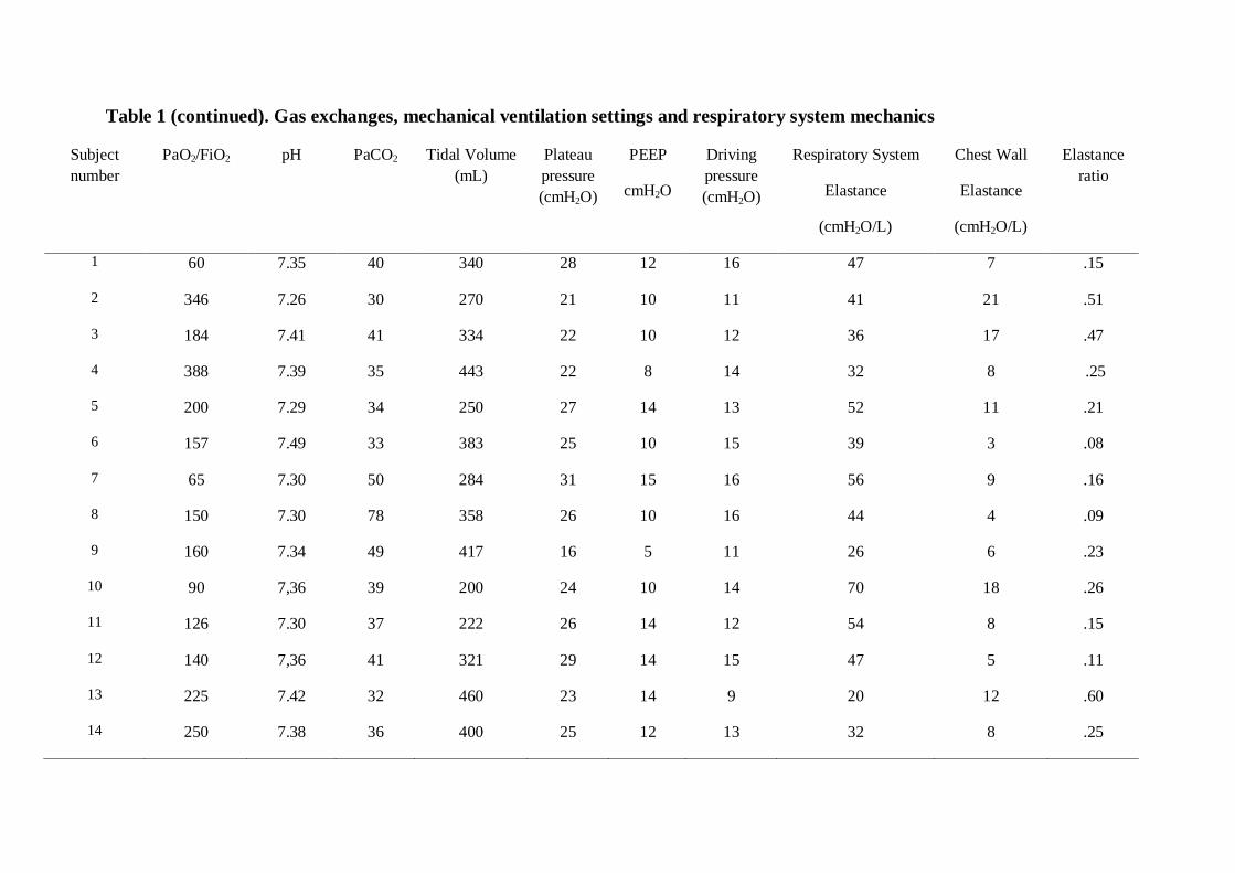
Table 1 (continued). Gas exchanges, mechanical ventilation settings and respiratory system mechanics
Subject
number
PaO2/FiO2 pH PaCO2 Tidal Volume
(mL)
Plateau
pressure
(cmH2O)
PEEP
cmH2O
Driving
pressure
(cmH2O)
Respiratory System
Elastance
(cmH2O/L)
Chest Wall
Elastance
(cmH2O/L)
Elastance
ratio
1 60 7.35 40 340 28 12 16 47 7 .15
2 346 7.26 30 270 21 10 11 41 21 .51
3 184 7.41 41 334 22 10 12 36 17 .47
4 388 7.39 35 443 22 8 14 32 8 .25
5 200 7.29 34 250 27 14 13 52 11 .21
6 157 7.49 33 383 25 10 15 39 3 .08
7 65 7.30 50 284 31 15 16 56 9 .16
8 150 7.30 78 358 26 10 16 44 4 .09
9 160 7.34 49 417 16 5 11 26 6 .23
10 90 7,36 39 200 24 10 14 70 18 .26
11 126 7.30 37 222 26 14 12 54 8 .15
12 140 7,36 41 321 29 14 15 47 5 .11
13 225 7.42 32 460 23 14 9 20 12 .60
14 250 7.38 36 400 25 12 13 32 8 .25

Mean±sd 181±96 7.35±0.06 41±12 334±81 25±4 11±3 13±2 42±13
10±5 .25±.16
F, female gender; M, male gender; SOFA, sepsis-related organ failure assessment score at inclusion; SAPS 2, simplified acute physiologic score 2 at inclusion; BLT, bilateral
lung transplantation; vvECMO, veno venous extracorporeal membrane oxygenation; ICU, intensive care unit; PEEP, positive end expiratory pressure; sd, standard deviation.
* At ICU admission.

Figure 1. Flow diagram of the included patients.

Figure 2. Representative tracing of volume, flow, airway, esophageal and non-dependent
pleural pressures during an occlusion test (white arrows).

Figure 3
(A)

Figure 3
(B)

Figure 4: Correlations and Bland and Altman analysis between transpulmonary pressures
Panel A: During inspiratory time
R2
=0.45, p=0.026
y=0.524x +13.07
R2
=0.604, p=0.002
y=0.446x +7.82
R2
=0.629, p=0.001 y=0.577x +10.75
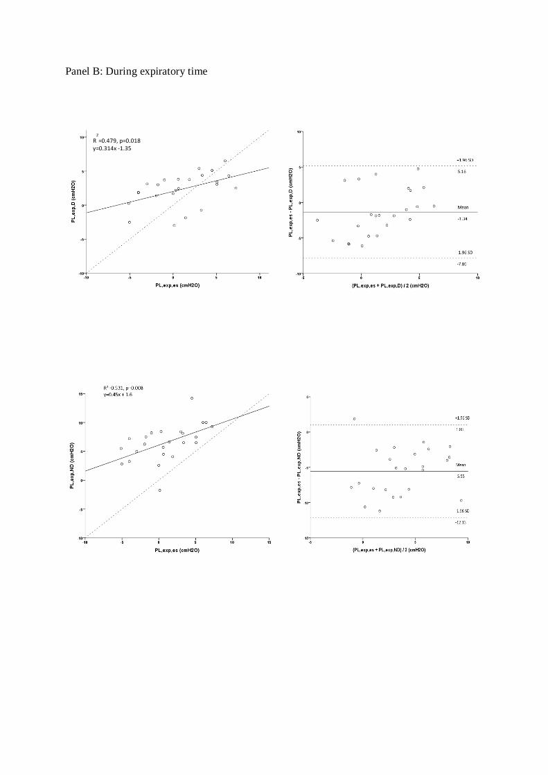
Panel B: During expiratory time
R2
=0.479, p=0.018
y=0.314x -1.35

Figure 5
(A)

Figures 5
(B)

Supplementary Figure 1
Picture of positioning of pleural catheters during surgery and corresponding post-
operative CT-scan in one patient.