Embed Size (px)
Citation preview
Essays in BiochEmistry
Other recent titles in the Essays in Biochemistry series:Amyloids in Health and Disease: volume 56edited by S. Perrett2014ISBN 978 1 85578 192 4
Autophagy: Molecules and Mechanisms: volume 55edited by J.D. Lane2013ISBN 978 1 85578 191 7
The Role of Non-Coding RNAs in Biology: volume 54edited by M.A. Lindsay and S. Griffiths-Jones2013ISBN 978 1 85578 190 0
Cell Polarity and Cancer: volume 53edited by A.D. Chalmers and R. Whitley2012ISBN 978 1 85578 189 4
Lysine-Based Post-Translational Modification of Proteins: volume 52edited by I. Scott2012ISBN 978 1 85578 185 6
Molecular Parasitology: volume 51edited by R. Docampo2011ISBN 978 1 85578 184 9
ABC Transporters: volume 50edited by F.J. Sharom2011ISBN 978 1 85578 181 8
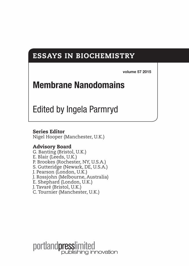
Membrane Nanodomains
Edited by Ingela Parmryd
Series EditorNigel Hooper (Manchester, U.K.)
Advisory BoardG. Banting (Bristol, U.K.)E. Blair (Leeds, U.K.)P. Brookes (Rochester, NY, U.S.A.)S. Gutteridge (Newark, DE, U.S.A.)J. Pearson (London, U.K.)J. Rossjohn (Melbourne, Australia)E. Shephard (London, U.K.)J. Tavaré (Bristol, U.K.)C. Tournier (Manchester, U.K.)
volume 57 2015
ESSAyS in BiochEmiStry
Essays in Biochemistry is published by Portland Press Limited on behalf of the Biochemical Society
Portland Press LimitedThird Floor, Charles Darwin House12 Roger StreetLondon WC1N 2JUU.K.Tel: +44 (0)20 7685 2410Fax: +44 (0)20 7685 2469 email: [email protected]
© The Authors; Journal compilation © 2015 Biochemical Society
All rights reserved. Apart from any fair dealing for the purposes of research or private study, or criticism or review, as permitted under the Copyright, Designs and Patents Act, 1998, this publication may be reproduced, stored or transmitted, in any forms or by any means, only with the prior permission of the publishers, or in the case of reprographic reproduction in accordance with the terms of the licences issued by the Copyright Licensing Agency. Inquiries concerning reproduction outside those terms should be sent to the publishers at the above-mentioned address.
Although, at the time of going to press, the information contained in this publication is believed to be correct, neither the authors nor the editors nor the publisher assumes any responsibility for any errors or omissions herein contained. Opinions expressed in this book are those of the authors and are not necessarily held by the Biochemical Society, the editors or the publisher.
All profits made from the sale of this publication are returned to the Biochemical Society for the promotion of the molecular life sciences.
British Library Cataloguing-in-Publication Data A catalogue record for this book is available from the British LibraryISBN 978-1-85578-193-1 ISSN (print) 0071 1365ISSN (online) 1744 1358
Typeset by Techset Composition Ltd, Salisbury, U.K.Printed in Great Britain by Cambrian Printers Ltd, Aberystwyth
© 2015 Biochemical Society
Preface ...........................................................................................xi Authors .......................................................................................xiii Abbreviations ............................................................................xix
1 Lipid domains in model membranes: a brief historical perspective ...............................................................................1Ole G. Mouritsen and Luis A. Bagatolli
Abstract .............................................................................................................. 1Introduction: models of membranes .................................................................. 2Lipids move centre stage ................................................................................... 4Models of lipid membranes ................................................................................ 5Macro-, meso- and nano-domains in lipid bilayers ............................................ 5Domains in one-, two- and several-component lipid bilayers ............................ 7The peculiar case of cholesterol-induced order ................................................. 9Domains induced by lipid–protein interactions ................................................ 11Non-equilibrium membranes ............................................................................ 12Biological membranes ...................................................................................... 13Conclusion ....................................................................................................... 14Summary .......................................................................................................... 14References ....................................................................................................... 14
2 microemulsions, modulated phases and macroscopic phase separation: a unified picture of rafts .........................21Ha Giang, Roie Shlomovitz and Michael Schick
Abstract ............................................................................................................ 21Introduction ...................................................................................................... 21Phase separation .............................................................................................. 22Modulated phases ............................................................................................ 23Length scales ................................................................................................... 25The system on a closed vesicle ....................................................................... 27Conclusions: putting it all together – vacuoles, rafts, the lot ........................... 28Summary .......................................................................................................... 30References ....................................................................................................... 30
3 Lipid bilayers: clusters, domains and phases .....................33David G. Ackerman and Gerald W. Feigenson
Abstract ............................................................................................................ 33Introduction: modelling the plasma membrane ................................................ 33
contEntS
vi Essays in Biochemistry volume 57 2015
© The Authors Journal compilation © 2015 Biochemical Society
The lipid bilayer as a state of matter ................................................................ 35The nature of small clusters and phase separation.......................................... 35Distinct domains that are not separate phases ................................................ 37Modelling the bilayer phase behaviour............................................................. 37Phase diagrams applicable to cell plasma membranes ................................... 38Four-component model membranes ................................................................ 40Conclusion ....................................................................................................... 40Summary .......................................................................................................... 41References ....................................................................................................... 41
4 cholesterol trafficking and distribution ...............................43David B. Iaea and Frederick R. Maxfield
Abstract ............................................................................................................ 43Introduction ...................................................................................................... 43Models of sterol–phospholipid interactions ..................................................... 44Distribution of sterols in mammalian cells ........................................................ 46Regulatory/biosynthetic mechanisms .............................................................. 47Intracellular trafficking of lipids and sterols ...................................................... 48Sterol transfer proteins in non-vesicular transport ........................................... 48Fluorescent probes for cholesterol trafficking .................................................. 49Consequences of extensive non-vesicular transport ....................................... 50Conclusion ....................................................................................................... 51Summary .......................................................................................................... 51References ....................................................................................................... 52
5 methods applicable to membrane nanodomain studies? ..................................................................................57Parham Ashrafzadeh and Ingela Parmryd
Abstract ............................................................................................................ 57Introduction ...................................................................................................... 57Detergent-resistant membranes ....................................................................... 58Giant plasma membrane vesicles .................................................................... 60Manipulation of cholesterol levels .................................................................... 62Probing membrane packing ............................................................................. 63Conclusions ...................................................................................................... 65Summary .......................................................................................................... 65References ....................................................................................................... 65
6 Super-resolution optical microscopy of lipid plasma membrane dynamics ............................................................69Christian Eggeling
Abstract ............................................................................................................ 69Plasma membrane dynamics ........................................................................... 69
Contents vii
© 2015 Biochemical Society
Far-field optical microscopy ............................................................................. 70STED-FCS ........................................................................................................ 72Conclusions ...................................................................................................... 77Summary .......................................................................................................... 78References ....................................................................................................... 78
7 microscopy of membrane lipids: how precisely can we define their distribution? .........................................81Sho Takatori and Toyoshi Fujimoto
Abstract ............................................................................................................ 81Introduction ...................................................................................................... 82Methods used for proteins may not be applicable and may even be
inappropriate to study lipids ......................................................................... 83Lipid-binding domains can be used for live imaging of membrane lipids........ 84Most lipids are refractory to chemical fixation ................................................. 85How does quick-freezing work? ....................................................................... 86How can quick-frozen specimens be processed? ........................................... 87Conclusion ....................................................................................................... 89Summary .......................................................................................................... 90References ....................................................................................................... 90
8 nanodomains in biological membranes .............................93Yuanqing Ma, Elizabeth Hinde and Katharina Gaus
Abstract ............................................................................................................ 93Introduction ...................................................................................................... 94Membrane models ........................................................................................... 94Experimental evidence for lipid rafts and other membrane models................. 98The future direction for studying membrane organization ............................. 101Conclusion ..................................................................................................... 102Summary ........................................................................................................ 103References ..................................................................................................... 103
9 membrane nanodomains: contribution of curvature and interaction with proteins and cytoskeleton ...............109Senthil Arumugam and Patricia Bassereau
Abstract .......................................................................................................... 109Introduction .................................................................................................... 109Phase separation and nanodomains .............................................................. 110Curvature and membrane nanodomains ........................................................ 111Cytoskeleton, membrane proteins and membrane nanodomains ................. 112Conclusion and future goals .......................................................................... 116Summary ........................................................................................................ 117References ..................................................................................................... 117
viii Essays in Biochemistry volume 57 2015
© The Authors Journal compilation © 2015 Biochemical Society
10 restoring synaptic vesicles during compensatory endocytosis ..........................................................................121Anne Gauthier-Kemper, Martin Kahms and Jürgen Klingauf
Abstract .......................................................................................................... 121Introduction .................................................................................................... 122Lipid and protein composition of SVs ............................................................ 124Recognition of synaptic vesicle cargo by sorting adaptors ........................... 125SV component retrieval by self-assembly ...................................................... 128Active zone integrity: clearance and scaffolds ............................................... 129Conclusion ..................................................................................................... 130Summary ........................................................................................................ 130References ..................................................................................................... 131
11 Functions of cholera toxin B-subunit as a raft cross-linker ..........................................135Charles A. Day and Anne K. Kenworthy
Abstract .......................................................................................................... 135Introduction .................................................................................................... 136Evidence that CTxB associates with lipid rafts and functions as a raft
cross- linker ................................................................................................. 137Biophysical studies of the dynamics of CTxB-cross-linked domains
in cell membranes ....................................................................................... 138Functional consequences of cross-linking for toxin entry.............................. 140Conclusions and future directions .................................................................. 141Summary ........................................................................................................ 142References ..................................................................................................... 143
12 nanodomains in early and later phases of Fcεri signalling ..............................................................................147David Holowka and Barbara Baird
Abstract .......................................................................................................... 147Introduction .................................................................................................... 147Nanodomains formed by antigen clustering of IgE–FcεRI complexes ........... 149Functional evidence for PI(4,5)P2 nanodomains relevant to Ca2+ signalling ....... 152Structural evidence for PI(4,5)P2 nanodomains relevant to Ca2+
signalling and exocytosis ............................................................................ 153Conclusions and future directions .................................................................. 157Summary ........................................................................................................ 159References ..................................................................................................... 159
Contents ix
© 2015 Biochemical Society
13 membrane nanodomains in t-cell antigen receptor signalling ..............................................................................165Konstantina Nika and Oreste Acuto
Abstract .......................................................................................................... 165Introduction .................................................................................................... 165The TCR signalling machine: a complex device organized into nanoclusters ... 167Discussion and conclusions ........................................................................... 172Summary ........................................................................................................ 173References ..................................................................................................... 173
14 membrane–cytoskeleton interactions in cholesterol- dependent domain formation ............................................177Jennifer N. Byrum and William Rodgers
Abstract .......................................................................................................... 177Introduction .................................................................................................... 178Soapy extracts to patchy membranes: evolution in the discovery
of cholesterol-dependent domains ............................................................. 178FRETing out lipid domains formed by the cytoskeleton ................................ 179Cytoskeleton mechanics in domain formation ............................................... 181CDM domains and the cytoskeleton in Src kinase regulation........................ 182Conclusions .................................................................................................... 184Summary ........................................................................................................ 185References ..................................................................................................... 185
15 caveolin-1, galectin-3 and lipid raft domains in cancer cell signalling ......................................................189Jay Shankar, Cecile Boscher and Ivan R. Nabi
Abstract .......................................................................................................... 189Introduction .................................................................................................... 189Caveolin-1: caveolae and other roles ............................................................. 192Tyrosine phosphorylated caveolin-1 ............................................................... 194Galectin-3, the galectin lattice and interaction with lipid rafts ....................... 195Co-ordinate Cav1–Gal3 signalling in cancer .................................................. 197Concluding remarks ....................................................................................... 198Summary ........................................................................................................ 198References ..................................................................................................... 199
index ............................................................................................... 203

© 2015 Biochemical Society
Membrane nanodomains, despite their well-documented role in cellular processes like signal-ling, protein sorting and vesicular trafficking, are largely ignored in textbooks. This is partly a reflection of membrane nanodomains being a very active research field where consensus has not yet been reached. Accordingly, the choice of possible topics for this volume on membrane nanodomains was wide open making my job very exciting. To give the readers a good over-view of membrane nanodomains, I found it important to include insights from diverse research disciplines ranging from theoretical physics to cell biology so this volume is very much interdisciplinary – the way science ought to be to facilitate breakthroughs. The commu-nities working on membrane nanodomains have a tradition of arranging cross-disciplinary activities, which has exposed me to excellent research and enabled me to meet the scientists behind the work, both of which were most helpful in my job as a Guest Editor.
Many definitions of membrane nanodomains have been suggested, but most scientists would probably associate the term with lipid–lipid and/or lipid–protein interactions. These interactions make membrane nanodomains dynamic liquid entities surrounded by a second type of dynamic liquid with continuous repartitioning of components between the domains and their surroundings. In cells new membrane components are continuously delivered and old ones removed which further contributes to the dynamics. In the plasma membrane there is the additional complexity of links between membrane lipids and proteins both in the extracel-lular matrix and in the cytoplasm. Moreover, cells are neither flat nor smooth and they move. Together this makes membrane nanodomains extremely challenging to study.
To understand how membrane nanodomains can form, chapters on what can physically drive lipid–lipid interactions, lipid–lipid avoidance, as well as lipid phase behaviour in model membranes, are an excellent starting point. In Chapter 1 Ole Mouritsen and Luis Bagatolli pro-vide a historical perspective on lipid domains in model membranes and their relation to mem-brane domains in biological membranes. In Chapter 2 Ha Giang, Roie Shlomovitz and Michael Schick discuss how modulated phases can result in microemulsions, fluids with structure. In Chapter 3 David Ackerman and Gerald Feigenson describe the differences between phase sep-aration and non-ideal mixing in one-phase systems, e.g. clusters. Cholesterol is a key player in membrane nanodomain formation in mammalian cells and in Chapter 4 David Iaea and Frederick Maxfield account for how cholesterol is trafficked and distributed.
To assess the results from cell studies, an understanding of the methods used is necessary and the next set of chapters cover both methodological details and what information can be obtain from specific methods. In Chapter 5 Parham Ashrafzadeh and myself discuss the strength and weaknesses of four methods commonly used in membrane nanodomain studies. In Chapter 6 Christian Eggeling accounts for FCS-STED and how it can be used to assess lipid dynamics in living cells. In Chapter 7 Sho Takatori and Toyoshi Fujimoto present how quick-freezing in combination with electron microscopy can capture the lipid organization in cells. In Chapter 8 Yuanqing Ma, Elizabeth Hinde and Katharina Gaus present some current models of how biological membranes are organized and how superesolution fluorescence microscopy can be used to increase our understanding of membrane nanodomains.
PrEFAcE
xii Essays in Biochemistry volume 57 2015
© The Authors Journal compilation © 2015 Biochemical Society
Lipids can have shapes giving them the propensity to accumulate in highly curved cellular regions which can drive the formation of membrane nanodomains. Moreover, actin filaments interact with membrane lipids in a dynamic fashion. In Chapter 9 Senthil Arumugam and Patricia Bassereau discuss how biological membranes are shaped by proteins and how this effects membrane heterogeneity. Vesicles are examples of membrane nanodomains with high curvature and a unique composition. How synaptic vesicles are restored during compensatory endocytosis is discussed by Anne Gauthier-Kemper, Martin Kahms and Jürgen Klingauf in Chapter 10. How the B-subunit of cholera toxin, a marker for ordered membrane nanodo-mains, operates by binding the ganglioside GM1 while both sensing and being able to generate curvature is discussed by Charles Day and Anne Kenworthy in Chapter 11.
Cell signalling is a field where membrane nanodomains have been extensively studied and it is well documented that cell signalling is accompanied by membrane rearrangement, in immune cells this is even apparent at the micron scale. The involvement of PI(4,5)P2 domains in mast cell signalling from the receptor FcεRI and changes in receptor diffusion upon its clus-tering are portrayed by David Holowka and Barbara Baird in Chapter 12. An account of the distinct position of the T-cell receptor in signalling and the involvement of membrane nano-domains in T-cell receptor signalling is provided by Konstantina Nika and Oreste Acuto in Chapter 13. How membrane–actin filament interactions create a compressive force that could lead to the formation of membrane nanodomains involved in segregating T-cell signalling molecules is presented by Jennifer Byrum and William Rodgers in Chapter 14. In Chapter 15 Jay Shankar, Cecile Boscher and Ivan Nabi discuss how the extracellular galectin lattice can lead to membrane nanodomain formation and how the lattice can contribute to signalling in cancer cells by interacting with caveolin-1.
I am grateful to whoever suggested that I edit this volume and to the Biochemical Society/Portland Press for following up the suggestion. I am very pleased with the line-up of contributors in the resulting volume and very glad that they could find the time to write the chapters. All contributors are world-leading experts and highly esteemed scientists in their respective fields and it was a privilege to have had the opportunity to work with all of them. My hope is that you readers will learn from and enjoy reading the chapters and I welcome you to the world of membrane nanodomains.
Ingela Parmryd,December 2014
© 2015 Biochemical Society
Ole Mouritsen is a Professor of biophysics at the University of Southern Denmark and direc-tor of the MEMPHYS-Center for Biomembrane Physics. His fields of specialization include statistical mechanics and thermodynamics, computer simulation techniques, phase transitions and critical phenomena, biomembrane physics and chemistry, interfacial physical chemistry, and soft matter physics, with applications within biomedicine, drug delivery and gastro sci-ences. He has published more than 300 scientific papers and a number of books, e.g. Life – As a Matter of Fat: the Emerging Science of Lipodomics (Springer, 2005) and recently three popular books on the science of cooking. He is the recipient of several prizes for research and research communication, most recently the RSC Bourke Award (2008) and the European Lipid Science Award (2011).
Luis Bagatolli obtained his M.Sc. (1991) and Ph.D. (1995) in chemistry from the University of Córdoba, Argentina. In 2002, he joined the MEMPHYS-Center for Biomembrane Physics at the University of Southern Denmark (SDU), Odense, Denmark. He is currently a Professor of biophotonics at SDU, leader of the Membrane Biophysics and Biophotonics Group, and director of DaMBIC (Danish Molecular Biomedical Imaging Center). His fields of specialization are biological physical chemistry, biomembranes and bio-imaging (fluorescence microscopy, non-linear phenomena). He has published more than 90 scientific papers and book chapters.
Ha Giang obtained her Ph.D. in Mechanical Engineering in 2013 at the California Institute of Technology. She is a Postdoctoral Fellow at the University of Washington, and is currently working on membrane asymmetry.
Roie Shlomovitz studied Physics and Chemistry as an undergraduate at The Hebrew University of Jerusalem and then earned his M.Sc. and Ph.D. in Physics at the Weizmann Institute of Science. He has been a postdoctoral researcher at UCLA in the departments of Chemistry and Biochemistry and Physics and Astronomy. He is now a Sackler Scholar at the University of Washington.
Michael Schick was a student of Felix Bloch at Stanford University. After a postdoctoral fellowship with Paul Zilsel at Case Western Reserve, he took a position in 1969 in the Department of Physics at the University of Washington. His interests have included adsorbed monolayers and their critical phenomena, wetting of thin films, microemulsions and the ordering of block copolymers and lipid bilayers. He is an avid amateur cellist.
David Ackerman received his B.A. in Physics, Mathematics and Integrated Sciences from Northwestern University in 2010, where he studied exoplanets under Fred Rasio. He is cur-rently a Ph.D. candidate in the field of Biophysics at Cornell University, where he works in Jerry Feigenson’s membrane physical chemistry laboratory. His thesis work focuses on using both coarse-grained and atomistic molecular dynamics to model phase separation in multiple-component membrane mixtures.
Jerry Feigenson is a Professor in the Cornell Department of Molecular and Cell Biology and Director of Graduate Studies for the Cornell Field of Biophysics. His 1968 B.S. in Chemistry is from the Rensselaer Polytechnic Institute, where he studied surface chemistry in
AuthorS
xiv Essays in Biochemistry volume 57 2015
© The Authors Journal compilation © 2015 Biochemical Society
the laboratory of Sydney Ross; his 1974 Ph.D. in Chemistry is from the California Institute of Technology, where he studied NMR with Sunney Chan. He joined the Cornell faculty in 1974, after brief postdoctoral study at Oxford University in the fluorescence laboratory of George Radda. He has been a visiting Professor of Physics at Keio University in Japan with Kazuhiko Kinosita, and a visiting scholar at Scripps Institution of Oceanography with Vic Vacquier.
David Iaea received a B.A. degree in Biochemistry from New York University. He was a research technician at Weill Cornell Medical College with Dr Timothy McGraw. During his time in the McGraw laboratory, he began to learn fluorescent microscopy and biochemical techniques to investigate protein trafficking. He entered the Tri-Institutional Program in Chemical Biology (TPCB) in 2010 and joined the Maxfield laboratory in 2012. His doctoral thesis focused on the structural and functional role of a sterol transfer protein.
Frederick Maxfield received a B.S. degree in Chemistry from Union College and a Ph.D. in Chemistry from Cornell University. He was a Postdoctoral Fellow at the National Cancer Institute with Ira Pastan. During his fellowship, he began to use quantitative fluorescence microscopy to study endocytosis. He continued these studies as a faculty member at New York University, at Columbia University, and at his current position as Professor and Chairman of Biochemistry at Weill Cornell Medical College in New York. His laboratory studies membrane trafficking with a recent emphasis on trafficking of lipids and cholesterol.
Parham Ashrafzadeh obtained his B.Sc. in Cell and Molecular Biology in 2006 from Tehran Azad University, Iran. He received his M.Sc. in Molecular Biology in 2009 from University Putra Malaysia (UPM), Malaysia. During his Master’s studies, he worked mainly on the development and evaluation of DNA vaccines. In 2010, he moved to Sweden and worked at Stockholm University and the Karolinska Institute on Neisseria pili usage in migration and role of histone H2B ubiquitylation. In 2012, he started his Ph.D. studies in Medical Cell Biology under the supervision of Dr Ingela Parmryd at Uppsala University. Currently, his research focuses on plasma membrane organization with emphasis on the relationship between actin filaments and ordered lipid domain formation at the plasma membrane. Apart from work, he enjoys travelling, hanging out with friends, playing basketball and playing football.
Ingela Parmryd obtained her M.Sc. in Chemistry in 1993 and received her Ph.D. in Biochemistry in 1999 from Stockholm University, Sweden. Between 1999 and 2003, she worked in the U.K., as a Postdoctoral Fellow at the National Institute for Medical Research and as a research associate at Imperial College. In 2003, she was appointed Assistant Professor in Cell Biology at Stockholm University and was promoted to Associate Professor/Docent in 2008. In 2011, she moved to Uppsala University where she took up a position as Guest Lecturer. Her research focuses on plasma membrane organization with emphasis on the importance of cell topography and lipid packing for cell signalling primarily using T-cells as the model system. The development of tools to quantify biological responses by image analysis is also a prominent feature of her research. She has several patents for RBNCC (replicate based noise corrected correlation), a method for which she received the Stockholm Inventor’s award in 2009 and is the CEO of the company No More Noise. When not working, Ingela enjoys spending time with her family, playing volleyball and playing the oboe.
Christian Eggeling holds a Ph.D. in Physics from the University of Göttingen, Germany. He performed his Ph.D. on single-molecule spectroscopy at the Max-Planck-Institute of Biophysical Chemistry (MPIbpc), Göttingen (supervised by C. Seidel). He then was a research scientist at Evotec, Hamburg, Germany, introducing single-molecule-based fluorescence
Authors xv
© 2015 Biochemical Society
spectroscopy techniques for high-throughput drug discovery. From 2003, he pushed optical nanoscopy and specifically the STED-FCS technology in the department of S. Hell, MPIbpc, who recently was rewarded the Nobel Prize in Chemistry. Since the end of 2012, he has been the principal investigator of the Nano-Immunology group in the MRC Human Immunology Unit and the scientific director of the Wolfson Imaging Centre at the Weatherall Institute of Molecular Medicine, University of Oxford. In September 2014, he was appointed Professor of Molecular Immunology of the University of Oxford.
Sho Takatori was born in 1982 in Japan, earned a Ph.D. in Pharmaceutical Sciences from the University of Tokyo in 2010 and became a Postdoctoral Research Fellow of the Japan Society for the Promotion of Science at Nagoya University. Research interests include the sub-organellar distribution of membrane proteins and lipids. Sho wishes to understand the build-ing principles of the beautiful and complex membrane architecture of organelles.
Toyoshi Fujimoto was born in 1954 in Kyoto, Japan, and graduated from Kyoto University School of Medicine in 1978. Since 1999, Toyoshi has been Professor and Chairman at Nagoya University Graduate School of Medicine. Main research interests are the nanoscale distribution of lipids in the membrane and the structure and function of lipid droplets. Toyoshi wishes to make discoveries by developing new electron microscopy techniques.
YuanQing Ma is currently a Ph.D. student at the Centre for Vascular Research under the supervision of Professor Katharina Gaus. He holds a Bachelor degree in Bioengineering from JiShou University in China and a Master’s from the University of Western Sydney, Australia, for which he engineered photoconvertible fluorescent proteins, which were developed into a patent. After cloning novel fluorescent proteins from jellyfish in Professor Takeharu Nagai’s laboratory in Japan for 1 year, he returned to Australia to pursue his Ph.D. in the Gaus labora-tory, where he is interested in developing sensors to measure membrane properties such as charges of the T-cell activation site, and using optogenetics to manipulate T-cell sensitivity.
Elizabeth Hinde is a Vice Chancellor Research Fellow at the University of New South Wales under the mentorship of Professor Gaus. She received her Ph.D. from the University of Melbourne in 2010 and then trained as a biophysicist during a postdoctoral appointment at the University of California, Irvine, under the mentorship of Professor Enrico Gratton. Her research is focused on the development of different fluorescence imaging technologies and methods of analysis, to probe the in vivo spatiotemporal dynamics of proteins in live cells. Her long-term research interest is to apply these methods to the study of how intracellular traffic and diffusion regulate biological function at the single -molecule level.
Katharina Gaus is an NHMRC Senior Research Fellow at the University of New South Wales. She received her Ph.D. from the University of Cambridge in 1999 and has led the Cell Membrane Biology group since 2005. Her group investigates signal transduction processes with advanced fluorescence microscopy approaches. She was awarded the Young Investigator Award from the Australia and New Zealand Society for Cell and Developmental Biology (2010), the Gottschalk Medal from the Australian Academy of Science (2012) and the New South Wales Science and Engineering Award for Excellence in Biological Sciences (2013).
Senthil Arumugam received his Master’s from the Tata Institute of Fundamental Research, Mumbai, India, in 2008. He then received his Ph.D. from the Technical University of Dresden and the Max-Planck-Institute of Cell Biology and Genetics, Dresden, Germany, in 2012. He worked on the dynamics and mechanics of FtsZ, a homologue of tubulin found in bacteria with Professor Petra Schwille. Senthil Arumugam is currently a joint postdoctoral
xvi Essays in Biochemistry volume 57 2015
© The Authors Journal compilation © 2015 Biochemical Society
researcher between the laboratories of Patricia Bassereau and Ludger Johannes at Institute Curie, where he works on membrane biophysics in living cells.
Patricia Bassereau is currently a CNRS Directrice de Recherche (equivalent to Professor) at the Institut Curie in Paris where she is the leader of the Membranes and Cellular Functions group. She started her career in soft matter. She worked for 7 years in Montpellier (GDPC) on the structure of self-assembled surfactant-based systems and for 1 year as a visiting scientist at the IBM Almaden Center (San Jose, CA, U.S.A.) on the structure of thin films of polymers. In 1993, she came to the Institut Curie. She first studied the interactions of soluble proteins with polymer monolayers, but soon after started to address questions related to physics of the cell. She has developed a multidisciplinary approach, largely based on synthetic biology and on the development and study of biomimetic systems, to understand the role of lipid membranes in important cellular functions such as intracellular trafficking, endo-exo-cytosis, transmem-brane transport of ions (‘active membranes’), protein diffusion or cell adhesion.
Anne Gauthier-Kemper studied Biochemistry and Biology at the Johann-Wolfgang-Goethe-University, Frankfurt am Maine, and the University of Osnabrück and obtained her Ph.D. at the University of Osnabrück in the Department of Neurobiology in 2011. Since then, she is working as a postdoc at the Institute of Medical Physics and Biophysics (IMPB) at the University of Münster in the group of Professor Dr Jürgen Klingauf. Her major interest is the analysis of protein clustering at the active zone in presynaptic boutons.
Martin Kahms studied Biochemistry at the University of Bochum and obtained his Ph.D. at the Max-Planck-Institute of Molecular Physiology in Dortmund. He did postdoctoral stud-ies together with Professor Reiner Peters at the Institute of Medical Physics and Biophysics at the University of Münster (IMPB) with research focus on structural and functional analysis of the nuclear pore complex. Since 2009, he has been in the group of Professor Dr Jürgen Klingauf at the IMPB studying exo- and endo-cytosis mechanisms at presynaptic boutons.
Jürgen Klingauf studied Biology and Physics at the Universities of Hamburg and Bonn. He did his Diploma work on the calcium control of secretion in secretory chromaffin cells and doctoral work with Professor Dr Erwin Neher at the Max-Planck-Institute for Biophysical Chemistry, Göttingen, and received his Ph.D. in Physics in 1999 from the University of Göttingen. As guest researcher and fellow of the Boehringer Ingelheim Fonds, he worked with Professor Richard W. Tsien in the Department of Molecular and Cellular Physiology at Stanford University from 1995 to 1998, where he started to work on the physiology of synaptic vesicle recycling in hippocampal neurons using imaging methods. After a short postdoc with Dr Neher, he became an independent research group leader at the Max-Planck-Institute for Biophysical Chemistry, Göttingen, in 2001, where he continued to work on the synaptic physi-ology of exo- and endo-cytosis. In 2008, he accepted the offer of the University of Münster for a full professorship and was appointed as Chair of the Institute of Medical Physics and Biophysics at the Medical Faculty.
Charles Day completed his B.Sc. at Gordon College and his doctorate in the Department of Molecular Physiology and Biophysics at Vanderbilt University in the labora-tory of Dr Anne Kenworthy. His research has focused on utilizing biophysics approaches and imaging techniques to characterize plasma membrane dynamics, organization and the gener-ation of clathrin-independent endocytic vesicles. He is currently a research fellow at the Mayo Clinic, where he is studying the interactions between dynamin and the cytoskeleton with Dr Mark McNiven.
Authors xvii
© 2015 Biochemical Society
Anne Kenworthy received her B.A. from Kenyon College and carried out graduate stud-ies in the Department of Cell Biology at Duke University Medical Center. After completing postdoctoral fellowships at The Johns Hopkins University and the National Institutes of Health, she joined the faculty in the Department of Molecular Physiology and Biophysics at Vanderbilt School of Medicine where she is currently an Associate Professor. Her research is focused on understanding the structure, dynamics, and function of caveolae and lipid rafts in cell membranes. Toward this goal, her group has developed quantitative approaches to study membrane domains and protein and lipid dynamics in cells using live-cell imaging.
David Holowka is Senior Scientist in the Department of Chemistry and Chemical Biology, Cornell University, Ithaca, NY. His research interests and current work include long-term efforts to understand molecular mechanisms by which cross-linking of IgE receptors on mast cells triggers complex cellular signalling processes that lead to important functional responses in immune host defence. Central to mast cell and other cell signalling responses is the mobilization of intracellular Ca2+ ions, and a component of Dr Holowka’s current work focuses on understanding this spatiotemporally complex process and its function roles in exo-cytosis, cytokine production, and host–pathogen interactions.
Barbara Baird is Professor in the Department of Chemistry and Chemical Biology, Cornell University, Ithaca, NY. She became fascinated by cell membrane function in college when the Singer–Nicholson model arrived to capture the interest of a diverse range of chem-ists, physicists and biologists. Since then, taking the route of biophysical chemistry, she has had the good fortune of linking up with David Holowka to investigate membrane participation in the functioning of IgE receptors on mast cells. Over the years, their joint research group at Cornell, together with their valuable collaborators, have steadily worked to develop higher-res-olution tools to elucidate both early and downstream signalling events initiated by activated IgE receptors, and how these are regulated and targeted by membrane interactions.
Konstantina Nika studied Biomedical Sciences at the University of Portsmouth, U.K., where she also obtained her Doctorate in 2001. She then joined the laboratory of Professor Thomas Mustelin at the Burnham Institute for Medical Research in La Jolla California, work-ing on the role of protein tyrosine phosphatases in T-cell activation. She has been working in Professor Acuto’s laboratory in the Sir William Dunn School of Pathology at Oxford U.K., since 2006, studying the proximal T-cell receptor signalling pathways.
Oreste Acuto studied biology and obtained a Doctorate degree in Immunology at the University of Rome. He then moved to Switzerland studying the biochemistry and function of immunoreceptors and membrane proteins at the Swiss Institute of Cancer Research (ETH) in Zurich and at the Swiss Institute of Cancer Research in Lausanne. Following that, he spent 6 years in the Dana-Farber Cancer Institue at Harvard Medical School, in Ellis Reinherz’s labo-ratory working in the molecular cloning and characterization of the T-cell receptor. He became Director of the Molecular Immunology Unit at the Pasteur Institute in Paris in 1998 and moved his laboratory to the University of Oxford in 2006 where he is currently Professor and Senior Research Fellow at the Sir William Dunn School of Pathology.
Jennifer Byrum is a Postdoctoral Research Fellow in the Department of Biochemistry and Molecular Biology at the University of Oklahoma Health Sciences Center. She obtained a B.S. in Biology in 2003 at Oklahoma City University, and completed her Ph.D. studies in 2012 in the Department of Pathology at the University of Oklahoma Health Sciences Center. Her thesis work focused on early T-cell signalling events in cholesterol-dependent domains,
xviii Essays in Biochemistry volume 57 2015
© The Authors Journal compilation © 2015 Biochemical Society
especially antigen-independent responses by T-cells during initial interactions with antigen-presenting cells. Since graduating, she is extending her research in the laboratory of Dr William Rodgers to study regulatory mechanisms of key components of the DNA damage response, and their contribution to genomic integrity.
William Rodgers began his studies of membrane structure and function in the labora-tory of Dr Michael Glaser at the University of Illinois, where he used fluorescence imaging to directly visualize and measure lipid-enriched domains in the erythrocyte plasma membrane. Upon completing his graduate studies in 1992, he moved to the laboratory Dr John K. Rose at the Yale University School of Medicine to study the role of lipid domains in T-cell signalling and its regulation. His research in the Rose laboratory provided the first evidence of regulation of membrane signalling proteins through their association with cholesterol-dependent domains. From Yale, he joined the Oklahoma Medical Research Foundation in 2008, spear-heading research that identified a critical role of the cell cytoskeleton in forming lipid domains in the plasma membrane, and showing that cytoskeleton-dependent domains attenuate Src family kinase activity in resting T-cells. In 2013, he moved to the Department of Biochemistry at the University of Oklahoma Health Science Center, where he continues to employ analytical fluorescence imaging methods to interrogate cell membrane structure.
Jay Shankar received his B.Sc. in Zoology at the University of Delhi, India, in 1998, Master’s in Biotechnology from Punjab University, Chandigarh, India, and Ph.D. in Allergy and Immunology from University of Delhi, India, in 2008. He also worked as a Research Scientist at the Indian Council of Medical Research, New Delhi, India, and as a Postdoctoral Fellow at the International Center for Genetic Engineering and Biotechnology, New Delhi, India. He joined Professor Nabi’s laboratory as a Postdoctoral Fellow in 2008. His work focuses on the role and importance of pseudopodia in cancer cell migration.
Cecile Boscher, as a Ph.D. student in the Rene-Marc Mege group, explored the mecha-nisms associated with cadherin-induced axon elongation. She showed how cadherins 11 and N are associated with FGF receptor signalling and how α-catenin provides a molecular clutch for cadherin adhesion. She joined Dr Nabi’s laboratory as a Postdoctoral Fellow, where she showed how caveolin-1 and galectin-3 lattice act together to promote EGF receptor and integrin cross-talk and how galectin-3 controls N-cadherin cell–cell junctions. She is now working in the Jean-Philippe Gratton group at the Université de Montréal exploring the antagonist roles of angiopoietin-1 and VEGF on endothelial cell adhesion and migration.
Ivan Nabi received his B.Sc. in Biochemistry at McGill University in 1983 and his Ph.D. in Cancer Metastasis at the Weizmann Institute of Science in 1989. After completing his post-doctoral training in Cell Biology at Cornell Medical College, he was appointed Assistant Professor and later promoted to Full Professor in the Department of Pathology and Cell Biology at the Université de Montréal (1992–2004). In 2004, he moved to the University of British Columbia where he is now a Professor in the Department of Cellular and Physiological Sciences and Director of the Imaging Facility in the Life Sciences Institute. His research focuses on the role of cellular domains in cancer progression and metastasis, i.e. the cell biol-ogy of cancer.

© 2015 Biochemical Society
aa amino acidsACAT acyl-CoA:cholesterol acyltransferaseAPC antigen-presenting cellAZ active zoneBODIPY boron–dipyrrometheneBRS basic-rich stretchCav1 caveolin-1CBM caveolin-binding motifCDM cholesterol-dependent membraneCDR3 complementary determining region 3chol cholesterolCLASP clathrin-associated sorting proteinCLIC clathrin-independent carrierCME clathrin-mediated endocytosisCNS central nervous systemCRAC Ca2+ release-activated Ca2+
CRD carbohydrate recognition domainCSD caveolin scaffolding domainCTxB cholera toxin B-subunitDHE dehydroergosterolDOPC dioleoylphosphatidylcholineDOPE dioleoylphosphtidylethanolamineDPPC dipalmitoylphosphatidylcholineDRM detergent-resistant membraneDSPC distearoylphosphatidylcholineDTC differentiated thyroid cancerdSTORM direct stochastic optical reconstruction microscopyEGF epidermal growth factorEGFR epidermal growth factor receptorEgr1 early growth response-1eNOS endothelial nitric oxide synthaseER endoplasmic reticulumERC endocytic recycling compartmentERK extracellular-signal-regulated kinaseERM ezrin, radixin and moesinFAK focal adhesion kinaseFCS fluorescence correlation spectroscopyFLIM fluorescent lifetime imaging microscopyfPALM fluorescence photo-activated localization microscopyGal3 galectin-3
ABBrEViAtionS
xx Essays in Biochemistry volume 57 2015
© The Authors Journal compilation © 2015 Biochemical Society
GAPs GTPase-activating proteinsGEEC glycosylphosphatidylinositol-enriched early endosomal compartmentGPCR G-protein-coupled receptorGP generalized polarizationGPI glycosylphosphatidylinositolGPMV giant plasma membrane vesicleGrb2 growth-factor-receptor-bound protein 2GUV giant unilamellar vesicleHA haemagglutininHMG 3-hydroxy-3-methyl-glutarylI(1,4,5)P3 inositol 1,4,5-trisphosphateIGF insulin-like growth factorIS immunological synapseITAM immunoreceptor tyrosine-based activation motif7KC 7-ketocholesterolLβ solid gelLAT linker for activation of T-cellsLck p56lck
Ld liquid disorderedLDL low-density lipoproteinLo liquid orderedmAb monoclonal antibodyMAPK mitogen-activated protein kinaseMBCD methyl-β-cyclodextrinMLV multilamellar vesicleMURC muscle-restricted coiled-coil proteinNBD nitrobenzoxadiazoleNM II non-muscle myosin IIOSBP oxysterol-binding proteinPA phosphatidic acidPALM photo-activated localization microscopyPC phosphatidylcholinePDGF platelet-derived growth factorPE phosphatidylethanolamineperi-AZ peri-active zonePH pleckstrin homologyPI phosphatidylinositolPI3K phosphoinositide 3-kinasePI(4,5)P2 phosphatidylinositol 4,5-bisphosphatePI(4)P5K phosphatidylinositol 4-phosphate 5-kinasePKC protein kinase CPLC phospholipase CPM plasma membranepMHC peptide-loaded major histocompatibility complexPOPC 1-palmitoyl-2-oleoylphosphatidylcholine
© 2015 Biochemical Society
Abbreviations xxi
POPE 1-stearoyl-2-oleoyl-phosphoethanolaminePS phosphatidylserinePTP protein tyrosine phosphatasePTRF polymerase I and transcript release factorRHM rehydration methodROCK Rho-associated protein kinaseRRetP readily retrievable poolSANS small-angle neutron scatteringSCAP SREBP-cleavage-activating proteinSDPR serum deprivation response proteinSDS sodium dodecyl sulfateSH2 Src homology 2SLP-76 Src homology 2 domain-containing leucocyte protein of 76 kDaSM sphingomyelinSOCE store-operated Ca2+ entrySOS Son of sevenlessSRBC serum deprivation response-related gene product that binds to C-kinaseSREBP sterol regulatory element-binding proteinStAR steroidogenic acute regulatory proteinStARD START domainSTART steroidogenic acute regulatory protein-related lipid transferSTED stimulated emission depletionSTORM stochastic optical reconstruction microscopySTxB Shiga toxin B-subunitSUV small unilamellar vesicleSV synaptic vesicleSV2 synaptic vesicle 2-related proteinsvFCS spot-variation FCSSyb2 synaptobrevin2Syp1 synaptophysin 1Syt1 synaptotagmin 1TCR T-cell receptorTfR transferrin receptorTGFβR transforming growth factor β receptorTIRF total internal reflectance fluorescenceTm melting temperatureTX Triton X-100v-ATPase v-type ATPaseWASP Wiskott–Aldrich syndrome proteinWAVE WASP verprolin homologousZAP70 TCR ζ-chain-associated protein kinase of 70 kDa