Embed Size (px)
Citation preview
REVIEW
Establishing neural crest identity: a gene regulatory recipeMarcos Simões-Costa and Marianne E. Bronner*
ABSTRACTThe neural crest is a stem/progenitor cell population that contributesto a wide variety of derivatives, including sensory and autonomicganglia, cartilage and bone of the face and pigment cells of the skin.Unique to vertebrate embryos, it has served as an excellent modelsystem for the study of cell behavior and identity owing to itsmultipotency, motility and ability to form a broad array of cell types.Neural crest development is thought to be controlled by a suite oftranscriptional and epigenetic inputs arranged hierarchically in a generegulatory network. Here, we examine neural crest development froma gene regulatory perspective and discuss how the underlyinggenetic circuitry results in the features that define this unique cellpopulation.
KEY WORDS: Gene regulation, Migration, Neural crest, Neural plateborder, Signaling, Transcription factors
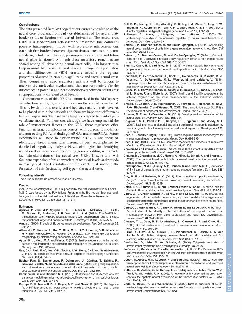
IntroductionThe neural crest is a remarkable cell type, notable for its ability tomigrate extensively and differentiate into numerous derivatives(Fig. 1A-E). An evolutionary innovation, the neural crest is thoughtto have been crucial for the origin and diversification of vertebrates(Gans and Northcutt, 1983). It has been a subject of great interest toembryologists due to its far-reaching migratory ability anddevelopmental plasticity, and is sometimes referred to as the‘fourth germ layer’ (Hall, 2000). Discovery of the neural crestchallenged important concepts pertaining to the development andevolution of the vertebrate body plan, especially when it wasrevealed that this ectodermal cell type could give rise tomesenchymal derivatives (Hall, 1999). Furthermore, identificationof the neural crest demonstrated that the establishment of progenitorfields during body part formation could include contributions fromcells incoming from distant locations.Over the past century, much has been learned through
experimental embryology about neural crest cell plasticity and itscontribution to various derivatives (Le Douarin and Kalcheim,1999). Transplantation experiments, first in amphibian embryos(Horstadius, 1950) and later in birds using the now classic quail-chick chimeras pioneered byLeDouarin (1973), have established thepathways followed by neural crest cells and the extensive array ofderivatives to which they contribute (Fig. 1F). These studies havemade the neural crest one of the best-studied progenitor cellpopulations of vertebrate embryos.In this Review, we first provide a brief overview of neural crest
development (for further details, see Le Douarin, 1982; Sauka-Spengler and Bronner-Fraser, 2008). We then describe the currentstate of knowledge regarding connections within the neural crestgene regulatory network (GRN), elaborating upon novel regulatory
interactions that have been characterized in the last few years. Wecompile information obtained from several studies and modelorganisms to sketch regulatory circuits controlling the differentsteps of neural crest development using the BioTapestry platform(Longabaugh et al., 2005, 2009) and discuss the importance ofepigenetic regulation in neural crest formation. Most interactionspresented in this version of the GRN were characterized in thecranial neural crest, although we have also included informationfrom other axial levels when it is consistent with network topology.In particular, we emphasize the data regarding direct interactionsobtained from cis-regulatory analysis, which allow in-depthcharacterization of the circuitry within the network.
A brief overview of neural crest developmentNeural crest cells are induced in the ectodermal germ layer duringgastrulation and initially reside in the neural plate border territory,which is positioned at the lateral edges of the central nervous system(Fig. 1B). During neurulation, this border territory elevates as theneural plate closes to form the neural tube (Fig. 1C,D). As aconsequence, nascent neural crest cells come to reside within thedorsal aspect of the apposing neural folds, initiating the expression ofneural crest genes, such as FoxD3 (Labosky and Kaestner, 1998) andSox10 (Southard-Smith et al., 1998), that reflect their specification asbona fide neural crest cells. After neural tube closure/cavitation, theysubsequently leave the central nervous system via an epithelial-to-mesenchymal transition (EMT) (Fig. 1D), resulting in theirtransformation into a migratory and multipotent progenitor cellpopulation (Fig. 1E) that undergoes some of the most far-reachingmigrations of any embryonic cell type, often moving long distancesalong highly stereotypic pathways.
Following migration, neural crest cells progressively differentiateinto distinct cell types according to environmental influencesencountered during their journey and at their final sites, where theycooperate with other cell populations to form appropriate tissues andorgans (Bronner and LeDouarin, 2012). They generate a multitudeof derivatives as diverse as elements of the craniofacial skeleton,sensory and autonomic ganglia of the peripheral nervous system,and pigment cells of the skin (Le Douarin, 1982) (Fig. 1F). Thus,vertebrate neural crest cells are defined by their origin at the neuralplate border, their ability to leave the neural tube via EMT and theirmultipotency.
Cell lineage-tracing experiments have identified the derivativesand pathways of migration of various neural crest populations (LeDouarin, 1982). The regionalization of the body axis from anteriorto posterior is reflected by neural crest subpopulations thatcontribute to some overlapping as well as axial level-specificderivatives. For example, cranial neural crest cells form the facialskeleton, including the upper and lower jaw and bones of the neck,as well as the glia and some neurons of the cranial sensory ganglia(Couly et al., 1998). Just below the head, vagal neural crest cellspopulate the outflow tract of the heart and enteric ganglia of the gut(Le Douarin and Teillet, 1973; Creazzo et al., 1998). The trunkneural crest contributes to the dorsal root and sympathetic ganglia of
Division of Biology and Biological Engineering, California Institute of Technology,Pasadena, CA 91125, USA.
*Author for correspondence ([email protected])
242
© 2015. Published by The Company of Biologists Ltd | Development (2015) 142, 242-257 doi:10.1242/dev.105445
DEVELO
PM
ENT
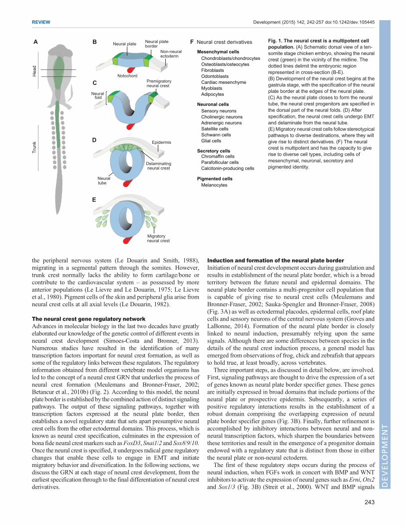
the peripheral nervous system (Le Douarin and Smith, 1988),migrating in a segmental pattern through the somites. However,trunk crest normally lacks the ability to form cartilage/bone orcontribute to the cardiovascular system – as possessed by moreanterior populations (Le Lievre and Le Douarin, 1975; Le Lievreet al., 1980). Pigment cells of the skin and peripheral glia arise fromneural crest cells at all axial levels (Le Douarin, 1982).
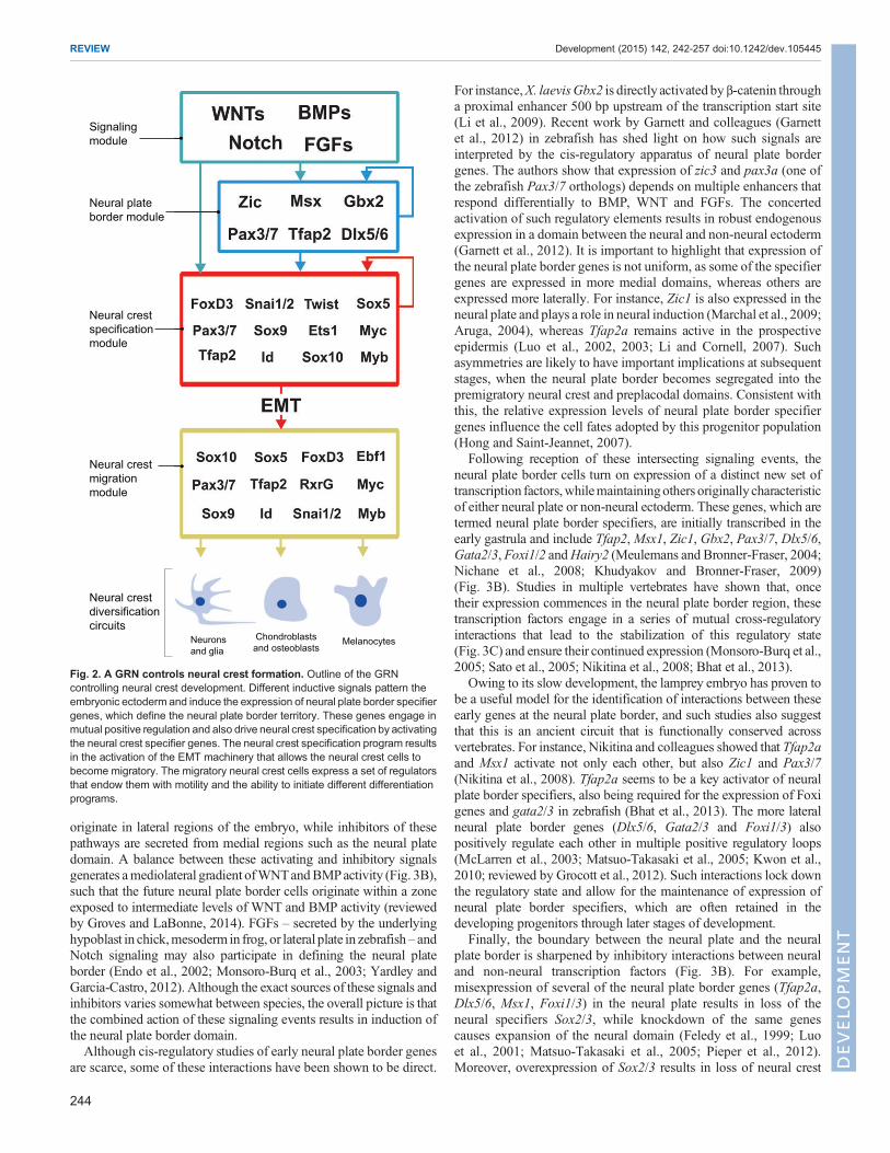
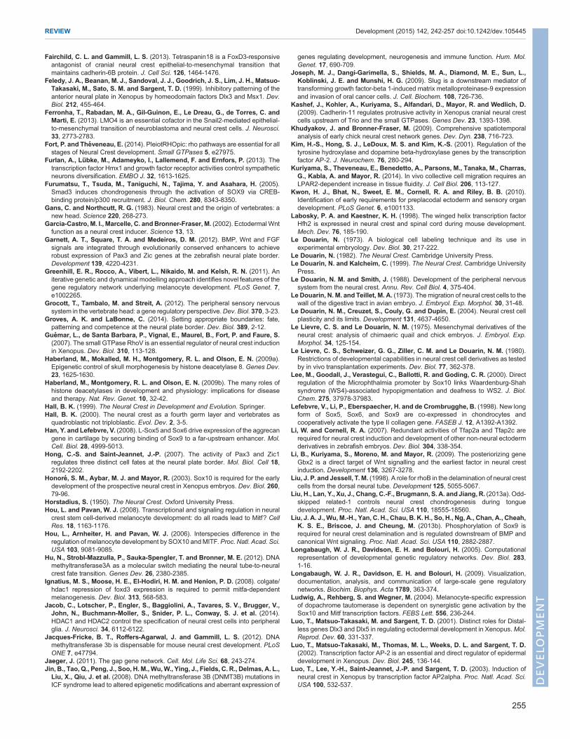
The neural crest gene regulatory networkAdvances in molecular biology in the last two decades have greatlyelaborated our knowledge of the genetic control of different events inneural crest development (Simoes-Costa and Bronner, 2013).Numerous studies have resulted in the identification of manytranscription factors important for neural crest formation, as well assome of the regulatory links between these regulators. The regulatoryinformation obtained from different vertebrate model organisms hasled to the concept of a neural crest GRN that underlies the process ofneural crest formation (Meulemans and Bronner-Fraser, 2002;Betancur et al., 2010b) (Fig. 2). According to this model, the neuralplate border is established by the combined action of distinct signalingpathways. The output of these signaling pathways, together withtranscription factors expressed at the neural plate border, thenestablishes a novel regulatory state that sets apart presumptive neuralcrest cells from the other ectodermal domains. This process, which isknown as neural crest specification, culminates in the expression ofbona fide neural crest markers such asFoxD3, Snai1/2 and Sox8/9/10.Once the neural crest is specified, it undergoes radical gene regulatorychanges that enable these cells to engage in EMT and initiatemigratory behavior and diversification. In the following sections, wediscuss the GRN at each stage of neural crest development, from theearliest specification through to the final differentiation of neural crestderivatives.
Induction and formation of the neural plate borderInitiation of neural crest development occurs during gastrulation andresults in establishment of the neural plate border, which is a broadterritory between the future neural and epidermal domains. Theneural plate border contains a multi-progenitor cell population thatis capable of giving rise to neural crest cells (Meulemans andBronner-Fraser, 2002; Sauka-Spengler and Bronner-Fraser, 2008)(Fig. 3A) as well as ectodermal placodes, epidermal cells, roof platecells and sensory neurons of the central nervous system (Groves andLaBonne, 2014). Formation of the neural plate border is closelylinked to neural induction, presumably relying upon the samesignals. Although there are some differences between species in thedetails of the neural crest induction process, a general model hasemerged from observations of frog, chick and zebrafish that appearsto hold true, at least broadly, across vertebrates.
Three important steps, as discussed in detail below, are involved.First, signaling pathways are thought to drive the expression of a setof genes known as neural plate border specifier genes. These genesare initially expressed in broad domains that include portions of theneural plate or prospective epidermis. Subsequently, a series ofpositive regulatory interactions results in the establishment of arobust domain comprising the overlapping expression of neuralplate border specifier genes (Fig. 3B). Finally, further refinement isaccomplished by inhibitory interactions between neural and non-neural transcription factors, which sharpen the boundaries betweenthese territories and result in the emergence of a progenitor domainendowed with a regulatory state that is distinct from those in eitherthe neural plate or non-neural ectoderm.
The first of these regulatory steps occurs during the process ofneural induction, when FGFs work in concert with BMP and WNTinhibitors to activate the expression of neural genes such asErni,Otx2and Sox1/3 (Fig. 3B) (Streit et al., 2000). WNT and BMP signals
Hea
dTr
unk
Neural fold
Epidermis
Premigratoryneural crest
Notochord
Neural plate Neural plateborder
Non-neuralectoderm
A B
C
D
E
Delaminatingneural crest
F Neural crest derivatives
Migratory neural crest
Neuraltube
Mesenchymal cells
Neuronal cells
Secretory cells
Pigmented cells
Chondroblasts/chondrocytesOsteoblasts/osteocytesFibroblastsOdontoblastsCardiac mesenchymeMyoblastsAdipocytes
Sensory neuronsCholinergic neuronsAdrenergic neuronsSatellite cellsSchwann cellsGlial cells
Chromaffin cellsParafollicular cellsCalcitonin-producing cells
Melanocytes
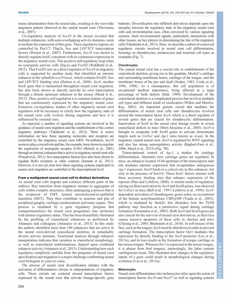
Fig. 1. The neural crest is a multipotent cellpopulation. (A) Schematic dorsal view of a ten-somite stage chicken embryo, showing the neuralcrest (green) in the vicinity of the midline. Thedotted lines delimit the embryonic regionrepresented in cross-section (B-E).(B) Development of the neural crest begins at thegastrula stage, with the specification of the neuralplate border at the edges of the neural plate.(C) As the neural plate closes to form the neuraltube, the neural crest progenitors are specified inthe dorsal part of the neural folds. (D) Afterspecification, the neural crest cells undergo EMTand delaminate from the neural tube.(E) Migratory neural crest cells follow stereotypicalpathways to diverse destinations, where they willgive rise to distinct derivatives. (F) The neuralcrest is multipotent and has the capacity to giverise to diverse cell types, including cells ofmesenchymal, neuronal, secretory andpigmented identity.
243
REVIEW Development (2015) 142, 242-257 doi:10.1242/dev.105445
DEVELO
PM
ENT
originate in lateral regions of the embryo, while inhibitors of thesepathways are secreted from medial regions such as the neural platedomain. A balance between these activating and inhibitory signalsgenerates amediolateral gradient ofWNTandBMPactivity (Fig. 3B),such that the future neural plate border cells originate within a zoneexposed to intermediate levels of WNT and BMP activity (reviewedby Groves and LaBonne, 2014). FGFs – secreted by the underlyinghypoblast in chick,mesoderm in frog, or lateral plate in zebrafish – andNotch signaling may also participate in defining the neural plateborder (Endo et al., 2002; Monsoro-Burq et al., 2003; Yardley andGarcia-Castro, 2012). Although the exact sources of these signals andinhibitors varies somewhat between species, the overall picture is thatthe combined action of these signaling events results in induction ofthe neural plate border domain.Although cis-regulatory studies of early neural plate border genes
are scarce, some of these interactions have been shown to be direct.
For instance,X. laevisGbx2 is directly activated by β-catenin througha proximal enhancer 500 bp upstream of the transcription start site(Li et al., 2009). Recent work by Garnett and colleagues (Garnettet al., 2012) in zebrafish has shed light on how such signals areinterpreted by the cis-regulatory apparatus of neural plate bordergenes. The authors show that expression of zic3 and pax3a (one ofthe zebrafish Pax3/7 orthologs) depends on multiple enhancers thatrespond differentially to BMP, WNT and FGFs. The concertedactivation of such regulatory elements results in robust endogenousexpression in a domain between the neural and non-neural ectoderm(Garnett et al., 2012). It is important to highlight that expression ofthe neural plate border genes is not uniform, as some of the specifiergenes are expressed in more medial domains, whereas others areexpressed more laterally. For instance, Zic1 is also expressed in theneural plate and plays a role in neural induction (Marchal et al., 2009;Aruga, 2004), whereas Tfap2a remains active in the prospectiveepidermis (Luo et al., 2002, 2003; Li and Cornell, 2007). Suchasymmetries are likely to have important implications at subsequentstages, when the neural plate border becomes segregated into thepremigratory neural crest and preplacodal domains. Consistent withthis, the relative expression levels of neural plate border specifiergenes influence the cell fates adopted by this progenitor population(Hong and Saint-Jeannet, 2007).
Following reception of these intersecting signaling events, theneural plate border cells turn on expression of a distinct new set oftranscription factors,whilemaintainingothers originally characteristicof either neural plate or non-neural ectoderm. These genes, which aretermed neural plate border specifiers, are initially transcribed in theearly gastrula and include Tfap2,Msx1, Zic1, Gbx2, Pax3/7, Dlx5/6,Gata2/3, Foxi1/2 andHairy2 (Meulemans and Bronner-Fraser, 2004;Nichane et al., 2008; Khudyakov and Bronner-Fraser, 2009)(Fig. 3B). Studies in multiple vertebrates have shown that, oncetheir expression commences in the neural plate border region, thesetranscription factors engage in a series of mutual cross-regulatoryinteractions that lead to the stabilization of this regulatory state(Fig. 3C) and ensure their continued expression (Monsoro-Burq et al.,2005; Sato et al., 2005; Nikitina et al., 2008; Bhat et al., 2013).
Owing to its slow development, the lamprey embryo has proven tobe a useful model for the identification of interactions between theseearly genes at the neural plate border, and such studies also suggestthat this is an ancient circuit that is functionally conserved acrossvertebrates. For instance, Nikitina and colleagues showed that Tfap2aand Msx1 activate not only each other, but also Zic1 and Pax3/7(Nikitina et al., 2008). Tfap2a seems to be a key activator of neuralplate border specifiers, also being required for the expression of Foxigenes and gata2/3 in zebrafish (Bhat et al., 2013). The more lateralneural plate border genes (Dlx5/6, Gata2/3 and Foxi1/3) alsopositively regulate each other in multiple positive regulatory loops(McLarren et al., 2003; Matsuo-Takasaki et al., 2005; Kwon et al.,2010; reviewed by Grocott et al., 2012). Such interactions lock downthe regulatory state and allow for the maintenance of expression ofneural plate border specifiers, which are often retained in thedeveloping progenitors through later stages of development.
Finally, the boundary between the neural plate and the neuralplate border is sharpened by inhibitory interactions between neuraland non-neural transcription factors (Fig. 3B). For example,misexpression of several of the neural plate border genes (Tfap2a,Dlx5/6, Msx1, Foxi1/3) in the neural plate results in loss of theneural specifiers Sox2/3, while knockdown of the same genescauses expansion of the neural domain (Feledy et al., 1999; Luoet al., 2001; Matsuo-Takasaki et al., 2005; Pieper et al., 2012).Moreover, overexpression of Sox2/3 results in loss of neural crest
Signalingmodule
Neural plateborder module
Neural crestspecificationmodule
Neural crestmigrationmodule
Neural crestdiversificationcircuits
Neuronsand glia
Chondroblastsand osteoblasts
Melanocytes
Fig. 2. A GRN controls neural crest formation. Outline of the GRNcontrolling neural crest development. Different inductive signals pattern theembryonic ectoderm and induce the expression of neural plate border specifiergenes, which define the neural plate border territory. These genes engage inmutual positive regulation and also drive neural crest specification by activatingthe neural crest specifier genes. The neural crest specification program resultsin the activation of the EMT machinery that allows the neural crest cells tobecome migratory. The migratory neural crest cells express a set of regulatorsthat endow them with motility and the ability to initiate different differentiationprograms.
244
REVIEW Development (2015) 142, 242-257 doi:10.1242/dev.105445
DEVELO
PM
ENT

and epidermal markers, suggesting that neural factors can alsorepress neural plate border specifiers (Wakamatsu et al., 2004;Rogers et al., 2009). Thus, the cis-regulatory interactions betweenneural genes and neural plate border specifiers result in a sharpboundary that will separate neural crest from central nervous systemprogenitors (Fig. 3C).
Establishing neural crest identity: the neural crest specifiergenesThe emergence of the neural crest from the neural plate border ismarked by the expression of a suite of genes dubbed neural crestspecifiers. Their expression is driven by the concerted action ofmore medial neural plate border specifiers and signaling pathwaysthat are active during the time of specification (Fig. 4). The neuralcrest specifiers have key functions. They activate the EMT effectorprogram, which will allow the neural crest to delaminate from the
ectoderm and become a migratory cell type (as discussed furtherbelow). Moreover, they also regulate each other positively tostabilize this regulatory state, which results in the maintenance of anumber of specifier genes in the delaminating and migratory neuralcrest. This is important, as the combination of these transcriptionfactors is thought to maintain the neural crest in an undifferentiatedstate (Sauka-Spengler and Bronner-Fraser, 2008). In fact, theplasticity and developmental potential of the neural crest is likely tobe encoded in the gene regulatory circuits that characterize thispopulation.
The first neural crest specifiers to be expressed in the nascentneural crest in the chick are FoxD3, Ets1 and Snai1/2 (Khudyakovand Bronner-Fraser, 2009). Neural crest expression of FoxD3 isregulated by the neural plate border specifiers Pax3/7 and Msx1,which directly bind to two regulatory regions that drive differentialexpression in the cranial and trunk neural crest cells of the chick
ANeural plate border BMPs
WNTs
NPBspecifiers
B Induction of the neural plate border
C Maintenance of the NPB specifier genes
Fig. 3. GRN module controlling the formation of the neural plate border. (A) The action of signaling systems such the BMP and WNT signaling pathwaysresults in activation of the neural plate border (NPB) genes in the margins of the neural plate. (B) In the neural ectoderm, FGF signaling drives neural inductionby activating proneural genes. In an intermediary territory located between the neural and non-neural ectoderm, WNTs and BMPs activate a number oftranscription factors dubbed neural plate border specifiers. Activity of WNTs and BMPs is hampered in the neural ectoderm by a number of inhibitory moleculesproduced by these cells. (C)While SoxB1 genes (Sox2 andSox3) are expressed in the early neural plate, the neural plate specifier transcription factors engage inmutual positive regulation, which stabilizes the neural plate border regulatory state. Direct interactions are indicated with solid lines, whereas dashed lines showpossible direct interactions inferred from gain- and loss-of-function studies.
245
REVIEW Development (2015) 142, 242-257 doi:10.1242/dev.105445
DEVELO
PM
ENT
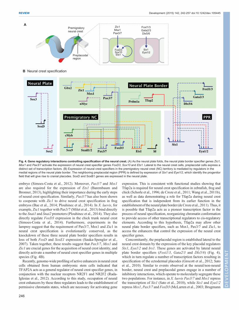
embryo (Simoes-Costa et al., 2012). Moreover, Pax3/7 and Msx1are also required for the expression of Ets1 (Barembaum andBronner, 2013), highlighting their importance during the early stepsof neural crest specification. Similarly, Pax3/7 has also been shownto cooperate with Zic1 to drive neural crest specification in frogembryos (Bae et al., 2014; Plouhinec et al., 2014). In X. laevis, forexample, Zic1 together with Pax3/7 (Milet et al., 2013) bind directlyto the Snai1 and Snai2 promoters (Plouhinec et al., 2014). They alsodirectly regulate FoxD3 expression in the chick trunk neural crest(Simoes-Costa et al., 2014). Furthermore, experiments in thelamprey suggest that the requirement of Pax3/7, Msx1 and Zic1 inneural crest specification is evolutionarily conserved, as theknockdown of these three neural plate border specifiers results inloss of both FoxD and SoxE1 expression (Sauka-Spengler et al.,2007). Taken together, these results suggest that Pax3/7, Msx1 andZic1 are crucial genes for the acquisition of neural crest identity, anddirectly activate a number of neural crest specifier genes in multiplespecies (Fig. 4B).Recently, genome-wide profiling of active enhancers in neural crest
cells obtained from human embryonic stem cells indicated thatTFAP2A acts as a general regulator of neural crest specifier genes, inconjunction with the nuclear receptors NR2F1 and NR2F2 (Rada-Iglesias et al., 2012). According to this study, occupation of neuralcrest enhancers by these three regulators leads to the establishment ofpermissive chromatin states, which are necessary for activating gene
expression. This is consistent with functional studies showing thatTfap2a is required for neural crest specification in zebrafish, frog andchick (Schorle et al., 1996; de Croze et al., 2011;Wang et al., 2011b),as well as data demonstrating a role for Tfap2a during neural crestspecification that is independent from its earlier function in theestablishmentof the neural plate border (deCroze et al., 2011).Thus, itis possible that Tfap2a acts as a pioneer transcription factor in theprocess of neural specification, reorganizing chromatin conformationto provide access of other transcriptional regulators to cis-regulatoryelements. According to this hypothesis, Tfap2a may allow otherneural plate border specifiers, such as Msx1, Pax3/7 and Zic1, toaccess the enhancers that control the expression of the neural crestspecifier genes.
Concomitantly, the preplacodal region is established lateral to theneural crest domain by the expression of the key placodal regulatorsSix1, Eya1/2 and Irx1. These genes are activated by lateral neuralplate border specifiers (Foxi1/3, Gata2/3 and Dlx5/6) (Fig. 4),which in turn regulate a number of transcription factors resulting inspecification of the ectodermal placodes (Grocott et al., 2012; Satoet al., 2010). Similar to events observed at the neural/non-neuralborder, neural crest and preplacodal genes engage in a number ofinhibitory interactions, which operate to molecularly segregate thesetwo populations. For instance, in X. laevis Pax3/7 andMsx1 repressthe transcription of Six1 (Sato et al., 2010), while Six1 and Eya1/2repressMsx1, Pax3/7 and FoxD3 (McLarren et al., 2003; Brugmann
FoxD3Sox10Ets1
Six1Eya1/2
Premigratoryneural crest
Zic1Msx1
Pax3/7
FoxI1/3Gata2/3Dlx5/6
Preplacodalregion
A
B Neural crest specification
Fig. 4. Gene regulatory interactions controlling specification of the neural crest. (A) As the neural plate folds, the neural plate border specifier genes Zic1,Msx1 and Pax3/7 activate the expression of neural crest specifier genes FoxD3, Sox10 and Ets1. Lateral to the neural crest cells, preplacodal cells express adistinct set of transcription factors. (B) Expression of neural crest specifiers in the premigratory neural crest (NC) territory is mediated by regulators in themedial regions of the neural plate border. The neighboring preplacodal region (PPR) is defined by expression of Six1 and Eye1/2, which identify the progenitorfield that will give rise to cranial placodes. SoxD and SoxB1 genes are expressed in the neural plate.
246
REVIEW Development (2015) 142, 242-257 doi:10.1242/dev.105445
DEVELO
PM
ENT
et al., 2004; Christophorou et al., 2009) in both chicken and frog.Interestingly, although molecular markers suggest a clear divisionbetween the neural crest and preplacodal domains, fate-mappingexperiments indicate that these two progenitor cell types are stilllargely intermingled. Labeling of individual cells in the late neuralplate border reveals that individual progenitor cells can give rise toboth neural crest and ectodermal cells (Selleck and Bronner-Fraser,1995), showing that, despite their expression of specifier genes, thefate of these cells is not yet fixed.Nevertheless, it is clear that the regulatory program of the neural
plate border cells operates to distinguish these cell types from itsonset: from the earliest stages, the key neural crest regulators aremore medially located relative to the placodal regulators, andinteractions between them might already be delineating thesegregation of neural crest and preplacodal domains. According tothis view, the mediolateral compartmentalization of the vertebrateectoderm would operate in a manner similar to the Drosophila gapgene GRN (Jaeger, 2011): morphogens polarize and divide theectoderm into broad territories, activating genes that will engage inmultiple cross-regulatory interactions, resulting in the progressiverestriction and delimitation of territories of cells sharing similarregulatory states.The importance of Msx1, Pax7 and Zic1 in neural crest
formation is clear, but other genes expressed in the neural plateborder, such as Myb, Myc and Prdm1a (Bellmeyer et al., 2003;Betancur et al., 2010b; Powell et al., 2013), have also beenimplicated in neural crest specification. Furthermore, signalinginput seems to be important for the expression of neural crestspecifier genes. Inhibition of WNT signaling after neural plateborder specification results in loss of expression of Snai2 andFoxD3 (Garcia-Castro et al., 2002) in chick embryos, while WNTshave been shown to act in concert with Zic1 and Pax3 to driveneural crest specification in the frog (Sato et al., 2005). This raisesthe interesting possibility that WNTs could act as permissivefactors in neural crest specification. However, cis-regulatoryanalysis has failed to uncover many direct TCF/LEF bindingsites that are crucial for neural crest specifier expression (Betancuret al., 2010b; Barembaum and Bronner, 2013; Simoes-Costa andBronner, 2013). The exception seems to be Snai2, which has aproximal enhancer that is directly activated by TCF/LEF and β-catenin (Vallin et al., 2001). Thus, although WNTs are thought toplay an important role in neural crest specification, the directunderlying mechanism of how this is mediated remains unclear.An intriguing possibility is that factors downstream of signalingsystems cooperate with transcription factors at the neural plateborder to mediate neural crest specification.Much like the neural plate border specifier genes, the neural
crest specifier genes positively cross-regulate each other. Suchinteractions are difficult to parse out given that they take place ina short period of time as the cells quickly transition to the nextregulatory state (Nikitina et al., 2008). A few examples of directregulatory interactions between neural crest specifier genesinclude the cranial-specific control of FoxD3 by Ets1 (Simoes-Costa et al., 2012). Nevertheless, it is clear that positiveregulatory loops result in the consolidation of the neural crestspecifier regulatory state. For instance, in the frog, Snai1/2 seemsto positively regulate Twist, FoxD3 and Sox9 (Aybar et al., 2003),while Foxd3 promotes Sox10 expression in mouse (Dottori et al.,2001). Sox10, on the other hand, has been shown to activateSnai2 and itself (Honoré et al., 2003), while Sox8 is important forthe expression of other SoxE genes in the same model in the frog(O’Donnell et al., 2006). Characterization of more direct
interactions in multiple species will be necessary to delineatethe hierarchy and logic controlling the expression of thesetranscription factors.
The process of neural crest specification results in a discrete cellpopulation, located between the neural plate and the preplacodalregion, that shares a common regulatory state. At these stages, thepremigratory neural crest expresses the full suite of specifier genes(including FoxD3, Snai2, Ets1, Sox8/9/10 and Pax3/7) as well assome of the border genes whose expression is maintained (Tfap2,Msx1, Zic1). This new regulatory state initiates drastic structuralchanges at a cellular level that result in delamination from the neuraltube via EMT.
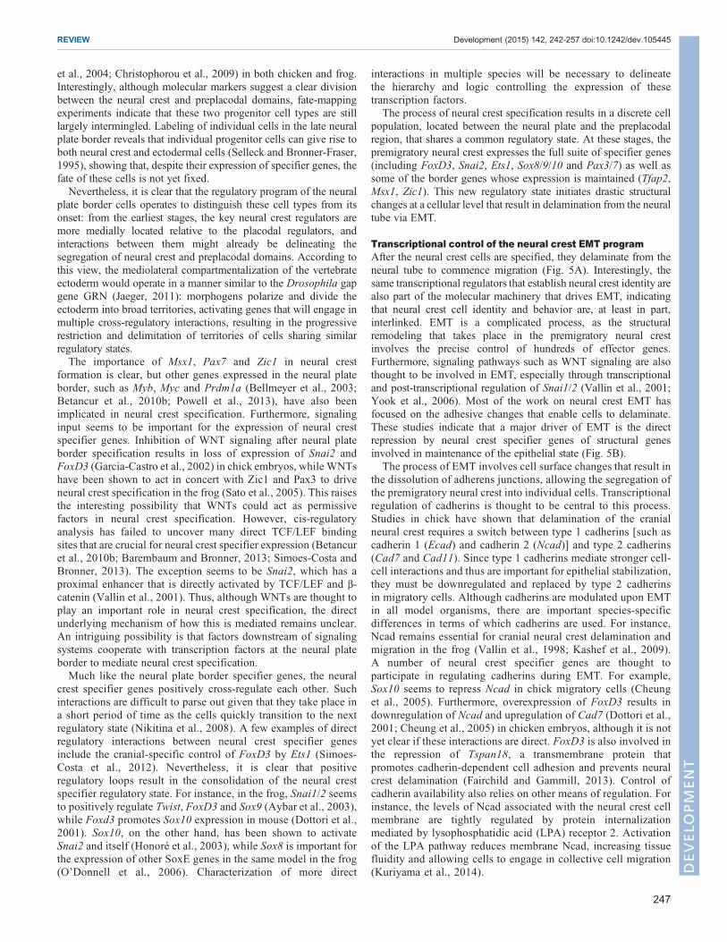
Transcriptional control of the neural crest EMT programAfter the neural crest cells are specified, they delaminate from theneural tube to commence migration (Fig. 5A). Interestingly, thesame transcriptional regulators that establish neural crest identity arealso part of the molecular machinery that drives EMT, indicatingthat neural crest cell identity and behavior are, at least in part,interlinked. EMT is a complicated process, as the structuralremodeling that takes place in the premigratory neural crestinvolves the precise control of hundreds of effector genes.Furthermore, signaling pathways such as WNT signaling are alsothought to be involved in EMT, especially through transcriptionaland post-transcriptional regulation of Snai1/2 (Vallin et al., 2001;Yook et al., 2006). Most of the work on neural crest EMT hasfocused on the adhesive changes that enable cells to delaminate.These studies indicate that a major driver of EMT is the directrepression by neural crest specifier genes of structural genesinvolved in maintenance of the epithelial state (Fig. 5B).
The process of EMT involves cell surface changes that result inthe dissolution of adherens junctions, allowing the segregation ofthe premigratory neural crest into individual cells. Transcriptionalregulation of cadherins is thought to be central to this process.Studies in chick have shown that delamination of the cranialneural crest requires a switch between type 1 cadherins [such ascadherin 1 (Ecad) and cadherin 2 (Ncad)] and type 2 cadherins(Cad7 and Cad11). Since type 1 cadherins mediate stronger cell-cell interactions and thus are important for epithelial stabilization,they must be downregulated and replaced by type 2 cadherinsin migratory cells. Although cadherins are modulated upon EMTin all model organisms, there are important species-specificdifferences in terms of which cadherins are used. For instance,Ncad remains essential for cranial neural crest delamination andmigration in the frog (Vallin et al., 1998; Kashef et al., 2009).A number of neural crest specifier genes are thought toparticipate in regulating cadherins during EMT. For example,Sox10 seems to repress Ncad in chick migratory cells (Cheunget al., 2005). Furthermore, overexpression of FoxD3 results indownregulation of Ncad and upregulation of Cad7 (Dottori et al.,2001; Cheung et al., 2005) in chicken embryos, although it is notyet clear if these interactions are direct. FoxD3 is also involved inthe repression of Tspan18, a transmembrane protein thatpromotes cadherin-dependent cell adhesion and prevents neuralcrest delamination (Fairchild and Gammill, 2013). Control ofcadherin availability also relies on other means of regulation. Forinstance, the levels of Ncad associated with the neural crest cellmembrane are tightly regulated by protein internalizationmediated by lysophosphatidic acid (LPA) receptor 2. Activationof the LPA pathway reduces membrane Ncad, increasing tissuefluidity and allowing cells to engage in collective cell migration(Kuriyama et al., 2014).
247
REVIEW Development (2015) 142, 242-257 doi:10.1242/dev.105445
DEVELO
PM
ENT
Since its discovery, Snai1/2 has been linked to EMT in manysystems, from cancer cell lines to gastrulating embryos (Nieto et al.,1994; Blanco et al., 2007). In the neural crest, Snai1/2 functions as atranscriptional repressor and plays an important role in thedownregulation of the type 1 cadherins during EMT. It has animportant role inNcad repression, where it cooperates with Lmo4 toinhibit transcription in both the neural crest and in cancer cells(Ferronha et al., 2013). Importantly, Snai1/2 also represses Cad6b(Taneyhill et al., 2007), a classical type 2 cadherin that needs to bedownregulated for neural crest delamination to occur (Coles et al.,2007). This is accomplished through direct interaction with a pair ofE-box sites located in the vicinity of the Kozak consensus sequence.An important partner of Snai1/2 during the process of EMT is thetranscription factor Sox9, which when phosphorylated physicallyinteracts with Snai1/2 to drive EMT (Cheung and Briscoe, 2003;Liu et al., 2013b).The two steps of EMT, namely delamination and cell dispersion,
seem to be partially uncoupled in neural crest development. In X.laevis, cranial crest cells detach from the neural tube as a group andwill only become truly mesenchymal at later stages when theycommence migration (Theveneau et al., 2010). The dissociation ofneural crest cells is mediated in part by Twist, which represses Ecadin the delaminating cells. Knockdown of Twist and its regulatorHif1α result in upregulation of Ecad and inhibition of cell dispersion(Barriga et al., 2013). In contrast to the frog, Twist is not expressedin the neural crest of amniotes during premigratory or migratorystages, and effects of Twist knockout on early neural crestdevelopment appear to be secondary to mesodermal effects (Chenand Behringer, 1995). This suggests that other transcriptionalregulators may exist in amniotes to help mediate EMT.One such factor appears to be the transcriptional regulator Zeb2
(also known as Sip1), which like Snai1/2 appears to act as arepressor. In chick, knockdown of Zeb2 results in maintenance ofEcad, which is normally repressed in migratory neural crest cells.This misregulation of Ecad does not prevent delamination from theneural tube but rather results in aggregates of adherent neural crest
cells in the vicinity of the neural tube (Rogers et al., 2013). Thus,while repression of Cad6b is required for loss of adhesion betweenthe neural crest population and the neural tube, Ecad mediates cell-cell adhesion between the neural crest cells themselves, and itsrepression, driven by Twist and/or Zeb2, is necessary for neuralcrest dissociation. Taken together, these findings suggest that neuralcrest EMT is a two-step process, with the first resulting in neuralcrest delamination from the neural tube, and the second involvingtheir acquisition of mesenchymal properties and migratorymorphology. The delamination process appears to involverepression of Cad6b by Snai2, which allows neural crest cells toleave the neural tube. The cell dispersion step involves repression ofEcad by Zeb2 and/or Twist, which allows separation of the neuralcrest into individual migratory cells.
In addition to the modulation of adhesion, other structural changesare necessary to allow for delamination and dispersion of themigratory cells. These include degradation of the basementmembraneby metalloproteases, favoring cell invasion (Theveneau and Mayor,2011). Evidence for the transcriptional control of these events isscarce, although Zeb2 and Snai1/2 are likely regulators of ADAMproteins and matrix metalloproteinases in the neural crest, given theirwell-established roles in basement membrane degradation in othercontexts (Joseph et al., 2009). Furthermore, changes in cytoskeletalorganization, which involve the polymerization of actin intomicrofilaments and their attachment to the cell membrane, arefundamental to cell dispersion (Yilmaz and Christofori, 2009). Theseevents might be mediated partly by RhoGTPases, which have animportant role in controlling actin dynamics (Liu and Jessell, 1998; Sitand Manser, 2011) and thus are likely to be involved in the cell shapechanges necessary for EMT. RhoB is strongly expressed in the neuralcrest in chick, and it seems to be transcriptionally activated by Sox5, amember of the SoxD family that is also required for expression of theneural crest specifiers Snai1/2, FoxD3 and Sox10 (Perez-Alcala et al.,2004). Although there has been some controversy as to the role ofRhoGTPases in EMT (Fort and Theveneau, 2014), they seem to beessential for neural crest cell detachment (Clay and Halloran, 2013)
A
Snai1/2FoxD3Sip1
B EMT
Fig. 5. Regulatory module responsible forneural crest EMT. Activity of Snai1/2, FoxD3,Twist and Zeb2 (Sip1) mediate changes in cell-cellinteraction that allow for delamination anddispersion of neural crest cells (NCC). Repressionof the epithelial cadherins Ncad, Ecad and Cad6band activation of the type 2 cadherins Cad11 andCad7 allow for changes in cell adhesion and EMT.
248
REVIEW Development (2015) 142, 242-257 doi:10.1242/dev.105445
DEVELO
PM
ENT
and might also impact neural crest specification by modulating theexpression of neural crest genes (Guemar et al., 2007). Additionalstudies are necessary to elucidate how the regulatory machineryinvolved in specification orchestrates cytoskeletal reorganization asneural crest cells leave the neural tube and acquiremigratory behavior.
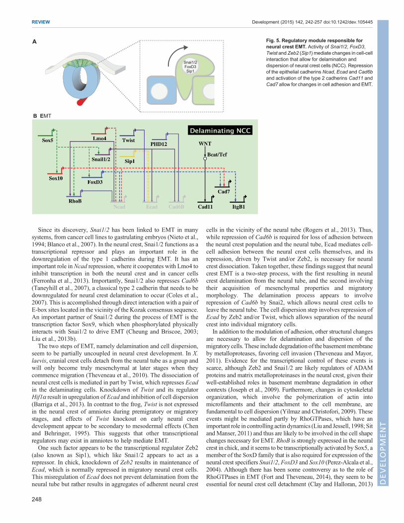
Regulatory interactions in the migratory neural crestAfter delamination, migrating neural crest cells assume a novelregulatory state, expressing a number of transcription factorsimportant for cell migration and for initiating the differentiationprograms that will lead to distinct derivatives (Fig. 6A). Theexpression of several neural crest specifier genes, such as FoxD3,Ets1 and Sox8/9/10, is retained in some or all migratory cells(Khudyakov and Bronner-Fraser, 2009; Betancur et al., 2010a).However, cis-regulatory analysis reveals important differencesbetween the GRNs of the premigratory and migratory states. Forinstance, FoxD3 expression in the premigratory cranial neural crestis controlled by an early enhancer, NC1, which initiates FoxD3expression in the newly closed cranial neural tube but quickly shutsdown as cells delaminate (Simoes-Costa et al., 2012). Maintenanceof FoxD3 expression in migratory cells is accomplished by theactivation of a different enhancer (NC2) (Simoes-Costa et al., 2012)that requires distinct regulatory inputs. Thus, even thoughpremigratory and migratory cells share a number of transcriptionfactors, the programs underlying the expression of the sameregulator can vary greatly as a function of time.Migratory neural crest cells are complex from a regulatory
viewpoint, as they are constantly exposed to different environmentalsignals and are also starting to differentiate into diverse derivatives.
Surprisingly, only a few transcriptional regulators had beenidentified within the migratory neural crest regulatory hub, largelydue to the fact that these cells intermingle with other cell types,making them difficult to isolate and characterize as a pure cellpopulation. Recent transcriptome analysis of pure populations ofSox10+ migratory neural crest cells from chick (Simoes-Costa et al.,2014) has greatly expanded this dataset. This study identified ∼50transcriptional regulators that are enriched in the migratory cranialneural crest when compared with the whole embryo (Simoes-Costaet al., 2014). This dataset provides a valuable resource, although therole of individual genes and how they fit in the neural crest GRNremains to be established.
Cis-regulatory analysis of genes within the neural crest migratoryregulatory module suggests that the expression of these transcriptionfactors is regulated by the neural crest specifier genes (Fig. 6B). Forexample, detailed analysis of the Sox10 genomic locus in chickrevealed that its expression in the migratory neural crest is mediatedby one of two enhancers, Sox10E2 and Sox10E1, which are active inthe cranial and trunk crest, respectively. The cranial Sox10E2enhancer is directly regulated by the neural crest specifier genesMyb,Ets1 and Sox9, which are sufficient to drive enhancer activity in naiveectoderm (Betancur et al., 2010b). These results are consistent withrecent results that place Ets1 and Sox9 as crucial regulators of themigratory module of the cranial neural crest GRN. Knockdown ofthese transcription factors in chick embryos results in loss of severalgenes that are normally expressed by the cranial migratory crest,including Lmo4, RxrG and Ltk (Simoes-Costa et al., 2014).Remarkably, Ets1 seems to be a crucial regulator in theestablishment of neural crest cephalic identity by mediating en
NCspecifiers
A
Migratory neural crest
SoxESnai1/2Lmo4
B Neural crest migration
Fig. 6. Neural crest specifiers drive the transition to themigratory neural crest regulatory state. (A) Action of neural crest specifiers transforms the identity ofthe neural crest as it becomes a migratory cell population. (B) Neural crest specifiers (including Sox9, cMyb and Ets1) cooperate to drive the expression of genesthat are strongly upregulated in the migratory neural crest, such as Sox10. This transcription factor positively regulates itself, which results in maintenance ofSox10 expression as the neural crest cells migrate throughout the embryo to give rise to different derivatives.
249
REVIEW Development (2015) 142, 242-257 doi:10.1242/dev.105445
DEVELO
PM
ENT
masse delamination from the neural tube, resulting in the wave-likemigration pattern observed in the cranial neural crest (Theveneauet al., 2007).Cis-regulatory analysis of Sox10 in the mouse revealed that
multiple enhancers, with semi-overlapping activity domains, seemto mediate the expression of this gene. These regulatory regions arecontrolled by Pax3/7, Tfap2a, Sox and LEF/TCF transcriptionfactors (Werner et al., 2007). Furthermore, Sox10 was shown todirectly regulate itself, consistent with its continuous expression inthe migratory neural crest. This positive self-regulatory loop relieson synergistic activity with Tfap2a and FoxD3 (Wahlbuhl et al.,2012). That FoxD3 acts as a direct regulator of Sox10 in migratorycells is supported by another study that identified an intronicenhancer in the zebrafish sox10 locus, which contains FoxD3, Soxand LEF/TCF binding sites (Dutton et al., 2008). Sox9, anotherSoxE gene that is maintained throughout neural crest migration,has also been shown to directly activate its own transcriptionthrough a distant upstream enhancer in the mouse (Mead et al.,2013). Thus, positive self-regulation is a common feature of genesthat are continuously expressed by the migratory neural crest.Extensive cis-regulatory studies of other migratory neural crestregulators will be necessary to clarify how the regulatory state ofthe neural crest cells evolves during migration and how it isinfluenced by external cues.As expected, a number of signaling systems are involved in the
guidance of neural crest migration and the establishment of the correctmigratory pathways (Takahashi et al., 2013). There is scarceinformation on how these signaling molecules and receptors arecontrolled by the migratory neural crest GRN. Nevertheless, Sox10seems toplayacrucial role andhas, forexample, been shown to regulatethe expression of neuregulin receptor Erbb3 (Britsch et al., 2001)through an intronic enhancer that is active inmigratory neural crest cells(Prasad et al., 2011). Sox transcription factors have also been shown toregulate Robo receptors in other contexts (Samant et al., 2011).However, it is not yet clear how these signaling systems implicated inneural crest migration are controlled at the transcriptional level.
From a multipotent neural crest cell to distinct derivativesAs neural crest cells migrate and colonize different parts of theembryo, they transition from migratory streams to aggregates ofcells within complex structures, often undergoing a process that isthe reciprocal of EMT, termed mesenchymal-to-epithelialtransition (MET). They then contribute to neurons and glia ofperipheral ganglia, cartilage condensations and many organs. Thisprocess is mediated by a gene regulatory program thatcompartmentalizes the neural crest progenitors into territorieswith distinct regulatory states. This has been beautifully illustratedby the profiling of craniofacial enhancers as performed byAttanasio and colleagues (Attanasio et al., 2013). In this studythe authors identified more than 100 enhancers that are active inthe neural crest-derived craniofacial skeleton, in remarkablydiverse activity domains. Functional analysis through enhancermanipulation indicates that variation in craniofacial morphology,as well as craniofacial malformations, depend upon combinedenhancer activity (Attanasio et al., 2013). Understanding how suchregulatory complexity unfolds from the programs of neural crestspecification andmigration is a major challenge confronting neuralcrest biologists in years to come.The process of neural crest diversification initiates with the
activation of differentiation circuits in subpopulations of migratorycells. These circuits are centered around transcription factorsexpressed by the neural crest that activate lineage-specific gene
batteries. Diversification into different derivatives depends upon theinterplay between the regulatory state of the migratory neural crestcells and environmental cues, often conveyed by various signalingsystems. Such environmental signals, particularly interactions withother tissues, are key players in determining the fate of the migratorycells (Takahashi et al., 2013). Here, we describe a subset of conservedregulatory circuits involved in neural crest cell differentiation,focusing on chondrocytes, melanocytes and neuronal cell types asexamples (Fig. 7).
ChondrocytesThe cranial neural crest has a crucial role in establishment of thecraniofacial skeleton, giving rise to the quadrate, Meckel’s cartilageand surrounding membrane bones, cartilage of the tongue, and themembrane bones of the jaw and skull (Noden, 1983; Couly et al.,1996, 1998). As a consequence, this cell population is ofexceptional medical importance, being affected in a largepercentage of birth defects (Hall, 1999). Development of thecraniofacial skeleton is a complicated process that involves multiplecell types and different kinds of ossification (Wilkie and Morriss-Kay, 2001). An important genetic circuit that mediates thedifferentiation of neural crest cells into chondrocytes revolvesaround the transcription factor Sox9, which is a direct regulator ofseveral genes that are crucial for chondrocytic differentiation.Inactivation of Sox9 in the neural crest lineage results in severecraniofacial defects in mice (Mori-Akiyama et al., 2003). Sox9 isthought to cooperate with SoxD genes to activate downstreamtargets such as Col2a1 and Agc1 (also known as Acan). In themigratory cranial neural crest, Sox9 is regulated by WNT signalingand also has strong autoregulatory activity (Bagheri-Fam et al.,2006; Mead et al., 2013) (Fig. 7B).
Transcriptional control of Agc1, a marker for cartilagedifferentiation, illustrates how cartilage genes are regulated. Inmice, an enhancer located 10 kb upstream of the transcription startsite mediates reporter expression that recapitulates endogenousAgc1 expression. Sox9 binds to a critical site in this enhancer, butonly in the presence of Sox5/6. These SoxC factors interact withthree accessory binding sites that enhance expression of thereporter (Han and Lefebvre, 2008). A similar mode of regulation,relying on direct activation by Sox9 and SoxD genes, was observedfor Col2a1 in mice (Bell et al., 1997; Lefebvre et al., 1998). Sox9-dependent activation of chondrogenic genes relies on recruitmentof the histone acetyltransferase CBP/p300 (Tsuda et al., 2003),which is mediated by Smad3; this illustrates how the TGFβpathway may function as a permissive signal during cartilageformation (Furumatsu et al., 2005). Both Sox9 and SoxD genes arealso crucial for the survival of neural crest derivatives, as their losscauses massive apoptosis of these cells in chicken and mice(Cheung et al., 2005; Bhattaram et al., 2010). In soft tissues of theface, such as the tongue, Sox9must be shut down in order to preventcartilage formation. The transcription factor Osr1 mediates thisrepression by directly binding to the Sox9 promoter (Liu et al.,2013a), and its loss results in the formation of ectopic cartilage inthe mouse tongue.WhereasOsr1 is expressed in the mouse tongue,it is absent from bird tongues; interestingly, the latter containcartilaginous tissue. This illustrates how changes in the regulatoryinputs of a gene could result in morphological changes duringevolution (Liu et al., 2013a).
MelanocytesNeural crest differentiation into melanocytes relies upon the action oftranscription factors Sox10 and Pax3/7 as well as signaling systems
250
REVIEW Development (2015) 142, 242-257 doi:10.1242/dev.105445
DEVELO
PM
ENT
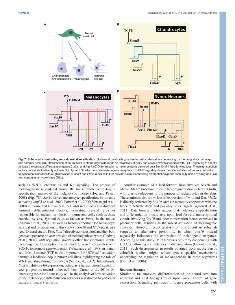
such as WNTs, endothelins and Kit signaling. The process ofmelanogenesis is centered around the transcription factor Mitf, aspecification marker of the melanocytic lineage (Hou and Pavan,2008) (Fig. 7C). Sox10 drives melanocyte specification by directlyactivatingMitf (Lee et al., 2000; Potterf et al., 2000; Verastegui et al.,2000) in mouse and human cell lines. Mitf in turn acts as a driver ofterminal differentiation factors, activating several enzymesresponsible for melanin synthesis in pigmented cells, such as thoseencoded by Dct, Tyr and Si (also known as Pmel) in the mouse(Murisier et al., 2007), as well as factors important for melanocytesurvival and proliferation. In this context, Sox10 andMitf operate in afeed-forward circuit. First, Sox10 directly activatesMitf, and then bothgenes cooperate todrive expression ofmelanogenic enzymes (Ludwiget al., 2004). Mitf regulation involves other transcriptional inputs,including the transcription factor Pax3/7, which cooperates withSOX10 to promote gene expression (Watanabe et al., 1998) in humancell lines. β-catenin/TCF is also important for MITF self-activationthrough a feedback loop in human cell lines, highlighting the role ofWNT signaling during this process (Saito et al., 2002). Interestingly,FoxD3 inhibits Mitf expression, acting as a transcriptional switch toveer progenitors towards other cell fates (Curran et al., 2010). Aninteresting topic for future study will be the analysis of how activationof the melanocytic differentiation networks is restricted to particularsubsets of neural crest cells.
Another example of a feed-forward loop involves Sox10 andMef2c. Mef2c knockout mice exhibit pigmentation defects at birth,with drastic reductions in the number of melanocytes in the skin.These animals also show loss of expression of Mitf and Dct. Mef2cis directly activated by Sox10, and subsequently cooperates with thelatter to activate itself and possibly other targets (Agarwal et al.,2011). Data from amniotes suggest that melanocyte specificationand differentiation mostly rely upon feed-forward transcriptionalcircuits involving Sox10 and other transcription factors expressed inprecursor cells, resulting in the robust activation of melanogenicenzymes. However, recent analysis of this circuit in zebrafishsuggests an alternative possibility, in which sox10 insteadnegatively influences the expression of melanogenic enzymes.According to this study, Mitf represses sox10 by cooperating withHDACs, allowing for melanocytic differentiation (Greenhill et al.,2011). Such discrepancies in results obtained from mouse versuszebrafish studies might reflect species-specific mechanismsunderlying the regulation of melanogenesis in these organisms(Hou et al., 2006).
Neuronal lineagesSimilar to melanocytes, differentiation of the neural crest intoneuronal and glial lineages relies upon Sox10 control of geneexpression. Signaling pathways influence progenitor cells with
Fig. 7. Subcircuits controlling neural crest diversification. (A) Neural crest cells give rise to distinct derivatives depending on their migratory pathwaysand external cues. (B) Differentiation of neural crest to chondrocytes depends on the activity of Sox9 and Sox5/6, which cooperate with TGFβ signaling to directlyactivate the cartilage differentiation genesCol2a1 andAgc1. (C) Differentiation tomelanocytes is centered on a Sox10-Mitf feed-forward loop. These transcriptionfactors cooperate to directly activate Dct, Tyr and Si, which encode melanogenic enzymes. (D) BMP signaling drives the differentiation of neural crest cellsto sympathetic neurons through activation of Ascl1 and Phox2b, which in turn activate a circuit controlling differentiation genes such as tyrosine hydroxylase (Th)and dopamine β-hydroxylase (Dbh).
251
REVIEW Development (2015) 142, 242-257 doi:10.1242/dev.105445
DEVELO
PM
ENT
respect to the cell type to which they contribute. For instance,neuregulin signaling biases neural crest progenitors to adopt aperipheral glial fate, whereas BMPs promote neuronaldifferentiation (Stemple and Anderson, 1993). Sympatheticneurons are specified by BMP signaling secreted by the dorsalaorta (Saito et al., 2012), which cooperates with Sox10 to activateexpression of the sympathetic neuron specifier genes Phox2b andAscl1 (Morikawa et al., 2009) (Fig. 7D and Fig. 8). Thesetranscription factors activate downstream targets, such as Gata3,Insm1 and Hand2, which mediate cell cycle control, maintenanceof survival and differentiation in sympathetic neurons (Rohrer,2011). Most of these data derive from genetic analyses ofknockout mice, and little is known about molecular mechanismsin this context. However, Tfap2a has been shown to directlyregulate tyrosine hydroxylase and dopamine β-hydroxylase and isthus a likely player in the differentiation of these cells (Kim et al.,2001). Further diversification of sympathetic neurons relies on thetranscription factor Hmx1, which drives differentiation towards anoradrenergic sympathetic fate. This regulator is repressed incholinergic neurons by Trkc (also known as Ntrk3) and Ret(Furlan et al., 2013).The above examples of diversification circuits by no means
represent an exhaustive survey. In addition to melanocytes,cartilage and sympathetic ganglia, neural crest cells form manyother derivatives, including sensory neurons, Schwann cells,adipocytes and smooth muscle cells. Generally, neural crestdiversification depends upon the interplay of the cell-intrinsictranscriptional machinery and its interpretation of environmentalcues conveyed by distinct signaling pathways. In this regard, SoxEgenes play a crucial role upstream of many neural crest celllineages, either by directly driving differentiation (e.g. as in thecase of Sox9 in chondrocyte differentiation) or by regulating thedrivers that activate differentiation gene batteries. Furthermore,other early neural crest GRN components (Tfap2, Pax3/7 andSox5/6) also participate later in the process of neural crestdiversification, highlighting that the neural crest transcriptionalmachinery can be repurposed, such that factors that act in themaintenance of pluripotency can be subsequently deployed indifferentiation programs.
Epigenetic regulation in neural crest formationAlthough we have focused on the role of transcription factors in theprogram underlying neural crest development, it has becomeincreasingly clear that this process contains additional layers ofregulatory complexity. For example, transcription factors oftenwork in conjunction with chromatin modifiers to activate or repressgene expression (Wang et al., 2011a; Strobl-Mazzulla and Bronner,2012). Similarly, gene expression can be modulated by post-transcriptional regulation mediated by RNA-binding proteins andsmall regulatory RNAs, as well as post-translational modifications oftranscription factors themselves by phosphorylation, sumoylation,and other changes that modify binding efficacy and stability (Taylorand LaBonne, 2007). As discussed below, DNA and histonemodifications have been shown to play particularly important rolesin both neural crest specification and EMT.DNA methylation marks, which are conferred upon the promoter
regions of genes by DNA methyltransferases (DNMTs), play animportant role in silencing genes and thus operate to restrict fatechoices during development (Okano et al., 1999; Smith andMeissner, 2013). In the neural crest, DNMTs have been shown to beimportant in the segregation of the neural crest from neural lineages.Dnmt3a, which is expressed in the neural plate border, directly
represses expression of the neural genes Sox2/3 bymethylating CpGislands in the neural crest progenitor territory. Knockdown ofDnmt3a results in the expansion of the neural plate and loss ofneural crest specification (Hu et al., 2012). Although it is not clearhow Dnmt3a is recruited to the promoter region of Sox2/3 in thedorsal neural tube, an intriguing possibility is that it may berecruited by transcription factors expressed in the neural crestprogenitor region. The role of its paralog Dnmt3b is less clear.Mutations in DNMT3B seem to be linked to human craniofacialdefects present in carriers of the ICF syndrome (Jin et al., 2008), andthere is evidence for a role of this regulator in craniofacialdevelopment in zebrafish (Rai et al., 2010). However, conditionalknockout of Dnmt3b in the mouse neural crest does not cause anobvious neural crest-related phenotype (Jacques-Fricke et al., 2012).Thus, there might be species-specific differences in the function ofthe enzyme.
Histone methylation affects gene regulation by definingrepressive or active transcriptional states. Methylation marks areattached to the histones of the nucleosomes by methyltransferasesand are removed by demethylases (Dambacher et al., 2010).Interestingly, removal of repressive methylation marks is necessaryfor the neural crest specification program. This is accomplished bythe histone demethylase Kdm4a (Jmjd2a), which clears these marksfrom the Sox10 and Snai2 promoters (Strobl-Mazzulla and Bronner,2012). At later stages of neural crest development, the histonedemethylase Phf8 acts in a similar manner, and demethylatespromoters of genes important for craniofacial development(Qi et al., 2010).
A different type of histone modification results from lysineacetylation, which is associated with active transcriptional states(Haberland et al., 2009b), and has been useful for investigatinggene regulation in the neural crest (Rada-Iglesias et al., 2012).During EMT, it is well known that Snail proteins directly represscadherins. In neural crest cells, the target of Snai2 is Cad6b, whoserepression is mediated by recruitment of the histone deacetylase(HDAC) repressive complex to the Cad6b promoter, which occursvia interactions between Snai2, Sin3a and the adaptor proteinPhd12 (also known as Phf12 or Pf1) (Strobl-Mazzulla andBronner, 2012). HDACs are also expressed in the migratoryneural crest (Simoes-Costa et al., 2014) and are involved in thediversification of derivatives. For example, HDACs act in therepression of foxd3 during melanophore specification in zebrafish(Ignatius et al., 2008) and have been shown to play important rolesin the development of peripheral glia (Jacob et al., 2014) andectomesenchymal derivatives (Haberland et al., 2009a) in themouse.
These examples demonstrate that epigenetic modifications are anintegral part of the gene regulatory machinery, playing a crucial rolein modulating gene expression. Like other progenitor cellpopulations, the neural crest is likely to undergo global chromatinremodeling as it is specified and differentiates to differentderivatives (Chen and Dent, 2014). However, the enzymes thatcatalyze either addition or removal of epigenetic marks are, for themost part, unable to interpret the genomic code. As a consequence,the epigenetic machinery depends upon the transcription factors ofthe GRN to find their destination in the genome and correctly marktheir target sequences. Indeed, studies have shown that epigeneticrepressive complexes are recruited to cis-regulatory regions andpromoters by transcription factors (Ropero and Esteller, 2007;Wang et al., 2011a; Tam and Weinberg, 2013). Thus, rather thanacting independently, these additional layers of regulation areintrinsically linked to the neural crest GRN.
252
REVIEW Development (2015) 142, 242-257 doi:10.1242/dev.105445
DEVELO
PM
ENT
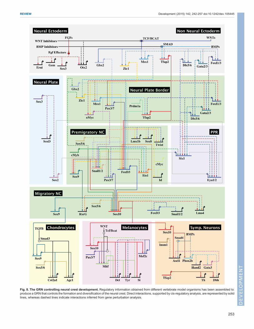
Fig. 8. The GRN controlling neural crest development. Regulatory information obtained from different vertebrate model organisms has been assembled toproduce aGRN that controls the formation and diversification of the neural crest. Direct interactions, supported by cis-regulatory analysis, are represented by solidlines, whereas dashed lines indicate interactions inferred from gene perturbation analysis.
253
REVIEW Development (2015) 142, 242-257 doi:10.1242/dev.105445
DEVELO
PM
ENT
ConclusionsThe data presented here knit together our current knowledge of theneural crest program, from early establishment of the neural plateborder to diversification into varied derivatives. The neural crestGRN is a feed-forward self-assembly ‘machine’ that combinespositive transcriptional inputs with repressive interactions thatestablish firm borders between adjacent tissues, such as non-neuralectoderm, ectodermal placode, presumptive neural crest and futureneural plate territories. Although these regulatory principles areshared among all developing neural crest cells, it is important tokeep in mind that the neural crest is not a homogenous populationand that differences in GRN structure underlie the regionalproperties observed in cranial, vagal, trunk and sacral neural crest.Thus, comparative gene regulatory analysis will be crucial touncover the molecular mechanisms that are responsible for thedifferences in potential and behavior observed between neural crestsubpopulations at different axial levels.Our current view of this GRN is summarized in the BioTapestry
visualization in Fig. 8, which focuses on the cranial neural crest.This is, by definition, overly simplified since many inputs have yetto be placed within the network and there are important differencesbetween organisms that have been largely collapsed here into a pan-vertebrate model. Furthermore, although we have emphasized therole of transcription factors in the GRN, these regulators oftenfunction in large complexes in concert with epigenetic modifiersand non-coding RNAs including lncRNAs and microRNAs. Futureexperiments will need to further refine this GRN and focus onidentifying direct interactions therein, as best accomplished bydetailed cis-regulatory analysis. New technologies for identifyingneural crest enhancers and performing genome editing will greatlyfacilitate elaboration of the neural crest GRN. This, in turn, willfacilitate expansion of this network to other axial levels and provideincreasingly detailed resolution of the events that underlie theformation of this fascinating cell type – the neural crest.
Competing interestsThe authors declare no competing financial interests.
FundingWork in the laboratory of M.E.B. is supported by the National Institutes of Health.M.S.-C. was funded by the Pew fellows Program in the Biomedical Sciences andby a grant from the National Institute of Dental and Craniofacial Research.Deposited in PMC for release after 12 months.
ReferencesAgarwal, P., Verzi, M. P., Nguyen, T., Hu, J., Ehlers, M. L., McCulley, D. J., Xu, S.-M., Dodou, E., Anderson, J. P., Wei, M. L. et al. (2011). The MADS boxtranscription factor MEF2C regulates melanocyte development and is a directtranscriptional target and partner of SOX10. Development 138, 2555-2565.
Aruga, J. (2004). The role of Zic genes in neural development.Mol. Cell. Neurosci.26,205-221.
Attanasio, C., Nord, A. S., Zhu, Y., Blow, M. J., Li, Z., Liberton, D. K., Morrison,H., Plajzer-Frick, I., Holt, A., Hosseini, R. et al. (2013). Fine tuning of craniofacialmorphology by distant-acting enhancers. Science 342, 1241006.
Aybar, M. J., Nieto, M. A. and Mayor, R. (2003). Snail precedes slug in the geneticcascade required for the specification and migration of the Xenopus neural crest.Development 130, 483-494.
Bae, C.-J., Park, B.-Y., Lee, Y.-H., Tobias, J. W., Hong, C.-S. and Saint-Jeannet,J.-P. (2014). Identification of Pax3 and Zic1 targets in the developing neural crest.Dev. Biol. 386, 473-483.
Bagheri-Fam, S., Barrionuevo, F., Dohrmann, U., Gunther, T., Schule, R.,Kemler, R., Mallo, M., Kanzler, B. andScherer, G. (2006). Long-range upstreamand downstream enhancers control distinct subsets of the complexspatiotemporal Sox9 expression pattern. Dev. Biol. 291, 382-397.
Barembaum, M. and Bronner, M. E. (2013). Identification and dissection of a keyenhancer mediating cranial neural crest specific expression of transcription factor,Ets-1. Dev. Biol. 382, 567-575.
Barriga, E. H., Maxwell, P. H., Reyes, A. E. and Mayor, R. (2013). The hypoxiafactor Hif-1alpha controls neural crest chemotaxis and epithelial to mesenchymaltransition. J. Cell Biol. 201, 759-776.
Bell, D. M., Leung, K. K. H., Wheatley, S. C., Ng, L. J., Zhou, S., Ling, K. W.,Sham, M. H., Koopman, P., Tam, P. P. L. and Cheah, K. S. E. (1997). SOX9directly regulates the type-II collagen gene. Nat. Genet. 16, 174-178.
Bellmeyer, A., Krase, J., Lindgren, J. and LaBonne, C. (2003). Theprotooncogene c-Myc is an essential regulator of neural crest formation inXenopus. Dev. Cell 4, 827-839.
Betancur, P., Bronner-Fraser, M. and Sauka-Spengler, T. (2010a). Assemblingneural crest regulatory circuits into a gene regulatory network. Annu. Rev. CellDev. Biol. 26, 581-603.
Betancur, P., Bronner-Fraser, M. and Sauka-Spengler, T. (2010b). Genomiccode for Sox10 activation reveals a key regulatory enhancer for cranial neuralcrest. Proc. Natl. Acad. Sci. USA 107, 3570-3575.
Bhat, N., Kwon, H.-J. and Riley, B. B. (2013). A gene network that coordinatespreplacodal competence and neural crest specification in zebrafish. Dev. Biol.373, 107-117.
Bhattaram, P., Penzo-Mendez, A., Sock, E., Colmenares, C., Kaneko, K. J.,Vassilev, A., DePamphilis, M. L., Wegner, M. and Lefebvre, V. (2010).Organogenesis relies on SoxC transcription factors for the survival of neural andmesenchymal progenitors. Nat. Commun. 1, 9.
Blanco, M. J., Barrallo-Gimeno, A., Acloque, H., Reyes, A. E., Tada, M., Allende,M. L., Mayor, R. and Nieto, M. A. (2007). Snail1a and Snail1b cooperate in theanterior migration of the axial mesendoderm in the zebrafish embryo.Development 134, 4073-4081.
Britsch, S., Goerich, D. E., Riethmacher, D., Peirano, R. I., Rossner, M., Nave,K.-A., Birchmeier, C. andWegner, M. (2001). The transcription factor Sox10 is akey regulator of peripheral glial development. Genes Dev. 15, 66-78.
Bronner, M. E. and LeDouarin, N. M. (2012). Development and evolution of theneural crest: an overview. Dev. Biol. 366, 2-9.
Brugmann, S. A., Pandur, P. D., Kenyon, K. L., Pignoni, F. and Moody, S. A.(2004). Six1 promotes a placodal fate within the lateral neurogenic ectoderm byfunctioning as both a transcriptional activator and repressor. Development 131,5871-5881.
Chen, Z. F. and Behringer, R. R. (1995). Twist is required in head mesenchyme forcranial neural tube morphogenesis. Genes Dev. 9, 686-699.
Chen, T. andDent, S. Y. R. (2014). Chromatin modifiers and remodellers: regulatorsof cellular differentiation. Nat. Rev. Genet. 15, 93-106.
Cheung, M. and Briscoe, J. (2003). Neural crest development is regulated by thetranscription factor Sox9. Development 130, 5681-5693.
Cheung, M., Chaboissier, M.-C., Mynett, A., Hirst, E., Schedl, A. and Briscoe, J.(2005). The transcriptional control of trunk neural crest induction, survival, anddelamination. Dev. Cell 8, 179-192.
Christophorou, N. A. D., Bailey, A. P., Hanson, S. and Streit, A. (2009). Activationof Six1 target genes is required for sensory placode formation. Dev. Biol. 336,327-336.
Clay, M. R. and Halloran, M. C. (2013). Rho activation is apically restricted byArhgap1 in neural crest cells and drives epithelial-to-mesenchymal transition.Development 140, 3198-3209.
Coles, E. G., Taneyhill, L. A. and Bronner-Fraser, M. (2007). A critical role forCadherin6B in regulating avian neural crest emigration. Dev. Biol. 312, 533-544.
Couly, G. F., Grapin-Bottom, A., Coltey, P. and Le Douarin, N. M. (1996). Theregeneration of the cephalic neural crest, a problem revisited: the regeneratingcells originate from the contralateral or from the anterior and posterior neural folds.Development 122, 3393-3407.
Couly, G., Grapin-Botton, A., Coltey, P., Ruhin, B. and Le Douarin, N. M. (1998).Determination of the identity of the derivatives of the cephalic neural crest:incompatibility between Hox gene expression and lower jaw development.Development 128, 3445-3459.
Creazzo, T. L., Godt, R. E., Leatherbury, L., Conway, S. J. and Kirby, M. L.(1998). Role of cardiac neural crest cells in cardiovascular development. Annu.Rev. Physiol. 60, 267-286.
Curran, K., Lister, J. A., Kunkel, G. R., Prendergast, A., Parichy, D. M. andRaible, D. W. (2010). Interplay between Foxd3 and Mitf regulates cell fateplasticity in the zebrafish neural crest. Dev. Biol. 344, 107-118.
Dambacher, S., Hahn, M. and Schotta, G. (2010). Epigenetic regulation ofdevelopment by histone lysine methylation. Heredity 105, 24-37.
de Croze, N., Maczkowiak, F. and Monsoro-Burq, A. H. (2011). Reiterative AP2aactivity controls sequential steps in the neural crest gene regulatory network.Proc.Natl. Acad. Sci. USA 108, 155-160.
Dottori, M., Gross, M. K., Labosky, P. and Goulding, M. (2001). The winged-helixtranscription factor Foxd3 suppresses interneuron differentiation and promotesneural crest cell fate. Development 128, 4127-4138.
Dutton, J. R., Antonellis, A., Carney, T. J., Rodrigues, F. S. L. M., Pavan, W. J.,Ward, A. and Kelsh, R. N. (2008). An evolutionarily conserved intronic regioncontrols the spatiotemporal expression of the transcription factor Sox10. BMCDev. Biol. 8, 105.
Endo, Y., Osumi, N. and Wakamatsu, Y. (2002). Bimodal functions of Notch-mediated signaling are involved in neural crest formation during avian ectodermdevelopment. Development 129, 863-873.
254
REVIEW Development (2015) 142, 242-257 doi:10.1242/dev.105445
DEVELO
PM
ENT
Fairchild, C. L. and Gammill, L. S. (2013). Tetraspanin18 is a FoxD3-responsiveantagonist of cranial neural crest epithelial-to-mesenchymal transition thatmaintains cadherin-6B protein. J. Cell Sci. 126, 1464-1476.
Feledy, J. A., Beanan, M. J., Sandoval, J. J., Goodrich, J. S., Lim, J. H., Matsuo-Takasaki, M., Sato, S. M. and Sargent, T. D. (1999). Inhibitory patterning of theanterior neural plate in Xenopus by homeodomain factors Dlx3 and Msx1. Dev.Biol. 212, 455-464.
Ferronha, T., Rabadan, M. A., Gil-Guinon, E., Le Dreau, G., de Torres, C. andMarti, E. (2013). LMO4 is an essential cofactor in the Snail2-mediated epithelial-to-mesenchymal transition of neuroblastoma and neural crest cells. J. Neurosci.33, 2773-2783.
Fort, P. and Theveneau, E. (2014). PleiotRHOpic: rho pathways are essential for allstages of Neural Crest development. Small GTPases 5, e27975.
Furlan, A., Lubke, M., Adameyko, I., Lallemend, F. and Ernfors, P. (2013). Thetranscription factor Hmx1 and growth factor receptor activities control sympatheticneurons diversification. EMBO J. 32, 1613-1625.
Furumatsu, T., Tsuda, M., Taniguchi, N., Tajima, Y. and Asahara, H. (2005).Smad3 induces chondrogenesis through the activation of SOX9 via CREB-binding protein/p300 recruitment. J. Biol. Chem. 280, 8343-8350.
Gans, C. and Northcutt, R. G. (1983). Neural crest and the origin of vertebrates: anew head. Science 220, 268-273.
Garcia-Castro, M. I., Marcelle, C. andBronner-Fraser, M. (2002). Ectodermal Wntfunction as a neural crest inducer. Science 13, 13.
Garnett, A. T., Square, T. A. and Medeiros, D. M. (2012). BMP, Wnt and FGFsignals are integrated through evolutionarily conserved enhancers to achieverobust expression of Pax3 and Zic genes at the zebrafish neural plate border.Development 139, 4220-4231.
Greenhill, E. R., Rocco, A., Vibert, L., Nikaido, M. and Kelsh, R. N. (2011). Aniterative genetic and dynamical modelling approach identifies novel features of thegene regulatory network underlying melanocyte development. PLoS Genet. 7,e1002265.
Grocott, T., Tambalo, M. and Streit, A. (2012). The peripheral sensory nervoussystem in the vertebrate head: a gene regulatory perspective.Dev. Biol. 370, 3-23.
Groves, A. K. and LaBonne, C. (2014). Setting appropriate boundaries: fate,patterning and competence at the neural plate border. Dev. Biol. 389, 2-12.
Guemar, L., de Santa Barbara, P., Vignal, E., Maurel, B., Fort, P. and Faure, S.(2007). The small GTPase RhoV is an essential regulator of neural crest inductionin Xenopus. Dev. Biol. 310, 113-128.
Haberland, M., Mokalled, M. H., Montgomery, R. L. and Olson, E. N. (2009a).Epigenetic control of skull morphogenesis by histone deacetylase 8. Genes Dev.23, 1625-1630.
Haberland, M., Montgomery, R. L. and Olson, E. N. (2009b). The many roles ofhistone deacetylases in development and physiology: implications for diseaseand therapy. Nat. Rev. Genet. 10, 32-42.
Hall, B. K. (1999). The Neural Crest in Development and Evolution. Springer.Hall, B. K. (2000). The neural crest as a fourth germ layer and vertebrates asquadroblastic not triploblastic. Evol. Dev. 2, 3-5.
Han, Y. and Lefebvre, V. (2008). L-Sox5 and Sox6 drive expression of the aggrecangene in cartilage by securing binding of Sox9 to a far-upstream enhancer. Mol.Cell. Biol. 28, 4999-5013.
Hong, C.-S. and Saint-Jeannet, J.-P. (2007). The activity of Pax3 and Zic1regulates three distinct cell fates at the neural plate border. Mol. Biol. Cell 18,2192-2202.
Honore, S. M., Aybar, M. J. and Mayor, R. (2003). Sox10 is required for the earlydevelopment of the prospective neural crest in Xenopus embryos. Dev. Biol. 260,79-96.
Horstadius, S. (1950). The Neural Crest. Oxford University Press.Hou, L. and Pavan, W. J. (2008). Transcriptional and signaling regulation in neuralcrest stem cell-derived melanocyte development: do all roads lead to Mitf? CellRes. 18, 1163-1176.
Hou, L., Arnheiter, H. and Pavan, W. J. (2006). Interspecies difference in theregulation of melanocyte development by SOX10 andMITF.Proc. Natl. Acad. Sci.USA 103, 9081-9085.
Hu, N., Strobl-Mazzulla, P., Sauka-Spengler, T. and Bronner, M. E. (2012). DNAmethyltransferase3A as a molecular switch mediating the neural tube-to-neuralcrest fate transition. Genes Dev. 26, 2380-2385.
Ignatius, M. S., Moose, H. E., El-Hodiri, H. M. and Henion, P. D. (2008). colgate/hdac1 repression of foxd3 expression is required to permit mitfa-dependentmelanogenesis. Dev. Biol. 313, 568-583.
Jacob, C., Lotscher, P., Engler, S., Baggiolini, A., Tavares, S. V., Brugger, V.,John, N., Buchmann-Moller, S., Snider, P. L., Conway, S. J. et al. (2014).HDAC1 and HDAC2 control the specification of neural crest cells into peripheralglia. J. Neurosci. 34, 6112-6122.
Jacques-Fricke, B. T., Roffers-Agarwal, J. and Gammill, L. S. (2012). DNAmethyltransferase 3b is dispensable for mouse neural crest development. PLoSONE 7, e47794.
Jaeger, J. (2011). The gap gene network. Cell. Mol. Life Sci. 68, 243-274.Jin, B., Tao, Q., Peng, J., Soo, H. M.,Wu,W., Ying, J., Fields, C. R., Delmas, A. L.,Liu, X., Qiu, J. et al. (2008). DNA methyltransferase 3B (DNMT3B) mutations inICF syndrome lead to altered epigenetic modifications and aberrant expression of
genes regulating development, neurogenesis and immune function. Hum. Mol.Genet. 17, 690-709.
Joseph, M. J., Dangi-Garimella, S., Shields, M. A., Diamond, M. E., Sun, L.,Koblinski, J. E. and Munshi, H. G. (2009). Slug is a downstream mediator oftransforming growth factor-beta 1-induced matrix metalloproteinase-9 expressionand invasion of oral cancer cells. J. Cell. Biochem. 108, 726-736.
Kashef, J., Kohler, A., Kuriyama, S., Alfandari, D., Mayor, R. and Wedlich, D.(2009). Cadherin-11 regulates protrusive activity in Xenopus cranial neural crestcells upstream of Trio and the small GTPases. Genes Dev. 23, 1393-1398.
Khudyakov, J. and Bronner-Fraser, M. (2009). Comprehensive spatiotemporalanalysis of early chick neural crest network genes. Dev. Dyn. 238, 716-723.
Kim, H.-S., Hong, S. J., LeDoux, M. S. and Kim, K.-S. (2001). Regulation of thetyrosine hydroxylase and dopamine beta-hydroxylase genes by the transcriptionfactor AP-2. J. Neurochem. 76, 280-294.
Kuriyama, S., Theveneau, E., Benedetto, A., Parsons, M., Tanaka, M., Charras,G., Kabla, A. and Mayor, R. (2014). In vivo collective cell migration requires anLPAR2-dependent increase in tissue fluidity. J. Cell Biol. 206, 113-127.
Kwon, H. J., Bhat, N., Sweet, E. M., Cornell, R. A. and Riley, B. B. (2010).Identification of early requirements for preplacodal ectoderm and sensory organdevelopment. PLoS Genet. 6, e1001133.
Labosky, P. A. and Kaestner, K. H. (1998). The winged helix transcription factorHfh2 is expressed in neural crest and spinal cord during mouse development.Mech. Dev. 76, 185-190.
Le Douarin, N. (1973). A biological cell labeling technique and its use inexperimental embryology. Dev. Biol. 30, 217-222.
Le Douarin, N. (1982). The Neural Crest. Cambridge University Press.Le Douarin, N. and Kalcheim, C. (1999). The Neural Crest. Cambridge University
Press.Le Douarin, N. M. and Smith, J. (1988). Development of the peripheral nervous
system from the neural crest. Annu. Rev. Cell Biol. 4, 375-404.Le Douarin, N. M. and Teillet, M. A. (1973). The migration of neural crest cells to the
wall of the digestive tract in avian embryo. J. Embryol. Exp. Morphol. 30, 31-48.Le Douarin, N. M., Creuzet, S., Couly, G. and Dupin, E. (2004). Neural crest cell
plasticity and its limits. Development 131, 4637-4650.Le Lievre, C. S. and Le Douarin, N. M. (1975). Mesenchymal derivatives of the
neural crest: analysis of chimaeric quail and chick embryos. J. Embryol. Exp.Morphol. 34, 125-154.
Le Lievre, C. S., Schweizer, G. G., Ziller, C. M. and Le Douarin, N. M. (1980).Restrictions of developmental capabilities in neural crest cell derivatives as testedby in vivo transplantation experiments. Dev. Biol. 77, 362-378.
Lee, M., Goodall, J., Verastegui, C., Ballotti, R. and Goding, C. R. (2000). Directregulation of the Microphthalmia promoter by Sox10 links Waardenburg-Shahsyndrome (WS4)-associated hypopigmentation and deafness to WS2. J. Biol.Chem. 275, 37978-37983.
Lefebvre, V., Li, P., Eberspaecher, H. and de Crombrugghe, B. (1998). New longform of Sox5, Sox6, and Sox9 are co-expressed in chondrocytes andcooperatively activate the type II collagen gene. FASEB J. 12, A1392-A1392.
Li, W. and Cornell, R. A. (2007). Redundant activities of Tfap2a and Tfap2c arerequired for neural crest induction and development of other non-neural ectodermderivatives in zebrafish embryos. Dev. Biol. 304, 338-354.
Li, B., Kuriyama, S., Moreno, M. and Mayor, R. (2009). The posteriorizing geneGbx2 is a direct target of Wnt signalling and the earliest factor in neural crestinduction. Development 136, 3267-3278.
Liu, J. P. and Jessell, T. M. (1998). A role for rhoB in the delamination of neural crestcells from the dorsal neural tube. Development 125, 5055-5067.
Liu, H., Lan, Y., Xu, J., Chang, C.-F., Brugmann, S. A. and Jiang, R. (2013a). Odd-skipped related-1 controls neural crest chondrogenesis during tonguedevelopment. Proc. Natl. Acad. Sci. USA 110, 18555-18560.
Liu, J. A. J.,Wu,M.-H., Yan, C. H., Chau, B. K. H., So, H., Ng, A., Chan, A., Cheah,K. S. E., Briscoe, J. and Cheung, M. (2013b). Phosphorylation of Sox9 isrequired for neural crest delamination and is regulated downstream of BMP andcanonical Wnt signaling. Proc. Natl. Acad. Sci. USA 110, 2882-2887.
Longabaugh, W. J. R., Davidson, E. H. and Bolouri, H. (2005). Computationalrepresentation of developmental genetic regulatory networks. Dev. Biol. 283,1-16.
Longabaugh, W. J. R., Davidson, E. H. and Bolouri, H. (2009). Visualization,documentation, analysis, and communication of large-scale gene regulatorynetworks. Biochim. Biophys. Acta 1789, 363-374.
Ludwig, A., Rehberg, S. and Wegner, M. (2004). Melanocyte-specific expressionof dopachrome tautomerase is dependent on synergistic gene activation by theSox10 and Mitf transcription factors. FEBS Lett. 556, 236-244.
Luo, T., Matsuo-Takasaki, M. and Sargent, T. D. (2001). Distinct roles for Distal-less genes Dlx3 and Dlx5 in regulating ectodermal development in Xenopus.Mol.Reprod. Dev. 60, 331-337.
Luo, T., Matsuo-Takasaki, M., Thomas, M. L., Weeks, D. L. and Sargent, T. D.(2002). Transcription factor AP-2 is an essential and direct regulator of epidermaldevelopment in Xenopus. Dev. Biol. 245, 136-144.
Luo, T., Lee, Y.-H., Saint-Jeannet, J.-P. and Sargent, T. D. (2003). Induction ofneural crest in Xenopus by transcription factor AP2alpha. Proc. Natl. Acad. Sci.USA 100, 532-537.
255
REVIEW Development (2015) 142, 242-257 doi:10.1242/dev.105445
DEVELO
PM
ENT
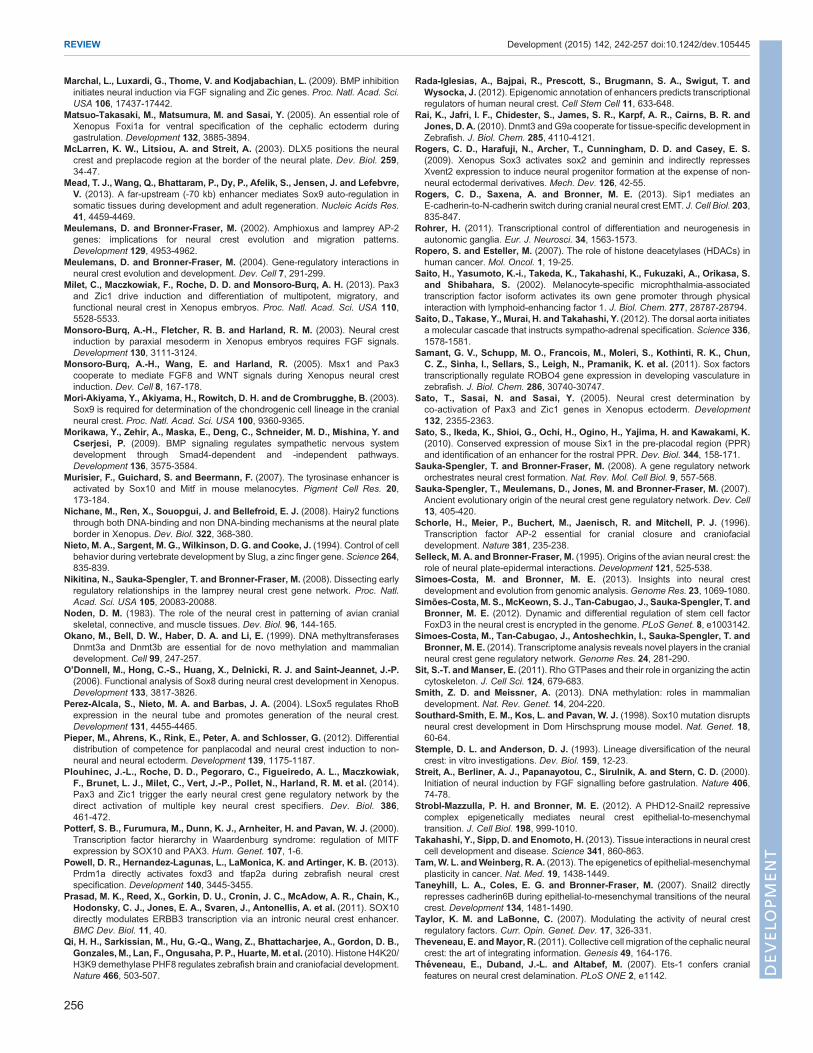
Marchal, L., Luxardi, G., Thome, V. and Kodjabachian, L. (2009). BMP inhibitioninitiates neural induction via FGF signaling and Zic genes. Proc. Natl. Acad. Sci.USA 106, 17437-17442.
Matsuo-Takasaki, M., Matsumura, M. and Sasai, Y. (2005). An essential role ofXenopus Foxi1a for ventral specification of the cephalic ectoderm duringgastrulation. Development 132, 3885-3894.
McLarren, K. W., Litsiou, A. and Streit, A. (2003). DLX5 positions the neuralcrest and preplacode region at the border of the neural plate. Dev. Biol. 259,34-47.
Mead, T. J., Wang, Q., Bhattaram, P., Dy, P., Afelik, S., Jensen, J. and Lefebvre,V. (2013). A far-upstream (-70 kb) enhancer mediates Sox9 auto-regulation insomatic tissues during development and adult regeneration. Nucleic Acids Res.41, 4459-4469.
Meulemans, D. and Bronner-Fraser, M. (2002). Amphioxus and lamprey AP-2genes: implications for neural crest evolution and migration patterns.Development 129, 4953-4962.
Meulemans, D. and Bronner-Fraser, M. (2004). Gene-regulatory interactions inneural crest evolution and development. Dev. Cell 7, 291-299.
Milet, C., Maczkowiak, F., Roche, D. D. and Monsoro-Burq, A. H. (2013). Pax3and Zic1 drive induction and differentiation of multipotent, migratory, andfunctional neural crest in Xenopus embryos. Proc. Natl. Acad. Sci. USA 110,5528-5533.
Monsoro-Burq, A.-H., Fletcher, R. B. and Harland, R. M. (2003). Neural crestinduction by paraxial mesoderm in Xenopus embryos requires FGF signals.Development 130, 3111-3124.
Monsoro-Burq, A.-H., Wang, E. and Harland, R. (2005). Msx1 and Pax3cooperate to mediate FGF8 and WNT signals during Xenopus neural crestinduction. Dev. Cell 8, 167-178.
Mori-Akiyama, Y., Akiyama, H., Rowitch, D. H. and de Crombrugghe, B. (2003).Sox9 is required for determination of the chondrogenic cell lineage in the cranialneural crest. Proc. Natl. Acad. Sci. USA 100, 9360-9365.
Morikawa, Y., Zehir, A., Maska, E., Deng, C., Schneider, M. D., Mishina, Y. andCserjesi, P. (2009). BMP signaling regulates sympathetic nervous systemdevelopment through Smad4-dependent and -independent pathways.Development 136, 3575-3584.
Murisier, F., Guichard, S. and Beermann, F. (2007). The tyrosinase enhancer isactivated by Sox10 and Mitf in mouse melanocytes. Pigment Cell Res. 20,173-184.
Nichane, M., Ren, X., Souopgui, J. and Bellefroid, E. J. (2008). Hairy2 functionsthrough both DNA-binding and non DNA-binding mechanisms at the neural plateborder in Xenopus. Dev. Biol. 322, 368-380.
Nieto, M. A., Sargent, M. G., Wilkinson, D. G. and Cooke, J. (1994). Control of cellbehavior during vertebrate development by Slug, a zinc finger gene. Science 264,835-839.
Nikitina, N., Sauka-Spengler, T. and Bronner-Fraser, M. (2008). Dissecting earlyregulatory relationships in the lamprey neural crest gene network. Proc. Natl.Acad. Sci. USA 105, 20083-20088.
Noden, D. M. (1983). The role of the neural crest in patterning of avian cranialskeletal, connective, and muscle tissues. Dev. Biol. 96, 144-165.
Okano, M., Bell, D. W., Haber, D. A. and Li, E. (1999). DNA methyltransferasesDnmt3a and Dnmt3b are essential for de novo methylation and mammaliandevelopment. Cell 99, 247-257.
O’Donnell, M., Hong, C.-S., Huang, X., Delnicki, R. J. and Saint-Jeannet, J.-P.(2006). Functional analysis of Sox8 during neural crest development in Xenopus.Development 133, 3817-3826.
Perez-Alcala, S., Nieto, M. A. and Barbas, J. A. (2004). LSox5 regulates RhoBexpression in the neural tube and promotes generation of the neural crest.Development 131, 4455-4465.
Pieper, M., Ahrens, K., Rink, E., Peter, A. and Schlosser, G. (2012). Differentialdistribution of competence for panplacodal and neural crest induction to non-neural and neural ectoderm. Development 139, 1175-1187.
Plouhinec, J.-L., Roche, D. D., Pegoraro, C., Figueiredo, A. L., Maczkowiak,F., Brunet, L. J., Milet, C., Vert, J.-P., Pollet, N., Harland, R. M. et al. (2014).Pax3 and Zic1 trigger the early neural crest gene regulatory network by thedirect activation of multiple key neural crest specifiers. Dev. Biol. 386,461-472.
Potterf, S. B., Furumura, M., Dunn, K. J., Arnheiter, H. and Pavan, W. J. (2000).Transcription factor hierarchy in Waardenburg syndrome: regulation of MITFexpression by SOX10 and PAX3. Hum. Genet. 107, 1-6.
Powell, D. R., Hernandez-Lagunas, L., LaMonica, K. and Artinger, K. B. (2013).Prdm1a directly activates foxd3 and tfap2a during zebrafish neural crestspecification. Development 140, 3445-3455.
Prasad, M. K., Reed, X., Gorkin, D. U., Cronin, J. C., McAdow, A. R., Chain, K.,Hodonsky, C. J., Jones, E. A., Svaren, J., Antonellis, A. et al. (2011). SOX10directly modulates ERBB3 transcription via an intronic neural crest enhancer.BMC Dev. Biol. 11, 40.
Qi, H. H., Sarkissian, M., Hu, G.-Q., Wang, Z., Bhattacharjee, A., Gordon, D. B.,Gonzales,M., Lan, F., Ongusaha, P. P., Huarte, M. et al. (2010). Histone H4K20/H3K9 demethylase PHF8 regulates zebrafish brain and craniofacial development.Nature 466, 503-507.
Rada-Iglesias, A., Bajpai, R., Prescott, S., Brugmann, S. A., Swigut, T. andWysocka, J. (2012). Epigenomic annotation of enhancers predicts transcriptionalregulators of human neural crest. Cell Stem Cell 11, 633-648.
Rai, K., Jafri, I. F., Chidester, S., James, S. R., Karpf, A. R., Cairns, B. R. andJones, D. A. (2010). Dnmt3 and G9a cooperate for tissue-specific development inZebrafish. J. Biol. Chem. 285, 4110-4121.
Rogers, C. D., Harafuji, N., Archer, T., Cunningham, D. D. and Casey, E. S.(2009). Xenopus Sox3 activates sox2 and geminin and indirectly repressesXvent2 expression to induce neural progenitor formation at the expense of non-neural ectodermal derivatives. Mech. Dev. 126, 42-55.
Rogers, C. D., Saxena, A. and Bronner, M. E. (2013). Sip1 mediates anE-cadherin-to-N-cadherin switch during cranial neural crest EMT. J. Cell Biol. 203,835-847.
Rohrer, H. (2011). Transcriptional control of differentiation and neurogenesis inautonomic ganglia. Eur. J. Neurosci. 34, 1563-1573.
Ropero, S. and Esteller, M. (2007). The role of histone deacetylases (HDACs) inhuman cancer. Mol. Oncol. 1, 19-25.
Saito, H., Yasumoto, K.-i., Takeda, K., Takahashi, K., Fukuzaki, A., Orikasa, S.and Shibahara, S. (2002). Melanocyte-specific microphthalmia-associatedtranscription factor isoform activates its own gene promoter through physicalinteraction with lymphoid-enhancing factor 1. J. Biol. Chem. 277, 28787-28794.
Saito, D., Takase, Y., Murai, H. and Takahashi, Y. (2012). The dorsal aorta initiatesa molecular cascade that instructs sympatho-adrenal specification. Science 336,1578-1581.
Samant, G. V., Schupp, M. O., Francois, M., Moleri, S., Kothinti, R. K., Chun,C. Z., Sinha, I., Sellars, S., Leigh, N., Pramanik, K. et al. (2011). Sox factorstranscriptionally regulate ROBO4 gene expression in developing vasculature inzebrafish. J. Biol. Chem. 286, 30740-30747.
Sato, T., Sasai, N. and Sasai, Y. (2005). Neural crest determination byco-activation of Pax3 and Zic1 genes in Xenopus ectoderm. Development132, 2355-2363.
Sato, S., Ikeda, K., Shioi, G., Ochi, H., Ogino, H., Yajima, H. and Kawakami, K.(2010). Conserved expression of mouse Six1 in the pre-placodal region (PPR)and identification of an enhancer for the rostral PPR. Dev. Biol. 344, 158-171.
Sauka-Spengler, T. and Bronner-Fraser, M. (2008). A gene regulatory networkorchestrates neural crest formation. Nat. Rev. Mol. Cell Biol. 9, 557-568.
Sauka-Spengler, T., Meulemans, D., Jones, M. and Bronner-Fraser, M. (2007).Ancient evolutionary origin of the neural crest gene regulatory network. Dev. Cell13, 405-420.
Schorle, H., Meier, P., Buchert, M., Jaenisch, R. and Mitchell, P. J. (1996).Transcription factor AP-2 essential for cranial closure and craniofacialdevelopment. Nature 381, 235-238.
Selleck, M. A. and Bronner-Fraser, M. (1995). Origins of the avian neural crest: therole of neural plate-epidermal interactions. Development 121, 525-538.
Simoes-Costa, M. and Bronner, M. E. (2013). Insights into neural crestdevelopment and evolution from genomic analysis.Genome Res. 23, 1069-1080.
Simões-Costa,M. S., McKeown, S. J., Tan-Cabugao, J., Sauka-Spengler, T. andBronner, M. E. (2012). Dynamic and differential regulation of stem cell factorFoxD3 in the neural crest is encrypted in the genome. PLoS Genet. 8, e1003142.
Simoes-Costa, M., Tan-Cabugao, J., Antoshechkin, I., Sauka-Spengler, T. andBronner, M. E. (2014). Transcriptome analysis reveals novel players in the cranialneural crest gene regulatory network. Genome Res. 24, 281-290.
Sit, S.-T. andManser, E. (2011). Rho GTPases and their role in organizing the actincytoskeleton. J. Cell Sci. 124, 679-683.
Smith, Z. D. and Meissner, A. (2013). DNA methylation: roles in mammaliandevelopment. Nat. Rev. Genet. 14, 204-220.
Southard-Smith, E. M., Kos, L. and Pavan, W. J. (1998). Sox10 mutation disruptsneural crest development in Dom Hirschsprung mouse model. Nat. Genet. 18,60-64.
Stemple, D. L. and Anderson, D. J. (1993). Lineage diversification of the neuralcrest: in vitro investigations. Dev. Biol. 159, 12-23.
Streit, A., Berliner, A. J., Papanayotou, C., Sirulnik, A. and Stern, C. D. (2000).Initiation of neural induction by FGF signalling before gastrulation. Nature 406,74-78.
Strobl-Mazzulla, P. H. and Bronner, M. E. (2012). A PHD12-Snail2 repressivecomplex epigenetically mediates neural crest epithelial-to-mesenchymaltransition. J. Cell Biol. 198, 999-1010.
Takahashi, Y., Sipp, D. and Enomoto, H. (2013). Tissue interactions in neural crestcell development and disease. Science 341, 860-863.
Tam,W. L. andWeinberg, R. A. (2013). The epigenetics of epithelial-mesenchymalplasticity in cancer. Nat. Med. 19, 1438-1449.
Taneyhill, L. A., Coles, E. G. and Bronner-Fraser, M. (2007). Snail2 directlyrepresses cadherin6B during epithelial-to-mesenchymal transitions of the neuralcrest. Development 134, 1481-1490.
Taylor, K. M. and LaBonne, C. (2007). Modulating the activity of neural crestregulatory factors. Curr. Opin. Genet. Dev. 17, 326-331.
Theveneau, E. andMayor, R. (2011). Collective cell migration of the cephalic neuralcrest: the art of integrating information. Genesis 49, 164-176.
Theveneau, E., Duband, J.-L. and Altabef, M. (2007). Ets-1 confers cranialfeatures on neural crest delamination. PLoS ONE 2, e1142.
256
REVIEW Development (2015) 142, 242-257 doi:10.1242/dev.105445
DEVELO
PM
ENT
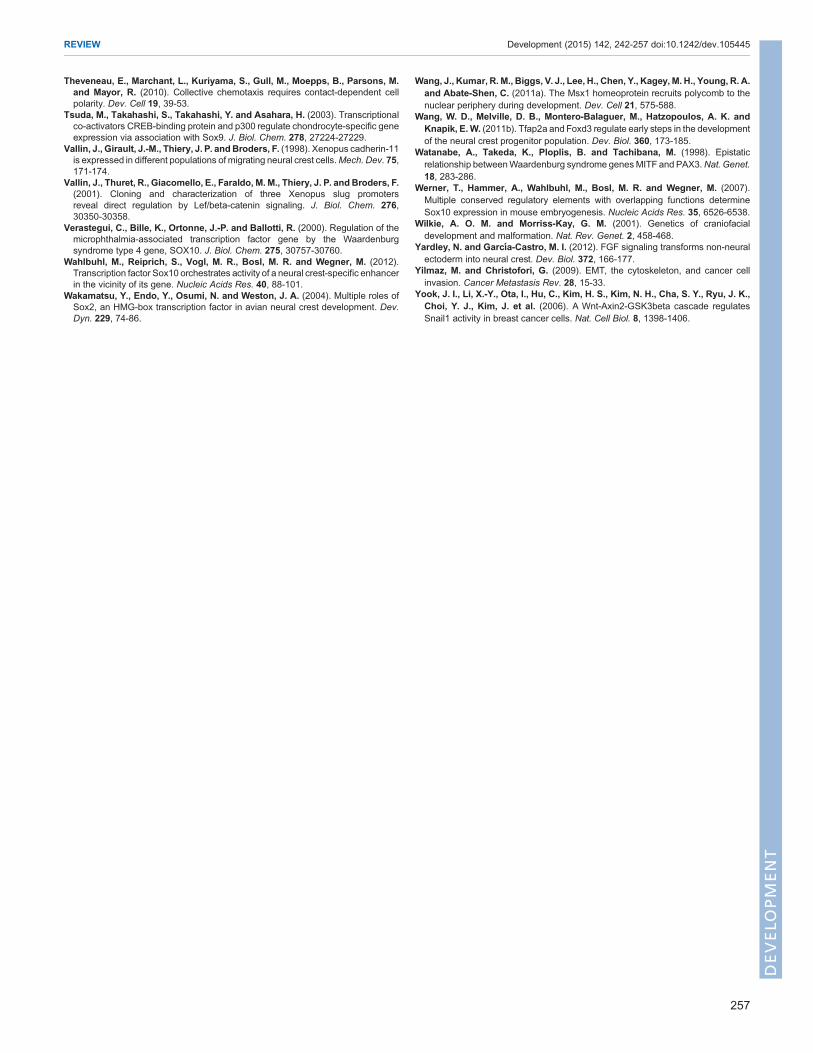
Theveneau, E., Marchant, L., Kuriyama, S., Gull, M., Moepps, B., Parsons, M.and Mayor, R. (2010). Collective chemotaxis requires contact-dependent cellpolarity. Dev. Cell 19, 39-53.
Tsuda, M., Takahashi, S., Takahashi, Y. and Asahara, H. (2003). Transcriptionalco-activators CREB-binding protein and p300 regulate chondrocyte-specific geneexpression via association with Sox9. J. Biol. Chem. 278, 27224-27229.
Vallin, J., Girault, J.-M., Thiery, J. P. andBroders, F. (1998). Xenopus cadherin-11is expressed in different populations of migrating neural crest cells.Mech. Dev. 75,171-174.
Vallin, J., Thuret, R., Giacomello, E., Faraldo, M. M., Thiery, J. P. and Broders, F.(2001). Cloning and characterization of three Xenopus slug promotersreveal direct regulation by Lef/beta-catenin signaling. J. Biol. Chem. 276,30350-30358.
Verastegui, C., Bille, K., Ortonne, J.-P. and Ballotti, R. (2000). Regulation of themicrophthalmia-associated transcription factor gene by the Waardenburgsyndrome type 4 gene, SOX10. J. Biol. Chem. 275, 30757-30760.
Wahlbuhl, M., Reiprich, S., Vogl, M. R., Bosl, M. R. and Wegner, M. (2012).Transcription factor Sox10 orchestrates activity of a neural crest-specific enhancerin the vicinity of its gene. Nucleic Acids Res. 40, 88-101.
Wakamatsu, Y., Endo, Y., Osumi, N. and Weston, J. A. (2004). Multiple roles ofSox2, an HMG-box transcription factor in avian neural crest development. Dev.Dyn. 229, 74-86.
Wang, J., Kumar, R. M., Biggs, V. J., Lee, H., Chen, Y., Kagey,M. H., Young, R. A.and Abate-Shen, C. (2011a). The Msx1 homeoprotein recruits polycomb to thenuclear periphery during development. Dev. Cell 21, 575-588.
Wang, W. D., Melville, D. B., Montero-Balaguer, M., Hatzopoulos, A. K. andKnapik, E.W. (2011b). Tfap2a and Foxd3 regulate early steps in the developmentof the neural crest progenitor population. Dev. Biol. 360, 173-185.
Watanabe, A., Takeda, K., Ploplis, B. and Tachibana, M. (1998). Epistaticrelationship betweenWaardenburg syndrome genesMITF and PAX3.Nat. Genet.18, 283-286.
Werner, T., Hammer, A., Wahlbuhl, M., Bosl, M. R. and Wegner, M. (2007).Multiple conserved regulatory elements with overlapping functions determineSox10 expression in mouse embryogenesis. Nucleic Acids Res. 35, 6526-6538.
Wilkie, A. O. M. and Morriss-Kay, G. M. (2001). Genetics of craniofacialdevelopment and malformation. Nat. Rev. Genet. 2, 458-468.
Yardley, N. and Garcıa-Castro, M. I. (2012). FGF signaling transforms non-neuralectoderm into neural crest. Dev. Biol. 372, 166-177.
Yilmaz, M. and Christofori, G. (2009). EMT, the cytoskeleton, and cancer cellinvasion. Cancer Metastasis Rev. 28, 15-33.
Yook, J. I., Li, X.-Y., Ota, I., Hu, C., Kim, H. S., Kim, N. H., Cha, S. Y., Ryu, J. K.,Choi, Y. J., Kim, J. et al. (2006). A Wnt-Axin2-GSK3beta cascade regulatesSnail1 activity in breast cancer cells. Nat. Cell Biol. 8, 1398-1406.
257
REVIEW Development (2015) 142, 242-257 doi:10.1242/dev.105445
DEVELO
PM
ENT