Embed Size (px)
Citation preview

RESEARCH Open Access
Establishment of functional epithelialorganoids from human lacrimal glandsSang Yun Jeong1†, Woo Hee Choi1,2†, Seong Gyeong Jeon1, Sookon Lee3, Jong-Moon Park4, Mira Park5,Hookeun Lee4, Helen Lew5* and Jongman Yoo1,2*
Abstract
Background: Tear deficiency due to lacrimal gland (LG) dysfunction is one of the major causes of dry eye disease(DED). Therefore, LG stem cell-based therapies have been extensively reported to regenerate injured lacrimal tissue;however, the number of stem cells in the LG tissue is low, and 2D long-term cultivation reduces the differentiationcapacity of stem cells. Nevertheless, 3D LG organoids could be an alternative for a DED therapy because it iscapable of prolonged growth while maintaining the characteristics of the LG tissue. Here, we report thedevelopment of LG organoids and their application as cell therapeutics.
Methods: Digested cells from human LG tissue were mixed with Matrigel and cultured in five different mediamodified from human prostate/salivary organoid culture media. After organoid formation, the growth, specificmarker expression, and histological characteristics were analyzed to authenticate the formation of LG organoids.The secretory function of LG organoids was confirmed through calcium influx or proteomics analysis afterpilocarpine treatment. To explore the curability of the developed organoids, mouse-derived LG organoids werefabricated and transplanted into the lacrimal tissue of a mouse model of DED.
Results: The histological features and specific marker expression of LG organoids were similar to those of normalLG tissue. In the pilocarpine-treated LG organoid, levels of internal Ca2+ ions and β-hexosaminidase, a lysosomalprotein in tear fluid, were increased. In addition, the secreted proteins from pilocarpine-treated lacrimal organoidswere identified through proteomics. More than 70% of the identified proteins were proven to exosome throughgene ontology analysis. These results indicate that our developed organoid was pilocarpine reactive, demonstratingthe function of LG. Additionally, we developed LG organoids from patients with Sjogren’s syndrome patients (SS)and confirmed that their histological features were similar to those of SS-derived LG tissue. Finally, we confirmedthat the mouse LG organoids were well engrafted in the lacrimal tissue two weeks after transplantation.
Conclusion: This study demonstrates that the established LG organoids resemble the characteristics of normal LGtissue and may be used as a therapy for patients with DED.
Keywords: Organoid, Lacrimal gland, Dry eye disease, Sjogren syndrome
© The Author(s). 2021 Open Access This article is licensed under a Creative Commons Attribution 4.0 International License,which permits use, sharing, adaptation, distribution and reproduction in any medium or format, as long as you giveappropriate credit to the original author(s) and the source, provide a link to the Creative Commons licence, and indicate ifchanges were made. The images or other third party material in this article are included in the article's Creative Commonslicence, unless indicated otherwise in a credit line to the material. If material is not included in the article's Creative Commonslicence and your intended use is not permitted by statutory regulation or exceeds the permitted use, you will need to obtainpermission directly from the copyright holder. To view a copy of this licence, visit http://creativecommons.org/licenses/by/4.0/.The Creative Commons Public Domain Dedication waiver (http://creativecommons.org/publicdomain/zero/1.0/) applies to thedata made available in this article, unless otherwise stated in a credit line to the data.
* Correspondence: [email protected]; [email protected]†Sang Yun Jeong and Woo Hee Choi contributed equally to this work.5Department of Ophthalmology, Bundang CHA Medical Center, CHAUniversity, Bundang-gu, Seongnam-si, Gyeonggi-do 13496, Republic of Korea1Department of Microbiology and CHA Organoid Research Center, CHAUniversity School of Medicine, 335 Pangyo-ro, Bundang-gu, Seongnam-si,Gyeonggi-do 13488, Republic of KoreaFull list of author information is available at the end of the article
Jeong et al. Stem Cell Research & Therapy (2021) 12:247 https://doi.org/10.1186/s13287-021-02133-y

BackgroundDry eye disease (DED) is a continuously disabling dis-ease occurring in 11%–22% of population, with about17% of them developing water-deficient dry eyes [1,2]. A previous study has reported that a severe typeof DED occurs in about half of the patients, present-ing structural and functional damage in the lacrimalgland (LG) [3]. DED is a multifactorial life-long de-bilitating disorder mainly caused by functional distur-bances in the LG [4]. Causes of glandular dysfunctionrange from deficiency and loss of tear film integrity,LG deterioration, to death of the secretory epithelialcells affected by hormonal imbalance, environmentalchanges, and autoimmune pathologies, leading toDED, a chronic condition [4].Furthermore, DED has also been defined as a multi-
factorial disease of the ocular surface characterized bythe lack of tear film stability. It is accompanied byocular symptoms in which unstable tear film, hyper-osmolarity, ocular surface inflammation and damage,and neurosensory abnormalities play pathologicalroles [5]. Its etiologies include causes secondary tosystemic autoimmune disorders such as Sjogren’s syn-drome (SS), rheumatoid arthritis, and systemic lupuserythematous, which are bothersome to doctors andpatients [6–10]. In conventional treatment, artificialtear eye drops are mainly used to moisturize the ocu-lar surface and offer additional lubrication [11]. Toaddress chronic inflammation symptoms, anti-inflammatory and topical immunosuppressive agentscould be considered [12]. However, their persistentuse is limited due to side effects [5, 12]. Other regen-erative strategies have recently been introduced to im-prove DED management [13].Interestingly, recent studies have demonstrated the
habitation of stem cells in exocrine glands such as saliv-ary [14], pancreatic [15, 16], prostate [17], and mammaryglands [18, 19]. However, reports on the existence ofstem cells in the mouse LG [20, 21] and human LGs arefewer than those in other exocrine glands.In contrast to LG cultures growing as a monolayer
in the order of preference on Matrigel, collagen, andHAM in two to three weeks, the formation of spher-oids including a mixed population of stem cells anddifferentiated cells has been reported in salivary glandcultures [22] and prostate spheres [23]. A single studyhas been conducted on spheroidal aggregation ofrabbit LG cells grown in the microgravity environ-ment of a rotary cell culture system [24]. Salivaryspheres including stem cells, can rescue gland func-tion, when they are transplanted into radiation-induced dry mouth animal models [22].To the best of our knowledge, there are few reports
of organoid establishment using stem cells in the
human LG. Under certain culture conditions, suspen-sion 3D cultures of human “lacrispheres” are main-tained and propagated for three to four weeks. Inaddition, lacrimal spheres can secrete quantifiablelevels of tear protein into conditioned media [23]. LGepithelial cells can form “spherules” with channel-likeconnections. The secretory lgA, lysozyme, and lacto-ferrin levels have explained the ductal origin in condi-tioned media [4].The structural and functional loss of the LG may be
managed by gland replacement and function restorationthrough cell therapy [23]. Organoid-based therapy con-taining stem cells would help recovery and differenti-ation into functionally competent cells, which could leadto damaged tissue regeneration.There is increasing evidence for the prevalence of
stem-like cells in the mouse LG [21, 25] that contributeto the reconstruction of the injured gland. We attemptedto establish cultures of human LG organoids and evalu-ated stem cell components by immunophenotyping,clonal assays, and real-time functional assays. We aimedto rebuild an organoid presenting a 3D LG tissue func-tional unit incorporating different cell types. It couldprovide a premise of treatment options for severe DEDmodels, including SS.
MethodsCell isolation and organoid formation from humanlacrimal gland tissueNormal LG tissues were obtained from non-damagedregions of the patients with eye-related disease. The tis-sues were chopped and washed with advanced DMEM/F12 (Gibco, Carlsbad, CA, USA) supplemented with 1%penicillin-streptomycin (Welgene, Gyeongsan-si, Korea)and then enzymatically digested with advanced DMEM/F12 supplemented with 0.125 mg/mL dispase II (Wako,Richmond, VA, USA), 0.1 mg/mL DNase I (Millipore-Sigma, Burlington, MA, USA), 0.125 mg/ml collagenaseII (Gibco, Carlsbad, CA, USA), and 1% penicillin-streptomycin for 1 h at 37 °C with shaking (150 rpm).After digestion, the supernatant was passed through a70-μm cell strainer (SPL, Pocheon-si, Gyeonggi-do,Korea) and pelleted by centrifugation at 200g for 5 min.The pellet was resuspended in culture media and mixedwith Matrigel (Corning, Corning, NY, USA) at a ratioof 1:1 (v:v), plated on a 48-well plate at a density of1X104 per well, and incubated with 5% CO2 at 37 °C for10 min to polymerize the matrices. The LG organoidswere cultured in five different media modified fromhuman prostate and salivary organoid culture medium[26, 27]. The components of each medium are listed inSupplementary Table 1. The culture medium was chan-ged every two to three days.
Jeong et al. Stem Cell Research & Therapy (2021) 12:247 Page 2 of 11

To confirm the origin of cells forming the organoid,epithelial cell adhesion molecule (EpCAM)-positive epi-thelial lineage cells were sorted using the MACS method(Miltenyibiotec, USA, 130-042-201). Briefly, single cellsfrom the lacrimal tissue were incubated with anti-EpCAM (Santacruz, CA, USA, sc-59906) for 1 h at 4 °C,washed with MACS buffer, and then incubatedwith anti-mouse IgG Microbeads (Miltenyibiotec, 130-048-402) for 30 min at 4 °C. After washing with PBS, theEpCAM-negative cells were passed through an MACScolumn, while the EpCAM-positive cells in the MACScolumn were isolated, washed, and then cultured inMatrigel.
Histology and immunofluorescenceTissues and organoids were washed with D-PBS (Welgene,Korea), fixed with 4% paraformaldehyde (Bio-solution, Seoul,Korea) for 30min, and embedded in paraffin. Paraffin sec-tions (6-μm-thick) were deparaffinized in xylene and hy-drated in a graded ethanol series. The samples were thenstained with H&E, Alcian blue, PAS staining kit (Abcam,Cambridge, MA, USA), and Masson’s trichrome staining kit(Dako, Santa Clara, CA, USA) according to the manufac-turers’ protocol. For immunofluorescence analysis, fixedsamples were cryoprotected by immersion in PBS supple-mented with 30% sucrose and 0.1% sodium azide at 4 °C.The cryoprotected samples were embedded in optimal cut-ting temperature (OCT, Sakura, Japan) compound, rapidlyfrozen in liquid nitrogen, and stored at − 80 °C until use. Sec-tions (4-μm-thick) of the frozen block were pre-blocked with5% normal horse serum (Vector, IL, USA) in Tris-bufferedsaline (Welgene) for 2 h at room temperature (RT) and incu-bated with primary antibody at 4 °C overnight. After washingwith PBS, the sections were incubated with secondary anti-body for 2 h at RT. For nuclear staining, the sections weretreated with 1 ug/ml Hoechst 33342 (MilliporeSigma,Burlington, MA, USA, 1 μg/ml) for 20min. Primary anti-bodies used for immunostaining included antibodiesagainst aquaporin 5 (AQP5; Abcam), alpha-smooth muscleactin (α-SMA; Biolegend, San Diego, CA, USA), vimentin(VIM; Cell signaling, Danvers, MA, USA), lysozyme (LYZ;Diagnostic biosystems, Pleasanton, CA, USA), E-cadherin(E-CAD; Santa Cruz Biotechnology, Dallas, TX, USA), anti-BrdU (Novus, Centennial, CO, USA), and Ki67 (Abcam,Cambridge, MA, USA). The secondary antibodies (ThermoFisher Scientific, Waltham, MA, USA) used included AlexaFluor 488 goat anti-rabbit IgG, Alexa Fluor 594 goat anti-mouse IgG, and Alexa Fluor 594 goat anti-rat IgG.
Total RNA isolation and quantitative RT-PCRTotal RNA was extracted from isolated tissues or orga-noids using MagListo™ 5M Cell Total RNA ExtractionKit (Bioneer, Daejeon Metropolitan City, Korea) follow-ing the manufacturer’s protocol. Thereafter, 1 μg of
RNA was used to synthesize cDNA using PrimeScript™RT Master Mix (TaKaRa, Kyoto City, Japan). Quantita-tive RT-PCR was performed with a Thermal CyclerDice® Real-Time System III (TaKaRa, Kyoto City, Japan)using SYBR® Premix Ex Taq™ II (TaKaRa). The PCRprimers sequences are listed in Supplementary Table 2.PCR experiments were carried out in triplicates.
Calcium flux assay with Fluo-4Ca2+ mobilization to the cytoplasm was detected using aFluo-4 Calcium Imaging Kit (Thermo Fisher Scientific)following the manufacturer’s protocol. Briefly, the orga-noids were treated with Fluo-4 AM for 15 min at 37 °C,followed by incubation at RT for 15 min. After washingwith PBS, the organoids were stimulated with 1 μg/mLpilocarpine (MilliporeSigma). Calcium signaling wasthen observed using a Nikon Eclipse Ti2 microscope(Nikon, Tokyo, Japan).
β-Hexosaminidase assayTo demonstrate the secretory function of LG organoids,the lysosomal enzyme N-acetyl-β-glucosaminidase(NAG), also known as α-galactosidase B, in organoidcultured medium was detected using an NAG assay kit(MilliporeSigma) following the manufacturer’s protocol.Briefly, organoids were incubated in serum-free DMEM/F12 for 2 h, treated with pilocarpine (1 μg/mL), and thenincubated at 37 °C in a 5% CO2 incubator for 24 h. Themedium was collected at 2 h and 24 h after pilocarpinetreatment and analyzed for NAG catalytic activity. Thereaction product was detected colorimetrically at 405 nmusing a microplate reader (Multiskan GO, ThermoFisher Scientific).
Transmission electron microscopy (TEM)The secretory proteins from organoids were detectedusing TEM . Briefly, the cultured organoids were washedwith D-PBS and fixed with 2% glutaraldehyde-paraformaldehyde in 0.1 M phosphate buffer (PB, pH7.4) for 12 h. After washing with 0.1 M PB, samples werepost-fixed with 1% OsO4 dissolved in 0.1M PB for 2 h,dehydrated in an ascending gradual ethanol series (50%–100%), infiltrated with propylene oxide, and embeddedwith a Poly/Bed 812 kit (Polysciences, PA, USA). Afterpure fresh resin embedding and polymerization at 65 °Cin an electron microscope oven (DOSAKA, Japan) for24 h, the Poly/Bed embedded samples were cut into ap-proximately 70-nm-thick sections with a Leica EM UC-7(Leica Microsystems, Wetzlar, Germany) equipped witha diamond knife (Diatome, PA, USA), transferred tocopper and nickel grids, contrast-stained with 6% uranylacetate and lead citrate (Fisher), and observed using atransmission electron microscope (JEOL, Japan) at 80 kVacceleration voltage.
Jeong et al. Stem Cell Research & Therapy (2021) 12:247 Page 3 of 11

Proteomic analysis of LG organoids secretomeTo identify the proteins secreted by LG organoids afterpilocarpine treatment, the culture medium washarvested 2 h after of pilocarpine treatment and analyzedthrough proteomics analysis [28]. In brief, proteins(200 μg) in the medium were digested using the filter-aided sample preparation (FASP) method with centrifu-gal filters (Millipore, MA, USA). After desalting thesamples with a Sep-Pak® Vac 1 cc C18 cartridge (Waters,MA, USA), the peptides were collected, purified, andquantified via LC-MS/MS analysis. LC-MS/MS assaywas performed using a Dionex Ultimate 3000 HPLCcoupled with a Q Exactive™ Hybrid Quadrupole-Orbitrap mass spectrometer (Thermo Fisher Scientific,Waltham, MA, USA). Raw MS/MS data were quantifiedusing MaxQuant (Max Planck Institute) and classified bygene ontology (GO) analysis. T test P < 0.05 and fold-change (> 2, < − 2) were applied to determine the differ-entially expressed proteins (DEPs) between the controland pilocarpine-treated groups.
Mouse dry eye model and organoid transplantationEight-week-old male C57BL/6 mice (Koatech, Pyeong-taek, Korea) or C57BL/6-Tg (CAG-EGFP)131Osb/Ley-SopJ mice (Nihon SLC, Shizuoka, Japan) mice wereused as DED models or for the manipulation of LGorganoids, respectively. The experimental protocol foranimal use was reviewed and approved by the CHAUniversity Institutional Animal Care and Use Commit-tee. The LG tissue was obtained from eGFP-Tg mice.Organoids were formed and cultured following thesame method used for human organoids. To create aninflammation-induced dry eye model, 15 μL concanav-alin A (ConA, 10 mg/mL in PBS, MilliporeSigma) wasinjected into the extra-orbital gland of wild-typemouse lacrimal tissue. The same volume of PBS wasinjected into the control group. Seven days after ConAinjection, cell clumps from GFP-expressing organoids(1 × 10⁴ cells/15 μL in advanced DMEM/F12/Matrigel)were injected into the extra-orbital gland space (Fig. 5).After two weeks, the mouse LG tissues were harvestedfor immunofluorescence analysis.
Statistical analysisStatistically significant differences were analyzed by Stu-dent’s t test of one-way analysis of variance (ANOVA)with post hoc Tukey test for multiple comparisons usingthe GraphPad Prism software package, version 3.0(GraphPad Prism, CA, USA). All experiments were con-ducted at least thrice. The number of independent ex-periments is indicated by n. Significance was consideredat p < 0.05.
ResultsGeneration of LG organoids from human tissuesTo generate LG organoids, we used human LG tissues andallowed them to self-organize within Matrigel (Fig. 1A). Thedissociated LG cells were embedded in Matrigel and thengrown respectively in five different conditions by modifyinghuman prostate or salivary gland organoid media. Mostorganoids were generated under all medium conditions. Inthe M-SA1 medium (salivary gland organoid media supple-mented with 10mM nicotinamide, 500 nM A83, and 100ng/mL noggin); however, the formation and growth oforganoids were maintained following passage, unlike thatin other media. Therefore, we defined M-SA1 medium asan LG organoid medium (LGOM); (Fig. 1B). In theLGOM, LG organoids expanded until passage 19 (Fig. 1B)and increased in size until day 15 (Fig. 1C). Histologicalanalysis showed that the organoids were similar to the LGtissues. Acinar cells were the major cell type in the LG tis-sue, and its secreted acidic mucosubstances were observed(Fig. 1D, H&E and Alcian blue staining, respectively).Secretory products such as glycogen and glycoprotein werealso observed in normal lacrimal tissue and organoids(Fig. 1D, PAS staining). Masson’s trichrome stainingshowed that keratin was the major ECM component oforganoids (Fig. 1D). These results showed that the orga-noids developed in this study were morphologically similarto the acinar cells of the LG tissue.Additionally, we confirmed the expression of EpCAM,
an epithelial lineage cell marker, in the lacrimal tissue(Fig. 1E). EpCAM-positive cells generated more LGorganoids than a non-sorted fraction, whereas organoidswere not formed in the EpCAM-negative cell fraction(Fig. 1F). A large amount of VIM, a gland-specificmarker, was observed in the organoids developed fromEpCAM-positive cells. These results showed that theorganoids originated from EpCAM-positive cells(Fig. 1G).
Human LG organoids recapitulate the structuralproperties of human tissuesThe developed LG organoids were compared with hu-man LG tissues. Specific markers such as AQP5, LYZ,E-CAD, VIM, and α-SMA were used for immunofluor-escence staining (Fig. 2A). VIM, AQP5, and LYZ ex-pression patterns in organoids were similar to those inacinar cells of LG tissues. Additionally, E-CAD and α-SMA expression patterns in LG organoids were similarto those in the tissues, although their expression levelswere lower than those in lacrimal tissues. Thesemarkers are known to be expressed in acinar cells, butnot in ductal cells. Therefore, the organoids formed inthis study are more similar to acinar cells than toductal cells in lacrimal tissues. Additionally, we con-firmed the proliferating cells in the LG organoid using
Jeong et al. Stem Cell Research & Therapy (2021) 12:247 Page 4 of 11

the BrdU assay (Fig. 2B). Four days after BrdU treat-ment, the BrdU-positive cells expressed Ki67 in theouter region of organoids, unlike the inner region. Inparticular, VIM expression was observed around BrdU-positive cells.To determine the cellular ultrastructure, LG orga-
noids were analyzed via TEM. In some organoids,cells (Fig. 2C, white dotted line) with secretory gran-ules (Fig. 2C, black arrowhead) were observed inorganoids. These characteristics are known to bethe features of acinar cells.
Human LG organoids recapitulate the structuralproperties of human LGsIn this study, we confirmed the secretory function of de-veloped organoids by pilocarpine stimulation. Afterstimulation with 1 μg/mL pilocarpine, Ca2+
concentration was increased in the cells of organoids(Fig. 3A). In the cells of the LG, increasing Ca2+ concen-tration are known to lead to tear secretion [29]. Add-itionally, secretion of β-hexaminidase, a lysosomalenzyme, was increased in pilocarpine-treated organoids(Fig. 3B). In particular, the secreted proteins were ob-served (Fig. 3C, red arrowhead), and cell-cell junctionwas widened (Fig. 3C, asterisk) in pilocarpine-treatedorganoids. These results indicate that the organoids de-veloped in the present study have a secretory function inresponse to the cholinergic agonist pilocarpine, similarto the LG tissue.
Human LG organoids recapitulate the functionalproperties of human LGsTo identify the proteins secreted by the LG orga-noids after pilocarpine treatment, the culture
Fig. 1 Generation of epithelial organoids from human lacrimal glands (LGs). A Scheme for the development and culture of lacrimal tissue-derivedorganoid. B Optimization of culture media following organoid formation and subculture. Representative images were obtained by lightmicroscopy after culturing for 3 days (Magnification; 10×, Scale bar; 500 µm). C The growth of organoid cultured in optimized LG organoidmedium (LGOM). Scale 100 μm. D A comparison of histological analysis between normal lacrimal tissue and formed LG organoid. H&E, Alcianblue, Masson’s trichrome, and PAS staining were performed for identifying the acini structure, mucosubstance secretion, connective tissue, andglycoprotein secretion, respectively (Magnification; 40×, Scale bar; 100 µm). E Confirmation of acinar cells (green; aquaporin 5; AQP5) andepithelial lineage (red; EpCAM) cells in lacrimal tissue (Magnification; 100×, Scale bar; 50 µm). F Organoid formation from EpCAM-positive cellfraction. Representative bright-field images (Magnification; 10×, Scale bar; 500 µm) were obtained after 4 days of cultivation and used forcounting the formed organoid (right, graph, N = 4). G Ki67, AQP5, and vimentin (VIM) expression was detected in the organoids and observedusing immunofluorescence (Magnification; 100×, Scale bar; 50 µm)
Jeong et al. Stem Cell Research & Therapy (2021) 12:247 Page 5 of 11

medium was collected and analyzed for proteomicsafter 2 h after PBS or pilocarpine treatment. A totalof 776 proteins were identified and quantified inboth groups, and the amount of the proteins wasadditionally secreted after pilocarpine treatment(Fig. 3D-E). In the pilocarpine-treated organoid,the upregulated cellular components were found tobe extracellular exosome/vehicles by GO analysis(Fig. 3E-F). Additionally, we identified 66 DEPs (fold-change >2), and most proteins (>70%) belonged toexosome/vehicle (Fig. 3G). Among the upregulatedproteins in the pilocarpine-treated group, the proteinsynthesis-related secretome (such as RPS and RPLfamily) was found to be increased. These results indi-cate that our developed lacrimal organoids mightturn on synthesis processes such as tear productionby the stimulator.
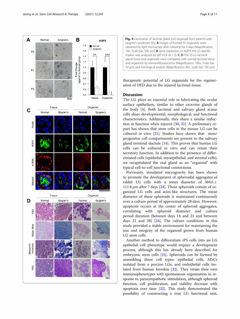
Generation of LG organoids derived from LGs of thepatients with SSTo advance our research, we developed LG orga-noids from the tissue of patients with SS, which are
referred to as SS organoids. SS organoids were gen-erated and cultured following the same methodused for normal organoids. SS organoids featured asmaller size and were cultured for shorter passages(up to passage 2), as compared to normal organoids(Fig. 4A). Expression of AQP5, an LG-specificmarker, was significantly lesser in the SS tissue andorganoids than that in the normal tissue and orga-noids (Fig. 4B, C). Moreover, histological analysisrevealed that SS organoids have higher fibroblasticproperties than the normal tissue and organoids,while their degree is similar to that of SS tissue(Fig. 4D). Morphologically, some structural damagesin acinar cells were found through H&E analysisand, dense blue regions representing collagen-stained areas were observed after Masson’s tri-chrome staining of SS organoids, similar to the SStissue, whereas the normal organoid showed an in-tact structure in the light blue area, as in the nor-mal tissue. These results suggest that our developedSS organoids could be a useful tool as an in vitromodel of mimetic SS disease.
Fig. 2 Structural recapitulation of human lacrimal gland (LG) organoids. A Immunofluorescence staining for specific Markers, such asvimentin (VIM), E-cadherin (E-CAD), aquaporin5 (AQP5), alpha-smooth muscle actin (α-SMA), and lysozyme (LYZ), was performed inhuman LG tissue and organoid cultured in LG organoid medium (LGOM). B In the organoid, the proliferating cells were confirmedvia BrdU assay. At 4 days after BrdU treatment, proliferating cell (BrdU and Ki76) and LG-specific (AQP5 and VIM) markers) weredetected in organoids (Magnification; 100×, Scale bar; 50 µm). C TEM analysis was performed to confirm cellular ultrastructure in theorganoid. Acinar cells (up; white dotted line) and secretory granules (down; red arrowhead) were detected in the LG organoid (Scalebar; 5 µm)
Jeong et al. Stem Cell Research & Therapy (2021) 12:247 Page 6 of 11

Engraftment of LG organoids in the DED mouse modelTo confirm the engraftment ability of LG organoids, theorganoids from the LG tissue of C57BL/6-GFP-Tg micewere prepared following the method used for humanorganoids. GFP-labeled organoids were formed (Fig. 5A,a–b), and their morphology was similar to that of the aci-nar cells of the mouse lacrimal tissue (Fig. 5A, c–d). Inthis study, we established an inflammation-based DED
mouse model by injecting ConA into the extra-orbitalgland of the lacrimal tissue and transplanting the formedGFP-organoids into the lacrimal tissue of the DEDmouse. GFP signals were observed in mouse lacrimaltissue after 14 days after transplantation (Fig. 5B). Add-itionally, we confirmed that AQP5, a water channelprotein for tear production, was expressed in the trans-planted organoids (Fig. 5B). These results suggest the
Fig. 3 Functional recapitulation of human lacrimal gland (LG) organoids. To confirm the secretory function of the organoid, A Ca2+ uptake(Magnification; 10×, Scale bar; 100 µm) and B released β-hexosaminidase was detected after 1 μg/mL pilocarpine treatment, and C the secretomereleased from the organoid was observed via TEM. Proteins secreted by pilocarpine were identified using proteomic analysis. Through the Dheatmap, E Venn diagram, F gene ontology (GO) term analysis, and identification of the differentially expressed proteins (DEPs), the proteins wereseen to be more secreted from pilocarpine-treated organoids than from the normal organoids, and most proteins belonged to exosome/vehicle
Jeong et al. Stem Cell Research & Therapy (2021) 12:247 Page 7 of 11
therapeutic potential of LG organoids for the regener-ation of DED due to the injured lacrimal tissue.
DiscussionThe LG plays an essential role in lubricating the ocularsurface epithelium, similar to other exocrine glands ofthe body [4]. Both lacrimal and salivary gland acinarcells share developmental, morphological, and functionalcharacteristics. Additionally, they share a similar reduc-tion in function when injured [30, 31]. A preliminary re-port has shown that stem cells in the mouse LG can becultured in vitro [21]. Studies have shown that stem/progenitor cell compartments are present in the salivarygland terminal ductule [14]. This proves that human LGcells can be cultured in vitro and can retain theirsecretory function. In addition to the presence of differ-entiated cells (epithelial, myoepithelial, and stromal cells),we recapitulated the real gland as an “organoid” withtypical cell-to-cell junctional connections.Previously, simulated microgravity has been shown
to promote the development of spheroidal aggregates ofrabbit LG cells with a mean diameter of 384.6 ±111.8 μm after 7 days [24]. These spheroids consist of or-ganized LG cells and acini-like structures. The meandiameter of these spheroids is maintained continuouslyover a culture period of approximately 28 days. However,apoptosis occurs at the center of spheroid aggregates,correlating with spheroid diameter and cultureperiod duration (between days 14 and 21 and betweendays 21 and 28) [24]. The culture conditions in thisstudy provided a stable environment for maintaining thesize and integrity of the organoid grown from humanLG stem cells.Another method to differentiate iPS cells into an LG
epithelial cell phenotype would require a developmentprocess, although this has already been described forembryonic stem cells [25]. Spheroids can be formed byassembling three cell types: epithelial cells, MSCsisolated from a porcine LGs, and endothelial cells iso-lated from human foreskin [32]. They retain their ownimmunophenotypes with spontaneous organization in re-sponse to parasympathetic stimulation, although spheroidfunction, cell proliferation, and viability decrease withapoptosis over time [32]. This study demonstrated thepossibility of constructing a true LG functional unit,
Fig. 4 Generation of lacrimal gland (LG) organoid from patients withSjogren’s syndrome (SS). A Images of formed SS organoids wereobtained by light microscopy after culturing for 3 days (Magnification;10×, Scale bar; 500 µm). B Gene expression of AQP5, the LG-specificmarker, was analyzed by qRT-PCR (N = 3). C, D The SS LG lacrimalgland tissue and organoids were compared with normal lacrimal tissueand organoids by immunofluorescence (Magnification; 100×, Scale bar;50 µm) and histological analysis (Magnification; 40×, Scale bar; 100 µm)
Jeong et al. Stem Cell Research & Therapy (2021) 12:247 Page 8 of 11

comprising lacrimal epithelial cells and MSCs from adultmammalian tissues in vitro.Much effort has been put into the culture of the LG
in vitro. For example, experiments in a micrograft envir-onment and the transfer of amniotic membranes havebeen performed. However, these methods have notshown expected results over an extended duration [33].We explored five media by modifying the salivary glandorganoid media and applied them to organoid culture.We finally concluded that M-SA1 medium is the bestLGOM because it resulted in the longest surface, max-imum length of organoids formed, and the best expan-sion. Similarly, murine LG stem cells can expressstemness markers (such as Nanog, Sox2, and Klf4),known as early lineage markers of all three germ layers
[33]. In this study, LG organoids from normal individ-uals presented gland-specific markers such as VIM, E-CAD, AQP5, and α-SMA, although their expression waslower than that in the tissue. As shown in Fig. 4, the ex-pression pattern of specific markers in organoids fromnormal and patients with SS reflects the difference be-tween the two tissues. Patients with SS, who produceless tear secretion than a normal person, cannot partici-pate in the Ca2+ signaling pathway, as they do not haveany Ca2+ channel in the myoepithelial cell plasma mem-brane [34]. Myoepithelial cells were altered in the pa-tients with SS and α-SMA from that in normalindividuals. These results suggest that our developed SSorganoids could be a useful tool for in vitro model ofmimetic SS.
Fig. 5 Engraftment of lacrimal gland (LG) organoid into dry eye mouse model. A (a–b) Mouse lacrimal organoids were formed from lacrimal tissue ofC57BL/6-GFP-Tg mice. The GFP signals were expressed in the formed organoid (Magnification; 10×, Scale bar; 500 µm) and A (c–d) their morphologywas compared to that of mouse LG tissue through H&E staining (Magnification; 20×, Scale bar; 500 µm). B Inflammation-based dry eye disease (DED)animal model was established by concanavalin A (ConA) injection into the extra-orbital gland of the mouse. GFP-labeled organoids were transplantedinto mouse lacrimal tissue after 7 days after ConA injection, and their engraftment was confirmed by GFP signal after 14 days after transplantation.Expression of aquaporin 5 (AQP5 ) was analyzed by immunofluorescence (Magnification; 40×, Scale bar; 100 µm)
Jeong et al. Stem Cell Research & Therapy (2021) 12:247 Page 9 of 11

In this study, we tried to confirm the secretoryfunction of the LG organoid and measured itssecretory function in vitro. Muscarinic receptorsshould be activated by various stimulants to inducetear secretion from the LG. Under these conditions,internal Ca2+ increase leads to tear secretion [35].Pilocarpine is a parasympathetic stimulant mainlyaffecting muscarinic M3 receptors. Since organoids donot have nerves, they are to be stimulated using pilo-carpine, an M3 receptor agonist. Therefore, internalCa2+ and β-hexosaminidase, a known lysosomal pro-tein in tear fluid, increased in the pilocarpine-treatedorganoid. In addition, the secreted proteins frompilocarpine-treated lacrimal organoids were identifiedthrough proteomics. These results indicate that ourdeveloped organoid is pilocarpine reactive, demon-strating the function of the LG.Moreover, this study preliminary demonstrated that
the LG organoid possessed a self-repair effect of stemcells in an inflammation-induced DED animal model.However, its future role merits further investigation.Although DED has a moderate prevalence globally,primary clinical management is still conservative usingartificial tear drops and lubricants [4]. TransplantedMSCs are well known to enhance corneal wound healingby trophic factor production and immune regulatoryeffects instead of direct transdifferentiation into epithe-lial cells [36–39]. As cell therapy prerequisites, theplausibility of using lacrimal organoids containing func-tional stem cells to rescue and repair functionally com-petent cells should be studied. Such studiesmay contribute to damaged tissue regeneration [23].We tried to determine the possibility of regenerating
the damaged LGs using lacrimal organoids after indu-cing inflammation using ConA in C57BL/6 mice. Wehypothesize that damages to the LG due to aging, hor-monal imbalance, or radiation may be treated usingorganoids. In summary, this study provided the first evi-dence for successful growth of fresh human LG orga-noids in vitro with an attempt toward functional unitformation while retaining secretory function. Furthervalidation is needed for the development of a function-ally competent secretory lacrimal organoid for potentialclinical application in severe DED, including auto-immune LG disorders.
ConclusionIn conclusion, we established lacrimal organoids fromhuman and mouse lacrimal tissues. The organoids estab-lished in this study recapitulate the structure and func-tion of human LGs, suggesting that organoids can beused as a tool for disease modeling and regenerativetherapy.
Supplementary InformationThe online version contains supplementary material available at https://doi.org/10.1186/s13287-021-02133-y.
Additional file 1: Table S1. The components of culture medium forlacrimal gland organoid. Table S2. The primers used for quantitative RT-PCR.
AbbreviationsDED: Dry eye disease; LGOM: Lacrimal gland organoid medium; PR: Mediumfor prostate organoid cultivation; SA: Medium for salivary organoidcultivation; M-SA1/M-SA2: Modified from salivary organoid culture medium;M-PRSA: Modified from prostate and salivary organoid culture medium;VIM: Vimentin; E-CAD: E-cadherin; AQP5: Aquaporin5; α-SMA: Alpha-smoothmuscle actin; LYZ: Lysozyme; SS: Sjogren’s syndrome; ConA: Concanavalin A
AcknowledgementsNot applicable.
Authors’ contributionsJ.S.Y. and C.W.H.: Collection and/or assembly of data, manuscript writing.J.S.G.: Collection and/or assembly of data, manuscript writing for revision. L.S.:Assembly of data. P.J.M. and L.H.: Proteomics analysis. P.M.: Assembly of data:L.H.: Conception and design/manuscript writing. Y.J.: Conception and design/manuscript writing/final approval of the manuscript. The authors read andapproved the final manuscript.
Authors’ informationSang Yun Jeong and Woo Hee Choi contributed equally to this work.
FundingThis work was supported by the Basic Science Research Program through theNational Research Foundation of Korea funded by the Ministry of Science, ICTand Future Planning, Republic of Korea (NRF-2018R1D1A102050030), by a grantof the Korea Health Technology R&D Project through the Korea Health IndustryDevelopment Institute, funded by the Ministry of Health & Welfare (HI16C0002and HI18C2458), and by the Industrial Strategic Technology DevelopmentProgram (20009773, Commercialization of 3D Multifunction Tissue MimeticsBased Drug Evaluation Platform) funded by the Ministry of Trade, Industry &Energy (MOTIE, Korea).
Availability of data and materialsThe datasets generated during and/or analyzed during the study areavailable from the corresponding author on reasonable request.
Ethics approval and consent to participateThis study was approved by the institutional review board (IRB) of CHABundang medical center, CHA University (CHAMC 2018-01-007) and carriedout with the written consent of all donors.
Consent for publicationNot applicable.
Competing interestsThe authors declared no potential conflicts of interest.
Author details1Department of Microbiology and CHA Organoid Research Center, CHAUniversity School of Medicine, 335 Pangyo-ro, Bundang-gu, Seongnam-si,Gyeonggi-do 13488, Republic of Korea. 2ORGANOIDSCIENCES, Ltd.,Seongnam, Gyeonggi-do 13488, Republic of Korea. 3Department ofRheumatology, Bundang CHA Medical Center, CHA University, Seongnam,Gyeonggi-do, Republic of Korea. 4Department of Pharmacology, GacheonUniversity, Incheon, Gyeonggi-do, Republic of Korea. 5Department ofOphthalmology, Bundang CHA Medical Center, CHA University, Bundang-gu,Seongnam-si, Gyeonggi-do 13496, Republic of Korea.
Jeong et al. Stem Cell Research & Therapy (2021) 12:247 Page 10 of 11
Received: 26 August 2020 Accepted: 1 January 2021
References1. Abelson MB, Ousler GW 3rd, Maffei C. Dry eye in 2008. Curr Opin
Ophthalmol. 2009;20(4):282–6.2. Lemp MA. Report of the National Eye Institute/Industry workshop on clinical
trials in dry eyes. CLAO J. 1995;21(4):221–32.3. Tiwari S, Bhatt A, Nagamodi J, et al. Aqueous deficient dry eye syndrome
post orbital radiotherapy: a 10-year retrospective study. Transl Vis SciTechnol. 2017;6(3):19.
4. Tiwari S, Ali MJ, Balla MM, et al. Establishing human lacrimal gland cultureswith secretory function. Plos One. 2012;7(1):e29458.
5. Craig JP, Nichols KK, Akpek EK, et al. TFOS DEWS II definition andclassification report. Ocul Surf. 2017;15(3):276–83.
6. Milner MS, Beckman KA, Luchs JI, et al. Dysfunctional tear syndrome: dryeye disease and associated tear film disorders - new strategies for diagnosisand treatment. Curr Opin Ophthalmol. 2017;27(Suppl 1):3–47.
7. Stevenson W, Chauhan SK, Dana R. Dry eye disease: an immune-mediatedocular surface disorder. Arch Ophthalmol. 2012;130(1):90–100.
8. Messmer EM. The pathophysiology, diagnosis, and treatment of dry eyedisease. Dtsch Arztebl Int. 2015;112(5):71–81 quiz 82.
9. The epidemiology of dry eye disease: report of the EpidemiologySubcommittee of the International Dry Eye WorkShop (2007). Ocul Surf2007;5(2):93–107.
10. Stenwall PA, Bergstrom M, Seiron P, et al. Improving the anti-inflammatoryeffect of serum eye drops using allogeneic serum permissive for regulatoryT cell induction. Acta Ophthalmol. 2015;93(7):654–7.
11. Song JK, Lee K, Park HY, et al. Efficacy of carboxymethylcellulose andhyaluronate in dry eye disease: a systematic review and meta-analysis.Korean J Fam Med. 2017;38(1):2–7.
12. Bron AJ, de Paiva CS, Chauhan SK, et al. TFOS DEWS II pathophysiologyreport. Ocul Surf. 2017;15(3):438–510.
13. Villatoro AJ, Fernandez V, Claros S et al. Regenerative therapies in dry eyedisease: from growth factors to cell therapy. Int J Mol Sci 2017;18(11).
14. Man YG, Ball WD, Marchetti L, et al. Contributions of intercalated duct cellsto the normal parenchyma of submandibular glands of adult rats. Anat Rec.2001;263(2):202–14.
15. Zulewski H, Abraham EJ, Gerlach MJ, et al. Multipotential nestin-positivestem cells isolated from adult pancreatic islets differentiate ex vivo intopancreatic endocrine, exocrine, and hepatic phenotypes. Diabetes. 2001;50(3):521–33.
16. Seaberg RM, Smukler SR, Kieffer TJ, et al. Clonal identification of multipotentprecursors from adult mouse pancreas that generate neural and pancreaticlineages. Nat Biotechnol. 2004;22(9):1115–24.
17. Tsujimura A, Koikawa Y, Salm S, et al. Proximal location of mouse prostateepithelial stem cells: a model of prostatic homeostasis. J Cell Biol. 2002;157(7):1257–65.
18. Kordon EC, Smith GH. An entire functional mammary gland may comprisethe progeny from a single cell. Development. 1998;125(10):1921–30.
19. Dontu G, Abdallah WM, Foley JM, et al. In vitro propagation andtranscriptional profiling of human mammary stem/progenitor cells. GenesDev. 2003;17(10):1253–70.
20. Zoukhri D, Fix A, Alroy J, et al. Mechanisms of murine lacrimal gland repairafter experimentally induced inflammation. Invest Ophthalmol Vis Sci. 2008;49(10):4399–406.
21. You S, Kublin CL, Avidan O, et al. Isolation and propagation ofmesenchymal stem cells from the lacrimal gland. Invest Ophthalmol Vis Sci.2011;52(5):2087–94.
22. Lombaert IM, Brunsting JF, Wierenga PK, et al. Rescue of salivary glandfunction after stem cell transplantation in irradiated glands. Plos One. 2008;3(4):e2063.
23. Tiwari S, Nair RM, Vamadevan P, et al. Establishing and characterizinglacrispheres from human lacrimal gland for potential clinical application.Graefes Arch Clin Exp Ophthalmol. 2018;256(4):717–27.
24. Schrader S, Kremling C, Klinger M, et al. Cultivation of lacrimal gland acinarcells in a microgravity environment. Br J Ophthalmol. 2009;93(8):1121–5.
25. Hirayama M, Liu Y, Kawakita T, et al. Cytokeratin expression in mouselacrimal gland germ epithelium. Exp Eye Res. 2016;146:54–9.
26. Drost J, Karthaus WR, Gao D, et al. Organoid culture systems for prostateepithelial and cancer tissue. Nat Protoc. 2016;11(2):347–58.
27. Maimets M, Rocchi C, Bron R, et al. Long-term in vitro expansion of salivarygland stem cells driven by Wnt signals. Stem Cell Rep. 2016;6(1):150–62.
28. Nam O, Park JM, Lee H, et al. De novo transcriptome profile ofcoccolithophorid alga Emiliania huxleyi CCMP371 at different calciumconcentrations with proteome analysis. Plos One. 2019;14(8):e0221938.
29. Imada T, Nakamura S, Hisamura R, et al. Serotonin hormonally regulateslacrimal gland secretory function via the serotonin type 3a receptor. SciRep. 2017;7(1):6965.
30. Zangrossi S, Marabese M, Broggini M, et al. Oct-4 expression in adult humandifferentiated cells challenges its role as a pure stem cell marker. Stem Cells.2007;25(7):1675–80.
31. Jez M, Ambady S, Kashpur O, et al. Expression and differentiation betweenOCT4A and its Pseudogenes in human ESCs and differentiated adultsomatic cells. Plos One. 2014;9(2):e89546.
32. Massie I, Spaniol K, Barbian A, et al. Development of lacrimal glandspheroids for lacrimal gland tissue regeneration. J Tissue Eng Regen Med.2018;12(4):e2001–9.
33. Ackermann P, Hetz S, Dieckow J, et al. Isolation and investigation ofpresumptive murine lacrimal gland stem cells. Invest Ophthalmol Vis Sci.2015;56(8):4350–63.
34. García-Posadas L, Hodges RR, Utheim TP, et al. Lacrimal Gland MyoepithelialCells Are Altered in a Mouse Model of Dry Eye Disease. Am J Pathol. 2020;190(10):2067–2079.
35. Imada T, Nakamura S, Hisamura R, et al. Serotonin hormonally regulateslacrimal gland secretory function via the serotonin type 3a receptor. SciRep. 2017;7(1):6965.
36. Zhang L, Coulson-Thomas VJ, Ferreira TG, et al. Mesenchymal stem cells fortreating ocular surface diseases. BMC Ophthalmol. 2015;15(Suppl 1):155.
37. Li GG, Zhu YT, Xie HT, et al. Mesenchymal stem cells derived from humanlimbal niche cells. Invest Ophthalmol Vis Sci. 2012;53(9):5686–97.
38. Lin KJ, Loi MX, Lien GS, et al. Topical administration of orbital fat-derivedstem cells promotes corneal tissue regeneration. Stem Cell Res Ther. 2013;4(3):72.
39. Rapoport DH, Schicktanz S, Gurleyik E, et al. Isolation and in vitro cultivationturns cells from exocrine human pancreas into multipotent stem-cells. AnnAnat. 2009;191(5):446–58.
Publisher’s NoteSpringer Nature remains neutral with regard to jurisdictional claims inpublished maps and institutional affiliations.
Jeong et al. Stem Cell Research & Therapy (2021) 12:247 Page 11 of 11