Embed Size (px)
Citation preview
Journal of Plant Pathology (2013), 95 (3), 559-569 Gonçalves et al. 559
ETIOLOGY OF PHAEOSPHAERIA LEAF SPOT DISEASE OF MAIZE
R.M. Gonçalves1, J.E.F. Figueiredo2, E.S. Pedro1, W.F. Meirelles2, R.P. Leite Junior3, A.V. Sauer1 and L.D. Paccola-Meirelles1
1Department of General Biology, State University of Londrina, Rod PR 445, Km 380, Cx. Postal 6001, CEP 86051-980, Londrina, PR, Brazil
2Molecular Biochemistry Laboratory, Embrapa Milho e Sorgo, Rod MG 424, Km 65, Cx. Postal 151, CEP 35701-970, Sete Lagoas, MG, Brazil
3Bacteriology Laboratory, Instituto Agronômico do Paraná, Rod Celso Garcia Cid, km 375, CEP 86047-902, Londrina, PR, Brazil
SUMMARY
Different fungal species and the bacterium Pantoea ananatis (Pa) have been reported as etiological agents of Phaeosphaeria leaf spot (PLS) disease. This work aimed at using molecular identification of the fungi and bac-teria occurring in PLS and its etiology. Genomic DNAs from (i) pool of each of the four stages of PLS lesions; (ii) bacteria and fungi isolated from lesions of natu-ral PLS (NPLS) and artificial injuries (AI); (iii) fungi isolated from lesions obtained from plants inoculated with Pa in greenhouse (GH) were used in PCR with universal primers for bacteria and fungal rRNA genes, and species-specific primers for Pa. Bacterial amplicons were observed at all stages of lesions and fungal ampli-cons in stages 3 and 4. Bacterial amplicons of pooled NPLS lesions were from Pa while fungal amplicons were from Phaeosphaeria sp. and Phoma sp. Bacteria from NPLS, GH and AI lesions were identified as Pa, Pa and Bacillus subtilis, respectively, while the fungi were Epicoccum nigrum, Leptosphaeria sacchari, Cochliobolus geniculatus, Pithomyces chartarum, Alternaria alternata, A. ricini, Gibberella intricans, G. fujikuroi, Phaeosphaeria sp., P. avenaria, Phoma sp., Phyllosticta sp., Sarocladium strictum, Glomerella graminicola, and Cochliobolus heterostrophus. Symptoms of PLS decreased by 90% with the use of oxytetracycline in foliar treatment of maize plants in the field, and its addition to culture me-dium completely inhibited the growth of Pa. The results strongly show that Pa is the causal agent of PLS disease and different species of opportunistic fungi appear late in the necrotic stages of lesions caused by Pa.
Key words: Maize leaf spot disease, etiology, Pantoea ananatis, diagnosis, PCR.
INTRODUCTION
The maize foliar disease, commonly known as Phaeos-phaeria leaf spot (PLS) as well as leaf spot of corn, maize leaf spot, maize white patch, white spot, and maize white spot (MWS) disease is widespread throughout Central and South America, Asia, and Africa (Rane et al., 1966; Shur-tleff, 1984; Carson et al., 1991; Casela, 1998; Carson, 1999).
The early stage of PLS disease is characterized by small dark green water-soaked leaf spots which may be circular, oval, elliptic, slightly elongate to oblong, 0.3 to 2.0 cm in diameter (Rane et al., 1966; Fantin, 1994; Carson, 1999; Paccola-Meirelles et al., 2001). Lesions are scattered over the leaf surface and have a chlorotic appearance, which lat-er turns pale green, straw-colored, bleached and necrotic (Rane et al., 1966; Shurtleff, 1984; Fantin, 1994; Paccola-Meirelles et al., 2001). These lesions may coalesce, become irregularly shaped, and blight the entire leaf. Perithecia and pycnidia may develop at the center of the necrotic le-sions in the late stages of the disease (Casela, 1998).
PLS can cause premature leaf drying thus shortening the plant growth cycle and reducing the size and weight of the grains (Pinto and Fernandes, 1995; Pinto, 2004). Although maize plants are generally affected at the end of the growing season, young plants may suffer a premature desiccation following more severe attacks (Cervelatti et al., 2002). Currently, PLS is considered a widely distributed and major foliar disease of maize in Brazil and South Af-rica (Fantin, 1994; Casela, 1998; Sibiya, 2009). In many countries, however, PLS is of minor importance (Carson, 1999). In Brazil, yield losses may exceed 60%, and the se-verity of infection is dependent on the susceptibility of the cultivars and favorable environmental conditions (Pinto and Fernandes, 1995). In the USA yield losses of 13% were reported (Carson, 2005).
In the last decade, the etiological agent of PLS has been the subject of much controversy and discussion. It was initially described in India as being the necrotrophic fungus Phaeosphaeria maydis (Henn.) Rane, Payak and Renfro (sin. Sphaerulinia maydis Henn.), anamorph Phyl-losticta sp. (Rane et al., 1966) Since then, several publi-cations reported PLS in Brazil accepting P. maydis as its
Corresponding author: L.D. Paccola-MeirellesFax: +55.43.3371.4191 E-mail: [email protected]
Edizioni ETS Pisa, 2013
6. JPP1602RP (Paccola).indd 559 06/11/13 16:07
560 Etiology of Phaeosphaeria leaf spot disease Journal of Plant Pathology (2013), 95 (3), 559-569
causal agent without isolation and identification of the pathogen, pathogenicity tests and fulfillment of Koch’s postulates (Fantin, 1994; Pinto, 2004; Brasil and Carvalho, 1998). Initially, disease diagnosis was made in Brazil based only on visual aspects of plant lesions in the field and vi-sual description of the sexual and asexual reproductive structures found in PLS lesions (Fantin, 1994). Moreover, P. maydis has not been found in most maize-growing Bra-zilian areas known for the high PLS incidence (Paccola-Meirelles et al., 2001; Cervelatti et al., 2002; Amaral et al., 2004, 2005; Carli, 2008) and several attempts to reproduce PLS symptoms by inoculating P. maydis in maize plants under controlled conditions have failed (Paccola-Meirelles et al., 2001; Cervelatti et al., 2002; Amaral et al., 2005). All such information has caused much confusion and serious doubts on PLS etiology. In addition, characteristic struc-tures of P. maydis (perithecia) or Phyllosticta (pycnidia) are hardly seen on PLS lesions.
In 2004, Phoma sorghina was described as a new fungal species associated with PLS on maize (Amaral et al., 2004). In another study (Amaral et al., 2005), P. sorghina, Phoma sp. (Plenodomus section), Phyllosticta spp., and Sporormi-ella sp. were postulated as the causal agents of PLS. How-ever, the incidence of each of these species was restricted to a specific environmental condition and varied according to growing regions and seasons of the year (Amaral et al., 2005). This led Amaral et al. (2005) to hypothesize that various pathogens are involved in induction of PLS-like symptoms in maize and that the environmental conditions could influence the predominance of a particular causal agent. In agreement with Amaral et al. (2005), Carli (2008) reported the occurrence of Phoma sorghina and Phoma sp. (Plenodomus section) associated with PLS in six different environments. The species P. sorghina was predominant in four locations and Phoma sp. was highly frequent in two.
A different organism, the bacterium Pantoea ananatis (Pa), was postulated as being the causal agent of PLS dis-ease (Paccola-Meirelles et al., 2001). These authors isolated only this bacterium from young PLS lesions of maize plants growing in the field. In addition, plants inoculated with Pa under controlled conditions showed typical symptoms of the disease 5 to 7 days post inoculation and the bacte-rium was re-isolated, suggesting its involvement in the early stages of the disease (Paccola-Meirelles et al., 2002). These results strengthened the conclusions reached by Paccola-Meirelles et al. (2001), demonstrating the role of Pa in the etiology of the disease. In that study, the authors coined a more appropriate name for this disease calling it maize white spot (MWS) in substitution of PLS. Subsequent stud-ies were inconclusive because they considered separately the bacterium or fungi present in PLS lesions (Pinto, 2004; Bomfeti et al., 2007, 2008; Duarte et al., 2009). Recently, the interaction between Pa and fungi was proposed as neces-sary for reproducing PLS symptoms (Vieira et al., 2009).
Because of the conflicting information on the etiology of PLS, more studies were deemed necessary so that, in
the present investigation, both bacteria and fungi present in PLS lesions were taken in to account for the first time. Experiments were carried out for: (i) establishing the mo-lecular identity of bacterial and fungal species present in different stages of PLS lesions in the field and greenhouse; (ii) discriminating the organisms that colonizing natural PLS lesions from those colonizing artificial maize lesions induced by liquid nitrogen damages under field conditions; (iii) evaluating the effect in vitro of a bactericide on Pa growth and its efficacy for the disease control in the field.
MATERIALS AND METHODS
Four experiments were carried out for isolation and molecular identification of bacterial and fungal species associated with PLS disease. The first experiment ad-dressed PCR-based diagnosis of bacterial and fungal spe-cies in four different developmental stages of PLS disease in field-grown plants. In the second experiment, leaves with PLS symptoms were collected from maize plants growing in the field and used for isolating bacteria and fungi associated with the four stages of PLS lesions. The third experiment was conducted in greenhouse and aimed at reproducing PLS symptoms in plants inoculated with Pa, and the isolation and molecular identification of fungi species colonizing the lesions induced by the bacterium. The fourth experiment aimed at identifying fungal spe-cies colonizing natural PLS lesions and artificial necrotic lesions in field-grown maize plants.
A fifth experiment was conducted to assess the effect of bactericide treatment for the suppression of PLS symp-toms in field-grown maize plants showing a high disease incidence. All experiments were repeated twice.
Characterization of PLS lesions and sampling. In the first experiment, leaves with PLS symptoms were col-lected from maize plants growing in the field at the tas-seling stage. The leaves were washed with neutral soap and dried with sterile paper. PLS lesions were detached from leaves with the aid of a scalpel and classified in four different disease stages according to Paccola-Meirelles et al. (2001): stage 1, dark green, water-soaked spot; stage 2, greyish lesion; stages 3 and 4, necrotic lesion without and with fungal reproductive structures, respectively. The differentiation between the third and the fourth stages was made using a magnifying glass. Pieces of symptomless tissues excised from infected leaves were used as control. Samples were superficially disinfected with 70% ethanol (1 min), 2% chloramine-T (4 min), 70% ethanol (30 sec) followed by three rinses with sterile distilled water (30 sec each) (Paccola-Meirelles et al., 2001). Total genomic DNA was isolated from approximately 150 mg of leaf tis-sue of each stage by the hexadecyltrimethylammonium bromide (CTAB) method plus 5% polyvinylpolypyrrol-idone (PVPP).
6. JPP1602RP (Paccola).indd 560 06/11/13 16:07
Journal of Plant Pathology (2013), 95 (3), 559-569 Gonçalves et al. 561
Isolation of microorganisms from PLS lesions. In the second experiment, leaves with PLS symptoms were col-lected from maize plants growing in the field at the tassel-ing stage and superficially disinfected as described. Water from the last wash was plated out on a suitable culture medium (trypticase soy agar, TSA) to test the efficiency of the disinfection procedure. The edges of each individual PLS lesions (approximately 1 cm) were removed, trans-ferred to TSA and incubated at 30°C in a moist chamber for 48 h with a photoperiod of 12 h light and 12 h dark. Leaf segments containing lesions at stage 4, showing fun-gal reproductive structures (pycnidia and perithecia), were washed with neutral soap, dried with paper, and placed in a moist chamber for 48 h with a photoperiod as above. The pycnidia/perithecia exudate containing spores were transferred to potato dextrose agar (PDA) and incubated at 25(±2)oC for fungal isolation. For genomic DNA isola-tion, approximately 200 mg of mycelium of each fungus grown on potato dextrose broth (PDB) were used.
Reproduction of PLS symptoms re-isolation of Pa and isolation of fungi under greenhouse conditions. In the third experiment, the reproduction of PLS symptoms under greenhouse conditions was obtained by inoculat-ing Pa on susceptible maize plants of the Brazilian hybrid HS200. Three pots with two plants each were used. A suspension of Pa (1×10⁶ CFU/ml) from a pure culture grown for 12 h at 30°C on TSB (trypticase soy broth) was inoculated by spraying maize leaves pre-injured with a sponge, until the point of dripping. Plants sprayed on-ly with sterile TSB served as controls. The plants were kept in a moist chamber for 72 h, then transferred to a greenhouse at 30°C and 70% relative humidity. After appearance of symptoms, Pa was re-isolated from typi-cal water-soaked lesions (stage 1). The plants were kept in the greenhouse until the lesions become necrotic, and fungi were isolated from pycnidia/perithecia from those lesions that progressed to stage 4. These fungi were used for molecular identification.
Artificial lesions and fungal isolation. In the fourth experiment, leaves of maize plants growing in the field were injured with liquid nitrogen splashes. Thirty artifi-cial injuries looking like stage 1 of PLS lesions were la-beled and monitored for approximately 30 days. Slices of 0.5 cm of thirty necrotic lesions with fungal reproductive structures (pycnidia/perithecia) were superficially dis-infected, as described, plated in PDA and incubated at 25(±2)°C for fungal isolation and later used for molecular identification.
DNA extraction, molecular identification and data analysis. Total genomic DNA from leaf lesions was ex-tracted by the modified hexadecyltrimethylammonium bromide (CTAB) protocol (Porebski et al., 1997). Genom-ic DNA of bacterial and fungal isolates were extracted
according to methods used by Gürtler and Stanisich (1996) and Shaolan et al. (2002), respectively. Genomic DNA of the fungus Neurospora crassa and FTA cards (What-man, USA) with preserved DNA from bacterial cultures of Pa reference strains (PNA 08-2, PNA 97-5 and PNA 99-13) were used as positive PCR controls. The bacterial 16S rRNA gene was amplified with the forward 16F27 (5'-AGAGTTTGATCCTGGCTCAG-3') and the reverse 16R1542 (5'-AAGGAGGTGATCCAAGCCGCA-3') uni-versal primers (Gürtler and Stanisich, 1996). The bacte-rial internal transcribed spacer (ITS) region of 16S-23S rRNA gene was amplified with the following species-specific pair of primers designed for Pa (Figueiredo and Paccola-Meirelles, 2012): ANAF (forward) (5'-CGTGAAACTACCCGTGTCTGTTGC-3') and ANAR (re-verse) (5'-TGCCAGGGCATCCACCGTGTACGCT-3'). Fungal ITS region of 18S-28S nuclear ribosomal rRNA gene was amplified with the forward ITS1 (5'-TCCGTAGGTGAACCTGCGG-3') and the reverse ITS4 (5'-TCCTCCGCTTATTGATATGC-3') universal primers (White et al., 1990). Genomic DNA of the fungus Neurospora crassa and genomic DNA of reference strains of Pa were used as positive controls and PCR reactions without DNA template were used as negative controls. PCR reactions were performed with 25 ng of genomic DNA plus 2.5 µl of 10× PCR buffer (20 mM Tris-HCl pH 8.4, 50 mM KCl), 2.0 µM of each primer, 25 mM dNTP, 2.5 mM MgCl2, and 1 U Taq DNA polymerase (Phoneu-tria, Brazil) in a total volume of 25 µl. PCR was performed in a model PTC-100 thermolcycler (MJ Research, USA) with the following conditions: one cycle for denaturation of DNA samples at 94ºC for 1 min, 30 cycles of 1 min at 94ºC, 1 min at 55ºC (annealing) and 1 min at 72ºC (ex-tension). Finally, reactions were incubated for 10 min at 72ºC. Amplified DNAs were analyzed by horizontal gel electrophoresis at 6 V/cm2 in 1.0% agarose gel (wt/v) in 1× TAE buffer (0.04 M Tris-Acetate, 0.001 M EDTA, pH 8.0) plus ethidium bromide (0.5 mg/l). Gels were visualized under UV light, photographed, and fingerprints were com-pared visually with the overview gels. The amplicons were cut out from gels and purified with the GeneClean kit II (BIO 101, USA). PCR products were directly sequenced at least two times in both directions using the PCR primers. The 16S internal primers 16F518 and 16R928 (Lane, 1991) or 968F (5'-AACGCGAAGAACCTTAC-3') and 1401R (5'-CGGTGTGTACAAGACCC-3') primers designed by Nübel et al. (1996) were used as an extra sequencing primers for bacteria. Sequencing reactions were performed with the BigDye Terminator v3.1 cycle sequencing kit ac-cording to the manufacturer’s instructions (Applied Bio-systems, USA), and DNA sequencing was resolved on an Applied Biosystems automatic sequencer ABI-377. Se-quence editing and contig sequences were generated with ClustalW (Thompson et al., 1994) and CAP3 programs (Huang and Madan, 1999) and similarity was evaluated by the BlastN program (Altschul et al., 1997) run against
6. JPP1602RP (Paccola).indd 561 06/11/13 16:07
562 Etiology of Phaeosphaeria leaf spot disease Journal of Plant Pathology (2013), 95 (3), 559-569
all available DNA sequences deposited in GenBank data-base (http://www.ncbi.nlm.nih.gov). Nucleotide sequences of bacteria and fungi were deposited in EMBL/GenBank/DDBJ nucleotide sequence data libraries under accession numbers shown in Tables 1, 2, and 3.
Antibiotic oxytetracicline for Pa and PLS disease control. The fifth experiment was conducted to test the inhibitory effect of the antibiotic Mycoshield (17% oxytetracycline) for controlling the growth of Pa in the laboratory and PLS disease in the field. In the laboratory, the Pa strain E19, isolated from maize plants with PLS lesions (stage 1) were cultured in the presence of differ-ent concentrations of the antibiotic (0.5, 1, 2, 3 and 3.5
g/l of culture medium). The bacterium growing in the same medium without the antibiotic was used as control. In the field, two maize hybrids (HS200 and DAS657), highly susceptible to PLS disease were sown in two rows of 10 plants each. After 30 days of growth, plants were sprayed three times with the antibiotic (2 g/l) with fif-teen days intervals between applications. Control plants were sprayed with water. Disease evaluations were made when the plants reached phenological stage 7, about 24 days after pollination. The first eight leaves of each plant were evaluated by counting the number of PLS lesions. Twenty plants of each hybrid were evaluated and the data were submitted to analysis of variance (ANOVA) in a 2×2 factorial design.
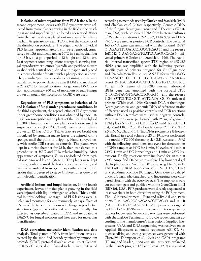
Fig. 1. Phaeosphaeria leaf spot (PLS) disease of maize. a. general appearance of the disease at advanced stage, and b, c, and d: le-sions at different stages. Stage 1: dark green water-soaked leaf spots; stage 2: greyish lesions; stage 3: lesions become necrotic and look like straw without visible fungal structures; and stage 4: necrotic lesions displaying fungal reproductive structures (pycnidia and perithecia).
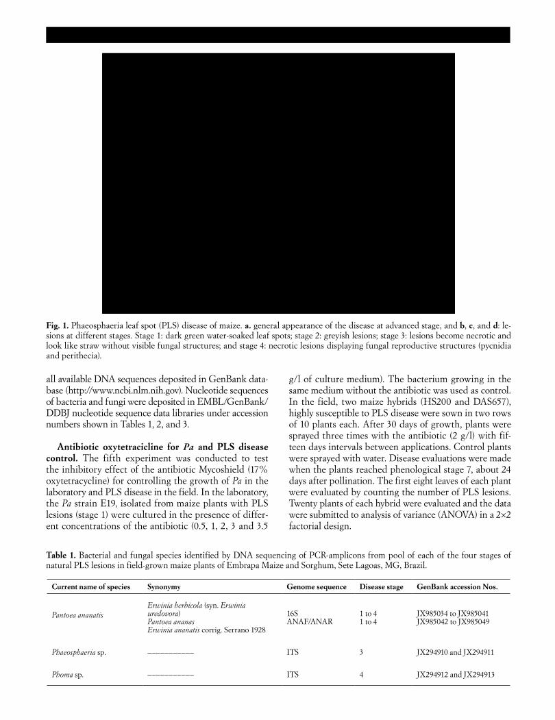
Table 1. Bacterial and fungal species identified by DNA sequencing of PCR-amplicons from pool of each of the four stages of natural PLS lesions in field-grown maize plants of Embrapa Maize and Sorghum, Sete Lagoas, MG, Brazil.
Current name of species Synonymy Genome sequence Disease stage GenBank accession Nos.
Pantoea ananatis
Erwinia herbicola (syn. Erwinia uredovora) Pantoea ananas Erwinia ananatis corrig. Serrano 1928
16S ANAF/ANAR
1 to 4 1 to 4
JX985034 to JX985041 JX985042 to JX985049
Phaeosphaeria sp.
–––––––––––
ITS
3
JX294910 and JX294911
Phoma sp.
–––––––––––
ITS
4
JX294912 and JX294913
6. JPP1602RP (Paccola).indd 562 06/11/13 16:07
Journal of Plant Pathology (2013), 95 (3), 559-569 Gonçalves et al. 563
RESULTS
The first of the five experiments carried out in the course of this study, encompassed the molecular iden-tification of bacteria and fungi associated with PLS, a disease that, as reported above, has a distinctive pattern of progression and may be separated into four distinct stages (Fig. 1). In stage 1, the lesions have dark green color and look like water-soaked spots; in stage 2, lesions are grayish; in stage 3, lesions become necrotic and look like straw; in stage 4, lesions are necrotic displaying fungal reproductive structures.
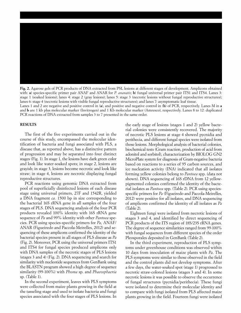
PCR reactions using genomic DNA extracted from pool of superficially disinfected lesions of each disease stage using universal primers, 27F and 1542R, yielded a DNA fragment ca. 1500 bp in size corresponding to the bacterial 16S rRNA gene in all samples of the four stages of PLS. DNA sequencing analysis of the four PCR products revealed 100% identity with 16S rRNA gene sequence of Pa and 99% identity with other Pantoea spe-cies. PCR using species-specific primers for Pa, ANAF/ANAR (Figueiredo and Paccola-Meirelles, 2012) and se-quencing of these amplicons confirmed the identity of the bacterial species present in all stages of PLS disease as Pa (Fig. 2). Moreover, PCR using the universal primers ITS1 and ITS4 for fungal species produced amplicons only with DNA samples of the necrotic stages of PLS lesions (stages 3 and 4) (Fig. 2). DNA sequencing and search for similarity with nucleotide sequences from GenBank using the BLASTN program showed a high degree of sequence similarity (99-100%) with Phoma sp. and Phaeosphaeria sp. (Table 1).
In the second experiment, leaves with PLS symptoms were collected from maize plants growing in the field at the tasseling stage and used to isolate bacteria and fungi species associated with the four stages of PLS lesions. In
the early stage of lesions (stages 1 and 2) yellow bacte-rial colonies were consistently recovered. The majority of necrotic PLS lesions at stage 4 showed pycnidia and perithecia, and different fungal species were isolated from those lesions. Morphological analysis of bacterial colonies, biochemical tests (Gram reaction, production of acid from adonitol and sorbitol), characterization by BIOLOG GN2 MicroPlate system for diagnosis of Gram-negative bacteria based on reactions to a series of 95 carbon sources, and ice nucleation activity (INA) indicated that all isolates forming yellow colonies belong to Pantoea spp. (data not shown). DNA sequencing of 16S rDNA from 12 yellow-pigmented colonies confirmed the identity of the bacte-rial isolates as Pantoea spp. (Table 2). PCR using species-specific primers for Pa (Figueiredo and Paccola-Meirelles, 2012) were positive for all isolates, and DNA sequencing of amplicons confirmed the identity of all isolates as Pa (Table 2).
Eighteen fungi were isolated from necrotic lesions of stages 3 and 4, and identified by direct sequencing of PCR products of the ITS region of 18S/23S rRNA genes. The degree of sequence similarities ranged from 99-100% with fungal sequences from different species of the order Pleosporales deposited in GenBank (Table 2).
In the third experiment, reproduction of PLS symp-toms under greenhouse conditions was observed within 10 days from inoculation of maize plants with Pa. The PLS symptoms were similar to those observed in the field and the control plants did not develop symptoms. After a few days, the water-soaked spot (stage 1) progressed to necrotic straw-colored lesions (stages 3 and 4). In some necrotic lesions it was possible to observe the occurrence of fungal structures (pycnidia/perithecia). These fungi were isolated to determine their molecular identity and to compare with fungi isolated from PLS affected maize plants growing in the field. Fourteen fungi were isolated
Fig. 2. Agarose gels of PCR products of DNA extracted from PSL lesions at different stages of development. Amplicons obtained with: a) species-specific primer pair ANAF and ANAR for P. ananatis; b) fungal universal primer pair ITS1 and ITS4. Lanes 3: stage 1 (soaked lesions); lanes 4: stage 2 (gray lesions); lanes 5: stage 3 (necrotic lesions without fungal reproductive structures); lanes 6: stage 4 (necrotic lesions with visible fungal reproductive structures); and lanes 7: asymptomatic leaf tissue. Lanes 1 and 2 are negative and positive control in (a), and positive and negative control in (b) of PCR, respectively. Lanes M in a and b are 1 kb plus molecular marker (Invitrogen) and 1 Kb molecular marker (Amresco), respectively. Lanes 8 to 12: duplicated PCR reactions of DNA extracted from samples 3 to 7 presented in the same order.
6. JPP1602RP (Paccola).indd 563 06/11/13 16:07
564 Etiology of Phaeosphaeria leaf spot disease Journal of Plant Pathology (2013), 95 (3), 559-569
from the necrotic lesions produced by inoculation with Pa under greenhouse conditions. The isolates were identi-fied by direct sequencing of the PCR products of the ITS region of 18S/28S rRNA genes. The degree of sequence similarities ranged from 99-100% with fungal sequences from different taxonomic species of the order Pleospo-rales deposited in GenBank (Table 2). Epicoccum nigrum
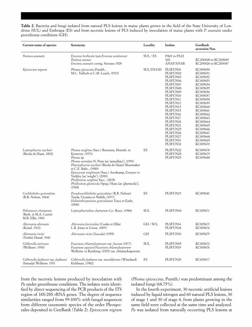
(Phoma epicoccina, Punith.) was predominant among the isolated fungi (68.75%).
In the fourth experiment, 30 necrotic artificial lesions induced by liquid nitrogen and 60 natural PLS lesions, 30 of stage 1 and 30 of stage 4, from plants growing in the same field were collected at the same time and analyzed. Pa was isolated from naturally occurring PLS lesions at
Table 2. Bacteria and fungi isolated from natural PLS lesions in maize plants grown in the field of the State University of Lon-drina (SUL) and Embrapa (ES) and from necrotic lesions of PLS induced by inoculation of maize plants with P. ananatis under greenhouse conditions (GH).
Current name of species Synonymy Locality Isolate GenBank accession Nos.
Pantoea ananatis Erwinia herbicola (syn.Erwinia uredovora) Pantoea ananas Erwinia ananatis corrig. Serrano 1928
SUL / ES PA01 to PA22 16S ANAF/ANAR
KC200048 to KC200069 KC200026 to KC200047
Epicoccum nigrum Phoma epicoccina Punith., M.C. Tulloch et C.M. Leach, (1972)
SUL/ES/GH PLSFUN01 PLSFUN02 PLSFUN03 PLSFUN06 PLSFUN07 PLSFUN08 PLSFUN09 PLSFUN10 PLSFUN11 PLSFUN12 PLSFUN13 PLSFUN15 PLSFUN16 PLSFUN17 PLSFUN18 PLSFUN21 PLSFUN25 PLSFUN26 PLSFUN27 PLSFUN31 PLSFUN32
KC005650 KC005651 KC005652 KC005653 KC005654 KC005655 KC005656 KC005657 KC005658 KC005659 KC005660 KC005661 KC005662 KC005663 KC005664 KC005665 KC005666 KC005667 KC005668 KC005669 KC005670
Leptosphaeria sacchari (Breda de Haan, 1892)
Phoma sorghina (Sacc.) Boerema, Dorenb. et Kesteren, (1973) Phoma sp. Phoma annulata N. Pons [as ‘annullata’], (1991) Phaeosphaeria sacchari (Breda de Haan) Shoemaker et C.E. Babc., (1989) Epicoccum sorghinum (Sacc.) Aveskamp, Gruyter et Verkley [as ‘sorghi’], (2010) Phyllosticta sorghina Sacc., (1878) Phyllosticta glumicola (Speg.) Hara [as ‘glumicola’], (1918)
ES PLSFUN22 PLSFUN28 PLSFUN29
KC005678 KC005679 KC005680
Cochliobolus geniculatus (R.R. Nelson, 1964)
Pseudocochliobolus geniculatus (R.R. Nelson) Tsuda, Ueyama et Nishih, (1977) Helminthosporium geniculatum Tracy et Earle, (1896)
ES PLSFUN23 KC005681
Pithomyces chartarum (Berk. et M.A. Curtis) M.B. Ellis, 1960
Leptosphaerulina chartarum Cec. Roux, (1986) SUL PLSFUN04 KC005671
Alternaria alternata (Keissl. 1912)
Alternaria fasciculata (Cooke et Ellis) L.R. Jones et Grout, (1897)
GH / SUL PLSFUN14 PLSFUN24
KC005673 KC005674
Alternaria ricini (Yoshii) Hansf. 1943
Alternaria ricini (Sawada) (1959) GH PLSFUN30 KC005675
Gibberella intricans (Wollenw. 1930)
Fusarium chlamydosporum var. fuscum (1977) Fusarium equiseti/Fusarium chlamydosporum Wollenw. et Reinking, (1925) var. chlamydosporum
SUL PLSFUN05 PLSFUN19
KC005672 KC005676
Gibberella fujikuroi var. fujikuroi (Sawada) Wollenw. 1931
Gibberella fujikuroi var. moniliformis (Wineland) Kuhlman, (1982)
ES PLSFUN20 KC005677
6. JPP1602RP (Paccola).indd 564 06/11/13 16:07
Journal of Plant Pathology (2013), 95 (3), 559-569 Gonçalves et al. 565
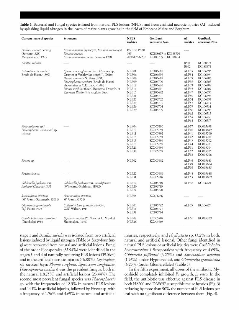
stage 1 and Bacillus subtilis was isolated from two artificial lesions induced by liquid nitrogen (Table 3). Sixty-four fun-gi were recovered from natural and artificial lesions. Fungi of the order Pleosporales (85.94%) were identified in the stages 3 and 4 of naturally occurring PLS lesions (39.06%) and in the artificial necrotic injuries (46.88%). Leptosphae-ria sacchari (syn: Phoma sorghina, Epicoccum sorghinum, Phaeosphaeria sacchari) was the prevalent fungus, both in the natural (18.75%) and artificial lesions (23.44%). The second most prevalent fungal species was Phaeosphaeria sp. with the frequencies of 12.5% in natural PLS lesions and 14.1% in artificial injuries, followed by Phoma sp. with a frequency of 1.56% and 4.69% in natural and artificial
injuries, respectively; and Phyllosticta sp. (3.2% in both, natural and artificial lesions). Other fungi identified in natural PLS lesions or artificial injuries were Cochliobolus heterostrophus (Pleosporales) with frequency of 4.69%, Gibberella fujikuroi (6.25%) and Sarocladium strictum (1.56%) (order Hypocreales), and Glomerella graminicola (6.25%) (order Glomerellales) (Table 3).
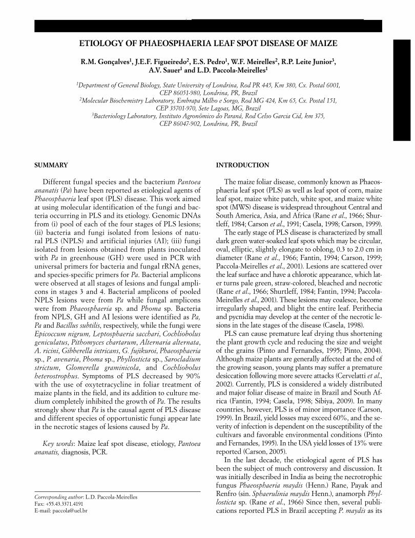
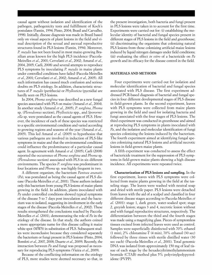
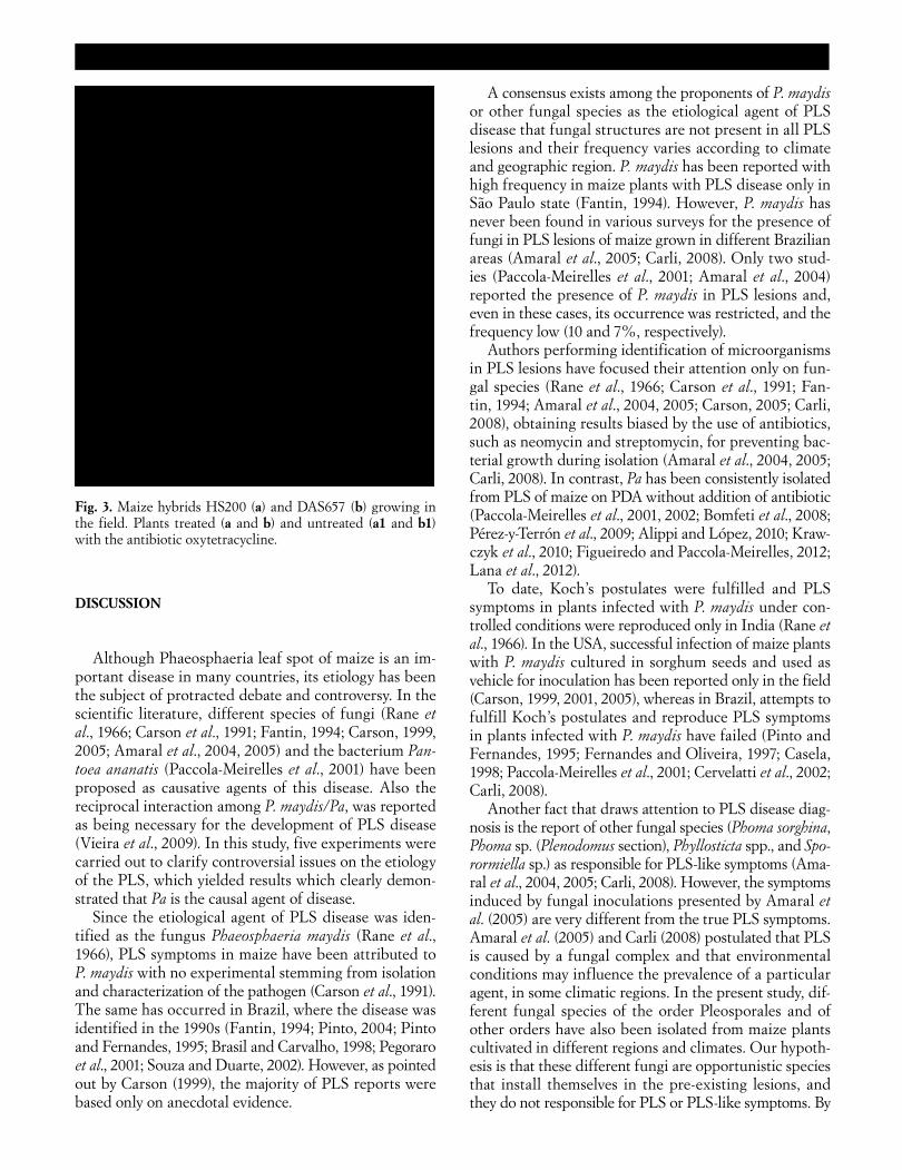
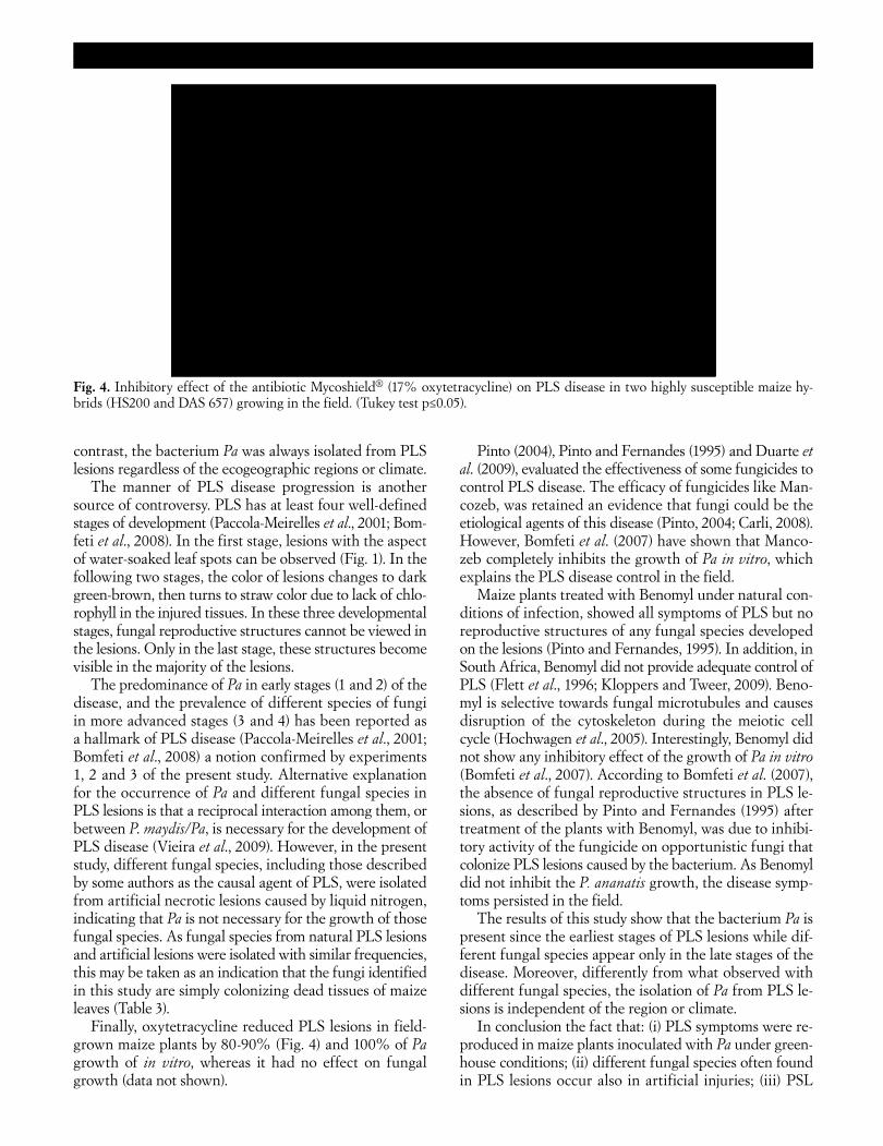
In the fifth experiment, all doses of the antibiotic My-coshield completely inhibited Pa growth, in vitro. In the field, the antibiotic was effective against PLS disease in both HS200 and DAS657 susceptible maize hybrids (Fig. 3) reducing by more than 90% the number of PLS lesions per leaf with no significant difference between them (Fig. 4).
Table 3. Bacterial and fungal species isolated from natural PLS lesions (NPLS), and from artificial necrotic injuries (AI) induced by splashing liquid nitrogen in the leaves of maize plants growing in the field of Embrapa Maize and Sorghum.
Current name of species Synonymy NPLS isolates
GenBank accession Nos.
AI isolates
GenBank accession Nos.
Pantoea ananatis corrig. (Serrano 1928) Mergaert et al. 1993
Erwinia ananas (synonym, Erwinia uredovora) Pantoea ananas Erwinia ananatis corrig. Serrano 1928
PA01 to PA30 16S ANAF/ANAR
KC188675 to KC188704 KC188705 to KC188734
–––
Bacillus subtilis ––– ––– ––– BS01 BS02
KC188673 KC188674
Leptosphaeria sacchari Breda de Haan, (1892)
Epicoccum sorghinum (Sacc.) Aveskamp, Gruyter et Verkley [as ‘sorghi’], (2010) Phoma annulata N. Pons (1991) Phaeosphaeria sacchari (Breda de Haan) Shoemaker et C.E. Babc. (1989) Phoma sorghina (Sacc.) Boerema, Dorenb. et Kesteren Phyllosticta sorghina Sacc.
NLF01 NLF06 NLF08 NLF09 NLF12 NLF14 NLF15 NLF21 NLF22 NLF23 NLF26 NLF29
KC106688 KC106699 KC106689 KC106700 KC106690 KC106691 KC106692 KC106701 KC106702 KC106703 KC106704 KC106705
ALF33 ALF34 ALF35 ALF36 ALF39 ALF45 ALF47 ALF50 ALF54 ALF57 ALF59 ALF60 ALF62 ALF63 ALF64
KC106693 KC106694 KC106706 KC106707 KC106708 KC106709 KC106695 KC106696 KC106697 KC106713 KC106714 KC106698 KC106715 KC106716 KC106717
Phaeosphaeria sp./ Phaeosphaeria avenaria f. sp. triticae
––– NLF04 NLF10 NLF11 NLF16 NLF17 NLF18 NLF25 NLF30
KC005690 KC005691 KC005692 KC005693 KC005694 KC005695 KC005696 KC005697
ALF37 ALF40 ALF41 ALF42 ALF43 ALF44 ALF51 ALF52 ALF58
KC005698 KC005699 KC005700 KC005701 KC005702 KC005703 KC005704 KC005705 KC005706
Phoma sp. ––– NLF02 KC005682 ALF46 ALF49 ALF56
KC005683 KC005684 KC005685
Phyllosticta sp. ––– NLF27 NLF31
KC005686 KC005687
ALF48 ALF53
KC005688 KC005689
Gibberella fujikuroi var. fujikuroi (Sawada) 1931
Gibberella fujikuroi var. moniliformis (Wineland) Kuhlman, (1982)
NLF19 NLF20 NLF24
KC106718 KC106719 KC106720
ALF38 KC106721
Sarocladium strictum (W. Gams) Summerb., (2011)
Acremonium strictum W. Gams, (1971)
NLF05 KC175286 ––– –––
Glomerella graminicola D.J. Politis 1975
Colletotrichum graminicola (Ces.) G.W. Wilson, 1914
NLF03 NLF13 NLF32
KC106722 KC106723 KC106724
ALF55 KC106725
Cochliobolus heterostrophus (Drechsler) 1934
Bipolaris maydis (Y. Nisik. et C. Miyake) Shoemaker, (1959)
NLF07 NLF28
KC005707 KC005708
ALF61 KC005709
6. JPP1602RP (Paccola).indd 565 06/11/13 16:07
566 Etiology of Phaeosphaeria leaf spot disease Journal of Plant Pathology (2013), 95 (3), 559-569
DISCUSSION
Although Phaeosphaeria leaf spot of maize is an im-portant disease in many countries, its etiology has been the subject of protracted debate and controversy. In the scientific literature, different species of fungi (Rane et al., 1966; Carson et al., 1991; Fantin, 1994; Carson, 1999, 2005; Amaral et al., 2004, 2005) and the bacterium Pan-toea ananatis (Paccola-Meirelles et al., 2001) have been proposed as causative agents of this disease. Also the reciprocal interaction among P. maydis/Pa, was reported as being necessary for the development of PLS disease (Vieira et al., 2009). In this study, five experiments were carried out to clarify controversial issues on the etiology of the PLS, which yielded results which clearly demon-strated that Pa is the causal agent of disease.
Since the etiological agent of PLS disease was iden-tified as the fungus Phaeosphaeria maydis (Rane et al., 1966), PLS symptoms in maize have been attributed to P. maydis with no experimental stemming from isolation and characterization of the pathogen (Carson et al., 1991). The same has occurred in Brazil, where the disease was identified in the 1990s (Fantin, 1994; Pinto, 2004; Pinto and Fernandes, 1995; Brasil and Carvalho, 1998; Pegoraro et al., 2001; Souza and Duarte, 2002). However, as pointed out by Carson (1999), the majority of PLS reports were based only on anecdotal evidence.
A consensus exists among the proponents of P. maydis or other fungal species as the etiological agent of PLS disease that fungal structures are not present in all PLS lesions and their frequency varies according to climate and geographic region. P. maydis has been reported with high frequency in maize plants with PLS disease only in São Paulo state (Fantin, 1994). However, P. maydis has never been found in various surveys for the presence of fungi in PLS lesions of maize grown in different Brazilian areas (Amaral et al., 2005; Carli, 2008). Only two stud-ies (Paccola-Meirelles et al., 2001; Amaral et al., 2004) reported the presence of P. maydis in PLS lesions and, even in these cases, its occurrence was restricted, and the frequency low (10 and 7%, respectively).
Authors performing identification of microorganisms in PLS lesions have focused their attention only on fun-gal species (Rane et al., 1966; Carson et al., 1991; Fan-tin, 1994; Amaral et al., 2004, 2005; Carson, 2005; Carli, 2008), obtaining results biased by the use of antibiotics, such as neomycin and streptomycin, for preventing bac-terial growth during isolation (Amaral et al., 2004, 2005; Carli, 2008). In contrast, Pa has been consistently isolated from PLS of maize on PDA without addition of antibiotic (Paccola-Meirelles et al., 2001, 2002; Bomfeti et al., 2008; Pérez-y-Terrón et al., 2009; Alippi and López, 2010; Kraw-czyk et al., 2010; Figueiredo and Paccola-Meirelles, 2012; Lana et al., 2012).
To date, Koch’s postulates were fulfilled and PLS symptoms in plants infected with P. maydis under con-trolled conditions were reproduced only in India (Rane et al., 1966). In the USA, successful infection of maize plants with P. maydis cultured in sorghum seeds and used as vehicle for inoculation has been reported only in the field (Carson, 1999, 2001, 2005), whereas in Brazil, attempts to fulfill Koch’s postulates and reproduce PLS symptoms in plants infected with P. maydis have failed (Pinto and Fernandes, 1995; Fernandes and Oliveira, 1997; Casela, 1998; Paccola-Meirelles et al., 2001; Cervelatti et al., 2002; Carli, 2008).
Another fact that draws attention to PLS disease diag-nosis is the report of other fungal species (Phoma sorghina, Phoma sp. (Plenodomus section), Phyllosticta spp., and Spo-rormiella sp.) as responsible for PLS-like symptoms (Ama-ral et al., 2004, 2005; Carli, 2008). However, the symptoms induced by fungal inoculations presented by Amaral et al. (2005) are very different from the true PLS symptoms. Amaral et al. (2005) and Carli (2008) postulated that PLS is caused by a fungal complex and that environmental conditions may influence the prevalence of a particular agent, in some climatic regions. In the present study, dif-ferent fungal species of the order Pleosporales and of other orders have also been isolated from maize plants cultivated in different regions and climates. Our hypoth-esis is that these different fungi are opportunistic species that install themselves in the pre-existing lesions, and they do not responsible for PLS or PLS-like symptoms. By
Fig. 3. Maize hybrids HS200 (a) and DAS657 (b) growing in the field. Plants treated (a and b) and untreated (a1 and b1) with the antibiotic oxytetracycline.
6. JPP1602RP (Paccola).indd 566 06/11/13 16:07
Journal of Plant Pathology (2013), 95 (3), 559-569 Gonçalves et al. 567
contrast, the bacterium Pa was always isolated from PLS lesions regardless of the ecogeographic regions or climate.
The manner of PLS disease progression is another source of controversy. PLS has at least four well-defined stages of development (Paccola-Meirelles et al., 2001; Bom-feti et al., 2008). In the first stage, lesions with the aspect of water-soaked leaf spots can be observed (Fig. 1). In the following two stages, the color of lesions changes to dark green-brown, then turns to straw color due to lack of chlo-rophyll in the injured tissues. In these three developmental stages, fungal reproductive structures cannot be viewed in the lesions. Only in the last stage, these structures become visible in the majority of the lesions.
The predominance of Pa in early stages (1 and 2) of the disease, and the prevalence of different species of fungi in more advanced stages (3 and 4) has been reported as a hallmark of PLS disease (Paccola-Meirelles et al., 2001; Bomfeti et al., 2008) a notion confirmed by experiments 1, 2 and 3 of the present study. Alternative explanation for the occurrence of Pa and different fungal species in PLS lesions is that a reciprocal interaction among them, or between P. maydis/Pa, is necessary for the development of PLS disease (Vieira et al., 2009). However, in the present study, different fungal species, including those described by some authors as the causal agent of PLS, were isolated from artificial necrotic lesions caused by liquid nitrogen, indicating that Pa is not necessary for the growth of those fungal species. As fungal species from natural PLS lesions and artificial lesions were isolated with similar frequencies, this may be taken as an indication that the fungi identified in this study are simply colonizing dead tissues of maize leaves (Table 3).
Finally, oxytetracycline reduced PLS lesions in field-grown maize plants by 80-90% (Fig. 4) and 100% of Pa growth of in vitro, whereas it had no effect on fungal growth (data not shown).
Pinto (2004), Pinto and Fernandes (1995) and Duarte et al. (2009), evaluated the effectiveness of some fungicides to control PLS disease. The efficacy of fungicides like Man-cozeb, was retained an evidence that fungi could be the etiological agents of this disease (Pinto, 2004; Carli, 2008). However, Bomfeti et al. (2007) have shown that Manco-zeb completely inhibits the growth of Pa in vitro, which explains the PLS disease control in the field.
Maize plants treated with Benomyl under natural con-ditions of infection, showed all symptoms of PLS but no reproductive structures of any fungal species developed on the lesions (Pinto and Fernandes, 1995). In addition, in South Africa, Benomyl did not provide adequate control of PLS (Flett et al., 1996; Kloppers and Tweer, 2009). Beno-myl is selective towards fungal microtubules and causes disruption of the cytoskeleton during the meiotic cell cycle (Hochwagen et al., 2005). Interestingly, Benomyl did not show any inhibitory effect of the growth of Pa in vitro (Bomfeti et al., 2007). According to Bomfeti et al. (2007), the absence of fungal reproductive structures in PLS le-sions, as described by Pinto and Fernandes (1995) after treatment of the plants with Benomyl, was due to inhibi-tory activity of the fungicide on opportunistic fungi that colonize PLS lesions caused by the bacterium. As Benomyl did not inhibit the P. ananatis growth, the disease symp-toms persisted in the field.
The results of this study show that the bacterium Pa is present since the earliest stages of PLS lesions while dif-ferent fungal species appear only in the late stages of the disease. Moreover, differently from what observed with different fungal species, the isolation of Pa from PLS le-sions is independent of the region or climate.
In conclusion the fact that: (i) PLS symptoms were re-produced in maize plants inoculated with Pa under green-house conditions; (ii) different fungal species often found in PLS lesions occur also in artificial injuries; (iii) PSL
Fig. 4. Inhibitory effect of the antibiotic Mycoshield® (17% oxytetracycline) on PLS disease in two highly susceptible maize hy-brids (HS200 and DAS 657) growing in the field. (Tukey test p≤0.05).
6. JPP1602RP (Paccola).indd 567 06/11/13 16:07
568 Etiology of Phaeosphaeria leaf spot disease Journal of Plant Pathology (2013), 95 (3), 559-569
disease is controlled by the oxytetracycline, strongly re-inforce the hypothesis that the PLS disease is caused by Pantoea ananatis as postulated by Paccola-Meirelles et al. (2001), rather than a specific fungus or a complex of fungal species as previously reported (Rane et al., 1966; Carson et al., 1991; Fantin, 1994; Amaral et al., 2004, 2005; Carson, 2005; Carli, 2008).
ACKNOWLEDGEMENTS
The authors gratefully acknowledge CNPq, Funda-ção Araucária, CAPES, UEL, and Embrapa for financial support.
REFERENCES
Alippi A.M., López A.C., 2010. First report of leaf spot disease of maize caused by Pantoea ananatis in Argentina. Plant Dis-ease 94: 487.
Altschul S.F., Madden T.L., Schaffer A.A., Zhang J., Zhang Z., Miller W., Lipman D.J., 1997. Gapped BLAST and PSI-BLAST: a new generation of protein database search pro-gram. Nucleic Acids Research 25: 3389-3402.
Amaral A.L., Carli M.L., Neto J.F.B., Soglio F.K., 2004. Phoma sorghina, a new pathogen associated with Phaeosphaeria leaf spot on maize in Brazil. Plant Pathology 53: 259.
Amaral A.L., Soglio F.K., Carli M.L., Neto J.F.B., 2005. Patho-genic fungi causing symptoms similar to Phaeosphaeria leaf spot of maize in Brazil. Plant Disease 89: 44-49.
Bomfeti C.A., Meirelles W.F., Souza-Paccola E.A., Casela C.R., Ferreira A.S., Marrie I.E., Paccola-Meirelles L.D., 2007. Evaluation of commercial chemical products, in vitro and in vivo in the control of foliar disease, maize white spot, caused by Pantoea ananatis. Summa Phytopathologica 33: 63-67.
Bomfeti C.A., Souza-Paccola E.A., Massola N.S., Marriel I.E., Meirelles W.F., Casela C.R., Paccola-Meirelles L.D., 2008. Localization of Pantoea ananatis inside lesions of maize white spot disease using transmission electron microscopy and molecular techniques. Tropical Plant Pathology 33: 1-6.
Brasil E.M., Carvalho Y., 1998. Comportamento de híbridos de milho em relação a Phaeosphaeria maydis em diferentes épo-cas de plantio. Pesquisa Agropecuária Brasileira 33: 1977-1981.
Carli M.L., 2008. Etiological and epidemiological aspects of the maize white leaf spot complex. Ms. Sci. Thesis. Universidade Federal do Rio Grande do Sul. Porto Alegre, Brazil.
Carson M.L., 1999. Vulnerability of U.S. maize germplasm to Phaeosphaeria leaf spot. Plant Disease 83: 462-464.
Carson M.L., 2001. Inheritance of resistance to Phaeosphaeria leaf spot of maize. Plant Disease 85: 798-800.
Carson M.L., 2005. Yield loss potential of Phaeospharia leaf spot of maize caused by Phaeosphaeria maydis in the United States. Plant Disease 89: 986-988.
Carson M.L., Goodman M.M., Glawe D.A., 1991. Phaeosphae-ria leaf spot of maize in Florida. Plant Disease 75: 968.
Casela C.R., 1998. The Phaeosphaeria leaf spot. In: Casela C.R., Renfro R., Krattiger A.F. (eds). Diagnosing Maize Diseases
in Latin America, pp.1-57. Isaa/Embrapa, Ithaca, NY, USA and Brasilia, BR, Brazil.
Cervelatti E.P., Paiva E., Meirelles W.F., Casela C.R., Fernandes F.T., Teixeira F.F., Paccola-Meirelles L.D., 2002. Character-ization of fungal isolates from pycnidia and pseudothecia from lesions of Phaeosphaeria leaf spot in maize. Revista Brasileira de Milho e Sorgo 1: 30-37.
Duarte R.P., Juliatti F.C., Freitas P.T., 2009. Efficacy of different fungicides on maize crop. Bioscience Journal 25: 101-111.
Fantin G.M., 1994. Mancha de phaeosphaeria, doença do milho que vem aumentando a sua importância. Biologico 56: 39.
Fernandes F.T., Oliveira E., 1997. Principais doenças na cultura do milho. Sete Lagoas, Embrapa-CNPMS, Circular 26.
Figueiredo J.E.F., Paccola-Meirelles L.D., 2012. Simple, rapid and accurate PCR-based detection of Pantoea ananatis in maize, sorghum and Digitaria sp. Journal of Plant Pathology 94: 663-667.
Flett B.C., Bensch M.J., Smit E., Fourie H., 1996. A Field Guide for Identification of Maize Diseases in South Africa. ARC-LNR, Potchefstroom, South Africa.
Gürtler V., Stanisich V.A., 1996. New approaches to typing and identification of bacteria by using the 16S-23S rDNA spacer region. Microbiology 142: 3-16.
Hochwagen A., Wrobel G., Cartron M., Demougin P., Nieder-hauser-Wiederkehr C., Boselli M.G., Primig M., Amon A., 2005. Novel response to microtubule perturbation in meio-sis. Molecular and Cellular Biology 25: 4767- 4781.
Huang X., Madan A., 1999. CAP3: A DNA sequence assembly program. Genome Research 9: 868-877.
Kloppers R., Tweer S., 2009. Phaeosphaeria leaf spot. Pannar Seed Factsheets, Pannar Seed Greytown, South Africa.
Krawczyk K., Kamasa J., Zwolinska A., Pospieszny H., 2010. First report of Pantoea ananatis associated with leaf spot dis-ease of maize in Poland. Journal of Plant Pathology 92: 807-811.
Lana U.G.P., Gomes E.A., Silva D.D., Costa R.V., Cota L.V., Parreira D.F., Souza I.R.P., Guimarães C.T., 2012. Detection and molecular diversity of Pantoea ananatis associated with white spot disease in maize, sorghum and crabgrass in Bra-zil. Journal of Phytopathology 160: 441-448.
Lane D.J., 1991. 16S/23S rRNA sequencing. In: Stackebrandt E., Goodfellow M., (eds). Nucleic Acid Techniques in Bacte-rial Systematics, pp.115-175. John Wiley and Sons, Chiches-ter, UK.
Nübel U., Engelen B., Felske A., Snaidr J., Wieshuber A., Amann R.I., Ludwig W., Backhaus H., 1996. Sequence het-erogeneities of genes encoding 16S rRNAs in Paenibacillus polymyxa detected by temperature gradient gel electropho-resis. Journal of Bacteriology 178: 5636-5643.
Paccola-Meirelles L.D., Ferreira A.S., Meirelles W.F., Marriel I.E., Casela C.R., 2001. Detection of a bacterium associated with a leaf spot disease of maize in Brazil. Journal of Phy-tophathology 149: 275-279.
Paccola-Meirelles L.D., Meirelles W.F., Parentoni S.N., Marriel I.E., Ferreira A.S., Casela C.R., 2002. Reaction of maize in-bred lines to a bacterium, Pantoea ananas, isolated from Pha-eosphaeria leaf spot lesions. Crop Breeding and Applied Bio-technology 2: 587-590.
6. JPP1602RP (Paccola).indd 568 06/11/13 16:07
Journal of Plant Pathology (2013), 95 (3), 559-569 Gonçalves et al. 569
Pegoraro D.G., Vacaro E., Nuss C.N., Soglio F.K., Sereno M.J.C.M., Neto J.F.B., 2001. Efeito da época de semeadura na mancha-foliar de Phaeosphaeria em milho. Pesquisa Ag-ropecuária Brasileira 36: 1037-1042.
Pérez-Y-Terrón R., Cuellan A., Muñoz-Rojas J., Castañeda-Lucio M., Hernández-Lucas I., Bustillos-Cristales R., Bautista-SosaL., Munive J.A., Caicedo-Rivas R., Fuentes-Ramírez L.E., 2009. Detection of Pantoea ananatis, causal agent of leaf spot disease of maize, in Mexico. Australasian Plant Dis-ease Notes 4: 96-99.
Pinto N.F.J.A., 2004. Controle químico de doenças foliares em milho. Revista Brasileira de Milho e Sorgo 3: 134-138.
Pinto N.F.J.A., Fernandes F.T., 1995. Avaliação de fungicidas no controle da mancha foliar de milho causada por Phyl-losticta sp. (Phaeosphaeria maydis). Fitopatologia Brasileira 20: 333.
Porebski S., Bailey L.G., Baum B.R., 1997. Modification of a CTAB DNA extraction protocol for plants containing high polysaccharide and polyphenol components. Plant Molecular Biology Reporter 15: 8-15.
Rane M.S., Payak M.M., Renfro B.L., 1966. A Phaeosphaeria leaf spot of maize. Indian Phytopathology Society Bulletin 3: 8-10.
Shaolan L., Bin Z., Liyuan Y., Zhiying L., Qi Z., Youwei C., 2002. An improved method for extracting fungal DNA. Journal of Yunnan University 24: 471-472.
Shurtleff M.C., 1984. Phaeosphaeria leaf spot. In: Shurtleff M.C. (ed.). Compendium of Corn Diseases, pp. 23-24. APS Press, St. Paul, MN, USA.
Sibiya J., 2009. Breeding investigations for resistance to Phaeos-phaeria leaf spot (PLS) and other important foliar diseases and study of yield stability in African maize germplasm. Ph.D. Thesis, University of Kwa-Zulu Natal, Pietermaritz-burg, Republic of South Africa.
Souza J.C., Duarte J.M., 2002. Reação de cultivares de milho a Phaeosphaeria maydis. Ciência e Agrotecnologia 26: 325-331.
Thompson J.D., Higgins D.G., Gibson T.J., 1994. CLUSTAL W: improving the sensitivity of progressive multiple se-quence alignment through sequence weighting, position specific gap penalties and weight matrix choice. Nucleic Acids Research 22: 4673-4680.
Vieira R.A., Tessmann D., Scapim C.A., Hata F.T., Rodovalho M.A., Barreto R.R., 2009. Genetic resistance of new popcorn hybrids to foliar diseases. Crop Breeding and Applied Biotech-nology 9: 140-146.
White T.J., Bruns T.D., Lee S., Taylor J., 1990. Amplification and direct sequencing of fungal ribosomal RNA genes for phylogenetics. In: Innis M.A., Gelfand D.H., Sninsky J.J., White T.J. (eds). PCR Protocols: a Guide to Methods and Applications, pp. 315-322. Academic Press, New York, NY, USA.
Received March 7, 2013Accepted April 20, 2013
6. JPP1602RP (Paccola).indd 569 06/11/13 16:07