Embed Size (px)
Citation preview

EtpE Binding to DNase X Induces Ehrlichial Entry via CD147 andhnRNP-K Recruitment, Followed by Mobilization of N-WASP andActin
Dipu Mohan Kumar,a Mingqun Lin,a Qingming Xiong,a Mathew James Webber,b Comert Kural,b,c Yasuko Rikihisaa
Department of Veterinary Biosciences,a Interdisciplinary Biophysics Graduate Program,b and Department of Physics,c The Ohio State University, Columbus, Ohio, USA
ABSTRACT Obligate intracellular bacteria, such as Ehrlichia chaffeensis, perish unless they can enter eukaryotic cells.E. chaffeensis is the etiological agent of human monocytic ehrlichiosis, an emerging infectious disease. To infect cells, Ehrlichiauses the C terminus of the outer membrane invasin entry-triggering protein (EtpE) of Ehrlichia (EtpE-C), which directly bindsthe mammalian cell surface glycosylphosphatidyl inositol-anchored protein, DNase X. How this binding drives Ehrlichia entry isunknown. Here, using affinity pulldown of host cell lysates with recombinant EtpE-C (rEtpE-C), we identified two new humanproteins that interact with EtpE-C: CD147 and heterogeneous nuclear ribonucleoprotein K (hnRNP-K). The interaction ofCD147 with rEtpE-C was validated by far-Western blotting and coimmunoprecipitation of native EtpE with endogenous CD147.CD147 was ubiquitous on the cell surface and also present around foci of rEtpE-C-coated-bead entry. Functional neutralizationof surface-exposed CD147 with a specific antibody inhibited Ehrlichia internalization and infection but not binding. Downregu-lation of CD147 by short hairpin RNA (shRNA) impaired E. chaffeensis infection. Functional ablation of cytoplasmic hnRNP-Kby a nanoscale intracellular antibody markedly attenuated bacterial entry and infection but not binding. EtpE-C also interactedwith neuronal Wiskott-Aldrich syndrome protein (N-WASP), which is activated by hnRNP-K. Wiskostatin, which inhibitsN-WASP activation, and cytochalasin D, which inhibits actin polymerization, inhibited Ehrlichia entry. Upon incubation withhost cell lysate, EtpE-C but not an EtpE N-terminal fragment stimulated in vitro actin polymerization in an N-WASP- andDNase X-dependent manner. Time-lapse video images revealed N-WASP recruitment at EtpE-C-coated bead entry foci. Thus,EtpE-C binding to DNase X drives Ehrlichia entry by engaging CD147 and hnRNP-K and activating N-WASP-dependent actinpolymerization.
IMPORTANCE Ehrlichia chaffeensis, an obligate intracellular bacterium, causes a blood-borne disease called human monocyticehrlichiosis, one of the most prevalent life-threatening emerging tick-transmitted infectious diseases in the United States. Thesurvival of Ehrlichia bacteria, and hence, their ability to cause disease, depends on their specific mode of entry into eukaryotichost cells. Understanding the mechanism by which E. chaffeensis enters cells will create new opportunities for developing effec-tive therapies to prevent bacterial entry and disease in humans. Our findings reveal a novel cellular signaling pathway triggeredby an ehrlichial surface protein called EtpE to induce its infectious entry. The results are also important from the viewpoint ofhuman cell physiology because three EtpE-interacting human proteins, DNase X, CD147, and hnRNP-K, are hitherto unknownpartners that drive the uptake of small particles, including bacteria, into human cells.
Received 5 October 2015 Accepted 7 October 2015 Published 3 November 2015
Citation Mohan Kumar D, Lin M, Xiong Q, Webber MJ, Kural C, Rikihisa Y. 2015. EtpE binding to DNase X induces ehrlichial entry via CD147 and hnRNP-K recruitment, followedby mobilization of N-WASP and actin. mBio 6(6):e01541-15. doi:10.1128/mBio.01541-15.
Editor Howard A. Shuman, University of Chicago
Copyright © 2015 Mohan Kumar et al. This is an open-access article distributed under the terms of the Creative Commons Attribution-Noncommercial-ShareAlike 3.0Unported license, which permits unrestricted noncommercial use, distribution, and reproduction in any medium, provided the original author and source are credited.
Address correspondence to Yasuko Rikihisa, [email protected].
This article is a direct contribution from a Fellow of the American Academy of Microbiology.
Human monocytic ehrlichiosis (HME) is one of the most prev-alent life-threatening emerging tick-borne zoonoses in the
United States (1). The disease was discovered in 1986 and wasdesignated a nationally notifiable disease in 1998 by the U.S Cen-ters for Disease Control and Prevention. HME is caused by infec-tion with Ehrlichia chaffeensis, an obligate intracellular bacteriumin the order Rickettsiales. E. chaffeensis replicates within humanmonocytes and macrophages and causes severe flulike symptomsaccompanied by hematologic abnormalities and signs of hepatitis.No vaccines exist for HME. The broad-spectrum antibiotic doxy-
cycline is the only drug effective for treating HME but is contra-indicated for pregnant women and children. Delayed initiation oftherapy, the presence of underlying illness, or immunosuppres-sion often lead to severe complications or even death (2). Theincidence of tick-borne zoonoses has risen continuously and dra-matically in the past 2 decades (3). The Centers for Disease Con-trol and Prevention confirmed 1,404 HME cases in 2014 and 1,509in 2013 (1), which is more than a 10-fold increase in HME inci-dence over a 15-year period. The 2011 U.S. Institute of Medicinereport “Critical Needs and Gaps in understanding prevention,
RESEARCH ARTICLE crossmark
November/December 2015 Volume 6 Issue 6 e01541-15 ® mbio.asm.org 1
on August 31, 2018 by guest
http://mbio.asm
.org/D
ownloaded from
amelioration, and resolution of Lyme and Other Tick-Borne Dis-eases” (4) pointed to the urgent need for research on HME.
E. chaffeensis entry into the human acute leukemia cell lineTHP-1 leads to productive infection and is dependent on host cellsurface lipid rafts and glycosylphosphatidyl inositol (GPI)-anchored proteins (5). After entry, E. chaffeensis replicates in anearly endosome-like compartment which contains early endo-some antigen 1 (EEA1), Rab5, transferrin receptor, and vacuolar-type H� ATPase (6) but does not contain lysosomal membrane-associated protein 1, CD63, or NADPH oxidase components (6–8). E. chaffeensis cells die if they fail to enter an appropriate hostcell. Previous studies using pharmacological inhibitors have sug-gested that the mechanism of E. chaffeensis entry is distinct fromthat of classic antimicrobial phagocytosis (5, 9).
E. chaffeensis outer membrane protein ECH1038, with highlystrain-conserved N- and C-terminal segments, is highly expressedat the stress-resistant and infectious stage of the E. chaffeensis in-tracellular developmental cycle (10). We previously namedECH1038 of Ehrlichia entry-triggering protein (EtpE) because wefound that EtpE is exposed on the E. chaffeensis surface and servesas an invasin for direct binding of its mammalian receptor DNaseX (DNase I-like protein 1), a GPI-anchored ubiquitous cell sur-face protein, to trigger infectious entry (11).
In patient blood specimens, E. chaffeensis is primarily seen inmonocytes and not in neutrophils (12); hence, human monocyticehrlichiosis is so named to distinguish it from other human ehrli-chioses caused by granulocytotropic species. E. chaffeensis hasbeen stably cultivated only in canine macrophage DH82 cells (13)or THP-1 cells (14). To apply transfection and mouse mutagenesistechniques and distinguish ehrlichial infectious entry from classicphagocytosis, we developed an effective E. chaffeensis culture sys-tem in nonphagocytic cell lines (human embryonic kidneyHEK293 cells and monkey endothelial RF/6A cells), as well asprimary phagocytic cells (human and canine peripheral bloodmonocytes and mouse bone marrow-derived macrophages [BM-DMs]) (11). An antibody (Ab) against the E. chaffeensis strain-conserved C-terminal segment of EtpE (EtpE-C) greatly inhibitsE. chaffeensis binding, entry, and infection of both phagocytic andnonphagocytic host cells in vitro, and immunization of mice withEtpE-C significantly inhibits infection (11). Although latex beadscannot bind or enter nonphagocytes, EtpE-C-coated beads enternonphagocytes as well as phagocytes, and the entry is blocked byseveral compounds that block E. chaffeensis entry (11). An anti-body against DNase X or small interfering RNA (siRNA)-mediated knockdown of DNase X significantly reducesE. chaffeensis binding and entry (11). Furthermore, the bacterialloads in the peripheral blood in experimentally infected DNaseX�/� mice are significantly reduced compared with those in in-fected wild-type mice, indicating that DNase X-mediated entry isalso relevant in vivo (11).
The actin cytoskeleton has crucial roles in particle uptake intophagocytes and nonphagocytes (15, 16), and a number of intra-cellular microbial pathogens have evolved ways to manipulate theactin cytoskeleton to facilitate infectious entry into host cells. Forexample, Salmonella induces plasma membrane ruffles to facili-tate host cell entry (17), Chlamydia causes actin remodeling, lead-ing to reorganization and hypertrophy of preexisting cell surfacemicrovilli to induce its entry (18), and Shigella causes retraction ofcell surface filopodia via actin remodeling to bring bacteria inclose contact with the cell body (19). E. chaffeensis and beads
coated with recombinant EtpE-C (rEtpE-C) induce filopodial ex-tensions on the host cell surface at the point of contact (11, 20),suggesting the involvement of actin cytoskeletal remodeling dur-ing the entry process. However, the mechanism of EtpE binding toDNase X to trigger infectious entry is unknown, especially consid-ering that the GPI-anchored DNase X does not span the plasmamembrane lipid bilayer.
Our present study is the first to investigate the details of theunique molecular interactions triggered by binding of an ehrli-chial invasin to host cells, which is required for host cell actinremodeling and the subsequent infectious entry of E. chaffeensis.Our study reveals a previously unknown signaling pathway in-duced by EtpE for actin cytoskeletal remodeling. EtpE is the firstexample of a protein that induces DNase X-dependent actin po-lymerization in vitro.
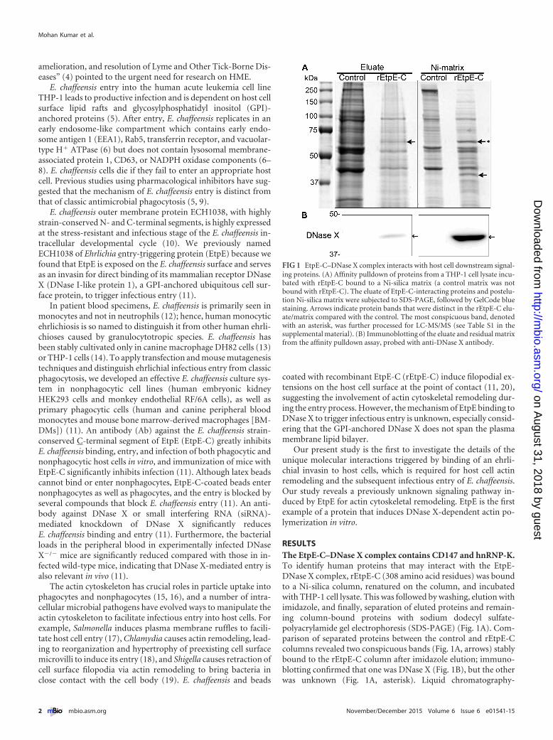
RESULTSThe EtpE-C–DNase X complex contains CD147 and hnRNP-K.To identify human proteins that may interact with the EtpE-DNase X complex, rEtpE-C (308 amino acid residues) was boundto a Ni-silica column, renatured on the column, and incubatedwith THP-1 cell lysate. This was followed by washing, elution withimidazole, and finally, separation of eluted proteins and remain-ing column-bound proteins with sodium dodecyl sulfate-polyacrylamide gel electrophoresis (SDS-PAGE) (Fig. 1A). Com-parison of separated proteins between the control and rEtpE-Ccolumns revealed two conspicuous bands (Fig. 1A, arrows) stablybound to the rEtpE-C column after imidazole elution; immuno-blotting confirmed that one was DNase X (Fig. 1B), but the otherwas unknown (Fig. 1A, asterisk). Liquid chromatography-
FIG 1 EtpE-C–DNase X complex interacts with host cell downstream signal-ing proteins. (A) Affinity pulldown of proteins from a THP-1 cell lysate incu-bated with rEtpE-C bound to a Ni-silica matrix (a control matrix was notbound with rEtpE-C). The eluate of EtpE-C-interacting proteins and postelu-tion Ni-silica matrix were subjected to SDS-PAGE, followed by GelCode bluestaining. Arrows indicate protein bands that were distinct in the rEtpE-C elu-ate/matrix compared with the control. The most conspicuous band, denotedwith an asterisk, was further processed for LC-MS/MS (see Table S1 in thesupplemental material). (B) Immunoblotting of the eluate and residual matrixfrom the affinity pulldown assay, probed with anti-DNase X antibody.
Mohan Kumar et al.
2 ® mbio.asm.org November/December 2015 Volume 6 Issue 6 e01541-15
on August 31, 2018 by guest
http://mbio.asm
.org/D
ownloaded from

coupled tandem mass spectrometry (LC-MS/MS) of this bandidentified two major proteins, namely, human CD147 (basigin/extracellular matrix metalloproteinase inducer; 26% coverage ofthe short form of CD147) and human heterogeneous nuclear ri-bonucleoprotein K (hnRNP-K; 44.3% coverage) (see Table S1 inthe supplemental material).
Mature human CD147 (i.e., lacking the 21-residue signal se-quence) contains 248 residues (short form; ubiquitously ex-pressed isoform of CD147) (21) or 368 residues (long form;retina-specific isoform of CD147) (22). CD147 (short isoform)contains two glycosylated N-terminal extracellular immunoglob-ulin domains of 185 residues, a 24-residue transmembrane do-main, and a 39-residue cytoplasmic region. Glycosylated humanCD147 (short isoform) can be detected with apparent molecularmasses ranging from 32 to 60 kDa by SDS-PAGE (23).
Human hnRNP-K is a 463-residue protein (51.3 kDa) and acomponent of the heterogeneous nuclear ribonucleoprotein (hn-RNP) complex that localizes both to the nucleus and cytoplasmand can interact with RNA, DNA, and various proteins (24). Twoother proteins identified with a MASCOT score above 100 (seeTable S1 in the supplemental material) were not investigated fur-ther because their interaction was tenuous owing to the relativelylow percentage of coverage in our LC-MS/MS analysis.
CD147 interacts with rEtpE-C as well as native EtpE, is re-cruited around rEtpE-C-coated-bead entry foci, and is requiredfor ehrlichial entry and infection but not for binding. Usingyeast two-hybrid analysis, we previously observed that DNase Xdirectly binds EtpE-C and that endogenous DNase X from THP-1cell lysate binds renatured rEtpE-C on a nitrocellulose membrane,as seen with far-Western blotting (11). In agreement with ourpulldown data (Fig. 1), far-Western blotting revealed that endog-enous CD147 from the THP-1 cell lysate bound renaturedrEtpE-C but not the control recombinant E. chaffeensis proteinECH0825 (rECH0825) that had been treated similarly (Fig. 2A).To validate this interaction with native EtpE, we performed coim-munoprecipitation with lysate of THP-1 cells that had been brieflyincubated with E. chaffeensis for 30 min; the lysate was incubatedwith protein A-agarose beads adsorbed with anti-rEtpE-C anti-body or control IgG. The precipitate, which was previously shownto contain DNase X (11), was probed for the presence of CD147.CD147 was present in the complex immunoprecipitated withanti-EtpE-C antibody but not with control IgG (Fig. 2B). CD147of THP-1 cells is likely glycosylated, because the molecular size ofCD147 was �50 kDa by immunoblotting (Fig. 2B), and this islikely the reason why CD147 was found in the same band withhnRNP-K by proteomics analysis (Fig. 1A).
To further study whether CD147 is present around the entryfoci of rEtpE-C-coated latex beads in the absence of other bacterialproteins, HEK293 cells (which are adherent and nonphagocytic,allowing better visualization of signal localization) were incubatedwith rEtpE-C-coated red fluorescent beads for 1 h, followed byimmunostaining with monoclonal antibody (MAb) MEM-M6/6directed against the membrane-proximal Ig2 domain of CD147.Fluorescence microscopy with deconvolution showed that CD147was detected ubiquitously on the plasma membrane and sur-rounded the entry foci of the coated beads (Fig. 2C). Althoughpretreatment of HEK293 cells with anti-CD147 antibody did notblock E. chaffeensis binding to HEK293 cells at 30 min postinfec-tion (p.i.) (Fig. 2D), it greatly inhibited E. chaffeensis internaliza-tion (Fig. 2E). This inhibition led to a significantly reduced bacte-
rial load at 48 h p.i. compared with the bacterial load followingcontrol IgG pretreatment (Fig. 2F). This result indicates thatCD147 is not a receptor for E. chaffeensis binding but, rather, isspecifically required for pathogen entry and for establishing infec-tion.
CD147 gene knockout in mice results in spermatocyte apopto-sis, degeneration of germ cells, and infertility (25), and thus,CD147 knockout mice are difficult to produce. Thus, to test therequirement of CD147 for E. chaffeensis entry, we established astable knockdown of CD147 via lentiviral-based transduction ofHEK293 cells with a CD147-specific short hairpin RNA (shRNA).Compared with using siRNA, shRNA provides sustainable knock-down of target genes with fewer off-target effects (26). CD147expression was markedly reduced in shRNA-transduced cells asobserved with immunoblotting (Fig. 2G). Moreover, immuno-blotting with an antibody against E. chaffeensis P28 (27) and quan-titative PCR (qPCR) of the E. chaffeensis 16S rRNA gene revealedsignificant reduction of E. chaffeensis infection at 76 h p.i. inCD147 knockdown cells compared with the level in control cells(Fig. 2G and H).
hnRNP-K is required for E. chaffeensis entry and infection.hnRNP-K is present in the nucleus, plasma membrane, and cyto-plasm of mammalian cells and acts as a docking platform to inte-grate signals from various signaling cascades (24). siRNA-mediated depletion of hnRNP-K results in cell death (28),suggesting that it is an indispensable protein. No knockout mousefor hnRNP-K has been reported, probably because hnRNP-K de-pletion leads to embryonic lethality in mice. Therefore, to studythe role of cytoplasmic hnRNP-K in ehrlichial entry and infection,we transfected cells with a plasmid encoding the nanoscale intra-cellular antibody (iAb) clone number 47 (iAb-47) that contains anhnRNP-K antigen-binding small fragment in the variable domainof the heavy-chain antibodies of camelids that naturally lack lightchains (29). iAb-47 has been shown to bind and confine hnRNP-Kin the nucleus of HT1080 fibrosarcoma cells without affecting cellviability, and iAb-47 also inhibits the chemotactic migration ofHT1080 cells (28). Similar to the results with HT1080 cells, trans-fection of HEK293 cells with an iAb-47-encoding plasmid resultedin hnRNP-K being confined to the nuclear region, with almost nocytoplasmic localization (Fig. 3A). E. chaffeensis binding was notaffected by functional ablation of hnRNP-K by iAb-47 (Fig. 3Band C), whereas internalization was strongly blocked (Fig. 3D andE). Immunoblotting of E. chaffeensis P28 in HEK293 cells trans-fected with the iAb-47 plasmid revealed a significant reduction inbacterial load compared with the load in cells transfected with thecontrol plasmid pEGFP (Fig. 3F). A similar result was observedwith qPCR (Fig. 3G), indicating that cytoplasmic hnRNP-K isrequired for E. chaffeensis entry and infection of host cells.
Activation of N-WASP and actin polymerization are re-quired for E. chaffeensis entry. Efficient nucleation of filamen-tous actin (F-actin) requires actin-nucleating factors, such as theactin-related protein 2 and3 (Arp2/3) complex, and engagementwith nucleation-promoting factors, such as neuronal Wiskott-Aldrich syndrome protein (N-WASP), which exists in an inactiveclosed conformation unless activated by specific stimuli (30, 31).It has been reported that hnRNP-K binds N-WASP and activatesthe Arp2/3 complex to nucleate actin polymerization in vitro (32).Therefore, we examined whether N-WASP activation is requiredfor E. chaffeensis entry into host cells. The cell-permeable chemicalinhibitor wiskostatin binds to the GTPase-binding domain of
CD147 and hnRNP-K Link EtpE-DNase X for Entry
November/December 2015 Volume 6 Issue 6 e01541-15 ® mbio.asm.org 3
on August 31, 2018 by guest
http://mbio.asm
.org/D
ownloaded from
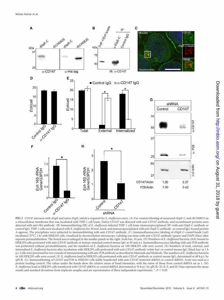
FIG 2 CD147 interacts with rEtpE and native EtpE, which is required for E. chaffeensis entry. (A) Far-western blotting of renatured rEtpE-C and rECH0825 ona nitrocellulose membrane that was incubated with THP-1 cell lysate. Native CD147 was detected with anti-CD147 antibody, and recombinant proteins weredetected with anti-His antibody. (B) Immunoblotting (IB) of E. chaffeensis-infected THP-1 cell lysate immunoprecipitated (IP) with anti-EtpE-C antibody orcontrol IgG. THP-1 cells were incubated with E. chaffeensis for 30 min, lysed, and immunoprecipitated with anti-EtpE-C antibody- or control IgG-bound proteinA-agarose. The precipitates were subjected to immunoblotting with anti-CD147 antibody. (C) Immunofluorescence labeling of rEtpE-C-coated beads (red)incubated (37°C, 1 h) with HEK293 cells, visualized by deconvolution microscopy. Labeling was done with anti-CD147 antibody (green) and DAPI (blue) aftersaponin permeabilization. The boxed area is enlarged in the smaller panels to the right. Scale bar, 10 �m. (D) Numbers of E. chaffeensis bacteria (Ech) bound toHEK293 cells pretreated with anti-CD147 antibody or isotype-matched control mouse IgG at 30 min p.i. Immunofluorescence labeling with anti-P28 antibodywas performed without permeabilization, and the numbers of E. chaffeensis bacteria on 100 HEK293 cells were scored. (E) Numbers of total, external, andinternalized E. chaffeensis bacteria after incubation with HEK293 cells pretreated with anti-CD147 antibody (white bar) or control mouse IgG (black bar) at 1 hp.i. Cells were processed for two rounds of immunostaining with anti-P28 antibody as described in Materials and Methods. The numbers of E. chaffeensis bacteriain 100 HEK293 cells were scored. (F) E. chaffeensis load in HEK293 cells pretreated with anti-CD147 antibody or control mouse IgG, determined at 48 h p.i. byqPCR. (G) Immunoblotting of CD147 and P28 in HEK293 cells stably transfected with anti-CD147 lentiviral shRNA or control shRNA. Actin was used as aprotein loading control. The values under the bands show the relative ratios of band intensities, with the ratios of those from control shRNA set as 1. (H)E. chaffeensis load in HEK293 cells transfected with CD147 shRNA or control shRNA determined at 76 h p.i. by qPCR. (D, E, F, and H) Data represent the meanresults and standard deviations from triplicate samples and are representative of three independent experiments. *, P � 0.05.
Mohan Kumar et al.
4 ® mbio.asm.org November/December 2015 Volume 6 Issue 6 e01541-15
on August 31, 2018 by guest
http://mbio.asm
.org/D
ownloaded from

N-WASP and thereby stabilizes its autoinhibited closed confor-mation (33). After pretreating DH82 cells with wiskostatin for30 min and then removing the wiskostatin, E. chaffeensis wasadded, and the bacterial load was determined at 50 h p.i. Although
pretreatment with 100 nM or 1 �M wiskostatin had little effect, aconcentration of 10 �M resulted in nearly complete inhibition ofinfection compared with that in the dimethyl sulfoxide (DMSO)solvent control, suggesting that N-WASP activation is required for
FIG 3 hnRNP-K is required for Ehrlichia entry and infection. (A) Immunofluorescence labeling of HEK293 cells transfected with iAb-47 plasmid or controlplasmid at 48 h, with saponin permeabilization. Labeling was done with anti-hnRNP-K antibody (red) and DAPI (blue). (B) E. chaffeensis (Ech) bound toHEK293 cells transfected with iAb-47 or control plasmid at 30 min p.i. Immunofluorescence labeling with anti-P28 antibody (red) and DAPI (blue) wasperformed without permeabilization. (C) Quantification of the results from an experiment similar to the one whose results are shown in panel B, done bycounting the E. chaffeensis bacteria in 100 HEK293 cells. (D) E. chaffeensis bacteria internalized by HEK293 cells transfected with iAb-47 or control plasmid at 2 hp.i. Cells were processed for two rounds of immunostaining with anti-P28 antibody, the first without permeabilization to detect external E. chaffeensis (red), andthe second with saponin permeabilization to detect total E. chaffeensis bacteria (green). Boxed areas are enlarged to the right. N, nucleus. (E) Quantification ofthe results from an experiment similar to the one whose results are shown in panel D, done by counting E. chaffeensis in 100 HEK293 cells. (F) Immunoblottingof P28 in HEK293 cells transfected with iAb-47 plasmid or the control plasmid at 56 h p.i. Host actin was used as a protein loading control. The values under thebands show the relative ratios of band intensities, with the ratios of those from control plasmid set as 1. (G) Quantification of the results from an experimentsimilar to the one whose results are shown in panel F, done by qPCR. (A, B, and D) Deconvolution microscopy. Scale bar, 5 �m; Merge/DIC, image merged withdifferential interference contrast. (C, E, and G) Data represent the mean results and standard deviations from triplicate samples and are representative of threeindependent experiments. *, P � 0.05.
CD147 and hnRNP-K Link EtpE-DNase X for Entry
November/December 2015 Volume 6 Issue 6 e01541-15 ® mbio.asm.org 5
on August 31, 2018 by guest
http://mbio.asm
.org/D
ownloaded from
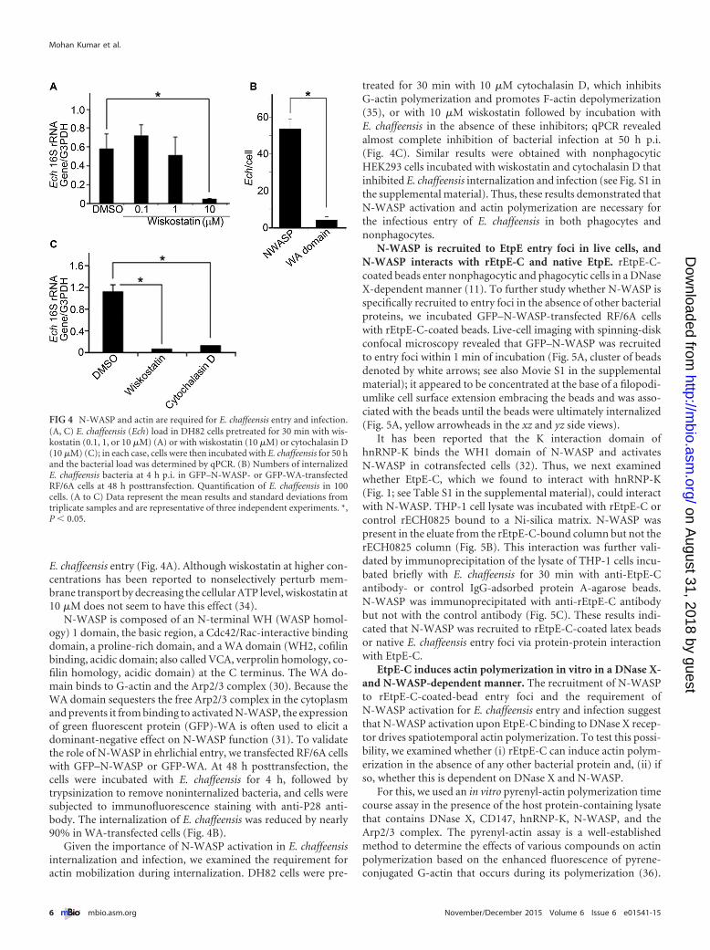
E. chaffeensis entry (Fig. 4A). Although wiskostatin at higher con-centrations has been reported to nonselectively perturb mem-brane transport by decreasing the cellular ATP level, wiskostatin at10 �M does not seem to have this effect (34).
N-WASP is composed of an N-terminal WH (WASP homol-ogy) 1 domain, the basic region, a Cdc42/Rac-interactive bindingdomain, a proline-rich domain, and a WA domain (WH2, cofilinbinding, acidic domain; also called VCA, verprolin homology, co-filin homology, acidic domain) at the C terminus. The WA do-main binds to G-actin and the Arp2/3 complex (30). Because theWA domain sequesters the free Arp2/3 complex in the cytoplasmand prevents it from binding to activated N-WASP, the expressionof green fluorescent protein (GFP)-WA is often used to elicit adominant-negative effect on N-WASP function (31). To validatethe role of N-WASP in ehrlichial entry, we transfected RF/6A cellswith GFP–N-WASP or GFP-WA. At 48 h posttransfection, thecells were incubated with E. chaffeensis for 4 h, followed bytrypsinization to remove noninternalized bacteria, and cells weresubjected to immunofluorescence staining with anti-P28 anti-body. The internalization of E. chaffeensis was reduced by nearly90% in WA-transfected cells (Fig. 4B).
Given the importance of N-WASP activation in E. chaffeensisinternalization and infection, we examined the requirement foractin mobilization during internalization. DH82 cells were pre-
treated for 30 min with 10 �M cytochalasin D, which inhibitsG-actin polymerization and promotes F-actin depolymerization(35), or with 10 �M wiskostatin followed by incubation withE. chaffeensis in the absence of these inhibitors; qPCR revealedalmost complete inhibition of bacterial infection at 50 h p.i.(Fig. 4C). Similar results were obtained with nonphagocyticHEK293 cells incubated with wiskostatin and cytochalasin D thatinhibited E. chaffeensis internalization and infection (see Fig. S1 inthe supplemental material). Thus, these results demonstrated thatN-WASP activation and actin polymerization are necessary forthe infectious entry of E. chaffeensis in both phagocytes andnonphagocytes.
N-WASP is recruited to EtpE entry foci in live cells, andN-WASP interacts with rEtpE-C and native EtpE. rEtpE-C-coated beads enter nonphagocytic and phagocytic cells in a DNaseX-dependent manner (11). To further study whether N-WASP isspecifically recruited to entry foci in the absence of other bacterialproteins, we incubated GFP–N-WASP-transfected RF/6A cellswith rEtpE-C-coated beads. Live-cell imaging with spinning-diskconfocal microscopy revealed that GFP–N-WASP was recruitedto entry foci within 1 min of incubation (Fig. 5A, cluster of beadsdenoted by white arrows; see also Movie S1 in the supplementalmaterial); it appeared to be concentrated at the base of a filopodi-umlike cell surface extension embracing the beads and was asso-ciated with the beads until the beads were ultimately internalized(Fig. 5A, yellow arrowheads in the xz and yz side views).
It has been reported that the K interaction domain ofhnRNP-K binds the WH1 domain of N-WASP and activatesN-WASP in cotransfected cells (32). Thus, we next examinedwhether EtpE-C, which we found to interact with hnRNP-K(Fig. 1; see Table S1 in the supplemental material), could interactwith N-WASP. THP-1 cell lysate was incubated with rEtpE-C orcontrol rECH0825 bound to a Ni-silica matrix. N-WASP waspresent in the eluate from the rEtpE-C-bound column but not therECH0825 column (Fig. 5B). This interaction was further vali-dated by immunoprecipitation of the lysate of THP-1 cells incu-bated briefly with E. chaffeensis for 30 min with anti-EtpE-Cantibody- or control IgG-adsorbed protein A-agarose beads.N-WASP was immunoprecipitated with anti-rEtpE-C antibodybut not with the control antibody (Fig. 5C). These results indi-cated that N-WASP was recruited to rEtpE-C-coated latex beadsor native E. chaffeensis entry foci via protein-protein interactionwith EtpE-C.
EtpE-C induces actin polymerization in vitro in a DNase X-and N-WASP-dependent manner. The recruitment of N-WASPto rEtpE-C-coated-bead entry foci and the requirement ofN-WASP activation for E. chaffeensis entry and infection suggestthat N-WASP activation upon EtpE-C binding to DNase X recep-tor drives spatiotemporal actin polymerization. To test this possi-bility, we examined whether (i) rEtpE-C can induce actin polym-erization in the absence of any other bacterial protein and, (ii) ifso, whether this is dependent on DNase X and N-WASP.
For this, we used an in vitro pyrenyl-actin polymerization timecourse assay in the presence of the host protein-containing lysatethat contains DNase X, CD147, hnRNP-K, N-WASP, and theArp2/3 complex. The pyrenyl-actin assay is a well-establishedmethod to determine the effects of various compounds on actinpolymerization based on the enhanced fluorescence of pyrene-conjugated G-actin that occurs during its polymerization (36).
FIG 4 N-WASP and actin are required for E. chaffeensis entry and infection.(A, C) E. chaffeensis (Ech) load in DH82 cells pretreated for 30 min with wis-kostatin (0.1, 1, or 10 �M) (A) or with wiskostatin (10 �M) or cytochalasin D(10 �M) (C); in each case, cells were then incubated with E. chaffeensis for 50 hand the bacterial load was determined by qPCR. (B) Numbers of internalizedE. chaffeensis bacteria at 4 h p.i. in GFP–N-WASP- or GFP-WA-transfectedRF/6A cells at 48 h posttransfection. Quantification of E. chaffeensis in 100cells. (A to C) Data represent the mean results and standard deviations fromtriplicate samples and are representative of three independent experiments. *,P � 0.05.
Mohan Kumar et al.
6 ® mbio.asm.org November/December 2015 Volume 6 Issue 6 e01541-15
on August 31, 2018 by guest
http://mbio.asm
.org/D
ownloaded from

Incubation of soluble rEtpE-C with THP-1 lysate indeed inducedpolymerization of pyrenyl-actin (Fig. 6). This actin polymeriza-tion specifically required the C terminus of EtpE, because recom-binant N-terminal EtpE (rEtpE-N), which does not bind or in-duce the entry of coated beads into nonphagocytes (11), did notinduce actin polymerization (Fig. 6). The effect of rEtpE-C onactin polymerization was abrogated by the addition of wiskosta-tin, indicating the requirement for N-WASP activation in EtpE-C-induced actin polymerization in vitro (Fig. 6). To determine theDNase X dependency of actin polymerization, we used the celllysate of wild-type or DNase X�/� mouse BMDMs rather than
THP-1 lysate. rEtpE-C-induced actin polymerization requiredDNase X, because the lysate of DNase X�/� BMDMs, which lacksDNase X, failed to elicit actin polymerization, whereas the lysate ofcongenic wild-type BMDMs elicited a polymerization profile sim-ilar to that of the THP-1 lysate (Fig. 6). Thus, EtpE-C inducedactin polymerization in the absence of any other bacterial proteinsand this is dependent on DNase X and N-WASP.
Taken together, these results imply that E. chaffeensis infectiousentry is induced by EtpE binding to DNase X, with subsequentrecruitment of CD147 and hnRNP-K to the entry foci, followed byspatiotemporal mobilization and activation of N-WASP (most
FIG 5 N-WASP is recruited to rEtpE-C-coated-bead entry foci, and it interacts with rEtpE-C and native EtpE. (A) Spinning-disk confocal microscopy imagesat four different time points from the three-dimensional time-lapse video of RF/6A cells transfected with GFP–N-WASP (cyan) and then incubated withrEtpE-C-coated beads (red). xy planes show the upper surface of an RF/6A cell that had internalized rEtpE-C-coated beads. Each white arrow indicates a clusterof N-WASP. Cell surface extensions surrounding the cluster of beads are highlighted with yellow arrows in the side views (xz and yz planes). Scale bar, 10 �m. (B)Affinity pulldown of THP-1 cell lysate incubated with rEtpE-C or rECH0825 bound to Ni-silica matrix. Bound proteins were eluted with imidazole and subjectedto immunoblot analysis with anti-N-WASP and anti-His antibodies. One lane between rEtpE-C and rECH0825 was deleted to save the space without alteringmolecular mass or exposure time. (C) THP-1 cells were incubated with E. chaffeensis for 30 min, lysed, and immunoprecipitated (IP) with anti-EtpE-C antibody-or control IgG-bound protein A-agarose. The immunoprecipitates were subjected to immunoblotting with anti-N-WASP antibody. (B and C) Numbers to theleft indicate kDa.
CD147 and hnRNP-K Link EtpE-DNase X for Entry
November/December 2015 Volume 6 Issue 6 e01541-15 ® mbio.asm.org 7
on August 31, 2018 by guest
http://mbio.asm
.org/D
ownloaded from

likely mediated by hnRNP-K) at the entry foci and spatiotemporalactin polymerization promoted by the activated N-WASP.
DISCUSSION
The present study reveals that EtpE (more specifically EtpE-C), anE. chaffeensis surface invasin that directly binds to the nontrans-membrane GPI-anchored protein DNase X (11), can induce actinpolymerization in an N-WASP activation-dependent manner. Inintact cells, the cell surface EtpE-DNase X complex and cytoplas-mic hnRNP-K and N-WASP are topologically separated by theplasma membrane, and thus, it is likely that the transmembraneglycoprotein CD147, which is recruited to entry foci, is responsi-ble for transducing the extracellular EtpE-DNase X binding sig-nals across the plasma membrane to engage hnRNP-K and acti-vate N-WASP-dependent actin polymerization to drive Ehrlichiaentry. This is a previously unknown signaling pathway for host celluptake/infection of obligate intracellular bacteria.
The only known physiologic function of DNase X is the uptakeand degradation of exogenous DNA (37). The present study dem-onstrates for the first time the molecular mechanism by whichDNase X mediates endocytosis by mammalian cells. This new in-formation suggests that interaction between CD147 (38) andDNase X (37), both of which are present in plasma membranelipid rafts, may facilitate ehrlichial uptake. This CD147-mediatedmechanism of E. chaffeensis entry may be related to other events ofactin dynamics, as CD147 has been reported to colocalize withF-actin and promote cytoskeletal rearrangements (39, 40) and themalarial ligand PfRh5 directly binds to CD147, which leads toparasite entry into erythrocytes (41).
The present work is the first to demonstrate the involvement ofhnRNP-K in the host cell entry of any pathogen or molecule. Re-combinant glutathione S-transferase (GST)– hnRNP-K was pre-viously shown to activate actin polymerization in an in vitropyrenyl-actin polymerization assay in the presence of the bovine
Arp2/3 complex and GST–N-WASP (32), suggesting thathnRNP-K links the EtpE-C–DNase X signal to N-WASP activa-tion and actin dynamics. hnRNP-K is involved in tumor metasta-sis and wound healing (28, 42), but its relevant signaling pathwaysare insufficiently defined. Therefore, our finding may help thediscovery of upstream regulators of hnRNP-K that elicit actin-based cytoskeletal reorganization, potentially unifying our under-standing of intracellular bacterial infection, tumor metastasis, andwound healing.
The involvement of hnRNP-K in ehrlichial entry is distinctfrom the classic mechanism of facultative intracellular bacterialentry, which involves the activation of Rho family GTPases anddownstream factors to promote actin polymerization, eitherthrough the secretion of type III effectors (e.g., Salmonella SopE,SopE2, and SptP) or direct binding of a bacterial ligand to itscognate host cell receptor with a known role in endogenous sub-strate adhesion and signaling (e.g., Listeria InlB/hepatocytegrowth factor and internalin A/E-cadherin and Yersinia inva-sin/�1 integrin) (43). It remains to be determined, however,whether the activation of Rho family GTPases has any role inehrlichial entry.
The mammalian WASP/WAVE family contains five members,namely, WASP, N-WASP, WAVE1, WAVE2, and WAVE3, whichintegrate signaling cascades that lead to Arp2/3-dependent actinpolymerization (44). N-WASP and WAVE2 are ubiquitously ex-pressed, whereas WASP is expressed exclusively in leukocytes andWAVE1 and WAVE3 are enriched in the brain (45). The entry ofListeria, Yersinia, Salmonella, and Chlamydia bacteria intononphagocytes depends on the Rac/WAVE complex pathway(46–49). On the other hand, phagocytosis of an avirulent strain ofYersinia pseudotuberculosis occurs through the activation ofN-WASP (50); moreover, a type III secretion effector, Yop2b,which is inoculated into target cells, is critical for evading phago-cytosis (51). N-WASP is involved in the internalization of Candiainto endothelial cells (52). hnRNP-K binds to the WH1 domain ofN-WASP (32), and ehrlichial hnRNP-K-dependent infectious en-try likely has coopted N-WASP rather than WAVE2, as the latterlacks a WH1 domain (53).
EtpE-C induced actin nucleation in vitro in a DNase X- andN-WASP-dependent manner. This is distinct from several bacte-rial proteins that are known to promote actin nucleation for actin-based motility by mimicking Cdc42, N-WASP, or Arp2/3 func-tions, such as Shigella autotransporter IcsA (31, 54), Listeria ActA(55), the spotted fever group Rickettsia WASP-like protein RickAand Sca2 autotransporter (56), SipC of Salmonella (57), and chla-mydial Tarp, a type III secretion effector (58).
E. chaffeensis tandem-repeat protein 120 (TRP120) (glycopro-tein 120 [gp120]) was the first protein proposed to mediateE. chaffeensis invasion of human cells, because Escherichia colitransformed with a plasmid encoding TRP120 can invade HeLacells (59). Antibodies to TRP120 can reduce the E. chaffeensis loadin vitro and in mice (60). In addition, an antibody against theE. chaffeensis surface-exposed lipoprotein OmpA inhibitsE. chaffeensis binding to and infection of THP-1 cells (10). Immu-nization of mice with recombinant P28, which is the E. chaffeensismajor outer membrane �-barrel protein (27) and functions as aporin (61), protects mice from E. chaffeensis challenge (27). Fur-thermore, immunization of mice with Ehrlichia muris P28 confersprotection from E. muris challenge (62). TRP120 is one of the
FIG 6 EtpE-C induces actin polymerization in vitro in an N-WASP- andDNase X-dependent manner. The results of a fluorometric assay of an in vitropyrenyl-actin polymerization time course are shown. The assay was performedin the presence of pyrenyl G-actin and the THP-1 lysate by adding rEtpE-C,rEtpE-N, or rEtpE-C plus 10 �M wiskostatin at 0 h or in the presence ofpyrenyl G-actin and mouse wild-type or DNase X�/� BMDM lysate by addingrEtpE-C at 0 h. The relative fluorescence of pyrenyl F-actin in each reactionmixture was plotted versus time.
Mohan Kumar et al.
8 ® mbio.asm.org November/December 2015 Volume 6 Issue 6 e01541-15
on August 31, 2018 by guest
http://mbio.asm
.org/D
ownloaded from

proteins on the surface of isolated E. chaffeensis cells, the other twoproteins being OmpA and VirB6-2, which are selectively degradedby E. chaffeensis HtrA protease upon treatment with the cell-permeable functional antagonist of cyclic-di-GMP; theE. chaffeensis transmembrane outer membrane proteins P28/Omp-1F, VirB9, and heat shock protein 60 (HSP60) were notdegraded by the treatment in the same sample (63). Because treat-ment of E. chaffeensis with the antagonist of cyclic-di-GMP blocksE. chaffeensis entry but not binding (63), this result suggests thatTRP120, OmpA, and VirB6-2 are not adhesins. Although some ofthese Ehrlichia surface proteins may mediate bacterial entry intocells, clear evidence for this has not been reported, and their re-spective human cell receptors and signaling pathways have notbeen identified.
Figure 7 presents our model for the mechanism underlying theinfectious entry of Ehrlichia. Upon EtpE binding to DNase X, thelateral redistribution of DNase X within lipid rafts brings CD147,which is enriched in lipid rafts, into association with the EtpE-DNase X complex. Because CD147 is a single-pass transmem-brane protein, it can relay a signal to the cytoplasmic side of thecell membrane and recruit hnRNP-K, which may culminate inN-WASP activation, actin polymerization, and filopodial exten-sion that surrounds the bacterium, leading to its uptake. Althoughfurther studies are necessary to determine detailed mechanisms ofassociations between these host cell proteins involved in facilitat-ing Ehrlichia entry, the present study provides insight into aunique signaling pathway for EtpE-mediated infectious entry ofan obligate intracellular bacterium.
MATERIALS AND METHODSEthics statement. BMDMs were established from wild-type and DNaseX�/� C57BL/6 mice as described previously (11) in accordance with TheOhio State University Institutional Animal Care and Use Committeeguidelines under approved e-protocol number 2009A0186. The Univer-sity program has Full Continued Accreditation by the Association forAssessment and Accreditation of Laboratory Animal Care International(AAALAC-I), number 000028, dated 6 February 2015, and has PublicHealth Services assurance renewal number A3261-01 through 28 Febru-ary 2019. The program is licensed by the USDA, 31-R-014, and is in fullcompliance with Animal Welfare Regulations until 1 August 2017.
Bacterial and host cell culture. E. chaffeensis type strain Arkansas waspropagated in DH82 cells, and host cell-free E. chaffeensis was obtained bycontrolled sonication as described previously (11). HEK293, RF/6A,THP-1 cells, and BMDMs were cultured as described previously (11).
Plasmids, recombinant proteins, and antibodies. The plasmids usedin the study, including those encoding GFP–N-WASP, GFP-WA, andiAb-47 (28), were purified with an EndoFree kit (Qiagen) and transfectedusing FuGene HD (Promega). The recombinant proteins rEtpE-C,rEtpE-N, and rECH0825 were prepared as described previously (11). Theprimary antibodies used in the study were mouse anti-EtpE-C antibody(11), rabbit anti-DNase X antibody (Abcam), anti-CD147 MAb (MEM-M6/6; Santa Cruz Biotechnology and Abcam), rabbit anti-N-WASP anti-body (Santa Cruz Biotechnology), anti-hnRNP-K MAb (Santa Cruz Bio-technology), rabbit anti-actin antibody (Sigma), and rabbit anti-E. chaffeensis P28 antibody (27). The secondary antibodies wereperoxidase-conjugated goat anti-mouse or anti-rabbit antibody (KPL)and goat anti-mouse or anti-rabbit IgG (Invitrogen) conjugated with Al-exa Fluor 488 (AF488) or AF555 (Invitrogen). DAPI (4=,6-diamidino-2-phenylindole) (Sigma) was used to stain E. chaffeensis DNA and/or hostcell DNA (nuclei).
Affinity pulldown and LC-MS/MS. For protein pulldown, histidine(His)-tagged rEtpE-C was bound to and renatured on a Ni-silica matrix(Promega). THP-1 cell lysate in NP-40 lysis buffer (150 mM NaCl, 50 mMTris-HCl, pH 7.4, 1% [wt/vol] NP-40, supplemented with 1% proteaseinhibitor cocktail set III [Calbiochem]) was applied to rEtpE-C-bound orcontrol matrix and incubated for 8 h at 4°C. Unbound proteins werewashed from the matrix with 50 mM sodium phosphate buffer (pH 7.4)containing 0.3 M NaCl and 1% (wt/vol) NP-40, followed by 15 mlphosphate-buffered saline (137 mM NaCl, 2.7 mM KCl, 8.1 mMNa2HPO4, 1 mM KH2PO4, pH 7.4) containing 10 mM imidazole and 1%NP-40. rEtpE-C and the bound protein complex were eluted with sodiumphosphate buffer containing 0.3 M NaCl and 250 mM imidazole. Theeluate and the postelution Ni-silica matrix were resuspended in 2� SDSsample buffer and subjected to SDS-PAGE. The gel was fixed and stainedwith GelCode blue (Thermo Fisher Scientific), and conspicuously differ-ent bands were cut out, trypsin digested, and subjected to LC-MS/MS atthe Mass Spectrometry and Proteomics Core Facility at The Ohio StateUniversity. Sequence information from the MS/MS data was processedwith Mascot Distiller to form a peak list and with the MASCOT MS/MSsearch engine and Turbo SEQUEST algorithm in BioWorks 3.1 software.Aliquots of samples were also subjected to immunoblotting with anti-DNase X antibody.
Far-Western blotting and coimmunoprecipitation. rEtpE-C andrECH0825 (5 �g) were separated with SDS-PAGE, transferred to a nitro-cellulose membrane, and renatured with serial guanidine-HCl treatment,followed by incubation with THP-1 cell lysate in NP-40 lysis buffer (11).After stringent washing, the membrane was incubated with anti-CD147antibody, followed by peroxidase-conjugated goat anti-mouse IgG. Themembrane was stripped with Restore Western blot stripping buffer(Thermo Fisher Scientific) and reprobed with peroxidase-conjugatedanti-His MAb (Sigma). THP-1 cells were incubated with E. chaffeensis for30 min and lysed in NP-40 lysis buffer. The lysate was immunoprecipi-tated with anti-EtpE-C antibody (2 �g)- or control mouse IgG (2 �g)-bound protein A-agarose beads. The precipitate was resuspended in 2�
FIG 7 Model for the mechanism of DNase X-mediated E. chaffeensis entry viainteraction with CD147 and hnRNP-K and the subsequent N-WASP-dependent actin polymerization. (1) Extracellular E. chaffeensis uses theC-terminal region of its surface protein EtpE to bind to DNase X on the hostcell surface. (2) Upon EtpE binding to DNase X, the lateral redistribution ofDNase X within dynamic lipid rafts brings CD147 into association with theEtpE-DNase X complex. (3) CD147 potentially relays the signal to the cyto-plasmic side of the cell membrane and recruits hnRNP-K to bind N-WASP,leading to activation of N-WASP (conformational change). (4) ActivatedN-WASP binds Arp2/3 actin nucleation complex. (5) This leads to spatiotem-poral actin polymerization and filopodium formation to internalizeE. chaffeensis into endosomes.
CD147 and hnRNP-K Link EtpE-DNase X for Entry
November/December 2015 Volume 6 Issue 6 e01541-15 ® mbio.asm.org 9
on August 31, 2018 by guest
http://mbio.asm
.org/D
ownloaded from
SDS sample buffer and subjected to immunoblotting with anti-CD147and anti-N-WASP antibodies.
CD147 neutralization and RNA interference and iAb-47 functionalablation. E. chaffeensis was added to HEK293 cells preincubated with10 �g/ml anti-CD147 MEM-M6/6 MAb (low endotoxin, azide free; Ab-cam) or control mouse MAb for 30 min at 25°C in serum-free Dulbecco’smodified Eagle medium (DMEM). Binding, internalization, and infectionwere determined at 30 min, 1 h, and 48 h p.i., respectively. HEK293 cells in24-well plates were transduced with 1 � 106 transducing units of CD147shRNA lentiviral transduction particles (Sigma) using 8 �g/ml Polybrene,and resistant cells were selected with three rounds of puromycin selection(2 �g/ml). The resistant clones were replated, lysed with NP-40 lysis buf-fer, and used to estimate the level of CD147 with immunoblotting. TheCD147 levels were normalized to the actin levels. Cells were incubatedwith E. chaffeensis for 30 min, 1 h, or 76 h to study binding, internalization,and infection, respectively. Coverslip cultures of HEK293 cells were trans-fected with 0.5 �g iAb-47 or control pEGFP-N1 plasmid. At 48 h post-transfection, cells were harvested, washed, fixed, and immunostained tostudy the expression pattern of hnRNP-K. Cells were incubated with hostcell-free E. chaffeensis for 30 min, 2 h, or 56 h to study binding, internal-ization, and infection, respectively.
Inhibitors of actin dynamics. DH82 or HEK293 cells in DMEM wereincubated with wiskostatin (Sigma) at 0.1 to 10 �M and cytochalasin D(Calbiochem) at 10 �M for 30 min. The cells were washed and replenishedwith complete DMEM prior to incubation with freshly isolated host cell-free E. chaffeensis. At 2 h p.i., the cells were washed to remove unbound orloosely attached bacteria and further incubated for 48 h without chemi-cals. For the internalization and infection study in HEK293 cells, cells werepretreated for 30 min with 2.5 �M cytochalasin D or 5 �M wiskostatin,followed by incubation with host cell-free E. chaffeensis. After removal ofchemicals by washing, the cells were cultured for an additional 1 and 48 h,respectively, for internalization and infection. To assess the role ofN-WASP in bacterial internalization, RF/6A cells were transfected withGFP–N-WASP or GFP-WA. At 48 h posttransfection, cells were incu-bated with host cell-free E. chaffeensis at a multiplicity of infection of 50 for4 h.
Binding, internalization, and infection assays. For the binding assay,samples were fixed with 3.5% paraformaldehyde and subjected to oneround of immunolabeling with anti-P28 antibody, whereas for internal-ization assays, two steps of labeling of fixed cells with anti-P28 antibodywere carried out: the first labeling step was performed without saponinpermeabilization to detect bound but not internalized E. chaffeensis usinganti-P28 antibody and AF488-conjugated anti-rabbit IgG, and the secondlabeling step was performed after permeabilization with saponin to detecttotal E. chaffeensis bacteria using AF555-conjugated anti-rabbit IgG (11).Fluorescence images were acquired using a DeltaVision deconvolutionmicroscope system (GE Healthcare). The number of bacteria on 100 cellswas scored. Overall infection was determined at 48 to 76 h p.i. by immu-noblotting with anti-P28 antibody or qPCR, using the E. chaffeensis 16SrRNA gene normalized to the host cell glycerol-3-phosphate dehydroge-nase (G3PDH) gene (10).
Cellular localization analysis. Sulfate-modified fluorescent red poly-styrene beads (0.5-�m diameter; Sigma) were coated with rEtpE-C asdescribed previously (11). Freshly prepared protein-coated beads wereadded to HEK293 cells at a ratio of approximately 50 beads per cell andincubated for 1 h at 37°C. Cells were fixed, permeabilized, and labeledwith mouse anti-CD147 MAb and AF488-conjugated goat anti-mouseIgG.
Live-cell imaging by spinning-disk confocal microscopy. RF/6A cellswere transfected with GFP–N-WASP, and at 23 h posttransfection, freshphenol red-free Leibovitz’s L-15 medium supplemented with 10% fetalbovine serum was added, and cells were incubated with flash red beads(0.51-�m diameter; Bangs Laboratories) coated with rEtpE-C (11). Sam-ples were moved to a live-cell imaging chamber at 37°C connected to aTI-E inverted research microscope (Nikon Instruments) controlled by
Nikon Elements software and equipped with a spinning-disk confocalunit (Yokogawa Electric), a 100� objective lens (Plan ApochromatLambda, NA 1.45; Nikon), and an electron multiplying charge-coupleddevice (EMCCD) camera (iXon DU897 Ultra; Andor Technology).Three-dimensional confocal image stacks were acquired at 1-min inter-vals for 2 h, with a step size of 0.1 �m along the z axis. The bead internal-ization movie (see Movie S1 in the supplemental material) was preparedwith ImageJ software by creating a stack of confocal images acquired at theupper surface of an RF/6A cell.
Pyrenyl-actin polymerization assay. The actin polymerization assaywas performed according to the manufacturer’s protocol (Cytoskeleton,Inc.). Purified rEtpE-C and rEtpE-N at 20 �g were used for the assay. Thebaseline fluorescence emitted by pyrenyl-actin in G-actin buffer (Cyto-skeleton, Inc.) or G buffer blank was measured at 37°C for 3 min. Controlbuffer, rEtpE-N, or rEtpE-C was added to the wells in the presence ofTHP-1 cell lysate or wild-type or DNase X�/� BMDM lysate with orwithout 10 �M wiskostatin in a total volume of 200 �l. Fluorescence wasmeasured every 30 s for 30 min using the medium photomultiplier settingwith an excitation wavelength of 355 nm, an emission wavelength of430 nm, and a cutoff of 420 nm. At the end of the reading, 10� actinpolymerization buffer (Cytoskeleton, Inc.) was added to all wells, and thefluorescence was read again to verify that the assay conditions were ap-propriate to detect polymerization. The raw data obtained were used togenerate a polymerization time course plot.
Statistical analysis. Statistical analysis was performed with an un-paired, two-tailed Student’s t test. A P value of �0.05 was consideredsignificant.
SUPPLEMENTAL MATERIALSupplemental material for this article may be found at http://mbio.asm.org/lookup/suppl/doi:10.1128/mBio.01541-15/-/DCSupplemental.
Movie S1, AVI file, 12.2 MB.Figure S1, TIF file, 1.3 MB.Table S1, DOCX file, 0.01 MB.
ACKNOWLEDGMENTS
We thank Renu Wadhwa (AIST, Tsukuba, Japan) for the iAb-47 plasmidand Michael Way (Cancer Research, United Kingdom) for providingGFP–N-WASP and GFP-WA plasmids. We thank Tim Vojt for assistancein preparing Fig. 7.
REFERENCES1. CDC. 2015. Table II. Provisional cases of selected notifiable diseases,
United States, weeks ending January 3, 2015 and December 28, 2013 (53rdweek). MMWR Morb Mortal Wkly Rep 63(53):ND-733-ND-746. http://www.cdc.gov/mmwr/preview/mmwrhtml/mm6353md.htm?s_cid�mm6353md_w#tab2.
2. Paddock CD, Childs JE. 2003. Ehrlichia chaffeensis: a prototypical emerg-ing pathogen. Clin Microbiol Rev 16:37– 64. http://dx.doi.org/10.1128/CMR.16.1.37-64.2003.
3. Paddock CD, Yabsley MJ. 2007. Ecological havoc, the rise of white-taileddeer, and the emergence of Amblyomma americanum-associated zoonosesin the United States. Curr Top Microbiol Immunol 315:289 –324. http://dx.doi.org/10.1007/978-3-540-70962-6_12.
4. Committee on Lyme Disease and Other Tick-Borne Diseases: The Stateof the Science; Institute of Medicine. 2011. Critical needs and gaps inunderstanding prevention, amelioration, and resolution of Lyme andother tick-borne diseases: the short-term and long-term outcomes. Work-shop report. National Academies Press, Washington, DC. http://www.nap.edu/read/13134/chapter/1.
5. Lin M, Rikihisa Y. 2003. Obligatory intracellular parasitism by Ehrlichiachaffeensis and Anaplasma phagocytophilum involves caveolae andglycosylphosphatidylinositol-anchored proteins. Cell Microbiol5:809 – 820. http://dx.doi.org/10.1046/j.1462-5822.2003.00322.x.
6. Mott J, Barnewall RE, Rikihisa Y. 1999. Human granulocytic ehrlichiosisagent and Ehrlichia chaffeensis reside in different cytoplasmic compart-ments in HL-60 cells. Infect Immun 67:1368 –1378.
7. Lin M, Rikihisa Y. 2007. Degradation of p22phox and inhibition of su-
Mohan Kumar et al.
10 ® mbio.asm.org November/December 2015 Volume 6 Issue 6 e01541-15
on August 31, 2018 by guest
http://mbio.asm
.org/D
ownloaded from

peroxide generation by Ehrlichia chaffeensis in human monocytes. CellM i c r o b i o l 9 : 8 6 1 – 8 7 4 . h t t p : / / d x . d o i . o r g / 1 0 . 1 1 1 1 / j . 1 4 6 2-5822.2006.00835.x.
8. Barnewall RE, Rikihisa Y, Lee EH. 1997. Ehrlichia chaffeensis inclusionsare early endosomes which selectively accumulate transferrin receptor.Infect Immun 65:1455–1461.
9. Lin M, Zhu MX, Rikihisa Y. 2002. Rapid activation of protein tyrosinekinase and phospholipase C-gamma2 and increase in cytosolic free cal-cium are required by Ehrlichia chaffeensis for internalization and growth inTHP-1 cells. Infect Immun 70:889 – 898. http://dx.doi.org/10.1128/IAI.70.2.889-898.2002.
10. Cheng Z, Miura K, Popov VL, Kumagai Y, Rikihisa Y. 2011. Insightsinto the CtrA regulon in development of stress resistance in obligatoryintracellular pathogen Ehrlichia chaffeensis. Mol Microbiol 82:1217–1234.http://dx.doi.org/10.1111/j.1365-2958.2011.07885.x.
11. Mohan Kumar D, Yamaguchi M, Miura K, Lin M, Los M, Coy JF,Rikihisa Y. 2013. Ehrlichia chaffeensis uses its surface protein EtpE to bindGPI-anchored protein DNase X and trigger entry into mammalian cells.PLoS Pathog 9:e1003666.
12. Maeda K, Markowitz N, Hawley RC, Ristic M, Cox D, McDade JE. 1987.Human infection with Ehrlichia canis, a leukocytic rickettsia. N Engl J Med316:853– 856. http://dx.doi.org/10.1056/NEJM198704023161406.
13. Dawson JE, Anderson BE, Fishbein DB, Sanchez JL, Goldsmith CS,Wilson KH, Duntley CW. 1991. Isolation and characterization of anEhrlichia sp. from a patient diagnosed with human ehrlichiosis. J ClinMicrobiol 29:2741–2745.
14. Barnewall RE, Rikihisa Y. 1994. Abrogation of gamma interferon-induced inhibition of Ehrlichia chaffeensis infection in human monocyteswith iron-transferrin. Infect Immun 62:4804 – 4810.
15. Allison AC, Davies P, De Petris S. 1971. Role of contractile microfila-ments in macrophage movement and endocytosis. Nat New Biol 232:153–155. http://dx.doi.org/10.1038/newbio232153a0.
16. Gottlieb TA, Ivanov IE, Adesnik M, Sabatini DD. 1993. Actin micro-filaments play a critical role in endocytosis at the apical but not the baso-lateral surface of polarized epithelial cells. J Cell Biol 120:695–710. http://dx.doi.org/10.1083/jcb.120.3.695.
17. Francis CL, Ryan TA, Jones BD, Smith SJ, Falkow S. 1993. Rufflesinduced by Salmonella and other stimuli direct macropinocytosis of bac-teria. Nature 364:639 – 642. http://dx.doi.org/10.1038/364639a0.
18. Carabeo RA, Grieshaber SS, Fischer E, Hackstadt T. 2002. Chlamydiatrachomatis induces remodeling of the actin cytoskeleton during attach-ment and entry into HeLa cells. Infect Immun 70:3793–3803. http://dx.doi.org/10.1128/IAI.70.7.3793-3803.2002.
19. Romero S, Grompone G, Carayol N, Mounier J, Guadagnini S, PrevostMC, Sansonetti PJ, Van Nhieu GT. 2011. ATP-mediated Erk1/2 activa-tion stimulates bacterial capture by filopodia, which precedes Shigella in-vasion of epithelial cells. Cell Host Microbe 9:508 –519. http://dx.doi.org/10.1016/j.chom.2011.05.005.
20. Zhang JZ, Popov VL, Gao S, Walker DH, Yu XJ. 2007. The develop-mental cycle of Ehrlichia chaffeensis in vertebrate cells. Cell Microbiol9:610 – 618. http://dx.doi.org/10.1111/j.1462-5822.2006.00812.x.
21. Iacono KT, Brown AL, Greene MI, Saouaf SJ. 2007. CD147 immuno-globulin superfamily receptor function and role in pathology. Exp MolPathol 83:283–295. http://dx.doi.org/10.1016/j.yexmp.2007.08.014.
22. Hanna SM, Kirk P, Holt OJ, Puklavec MJ, Brown MH, Barclay AN.2003. A novel form of the membrane protein CD147 that contains an extraIg-like domain and interacts homophilically. BMC Biochem 4:17. http://dx.doi.org/10.1186/1471-2091-4-17.
23. Jia L, Zhou H, Wang S, Cao J, Wei W, Zhang J. 2006. Deglycosylationof CD147 down-regulates matrix metalloproteinase-11 expression andthe adhesive capability of murine hepatocarcinoma cell HcaF in vitro.IUBMB Life 58:209 –216. http://dx.doi.org/10.1080/15216540600719580.
24. Mikula M, Dzwonek A, Karczmarski J, Rubel T, Dadlez M, Wyrwicz LS,Bomsztyk K, Ostrowski J. 2006. Landscape of the hnRNP K protein-protein interactome. Proteomics 6:2395–2406. http://dx.doi.org/10.1002/pmic.200500632.
25. Chen H, Fok KL, Jiang X, Jiang J, Chen Z, Gui Y, Chan HC, Cai Z. 2012.CD147 regulates apoptosis in mouse spermatocytes but not spermatogo-nia. Hum Reprod 27:1568 –1576. http://dx.doi.org/10.1093/humrep/des050.
26. Rao DD, Vorhies JS, Senzer N, Nemunaitis J. 2009. siRNA vs. shRNA:similarities and differences. Adv Drug Deliv Rev 61:746 –759. http://dx.doi.org/10.1016/j.addr.2009.04.004.
27. Ohashi N, Zhi N, Zhang Y, Rikihisa Y. 1998. Immunodominant majorouter membrane proteins of Ehrlichia chaffeensis are encoded by a poly-morphic multigene family. Infect Immun 66:132–139.
28. Inoue A, Sawata SY, Taira K, Wadhwa R. 2007. Loss-of-function screen-ing by randomized intracellular antibodies: identification of hnRNP-K asa potential target for metastasis. Proc Natl Acad Sci U S A 104:8983– 8988.http://dx.doi.org/10.1073/pnas.0607595104.
29. Jobling SA, Jarman C, Teh MM, Holmberg N, Blake C, Verhoeyen ME.2003. Immunomodulation of enzyme function in plants by single-domainantibody fragments. Nat Biotechnol 21:77– 80. http://dx.doi.org/10.1038/nbt772.
30. Campellone KG, Welch MD. 2010. A nucleator arms race: cellular controlof actin assembly. Nat Rev Mol Cell Biol 11:237–251. http://dx.doi.org/10.1038/nrm2867.
31. Moreau V, Frischknecht F, Reckmann I, Vincentelli R, Rabut G, Stew-art D, Way M. 2000. A complex of N-WASP and WIP integrates signallingcascades that lead to actin polymerization. Nat Cell Biol 2:441– 448. http://dx.doi.org/10.1038/35017080.
32. Yoo Y, Wu X, Egile C, Li R, Guan JL. 2006. Interaction of N-WASP withhnRNPK and its role in filopodia formation and cell spreading. J BiolChem 281:15352–15360. http://dx.doi.org/10.1074/jbc.M511825200.
33. Peterson JR, Bickford LC, Morgan D, Kim AS, Ouerfelli O, KirschnerMW, Rosen MK. 2004. Chemical inhibition of N-WASP by stabilizationof a native autoinhibited conformation. Nat Struct Mol Biol 11:747–755.http://dx.doi.org/10.1038/nsmb796.
34. Guerriero CJ, Weisz OA. 2007. N-WASP inhibitor wiskostatin nonselec-tively perturbs membrane transport by decreasing cellular ATP levels. AmJ Physiol Cell Physiol 292:C1562–C1566. http://dx.doi.org/10.1152/ajpcell.00426.2006.
35. Casella JF, Flanagan MD, Lin S. 1981. Cytochalasin D inhibits actinpolymerization and induces depolymerization of actin filaments formedduring platelet shape change. Nature 293:302–305. http://dx.doi.org/10.1038/293302a0.
36. Cooper JA, Walker SB, Pollard TD. 1983. Pyrene actin: documentationof the validity of a sensitive assay for actin polymerization. J Muscle ResCell Motil 4:253–262. http://dx.doi.org/10.1007/BF00712034.
37. Shiokawa D, Matsushita T, Shika Y, Shimizu M, Maeda M, Tanuma S.2007. DNase X is a glycosylphosphatidylinositol-anchored membrane en-zyme that provides a barrier to endocytosis-mediated transfer of a foreigngene. J Biol Chem 282:17132–17140. http://dx.doi.org/10.1074/jbc.M610428200.
38. Tang W, Chang SB, Hemler ME. 2004. Links between CD147 function,glycosylation, and caveolin-1. Mol Biol Cell 15:4043– 4050. http://dx.doi.org/10.1091/mbc.E04-05-0402.
39. Schlosshauer B, Bauch H, Frank R. 1995. Neurothelin: amino acid se-quence, cell surface dynamics and actin colocalization. Eur J Cell Biol68:159 –166.
40. Qian A, Zhang W, Cao J, Yang P, Gao X, Wang Z, Xu H, Weng Y,Shang P. 2008. Downregulation of CD147 expression alters cytoskeletonarchitecture and inhibits gelatinase production and SAPK pathway in hu-man hepatocellular carcinoma cells. J Exp Clin Cancer Res 27:50. http://dx.doi.org/10.1186/1756-9966-27-50.
41. Crosnier C, Bustamante LY, Bartholdson SJ, Bei AK, Theron M,Uchikawa M, Mboup S, Ndir O, Kwiatkowski DP, Duraisingh MT,Rayner JC, Wright GJ. 2011. Basigin is a receptor essential for erythrocyteinvasion by Plasmodium falciparum. Nature 480:534 –537. http://dx.doi.org/10.1038/nature10606.
42. Gao R, Yu Y, Inoue A, Widodo N, Kaul SC, Wadhwa R. 2013. Heter-ogeneous nuclear ribonucleoprotein K (hnRNP-K) promotes tumor me-tastasis by induction of genes involved in extracellular matrix, cell move-ment, and angiogenesis. J Biol Chem 288:15046 –15056. http://dx.doi.org/10.1074/jbc.M113.466136.
43. Pizarro-Cerdá J, Cossart P. 2006. Bacterial adhesion and entry into hostcells. Cell 124:715–727. http://dx.doi.org/10.1016/j.cell.2006.02.012.
44. Higgs HN, Pollard TD. 2001. Regulation of actin filament network for-mation through ARP2/3 complex: activation by a diverse array of proteins.Annu Rev Biochem 70:649 – 676. http://dx.doi.org/10.1146/annurev.biochem.70.1.649.
45. Takenawa T, Miki H. 2001. WASP and WAVE family proteins: key mol-ecules for rapid rearrangement of cortical actin filaments and cell move-ment. J Cell Sci 114:1801–1809.
46. Bosse T, Ehinger J, Czuchra A, Benesch S, Steffen A, Wu X, Schloen K,Niemann HH, Scita G, Stradal TE, Brakebusch C, Rottner K. 2007.
CD147 and hnRNP-K Link EtpE-DNase X for Entry
November/December 2015 Volume 6 Issue 6 e01541-15 ® mbio.asm.org 11
on August 31, 2018 by guest
http://mbio.asm
.org/D
ownloaded from
Cdc42 and phosphoinositide 3-kinase drive Rac-mediated actin polymer-ization downstream of c-Met in distinct and common pathways. Mol CellBiol 27:6615– 6628. http://dx.doi.org/10.1128/MCB.00367-07.
47. Alrutz MA, Srivastava A, Wong KW, D’Souza-Schorey C, Tang M,Ch’Ng L, Snapper SB, Isberg RR. 2001. Efficient uptake of Yersiniapseudotuberculosis via integrin receptors involves a Rac1-Arp 2/3 pathwaythat bypasses N-WASP function. Mol Microbiol 42:689 –703. http://dx.doi.org/10.1046/j.1365-2958.2001.02676.x.
48. Carabeo RA, Grieshaber SS, Hasenkrug A, Dooley C, Hackstadt T.2004. Requirement for the Rac GTPase in Chlamydia trachomatis inva-sion of non-phagocytic cells. Traffic 5:418 – 425. http://dx.doi.org/10.1111/j.1398-9219.2004.00184.x.
49. Humphreys D, Davidson AC, Hume PJ, Makin LE, Koronakis V. 2013.Arf6 coordinates actin assembly through the WAVE complex, a mecha-nism usurped by salmonella to invade host cells. Proc Natl Acad Sci U S A110:16880 –16885. http://dx.doi.org/10.1073/pnas.1311680110.
50. McGee K, Zettl M, Way M, Fällman M. 2001. A role for N-WASP ininvasin-promoted internalisation. FEBS Lett 509:59 – 65. http://dx.doi.org/10.1016/S0014-5793(01)03139-8.
51. Rosqvist R, Bolin I, Wolf-Watz H. 1988. Inhibition of phagocytosis inYersinia pseudotuberculosis: a virulence plasmid-encoded ability involv-ing the Yop2b protein. Infect Immun 56:2139 –2143.
52. Shintaku T, Glass KA, Hirakawa MP, Longley SJ, Bennett RJ, Bliss JM,Shaw SK. 2013. Human endothelial cells internalize Candida parapsilosisvia N-WASP-mediated endocytosis. Infect Immun 81:2777–2787. http://dx.doi.org/10.1128/IAI.00535-13.
53. Suetsugu S, Hattori M, Miki H, Tezuka T, Yamamoto T, Mikoshiba K,Takenawa T. 2002. Sustained activation of N-WASP through phosphor-ylation is essential for neurite extension. Dev Cell 3:645– 658. http://dx.doi.org/10.1016/S1534-5807(02)00324-6.
54. Egile C, Loisel TP, Laurent V, Li R, Pantaloni D, Sansonetti PJ, CarlierMF. 1999. Activation of the CDC42 effector N-WASP by the Shigella flex-neri IcsA protein promotes actin nucleation by Arp2/3 complex and bac-
terial actin-based motility. J Cell Biol 146:1319 –1332. http://dx.doi.org/10.1083/jcb.146.6.1319.
55. Schirmer EC, Yates JR III, Gerace L. 2003. MudPIT: A powerful pro-teomics tool for discovery. Discov Med 3:38 –39.
56. Harlander RS, Way M, Ren Q, Howe D, Grieshaber SS, Heinzen RA.2003. Effects of ectopically expressed neuronal Wiskott-Aldrich syndromeprotein domains on Rickettsia rickettsii actin-based motility. Infect Im-mun 71:1551–1556. http://dx.doi.org/10.1128/IAI.71.3.1551-1556.2003.
57. Suzuki K. 2013. Selective autophagy in budding yeast. Cell Death Differ20:43– 48. http://dx.doi.org/10.1038/cdd.2012.73.
58. Siren J, Sareneva T, Pirhonen J, Strengell M, Veckman V, Julkunen I,Matikainen S. 2004. Cytokine and contact-dependent activation of natu-ral killer cells by influenza A or Sendai virus-infected macrophages. J GenVirol 85:2357–2364. http://dx.doi.org/10.1099/vir.0.80105-0.
59. Popov VL, Yu X, Walker DH. 2000. The 120-kDa outer membraneprotein of Ehrlichia chaffeensis: preferential expression on dense-core cellsand gene expression in Escherichia coli associated with attachment andentry. Microb Pathog 28:71– 80. http://dx.doi.org/10.1006/mpat.1999.0327.
60. Kuriakose JA, Zhang X, Luo T, McBride JW. 2012. Molecular basis ofantibody mediated immunity against Ehrlichia chaffeensis involvesspecies-specific linear epitopes in tandem repeat proteins. Microbes Infect14:1054 –1063. http://dx.doi.org/10.1016/j.micinf.2012.05.012.
61. Kumagai Y, Huang H, Rikihisa Y. 2008. Expression and porin activity ofP28 and OMP-1F during intracellular Ehrlichia chaffeensis development. JBacteriol 190:3597–3605. http://dx.doi.org/10.1128/JB.02017-07.
62. Thomas S, Thirumalapura NR, Crocquet-Valdes PA, Luxon BA,Walker DH. 2011. Structure-based vaccines provide protection in amouse model of ehrlichiosis. PLoS One 6:e27981. http://dx.doi.org/10.1371/journal.pone.0027981.
63. Kumagai Y, Matsuo J, Hayakawa Y, Rikihisa Y. 2010. Cyclic di-GMPsignaling regulates invasion by Ehrlichia chaffeensis of human monocytes.J Bacteriol 192:4122– 4133. http://dx.doi.org/10.1128/JB.00132-10.
Mohan Kumar et al.
12 ® mbio.asm.org November/December 2015 Volume 6 Issue 6 e01541-15
on August 31, 2018 by guest
http://mbio.asm
.org/D
ownloaded from