Embed Size (px)
Citation preview

SHORT REPORT Open Access
ETV2/ER71 regulates the generation ofFLK1+ cells from mouse embryonic stemcells through miR-126-MAPK signalingJu Young Kim1,2,3, Dong Hun Lee1,2, Joo Kyung Kim1,2, Hong Seo Choi1, Bhakti Dwivedi5, Manali Rupji5,Jeanne Kowalski5,6,7, Stefan J. Green8, Heesang Song1,9, Won Jong Park1, Ji Young Chang1, Tae Min Kim10 andChangwon Park1,2,3,4*
Abstract
Previous studies including ours have demonstrated a critical function of the transcription factor ETV2 (ets variant 2;also known as ER71) in determining the fate of cardiovascular lineage development. However, the underlyingmechanisms of ETV2 function remain largely unknown. In this study, we demonstrated the novel function of themiR (micro RNA)-126-MAPK (mitogen-activated protein kinase) pathway in ETV2-mediated FLK1 (fetal liver kinase 1;also known as VEGFR2)+ cell generation from the mouse embryonic stem cells (mESCs). By performing a series ofexperiments including miRNA sequencing and ChIP (chromatin immunoprecipitation)-PCR, we found that miR-126is directly induced by ETV2. Further, we identified that miR-126 can positively regulate the generation of FLK1+ cellsby activating the MAPK pathway through targeting SPRED1 (sprouty-related EVH1 domain containing 1). Further,we showed evidence that JUN/FOS activate the enhancer region of FLK1 through AP1 (activator protein 1) bindingsequences. Our findings provide insight into the novel molecular mechanisms of ETV2 function in regulatingcardiovascular lineage development from mESCs.
Keywords: ETV2/ER71, FLK1+ cells, miR-126, EGFL7, Embryonic stem cells
IntroductionThe ETS (E26 transformation-specific or E-twenty-sixspecific sequence) transcription factor family membersplay key roles in diverse biological events including cellsurvival, cancer, vascular-angiogenesis and hematopoiesismainly through the interaction between their conservedETS DNA binding domain and the consensus sequence(5′-GGAA/T-3′) present in the regulatory elements oftheir target genes [1, 2]. ETV2 (ets variant 2; also knownas ER71), a member of the ETS transcription factor family,has been reported as an indispensable factor in establish-ing the cardiovascular system [3, 4]. We previously dem-onstrated that deficiency in Etv2 led to embryonic lethalitydue to a complete lack of both vascular and hematopoietic
compartments [5, 6]. We also showed that ETV2 can dir-ectly bind the promoters/enhancers of endothelial andhematopoietic genes including Flk1 (fetal liver kinase 1;also known as VEGFR2) [5–7]. Together with the findingsthat ETV2 can induce de novo generation of FLK1+ cells,the multipotent progenitor for blood, endothelial and car-diac lineages from mouse embryonic stem cells (mESCs)[5], these results strongly suggest the critical function ofETV2 for the establishment of the cardiovascular system.Reports from other groups further support the importanceof ETV2 in this process [8–11]. Regarding the regulatorymechanisms of ETV2 functions, several studies examiningthe ETV2 binding proteins have been reported. For ex-ample, it was shown that the interaction between ETV2and FOXC2 (forkhead box protein C2) plays an importantrole in regulating several key genes of the endothelial andhematopoietic lineages [12, 13]. Also, our recent study re-vealed the functional significance of the ETV2-OVOL2(ovo-like zinc finger 2) interaction in generating FLK1+
cells and its further differentiation into the hematopoietic
© The Author(s). 2019 Open Access This article is distributed under the terms of the Creative Commons Attribution 4.0International License (http://creativecommons.org/licenses/by/4.0/), which permits unrestricted use, distribution, andreproduction in any medium, provided you give appropriate credit to the original author(s) and the source, provide a link tothe Creative Commons license, and indicate if changes were made. The Creative Commons Public Domain Dedication waiver(http://creativecommons.org/publicdomain/zero/1.0/) applies to the data made available in this article, unless otherwise stated.
* Correspondence: [email protected] of Pediatrics, Emory University School of Medicine, 2015Uppergate Dr. Atlanta, Atlanta, GA 30322, USA2Children’s Heart Research & Outcomes (HeRO) Center, Children’s Healthcareof Atlanta & Emory University, Atlanta, GA, USAFull list of author information is available at the end of the article
Kim et al. Stem Cell Research & Therapy (2019) 10:328 https://doi.org/10.1186/s13287-019-1466-8

and endothelial cells [14]. However, the detailed molecularinsight into the ETV2 function remains largely unknown.To better understand the machinery of ETV2 that
regulates the FLK1+ cell generation from mESCs, weprofiled miRNAs (micro RNAs) that are differentiallyregulated by ETV2 and found miR-126 as one of thedirect downstream players of ETV2. We subsequentlyinvestigated the molecular mechanism of the miR-126/MAPK (mitogen-activated protein kinase) pathway inETV2-mediated FLK1+ cell generation.
Materials and methodsComplete materials and methods are presented inAdditional file 1: Supplemental materials and methods.
ResultsAnalysis of ETV2-mediated miRNAsTo gain a novel insight into the molecular mechanisms ofETV2 function in FLK1+ cell generation, we performedmiRNA profiling analysis. FLK1+ cells from doxycycline(Dox)-inducible ETV2 in mESC (herein, iFLAG-ETV2ESCs) [14] at day 3.5 of differentiation ±Dox were FACS(fluorescence-activated cell sorting)-sorted and subjected tomiRNA sequencing (Fig. 1a). The miRNAs with ≥ 1.5 foldchange and a false discovery rate (FDR) ≤ 0.05 were consid-ered to be significantly differentially expressed, resulting ina total of 67 miRNAs of interest that were subsequentlysubjected to unsupervised hierarchical clustering (Fig. 1b,c). GO (gene ontology) term analysis indicated that theETV2-mediated miRNAs could be involved in diverse bio-logical events with embryo development, cell differentiationand anatomical structure development being top ranked(Fig. 1d). Signaling pathways such as MAPK, RAP1 (ras-as-sociated protein 1) and WNT (wingless-related integrationsite) were identified as the major regulatory network of themiRNAs, all of which are critical for cardiovascular devel-opment (Additional file 2: Tables S1 and S2). Some of thedifferentially expressed miRNAs were validated by qRT-PCR (Fig. 1e and Additional file 3: Figure S1).Among the differentially regulated miRNAs, miRNA-
126 drew our attention as an important player of ETV2-mediated FLK1+ cell generation due to its function invascular development and hematopoiesis [15–17]. Inde-pendently, our previous reports showed that Egfl7 (egf-like domain multiple 7) [18], the host gene of miR-126,is one of the most upregulated genes upon ETV2 over-expression [6, 7]. Accordingly, we sought to determinethe functional consequence of miR-126 in regulatingETV2-mediated FLK1+ cell generation.
ETV2 directly activates the expression of miR-126 throughEgfl7 promoterFirst, we found a significant increment of the expressionof both miR-126 and Egfl7 in differentiating iFLAG-
ETV2 ESCs upon Dox treatment (Figs. 1e and 2a). Next,we examined whether ETV2 can directly activate thepromoter of Egfl7, thus inducing the expression of miR-126. From the literature search and our own investiga-tion, we found the highly conserved upstream region ofthe transcription start site in mouse and human EGFL7that contains two conserved potential ETS binding sites(Fig. 2b). By performing the promoter assay, we revealedthat the overexpression of Etv2 significantly increasedthe activity of the Egfl7 promoter. However, the Egfl7promoter construct with mutations on one putative ETSsite failed to respond to ETV2 (Fig. 2c). The results werefurther corroborated by chromatin immunoprecipitation(ChIP)-PCR assay, confirming in vivo occupancy ofETV2 in Egfl7 promoter (Fig. 2d). Taken together, weconclude that the expression of Egfl7 and thus miR-126is directly regulated by ETV2 in differentiating mESCs.
The miR-126/MAPK pathway plays an important role forETV2-induced FLK1+ cell generationMiR-126 (especially, miR-126-3p) can regulate theMAPK pathway through the suppression of the SPRED1expression, a negative regulator of the MAPK pathway[15, 19]. Together with the report that treatment ofbFGF (basic fibroblast growth factor), an agonist of FGFreceptor-mediated signaling including MAPK [20], in-creases the generation of the FLK1+ cells from mESCs[21], we hypothesized that ETV2 induces FLK1+ cellspartly through the miR-126-MAPK pathway by sup-pressing the expression of SPRED1. As shown in Fig. 3aand b, while SPRED1 (sprouty related EVH1 domaincontaining 1) was significantly reduced in response toETV2, augmented phospho-ERK1/2 (extracellular signal-regulated kinases 1/2) level was evident upon the ETV2overexpression in differentiating mESCs. Interestingly,Etv2−/− ESCs showed an increased level of SPRED1 ex-pression with concomitant decrease of phosphorylatedERK1/2. Further, we found that ETV2-induced FLK1+
cell generation was significantly reduced in the presenceof U0126, a MAPK inhibitor, compared to the control(Fig. 3c). To corroborate the findings, we transfectedDox-inducible MAP2K1-8E, a catalytically inactive formof MAP2K1 [22] into iFLAG-ETV2 mESCs in whichboth ETV2 and MAP2K1-8E are co-overexpressed uponDox treatment. In agreement with the inhibitor treat-ment (Fig. 3c), the co-expression of MAPK2K1-8E andETV2 led to a decreased generation of FLK1+ cells, com-pared to the group in which ETV2 only is overexpressed(Fig. 3d). Additionally, we went on to show that overex-pression of SPRED1 was able to inhibit generation ofFLK1+ cells upon the overexpression of ETV2 (Fig. 3d).These results clearly suggest that the SPRED1/MAPKpathway plays an important role for ETV2-inducedFLK1+ cell generation from mESCs.
Kim et al. Stem Cell Research & Therapy (2019) 10:328 Page 2 of 9

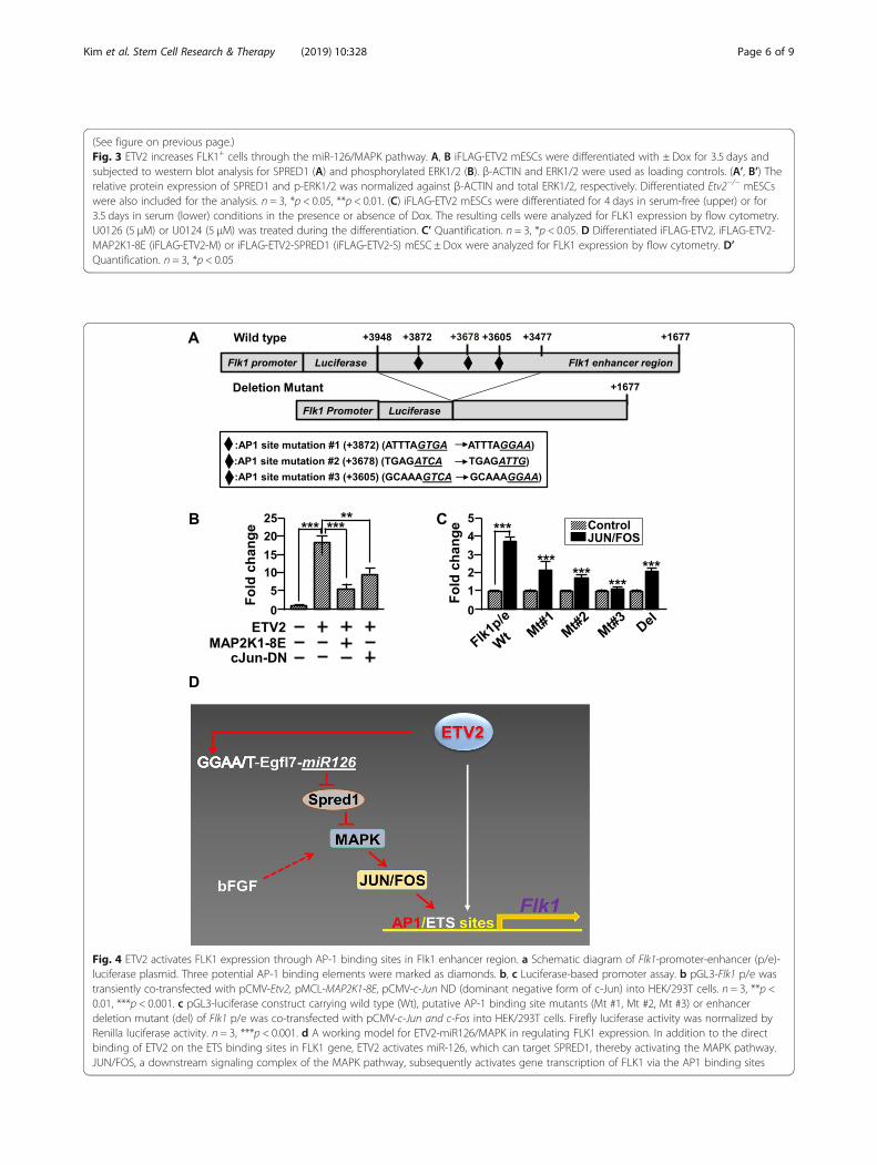
Direct activation of FLK1 gene expression by the MAPKpathwayTo get a detailed insight into how the MAPK pathwayactivated by ETV2 regulates the generation of FLK1+
cells, we examined the regulatory elements such as pro-moter and enhancer of Flk1 gene [23] and found severalpotential activator protein 1 (AP1) binding sequences inFlk1 enhancer which is critical for controlling the en-dogenous expression of Flk1 (Fig. 4a). AP1 (activatorprotein 1), a heterodimeric transcriptional complex con-sisting of JUN and FOS proteins, conveys the importantfunctions for multiple biological processes such as differ-entiation, proliferation and apoptosis [24]. Given thefindings that AP1 complex acts as a downstream targetof the MAPK pathway [25], these suggest an important
mechanistic link between ETV2 and FLK1 gene ex-pression via the MAPK-AP1 pathway. Therefore, wefirst performed a luciferase-based promoter assay andrevealed that the activity of the Flk1-promoter/enhan-cer (p/e) was increased in response to ETV2 or c-JUN/FOS, a downstream of the MAPK pathway(Fig. 4b, c). In contrast, both MKAP2K1-8E and theJUN dominant negative mutant, c-JUN DN [26],inhibited ETV2 function in activating Flk1-p/e(Fig. 4b). Further, we showed that mutations on theputative AP-1 binding site in the Flk1-p/e led to asignificantly reduced luciferase activity induced by c-JUN/FOS (Fig. 4c). Collectively, these results suggestthat the MAPK pathway can activate the expressionof Flk1 gene partly through c-JUN/FOS.
Fig. 1 Analysis of ETV2-regualted miRNA expression in FLK1+ cells. a Schematic diagram of miRNA sequencing experiment. Doxycycline-inducible(iFLAG-ETV2) mESCs were differentiated, treated with ± Doxycycline (Dox) at day 1 and sorted for FLK1+ cells at day 3.5. RNAs from the sortedcells were subjected to miRNA sequencing and analysis. b Volcano plot showing the log2 fold change between +Dox versus −Dox against the−log10 FDR-adjusted p value for each miRNA. miRNAs (FDR ≤ 0.05) with fold change of ≥ 1.5 (in red; upregulated) and ≤ − 1.5 (in green;downregulated) were highlighted and selected. c Heatmap of the selected miRNAs in response to overexpression of ETV2. miRNAs upregulatedand downregulated in +DOX were indicated with gray and black bars, respectively. d Gene Ontology (GO) categories of selected miRNAs byDIANA miRpath analysis. Bars indicate the significance level of miRNA target genes and interactions. e Differentiated iFLAG-ETV2 mESCs at day 3.5were subjected to qRT-PCR analysis. n = 3, ***p < 0.001
Kim et al. Stem Cell Research & Therapy (2019) 10:328 Page 3 of 9

DiscussionIn our study, several layers of novel findings on ETV2function were made. First, we reported genome-widemiRNA profiles in FLK1+ cells generated in response toETV2, providing a new research resource. Second, wedemonstrated the functional significance of the miR-126/MAPK pathway in ETV2-mediated FLK1+ cell gen-eration. Third, we also showed a direct activation of Flk1enhancer by the JUN/FOS complex. Overall, our resultsreveal that the miR-126/MAPK pathway constitutes anovel mechanism responsible for ETV2-mediated Flk1expression (Fig. 4d).Our miRNA profiling results suggest that ETV2 regu-
lates development of FLK1+ cells, hematopoietic andendothelial cell lineages as well as cardiomyocytesthrough miRNAs. For example, miR-10b, one of thedownregulated miRNAs by ETV2, is a critical regulatorfor vessel development through targeting FLT-1 (fms-re-lated tyrosine kinase 1), which can inhibit VEGF (vascu-lar endothelial growth factor)-FLK1 signaling [27]. Incontrast, miR-146a was upregulated in response toETV2 expression. Interestingly, miR-146 can target
CXCR4 (C-X-C chemokine receptor type 4), a receptorof SDF1 (stromal cell-derived factor 1), whose signalingis important during heart development and promotescardiogenesis from pluripotent stem cells [28–31]. An-other downregulated miRNA by ETV2, miR-106, hasbeen reported as a critical player for cardiac develop-ment as evidenced by in utero lethality displaying severecardiac defects in compound knockout of mouse miR-106~25 and miR-17~92 cluster [32]. Thus, determiningbiological consequences of the links between ETV2 andETV2-dependent miRNAs for cardiovascular lineage de-velopment would be an interesting topic for furtherstudies.Studies have shown that miR-126 plays important
functions in cardiovascular development and angiogen-esis [15–17]. In this study, we provided evidence of theunknown function of miR-126 in generating the firstemerging FLK1+ cells. Our results showed that ETV2 viaa direct binding on the Egfl7 promoter can induce theexpression of miR-126. In the subsequent studies, inhib-ition of the miR-126/MAPK pathway leads to impair-ment of ETV2-mediated FLK1+ cell generation. Thus,
Fig. 2 ETV2 upregulates miR126 expression through direct binding on Egfl7 promoter. a Expression analysis. iFLAG-ETV2 mESCs weredifferentiated for 3.5 days ± Dox treatment and were subjected to gene expression analysis. n = 3, *p < 0.05. b Schematic diagram of Egfl7promoter-luciferase plasmid. Two potential ETS binding elements were marked as diamonds, and their mutant sequences were shown in thebottom. c HEK/293T cells were transiently co-transfected with pCMV-Etv2 and pGL3-luciferase constructs carrying wild type (Wt), or mutants ofEgfl7 promoter. Firefly luciferase activity was normalized by Renilla luciferase activity. n = 3. ***p < 0.001. d iFLAG-ETV2 mESCs were differentiatedin the presence of Dox for 3.5 days and then subjected to ChIP-PCR assay. Rabbit anti-FLAG or IgG antibody was used for theimmunoprecipitation. n = 3, **p < 0.01
Kim et al. Stem Cell Research & Therapy (2019) 10:328 Page 4 of 9
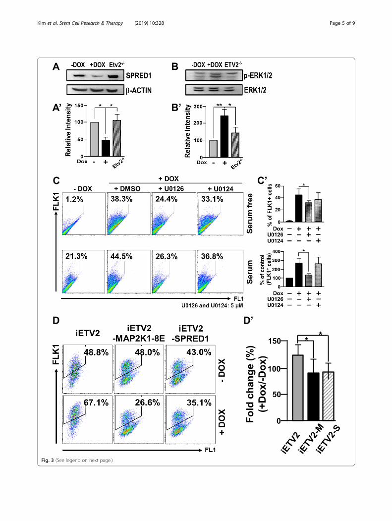
Fig. 3 (See legend on next page.)
Kim et al. Stem Cell Research & Therapy (2019) 10:328 Page 5 of 9
(See figure on previous page.)Fig. 3 ETV2 increases FLK1+ cells through the miR-126/MAPK pathway. A, B iFLAG-ETV2 mESCs were differentiated with ± Dox for 3.5 days andsubjected to western blot analysis for SPRED1 (A) and phosphorylated ERK1/2 (B). β-ACTIN and ERK1/2 were used as loading controls. (A’, B’) Therelative protein expression of SPRED1 and p-ERK1/2 was normalized against β-ACTIN and total ERK1/2, respectively. Differentiated Etv2−/− mESCswere also included for the analysis. n = 3, *p < 0.05, **p < 0.01. (C) iFLAG-ETV2 mESCs were differentiated for 4 days in serum-free (upper) or for3.5 days in serum (lower) conditions in the presence or absence of Dox. The resulting cells were analyzed for FLK1 expression by flow cytometry.U0126 (5 μM) or U0124 (5 μM) was treated during the differentiation. C’ Quantification. n = 3, *p < 0.05. D Differentiated iFLAG-ETV2, iFLAG-ETV2-MAP2K1-8E (iFLAG-ETV2-M) or iFLAG-ETV2-SPRED1 (iFLAG-ETV2-S) mESC ± Dox were analyzed for FLK1 expression by flow cytometry. D’Quantification. n = 3, *p < 0.05
Fig. 4 ETV2 activates FLK1 expression through AP-1 binding sites in Flk1 enhancer region. a Schematic diagram of Flk1-promoter-enhancer (p/e)-luciferase plasmid. Three potential AP-1 binding elements were marked as diamonds. b, c Luciferase-based promoter assay. b pGL3-Flk1 p/e wastransiently co-transfected with pCMV-Etv2, pMCL-MAP2K1-8E, pCMV-c-Jun ND (dominant negative form of c-Jun) into HEK/293T cells. n = 3, **p <0.01, ***p < 0.001. c pGL3-luciferase construct carrying wild type (Wt), putative AP-1 binding site mutants (Mt #1, Mt #2, Mt #3) or enhancerdeletion mutant (del) of Flk1 p/e was co-transfected with pCMV-c-Jun and c-Fos into HEK/293T cells. Firefly luciferase activity was normalized byRenilla luciferase activity. n = 3, ***p < 0.001. d A working model for ETV2-miR126/MAPK in regulating FLK1 expression. In addition to the directbinding of ETV2 on the ETS binding sites in FLK1 gene, ETV2 activates miR-126, which can target SPRED1, thereby activating the MAPK pathway.JUN/FOS, a downstream signaling complex of the MAPK pathway, subsequently activates gene transcription of FLK1 via the AP1 binding sites
Kim et al. Stem Cell Research & Therapy (2019) 10:328 Page 6 of 9

our results suggest a novel post-transcriptional regula-tory mechanism of ETV2 in generating FLK1+ cell. Al-though EGFL7 is co-transcribed with miR-126 uponETV2 expression, we rule out the potential contributionof EGFL7 to inducing FLK1+ cell generation due to thedispensable function of EGFL7 in early stages of em-bryogenesis and embryonic vasculature formation asdemonstrated by knockout studies in mice and knock-down experiments in differentiating ESCs [18, 33].Taken together, it is evident that miR-126 acting down-stream of ETV2 plays important roles in the establish-ment of the cardiovascular system.The molecular insights into Flk1 gene expression (i.e.,
determining its upstream regulators) has not been an ac-tive research area, despite its critical function inhematopoietic and vascular system. Regarding this, wehave demonstrated that ETV2 directly binds ETS bind-ing elements present on both promoter and enhancer ofFlk1 and activates its expression [5]. The subsequentChIP sequencing analysis demonstrated that a widerange of genes critical for endothelial cell and blood cellsare direct downstream targets of ETV2 [7]. In thiscurrent study, we revealed an important function ofMAPK activity in regulating Flk1 gene expression. Over-expression of ETV2 activates the MAPK activity, whileinhibition of the MAPK pathway decreases ETV2-mediated FLK1+ cell generation. Further, we showedthat the MAPK pathway is able to directly upregulatethe expression of Flk1 via a direct binding of JUN/FOSon Flk1 enhancer. Thus, we envision that ETV2 inducesFlk1 gene expression in a bimodal manner, one by acti-vating the miR-126/MAPK pathway and the other by itsdirect binding on Flk1 gene regulatory elements. Inmouse embryos, activated ERK1/2 is detected in theblood islands, the first sites of blood and endothelial celldevelopment and later in the dorsal aorta and intersomi-tic vessels [34]. Further, Erk2-deficient mouse embryosare incompatible with proper mesoderm formation [35,36]. Sprouty/Spred proteins, the negative effectors forRas/MAPK pathway [19], have inhibitory functions ingenerating mesoderm and blood as well as vessel devel-opment [37–40], further suggesting important functionsof the MAPK pathway in emergence of mesoderm withhematopoietic and endothelial potential. Consideringthat the enhanced protein stability of ETV2 by OVOL2is thought to be one mechanism for ETV2-mediatedFlk1 gene transcription [14], it would be interesting tosee if the interaction between ETV2 and OVOL2 canpositively regulate the miR-126/MAPK pathway in thisprocess. In addition, the findings that ETV2 interactswith OVOL2 [14] or FOXC2 [12] and that ETV2 andJUN/FOS acts on Flk1 promoter and enhancer suggest ahypothesis that ETV2 can form a transcriptional com-plex with OVOL2, FOXC2, and/or AP1 to regulate the
expression of Flk1, which could provide an in-depth andnovel insight into molecular mechanisms behind theETV2-FLK1 axis.In conclusion, we reported miRNA profiles regulated
by ETV2 in generating FLK1+ cells from mESCs. Fur-ther, we showed that ETV2 can regulate the expressionof Flk1 through the miR126/MAPK pathway. Thesefindings could provide a novel insight into the mecha-nisms of how ETV2 regulates the development of thecardiovascular system. Studying the functions of othermiRNAs identified in this research would be importantfor further deciphering the ETV2-miRNA regulatorymechanisms in FLK1+ cell and cardiovascular lineagegeneration. Since embryonic events can often play a crit-ical role in adults, examination of our findings in patho-physiological angiogenesis would be warranted.
Supplementary informationSupplementary information accompanies this paper at https://doi.org/10.1186/s13287-019-1466-8.
Additional file 1. Supplemental materials and methods
Additional file 2: Table S1. Gene Ontology (GO) categories for genestargeted by significant differentially expressed miRNA’s. Table S2. KEGGcategories for genes targeted by significant differentially expressedmiRNA’s. Table S3. Primer sequences.
Additional file 3: Figure S1. Analysis on miR-126-5p in response toETV2. Differentiated iFLAG-ETV2 mESCs at day 3.5 were subjected to qRT-PCR analysis. n=3, **p<0.01.
AbbreviationsAP1: Activator protein 1; ChIP: Chromatin immunoprecipitation; c-JUNDN: JUN dominant negative mutant; CXCR4: C-X-C chemokine receptor type4; Dox: Doxycycline; EGFL7: Egf-like domain multiple 7; ERK1/2: Extracellularsignal-regulated kinases 1/2; ETS: E26 transformation-specific; ETV2: Etsvariant 2; FACS: Fluorescence-activated cell sorting; FDR: False discovery rate;FLK1: Fetal liver kinase 1; Flk1-p/e: Flk1-promoter/enhancer; FLT-1: Fms-related tyrosine kinase 1; FOXC2: Forkhead box protein C2; GO: Geneontology; iFLAG-ETV2: Doxycycline-inducible FLAG-ETV2; MAPK: Mitogen-activated protein kinase; mESCs: Mouse embryonic stem cells; OVOL2: Ovo-like zinc finger 2; RAP1: Ras-associated protein 1; SDF1: Stromal cell-derivedfactor 1; SPRED1: Sprouty-related EVH1 domain containing 1; VEGF: Vascularendothelial growth factor; WNT: Wingless-related integration site
AcknowledgementsWe would like to thank Dr. Seyoung Oh for initial technical support. We alsowould like to thank Emory’s Pediatrics/Winship Flow Cytometry Core.
Authors’ contributionsJYK, DHL, JKK, HSC, HS, WJP, JYC, and TMK contributed to the collection andassembly of data. JYK and DHL contributed to the design, data analysis,interpretation, and manuscript writing. BD, MR, JK, and SJG contributed tothe data analysis. CP contributed to the conception, design, collection andassembly of data, financial support, data interpretation, and manuscriptwriting. JYK and CP contributed to the final approval of manuscript. Allauthors read and approved the final manuscript.
FundingResearch reported in this publication was supported in part by theBiostatistics and Bioinformatics Shared resource of Winship Cancer Instituteof Emory University and NIH/NCI under award number P30CA13829. Thiswork was also supported by Basic Science Research Program through theNational Research Foundation of Korea (NRF) funded by the Ministry ofEducation (2018R1D1A1A02085481) (T.M.K) and the March of Dimes
Kim et al. Stem Cell Research & Therapy (2019) 10:328 Page 7 of 9

Foundation, 5-FY12-44 (to C.P.), the American Heart Association,11SDG7390074 (to C. P.), the Children’s Miracle Network, 660085–1116 (toC.P.), Children’s Heart Research and Outcomes Center and Children’s Health-care of Atlanta 00060337 (to C.P.) and NIH HL119291 (to C.P.).
Availability of data and materialsThe datasets used and/or analyzed during the current study are availablefrom the corresponding author on reasonable request.
Ethics approval and consent to participateNot applicable.
Consent for publicationNot applicable.
Competing interestsThe authors declare that they have no competing interests.
Author details1Department of Pediatrics, Emory University School of Medicine, 2015Uppergate Dr. Atlanta, Atlanta, GA 30322, USA. 2Children’s Heart Research &Outcomes (HeRO) Center, Children’s Healthcare of Atlanta & EmoryUniversity, Atlanta, GA, USA. 3Molecular and Systems Pharmacology Program,Emory University, Atlanta, GA, USA. 4Biochemistry, Cell Biology andDevelopmental Biology Program, Emory University, Atlanta, GA, USA.5Winship Cancer Institute, Emory University, Atlanta, GA, USA. 6Departmentof Biostatistics and Bioinformatics, Emory University, Atlanta, GA, USA.7Present Address: Department of Oncology, University of Texas at Austin,Austin, TX 78712, USA. 8Sequencing Core, Research Resources Center,University of Illinois at Chicago, Chicago, IL, USA. 9Department ofBiochemistry and Molecular Biology, Chosun University School of Medicine,Gwangju, IL, Republic of Korea. 10Graduate School of InternationalAgricultural Technology and Institute of Green-Bio Science and Technology,Seoul National University, Pyeongchang, Gangwon-do, Republic of Korea.
Received: 1 August 2019 Revised: 28 September 2019Accepted: 22 October 2019
References1. Hsu T, Trojanowska M, Watson DK. Ets proteins in biological control and
cancer. J Cell Biochem. 2004;91(5):896–903.2. Findlay VJ, LaRue AC, Turner DP, Watson PM, Watson DK. Understanding the
role of ETS-mediated gene regulation in complex biological processes. AdvCancer Res. 2013;119:1–61.
3. Oh SY, Kim JY, Park C. The ETS factor, ETV2: a master regulator for vascularendothelial cell development. Mol Cells. 2015;38(12):1029–36.
4. Lee DH, Kim TM, Kim JK, Park C. ETV2/ER71 transcription factor as atherapeutic vehicle for cardiovascular disease. Theranostics.2019;9(19):5694–705.
5. Lee D, Park C, Lee H, Lugus JJ, Kim SH, Arentson E, Chung YS, Gomez G,Kyba M, Lin S, Janknecht R, Lim DS, Choi K. ER71 acts downstream of BMP,notch, and Wnt signaling in blood and vessel progenitor specification. CellStem Cell. 2008;2(5):497–507.
6. Liu F, Kang I, Park C, Chang LW, Wang W, Lee D, Lim DS, Vittet D, NerbonneJM, Choi K. ER71 specifies Flk-1+ hemangiogenic mesoderm by inhibitingcardiac mesoderm and Wnt signaling. Blood. 2012;119(14):3295–305.
7. Liu F, Li D, Yu YY, Kang I, Cha MJ, Kim JY, Park C, Watson DK, Wang T, ChoiK. Induction of hematopoietic and endothelial cell program orchestrated byETS transcription factor ER71/ETV2. EMBO Rep. 2015;16(5):654–69.
8. Ferdous A, Caprioli A, Iacovino M, Martin CM, Morris J, Richardson JA, Latif S,Hammer RE, Harvey RP, Olson EN, Kyba M, Garry DJ. Nkx2-5 transactivatesthe Ets-related protein 71 gene and specifies an endothelial/endocardialfate in the developing embryo. Proc Natl Acad Sci U S A. 2009;106(3):814–9.
9. Kataoka H, Hayashi M, Nakagawa R, Tanaka Y, Izumi N, Nishikawa S, Jakt ML,Tarui H, Nishikawa S. Etv2/ER71 induces vascular mesoderm from Flk1+PDGFRalpha+ primitive mesoderm. Blood. 2011;118(26):6975–86.
10. Neuhaus H, Muller F, Hollemann T. Xenopus er71 is involved in vasculardevelopment. Dev Dyn. 2010;239(12):3436–45.
11. Sumanas S, Lin S. Ets1-related protein is a key regulator of vasculogenesis inzebrafish. PLoS Biol. 2006;4(1):e10.
12. De Val S, Chi NC, Meadows SM, Minovitsky S, Anderson JP, Harris IS, EhlersML, Agarwal P, Visel A, Xu SM, Pennacchio LA, Dubchak I, Krieg PA, StainierDY, Black BL. Combinatorial regulation of endothelial gene expression byets and forkhead transcription factors. Cell. 2008;135(6):1053–64.
13. Veldman MB, Lin S. Etsrp/Etv2 is directly regulated by Foxc1a/b in thezebrafish angioblast. Circ Res. 2012;110(2):220–9.
14. Kim JY, Lee RH, Kim TM, Kim DW, Jeon YJ, Huh SH, Oh SY, Kyba M, KataokaH, Choi K, Ornitz DM, Chae JI, Park C. OVOL2 is a critical regulator of ER71/ETV2 in generating FLK1+, hematopoietic, and endothelial cells fromembryonic stem cells. Blood. 2014;124(19):2948–52.
15. Fish JE, Santoro MM, Morton SU, Yu S, Yeh RF, Wythe JD, Ivey KN, BruneauBG, Stainier DY, Srivastava D. miR-126 regulates angiogenic signaling andvascular integrity. Dev Cell. 2008;15(2):272–84.
16. Wang S, Aurora AB, Johnson BA, Qi X, McAnally J, Hill JA, Richardson JA,Bassel-Duby R, Olson EN. The endothelial-specific microRNA miR-126governs vascular integrity and angiogenesis. Dev Cell. 2008;15(2):261–71.
17. Sturgeon CM, Chicha L, Ditadi A, Zhou Q, McGrath KE, Palis J, HammondSM, Wang S, Olson EN, Keller G. Primitive erythropoiesis is regulated by miR-126 via nonhematopoietic Vcam-1+ cells. Dev Cell. 2012;23(1):45–57.
18. Kuhnert F, Mancuso MR, Hampton J, Stankunas K, Asano T, Chen CZ, KuoCJ. Attribution of vascular phenotypes of the murine Egfl7 locus to themicroRNA miR-126. Development. 2008;135(24):3989–93.
19. Wakioka T, Sasaki A, Kato R, Shouda T, Matsumoto A, Miyoshi K, Tsuneoka M,Komiya S, Baron R, Yoshimura A. Spred is a Sprouty-related suppressor ofRas signalling. Nature. 2001;412(6847):647–51.
20. Ornitz DM, Itoh N. The fibroblast growth factor signaling pathway. WileyInterdiscip Rev Dev Biol. 2015;4(3):215–66.
21. Faloon P, Arentson E, Kazarov A, Deng CX, Porcher C, Orkin S, Choi K. Basicfibroblast growth factor positively regulates hematopoietic development.Development. 2000;127(9):1931–41.
22. Mansour SJ, Matten WT, Hermann AS, Candia JM, Rong S, Fukasawa K,Vande Woude GF, Ahn NG. Transformation of mammalian cells byconstitutively active MAP kinase kinase. Science. 1994;265(5174):966–70.
23. Kappel A, Ronicke V, Damert A, Flamme I, Risau W, Breier G. Identification ofvascular endothelial growth factor (VEGF) receptor-2 (Flk-1) promoter/enhancer sequences sufficient for angioblast and endothelial cell-specifictranscription in transgenic mice. Blood. 1999;93(12):4284–92.
24. Bejjani F, Evanno E, Zibara K, Piechaczyk M, Jariel-Encontre I. The AP-1transcriptional complex: local switch or remote command? Biochim BiophysActa Rev Cancer. 2019;1872(1):11–23.
25. Maik-Rachline G, Hacohen-Lev-Ran A, Seger R. Nuclear ERK: mechanism oftranslocation, substrates, and role in cancer. Int J Mol Sci. 2019;20(5):1194.
26. Wang ZY, Sato H, Kusam S, Sehra S, Toney LM, Dent AL. Regulation of IL-10gene expression in Th2 cells by Jun proteins. J Immunol. 2005;174(4):2098–105.
27. Hassel D, Cheng P, White MP, Ivey KN, Kroll J, Augustin HG, Katus HA,Stainier DY, Srivastava D. MicroRNA-10 regulates the angiogenic behavior ofzebrafish and human endothelial cells by promoting vascular endothelialgrowth factor signaling. Circ Res. 2012;111(11):1421–33.
28. Chiriac A, Terzic A, Park S, Ikeda Y, Faustino R, Nelson TJ. SDF-1-enhancedcardiogenesis requires CXCR4 induction in pluripotent stem cells. JCardiovasc Transl Res. 2010;3(6):674–82.
29. Ma Q, Jones D, Borghesani PR, Segal RA, Nagasawa T, Kishimoto T, BronsonRT, Springer TA. Impaired B-lymphopoiesis, myelopoiesis, and derailedcerebellar neuron migration in CXCR4- and SDF-1-deficient mice. Proc NatlAcad Sci U S A. 1998;95(16):9448–53.
30. Nagasawa T, Hirota S, Tachibana K, Takakura N, Nishikawa S, Kitamura Y,Yoshida N, Kikutani H, Kishimoto T. Defects of B-cell lymphopoiesis andbone-marrow myelopoiesis in mice lacking the CXC chemokine PBSF/SDF-1.Nature. 1996;382(6592):635–8.
31. Zou YR, Kottmann AH, Kuroda M, Taniuchi I, Littman DR. Function of thechemokine receptor CXCR4 in haematopoiesis and in cerebellardevelopment. Nature. 1998;393(6685):595–9.
32. Ventura A, Young AG, Winslow MM, Lintault L, Meissner A, Erkeland SJ,Newman J, Bronson RT, Crowley D, Stone JR, Jaenisch R, Sharp PA, Jacks T.Targeted deletion reveals essential and overlapping functions of the miR-17through 92 family of miRNA clusters. Cell. 2008;132(5):875–86.
33. Durrans A, Stuhlmann H. A role for Egfl7 during endothelial organization inthe embryoid body model system. J Angiogenes Res. 2010;2:4.
34. Corson LB, Yamanaka Y, Lai KM, Rossant J. Spatial and temporal patterns ofERK signaling during mouse embryogenesis. Development. 2003;130(19):4527–37.
Kim et al. Stem Cell Research & Therapy (2019) 10:328 Page 8 of 9

35. Saba-El-Leil MK, Vella FD, Vernay B, Voisin L, Chen L, Labrecque N, Ang SL,Meloche S. An essential function of the mitogen-activated protein kinaseErk2 in mouse trophoblast development. EMBO Rep. 2003;4(10):964–8.
36. Yao Y, Li W, Wu J, Germann UA, Su MS, Kuida K, Boucher DM. Extracellularsignal-regulated kinase 2 is necessary for mesoderm differentiation. ProcNatl Acad Sci U S A. 2003;100(22):12759–64.
37. Muhl B, Hagele J, Tasdogan A, Loula P, Schuh K, Bundschu K. SPREDs(Sprouty related proteins with EVH1 domain) promote self-renewal andinhibit mesodermal differentiation in murine embryonic stem cells. DevDyn. 2015;244(4):591–606.
38. Nobuhisa I, Kato R, Inoue H, Takizawa M, Okita K, Yoshimura A, Taga T.Spred-2 suppresses aorta-gonad-mesonephros hematopoiesis by inhibitingMAP kinase activation. J Exp Med. 2004;199(5):737–42.
39. Sivak JM, Petersen LF, Amaya E. FGF signal interpretation is directed bySprouty and Spred proteins during mesoderm formation. Dev Cell. 2005;8(5):689–701.
40. Taniguchi K, Kohno R, Ayada T, Kato R, Ichiyama K, Morisada T, Oike Y,Yonemitsu Y, Maehara Y, Yoshimura A. Spreds are essential for embryoniclymphangiogenesis by regulating vascular endothelial growth factorreceptor 3 signaling. Mol Cell Biol. 2007;27(12):4541–50.
Publisher’s NoteSpringer Nature remains neutral with regard to jurisdictional claims inpublished maps and institutional affiliations.
Kim et al. Stem Cell Research & Therapy (2019) 10:328 Page 9 of 9


![ETV2/ER71 Transcription Factor as a Therapeutic Vehicle ...into endothelial and hematopoietic cells , display [23] enriched expression of , compared to FLK1Etv2-cells as demonstrated](https://img.pdfslide.net/doc/110x75/5ea650c1992abe6d41291ffc/etv2er71-transcription-factor-as-a-therapeutic-vehicle-into-endothelial-and.jpg)