Embed Size (px)
Citation preview
Eukaryotic organism
Premedical IV
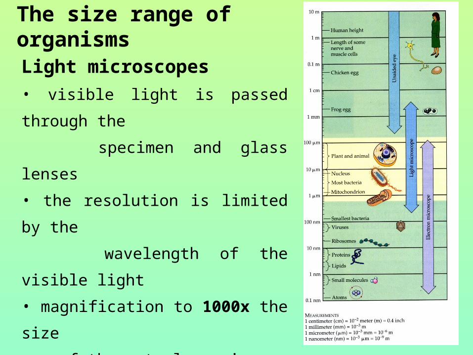
The size range of organisms
Light microscopes
• visible light is passed through the
specimen and glass lenses
• the resolution is limited by the
wavelength of the visible light
• magnification to 1000x the size
of the actual specimen
Resolving power - the minimum
distance two points, which can be
distiquished.
Electron microscope
– focused a beam (current) of electrons, have the
wavelength much shorter than visible light, 1 nm (0.1nm)
TEM transmission: the beam through a thin specimen -
ultrastructure
SEM scanning: the electron beam scans the surface of
the sample
• use the electromagnets instead of glass lenses
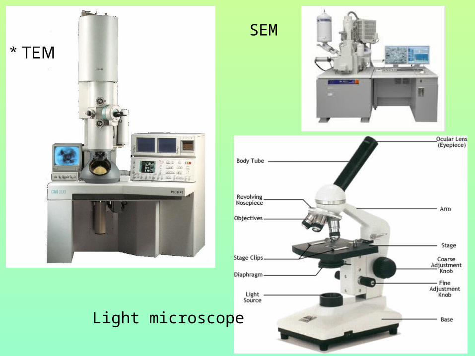
SEM
Light microscope
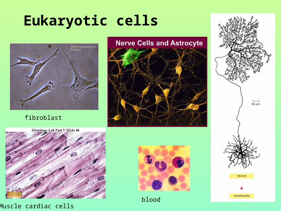
Eukaryotic cells
Muscle cardiac cells
fibroblast
blood
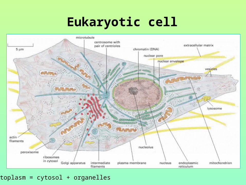
Eukaryotic cell
Cytoplasm = cytosol + organelles
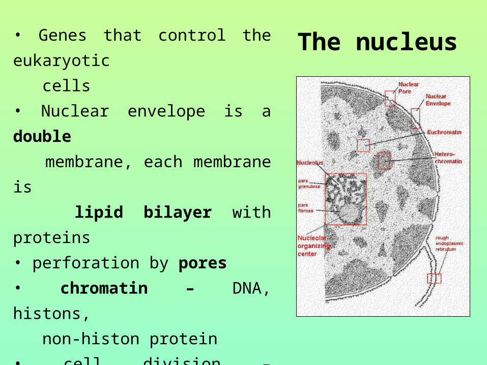
The nucleus• Genes that control the eukaryotic
cells
• Nuclear envelope is a double
membrane, each membrane is
lipid bilayer with proteins
• perforation by pores
• chromatin – DNA, histons,
non-histon protein
• cell division – chromatin
condensate to chromosomes
• the nucleolus – synthesis of
ribosomes components
• The nucleus control protein synthesis by sending
molecular messengers in the form RNA – mRNA -
messenger - TRANSCRIPTION
• is synthesized in nucleus according the DNA
• in ribosomes is genetic information translate into the
primary structure of a specific protein - TRANSLATION
• free ribosomes – suspended in the cytosol, function of
protein in cytosol
• bound ribosomes are attached to outside membrane
network called the endoplazmatic reticulum;
make proteins destined into membrane and for export from
the cell (secretion)

Ribosomes
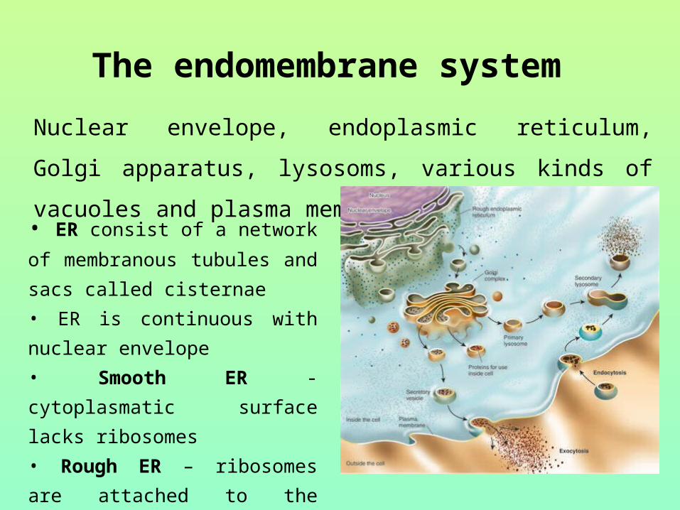
The endomembrane system
Nuclear envelope, endoplasmic reticulum, Golgi apparatus,
lysosoms, various kinds of vacuoles and plasma membrane
• ER consist of a network of
membranous tubules and sacs
called cisternae
• ER is continuous with nuclear
envelope
• Smooth ER - cytoplasmatic
surface lacks ribosomes
• Rough ER – ribosomes are
attached to the cytoplasmatic side

Function of smooth ER – synthesis of lipids (phospholipids,
steroids), metabolism of carbohydrates (glycogen) and
detoxification of drugs (barbiturates) and poisons
Function of rough ER – secretion of proteins, glycoproteins
formation of transport vesicules to other components of
endomembrane system
Golgi apparatus – sorting cell products, they are modified
and stored (removes sugar monomers and product diverse
oligosaccharides)
two poles are reffered to as the cis face ad trans face

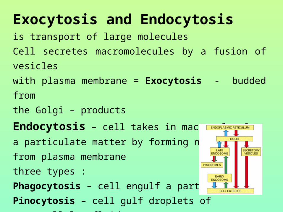
Exocytosis and Endocytosis is transport of large molecules
Cell secretes macromolecules by a fusion of vesicles
with plasma membrane = Exocytosis - budded from
the Golgi – products
Endocytosis – cell takes in macromolecules a particulate
matter by forming new vesicles from plasma membrane
three types :
Phagocytosis – cell engulf a particle
Pinocytosis – cell gulf droplets of
extracellular fluid
Receptor-mediated endocytosis is very
specific – receptor and ligand

Lysosomes are digestive compartments
• membrane bounded sac of
hydrolytic enzymes
• enzymes hydrolyze in acidic
environment (pH 5) proteins,
polysaccharides, fats and nucleic
acids
• function is intracellular digestion of
food particles, smaller organisms and
organic components engulfing by
phagocytosis and own organic old
material by autophagy

Mitochondria and chloroplasts
Vacuoles, vesicles • membrane–bounded sacs • vacuoles have various functions: food vacuoles contractile vacuoles tonoplast
• Convert energy (ATP) that cells use for work
Mitochondria are the sites of cellular respiration
Chloroplasts are sites of photosynthesis
• Semiautonomic organelles, that grow and reproduce
within the cell
• contain own DNA (prokaryotic origin)
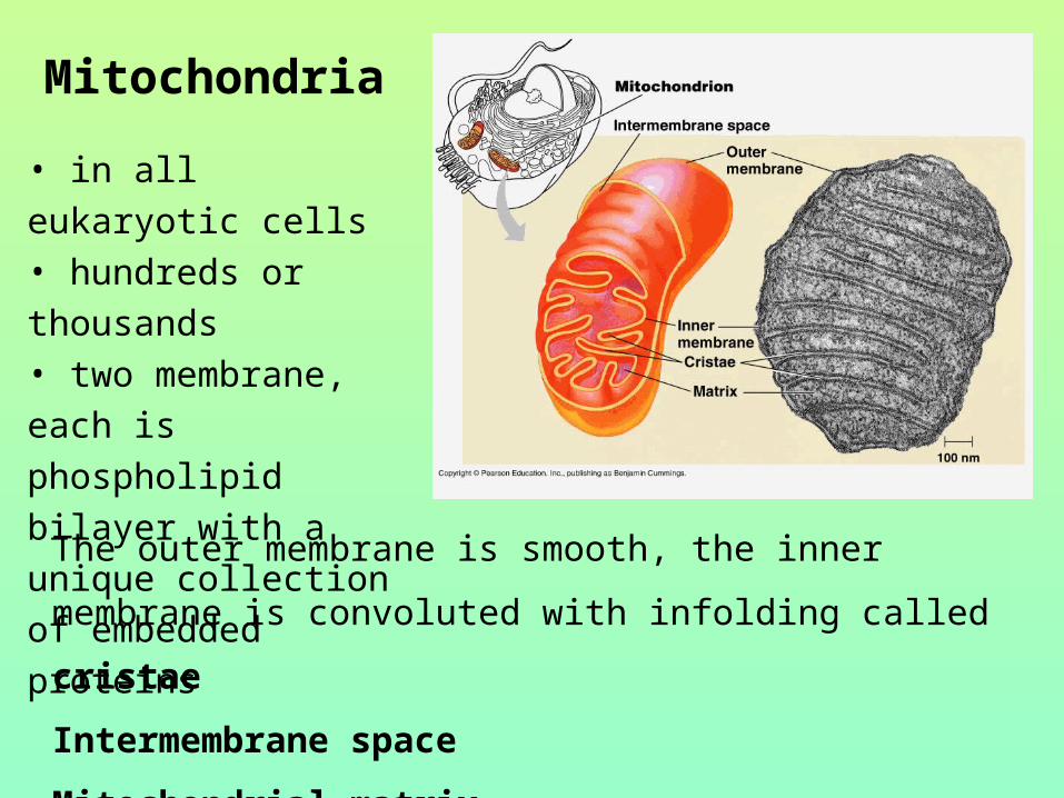
Mitochondria
• in all eukaryotic cells• hundreds or thousands• two membrane, each
is phospholipid bilayer
with a unique collection
of embedded proteins
The outer membrane is smooth, the inner membrane is
convoluted with infolding called cristae
Intermembrane space
Mitochondrial matrix

Chloroplast A member of plant organelles
family called plastids:
leukoplast
chromoplasts
chloroplasts
thylakoids
Inner membranous
system, outside of it
is stroma
• photosynthesis
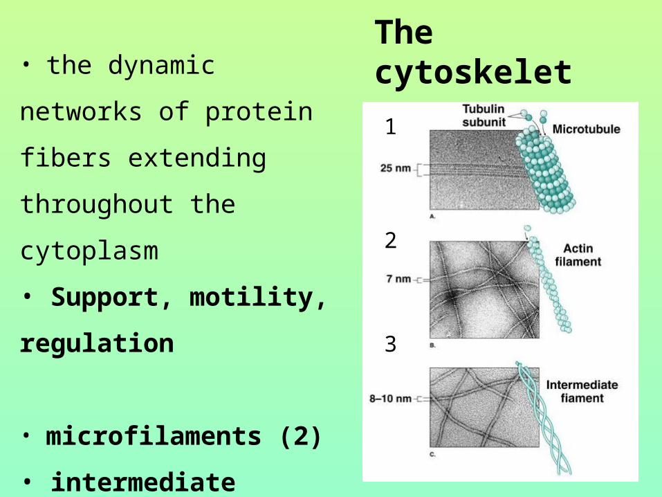
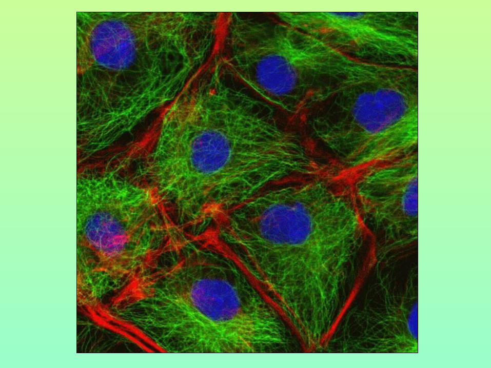
The cytoskelet• the dynamic networks of
protein fibers extending
throughout the cytoplasm
• Support, motility,
regulation
• microfilaments (2)
• intermediate filaments (3)
• microtubules (1)
1
2
3
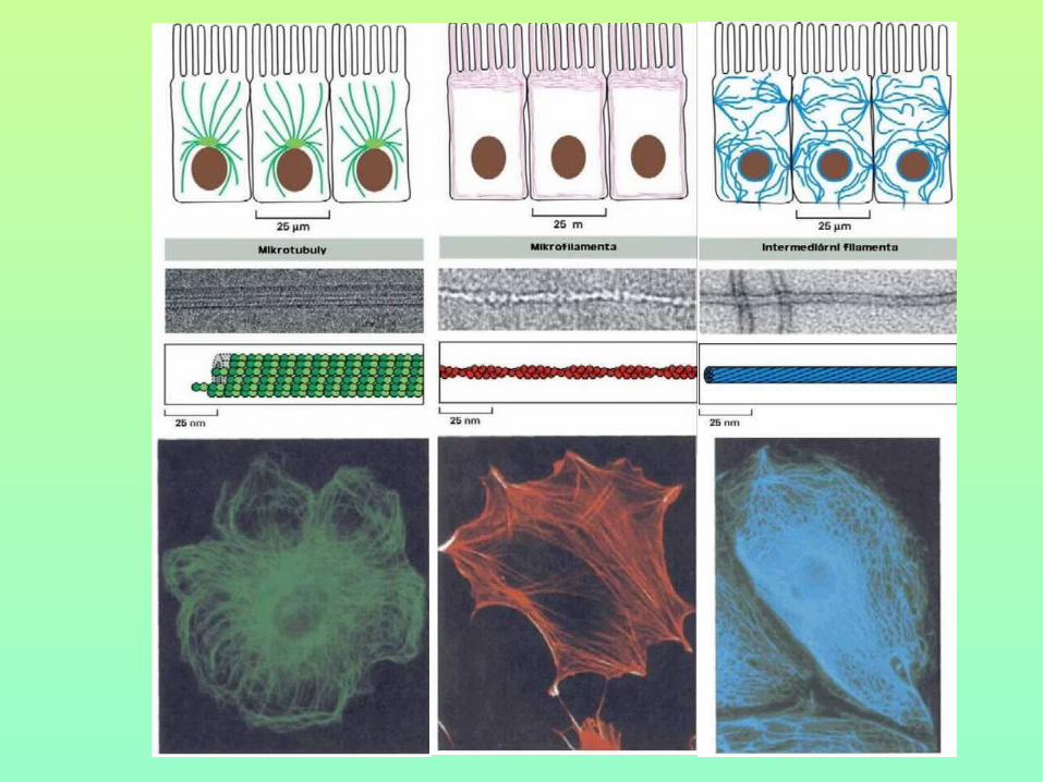
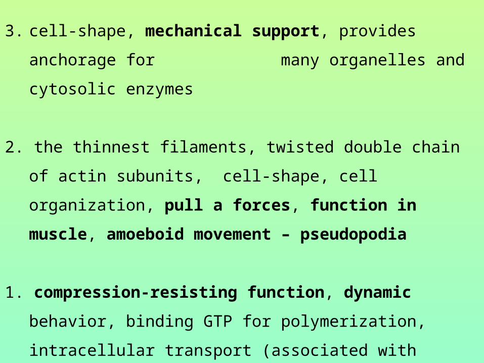
3. cell-shape, mechanical support, provides anchorage for
many organelles and cytosolic enzymes
2. the thinnest filaments, twisted double chain of actin subunits,
cell-shape, cell organization, pull a forces, function in
muscle, amoeboid movement – pseudopodia
1. compression-resisting function, dynamic behavior, binding
GTP for polymerization, intracellular transport (associated with
dyneins and kinesins, they transport organelles like
mitochondria or vesicles, the axoneme of cilia and flagella,
the mitotic spindle
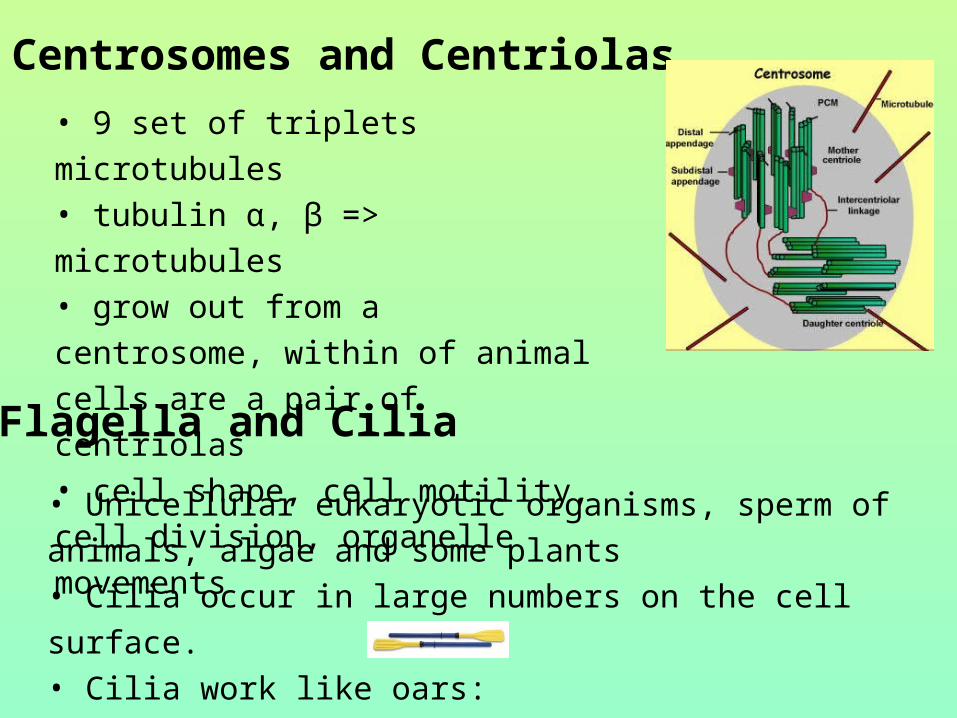
Centrosomes and Centriolas
• 9 set of triplets microtubules • tubulin α, β => microtubules• grow out from a centrosome, within of
animal cells are a pair of centriolas• cell shape, cell motility, cell division,
organelle movements
Flagella and Cilia
• Unicellular eukaryotic organisms, sperm of animals, algae
and some plants• Cilia occur in large numbers on the cell surface.• Cilia work like oars:

• Flagella are longer and are usually limited to just one or few • the motor molecule
called dynein• basal body identical
to centriole• 9 doublets of outer
microtubules• one doublet of inner
microtubule
Flagellum
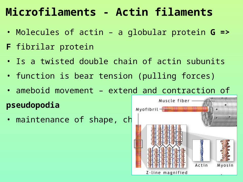
Microfilaments - Actin filaments
• Molecules of actin – a globular protein G => F fibrilar protein
• Is a twisted double chain of actin subunits
• function is bear tension (pulling forces)
• ameboid movement – extend and contraction of pseudopodia
• maintenance of shape, changes of shape
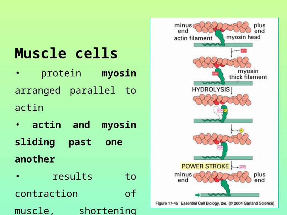
Muscle cells• protein myosin arranged
parallel to actin
• actin and myosin sliding
past one another
• results to contraction of
muscle, shortening the cell

Plant Cells:
have chloroplast use photosynthesis have cell wall one large vacuole are rectangular
Animal Cells:
don't have chloroplast no cell wall one or more small vacuole either circular or have irregular shape
Cellulose of plant cell walls helps to plant cells to allow high pressure to build inside of it, without bursting. A plant cell has to be able to accept large amounts of liquid through osmosis, without being destroyed. An animal cell does not have this cell wall. If you start to fill the animal cell with too much distilled water or other fluid, it will eventually pop.

Campbell, Neil A., Reece, Jane B., Cain Michael L., Jackson, Robert B., Minorsky, Peter V., Biology, Benjamin-Cummings Publishing Company, 1996 –2010.