Embed Size (px)
Citation preview
Sede di Napol i
EUROPEAN SCHOOL OF MOLECULAR MEDICINE
NAPLES SITE ndash Scientific Coordinator Prof Francesco Salvatore
UNIVERSITArsquo DEGLI STUDI DI NAPOLI ldquoFEDERICOIIrdquo
PhD in Molecular Medicine
Curriculum Human Genetics
II Ciclo
Title of the Thesis Inhibition of the Sonic Hedgehog pathway as a strategy to treat ocular
neovascularization in animal models
Supervisor PhD student
Prof Alberto Auricchio Dr Gabriella Cotugno
Internal Supervisor
Prof Lucio Pastore
Extrernal Supervisor
Prof Robin Ali
Naples 3032009
1
TABLE OF CONTENTS
1 Abstractpg7
2 Introduction9
21 The eye structure and functionhelliphelliphelliphelliphelliphelliphelliphelliphelliphelliphelliphelliphelliphelliphelliphelliphelliphelliphellip9
22 Organization and development of ocular vasculaturehelliphelliphelliphelliphelliphelliphelliphelliphelliphellip12
23 Ocular Neovascularization and related diseaseshelliphelliphelliphelliphelliphelliphelliphelliphelliphelliphelliphelliphellip14
Age Related Macular Degeneration and Choroidal Neovascularizationhelliphelliphelliphellip15
Retinal Neovascularizationhelliphelliphelliphelliphelliphelliphelliphelliphelliphelliphelliphelliphelliphelliphelliphelliphelliphelliphelliphelliphellip16
Retinopathy of prematurity helliphelliphelliphelliphelliphelliphelliphelliphelliphelliphelliphelliphelliphelliphelliphelliphelliphelliphelliphelliphellip17
Diabetes Mellitus and Proliferative Diabetic Retinopathyhelliphelliphelliphelliphelliphelliphelliphellip17
24 Treatment of ocular neovascularization helliphelliphelliphelliphelliphelliphelliphelliphelliphelliphelliphelliphelliphelliphelliphellip18
25 Animal models of ocular neovascularization helliphelliphelliphelliphelliphelliphelliphelliphelliphelliphelliphelliphelliphellip20
26 Experimental therapies for ocular neovascularizationhelliphelliphelliphelliphelliphelliphelliphelliphelliphelliphellip22
27 Gene therapy and ocular gene transfer helliphelliphelliphelliphelliphelliphelliphelliphelliphelliphelliphelliphelliphelliphelliphellip 24
28 Sonic Hedgehog and ocular Neovascularization helliphelliphelliphelliphelliphelliphelliphelliphelliphelliphelliphelliphellip26
3 Aim of the thesishelliphelliphelliphelliphelliphelliphelliphelliphelliphelliphelliphelliphelliphelliphelliphelliphelliphelliphelliphelliphelliphelliphelliphelliphellip28
4 Materials and Methodshelliphelliphelliphelliphelliphelliphelliphelliphelliphelliphelliphelliphelliphelliphelliphelliphelliphelliphelliphelliphelliphelliphellip29
41 Vector Construction and Productionhelliphelliphelliphelliphelliphelliphelliphelliphelliphelliphelliphelliphelliphelliphelliphelliphellip29
42 Anti-Shh siRNA design and productionhelliphelliphelliphelliphelliphelliphelliphelliphelliphelliphelliphelliphelliphelliphelliphellip31
43 Diabetes mouse model vectors administration AP20187 stimulation blood and
tissue collectionhelliphelliphelliphelliphelliphelliphelliphelliphelliphelliphelliphelliphelliphelliphelliphelliphelliphelliphelliphelliphelliphelliphelliphelliphelliphellip31
44 Mouse models of ocular NV vectors administration cyclopamine and siRNA
administration eyes collectionhelliphelliphelliphelliphelliphelliphelliphelliphelliphelliphelliphelliphelliphelliphelliphelliphelliphelliphellip33
2
45 Retinal angiography immunofluorescence of whole mount preparation in vivo
fluorescein angiography and quantification of CNV areahelliphelliphelliphelliphelliphelliphelliphelliphellip34
46 Hepatic glycogen measurementhelliphelliphelliphelliphelliphelliphelliphelliphelliphelliphelliphelliphelliphelliphelliphelliphelliphelliphellip36
47 In vivo glucose utilization indexhelliphelliphelliphelliphelliphelliphelliphelliphelliphelliphelliphelliphelliphelliphelliphelliphelliphelliphellip36
48 Cell culture plasmid and siRNA transfection AAV transduction cells and media
collectionhelliphelliphelliphelliphelliphelliphelliphelliphelliphelliphelliphelliphelliphelliphelliphelliphelliphelliphelliphelliphelliphelliphelliphelliphelliphelliphelliphellip37
49 C3H10T12 osteoblastic differentiation and Alkaline Phosphatase assayhelliphelliphellip38
410 Anti-myc co-immunoprecipitationhelliphelliphelliphelliphelliphelliphelliphelliphelliphelliphelliphelliphelliphelliphelliphelliphelliphellip39
411 Western blot analysishelliphelliphelliphelliphelliphelliphelliphelliphelliphelliphelliphelliphelliphelliphelliphelliphelliphelliphelliphelliphelliphelliphellip39
412 Localization of HIP and BRDU labeled siRNA in the eyehelliphelliphelliphelliphelliphelliphelliphelliphellip41
413 RNA Extraction Semiquantitative RT-PCR and Quantitative Real-Time PCR42
414 In situ hybridizationhelliphelliphelliphelliphelliphelliphelliphelliphelliphelliphelliphelliphelliphelliphelliphelliphelliphelliphelliphelliphelliphelliphellip43
415 Histologyhelliphelliphelliphelliphelliphelliphelliphelliphelliphelliphelliphelliphelliphelliphelliphelliphelliphelliphelliphelliphelliphelliphelliphelliphelliphelliphelliphellip44
415 Statistical analysishelliphelliphelliphelliphelliphelliphelliphelliphelliphelliphelliphelliphelliphelliphelliphelliphelliphelliphelliphelliphelliphelliphelliphellip44
5 Resultshelliphelliphelliphelliphelliphelliphelliphelliphelliphelliphelliphelliphelliphelliphelliphelliphelliphelliphelliphelliphelliphelliphelliphelliphelliphelliphelliphelliphelliphellip46
51 Gene transfer for pharmacological regulation of the insulin receptor signallinghellip46
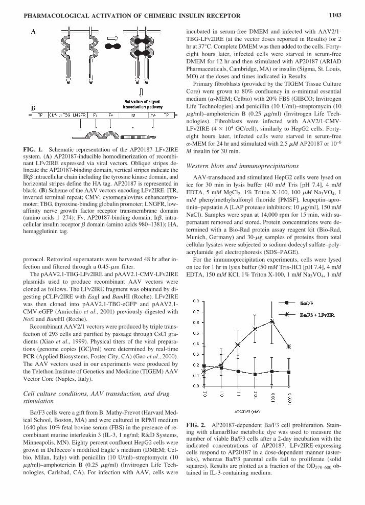
Generation of a pharmacologically regulated chimeric insulin receptorhelliphelliphelliphellip46
AP20187-dependent LFv2IRE activation in liver and muscle transduced with
AAV vectorshelliphelliphelliphelliphelliphelliphelliphelliphelliphelliphelliphelliphelliphelliphelliphelliphelliphelliphelliphelliphelliphelliphelliphelliphelliphelliphellip48
AP20187 induces insulin-like actions in muscle and liver of NOD mice transduced
with AAV vectorshelliphelliphelliphelliphelliphelliphelliphelliphelliphelliphelliphelliphelliphelliphelliphelliphelliphelliphelliphelliphelliphelliphelliphelliphellip52
52 Evaluation of the involvement of the Sonic Hedgehog pathway in ocular
neovascular diseaseshelliphelliphelliphelliphelliphelliphelliphelliphelliphelliphelliphelliphelliphelliphelliphelliphelliphelliphelliphelliphelliphelliphelliphellip56
Sonic Hedgehog pathway is involved in physiological and pathological ocular
vessel developmenthelliphelliphelliphelliphelliphelliphelliphelliphelliphelliphelliphelliphelliphelliphelliphelliphelliphelliphelliphelliphelliphelliphelliphellip56
Systemic pharmacological inhibition of Shh pathway reduces retinal and choroidal
neovascularizationhelliphelliphelliphelliphelliphelliphelliphelliphelliphelliphelliphelliphelliphelliphelliphelliphelliphelliphelliphelliphelliphelliphelliphellip59
3
53 Development of nucleic acid-based strategies for specific inhibition
of Shh pathwayhelliphelliphelliphelliphelliphelliphelliphelliphelliphelliphelliphelliphelliphelliphelliphelliphelliphelliphelliphelliphelliphelliphelliphelliphellip64
Intraocular delivery of HIP-Δ-22 and of siRNA2 in ROP micehelliphelliphelliphelliphelliphellip70
Intraocular delivery of HIP-Δ-22 and siRNA2 results in efficient inhibition of Shh
pathwayhelliphelliphelliphelliphelliphelliphelliphelliphelliphelliphelliphelliphelliphelliphelliphelliphelliphelliphelliphelliphelliphelliphelliphelliphelliphelliphelliphellip72
Impact of intraocular inhibition of the Shh pathway on ocular NVhelliphelliphelliphelliphellip75
6 Discussionhelliphelliphelliphelliphelliphelliphelliphelliphelliphelliphelliphelliphelliphelliphelliphelliphelliphelliphelliphelliphelliphelliphelliphelliphelliphelliphelliphellip77
7 Conclusionshelliphelliphelliphelliphelliphelliphelliphelliphelliphelliphelliphelliphelliphelliphelliphelliphelliphelliphelliphelliphelliphelliphelliphelliphelliphelliphelliphellip83
8 Referenceshelliphelliphelliphelliphelliphelliphelliphelliphelliphelliphelliphelliphelliphelliphelliphelliphelliphelliphelliphelliphelliphelliphelliphelliphelliphelliphelliphellip84
9 Attached PDFs
4
LIST OF ABBREVIATIONS
ONL outer nuclear layer
INL inner nuclear layer
GCL ganglion cell layer
OPL outher plexiform layer
IPL inner plexiform layer
RPE retinal pigment epithelium
NV neovascularization
CNV choroidal neovascularization
AMD age related macular degeneration
PDR proliferative diabetic retinopathy
VEGF vascular endothelial growth factor
DM diabetes mellitus
ROP retinopathy of prematurity
AAV adeno associated virus
HIP hedgehog interacting protein
CYCL cyclopamine
Shh Sonic Hedgehog
5
FIGURE INDEX
page
Figure 1 schematic representation of the eyehelliphelliphelliphelliphelliphelliphelliphelliphelliphelliphelliphelliphelliphelliphelliphelliphellip10
Figure 2 Schematic representation of retinal layers helliphelliphelliphelliphelliphelliphelliphelliphelliphelliphelliphelliphelliphellip12
Figure 3 Distribution of retinal and choroidal vasculaturehelliphelliphelliphelliphelliphelliphelliphelliphelliphelliphelliphellip13
Figure 4 Localization of choroidal neovascular tufts helliphelliphelliphelliphelliphelliphelliphelliphelliphelliphelliphelliphelliphellip15
Figure 5 Representation of an eye with CNV subjected to laser photocoagulation helliphellip19
Figure 6 Evaluation of retinal neovascularization in ROP micehelliphelliphelliphelliphelliphelliphelliphelliphelliphellip21
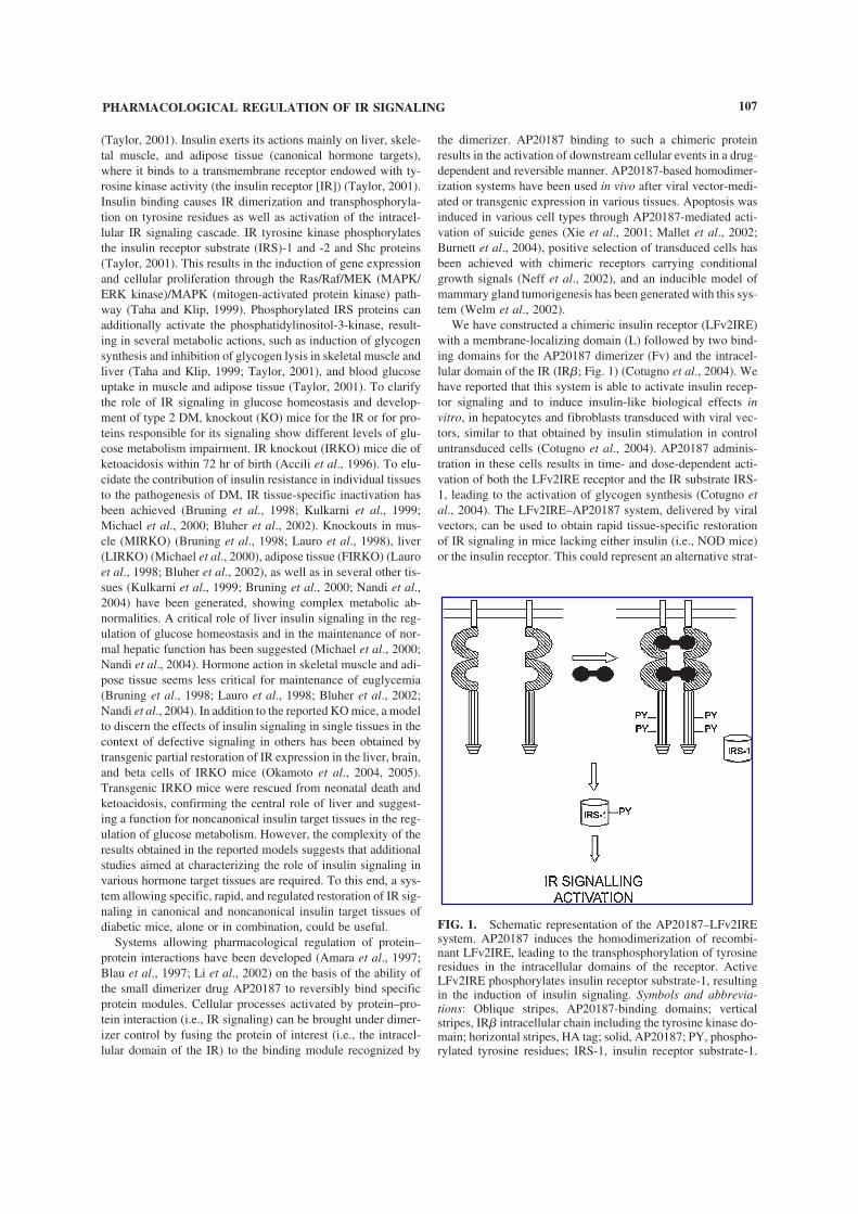
Figure 7 Schematic representation of the AP20187ndashLFv2IRE systemhelliphelliphelliphelliphelliphelliphellip47
Figure 8 Protein tyrosine phosphorylation in AAV-transduced livers upon
AP20187 administration time dependency of protein phosphorylationhelliphelliphelliphellip49
Figure 9 LFv2IRE expression and protein tyrosine phosphorylation in
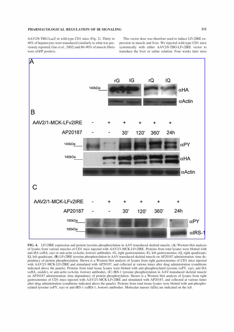
AAV-transduced skeletal muscleshelliphelliphelliphelliphelliphelliphelliphelliphelliphelliphelliphelliphelliphelliphelliphelliphelliphelliphellip51
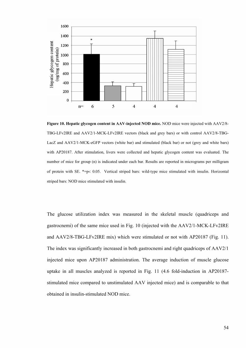
Figure 10 Hepatic glycogen content in AAV-injected NOD micehelliphelliphelliphelliphelliphelliphelliphelliphellip54
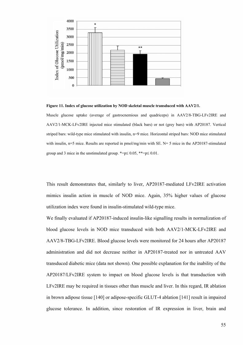
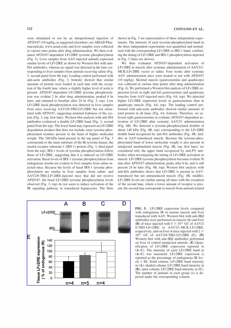
Figure 11 Index of glucose utilization by NOD skeletal muscle transduced
with AAV21helliphelliphelliphelliphelliphelliphelliphelliphelliphelliphelliphelliphelliphelliphelliphelliphelliphelliphelliphelliphelliphelliphelliphelliphelliphellip55
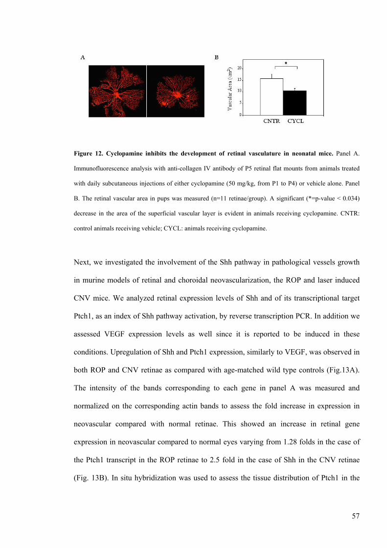
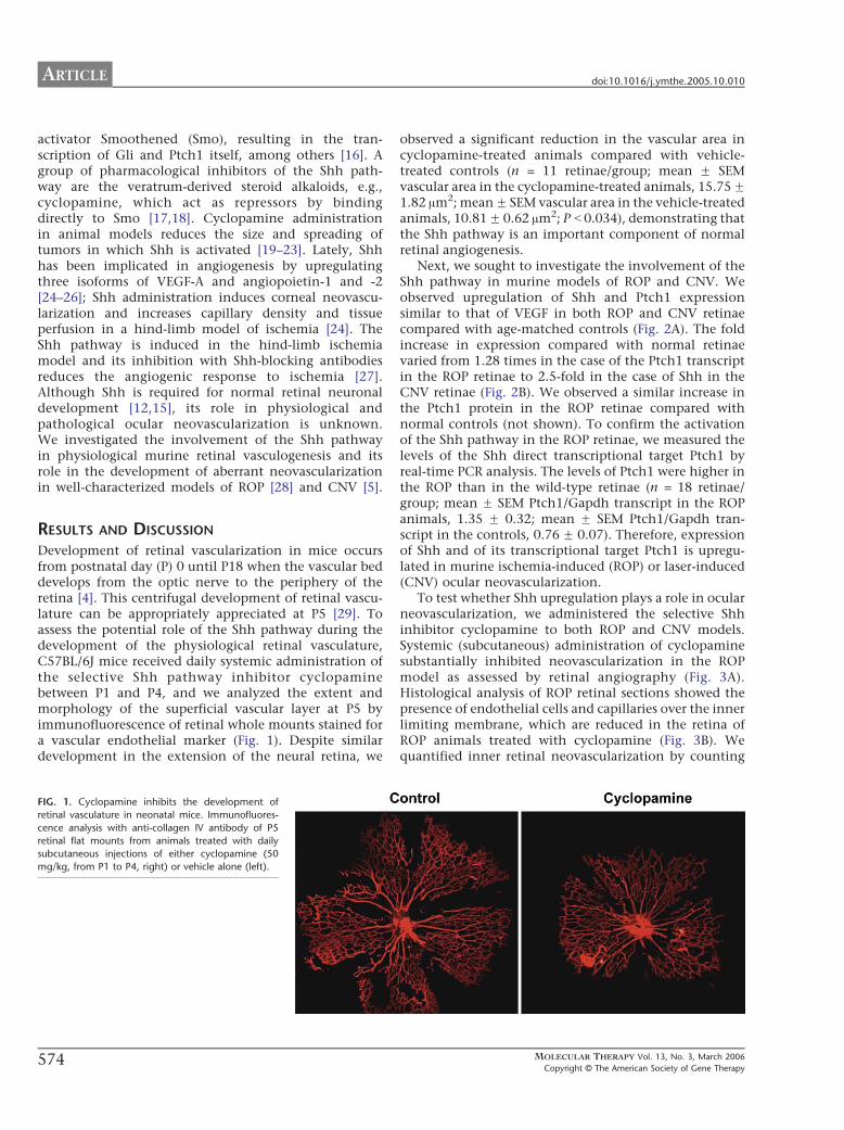
Figure 12 Cyclopamine inhibits the development of retinal vasculature in
neonatal micehelliphelliphelliphelliphelliphelliphelliphelliphelliphelliphelliphelliphelliphelliphelliphelliphelliphelliphelliphelliphelliphelliphelliphelliphelliphelliphellip57
Figure 13 Upregulation of the Shh pathway in the retina of animal models
with neovascular diseasehelliphelliphelliphelliphelliphelliphelliphelliphelliphelliphelliphelliphelliphelliphelliphelliphelliphelliphelliphelliphelliphellip58
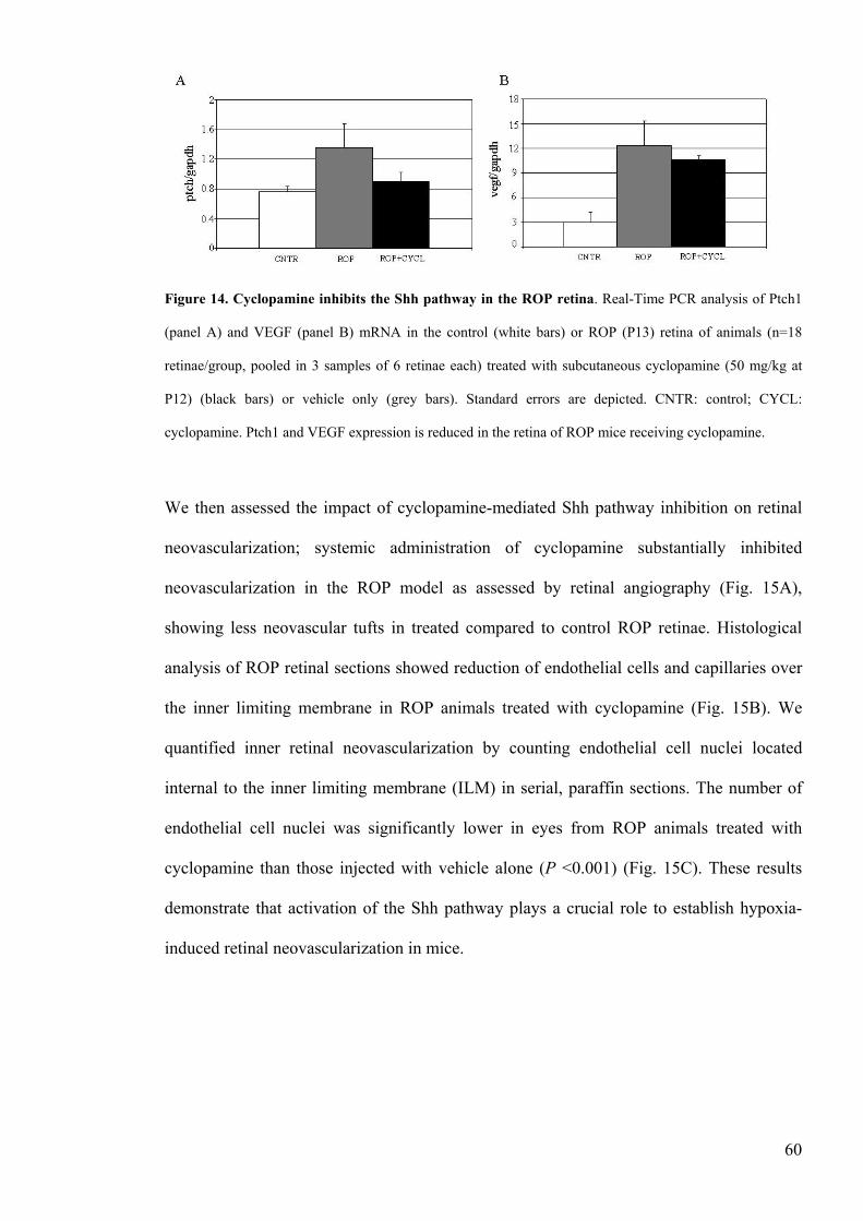
Figure 14 Cyclopamine inhibits the Shh pathway in the ROP retinahelliphelliphelliphelliphelliphelliphelliphellip60
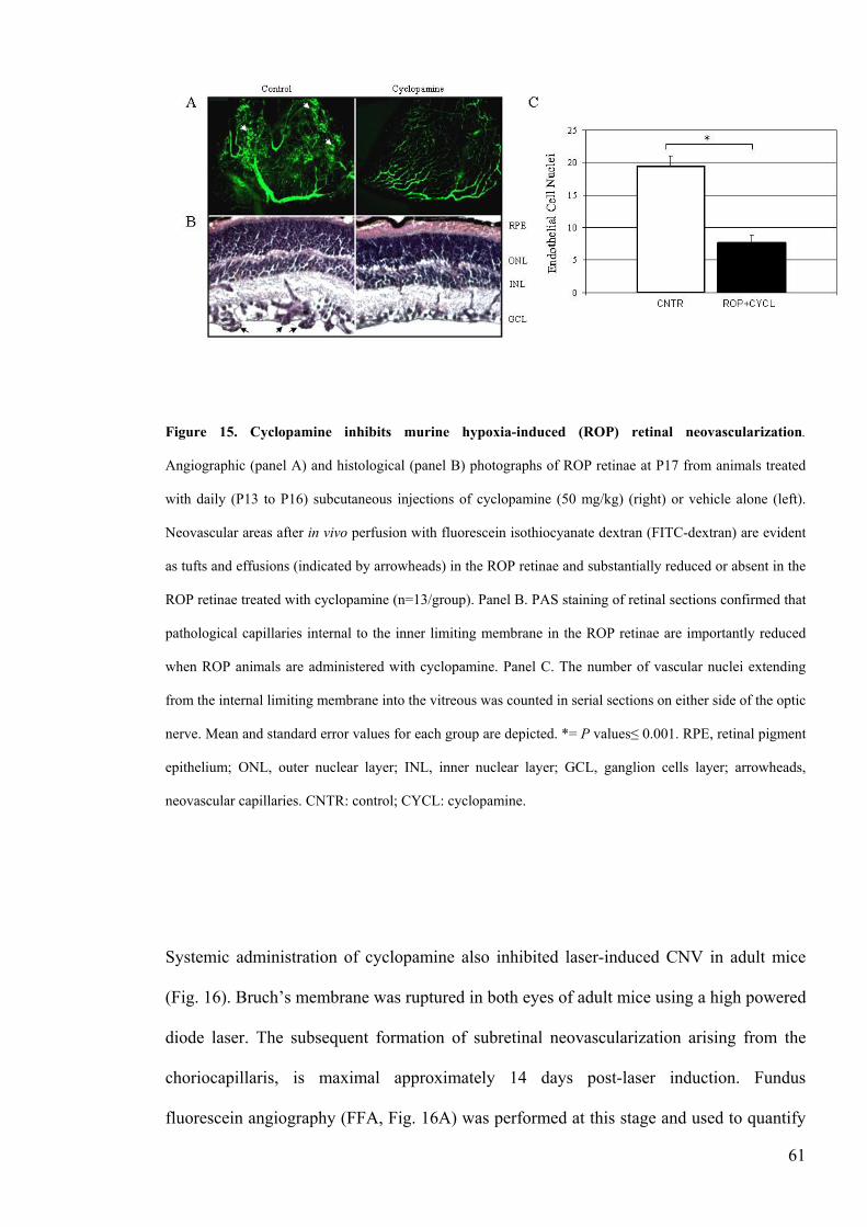
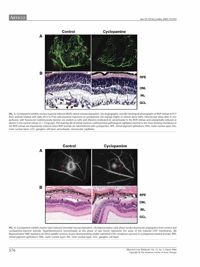
Figure 15 Cyclopamine inhibits murine hypoxia-induced (ROP) retinal
neovascularizationhelliphelliphelliphelliphelliphelliphelliphelliphelliphelliphelliphelliphelliphelliphelliphelliphelliphelliphelliphelliphelliphelliphelliphellip61
Figure 16 Cyclopamine inhibits murine laser-induced choroidal neovascularizationhelliphellip63
Figure 17 Schematic representation of strategies for inhibition of Shh actionhelliphelliphelliphellip64
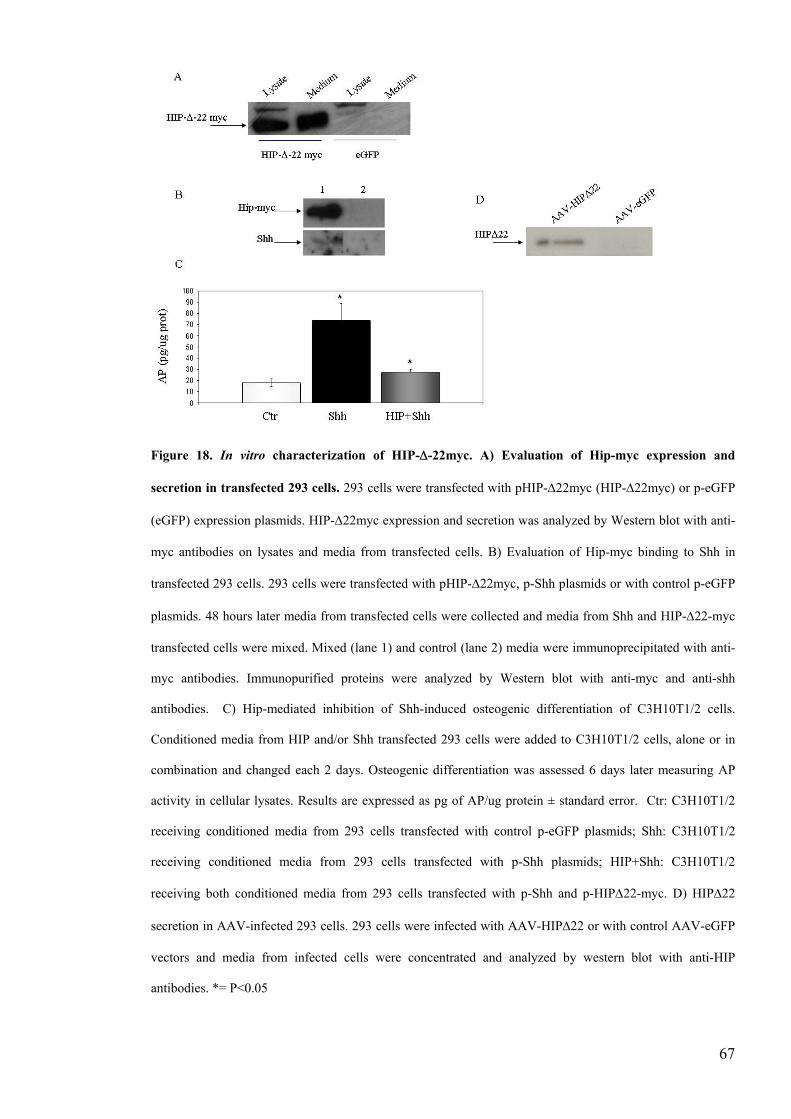
Figure 18 In vitro characterization of HIP-Δ-22mychelliphelliphelliphelliphelliphelliphelliphelliphelliphelliphelliphelliphelliphellip67
6
Figure 19 Shh siRNA reduces Shh expression and activity in vitrohelliphelliphelliphelliphelliphelliphelliphellip69
Figure 20 Efficient intraocular delivery of anti-Shh moleculeshelliphelliphelliphelliphelliphelliphelliphelliphelliphellip71
Figure 21 Shh siRNA reduces Shh expression in vivo in rop micehelliphelliphelliphelliphelliphelliphelliphelliphellip72
Figure 22 Shh siRNA and HIP-Δ-22 reduce Ptch1 expression in vivo in the
ROP retinahelliphelliphelliphelliphelliphelliphelliphelliphelliphelliphelliphelliphelliphelliphelliphelliphelliphelliphelliphelliphelliphelliphelliphelliphelliphelliphelliphellip74
Figure 23 AAV-mediated HIP-Δ22 expression in ROP retinae reduces Shh
induced Ptch1 expressionhelliphelliphelliphelliphelliphelliphelliphelliphelliphelliphelliphelliphelliphelliphelliphelliphelliphelliphelliphelliphelliphellip75
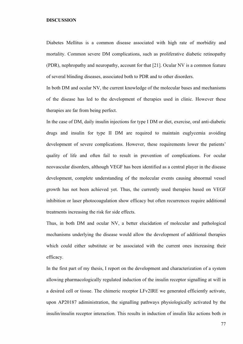
Figure 24 Intraocular inhibition of the Shh pathway does not impact
on retinal neovascularizationhelliphelliphelliphelliphelliphelliphelliphelliphelliphelliphelliphelliphelliphelliphelliphelliphelliphelliphelliphelliphellip76
7
ABSTRACT
Ocular neovascularization (NV) is a feature of several common retinal and choroidal
blinding diseases including proliferative diabetic retinopathy and age-related macular
degeneration Unbalanced production of pro- vs anti-angiogenic molecules in the eye
causes abnormal vessel growth Although several pro-angiogenic pathways leading to
ocular NV have been elucidated the identification of novel molecules involved in this
complex process is desirable to better understand the disease pathogenesis and to develop
efficient therapeutic strategies To this aim we investigated the role of the morphogen
Sonic Hedgehog (Shh) in the development of ocular NV
We observed that the Shh pathway is activated in the retina of the retinopathy of
prematurity (ROP) and the laser-induced choroidal NV (CNV) murine models of retinal
and choroidal neovascularization respectively We show that systemic administration of
cyclopamine a Shh pathway inhibitor results in reduction of pathological vascularization
in both models suggesting that activation of the Shh pathway plays an important role in
the ocular NV process We then developed two nucleic acid-based systems for specific Shh
inhibition in the retina a Shh-decoy receptor (HIP-Δ-22) able to bind and sequester Shh
inhibiting its pathway and short interfering RNAs (siRNA) able to reduce gt70 Shh
expression levels in vitro Both HIP-Δ-22 and the siRNA inhibited Shh-induced osteogenic
differentiation of the mesenchymal cell line C3H10T12 In the ROP retina adeno-
associated viral vector-mediated HIP-Δ-22 delivery or periocular injections of Shh siRNA
resulted in efficient inhibition of the Shh pathway but not of retinal neovascularization
even when the two strategies were combined Stronger inhibition of the Shh pathway may
be required to reduce retinal NV in the ROP model Alternatively the inhibition of ocular
NV observed following systemic cyclopamine administration may result from secondary
extraocular effects of the Shh pathway blockade These results suggest Shh as a potential
8
therapeutic target for the treatment of ocular NV Thorough characterization of Shh role in
ocular NV is required for the development of an appropriate therapeutic strategy
9
INTRODUCTION
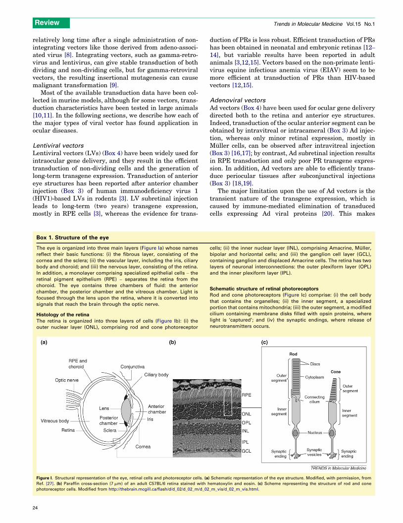
The Eye structure and function
The eye is a complex organ with the function of capturing light allowing vision
It is organized into three main layers (Fig 1) [1]
- A fibrous external layer with structural and protective functions
It consists of the sclera a protective layer located on the posterior part of the eye and the
cornea which is an outer continuation of the sclera and is transparent in order to allow the
light to enter the eye Because transparency is of prime importance the cornea does not
have blood vessels it receives nutrients via diffusion from the tear fluid at the outside and
the aqueous humour at the inside
-A vascular layer supplying nutrients to the eye structures
It includes the choroid a pigmented vascularized layer located between the sclera and the
retina (see below) and the iris a thin diaphragm composed mostly of connective tissue and
smooth muscle situated behind the cornea In the middle of the iris is the pupil a circular
hole that regulates the amount of light passing through to the retina which is at the back of
the eye The light that enters the eye is refracted on the retina by the crystalline lens a
transparent structure located immediately behind the iris it is suspended in place by
suspensory ligaments connected to the ciliary body a muscular ring that regulates the lens
shape to change the focal distance of the eye so that it can focus on objects at various
distances
-A nervous layer consisting of the retina representing the light sensitive part of the eye
(Fig 2)
Retina itself is organized into three layers of cells the outer nuclear layer (ONL)
containing rod and cone photoreceptors the inner nuclear layer (INL) comprising
Amacrine Muller bipolar and horizontal cells and the ganglion cell layer (GCL)
containing ganglion cells and two layers of neuronal interconnections the outer plexiform
layer (OPL) and the inner plexiform layer (IPL)
In addition a monolayer comprising specialized epithelial cells ndash the retinal pigment
epithelium (RPE) ndash separates the retina from the choroid The membrane located between
the RPE and the choroid is called Bruchrsquos membrane
Figure 1 schematic representation of the eye The eye is a complex organ organized into three main
layers a fibrous external layer consisting of the cornea and the sclera a vascular layer containing the
choroids the iris and the ciliary body a nervous layer consisting of the retina Three chambers containing
fluid are delimited the anterior the posterior and the vitreal chamber
Photoreceptors in the retina are a specialized type of neuron able to convert light stimuli
into electric impulses These signals are then transmitted through the bipolar cells to
ganglion cells whose axons leave the retina from the optic disk to form the optic nerve
Thus visual information is carried from the eye to the visual centres of the brain
Muller cells represent the principal glial cell of the retina They form architectural support
structures across the thickness of the retina and form the so called outer and inner limiting
10
11
membranes (OLM and ILM) (Fig 2) Muller cell bodies sit in the inner nuclear layer and
project irregularly thick and thin processes in either direction to the outer limiting
membrane and to the inner limiting membrane Muller cell processes insinuate between
cell bodies of the neurons in the nuclear layers and envelope groups of neural processes in
the plexiform layers The outer limiting membrane is formed by junctions between Muller
cells and other Muller and photoreceptor cells The inner limiting membrane on the other
hand is formed by the conical endfeet of the Muller cells
The eye is divided into three main spaces or chambers (Fig 1) The largest is the vitreous
chamber between the lens and the retina filled with the amorphous and somewhat
gelatinous material of the vitreous body This material serves mainly to maintain the eyes
shape The anterior and posterior chambers also play a major role in maintaining the eye
normal shape by balancing the production and drainage of aqueous humor the fluid which
fills both of them These two fluid-filled chambers are separated from each other by the iris
and are in communication via the pupil the anterior chambers boundaries are the cornea
and the iris the posterior chamber is demarcated by the iris and the lens (Fig 1)
Figure 2 Schematic representation of retinal layers The different layers of the retina are shown and listed
on the right Outer segments of photoreceptor (PRs) are specialized membrane structures where the light is
captured
Organization and development of the ocular vasculature
In most mammals the adult retina is vascularized by two independent circulatory systems
the choroid and the retinal vessels (Fig3) During the initial development of the eye the
oxygenation of the retina is ensured by the choroidal vessels and the hyaloid system [2]
The vascularization of the retina itself occurs only during late gestation and is restricted to
the inner part of the retina with the outer retina completely avascular to ensure visual
function [2] The hyaloid vessel system is a dense but transient intraocular circulatory
system that undergoes progressive and nearly complete regression during the latest stage of
ocular development as the lens the vitreous and the retina mature [3]
12
Figure 3 Distribution of retinal and choroidal vasculature The adult retina receives oxygen and nutrients
from choroidal vessels (on the top) and from two different retinal vascular beds the deep vascular layer at
the junction between outer plexiform layer and inner nuclear layer and the superficial vascular bed in the
inner part of the retina
The choroidal vascular system forms during early development deriving from the neural
tube vessels and extending around the outer layer of the optic cup During the second and
third month of gestation this primitive plexus is then organized in a complex vascular
network that remains separate from neural retina by the basement membrane of the RPE
[2] The development of choroidal vasculature depends on the presence of differentiated
RPE cells and their production of inductive signals such as Vascular Endothelial Growth
Factor (VEGF) and basic Fibroblast Growth Factor (bFGF) [2]
Retinal vasculature development in humans starts at the fourth month of gestation with
the primitive vessels emerging form the optic disk and extending during the next four
months to the periphery of the retina [2] The formation and maturation of retinal vascular
network is completed only after birth This network is organized into two planar layers a
deep vascular plexus at the junction between the INL and the OPL and a superficial
vascular network on the inner surface of the retina (Fig 3) [1] Retinal vessels
development follows the differentiation of neural cells as retina matures an increase in
13
14
neuronal activity with increased metabolic demand leads to development of physiological
hypoxia in the avascular retina [4] This hypoxic condition induces VEGF production by
two different types of microglial cells the astrocytes located in the ganglion cell layer of
the retina and the Muller cells in the INL [5] VEGF expression can be indeed induced by
hypoxia through the activation of a hypoxia-inducible transcription factor (HIF) [6]
VEGF in turn induces vascular growth with sprouting of endothelial cells towards retinal
edges Behind the front of vascularization the increased oxygen supply suppresses VEGF
expression thereby preventing excessive vascular growth [2] The absence of VEGF a
well known endothelial cell survival factor can induce apoptosis of endothelial cells and
thus obliteration of undifferentiated vessel allowing remodeling of capillary network in
order to meet the metabolic needs of the retina [7]
Ocular Neovascularization and related diseases
Different pathological conditions are characterized by abnormal vessel growth in the eye a
phenomenon called ocular neovascularization The neo-vessels can derive from different
ocular vascular beds choroidal neovascularization (CNV) involves the choroidal
vasculature while retinal neovascularization (NV) affects the retinal vasculature
Unbalanced production of pro-angiogenic signals including VEGF angiopoietins [8] or
insulin-like growth factor-1 (Igf-1) [9] and anti-angiogenic molecules such as Pigment
Epithelial Derived Factor (PEDF) [10] in the eye induces vessel growth in these
conditions The newly formed vessels do not generate an organized vascular network and
growth irregularly In addition their permeability is altered and this usually leads to
haemorrhages and damage to ocular tissues [2]
Age Related macular Degeneration and Choroidal Neovascularization
Age related macular degeneration (AMD) is the most common cause of blindness in
individuals older than 65 years in developed countries AMD is a degenerative disorder of
the retina affecting the macula an anatomic structure of the primate retina with the highest
cone photoreceptors concentration and responsible for acute central vision the key lesion
of ARM is the formation of drusen aggregations of hyaline material located between
Bruchrsquos membrane and the retinal pigment epithelium This is associated with atrophy and
depigmentation of the overlying retinal pigment epithelium [11]
AMD is classified into two major forms the dry (non-exudative) and the wet (exudative)
type Dry AMD is due to a slow and progressive degeneration of the photoreceptors with
RPE hypo- or hyper-pigmentation and gradual failure of central vision [11]
Wet AMD is characterized by the pathologic outgrowth of new vessels from the choroid
(CNV) This type of macular degeneration may have rapid and devastating effects upon
vision In contrast with patients with dry AMD in whom impairment of vision is gradual
central vision may be lost over the course of a few days due to the neo-formed vascular
tufts that extend in the subretinal space causing accumulation of fluid or blood in the
posterior part of the retina [211] This can lead to the detachment of the RPE or the retina
resulting in vision loss (Fig 4)
Figure 4 Localization of choroidal neovascular tufts Choroidal neovasularization (CNV) is characterized
by abnormal vessels growth between the retina and the choroid leading to retinal detachment and blindness
15
16
It is not clear what is the primary stimulus for the development of CNV It is possible that
an hypoxic condition of the retina is involved maybe alteration of choroidal blood flow or
the thickening of Bruchrsquos membrane with lipophilic material could result in decreased
diffusion of oxygen from the choroids to the RPE and retina but therersquos no clear data to
proof this hypotesis [12]
The most common pathologic finding in wet AMD is accumulation of abnormal
extracellular matrix and thickening of Bruchrsquos membrane which can cause increased
secretion of pro-angiogenic growth factors from RPE cells such as VEGF and Fibroblast
Growth Factor 2 (FGF2) contributing to CNV development [12]
Retinal Neovascularization
In normal circumstances the blood vessels of the adult retina are quiescent with respect to
growth [13] However several pathological conditions are characterized by rapid and
abnormal retinal vessels proliferation including proliferative diabetic retinopathy (PDR)
and retinopathy of prematurity (ROP) with the new vessels usually growing outside the
retina and in the vitreous [2] All these conditions are characterized by the presence of non-
perfused and therefore hypoxic retinal tissues as a precedent to the NV [2] increased
VEGF levels in the retina and vitreous of patients and animal models with ischemic
retinopaties have been found suggesting that this factor might have a role in NV
development [12] Indeed VEGF inhibition results in reduction of retinal NV in animal
models and humans and its ectopic expression in PRs is sufficient to stimulate NV in
murine retina [141516]
17
Retinopathy of prematurity
Since vascularization of the human retina takes place in the final trimester of gestation a
premature infant has an incompletely vascularized retina in which ldquophysiologic hypoxiardquo
has induced VEGF expression Placement of an infant into high oxygen to alleviate
respiratory distress suppresses VEGF expression leading to the cessation of vessel growth
a phase of ROP termed vaso-obliteration Once the infant is returned to room air the
retina lacking its normal vascular network becomes hypoxic leading to VEGF
upregulation and abnormal new vessels growth [2] Often the neovascular processes
regress spontaneously in 6-12 weeks [17]
Diabetes Mellitus and Proliferative Diabetic Retinopathy
One of the most common causes of ocular NV is Proliferative Diabetic Retinopathy (PDR)
which is a complication of Diabetes Mellitus (DM) DM is a metabolic disease
characterized by elevated blood glucose levels (hyperglycaemia) resulting from defects in
either insulin secretion or action Insulin is produced by pancreatic beta cells and released
in response to stimuli such as increases in circulating glucose levels Insulin exerts its
actions mainly on liver skeletal muscle and adipose tissue (canonical hormone targets)
where it binds to a transmembrane receptor endowed with tyrosine kinase activity (IR)
[18] Insulin binding causes IR dimerization and transphosphorylation upon tyrosine
residues as well as activation of the intracellular IR signalling cascade IR tyrosine kinase
phosphorylates the insulin receptor substrate (IRS)-1 and -2 and shc proteins [18] This
results in the induction of gene expression and cellular proliferation through the
RasRafMEKMAPK pathway [19] Phosphorylated IRS proteins can additionally activate
the phosphaditilinositol-3-kinase resulting in several metabolic actions such as induction
of glycogen synthesis and inhibition of glycogen lysis in skeletal muscle and liver [1819]
and blood glucose uptake in muscle and adipose tissue [18] thus resulting in reduction of
glycaemia Insulin deficiency due to autoimmune destruction of pancreatic β-cells causes
type 1 DM [20] This condition is treated by daily subcutaneous injection of recombinant
18
insulin The most common type 2 DM is caused by insulin resistance in the hormone target
tissues combined with deficient hormone secretion by pancreatic β-cells [18] The deriving
hyperglycemia can be controlled by diet and exercise oral anti-diabetic drugs or insulin
injections [18] The metabolic and biochemical changes associated with DM such as
hyperglycemia associates with protein glycosilation and alteration of several metabolic
pathways increased levels of sorbitol and reduced synthesis of phosphoinositides [21] All
of these changes are related to induction of severe complications of the DM such as PDR
Diabetic Nephropathy and Neuropathy as well as cataract and increased risk for
atherosclerosis development [21]
Ocular pathology is one of the most devastating complications of Diabetes Mellitus (DM
see below) PDR associates with changes in the retinal vasculature including vessel
dilation increased permeability basement membrane thickening loss of pericytes and
formation of microaneurysms [2] These vascular changes reflect the chronic damage
sustained by the vasculature as a result of metabolic alterations including hyperglycemia
associated with DM and lead to vascular dysfunction and loss [2] The ischemia that results
from the loss of vessel perfusion leads to increased expression of pro-angiogenic factors
and vessel growth The new vessels growing outside the retina into the vitreous are leaky
due in part to the permeability-inducing effects of VEGF that is up-regulated in the
hypoxic retina Formation of a fibrous membrane in combination with traction caused by
vitreous attachments can lead to retinal detachment and blindness [2]
Treatment of ocular neovascularization
Clinical management of ocular neovascularization is performed with different therapeutic
strategies Laser photocoagulation is widely used for the treatment of these conditions it
uses the heat generated with a laser on specific regions of the eye to seal or destroy
abnormal leaking blood vessels in the retina or the choroid
Laser therapy is destructive by design indeed some retinal tissue is intentionally destroyed
(sacrificed) in order to preserve the function of other more visually important areas
thereby reducing the chance of more serious vision loss and blindness As a result patients
very often experience a loss of peripheral (side) vision abnormal blind spots and reduced
ability to see at night or in dimly lit environments (Fig 5)
Figure 5 Representation of an eye with CNV subjected to laser photocoagulation The heat generated
by a laser is directed to specific regions of the retina (A)This heat cauterizes the CNV seals it and stops it
from growing leaking and bleeding However tissues in and around the CNV process are also cauterized
and following treatment a scar will form creating a permanent blind spot in the field of vision (B)
Recent advances in the elucidation of the molecular mechanisms underlying ocular
neovascularization led to the identification of VEGF as a central player in the development
of both retinal and choroidal NV This have allowed the development of
biopharmacological treatment of ocular NV based on inhibition of VEGF action Three
different anti-VEGF agents have been produced and extensively tested for their ability to
reduce ocular neovascularization associated with different pathological conditions A
pegylated aptamer (pegaptanib) a monoclonal antibody (bevacizumab) and an antibody
fragment (ranibizumab) targeting human VEGF have been produced and administered to
patients with retinal or choroidal NV in several clinical trials [222324252627] These
19
20
drugs are currenty used in clinical practice [22] resulting in regression of
neovascularization in patients with different ocular NV diseases [2324252627] In most
cases anti-VEGF molecules are delivered via intravitreal injections [2324252627] and
require repeated administration to result in significant therapeutic efficacy In addition the
therapeutic effect is often transient with additional progression of the neovascularization
after the termination of the therapy In addition intravitreal injection is an invasive
procedure associated with potentially serious complications such as endophtalmitis or
retinal detachment which may be significant for patients requiring serial treatments over
many years [282930]
Animal Models of Retinal Neovascularization
Animal models of retinal and choroidal neovascularization have been generated an
extensively used to improve knowledge about molecular bases of ocular neovascular
diseases and to test efficacy of experimental therapies for these conditions
Two types of animal models of retinal neovascularization exist the most commonly used
is the Retinopathy of Prematurity (ROP) mouse in which a condition similar to what is
observed in premature infants developing retinal neovascularization is generated [31] In
mice retinal vessels development takes place after birth with the growing vessels
extending from the optic disk and reaching retinal edges at postnatal day (P-) 17 Thus the
vascular network of murine retina at P7 closely resembles that of premature infants with
ongoing regression of hyaloid vessels and incomplete development of retinal vasculature
to induce NV mice are exposed to high oxygen percentage (75) from P7 to P12 this
reduces the physiological hypoxia normally present in the retina at this time point blocking
the normal retinal vessels growth When mice are returned to room air the retina showing
incomplete vasculature becomes hypoxic and this leads to de-regulated activation of pro-
angiogenic stimuli and induction of retinal neovascularization [31] Retinal NV develops in
100 of these mice between P17 and P21 Murine ROP retina shows a non-perfused
central region and peripheral neovascular tissue with vascular tufts extending beyond the
internal limiting membrane into the vitreous [31] retinal NV in this model can be assessed
by intracardiac perfusion with fluorescein-labelled high molecular weight albumin
followed by analysis of retinal flat mounts under a fluorescence microscope (Fig 6A) In
addition counting the number of endothelial cell nuclei on the vitreal side of the inner
limiting membrane in retinal cross sections allows precise quantification of NV (Fig 6B)
Retinal NV can be induced even in rats [32] newborn rats are exposed to variable oxygen
between 40 an 80 in a cyclic fashion for 14 days and then brought to room air for 4
days About 62 of the animals develop retinal NV in these settings [33]
Figure 6 Evaluation of retinal neovascularization in ROP mice
A) Retinal flat mount of fluorescein-perfused ROP mice showing the classical appearance of retinal vessels
with absence of vessels in the central part and disorganized vascular network at the periphery Regions of
hyperfluorescence represent points of fluorescein effusion due to vessels leakiness (white arrows)
B) Paraffin cross sections of ROP retina showing neo-vessels on the vitreal side of the inner limining
membrane (black arrows) The number of neo-vascular nuclei can be counted to quantify the extent of retinal
NV
21
22
The other types of retinal neovascularization models is obtained without oxygen exposure
in spontaneous hypertensive rats with extensive retinal degeneration in which retinal
vessels first migrate towards the RPE and then grow beyond the inner limiting membrane
similarly transgenic mice expressing VEGF in photoreceptors show new vessels arising
from retinal vasculature and growing in the subretinal space demostrating that increased
expression of VEGF in the retina can stimulate intraretinal and subretinal NV [14]
The most commonly used model of choroidal neovascularization is the laser induced
model in which rupture of the Bruchrsquos membrane is caused by laser photocoagulation This
results in inflammatory response to the laser injury and CNV
This strategy has been used to induce CNV in primates [34] rats [3536] rabbits [37] and
mice [38] Despite similarities with AMD-associated CNV in humans the laser model may
not be appropriate for studies of mechanisms of initiation of CNV since therersquos acute
extensive damage of retinal tissue and Bruchrsquos membrane with the laser treatment that is
not seen in clinical CNV However this model has been extensively used to assess efficacy
of anti-neovascular therapies The choroidal neovascularization can be evaluated by
Fundus Fluorescein Angiograms (FFA) and measurement of the areas of hyperfluorescence
or by evaluation of subretinal CNV complexes in paraffin cross sections [12]
Experimental therapies for ocular neovascularization
Since actual therapies for ocular NV despite showing therapeutic efficacy have several
side-effects and often result in relapses strategies for safe and long term inhibition of
ocular neovascularization based on ocular gene transfer of anti-angiogenic factors are
being evaluated (see attached PDFs [1516] ) Molecules able to inhibit VEGF expression
or action represent a promising tool to this aim given the proven involvement of VEGF in
different neovascular pathologies of the eye Long term intraocular production of anti-
VEGF molecules can be achieved by intraocular gene transfer via viral vectors (see
23
below) The soluble form of the Flt-1 VEGF receptor (sFlt-1) which acts as an endogenous
specific inhibitor of VEGF has been delivered to the eye via intra- or peri-ocular injection
of different viral vectors resulting in reduction of NV in various models of CNV and
retinal NV [39404142] In addition the inibition of VEGF gene expression at the level of
the messenger RNA has been achieved in ocular NV models Short RNA duplexes called
short interfering RNAs (siRNAs) can cause the sequence specific degradation of a target
mRNA The siRNA can be exogenously administered or produced in situ from longer
precursors (short hairpin RNA shRNA) that can be expressed in the target cells (ie
delivered by a gene therapy vector) and cleaved to produce the siRNA by intracellular
protein complexes [4344] SiRNA and viral-vector delivered shRNA directed to VEGF or
molecules involved in VEGF signalling pathways have been tested in murine models of
ocular NV resulting in inhibition of both retinal and choroidal NV [454647] In addition
to anti-VEGF molecules molecules endowed with anti-angiogenic activity are being tested
for their ability to inhibit ocular NV Among them pigment epithelium-derived factor
(PEDF) is one of the most representative PEDF is an anti-angiogenic molecule responsible
for inducing and maintaining the avascularity of the cornea and vitreous compartments in
physiological conditions [10] PEDF gene transfer inhibits both retinal and choroidal NV
in animal models [39484950] The results obtained in pre-clinical studies allowed the
development of a phase I clinical trial in patients with AMD-associated choroidal NV
(CNV) based on intravitreal injections of viral vectors encoding PEDF No major toxic
effects were associated with vector administration and preliminary therapeutic efficacy has
been reported at the highest vector dose [51] The identification of additional
antiangiogenic factors such as angiostatin [52] endostatin [53] and tissue inhibitor of
metalloprotease (TIMP)-3 [54] has provided novel tools to inhibit ocular NV Angiostatin
is a proteolytic fragment of plasminogen encompassing the first four kringle domains of
the molecule Angiostatin [55] and its recombinant derivative K1K3 (containing only the
first three kringles) [56] have antiangiogenic properties and their intraocular expression
24
obtained with viral vector mediated gene transfer resulted in significant reduction of
choroidal and retinal NV in animal models [57] Endostatin is a cleavage product of
collagen XVIII that is able to reduce choroidal NV when delivered systemically [58]
TIMP3 is a potent angiogenesis inhibitor able to block VEGF signalling [58] Viral vector-
mediated expression of these factors in the eye resulted in inhibition of ischemia-induced
retinal NV [58]
Although inhibition of VEGF seems a powerful strategy for treatment of ocular NV the
identification of additional molecules involved in neovascular processes andor showing
anti-angiogenic properties would allow development of additional therapeutic strategies
that alone or in combination with anti-VEGF molecules could allow effective and long
term inhibition of ocular NV in different conditions to this aim the development of
systems able to provide efficiently and long-term intraocular anti-angiogenic factors
represents a requirement
Gene therapy and ocular gene transfer
Long term intra-ocular production of a desired molecule can be achieved by introduction
of genetic material encoding for the protein into target cells of the eye (gene transfer) This
is usually done using viral vectors generated by modification of parental viruses the viral
genome is partially or completely deleted of viral genes which are generally substituted by
an expression cassette containing the coding sequence for the desired protein downstream
of an ubiquitous or a tissue specific promoter Different viral vectors able to efficiently
transduce ocular cells are available [16]
For most vectors the administration route to be used is largely dependent on the targeted
ocular cell type Subretinal injections expose the outer retina (PRs and RPE) whereas
intravitreal injections expose the anterior retina (retinal ganglion cells) to the nucleic acid-
based therapeutic Vectors commonly used for ocular gene transfer are adenoviral
25
lentiviral and adeno-associated viral (AAV) vectors as we reviewed in the attached PDF
[16] Among these vectors AAV represent the most promising ones given their ability to
efficiently transduce various ocular cell types resulting in long lasting expression of the
encoded gene (transgene) Generation of AAV vectors is obtained by deletion of all viral
coding sequences and insertion of the expression cassette between the inverted terminal
repeats (ITRs) of the viral genome The existence of dozens of adeno-associated virus
serotypes has allowed generation of hybrid vectors the same AAV vector genome (usually
derived from AAV serotype 2) is included in external surface proteins (capsids) from other
AAV serotypes the resulting recombinant vectors are indicated as lsquoAAV2nrsquo with the first
number indicating the genome (ie AAV2 in this case) and the second the capsid [59]
different rAAV serotypes have different tropism and transduction characteristics The
ability of the various AAV serotypes to transduce ocular structures has been extensively
documented with vectors encoding marker proteins showing that a combination of
serotypes injection route and promoters allows selective transduction of different cellular
populations The viral serotypes AAV25 AAV27 AAV28 and AAV29 are the most
efficient for transduction of PRs after subretinal injection AAV29 vectors in addition to
PRs efficiently transduce Muller cells [60] while transduction of ganglion cells can be
achieved by intravitreal injection of either AAV22 or AAV28 vectors [61] RPE is
efficiently transduced by most AAV serotypes upon subretinal injection those that have a
predominant RPE tropism in the murine retina are AAV21 and AAV24 [596263]
AAV21-mediated RPE transduction has been used as a strategy for intraocular delivery of
secreted molecules by inducing the production of the desired factor in the RPE cells
resulting in its secretion into ocular chambers [64]
In addition several reports have shown AAV vectors ability to efficiently transduce for
long-term several other organs including brain [656667] β-cells [68] skeletal muscle
[69] and liver [70] after systemic or local injections Systemic administration of AAV21
vectors results in body-wide and robust skeletal muscle transduction [71] Similarly
26
administration of vectors with AAV8 capsids (AAV28) results in high levels of liver
transduction [72]
Sonic hedgehog and ocular neovascularization
The current knowledge of the pathogenetic mechanisms underlying ocular neovascular
diseases has allowed to develop therapies based on biological drugs Nevertheless
identification of new molecular players and definition of their hierarchy in this process will
allow to better understand the molecular bases of these disorders and to develop of
additional effective therapies to be combined with or substituted to those actually used to
achieve better efficacy
Sonic hedgehog (Shh) is a secreted morphogen implicated in a multiplicity of
developmental and post-natal processes [7374] Together with the other hedgehog genes
(Indian and Desert Hedgehog) it is crucial for the formation of lung limb gut and bone
[7576777879808182] in addition its signalling regulates the proliferation of distinct
cell types via direct activation of genes involved in the progression of the cell cycle
[8384] In adult tissues several evidences suggest that uncontrolled activation of the Shh
pathway results in specific types of cancer of brain [8586] skin [878889] pancreas [90]
and lung [91]
Shh exerts its action through the binding to a transmembrane receptor (Patched Ptch1) In
the absence of ligand the Shh signalling pathway is inactive In this case Ptch1 inhibits
the activity of Smoothened (Smo) a seven transmembrane protein The transcription factor
Gli a downstream component of Shh signalling is prevented from entering the nucleus
through interactions with cytoplasmic proteins including Fused and Suppressor of fused
(Sufu) As a consequence transcriptional activation of Hh target genes is repressed
Activation of the pathway is initiated through binding of Sonic hedgehog to Ptch1 Ligand
binding results in de-repression of Smo thereby activating a cascade that leads to the
27
translocation of the active form of the transcription factor Gli to the nucleus [74] Nuclear
Gli activates target gene expression including Ptch1 and Gli itself [74] as well as
Hedgehog interacting protein (Hip) a Shh binding membrane glycoprotein that attenuates
ligand diffusion and so acts as negative regulator of Shh pathway [92] In the eye Shh is
expressed throughout retinal development acting as a precursor cell mitogen [93] while in
differentiated retina it localizes to the ganglion cell layer [939495] Correct retinal
development seems to depend from Shh signalling from ganglion cells [959697] The
subsets of retinal cells that respond to Shh signaling are ganglion cells [98] and astrocytes
([99] in the inner retina and Muller glial cells [95] in the INL expressing Ptch1
The hedgehog pathway can be blocked by using cyclopamine a veratrum-derived steroid
alkaloid which act as antagonists by binding and inhibiting Smo [100] Cyclopamine
administration in animal models reduces the size and spreading of tumors in which Shh is
activated [90101102103104]
In addition to the roles reported here Shh has been implicated in vascularization of
embryonic tissues such as lung [77] expression of Shh receptor Ptch1 on adult
cardiovascular tissues has been found allowing these cells to respond to Shh exogenous
administration [105] Thus Shh seems to be implicated in angiogenesis indeed it is able to
upregulate angiogenic factors including VEGF and angiopoietins 1 and 2 in cultured
fibroblasts [105106] In addition its exogenous administration induces corneal
neovascularization [105] and increases capillary density and tissue perfusion in a murine
model of hind-limb ischemia [107] The Shh pathway is induced in the hind-limb model of
ischemia reperfusion and its inhibition with Shh-blocking antibodies reduces the
angiogenic response to ischemia [107]
Although Shh is required for normal retinal neuronal development [95] [96] [97] its role in
physiological and pathological ocular neovascularization was unknown
28
AIM OF THE THESIS
Diabetes Mellitus is a common disease affecting over 200 million individuals in the world
Severe complications of DM include proliferative diabetic retinopathy (PDR) which
together with wet AMD are associated with ocular NV and represent the most common
causes of vision loss in developed countries
The work of my thesis had two different but related aims 1) to generate gene transfer-
based strategies to obtain glucose homeostasis in DM 2) To develop new therapeutic
strategies for the treatment of ocular neovascular diseases
Towards the first aim I have developed and characterized a gene transfer-based system for
pharmacological regulation of the insulin receptor signalling to selectively mimic insulin
action on a desired insulin target tissue this system represents a tool for studying the role
of insulin action on a specific tissue and to induce glucose uptake and homeostasis as
treatment of DM thus overcoming the requirement of daily insulin injections in type I DM
patients
Toward the second aim we hypotesized that the Shh pathway is implicated in physiological
and pathological ocular NV and applied various strategies for systemic or intraocular
inhibition of the Shh pathway thus assessing its role in ocular vascular development and
developing therapeutic approaches based on Shh blockade for the treatment of retinal and
choroidal NV
29
MATERIALS AND METHODS
Vector Construction and Production
pCLFv2IRE is a CMV expression vector encoding a fusion protein containing the
extracellular and transmembrane portions (amino acids 1-270) of the human low affinity
nerve growth factor receptor (LNGFR) fused to two F36V-FKBP12 ligand binding
domains followed by the cytoplasmic domain of the human insulin receptor and a C-
terminal hemaglutinin epitope (HA) Details of the LNGFR- F36V-FKBP fusion sequences
and expression vector have been described [108109110] The Insulin Receptor
cytoplasmic domain (amino acids 980-1382) was isolated by PCR from a cDNA library
prepared by RT-PCR from human skeletal muscle total RNA (Clontech Palo Alto CA)
The following primers were used 5-
AGCTTCTAGAAGAAAGAGGCAGCCAGATGGGCCGCTG-3 (Forward) and 5-
AGCTACTAGTGGAAGGATTGGACCGAGGCAAGGTC-3 (Reverse) The PCR
product was cleaved with XbaI and SpeI prior to insertion at an XbaI site between the
FKBP and epitope sequences in pCLFv2IRE
The pAAV21-TBG-LFv2IRE pAAV21-MCK-LFv2IRE pAAV21-CMV-HIP-Δ22 and
pAAV21-CMV-HIP-Δ22-myc plasmids used to produce recombinant AAV vectors were
cloned as follows The LFv2IRE fragment was obtained digesting pCLFv2IRE with Eag1
and BamH1 (Roche Basel Switzerland) LFv2IRE was then cloned into pAAV21-TBG-
eGFP [111] previously digested with Not1 and BamH1 (Roche Basel Switzerland)
The 135 Kb muscle specific promoter from the human muscle creatine kinase (MCK)
gene [112] was PCR amplified from human genomic DNA The primers used are the
following 5rsquo-aattagctagctgggaaagggctgggc-3rsquo (Forward) and 5rsquo-
aaatacggccgaggtgacactgacccaa-3rsquo (Reverse) containing the NheI and PstI restriction sites
30
respectively The resulting PCR product was digested NheI-PstI (Roche Basel
Switzerland) and cloned into the pAAV21-TBG-LFv2IRE plasmid previously digested
with the same enzymes to remove the TBG sequence
The HIP-Δ22 sequence was generated by deleting the last 22 codons of the murine HIP
coding sequence this was performed by PCR on C57Bl6 retinal embrionic cDNA with the
following primers Fw- AAGCGGCCGC-
ATGCTGAAGATGCTCTCGTTTAAGCTGCTA Rev- AAGGATCCC-
TACCTGGTCACTCTGCGGACGTT containing Not1 and BamH1 restriction sites
respectively The PCR product was inserted in the Topo Cloning 21 vector (Invitrogen
Life Technologies Carlsbad CA) as suggested by manifacturer sequenced and digested
Not1BamH1 The HIP- Δ22-myc sequence was generated in the same way but we used a
different Rev-primer containing the myc tag sequence a new stop codon and the BamHI
restriction site whose sequence is the following
AAGGATCCCTACAGATCTTCTTCAGAAATAAGTTTTTGTTCCCTGGTCACTCTG
CGGACGTTCCTGTCC
The HIP- Δ22 and HIP- Δ22-myc sequences were then cloned into pAAV21-CMV-eGFP
[111] plasmid previously digested Not1BamH1
The pShh expression plasmid was generated by PCR amplification of human Shh coding
sequence from human retinal cDNA (Clontech Palo Alto CA) with specific primers The
PCR product was inserted in the Topo Cloning 21 vector (Invitrogen Life Technologies
Carlsbad CA) sequenced digested Not1BamH1 and then cloned into pAAV21-CMV-
eGFP [111] plasmid
Recombinant AAV vectors were produced by the TIGEM AAV Vector Core by triple
transfection of 293 cells and purified by CsCl2 gradients [113] Physical titers of the viral
preparations (genome copies gcml) were determined by Real Time PCR (Perkin Elmer
Foster City CA) [114]
31
Anti-Shh siRNA design and production
Five different 19-21nt siRNA oligos targeting regions of sequence identity between human
and murine Shh mRNA were designed using the online Dharmacon siDESIGN center
(wwwdharmaconcom) The 5rsquo-3rsquo target sequence for each siRNA is 1
UUAGCCUACAAGCAGUUUA 2 UGGCGGUCAAGUCCAGCUGAA 3
AAGCUGACCCCUUUAGCCU 4 UUACAACCCCGACAUCAUA 5
GAAGGUCUUCUACGUGAUC Control siRNA targeting eGFP were designed (target
sequence CGAGAAGCGCGAUCACAUG) All of these sequences were blasted against
human and murine genomes to ensure they do not recognize additional sequences The
siRNA were sinthetized by Dharmacon (Lafayette CO) ldquoA4 optionrdquo was used for in vitro
studies while for in vivo administration the ldquoin vivo optionrdquo was used and siRNA were
resuspended in sterile PBS (Invitrogen Life Technology Carlsbad CA) For localization of
siRNA2 in the retina we used BrdU labelled siRNA 2 as previously reported [115] the
siRNA oligos containing BrdU at the 3rsquo end of both sense and antisense strand were
sintetized by Sigma-Proligo (The Woodlands TX USA)
Diabetes Mellitus mouse model vectors administration AP20187 stimulation blood
and tissue collection
To evaluate LFv2IRE expression and tyrosine phosphorylation 4 weeks old CD1 mice
(Harlan Italy S Pietro al Natisone Italy) were injected into the tail vein with 5x10¹¹GC of
the AAV28-TBG-LFv2IRE or AAV21-MCK-LFv2IRE vectors Four weeks later mice
were stimulated or not by intraperitoneal injection of 10 mgkg AP20187 as described
[116117118119120] (ARIAD Pharmaceuticals Cambridge MA wwwariadcom)
32
Liver or muscles were collected at the time points reported in the Results section for
further analysis
NOD mice (Harlan Italy S Pietro al Natisone Italy) were used for the evaluation of the
biological effects of the LFv2IREAP20187 system These mice spontaneously develop
autoimmune insulin-dependent DM between 11 and 15 weeks of age [121] 11-week old
female mice were injected or not with a mix of the AAV28-TBG-LFv2IRE and AAV21-
MCK-LFv2IRE or of the control AAV28-TBG-LacZ and AAV21-MCK-eGFP vectors
(5x10¹¹GCmouse) Plasma glucose levels were monitored weekly by a glucometer (Accu-
Check active Roche) on blood samples obtained via eye bleeding according to
manufacturerrsquos instructions Four weeks after AAV vector injection mice with plasma
glucose levels higher than 250 mgdl were selected and further studied for the evaluation of
hepatic glycogen content and muscle glucose uptake Mice were stimulated or not with
intraperitoneal injection of 10mgkg of AP20187 eighteen and six hours (when they were
fasted to avoid variations in plasma glucose levels) before receiving intravenous injection
of 1μCi of 2-Deoxy[1-3H] glucose (2-DG Amersham Pharmacia Biotech Piscataway NJ)
About 70 μl of blood were collected 1 10 20 and 30 minutes after the injection via eye
bleeding added to 10μl of 5M EDTA and centrifuged at 10000 rpm for 10 minutes
Supernatant were then collected and frozen Skeletal muscles (gastrocnemi and quadriceps)
and livers were dissected 30 minutes after the 2-DG injection and frozen
Control uninjected NOD and CD1 mice were stimulated with insulin (Humulin 075 Ukg
Eli Lilly Indianapolis IN) and hepatic glycogen content and muscle glucose uptake were
measured as described
33
Mouse models of ocular NV vectors administration cyclopamine and siRNA
administration eyes collection
For ocular neovascularization experiments we used murine models of ischemia induced
retinal NV (the ROP mice [31]) and laser induced choroidal NV (the CNV mice [38]) For
generation of the ROP model we used C57BL6J mice (Harlan Italy S Pietro al Natisone
Italy) When reported newborn mice (P2-P3) received subretinal injection of 1x10⁹ gc of
AAV21-CMV-HIP-Δ22 vectors in the right eye and AAV21-CMV-eGFP control vectors
[111] in the left eye To induce retinal NV mice were kept in a chamber with PO2 between
75 and 78 from postnatal day (P) 7 to P12 to block retinal vessels growth [31] At P12
mice were returned to room air until P17 to induce hypoxia in the retina allowing
development of neovascularization [31] When stated ROP mice received daily injections
of either 50mgkg cyclopamine or vehicle alone from P12 to P17 Cyclopamine (Toronto
Research Chemicals Toronto Canada and Biomol Research Labs Plymouth Meeting PA)
was resuspended and administrated as described by Berman et al [102] P17 ROP mice
were deeply anesthetized with avertin (222-tribromoethanol Sigma-Aldrich Milan Italy)
for retinal angiography andor eyes and tissues collection To confirm a role for Shh in
physiological retinal vessels development wild type C57BL6 mice were injected daily
with 50mgkg cyclopamine or vehicle alone from P1 to P4 eyes were then collected at P5
For the Shh RNA interference studies siRNA2 or control siRNA were administered via
subconjunctival injections [39] to ROP mice Briefly the lids were open with a forceps if
required and conjunctiva was lifted up The siRNA was injected under the conjunctiva with
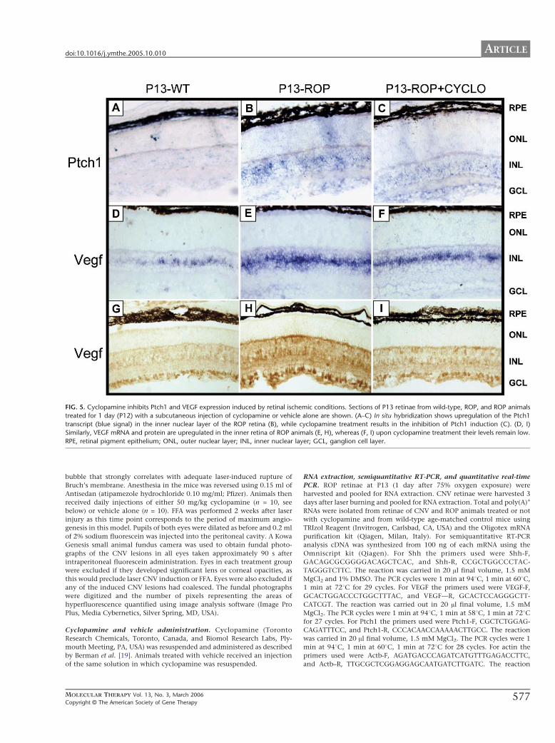
a Hamilton syringe and 33G needle For ISH Western blot analysis of Shh expression and
Ptch1 real time 3 μg of siRNA2 were injected in the right eye and the same amount of
control siRNA was injected in the left eye in P12 ROP mice eyes were collected and
retinae were dissected at P13 or at P14 for analysis To assess inhibition of retinal NV
mice received 3 or 6 μg of siRNA2 or control siRNA at P12 P14 and at P15 mice were
34
then sacrificed at P17 and eye collected for further analysis Results deriving from mice
receiving 3 or 6 μg of siRNA were pulled since no difference was observed
CNV was induced in adult C57BL6 mice as follows mice were anesthetized with an
intraperitoneal injection of 015 ml of a mixture of Domitor 1 mgml (medetomidine
hydrochloride Pfizer Pharmaceuticals Kent UK) and ketamine (100 mgml Fort Dodge
Animal Health Southampton UK) mixed with sterile water for injections in the ratio
5342 The pupils of all animals were dilated using topical 1 tropicamide and 25
phenylephrine (Chauvin Pharmaceuticals Essex UK) A slit-lamp mounted diode laser
system (wavelength 680 nm Keeler UK) was used to deliver 3 laser burns to the retinas of
each eye approximately 3-4 disc diameters from the optic disc avoiding major retinal
vessels (laser settings 210 mW 100 ms duration 100 μm diameter) These settings
consistently generate a subretinal gas bubble which strongly correlates with adequate laser-
induced rupture of Bruchrsquos membrane Anesthesia in mice was reversed using 015ml of
Antisedan (atipamezole hydrochloride 010 mgml Pfizer Kent UK) Animals then
received daily injections of either 50mgkg cyclopamine (n=10) or vehicle alone (n=10)
Fluorescein Fundus Angiogram (FFA see below) was performed 2 weeks after laser injury
as this time point corresponds to the period of maximum angiogenesis in this model
Retinal angiography immunofluorescence of whole mount preparation in vivo
fluorescein angiography and quantification of CNV area
Retinal angiography was performed by transcardiac perfusion with 15 ml of a 50 mgml
solution of 2 million molecular weight fluorescein isothyocyanate dextran (FITC-dextran
Sigma-Aldrich Milano Italy) in phosphate buffered saline (PBS) High molecular weight
dextran conjugated to fluorescein is retained in vessels that are fluorescently labelled
[31] In neovascular retina the newly formed vessels are leaky and retinal hyper-
fluorescence is observed due to fluorescein effusion [31] In addition neovascular tufts
35
corresponding to vessels extending beyond the internal limiting membrane into the
vitreous are evident [31] Retinae were dissected and flat-mounted and retinal vasculature
examined using a fluorescent dissection microscope (Leica Microsystems Milano Italy)
For immunofluorescence on whole-mount preparations ROP eyes (P5) were removed and
fixed in 4 (wv) paraformaldehyde in PBS The retinae were dissected and fixed in ice-
cold methanol for 10 min After incubating in PBS containing 50 fetal calf serum (FCS)
and 1 (wv) Triton X-100 for at least 1hr at room temperature the retinae were incubated
overnight at room temperature with a rabbit anti-mouse collagen IV antibody to label
vessels [122] (Chemicon Milano Italy) diluted 1200 in blocking buffer Retinae were
washed for 1 hr in PBS incubated for 2 hr at room temperature with Alexa Fluor 594-
conjugated goat anti-rabbit IgG secondary antibody (1200 dilution in blocking buffer
Molecular Probes Invitrogen Carlsbad CA) washed for 1 hr and mounted The area of
the retinal vasculature was measured with imageJ 132j software (Wayne Rasband National
Institute of Health Bethesda MD httprsbinfonihgovij)
For FFA pupils of both eyes were dilated as before and 02 ml of 2 sodium fluorescein
was injected into the peritoneal cavity A Kowa Genesis small animal fundus camera was
used to obtain fundal photographs of the CNV lesions in all eyes taken approximately 90
seconds after intraperitoneal fluorescein administration Eyes in each treatment group were
excluded if they developed significant lens or corneal opacities as this would preclude
laser CNV induction or FFA Eyes were also excluded if any of the induced CNV lesions
had coalesced The fundal photographs were digitized and the number of pixels
representing the areas of hyperfluorescence quantified using image analysis software
(Image Pro Plus Media Cybernetics Silver Spring MD USA)
36
Hepatic glycogen measurement
Hepatic glycogen contents was measured by a spectrophotometric assay [123] Briefly
tissues were solubilized in 01SDS then 12 volume of saturated Na2SO4 and 12 volume
of 95 ethanol were added The samples were chilled on ice for 30 minutes and then
centrifuged at 4 degC The pellet was rehydrated and 5 phenol and H2SO4 were added The
samples were left at room temperature for 10 minutes and incubated at 30degC for 20
minutes Finally absorbance at 490 nm was measured The results are expressed in
micrograms of glycogen per milligram of protein
In vivo glucose utilization index
The specific blood 2-DG clearance was determined using the Somogyi procedure as
previously reported [124] This method [125] is based on biochemical properties of 2-
deoxiglucose that is transported by the same carrier that the glucose and is also
phosphorilated by hexokinases This 2-deoxiglucose-6-phosphate (2-DG-6) can not be
further metabolized and remains inside tissues
A tracer dose (1microCi) of 2-deoxy[1-3H] deoxy-D-glucose (2-DG) was injected
intravenously in anaesthetized mice and its concentration was monitored in blood with a β-
counter on 25 microl blood samples obtained 1 10 20 and 30 min after injection Total
(labelled and unlabeled) serum glucose levels were measured with Amplex Red
GlucoseGlucose Oxidase Assay Kit (Invitrogen Life Technologies Carlsbad CA)
Skeletal muscle (gastrocnemius and quadriceps) samples were removed 30 min after
injection and the accumulation of radiolabeled compounds was measured by disgregation
of the tissue and β-counter measurement [125] The amount of 2-DG-6 phosphate per
milligram of protein was divided by the integral of the ratio between the concentration of
37
2-DG and the unlabeled glucose measured in the serum The index of glucose utilization
results are expressed as picomoles of 2DG per milligram of protein per minute
Cell culture plasmid and siRNA transfection AAV transduction cells and media
collection
Human embryonic kidney (Hek293) cells were used to assess expression and secretion of
HIP-Δ22-myc receptor and for production of Shh and HIP-Δ22 conditioned media 293
cells were cultured in DMEM (Invitrogen Life Technologies Carlsbad CA) 10 Fetal
Bovine Serum (FBS Gibco Invitrogen Life Technologies Carlsbad CA) 1
penicillinstreptomycin (Euroclone Celbio Milan Italy ) and transfected with Fugene 6
reagent (Roche Basel Switzerland) as suggested by manufacturer For conditioned media
production 48h after transfection cells were washed and serum free DMEM was added
12h later conditioned media were collected centrifuged at 3000rmp for 5rsquo in a
microcentrifuge to remove cells and stored at-20degC For Western blot analysis transfected
cells were collected and lysed in lysis buffer (40 mM Tris ph74 4mM EDTA 5mM
MgCl2 1 Triton X100 100 μM Na3VO4 1 mM PMSF 10 μgml Leupeptin-Aprotinin-
Pepstatin A-LAP-protease inhibitors 150mM NaCl) with standard procedures For AAV
infection 293 cells were incubated in serum-free DMEM and infected with AAV21-
CMV-HIP-Δ22 vectors (1x10⁴ gccell) for 2h at 37degC Complete DMEM was then added
to the cells 48h later cells were washed and incubated in DMEM serum free for 12h
media were then collected 500ul of each medium was concentrated with vivaspin
(Vivascience Littleton MA) as suggested by manufacturer and subjected to Western blot
analysis For siRNAs selection 293 cells were plated in MW12 plates 80 confluent cells
were transfected with the pShh plasmid using Fugene 6 reagent (Roche Basel
38
Switzerland) 24h later the same cells were transfected with each of the five siRNAs
targeting Shh or with control siRNAs using Lipofectamine 2000 (Invitrogen Life
Technologies Carlsbad CA) 5pmol of each siRNA were used After additional 24h
transfected cells were collected lysed in lysis buffer and subjected to Western blot
analysis
C3H10T12 osteoblastic differentiation and Alkaline Phosphatase assay
Members of the hedgehog gene family have been shown to regulate skeletal formation in
vertebrates affecting both chondrocyte [126] and osteoblast differentiation [7580] In
vitro Shh induces alkaline phosphatase (AP) a marker of osteoblast differentiation in the
mouse mesenchymal cell line C3H10T12 [127128] Indeed osteoblast differentiation of
these cells has been widely used as tool to quantitatively measure Shh activity by
assessment of AP expression [129] C3H10T12 were cultured in BME (Invitrogen Life
Technologies Carlsbad CA) supplemented with 2mM L-glutamine (Gibco Invitrogen
Life Technologies Carlsbad CA) 15 gL sodium bicarbonate (Gibco Invitrogen Life
Technologies Carlsbad CA) 10 heat-inactivated FBS (Gibco Invitrogen Life
Technologies Carlsbad CA) For differentiation experiments 1x10⁴cellscm2 were plated
in MW12 plates For experiments with conditioned media 500 μl of Shh containing
medium + 500 μl of HIP-Δ22 or eGFP conditioned medium was added Control cells
received eGFP medium alone Conditioned media were changed each 2 days 6 days later
cells were stained for AP expression or collected for AP assay For siRNA experiments
C3H10T2 were transfected with pShh using Fugene 6 reagent (Roche Basel
Switzerland) 24h later and every 2 days cells were transfected with 5pmol siRNA2 or
control siRNA using lipofectamine 2000 (Invitrogen Life Technologies Carlsbad CA) as
suggested by manufacturer 6 days later cells were stained for AP expression or collected
39
for AP assay AP staining was performed using Leukocyte alkaline phosphatase kit
(Sigma-Aldrich St Louis MO) as suggested from manufacturer For AP assay cells were
resuspended in a buffer containing 50mM TrisHCl pH 75 and 01 triton cells were then
lysed by 3 cycles of freeze-thaw in dry ice37degC Lysates were centrifuged at 14000 rpm
for 15rsquo supernatant were collected protein concentration was determined with Bio-Rad
Protein Assay Reagent kit (Bio-Rad Munchen Germany) and 10ug of each sample was
used to measure AP levels with the SEAP reporter gene kit (Roche Basel Switzerland) as
suggested by manufacturer
Anti-myc co-immunoprecipitation
For anti-myc co-immunoprecipitation conditioned media from pShh or pAAV21-CMV-
HIP-Δ22-myc transfected 293 cells were mixed 11 as control 1ml of medium from eGFP
transfected cells was used 15 μg of anti-myc antibodies (Clontech Palo Alto CA) were
added to each sample and incubated at 4degC over night (ON) The day after protein A-
sepharose (25ul Sigma-Aldrich St Louis MO) was added and samples incubated at 4degC
for 4h Finally samples were centrifuged at 3000 rpm for 5rsquo pellets were washed 3 times
with wash buffer (25mM Hepes pH 76 01mM EDTA 100mM NaCl 01 NP40)
resuspended in 50 μl of sample buffer (4 SDS 20 Glycerol 10 β-Mercaptoethanol
0125M TrisHcl pH 68 0004 Bromophenol Blue) and subjected to Western blot
analysis with anti-Shh or anti-myc antibodies
Western blot analysis
For Western blot analysis muscles and livers from AAV injected CD1 mice were
omogenized and lysed on ice for 30 min in lysis buffer (40 mM Tris ph74 4mM EDTA
40
5mM MgCl2 1 Triton X100 100 μM Na3VO4 1 mM PMSF 10 μgml Leupeptin-
Aprotinin-Pepstatin A-LAP-protease inhibitors 150mM NaCl) Samples were spun at
14000 rpm for 15 min the supernatant removed and stored at ndash80degC ROP retinae were
collected al P13 for Ptch1 western blot and at P13 and P14 for Shh Western blot For anti-
Shh and Ptch1 Western blot retinae were disgregated in lysis buffer by pipetting and
incubated on ice for 30rsquo samples were spun at 14000 rpm for 15rsquo and supernatant was
collected Protein concentrations from tissue and cell lysates were determined by Bio-Rad
Protein Assay Reagent kit (Bio-Rad Munchen Germany) Proteins from total lysates or
media from transfected cells were submitted to SDS-PAGE on 7 polyacrylamide gels
for HA PY and IRS-1 protein analysis for Ptch1 HIP-Δ22-myc and HIP-Δ-22 analysis
10 gels were used while for Shh Western blot proteins were separated on a 12 gel
After separation proteins were transferred to a PVDF membrane (Millipore Billerica
MA) The filter was incubated with anti-HA (12000 dilution) (Sigma-Aldrich St Louis
MO) anti-phosphotyrosine (PY 11000 dilution) (Santa Cruz Biotechnology Santa Cruz
CA) anti-IRS-1 (11000 dilution) (Santa Cruz Biotechnology) anti-actin (11000 dilution)
(Santa Cruz Biotechnology) anti-Shh (11000 dilution Santa Cruz Biotechnology) anti-
Ptch1 (11000 dilution Santa Cruz Biotechnology) anti-myc (upstate 11000 dilution)
anti-HIP (11000 dilution RampD Minneapolis MN) antibodies Mouse anti-PY and anti-
HIP antibodies were detected with HRP-conjugated anti-mouse antibodies (Sigma St
Louis MO) rabbit anti-HA anti-IRS-1 anti Shh and anti-Ptch1 were detected with HRP-
conjugated-anti-rabbit antibodies (Amersham Piscataway NJ) Goat anti-actin were
detected with HRP-conjugated-anti-goat antibodies (Santa Cruz Biotechnology) Finally
the protein-antibodies complexes were revealed by ECL-Pico chemioluminescent reaction
(Celbio Milan Italy) according to manufacturerrsquos instructions
41
Localization of HIP and BRDU labeled siRNA in the eye
AAV21-CMV-HIP-Δ22 injected eyes and control eyes receiving AAV21-CMV-eGFP
vectors were collected at P13 fixed in PFA 4 for 12h embedded in OCT and
cryosectioned sections were then permeabilized in PBS containing 01 triton (Carlo
Erba Milan Italy) blocked for 1h at RT in PBS 01 Triton 10 FBS (Gibco
Invitrogen Life Technologies Carlsbad CA) 01 BSA (Sigma-Aldrich St Louis MO)
and incubated ON with anti-HIP antibody (RampD Minneapolis MN) diluted 1100 in
blocking solution The day after sections were washed in PBS 01 Triton and incubated
with Cy3-labeled anti-rat secondary antibody (Molecular Probes Invitrogen Life
Technologies Carlsbad CA) Slides were then washed and mounted with vectashield
(Vinci Biochem Firenze Italy) HIP signal was observed under a fluorescence microscope
(Zeiss Milano Italy)
BrdU labelled siRNA2 were injected subconjunctivally in P9 mice (5 μg of siRNAeye)
injected eyes or control uninjected eyes were collected 1 and 2 days after siRNA injection
fixed in 4 PFA embedded in OCT sectioned and stained for BrdU as follows sections
were post-fixed in PFA 4 for 15rsquo and washed in PBS Endogenous peroxidase were
inactivated by incubating sections in 05 H2O2 in EtOH for 15rsquo After PBS washing
sections were denaturated in 2N HCl 05 Triton at 37degC for 15rsquo NaCl was neutralized in
01 Sodium Tetraborate for 30rsquo at RT sections were then incubated in blocking buffer
(PBS 10 FBS 01 Triton) for 30rsquo and ON with anti-BrdU antibody (diluted 1100 in
blocking solution Sigma-Aldrich St Louis MO) Tha day after sections were washed in
blocking buffer and incubated with anti-mouse biotinilated secondary antibody (11000 in
blocking buffer Vector laboratory CA USA) for 1h at RT The reaction was developed
using the Vectastained Elite ABC-Peroxidase Kit (Vector laboratory CA USA) followed
by 30min DAB staining (Vector laboratory CA USA) finally sections were mounted
with Eukitt (Kaltek Padova Italy)
42
RNA Extraction Semiquantitative RT-PCR and Quantitative Real-Time PCR
ROP retinae at P13 (one day after 75 oxygen exposure) were harvested for RNA
extraction CNV retinae were harvested three days after laser burning and pulled for RNA
extraction Total and polyA+ RNA were isolated from retinae of CNV and ROP animals
treated or not with cyclopamine and of wild-type age-matched control mice using TRIzol
Reagent (Invitrogen Life Technologies Carlsbad CA) and Oligotex mRNA Purification
Kit (Qiagen Milano Italy) For semi quantitative RT-PCR analysis cDNA was synthesized
from 100ng of each mRNA using the Omniscript kit (Quiagen Milano Italy) For Shh the
primers used were Shh-FGACAGCGCGGGGACAGCTCAC and Shhndash
RCCGCTGGCCCTACTAGGGTCTTC The reaction was carried in 20ul final volume
with 15mM MgCl2 and 1 DMSO The PCR cycles were 1min at 94degC 1 min at 60degC 1
min at 72degC for 29 times For VEGF the primers used were VEGFndashF
GCACTGGACCCTGGCTTTAC and VEGFndashRGCACTCCAGGGCTTCATCGT The
reaction was carried in 20 ul final volume with 15mM MgCl2 The PCR cycles were 1
min at 94degC 1 min at 58degC 1 min at 72degC for 27 times For Ptch1 the primers used were
Ptch1-F CGCTCTGGAGCAGATTTCC Ptch1ndashR CCCACAACCAAAAACTTGCC
The reaction was carried in 20 ul final volume 15mM MgCl2 The PCR cycles were 1
min at 94degC 1 min at 60degC 1 min at 72degC for 28 times For Actin the primers used were
Actβ-F AGATGACCCAGATCATGTTTGAGACCTTC and ActβndashR
TTGCGCTCGGAGGAGCAATGATCTTGATC The reaction was carried in 20 ul final
volume with 15mM MgCl2 The PCR cycles were 1 min at 94degC 1 min at 60degC 1 min at
72degC for 28 times The measurement of the band intensities was performed with the
Quantity One 411 software included in the Gel Doc 2000 gel documentation system (Bio-
Rad Milano Italy) Real-time PCR analysis was performed on mRNA extracted from the
retinae of the above mentioned mice in order to analyze the Shh Ptch1 and VEGF
43
transcripts All primers and probes were synthesized using the Applied Biosystems
ldquoAssays-bydesignrdquo software and indeed met the established criteria for TaqMan probes
(Applied Biosystems Foster City CA) Each probe was labeled with FAM at the 5rsquo end
and MGB at the 3rsquo end All reactions (30 ul) were performed with 100 to 200 ng of
mRNA 15 ul of Master Mix Reagent Kit (Applied Biosystems Foster City CA) 120
pmol of TaqMan probe and 10 uM of each specific primer The following amplification
conditions were used 10 min at 25degC 30 min at 48degC and 10 min at 95degC These
conditions were followed by 40 cycles of denaturation for 15 s at 95degC and annealing for 1
min at 60degC The amplification was performed using the ABI Prism 7000HT sequence
detection system (Applied Biosystems Foster City CA) equipped with a 96-well thermal
cycler Data were collected and analyzed with the Sequence Detector software (version
20 Applied Biosystems Foster City CA) All the reactions were performed in triplicate
and were normalized against Gapdh detected with specific primersprobes (Applied
Biosystems Foster City CA) labeled with VIC at the 5rsquo end and with TAMRA at the 3rsquo
end
In situ hybridization
For in situ hybridization eyes from P13 ROP mice were fixed in 4 PFA embedded in
OCT and cryosectioned at 12-14 μm Sections from different eyes were examined for each
probe images shown are representative of that seen all eyes examined (see results section)
Ptch1 probes were sinthetized by a pBSIIKS+ plasmid (Invitrogen Life Technologies
Carlsbad CA) containing the last 841 bp of murine Ptch1 coding sequence VEGF probes
were produced using a pCRII Topo plasmid (Invitrogen Life Technologies Carlsbad CA)
containing the sequence from 185 to 572 bp of murine VEGF Antisense and sense
digoxygenin (DIG)-labeled riboprobes were generated as follows plasmids were linearized
and sense probes were synthesized using T7 RNA polymerase (Roche Basel Switzerland)
44
for Ptch1 probe and SP6 RNA polymerase (Roche Basel Switzerland) for VEGF probe
(Roche Basel Switzerland) Antisense probes were produced using T3 RNA polymerase
(Roche Basel Switzerland) for Ptch1 and T7 RNA polymerase for VEGF (Roche Basel
Switzerland) Probe synthesis was carried out following manufactures guidelines Slides
were permeabilized with Ripa buffer (150mM NaCl 1 NP40 05 Na Deoxicolate
01 SDS 1mM EDTA 50mM TrisHCl pH 8) incubated ON in hybridizations solution
(50 Formammide 5x SSC 5x denhards 500μgml salmon sperm DNA 250 μgml Yeast
RNA) containing probes (300 ngml) at 70degC and the signal was detected with AP-labeled
anti-DIG antibodies (Roche Basel Switzerland) as suggested my manufacturer Finally
signal was developed by BCIPNBT colorimetric AP substrate (Sigma-Aldrich St Louis
MO)
Histology
Eyes from ROP mice sacrificed at P17-19 were enucleated and fixed in 4 PFA Eyes
were embedded in paraffin sectioned at 6 μm and stained with periodic-acid-Schiff and
hematoxylin The number of retinal vascular endothelial cell nuclei on the vitreous surface
of the internal limiting membrane was counted Six to eight sectionseye were counted and
the counts were averaged Some eyes in which CNV was induced were enucleated 14 days
after laser injury Following overnight fixation in 10 neutral buffered formalin they were
processed and embedded in paraffin Serial 6μm sections were cut and stained with
hematoxylin and eosin and examined using light microscopy
Statistical analysis
Statistical analysis of differences between groups was performed using the paired
Studentrsquos t-test using the microsoft excel t-test function Significance (ple005) is shown as
45
single asterisks Where p is le001 two asterisks have been used as described in the legend
to the figures For the CNV mice groups Shapiro-Wilk and Drsquoagostino and Pearson
omnibus normality tests confirmed the non-normal distribution of CNV area data A non-
parametric test for unpaired samples (Mann Whitney U test) was therefore used to analyze
significance of differences (P lt 005)
46
RESULTS
Gene transfer for pharmacological regulation of the insulin receptor signalling
Generation of a pharmacologically regulated chimeric insulin receptor
To obtain pharmacological activation of the insulin receptor signalling in a desired cell or
tissue we used a recently developed system allowing to pharmacologically regulate
proteinndashprotein interactions such as the homodimerization of growth factor receptors with
tyrosine kinase activity [108130131132] This system is based on the ability of a small
orally bioavailable molecule dimerizer drug AP20187 to bind to a specific protein module
contained in the cytoplasmic FKBP12 protein Any cellular process activated by proteinndash
protein interaction (such as IR activation) can in principle be brought under dimerizer
control by fusing the protein of interest (ie the intracellular domain of IR) to the
FKBP12 binding domain recognized by the dimerizer Addition of the dimerizer then
cross-links the chimeric signalling protein thus activating those pathways induced by the
protein homodimerization (Fig 7)
We generated a chimeric insulin receptor (LFv2IRE) protein responsive to AP20187 by
fusing the cytoplasmic domain of the human insulin receptor (IR) to two AP20187-binding
domains (Fv) and to one C-terminal epitope tag (E) The chimeric protein was fused to an
N-terminal sequence including the low affinity nerve growth factor receptor (LNGFR)
extracellular and transmembrane domains (L) to localize it to the plasma membrane (Fig
7)
Figure 7 Schematic representation of the AP20187ndashLFv2IRE system We constructed a chimeric
receptor containing the intracellular domain of the insulin receptor (IRβ) including its tyrosine kinase
domain fused to two dimerization domains (Fv) which are binding domains for the small dimerizer drug
AP20187 Addition of AP20187 results in dimerization of the chimeric receptor and induction of intracellular
signalling HA hemagglutinin tag L transmembrane domain of the low affinity nerve growth factor
receptor
We already reported that the AP20187-LFv2IRE system is able to activate the insulin
receptor signalling and to induce insulin-like biological effects in vitro in hepatocytes and
fibroblasts transduced with AAV vectors (see attached PDF [130]) AP20187
administration in these cells resulted in time- and dose-dependent activation of both the
LFv2IRE receptor and the IR substrate IRS-1 leading to the activation of glycogen
synthesis (see attached PDF [130]) Then we used AAV vectors to induce LFv2IRE
expression in liver and muscle of normal and diabetic mice to evaluate the AP20187-
dependent activation of the chimeric receptor and the induction of the insulin signalling
and actions in two of the main hormone target tissues We used nonobese diabetic (NOD)
47
48
mice which spontaneously develop autoimmune insulin-dependent DM [121] and
therefore are widely used animal models of type 1 DM
AP20187-dependent LFv2IRE activation in liver and muscle transduced with AAV
vectors
To assess the ability of the AP20187 dimerizer to activate LFv2IRE in vivo we transduced
liver and muscle with AAV vectors encoding LFv2IRE under the control of liver or muscle
specific promoters (the thyroxin binding globulin-TBG and muscle creatine kinase-MCK
promoters respectively) AAV21 and 28 vectors were used to transduce muscle and liver
respectively The LFv2IRE receptor contains an HA tag after the IR intracellular domain
allowing its recognition with specific anti-HA antibodies (Fig 7) The dose of AAV
vectors administered systemically in this and the following experiments (5x10¹¹ genome
copies GCmouse) is optimal for both liver and muscle transduction [71133] We injected
wild type CD1 mice systemically with either AAV28-TBG-LFv2IRE vectors to transduce
the liver or saline solution Four weeks later mice were stimulated or not with an
intraperitoneal AP20187 injection (10 mgkg as suggested elsewhere wwwariadcom)
and livers were collected at different time points after drug administration We then
evaluated AP20187-dependent LFv2IRE tyrosine phosphorylation (Fig 8) Livers from
AAV injected animals expressed similar levels of LFv2IRE as shown by Western blot with
anti-HA antibodies while no signal was detected in the lane corresponding to livers from
animals receiving saline (Fig 8 middle panel) AP20187-dependent LFv2IRE tyrosine
phosphorylation was evident two hours after drug administration peaked 6 hours later and
returned to baseline after 24 hours (Fig 8 upper panel) Low LFv2IRE basal
phosphorylation was detected in livers from mice receiving AAV28-TBG-LFv2IRE but
not stimulated with AP20187 suggesting minimal leakiness of the system (Fig 8 upper
panel first lane)
Figure 8 Protein tyrosine phosphorylation in AAV-transduced livers upon AP20187 administration
time dependency of protein phosphorylation Western blot analysis of lysates from livers of CD1 mice
injected with AAV28-TBG-LFv2IRE stimulated with AP20187 and collected at different times after drug
administration (reported on the top of the figure) Proteins from total lysates were blotted with anti-P-tyrosine
(αPY upper panel) anti-HA (αΗΑ middle panel) or anti IRS-1 (αIRS-1 lower panel) antibodies Molecular
masses (kDa) are indicated on the left
Western blot analysis with anti-HA antibodies evidenced a double LFv2IRE band (Fig 8
middle panel) The lower band may represent an LFv2IRE degradation product that does
not include some tyrosine-phosphorylated residues present in the band of higher molecular
weight The 180 kDa band present in the upper panel of figure 8 corresponds to the main
substrate of the IR tyrosine kinase the insulin receptor substrate-1 (IRS-1) protein (Fig 8
lower panel) IRS-1 levels of tyrosine phosphorylation follow those of LFv2IRE
suggesting that it is induced upon LFv2IRE activation Basal levels of IRS-1 tyrosine
phosphorylation from endogenous insulin is evident in livers from saline injected mice
Since the levels of basal IRS-1 tyrosine phosphorylation are similar in livers from saline-
and AAV28-TBG-LFv2IRE-injected mice that did not receive AP20187 the basal
LFv2IRE tyrosine phosphorylation levels observed (Fig8 upper panel) do not seem to
induce activation of the IR signaling pathway in transduced hepatocytes 49
50
We then evaluated AP20187-dependent activation of LFv2IRE in muscle following
systemic administration of AAV21-MCK-LFv2IRE vectors or saline Four weeks after
AAV systemic administration mice were treated or not with AP20187 (10 mgkg) Skeletal
muscles (gastrocnemi and quadriceps) were collected at different time points after drug
administration (Fig 9) We performed Western blot analysis of LFv2IRE expression levels
on right and left gastrocnemi and quadriceps from AAV injected mice (Fig 9A upper
panel) We detected higher LFv2IRE expression levels in gastrocnemi than quadriceps
muscles (Fig 9A upper panel) The loading control performed with anti-actin antibodies
showed similar amounts of total protein in all lanes (Fig 9A lower panel)
Therefore we selected right gastrocnemi to evaluate AP20187-dependent activation of
LFv2IRE following AAV21 systemic administration (Fig 9B) We detected a tyrosine
phosphorylated doublet of about 140 kDa (Fig 9B upper panel) corresponding to the
LFv2IRE double band recognized by anti-HA antibodies (Fig 9B lower panel) in AAV
transduced muscles Since the tyrosine phosphorylated band of lower molecular weight is
also present in uninjected unstimulated muscles (Fig 9B upper panel first lane) we only
considered the upper band recognized by the anti-PY antibodies when investigating the
timing of LFv2IRE activation in muscle LFv2IRE tyrosine phosphorylation becomes
evident 30 minutes after AP20187 administration peaks after 6 hours and is still present 24
hours later (Fig 9B upper panel) Western blot analysis with anti-HA antibodies shows
that LFv2IRE is present in AAV transduced but not untransduced muscles (Fig 9B lower
panel) LFv2IRE levels are similar among all lanes with the exception of the second lane
corresponding to muscles from animals treated with AAV21-MCK-LFv2IRE but not
stimulated with AP20187 where a lower amount of receptor is present This weak
difference in LFv2IRE levels however cannot account for the almost absent LFv2IRE
tyrosine phosphorylation (Fig 9B upper panel second lane) The 180 kDa band
corresponding to IRS-1 (Fig 9C lower panel) has tyrosine phosphorylation levels that
increase 30 minutes after AP20187 administration remain high after 120 minutes and then
decrease after 6 hours (Fig 9C upper panel)
Figure 9 LFv2IRE expression and protein tyrosine phosphorylation in AAV-transduced skeletal
muscles A) Western blot analysis of lysates from different muscles of CD1 mice injected with AAV21-
MCK-LFv2IRE Proteins from total lysates were blotted with anti-HA (αΗΑ) antibodies (rG right
gastrocnemious lG left gastrocnemious rQ right quadricep lQ left quadricep) B) LFv2IRE tyrosine
phosphorylation in AAV-transduced skeletal muscle upon AP20187 administration time dependency of
protein phosphorylation Western blot analysis of lysates from right gastrocnemi of CD1 mice injected with
AAV21-MCK-LFv2IRE and stimulated with AP20187 collected at different times after drug administration
(reported on the top of the figure) Proteins from total tissue lysates were blotted with anti-P-tyrosine (αPY
upper panel) or anti-HA (αΗΑ lower panel) antibodies C) IRS-1 tyrosine phosphorylation in AAV-
transduced skeletal muscle upon AP20187 administration time dependency of protein phosphorylation
Western blot analysis of lysates from right gastrocnemi of CD1 mice injected with AAV21-MCK-LFv2IRE
and stimulated with AP20187 collected at different times after drug administration (reported on the top of the
figure) Proteins from total tissue lysates were blotted with anti-P-tyrosine (αPY upper panel) or anti-IRS-1
(αIRS-1 lower panel) antibodies Molecular masses (kDa) are indicated on the left of each panel
51
52
This suggests that AP20187 administration triggers LFv2IRE activation which
phosphorylates IRS-1 upon tyrosine residues The IRS-1 activation in muscle occurs before
the levels of LFv2IRE phosphorylation peacks and is rapidly reverted before the receptor
phosphorylation returns to baseline These results confirm that AAV21 and AAV28
vectors are able to strongly transduce murine muscle and liver with LFv2IRE In addition
our data indicate that AP20187 induces LFv2IRE transphosphorylation in both tissues
transduced with AAV vectors This occurs rapidly after drug administration and is reverted
to baseline levels 24 hours after AP20187 injection in liver but not in muscle suggesting a
possible difference in drug clearance from the two tissues The timing of LFv2IRE
activation in vivo is in accordance with AP20187 half-life that is 8 hours in murine serum
(V Rivera ARIAD Pharmaceuticals personal communication) The activated receptor
induces the IR signaling in both transduced tissues since its activation results in IRS-1
phosphorylation with kinetics identical to LFv2IRE in liver and similar in muscle
However the kinetics of LFv2IRE activation upon AP20187 administration do not
perfectly mirror those of the physiological insulin-mediated IR activation which occurs
few minutes after meal assumptions and returns to baseline in less than two hours [18] It is
possible that the development of AP derivatives with half-life and biodistribution different
from AP20187 may overcome this delay
AP20187 induces insulin-like actions in muscle and liver of NOD mice transduced with
AAV vectors
To investigate the ability of LFv2IRE to induce insulin-like actions in vivo we used a
model in which there is no endogenous insulin signaling IR knockout mice die in the first
days of life [134] in other models of type 2 DM ie obob and dbdb mice [135] the cause
of insulin resistance is unclear [136137138139] Therefore we decided to use NOD
mice a murine model of type 1 DM [121] We induced LFv2IRE expression in muscle and
53
liver of adult diabetic NOD mice through systemic injection of a mix of the AAV21-
MCK-LFv2IRE and AAV28-TBG-LFv2IRE vectors (5x10¹¹GC of each vectormouse) A
control group of animals received the same dose of the AAV28-TBG-LacZ and AAV21-
MCK-eGFP vector mix One month later we evaluated the AP20187-dependent increase in
glycogen synthesis and circulating glucose uptake as index of insulin-like signalling in the
transduced tissues We selected liver to evaluate glycogen synthesis Since glucose uptake
in liver is not insulin-dependent [18] we used muscle to evaluate the induction of glucose
uptake Fig 10 shows that liver glycogen levels in mice expressing LFv2IRE and
stimulated with AP20187 are significantly higher than in unstimulated mice in which
glycogen levels are similar to those measured in control mice
In addition the effect of AP20187 in mice expressing LFv2IRE is almost superimposable
to that of insulin treatment (075 Ukg body weight) in NOD mice (Fig 10) This was 35
lower however compared to the glycogen content measured in insulin-treated wild-type
controls Our results demonstrate that AP20187 administration induces glycogen synthesis
in liver expressing LFv2IRE similarly to insulin [18] and confirms that the basal levels of
LFv2IRE tyrosine phosphorylation observed in the absence of AP20187 do not impact on
this aspect of liver glucose metabolism
Figure 10 Hepatic glycogen content in AAV-injected NOD mice NOD mice were injected with AAV28-
TBG-LFv2IRE and AAV21-MCK-LFv2IRE vectors (black and grey bars) or with control AAV28-TBG-
LacZ and AAV21-MCK-eGFP vectors (white bar) and stimulated (black bar) or not (grey and white bars)
with AP20187 After stimulation livers were collected and hepatic glycogen content was evaluated The
number of mice for group (n) is indicated under each bar Results are reported in micrograms per milligram
of protein with SE =plt 005 Vertical striped bars wild-type mice stimulated with insulin Horizontal
striped bars NOD mice stimulated with insulin
The glucose utilization index was measured in the skeletal muscle (quadriceps and
gastrocnemi) of the same mice used in Fig 10 (injected with the AAV21-MCK-LFv2IRE
and AAV28-TBG-LFv2IRE mix) which were stimulated or not with AP20187 (Fig 11)
The index was significantly increased in both gastrocnemi and right quadriceps of AAV21
injected mice upon AP20187 administration The average induction of muscle glucose
uptake in all muscles analyzed is reported in Fig 11 (46 fold-induction in AP20187-
stimulated mice compared to unstimulated AAV injected mice) and is comparable to that
obtained in insulin-stimulated NOD mice
54
Figure 11 Index of glucose utilization by NOD skeletal muscle transduced with AAV21
Muscle glucose uptake (average of gastrocnemious and quadriceps) in AAV28-TBG-LFv2IRE and
AAV21-MCK-LFv2IRE injected mice stimulated (black bars) or not (grey bars) with AP20187 Vertical
striped bars wild-type mice stimulated with insulin n=9 mice Horizontal striped bars NOD mice stimulated
with insulin n=5 mice Results are reported in pmolmgmin with SE N= 5 mice in the AP20187-stimulated
group and 3 mice in the unstimulated group =ple 005 =ple 001
This result demonstrates that similarly to liver AP20187-mediated LFv2IRE activation
mimics insulin action in muscle of NOD mice Again 35 higher values of glucose
utilization index were found in insulin-stimulated wild-type mice
We finally evaluated if AP20187-induced insulin-like signalling results in normalization of
blood glucose levels in NOD mice transduced with both AAV21-MCK-LFv2IRE and
AAV28-TBG-LFv2IRE Blood glucose levels were monitored for 24 hours after AP20187
administration and did not decrease neither in AP20187-treated nor in untreated AAV
transduced diabetic mice (data not shown) One possible explanation for the inability of the
AP20187LFv2IRE system to impact on blood glucose levels is that transduction with
LFv2IRE may be required in tissues other than muscle and liver In this regard IR ablation
in brown adipose tissue [140] or adipose-specific GLUT-4 ablation [141] result in impaired
glucose tolerance In addition since restoration of IR expression in liver brain and
55
56
pancreatic β-cells of IR ko mice is sufficient to rescue the lethality and prevent
hyperglycemia in this model [142143] mechanisms other than the insulin-dependent
glucose uptake in canonical insulin target tissues could contribute to the regulation of
circulating glucose levels Despite the LFv2IRE ability to induce IRS-1 activation
resulting in insulin-like biological actions in both muscle and liver we cannot exclude that
the LFv2IRE-AP20187 system does not activate some IR targets downstream of IRS-1 or
has a different turn-overhalf life compared to the endogenous insulin receptor therefore
failing to normalize glucose levels in diabetic models Alternatively LFv2IRE tyrosine
phosphorylation levels or timing different from the endogenous IR (as we show in Fig 8
and 9) could be responsible for the absence of impact on blood glucose levels
Evaluation of the involvement of the Sonic Hedgehog pathway in ocular neovascular
diseases
Sonic Hedgehog pathway is involved in physiological and pathological ocular vessel
development
To assess the potential role of the Shh pathway during the development of the
physiological retinal vasculature wild type C57BL6J mice received daily systemic
administration of the selective Shh pathway inhibitor cyclopamine between post-natal day
(p) 1 and p4 a time point at which retinal vascular network is developing At p5 we
evaluated the extent and morphology of the superficial retinal vascular layer by
immunofluorescence of retinal whole-mounts stained for a vascular endothelial marker
(Fig 12) Despite a similar development in the extension of the neural retina a reduced
vascular area was observed in cyclopamine treated animals when compared with vehicle-
treated controls (Fig 12A) The extension of retinal vasculature was measured confirming
a significant reduction of the area of vessels development (Fig 12B) thus suggesting that
the Shh pathway is an important component of normal retinal vasculogenesis
Figure 12 Cyclopamine inhibits the development of retinal vasculature in neonatal mice Panel A
Immunofluorescence analysis with anti-collagen IV antibody of P5 retinal flat mounts from animals treated
with daily subcutaneous injections of either cyclopamine (50 mgkg from P1 to P4) or vehicle alone Panel
B The retinal vascular area in pups was measured (n=11 retinaegroup) A significant (=p-value lt 0034)
decrease in the area of the superficial vascular layer is evident in animals receiving cyclopamine CNTR
control animals receiving vehicle CYCL animals receiving cyclopamine
Next we investigated the involvement of the Shh pathway in pathological vessels growth
in murine models of retinal and choroidal neovascularization the ROP and laser induced
CNV mice We analyzed retinal expression levels of Shh and of its transcriptional target
Ptch1 as an index of Shh pathway activation by reverse transcription PCR In addition we
assessed VEGF expression levels as well since it is reported to be induced in these
conditions Upregulation of Shh and Ptch1 expression similarly to VEGF was observed in
both ROP and CNV retinae as compared with age-matched wild type controls (Fig13A)
The intensity of the bands corresponding to each gene in panel A was measured and
normalized on the corresponding actin bands to assess the fold increase in expression in
neovascular compared with normal retinae This showed an increase in retinal gene
expression in neovascular compared to normal eyes varying from 128 folds in the case of
the Ptch1 transcript in the ROP retinae to 25 fold in the case of Shh in the CNV retinae
(Fig 13B) In situ hybridization was used to assess the tissue distribution of Ptch1 in the
57
ROP retinas Ptch1 transcript was upregulated in the inner nuclear layer of the ROP retinae
with a gradient higher in the central than in the peripheral retina (Fig 13C)
Figure 13 Upregulation of the Shh pathway in the retina of animal models with neovascular disease
Panel A RNA from 6 animals per group was isolated from whole retinae retrotranscribed and PCR-
amplified with specific primers under semi-quantitative conditions Each lane is representative of 3 animals
(6 retinae) Bands corresponding to Shh Ptch1 and VEGF are more abundant in the samples from the CNV
and ROP than from the control retinae Panel B Fold-increase of Shh Ptch1 and VEGF expression in the
ROP (black bars) and CNV (white bars) relative to control samples The intensity of the bands in panel A
was quantified the values from the Shh Ptch1 and VEGF bands normalized by those from the Actin bands
and compared between the ROP or CNV groups and control retinae Panel C In situ hybridization of Ptch1
on P13 retinae in normal control (upper panel) and ROP retina (lower panel) reveals upregulation of the
Ptch1 transcript (blue signal) in the inner retina following hypoxia Each picture is representative of two eyes
Panel D Western blot analysis for Ptch1 protein in P13 wild type (first three lanes) and ROP (last three
lanes) retinae shows induction of Ptch1 protein in neovascular eyes
58
59
In addition an increase in the Ptch1 protein was observed in the ROP retinae when
compared with normal controls analyzed by Western blot (Fig 13D) Therefore
expression of Shh and of its transcriptional target Ptch1 is upregulated in murine ischemia-
induced (ROP) or laser-induced (CNV) ocular neovascularization suggesting an
involvement of the Shh pathway in ocular neovascular processes
Systemic pharmacological inhibition of Shh pathway reduces retinal and choroidal
neovascularization
To confirm that Shh upregulation plays a role in ocular neovascularization we inhibited
Shh pathway in ROP and CNV mice by systemic (subcutaneous) administration of the Shh
inhibitor cyclopamine We confirmed the inhibition of the Shh pathway after cyclopamine
administration in the ROP retina by measuring the mRNA levels of Ptch1 by Real Time
PCR VEGF expression was assessed as well Both transcripts were up-regulated in ROP
compared to control retinae The levels of Ptch1 (Fig 14A) and to a lesser extent of VEGF
(Fig 14B) were lower in the cyclopamine-treated than untreated eyes (Fig 14) confirming
the inhibition of the Shh pathway by cyclopamine
Figure 14 Cyclopamine inhibits the Shh pathway in the ROP retina Real-Time PCR analysis of Ptch1
(panel A) and VEGF (panel B) mRNA in the control (white bars) or ROP (P13) retina of animals (n=18
retinaegroup pooled in 3 samples of 6 retinae each) treated with subcutaneous cyclopamine (50 mgkg at
P12) (black bars) or vehicle only (grey bars) Standard errors are depicted CNTR control CYCL
cyclopamine Ptch1 and VEGF expression is reduced in the retina of ROP mice receiving cyclopamine
We then assessed the impact of cyclopamine-mediated Shh pathway inhibition on retinal
neovascularization systemic administration of cyclopamine substantially inhibited
neovascularization in the ROP model as assessed by retinal angiography (Fig 15A)
showing less neovascular tufts in treated compared to control ROP retinae Histological
analysis of ROP retinal sections showed reduction of endothelial cells and capillaries over
the inner limiting membrane in ROP animals treated with cyclopamine (Fig 15B) We
quantified inner retinal neovascularization by counting endothelial cell nuclei located
internal to the inner limiting membrane (ILM) in serial paraffin sections The number of
endothelial cell nuclei was significantly lower in eyes from ROP animals treated with
cyclopamine than those injected with vehicle alone (P lt0001) (Fig 15C) These results
demonstrate that activation of the Shh pathway plays a crucial role to establish hypoxia-
induced retinal neovascularization in mice
60
Figure 15 Cyclopamine inhibits murine hypoxia-induced (ROP) retinal neovascularization
Angiographic (panel A) and histological (panel B) photographs of ROP retinae at P17 from animals treated
with daily (P13 to P16) subcutaneous injections of cyclopamine (50 mgkg) (right) or vehicle alone (left)
Neovascular areas after in vivo perfusion with fluorescein isothiocyanate dextran (FITC-dextran) are evident
as tufts and effusions (indicated by arrowheads) in the ROP retinae and substantially reduced or absent in the
ROP retinae treated with cyclopamine (n=13group) Panel B PAS staining of retinal sections confirmed that
pathological capillaries internal to the inner limiting membrane in the ROP retinae are importantly reduced
when ROP animals are administered with cyclopamine Panel C The number of vascular nuclei extending
from the internal limiting membrane into the vitreous was counted in serial sections on either side of the optic
nerve Mean and standard error values for each group are depicted = P valuesle 0001 RPE retinal pigment
epithelium ONL outer nuclear layer INL inner nuclear layer GCL ganglion cells layer arrowheads
neovascular capillaries CNTR control CYCL cyclopamine
Systemic administration of cyclopamine also inhibited laser-induced CNV in adult mice
(Fig 16) Bruchrsquos membrane was ruptured in both eyes of adult mice using a high powered
diode laser The subsequent formation of subretinal neovascularization arising from the
choriocapillaris is maximal approximately 14 days post-laser induction Fundus
fluorescein angiography (FFA Fig 16A) was performed at this stage and used to quantify
61
62
the areas of induced CNV in cyclopamine treated and vehicle-only treated animals
Systemic cyclopamine delivery resulted in significant inhibition of CNV formation
compared with vehicle-only control animals (Fig 16 C)
The results shown in this section demonstrate that activation of the Shh pathway is an
important component in the development of both mature and aberrant retinal vessels Shh
Ptch1 and VEGF are upregulated in murine models of ocular neovascularization and
systemic pharmacological inhibition of the Shh pathway significantly reduces angiogenesis
in both contexts Thus we suppose that this pathway may represent a novel and important
target to which pharmacological or gene-based strategies for ischemic retinopathies and
exudative AMD could be developed
Figure 16 Cyclopamine inhibits murine laser-induced choroidal neovascularization Panel A
Representative early phase fundus fluorescein angiograms (FFAs) from control and cyclopamine injected
animals Hyperfluorescence (arrowheads) at this phase of dye transit represent the areas of the induced CNV
membranes Panel B Representative H amp E stained 6μm thick paraffin sections of eyes demonstrating
smaller subretinal CNV complexes (arrows) in cyclopamine treated animals Panel C CNV complexes in
animals receiving daily cyclopamine (n=39 mean 20789 plusmn 2627 pixels) were 591 smaller than those in
vehicle-only treated animals (n=37 mean 50874 plusmn 10989 pixels) = P lt 005 (Abbreviations as before
RPE retinal pigment epithelium ONL outer nuclear layer INL inner nuclear layer GCL ganglion cells
layer) Standard errors are depicted
CNTR control CYCL cyclopamine
63
Development of nucleic acid-based strategies for specific inhibition of Shh pathway
The data reported in the previous sections indicate that Shh pathway plays a role in
pathological induction of neovascularization and thus represents a new potential
therapeutic target for diseases characterized by ocular NV
Systemic administration of cyclopamine cannot be considered of therapeutic interest for
treatment of ocular neovascular conditions because of possible side effects related to
systemic inhibition of Shh pathway as well as possible unknown systemic actions of
cyclopamine different from inhibition of Shh
Thus we developed two different strategies for specific intraocular inhibition of Shh to
both confirm its involvement in ocular neovascular diseases and to provide strategies for
its specific inhibition to be eventually used in therapeutic settings (Fig 17)
Figure 17 Schematic representation of strategies for inhibition of Shh action
(A) RNA interference can be used to reduce Shh expression by hypoxic cells (B) A soluble decoy receptor
for Shh can be used to block its extracellular diffusion preventing its binding to the Ptch1 receptor This has
been generated by deleting the transmembrane domain of the Hedgehog interacting protein Hip (HIP-
Δ22myc)
64
65
To inhibit Shh action we generated a soluble decoy receptor (HIP-Δ22) by deleting the
transmembrane domain of the Hedgehog Interacting Protein (HIP) a membrane
glycoprotein physiologically binding and sequestering Shh [92] Deletion of the last 22 C-
terminal aminoacids results in efficient secretion of HIP protein as reported [92] In
addition we added a myc tag at the C-terminal of the protein to allow detection with anti-
myc antibodies generating the HIP-Δ22-myc receptor (Fig 17B)
In the second strategy to inhibit Shh expression we used short interfering RNAs (siRNA)
21-23nt dsRNA duplexes able to silence Shh expression in a sequence specific manner
(Fig 17A) [44144145] We designed five different siRNA oligos (siRNA1 to 5)
targeting regions of sequence complementarity between human and murine Shh mRNA
We then tested both systems in vitro for their ability to inhibit Shh pathway We first
confirmed that HIP-Δ22-myc is efficiently expressed and secreted in 293 cells transfected
with constructs encoding the decoy receptor (pHIP-Δ22-myc) Western blot analysis with
anti-myc antibodies showed the presence of HIP-Δ22-myc in both cell lysates and media of
transfected but not control cells as expected (Fig 18A) To assess the ability of the decoy
receptor to bind Shh we performed anti-myc co-immunoprecipitations (co-IP) on culture
media from cells transfected with pHIP-Δ22-myc or with a plasmid encoding Shh (pShh)
HIP-Δ22 and Shh containing media were mixed and subjected to co-IP As controls media
from cells expressing eGFP were used Western blot analysis with anti-myc and anti-Shh
antibodies of immuno-purified complexes revealed presence of both HIP-Δ22-myc and
Shh (Fig 18B) confirming that once secreted the decoy receptor we generated is able to
bind Shh in vitro
Finally we used conditioned media from transfected cells expressing HIP-Δ22-myc Shh
or eGFP to assess the ability of the decoy receptor to block Shh action in vitro We used
the murine mesenchimal C3H10T12 cells which are able to differentiate in osteoblasts
and express alkaline phosphatase (AP) upon Shh addition [129] When these cells were
66
incubated with Shh conditioned media the AP expression increased significantly (Fig
18C) We observed consistent reduction of AP expression when conditioned medium
containing HIP-Δ22-myc was added to the Shh containing medium (Fig 18 C) suggesting
that the decoy receptor sequesters Shh and inhibits its action in these settings Similar
results were obtained using conditioned media containing HIP-Δ22 (devoid of the myc
tag data not shown) Then we generated AAV21 vectors encoding HIP-Δ22 (AAV-HIP-
Δ22) Western blot analysis on culture media from 293 cells infected with AAV-HIP-Δ22
confirmed expression and secretion of the decoy receptor upon infection (Fig 18D)
Figure 18 In vitro characterization of HIP-Δ-22myc A) Evaluation of Hip-myc expression and
secretion in transfected 293 cells 293 cells were transfected with pHIP-Δ22myc (HIP-Δ22myc) or p-eGFP
(eGFP) expression plasmids HIP-Δ22myc expression and secretion was analyzed by Western blot with anti-
myc antibodies on lysates and media from transfected cells B) Evaluation of Hip-myc binding to Shh in
transfected 293 cells 293 cells were transfected with pHIP-Δ22myc p-Shh plasmids or with control p-eGFP
plasmids 48 hours later media from transfected cells were collected and media from Shh and HIP-Δ22-myc
transfected cells were mixed Mixed (lane 1) and control (lane 2) media were immunoprecipitated with anti-
myc antibodies Immunopurified proteins were analyzed by Western blot with anti-myc and anti-shh
antibodies C) Hip-mediated inhibition of Shh-induced osteogenic differentiation of C3H10T12 cells
Conditioned media from HIP andor Shh transfected 293 cells were added to C3H10T12 cells alone or in
combination and changed each 2 days Osteogenic differentiation was assessed 6 days later measuring AP
activity in cellular lysates Results are expressed as pg of APug protein plusmn standard error Ctr C3H10T12
receiving conditioned media from 293 cells transfected with control p-eGFP plasmids Shh C3H10T12
receiving conditioned media from 293 cells transfected with p-Shh plasmids HIP+Shh C3H10T12
receiving both conditioned media from 293 cells transfected with p-Shh and p-HIPΔ22-myc D) HIPΔ22
secretion in AAV-infected 293 cells 293 cells were infected with AAV-HIPΔ22 or with control AAV-eGFP
vectors and media from infected cells were concentrated and analyzed by western blot with anti-HIP
antibodies = Plt005
67
68
For the RNA interference strategy we first analyzed the five siRNA oligos we designed
for their ability to inhibit Shh expression in vitro 293 cells were first transfected with pShh
and then co-transfected with each of the five siRNA we designed or with control siRNAs
Shh expression levels in treated and control cells were assessed by Western blot All the
siRNA efficiently reduced Shh expression (Fig 19A) the siRNA2 showed the strongest
Shh inhibition as assessed by measuring the intensity of the Shh bands (Fig 19B) in 3
independent experiments We then selected the siRNA2 as Shh siRNA for all the
subsequent experiments We again used C3h10T12 cells to evaluate the ability of Shh
siRNA 2 to inhibit Shh activity Cells were transfected with pShh and then co-transfected
with siRNA 2 or with control siRNAs AP expression induced by pShh transfection was
significantly reduced when siRNA 2 was co-transfected together with pShh confirming
that siRNA2-mediated inhibition of Shh expression results in inhibition of its activity in
this setting (Fig 19 C D)
Figure 19 Shh siRNA reduces Shh expression and activity in vitro
A) Reduction of Shh protein levels following siRNA treatment of 293 cells 293 cells were transiently
trasfected with the p-Shh expression plasmid and 24 hours later trasfected independently with each of the 5
siRNAs we designed or with a control siRNA (CTR) The levels of Shh expression in transfected cells were
evaluated by Western blot analysis (upper panel) Protein loading was normalized with anti-actin antibodies
(lower panel) B) Measurement of Shh levels shown in panel A The intensity of the bands in panel A was
quantified with the ImageJ software and Shh values were normalized by actin in each lane Results are
reported as of Shh expression relative to cells transfected with the control siRNA (CTR lane) The average
of three independent experiments is shown The siRNA 2 results in strong inhibition of Shh expression C
D) siRNA-mediated inhibition of Shh-induced osteogenic differentiation of C3H10T12 cells Alkaline
phosphatase expression in transfected C3H10T12 cells C3H10T12 cells were transfected with p-Shh and
co-transfected each two days with Shh siRNA 2 or control siRNAs Osteogenic differentiation was assessed
5 days later measuring alkaline phosphatase (AP) expression by hystochemical staining (blu staining A) and
AP activity in cellular lysates (B) Reduction of AP expression upon siRNA2 transfection is evident Results
are reported in pg of APmg protein plusmn standard error (B) Ctr C3H10T12 cells receiving control siRNA
pShh+Ctr C3H10T12 receiving pShh and control siRNA pShh+2 C3H10T12 receiving pShh and
siRNA2 =Plt005
69
70
Intraocular delivery of HIP-Δ-22 and of siRNA2 in ROP mice
Since the anti-Shh molecules we developed showed ability to block Shh patway in vitro
we decided to deliver the HIP-Δ22 receptor and the siRNA2 to the eye of ROP mice to
assess if specific intraocular inhibition of Shh pathway can result in reduction of NV in this
model
The HIP-Δ22 receptor was delivered via subretinal injection of AAV-HIP-Δ22 vectors in
p2 ROP mice Its intraocular expression was assessed at p13 by anti-HIP
immunofluorescence on retinal cross sections HIP-Δ22 expression was localized to the
RPE cell layer as expected from the AAV21 serotype retinal tropism [62] (Fig 20A) For
intraocular delivery of siRNA2 we decided to inject mice at p12 when they exit from the
ROP chamber This time point was selected because given the expected short half-life of
the nude siRNA in the ocular fluids [115] the exit from the hyperoxic chamber
corresponds to the activation of the Shh pathway in the ROP retina (see previous section)
Since in our experience intraocular injections performed at p12 in ROP mice result in
inhibition of retinal NV development (unpublished data) we injected the siRNA
periocularly under the conjunctiva since nude siRNA injected periocularly are able to
enter the eye and concentrate in the retina [115] To confirm this we first injected p9 mice
with BrdU labeled siRNA2 and assessed intraocular localization of the oligo by anti-
BrdU staining (Fig 20B) We detected retinal siRNA specific staining both one and two
days after the siRNA injection with the strongest signal observed in the inner retina two
days after the injection
Figure 20 Efficient intraocular delivery of anti-Shh molecules
A) Intraocular expression of HIP-Δ22 in AAV injected eyes Newborn C57BL6 mice were injected
subretinally with AAV-HIPΔ22 vectors At postnatal day 13 eyes from injected animals were collected
cryosectioned and immunefluorecence with anti-HIP antibodies was performed Arrow point to HIP staining
(panel on the left) in red nuclear staining with DAPI is shown in blue Right panel control retina from
uninjected mice stained with anti-HIP antibody confirms the specificity of the staining B) Intraocular
localization of siRNA2 upon periocular injection Postnatal day 9 mice were injected under the conjunctiva
with BrdU labeled siRNA2 (left and middle panel) or uninjected (ctr right panel) One or two days after
the injection mice were killed eye collected and intraocular siRNA localization was assessed by anti-BrdU
IHC siRNa specific signal is detected in inner retina (arrow) Pi post injection
Thus we decided to inject the siRNA in ROP mice at p12 by subconjunctival injections
performed every other day when inhibition of Shh expression for more than two days was
needed
This preliminary evaluations confirmed that AAV-mediated intraocular HIP-Δ22 gene
delivery and periocular injection of siRNA2 result in efficient intraocular delivery of the
anti-Shh molecules (Fig 20)
71
Intraocular delivery of HIP-Δ-22 and siRNA2 results in efficient inhibition of Shh
pathway
The ability of the two strategies we designed to efficiently inhibit Shh pathway in vivo in
ROP retina was then confirmed by evaluating the expression levels of Shh in the retina of
siRNA injected eyes and the expression of the Shh target gene Ptch1 in both siRNA2 and
HIP-Δ22 treated eyes
Shh expression levels were assessed by Western blot analysis on ROP retinae receiving
siRNA2 or control siRNA at p12 and collected one and two days after the subconjunctival
injection As expected from the previous experiments the Western blot and the
quantification of the observed bands showed reduction of Shh levels in treated compared to
control eyes (40 to 55 reduction) with the strongest inhibition obtained two days after the
siRNA delivery (Fig 21) Similar results were obtained when we injected a mix of the
siRNA1 and 2 in the same settings (data not shown) so we decided to use the siRNA2
alone for further experiments
Figure 21 Shh siRNA reduces Shh expression in vivo in rop mice A) Western blot analysis of Shh
expression levels in the retina of ROP mice injected with siRNA P12 ROP C57Bl6 mice were injected under
the conjunctiva with siRNA 2 in the right eye (2) and with a control siRNA (ctr) in the left eye One and
two days after the injection mice were killed and retinae dissected for Western blot analysis of Shh
expression levels Protein loading is normalized with anti-tubulin antibodies (Tub lower panel) B)
Quantification of the Shh expression shown in panel A The intensity of the bands in panel A was quantified
and Shh values were normalized with tubulin in each lane The stronger reduction of Shh expression is
72
73
observed 2 days after siRNA2 administration Results are reported as percent of Shh expression relative to
the eyes receiving control siRNA (ctr lane and grey bars) plusmn standard error Four animals were analyzed in
each group pi post injection
Ptch1 in situ hybridization was then performed on ROP retinae injected with siRNA2 or
AAV-HIP-Δ22 to assess if inhibition of Shh was associated with decreased Ptch1 levels
Strong induction of Ptch1 was observed in p13 ROP compared to normal retinae (Fig 22 A
and B) while reduced expression of this gene was detected in ROP retinae treated with
siRNA2 (40 reduction) (Fig 22A) or with AAV-HIP-Δ22 (35 reduction) (Fig 22B)
In addition Ptch1 real time PCR performed on ROP retinae treated with AAV-HIP-Δ22
showed inhibition of Ptch1 expression similar to what observed with ISH (Fig 23) Ptch1
expression was upregulated in ROP compared to wild type retinae When AAV-HIP-Δ22
vectors were delivered to the ROP retinae Ptch1 expression decreased at levels similar to
those observed in wild type retinae These results confirmed that the two Shh inhibiting
strategies we have developed both result in efficient inhibition of the Shh pathway in the
ROP retina
Figure 22 Shh siRNA and HIP-Δ-22 reduce Ptch1 expression in vivo in the ROP retina
A) siRNA2 injection in ROP retinae reduces Shh induced Ptch1 expression Ptch1 In Situ Hybridization
(ISH) analysis of wild type and ROP eyes injected with siRNA 2 or control siRNAs Postnatal day 12 (p12)
ROP mice were injected subpalpebrally with the siRNA 2 in the right eye (right panel) and with a control
siRNA (ctr) in the left eye (middle panel) Wild-type p12 mice were injected in right and left eyes with
control siRNAs (left panel) One day after the injection mice were killed and eyes collected for Ptch1 ISH
analysis B) AAV-mediated HIP-Δ-22 expression in ROP retinae reduces Shh induced Ptch1 expression
ROP mice at postnatal day 1 (P1) were injected under the retina of the right eye with AAV-HIPΔ22 and in
the left eye with a control vector encoding eGFP (AAV-eGFP) Wild type mice were injected in both eyes
with AAV-eGFP After induction of retinal neovascularization P13 mice were killed and retinal Ptch1
expression analyzed by ISH Ptch1 expression is upregulated in ROP retinae and reduced upon HIP-Δ22 or
siRNA 2 delivery Each picture is representative of 3-4 eyes ONL outer nuclear layer INL Inner nuclear
Layer GCL ganglion cell layer Arrows on the right point to region of positive signal in the INL and GCL
74
Fig 23 AAV-mediated HIP-Δ22 expression in ROP retinae reduces Shh induced Ptch1 expression
ROP mice at postnatal day 1 (P1) were injected under the retina of the right eye with AAV-HIPΔ22 vectors
(ROP+HIP) and in the left eye with a control vector encoding eGFP (AAV-eGFP ROP) After induction of
retinal neovascularization P13 mice were killed and retinal Ptch1 expression analyzed by Real time PCR
Results are reported as 2^-DCt WT retinae from wild type mice injected with AAV-eGFP The number of
retinae in each group is reported on each bar
Impact of intraocular inhibition of the Shh pathway on ocular NV
Given the efficient inhibiton of the Shh pathway obtained in ROP retina by both siRNA2
and HIP-Δ22 intraocular delivery we assessed the ability of these two strategies to inhibit
ocular neovascularization in ROP mice
Newborn ROP mice were injected subretinally with AAV-HIPΔ22 vectors or with control
AAV-eGFP vectors (Fig 24 A) In another group of ROP animals we injected siRNA2 or
control siRNA at p12 and every other day until p17-19 (Fig 24 B) In both groups no
significant reduction in the number of neovascular nuclei was observed in treated
compared to control eyes (Fig 24 A and B) To assess if the lack of efficacy was due to
insufficient level of inhibition of Shh we injected ROP mice at birth with AAV-HIP-Δ22
or control vectors and co-injected the same mice at p12 with siRNA2 or control siRNAs
75
This was done to potentially obtain stronger inhibition of Shh pathway As reported in Fig
24C we did not obtain reduction of retinal neovascularization These results show that
intraocular inhibition of the Shh pathway does not result in significant inhibition of the
retinal NV observed in the ROP model
Figure 24 Intraocular inhibition of the Shh pathway does not impact on retinal neovascularization
A) ROP mice were injected at birth with AAV-HIP-Δ22 or control vectors and retinal neovascularization was
assessed at p19 by counting the number of endothelial cell nuclei on the vitreal side of the inner limiting
membrane B) ROP mice were injected periocularly with siRNA2 or with control (CTR) siRNAs every
other day from p12 to p19 when retinal neovascularization was assessed C) ROP mice were injected at birth
with AAV-HIP-Δ22 or control vectors and re-injected periocularly with siRNA2 or with control (CTR)
siRNAs from p12 to p19 when retinal neovascularization was assessed No difference in the number of
endothelial cell nuclei was evident between treated and control eyes in each group The number of eyes in
each group is reported in each bar
76
77
DISCUSSION
Diabetes Mellitus is a common disease associated with high rate of morbidity and
mortality Common severe DM complications such as proliferative diabetic retinopathy
(PDR) nephropathy and neuropathy account for that [21] Ocular NV is a common feature
of several blinding diseases associated both to PDR and to other disorders
In both DM and ocular NV the current knowledge of the molecular bases and mechanisms
of the disease has led to the development of therapies used in clinic However these
therapies are far from being perfect
In the case of DM daily insulin injections for type I DM or diet exercise oral anti-diabetic
drugs and insulin for type II DM are required to maintain euglycemia avoiding
development of severe complications However these requirements lower the patientsrsquo
quality of life and often fail to result in prevention of complications For ocular
neovascular disorders although VEGF has been identified as a central player in the disease
development complete understanding of the molecular events causing abnormal vessel
growth has not been achieved yet Thus the currently used therapies based on VEGF
inhibition or laser photocoagulation show efficacy but often recurrences require additional
treatments increasing the risk for side effects
Thus in both DM and ocular NV a better elucidation of molecular and pathological
mechanisms underlying the disease would allow the development of additional therapies
which could either substitute or be associated with the current ones increasing their
efficacy
In the first part of my thesis I report on the development and characterization of a system
allowing pharmacologically regulated induction of the insulin receptor signalling at will in
a desired cell or tissue The chimeric receptor LFv2IRE we generated efficiently activate
upon AP20187 adiministration the signalling pathways physiologically activated by the
insulininsulin receptor interaction This results in induction of insulin like actions both in
78
vitro when LFv2IRE is expressed in cultured cells via AAV infection (see attached PDF
and [130]) and in vivo when expressed in muscle and liver of AAV-injected wild type and
diabetic mice as reported in this thesis (see attached PDF [146]) The results of the
extensive characterization we performed suggest that this system is a powerful tool to
mimic insulin action in a desired tissue at will allowing the study of the role of the
hormone on canonical and non-canonical insulin target tissues This could be helpful in the
context of clarifying the contribution of insulin resistance in individual tissues to the
pathogenesis of type II DM Indeed to this aim several mouse models with complete or
tissue specific IR inactivation have been generated by several groups
[134147148149150151152] but the complexity of the results obtained in these models
suggested that additional studies are required to characterize the role of insulin action on
various hormone target tissues Our system allowing specific rapid and regulated
restoration of the IR signaling in canonical and non canonical insulin target tissues of
diabetic mice alone or in combination could be useful for that In addition AAV-
mediated LFv2IRE expression in insulin target tissues coupled to AP20187
administration could be used to restore glucose homeostasis in diabetic animal models and
possibly in patients To test this hypothesis we expressed the LFv2IRE receptor in muscle
and liver of diabetic NOD mice despite induction of insulin like action in both tissues
upon AP20187 administration (Fig 10 and 11) we did not observe reduction of serum
glucose levels This suggests that insulin action in muscle and liver is not sufficient to
reduce hyperglycaemia in diabetic individuals pointing to the importance of other tissues
(both canonic and non-canonic insulin targets) in glucose homeostasis regulation
However a more detailed characterization of the LFv2IREAP20187 targets is required to
exclude that the lack of activation of targets other than IRS-1 is responsible for the
observed inability to obtain glucose homeostasis
In the second part of my thesis we assessed the involvement of the Shh pathway in the
induction of ocular neovascular diseases In addition to its morphogenic functions in
79
embryonic development this molecule has been reported to induce the expression of
VEGF as well as other pro-angiogenic factors [105106] thus it could be involved in the
induction of pro-angiogenic processes in the eye as reported for other tissues [105] We
hypothesized that Shh activation could occur both in physiological and pathological retinal
vessel development
Our data suggesting the involvement of Shh pathway in retinal vessels development and
proving its activation in the retina of animal models of ocular NV (Fig 12 and 13) support
this hypothesis The evidence that systemic inhibition of this pathway through
administration of the alkaloid cyclopamine results in reduction of retinal and choroidal NV
in animal models (Fig 15 and 16) point to Shh as a potential novel therapeutic target for
the treatment of ocular NV (see attached PDF [153]) To confirm this we developed two
systems for specific intraocular inhibition of Shh pathway a Shh decoy receptor (HIP-Δ22)
delivered intraocularly with AAV vectors and a Shh siRNA (siRNA2) which was
injected as nude RNA duplex These anti-Shh molecules were delivered to the retina of
ROP mice to test their ability to block Shh pathway in vivo SiRNA2 upon periocular
injection localizes to the inner retina (Fig 20) HIP-Δ22 is expressed from RPE cells upon
AAV infection (Fig 20) but should be secreted from producing cells thus reaching other
regions of the eye where its action could be required We couldnrsquot detect HIP-Δ22
expression in retinal regions other than RPE cells probably due to low sensitivity of the
antibody used
Upregulation of Ptch1 transcript representing activation of Shh pathway [154] is evident
in the INL (probably Muller cells) and in the GCL (ganglion cells andor astrocytes) of
ROP retinae (Fig 13 and 22) as expected by its expression pattern in post-natal retina (see
introduction section) Shh expressed by ganglion cells upregulated in hypoxic retina could
stimulate production of pro-angiogenic factors from its target cells (Muller ganglion and
astrocyte cells) which indeed are the cells responsible for retinal VEGF production during
physiological and pathological retinal vessels growth [5155] Thus inhibition of ganglion
80
cells derived Shh would be required to impact on this process intraocular injection of our
anti-Shh molecules should allow that Indeed Ptch1 expression in the INL and GCL was
reduced upon intraocular delivery of siRNA2 or HIP-Δ22 (Fig 22 and 23) Ptch1 is a Shh
transcriptional target and is commonly considered as a marker of hedgehog pathway
activation in response to Shh and other Hedgehog proteins [105154156157]
Thus Ptch1 reduced expression evident in Fig 22 confirms inhibition of the Shh pathway
in the desired retinal regions Despite this inhibition we could not observe the same
efficacy in reduction of NV as observed with cyclopamine administration It is possible
that stronger intraocular inhibition of the pathway may be required to reduce retinal NV in
the ROP model Ptch1 expression in ROP retinae injected with AAV-HIP-Δ22 vectors or
with siRNA2 was similar to expression levels observed in normal retinae as suggested by
real time PCR analysis (Fig 23) and ISH (Fig 22) for Ptch1 transcript indicating that the
anti-Shh molecules we developed are able to reinstate Shh pathway to physiological levels
of activation However it is possible that in the ROP retina a level of Shh pathway
activation lower than normal is required to inhibit NV To address this issue and to obtain
a more robust Shh inhibition than that achieved by single strategies we co-administered
the siRNA2 and the decoy HIP-Δ22 receptor in ROP mice to obtain higher inhibition of
Shh action but again no NV reduction was observed (Fig 24C)
Alternatively the inhibition of ocular NV observed following systemic cyclopamine
administration may result from secondary extraocular effects of the Shh pathway blockade
or to other unknown actions of this drug independent of Shh pathway inhibition as
suggested in human breast cancer cells [158] In addition cyclopamine inhibits the
activity of all hedgehog proteins [74] even though Shh is the mostly expressed hedgehog
protein in the eye and has been reported to induce vascular growth we cannot exclude that
other hedgehog proteins such as Ihh that is expressed in the eye as well [94] can induce
retinal vessels growth when Shh is blocked The decoy receptor we designed should indeed
bind all hedgehog proteins as the wild type HIP protein does [92] while the anti-Shh
81
siRNA we designed is Shh specific It is possible that stronger inhibition of additional
hedgehog proteins than that obtained here is required to inhibit retinal NV The reduction
of Ptch1 expression in ROP retina to wild type levels suggests that this is not the point
since its expression is induced by all hedgehog proteins [157] However the presence of
additional unidentified factors involved in hedgehog signalling which could differently
interact with the various hedgehog proteins has been supposed [105157] Identification of
these eventual players and study of their role in retinal NV development may help to
clarify our results
Our data suggest that despite Shh involvement in angiogenesis [77105106107] and more
specifically in ocular neovascular disorders [153] a better characterization of its role and
of that of the other hedgehog proteins in these conditions is required to obtain therapeutic
success through their inhibition In addition extensive evaluation of the possible toxic
effects resulting from Shh pathway systemic or intraocular inhibition will be required
given the evidence for the important role of Shh in embryonic and adult tissues
development and function [94159160161162163] The anti-Shh molecules we
developed and characterized in this work are available to specifically inhibit the Shh
pathway at various levels either intraocularly or systemically allowing a better
characterization of its role in ocular neovascular disorders as well as the possible toxicity
from specific Shh pathway inhibition
Finally our system represents a tool allowing specific short and long term inhibition of
Shh pathway which could be useful in other contexts
-Shh pathway inhibition has been suggested as therapeutic strategy for the treatment of
different tumors whose growth is related to hedgehog pathway activation such as prostate
cancer [156] medulloblastoma basal cell carcinoma and others [74164] The molecules
we developed could thus represent strategies alternative to chemical inhibitors of the Shh
pathway in the treatment of specific tumors
82
- The early embryonic lethality of mice lacking Shh [75] does not allow to understand the
role of its pathway in embryonic and adult tissues Tissue-specific Shh knock-out
[165166] have been generated to overcome this limit Our somatic gene transfer-based
system represents an alternative tool to obtain Shh specific inactivation in a desired tissue
andor at a desired time point
83
CONCLUSIONS
During the work of my PhD thesis I have developed and extensively characterized systems
allowing the study of the molecular bases of common diseases such as Diabetes Mellitus
(DM 1) and ocular Neovascularization (NV 2)
1) we generated a chimeric LFv2IRE receptor that can be pharmacologically activated by
an orally bioavailable drug the rapamycin derivative AP20187 LFv2IRE expressed via
AAV vectors in a desired tissue allows AP20187-dependent induction of insulin-like
actions This system represents a powerful tool to study the role of insulin on single
tissues In addition it could be used to pharmacologically modulate glucose homeostasis in
diabetic organisms mimicking insulin action this could be achieved by AAV-mediated
LFv2IRE expression in selected tissues followed by AP20187 administration
2) we developed different strategies for efficient systemic and intraocular inhibition of the
Shh pathway that is activated in the retina of animal models of retinal and choroidal NV
Reduction of ocular NV is obtained when Shh signalling is inhibited systemically by the
alkaloid cyclopamine but not when specific inhibition of Shh action through intraocular
delivery of anti-Shh molecules is achieved These data identify Shh as a potential
therapeutic target for treatment of ocular neovascular disorders but suggest that better
characterization of its role in ocular NV development is required to allow the set up of
efficient therapeutic strategies for these disorders
In conclusion the systems we describe represent useful tools to improve knowledge on the
role of the different insulin target tissues in glucose homeostasis for DM and of Shh
pathway in pathological vessels growth for ocular NV In addition these systems can be
used for development of new and efficient therapeutic strategies for the treatment of these
disorders
84
REFERENCES
1 Goglia G (1999) Anatomia Umana Piccin-Nuova Libraria
2 Saint-Geniez M DAmore PA (2004) Development and pathology of the hyaloid
choroidal and retinal vasculature Int J Dev Biol 48 1045-1058
3 Zhu M Madigan MC van Driel D Maslim J Billson FA et al (2000) The human
hyaloid system cell death and vascular regression Exp Eye Res 70 767-776
4 Chan-Ling T Gock B Stone J (1995) The effect of oxygen on vasoformative cell
division Evidence that physiological hypoxia is the stimulus for normal retinal
vasculogenesis Invest Ophthalmol Vis Sci 36 1201-1214
5 Stone J Itin A Alon T Peer J Gnessin H et al (1995) Development of retinal
vasculature is mediated by hypoxia-induced vascular endothelial growth factor
(VEGF) expression by neuroglia J Neurosci 15 4738-4747
6 Ozaki H Hayashi H Vinores SA Moromizato Y Campochiaro PA et al (1997)
Intravitreal sustained release of VEGF causes retinal neovascularization in rabbits
and breakdown of the blood-retinal barrier in rabbits and primates Exp Eye Res 64
505-517
7 Alon T Hemo I Itin A Peer J Stone J et al (1995) Vascular endothelial growth factor
acts as a survival factor for newly formed retinal vessels and has implications for
retinopathy of prematurity Nat Med 1 1024-1028
8 Yancopoulos GD Davis S Gale NW Rudge JS Wiegand SJ et al (2000) Vascular-
specific growth factors and blood vessel formation Nature 407 242-248
9 Ruberte J Ayuso E Navarro M Carretero A Nacher V et al (2004) Increased ocular
levels of IGF-1 in transgenic mice lead to diabetes-like eye disease J Clin Invest
113 1149-1157
85
10 Dawson DW Volpert OV Gillis P Crawford SE Xu H et al (1999) Pigment
epithelium-derived factor a potent inhibitor of angiogenesis Science 285 245-248
11 OShea JG (1996) Age-related macular degeneration a leading cause of blindness Med
J Aust 165 561-564
12 Campochiaro PA (2000) Retinal and choroidal neovascularization J Cell Physiol 184
301-310
13 Engerman RL Pfaffenbach D Davis MD (1967) Cell turnover of capillaries Lab
Invest 17 738-743
14 Okamoto N Tobe T Hackett SF Ozaki H Vinores MA et al (1997) Transgenic mice
with increased expression of vascular endothelial growth factor in the retina a new
model of intraretinal and subretinal neovascularization Am J Pathol 151 281-291
15 Allocca M Tessitore A Cotugno G Auricchio A (2006) AAV-mediated gene transfer
for retinal diseases Expert Opin Biol Ther 6 1279-1294
16 Colella P Cotugno G Auricchio A (2009) Ocular gene therapy current progress and
future prospects Trends Mol Med 15 23-31
17 Sasidharan CK Kumar MS Anoop P Syamala B Das BN (2003) Spontaneous
regression of retinopathy of prematurity Indian J Pediatr 70 359-360
18 Taylor SIK SEPorteD (2001) Type 2 Diabetes Mellitus In Scriver Beaudet ALS
W SValle D editor The Metabolic amp Molecular Bases of Inherited Disease
eighth edition ed McGraw-Hill pp 1407-1470
19 Taha C Klip A (1999) The insulin signaling pathway J Membr Biol 169 1-12
20 Maclaren NKK A (2001) Type 1 Diabetes Mellitus In Scriver CRB A LSly W
SValleD editor The Metabolic amp Molecular Bases of Inherited Disease
21 Del Monte U (1998) Il diabete mellito In Piccin CE editor Fisiopatologia generale
Padova Piccin Nuova Libraria pp 538-558
22 Simo R Hernandez C (2008) Intravitreous anti-VEGF for diabetic retinopathy hopes
and fears for a new therapeutic strategy Diabetologia 51 1574-1580
86
23 Nguyen QD Shah SM Hafiz G Do DV Haller JA et al (2008) Intravenous
bevacizumab causes regression of choroidal neovascularization secondary to
diseases other than age-related macular degeneration Am J Ophthalmol 145 257-
266
24 Adamis AP Altaweel M Bressler NM Cunningham ET Jr Davis MD et al (2006)
Changes in retinal neovascularization after pegaptanib (Macugen) therapy in
diabetic individuals Ophthalmology 113 23-28
25 Eter N Krohne TU Holz FG (2006) New pharmacologic approaches to therapy for
age-related macular degeneration BioDrugs 20 167-179
26 Campochiaro PA Hafiz G Shah SM Nguyen QD Ying H et al (2008) Ranibizumab
for macular edema due to retinal vein occlusions implication of VEGF as a critical
stimulator Mol Ther 16 791-799
27 Spaide RF Fisher YL (2006) Intravitreal bevacizumab (Avastin) treatment of
proliferative diabetic retinopathy complicated by vitreous hemorrhage Retina 26
275-278
28 Gragoudas ES Adamis AP Cunningham ET Jr Feinsod M Guyer DR (2004)
Pegaptanib for neovascular age-related macular degeneration N Engl J Med 351
2805-2816
29 Brown DM Kaiser PK Michels M Soubrane G Heier JS et al (2006) Ranibizumab
versus verteporfin for neovascular age-related macular degeneration N Engl J Med
355 1432-1444
30 Heier JS Boyer DS Ciulla TA Ferrone PJ Jumper JM et al (2006) Ranibizumab
combined with verteporfin photodynamic therapy in neovascular age-related
macular degeneration year 1 results of the FOCUS Study Arch Ophthalmol 124
1532-1542
31 Smith LE Wesolowski E McLellan A Kostyk SK DAmato R et al (1994) Oxygen-
induced retinopathy in the mouse Invest Ophthalmol Vis Sci 35 101-111
87
32 Penn JS Tolman BL Lowery LA (1993) Variable oxygen exposure causes preretinal
neovascularization in the newborn rat Invest Ophthalmol Vis Sci 34 576-585
33 Penn JS Tolman BL Henry MM (1994) Oxygen-induced retinopathy in the rat
relationship of retinal nonperfusion to subsequent neovascularization Invest
Ophthalmol Vis Sci 35 3429-3435
34 Ohkuma H Ryan SJ (1982) Vascular casts of experimental subretinal
neovascularization in monkeys a preliminary report Jpn J Ophthalmol 26 150-
158
35 Dobi ET Puliafito CA Destro M (1989) A new model of experimental choroidal
neovascularization in the rat Arch Ophthalmol 107 264-269
36 Frank RN Das A Weber ML (1989) A model of subretinal neovascularization in the
pigmented rat Curr Eye Res 8 239-247
37 elDirini AA Ogden TE Ryan SJ (1991) Subretinal endophotocoagulation A new
model of subretinal neovascularization in the rabbit Retina 11 244-249
38 Tobe T Ortega S Luna JD Ozaki H Okamoto N et al (1998) Targeted disruption of
the FGF2 gene does not prevent choroidal neovascularization in a murine model
Am J Pathol 153 1641-1646
39 Gehlbach P Demetriades AM Yamamoto S Deering T Duh EJ et al (2003)
Periocular injection of an adenoviral vector encoding pigment epithelium-derived
factor inhibits choroidal neovascularization Gene Ther 10 637-646
40 Rota R Riccioni T Zaccarini M Lamartina S Gallo AD et al (2004) Marked
inhibition of retinal neovascularization in rats following soluble-flt-1 gene transfer
J Gene Med 6 992-1002
41 Lamartina S Cimino M Roscilli G Dammassa E Lazzaro D et al (2007) Helper-
dependent adenovirus for the gene therapy of proliferative retinopathies stable
gene transfer regulated gene expression and therapeutic efficacy J Gene Med 9
862-874
88
42 Allocca M Doria M Petrillo M Colella P Garcia-Hoyos M et al (2008) Serotype-
dependent packaging of large genes in adeno-associated viral vectors results in
effective gene delivery in mice J Clin Invest 118 1955-1964
43 Xia H Mao Q Paulson HL Davidson BL (2002) siRNA-mediated gene silencing in
vitro and in vivo Nat Biotechnol 20 1006-1010
44 McManus MT Sharp PA (2002) Gene silencing in mammals by small interfering
RNAs Nat Rev Genet 3 737-747
45 Reich SJ Fosnot J Kuroki A Tang W Yang X et al (2003) Small interfering RNA
(siRNA) targeting VEGF effectively inhibits ocular neovascularization in a mouse
model Mol Vis 9 210-216
46 Campochiaro PA (2006) Potential applications for RNAi to probe pathogenesis and
develop new treatments for ocular disorders Gene Ther 13 559-562
47 Cashman SM Bowman L Christofferson J Kumar-Singh R (2006) Inhibition of
choroidal neovascularization by adenovirus-mediated delivery of short hairpin
RNAs targeting VEGF as a potential therapy for AMD Invest Ophthalmol Vis Sci
47 3496-3504
48 Mori K Gehlbach P Ando A McVey D Wei L et al (2002) Regression of ocular
neovascularization in response to increased expression of pigment epithelium-
derived factor Invest Ophthalmol Vis Sci 43 2428-2434
49 Mori K Gehlbach P Yamamoto S Duh E Zack DJ et al (2002) AAV-mediated gene
transfer of pigment epithelium-derived factor inhibits choroidal neovascularization
Invest Ophthalmol Vis Sci 43 1994-2000
50 Saishin Y Silva RL Saishin Y Kachi S Aslam S et al (2005) Periocular gene
transfer of pigment epithelium-derived factor inhibits choroidal neovascularization
in a human-sized eye Hum Gene Ther 16 473-478
51 Campochiaro PA Nguyen QD Shah SM Klein ML Holz E et al (2006) Adenoviral
vector-delivered pigment epithelium-derived factor for neovascular age-related
89
macular degeneration results of a phase I clinical trial Hum Gene Ther 17 167-
176
52 OReilly MS Holmgren L Shing Y Chen C Rosenthal RA et al (1994) Angiostatin
a novel angiogenesis inhibitor that mediates the suppression of metastases by a
Lewis lung carcinoma Cell 79 315-328
53 OReilly MS Boehm T Shing Y Fukai N Vasios G et al (1997) Endostatin an
endogenous inhibitor of angiogenesis and tumor growth Cell 88 277-285
54 Qi JH Ebrahem Q Moore N Murphy G Claesson-Welsh L et al (2003) A novel
function for tissue inhibitor of metalloproteinases-3 (TIMP3) inhibition of
angiogenesis by blockage of VEGF binding to VEGF receptor-2 Nat Med 9 407-
415
55 Meneses PI Hajjar KA Berns KI Duvoisin RM (2001) Recombinant angiostatin
prevents retinal neovascularization in a murine proliferative retinopathy model
Gene Ther 8 646-648
56 Cao Y Ji RW Davidson D Schaller J Marti D et al (1996) Kringle domains of
human angiostatin Characterization of the anti-proliferative activity on endothelial
cells J Biol Chem 271 29461-29467
57 Lai CC Wu WC Chen SL Xiao X Tsai TC et al (2001) Suppression of choroidal
neovascularization by adeno-associated virus vector expressing angiostatin Invest
Ophthalmol Vis Sci 42 2401-2407
58 Auricchio A Behling K Maguire A OConnor E Bennett J et al (2002) Inhibition of
Retinal Neovascularization by Intraocular Viral-Mediated Delivery of Anti-
angiogenic Agents Mol Ther 6 490
59 Auricchio A (2003) Pseudotyped AAV vectors for constitutive and regulated gene
expression in the eye Vision Res 43 913-918
90
60 Allocca M Mussolino C Garcia-Hoyos M Sanges D Iodice C et al (2007) Novel
adeno-associated virus serotypes efficiently transduce murine photoreceptors J
Virol 81 11372-11380
61 Lebherz C Maguire A Tang W Bennett J Wilson JM (2008) Novel AAV serotypes
for improved ocular gene transfer J Gene Med 10 375-382
62 Auricchio A Kobinger G Anand V Hildinger M OConnor E et al (2001) Exchange
of surface proteins impacts on viral vector cellular specificity and transduction
characteristics the retina as a model Hum Mol Genet 10 3075-3081
63 Rolling F Le Meur G Stieger K Smith AJ Weber M et al (2006) Gene therapeutic
prospects in early onset of severe retinal dystrophy restoration of vision in RPE65
Briard dogs using an AAV serotype 4 vector that specifically targets the retinal
pigmented epithelium Bull Mem Acad R Med Belg 161 497-508 discussion 508-
499
64 Auricchio A Behling KC Maguire AM OConnor EM Bennett J et al (2002)
Inhibition of retinal neovascularization by intraocular viral-mediated delivery of
anti-angiogenic agents Mol Ther 6 490-494
65 Kaplitt MG Leone P Samulski RJ Xiao X Pfaff DW et al (1994) Long-term gene
expression and phenotypic correction using adeno-associated virus vectors in the
mammalian brain Nat Genet 8 148-154
66 Bartlett JS Samulski RJ McCown TJ (1998) Selective and rapid uptake of adeno-
associated virus type 2 in brain Hum Gene Ther 9 1181-1186
67 Xu R Janson CG Mastakov M Lawlor P Young D et al (2001) Quantitative
comparison of expression with adeno-associated virus (AAV-2) brain-specific gene
cassettes Gene Ther 8 1323-1332
68 Wang Z Zhu T Rehman KK Bertera S Zhang J et al (2006) Widespread and Stable
Pancreatic Gene Transfer by Adeno-Associated Virus Vectors via Different Routes
Diabetes 55 875-884
91
69 Xiao X Li J Samulski RJ (1996) Efficient long-term gene transfer into muscle tissue
of immunocompetent mice by adeno-associated virus vector J Virol 70 8098-
8108
70 Grimm D Pandey K Nakai H Storm TA Kay MA (2006) Liver transduction with
recombinant adeno-associated virus is primarily restricted by capsid serotype not
vector genotype J Virol 80 426-439
71 Denti MA Rosa A DAntona G Sthandier O De Angelis FG et al (2006) Body-wide
gene therapy of Duchenne muscular dystrophy in the mdx mouse model Proc Natl
Acad Sci U S A 103 3758-3763
72 Sarkar R Tetreault R Gao G Wang L Bell P et al (2004) Total correction of
hemophilia A mice with canine FVIII using an AAV 8 serotype Blood 103 1253-
1260
73 Ming JE Roessler E Muenke M (1998) Human developmental disorders and the Sonic
hedgehog pathway Mol Med Today 4 343-349
74 Pasca di Magliano M Hebrok M (2003) Hedgehog signalling in cancer formation and
maintenance Nat Rev Cancer 3 903-911
75 Chiang C Litingtung Y Lee E Young KE Corden JL et al (1996) Cyclopia and
defective axial patterning in mice lacking Sonic hedgehog gene function Nature
383 407-413
76 Johnson RL Tabin CJ (1997) Molecular models for vertebrate limb development Cell
90 979-990
77 Pepicelli CV Lewis PM McMahon AP (1998) Sonic hedgehog regulates branching
morphogenesis in the mammalian lung Curr Biol 8 1083-1086
78 Ramalho-Santos M Melton DA McMahon AP (2000) Hedgehog signals regulate
multiple aspects of gastrointestinal development Development 127 2763-2772
79 St-Jacques B Dassule HR Karavanova I Botchkarev VA Li J et al (1998) Sonic
hedgehog signaling is essential for hair development Curr Biol 8 1058-1068
92
80 St-Jacques B Hammerschmidt M McMahon AP (1999) Indian hedgehog signaling
regulates proliferation and differentiation of chondrocytes and is essential for bone
formation Genes Dev 13 2072-2086
81 Bitgood MJ Shen L McMahon AP (1996) Sertoli cell signaling by Desert hedgehog
regulates the male germline Curr Biol 6 298-304
82 Parmantier E Lynn B Lawson D Turmaine M Namini SS et al (1999) Schwann cell-
derived Desert hedgehog controls the development of peripheral nerve sheaths
Neuron 23 713-724
83 Duman-Scheel M Weng L Xin S Du W (2002) Hedgehog regulates cell growth and
proliferation by inducing Cyclin D and Cyclin E Nature 417 299-304
84 Mill P Mo R Fu H Grachtchouk M Kim PC et al (2003) Sonic hedgehog-dependent
activation of Gli2 is essential for embryonic hair follicle development Genes Dev
17 282-294
85 Reifenberger J Wolter M Weber RG Megahed M Ruzicka T et al (1998) Missense
mutations in SMOH in sporadic basal cell carcinomas of the skin and primitive
neuroectodermal tumors of the central nervous system Cancer Res 58 1798-1803
86 Taylor MD Liu L Raffel C Hui CC Mainprize TG et al (2002) Mutations in SUFU
predispose to medulloblastoma Nat Genet 31 306-310
87 Oro AE Higgins KM Hu Z Bonifas JM Epstein EH Jr et al (1997) Basal cell
carcinomas in mice overexpressing sonic hedgehog Science 276 817-821
88 Wolter M Reifenberger J Sommer C Ruzicka T Reifenberger G (1997) Mutations in
the human homologue of the Drosophila segment polarity gene patched (PTCH1)
in sporadic basal cell carcinomas of the skin and primitive neuroectodermal tumors
of the central nervous system Cancer Res 57 2581-2585
89 Xie J Murone M Luoh SM Ryan A Gu Q et al (1998) Activating Smoothened
mutations in sporadic basal-cell carcinoma Nature 391 90-92
93
90 Thayer SP di Magliano MP Heiser PW Nielsen CM Roberts DJ et al (2003)
Hedgehog is an early and late mediator of pancreatic cancer tumorigenesis Nature
425 851-856
91 Zochbauer-Muller S Gazdar AF Minna JD (2002) Molecular pathogenesis of lung
cancer Annu Rev Physiol 64 681-708
92 Chuang PT McMahon AP (1999) Vertebrate Hedgehog signalling modulated by
induction of a Hedgehog-binding protein Nature 397 617-621
93 Jensen AM Wallace VA (1997) Expression of Sonic hedgehog and its putative role as
a precursor cell mitogen in the developing mouse retina Development 124 363-
371
94 Takabatake T Ogawa M Takahashi TC Mizuno M Okamoto M et al (1997)
Hedgehog and patched gene expression in adult ocular tissues FEBS Lett 410
485-489
95 Wang YP Dakubo G Howley P Campsall KD Mazarolle CJ et al (2002)
Development of normal retinal organization depends on Sonic hedgehog signaling
from ganglion cells Nat Neurosci 5 831-832
96 Black GC Mazerolle CJ Wang Y Campsall KD Petrin D et al (2003) Abnormalities
of the vitreoretinal interface caused by dysregulated Hedgehog signaling during
retinal development Hum Mol Genet 12 3269-3276
97 Dakubo GD Wang YP Mazerolle C Campsall K McMahon AP et al (2003) Retinal
ganglion cell-derived sonic hedgehog signaling is required for optic disc and stalk
neuroepithelial cell development Development 130 2967-2980
98 Sanchez-Camacho C Bovolenta P (2008) Autonomous and non-autonomous Shh
signalling mediate the in vivo growth and guidance of mouse retinal ganglion cell
axons Development 135 3531-3541
99 Wallace VA Raff MC (1999) A role for Sonic hedgehog in axon-to-astrocyte
signalling in the rodent optic nerve Development 126 2901-2909
94
100 Chen JK Taipale J Cooper MK Beachy PA (2002) Inhibition of Hedgehog signaling
by direct binding of cyclopamine to Smoothened Genes Dev 16 2743-2748
101 Watkins DN Berman DM Burkholder SG Wang B Beachy PA et al (2003)
Hedgehog signalling within airway epithelial progenitors and in small-cell lung
cancer Nature 422 313-317
102 Berman DM Karhadkar SS Hallahan AR Pritchard JI Eberhart CG et al (2002)
Medulloblastoma growth inhibition by hedgehog pathway blockade Science 297
1559-1561
103 Berman DM Karhadkar SS Maitra A Montes De Oca R Gerstenblith MR et al
(2003) Widespread requirement for Hedgehog ligand stimulation in growth of
digestive tract tumours Nature 425 846-851
104 Karhadkar SS Bova GS Abdallah N Dhara S Gardner D et al (2004) Hedgehog
signalling in prostate regeneration neoplasia and metastasis Nature 431 707-712
105 Pola R Ling LE Silver M Corbley MJ Kearney M et al (2001) The morphogen
Sonic hedgehog is an indirect angiogenic agent upregulating two families of
angiogenic growth factors Nat Med 7 706-711
106 Lawson ND Vogel AM Weinstein BM (2002) sonic hedgehog and vascular
endothelial growth factor act upstream of the Notch pathway during arterial
endothelial differentiation Dev Cell 3 127-136
107 Pola R Ling LE Aprahamian TR Barban E Bosch-Marce M et al (2003) Postnatal
recapitulation of embryonic hedgehog pathway in response to skeletal muscle
ischemia Circulation 108 479-485
108 Amara JF Clackson T Rivera VM Guo T Keenan T et al (1997) A versatile
synthetic dimerizer for the regulation of protein-protein interactions Proc Natl
Acad Sci U S A 94 10618-10623
95
109 Clackson T Yang W Rozamus LW Hatada M Amara JF et al (1998) Redesigning
an FKBP-ligand interface to generate chemical dimerizers with novel specificity
Proc Natl Acad Sci U S A 95 10437-10442
110 Thomis DC Marktel S Bonini C Traversari C Gilman M et al (2001) A Fas-based
suicide switch in human T cells for the treatment of graft-versus-host disease
Blood 97 1249-1257
111 Auricchio A Hildinger M OConnor E Gao GP Wilson JM (2001) Isolation of
highly infectious and pure adeno-associated virus type 2 vectors with a single-step
gravity-flow column Hum Gene Ther 12 71-76
112 Dunant P Larochelle N Thirion C Stucka R Ursu D et al (2003) Expression of
dystrophin driven by the 135-kb MCK promoter ameliorates muscular dystrophy
in fast but not in slow muscles of transgenic mdx mice Mol Ther 8 80-89
113 Xiao W Chirmule N Berta SC McCullough B Gao G et al (1999) Gene therapy
vectors based on adeno-associated virus type 1 J Virol 73 3994-4003
114 Gao G Qu G Burnham MS Huang J Chirmule N et al (2000) Purification of
recombinant adeno-associated virus vectors by column chromatography and its
performance in vivo Hum Gene Ther 11 2079-2091
115 Shen J Samul R Silva RL Akiyama H Liu H et al (2006) Suppression of ocular
neovascularization with siRNA targeting VEGF receptor 1 Gene Ther 13 225-234
116 Burnett SH Kershen EJ Zhang J Zeng L Straley SC et al (2004) Conditional
macrophage ablation in transgenic mice expressing a Fas-based suicide gene J
Leukoc Biol 75 612-623
117 Mallet VO Mitchell C Guidotti JE Jaffray P Fabre M et al (2002) Conditional cell
ablation by tight control of caspase-3 dimerization in transgenic mice Nat
Biotechnol 20 1234-1239
118 Neff T Horn PA Valli VE Gown AM Wardwell S et al (2002) Pharmacologically
regulated in vivo selection in a large animal Blood 100 2026-2031
96
119 Welm BE Freeman KW Chen M Contreras A Spencer DM et al (2002) Inducible
dimerization of FGFR1 development of a mouse model to analyze progressive
transformation of the mammary gland J Cell Biol 157 703-714
120 Xie X Zhao X Liu Y Zhang J Matusik RJ et al (2001) Adenovirus-mediated
tissue-targeted expression of a caspase-9-based artificial death switch for the
treatment of prostate cancer Cancer Res 61 6795-6804
121 Makino S Kunimoto K Muraoka Y Mizushima Y Katagiri K et al (1980) Breeding
of a non-obese diabetic strain of mice Jikken Dobutsu 29 1-13
122 Franciosi S De Gasperi R Dickstein DL English DF Rocher AB et al (2007)
Pepsin pretreatment allows collagen IV immunostaining of blood vessels in adult
mouse brain J Neurosci Methods 163 76-82
123 Keppler D DK (198311) Methods of Enzymatic Analysis Poly-Oligo- and
Disaccharides 3rd ed In Bergmeyer H editor Methods of Enzymatic Analysis
New York NY Academic Press Inc
124 Somogyi M ( 1945) Determination of blood sugar J Biol Chem 160 69ndash73
125 Ferre P Leturque A Burnol AF Penicaud L Girard J (1985) A method to quantify
glucose utilization in vivo in skeletal muscle and white adipose tissue of the
anaesthetized rat Biochem J 228 103-110
126 Vortkamp A Lee K Lanske B Segre GV Kronenberg HM et al (1996) Regulation
of rate of cartilage differentiation by Indian hedgehog and PTH-related protein
Science 273 613-622
127 Nakamura T Aikawa T Iwamoto-Enomoto M Iwamoto M Higuchi Y et al (1997)
Induction of osteogenic differentiation by hedgehog proteins Biochem Biophys
Res Commun 237 465-469
128 Spinella-Jaegle S Rawadi G Kawai S Gallea S Faucheu C et al (2001) Sonic
hedgehog increases the commitment of pluripotent mesenchymal cells into the
97
osteoblastic lineage and abolishes adipocytic differentiation J Cell Sci 114 2085-
2094
129 Zeng X Goetz JA Suber LM Scott WJ Jr Schreiner CM et al (2001) A freely
diffusible form of Sonic hedgehog mediates long-range signalling Nature 411
716-720
130 Cotugno G Pollock R Formisano P Linher K Beguinot F et al (2004)
Pharmacological regulation of the insulin receptor signaling pathway mimics
insulin action in cells transduced with viral vectors Hum Gene Ther 15 1101-
1108
131 Blau CA Peterson KR Drachman JG Spencer DM (1997) A proliferation switch for
genetically modified cells Proc Natl Acad Sci U S A 94 3076-3081
132 Li ZY Otto K Richard RE Ni S Kirillova I et al (2002) Dimerizer-induced
proliferation of genetically modified hepatocytes Mol Ther 5 420-426
133 Gao GP Alvira MR Wang L Calcedo R Johnston J et al (2002) Novel adeno-
associated viruses from rhesus monkeys as vectors for human gene therapy Proc
Natl Acad Sci U S A 99 11854-11859
134 Accili D Drago J Lee EJ Johnson MD Cool MH et al (1996) Early neonatal death
in mice homozygous for a null allele of the insulin receptor gene Nat Genet 12
106-109
135 Meinders AE Toornvliet AC Pijl H (1996) Leptin Neth J Med 49 247-252
136 Kahn BB Flier JS (2000) Obesity and insulin resistance J Clin Invest 106 473-481
137 Shimomura I Matsuda M Hammer RE Bashmakov Y Brown MS et al (2000)
Decreased IRS-2 and increased SREBP-1c lead to mixed insulin resistance and
sensitivity in livers of lipodystrophic and obob mice Mol Cell 6 77-86
138 Haluzik M Colombo C Gavrilova O Chua S Wolf N et al (2004) Genetic
background (C57BL6J versus FVBN) strongly influences the severity of diabetes
and insulin resistance in obob mice Endocrinology 145 3258-3264
98
139 Werner ED Lee J Hansen L Yuan M Shoelson SE (2004) Insulin resistance due to
phosphorylation of insulin receptor substrate-1 at serine 302 J Biol Chem 279
35298-35305
140 Guerra C Navarro P Valverde AM Arribas M Bruning J et al (2001) Brown
adipose tissue-specific insulin receptor knockout shows diabetic phenotype without
insulin resistance J Clin Invest 108 1205-1213
141 Abel ED Peroni O Kim JK Kim YB Boss O et al (2001) Adipose-selective
targeting of the GLUT4 gene impairs insulin action in muscle and liver Nature
409 729-733
142 Okamoto H Obici S Accili D Rossetti L (2005) Restoration of liver insulin signaling
in Insr knockout mice fails to normalize hepatic insulin action J Clin Invest 115
1314-1322
143 Okamoto H Nakae J Kitamura T Park BC Dragatsis I et al (2004) Transgenic
rescue of insulin receptor-deficient mice J Clin Invest 114 214-223
144 Dykxhoorn DM Novina CD Sharp PA (2003) Killing the messenger short RNAs
that silence gene expression Nat Rev Mol Cell Biol 4 457-467
145 Elbashir SM Harborth J Lendeckel W Yalcin A Weber K et al (2001) Duplexes of
21-nucleotide RNAs mediate RNA interference in cultured mammalian cells
Nature 411 494-498
146 Cotugno G Formisano P Giacco F Colella P Beguinot F et al (2007) AP20187-
mediated activation of a chimeric insulin receptor results in insulin-like actions in
skeletal muscle and liver of diabetic mice Hum Gene Ther 18 106-117
147 Bruning JC Michael MD Winnay JN Hayashi T Horsch D et al (1998) A muscle-
specific insulin receptor knockout exhibits features of the metabolic syndrome of
NIDDM without altering glucose tolerance Mol Cell 2 559-569
99
148 Lauro D Kido Y Castle AL Zarnowski MJ Hayashi H et al (1998) Impaired
glucose tolerance in mice with a targeted impairment of insulin action in muscle
and adipose tissue Nat Genet 20 294-298
149 Kulkarni RN Bruning JC Winnay JN Postic C Magnuson MA et al (1999) Tissue-
specific knockout of the insulin receptor in pancreatic beta cells creates an insulin
secretory defect similar to that in type 2 diabetes Cell 96 329-339
150 Michael MD Kulkarni RN Postic C Previs SF Shulman GI et al (2000) Loss of
insulin signaling in hepatocytes leads to severe insulin resistance and progressive
hepatic dysfunction Mol Cell 6 87-97
151 Bluher M Michael MD Peroni OD Ueki K Carter N et al (2002) Adipose tissue
selective insulin receptor knockout protects against obesity and obesity-related
glucose intolerance Dev Cell 3 25-38
152 Nandi A Kitamura Y Kahn CR Accili D (2004) Mouse models of insulin resistance
Physiol Rev 84 623-647
153 Surace EM Balaggan KS Tessitore A Mussolino C Cotugno G et al (2006)
Inhibition of ocular neovascularization by hedgehog blockade Mol Ther 13 573-
579
154 Tabin CJ McMahon AP (1997) Recent advances in hedgehog signalling Trends Cell
Biol 7 442-446
155 Shima DT Gougos A Miller JW Tolentino M Robinson G et al (1996) Cloning
and mRNA expression of vascular endothelial growth factor in ischemic retinas of
Macaca fascicularis Invest Ophthalmol Vis Sci 37 1334-1340
156 Sheng T Li C Zhang X Chi S He N et al (2004) Activation of the hedgehog
pathway in advanced prostate cancer Mol Cancer 3 29
157 Pathi S Pagan-Westphal S Baker DP Garber EA Rayhorn P et al (2001)
Comparative biological responses to human Sonic Indian and Desert hedgehog
Mech Dev 106 107-117
100
158 Zhang X Harrington N Moraes RC Wu MF Hilsenbeck SG et al (2008)
Cyclopamine inhibition of human breast cancer cell growth independent of
Smoothened (Smo) Breast Cancer Res Treat
159 Elia D Madhala D Ardon E Reshef R Halevy O (2007) Sonic hedgehog promotes
proliferation and differentiation of adult muscle cells Involvement of MAPKERK
and PI3KAkt pathways Biochim Biophys Acta 1773 1438-1446
160 Wang L Zhang ZG Gregg SR Zhang RL Jiao Z et al (2007) The Sonic hedgehog
pathway mediates carbamylated erythropoietin-enhanced proliferation and
differentiation of adult neural progenitor cells J Biol Chem 282 32462-32470
161 Ishizuya-Oka A Hasebe T (2008) Sonic hedgehog and bone morphogenetic protein-4
signaling pathway involved in epithelial cell renewal along the radial axis of the
intestine Digestion 77 Suppl 1 42-47
162 Zavros Y (2008) The adventures of sonic hedgehog in development and repair IV
Sonic hedgehog processing secretion and function in the stomach Am J Physiol
Gastrointest Liver Physiol 294 G1105-1108
163 Varas A Hernandez-Lopez C Valencia J Mattavelli S Martinez VG et al (2008)
Survival and function of human thymic dendritic cells are dependent on autocrine
Hedgehog signaling J Leukoc Biol 83 1476-1483
164 Romer JT Kimura H Magdaleno S Sasai K Fuller C et al (2004) Suppression of
the Shh pathway using a small molecule inhibitor eliminates medulloblastoma in
Ptc1(+-)p53(--) mice Cancer Cell 6 229-240
165 Lan Y Jiang R (2009) Sonic hedgehog signaling regulates reciprocal epithelial-
mesenchymal interactions controlling palatal outgrowth Development 136 1387-
1396
166 Komada M Saitsu H Kinboshi M Miura T Shiota K et al (2008) Hedgehog
signaling is involved in development of the neocortex Development 135 2717-
2727
HUMAN GENE THERAPY 151101ndash1108 (November 2004)copy Mary Ann Liebert Inc
Pharmacological Regulation of the Insulin Receptor Signaling Pathway Mimics Insulin Action in Cells
Transduced with Viral Vectors
GABRIELLA COTUGNO1 ROY POLLOCK2 PIETRO FORMISANO3 KATJA LINHER2
FRANCESCO BEGUINOT3 and ALBERTO AURICCHIO1
ABSTRACT
Diabetes mellitus derives from either insulin deficiency (type I) or resistance (type II) Homozygous mutationsin the insulin receptor (IR) gene cause the rare leprechaunism and RabsonndashMendenhall syndromes severeforms of hyperinsulinemic insulin resistance for which no therapy is currently available Systems have beendeveloped that allow proteinndashprotein interactions to be brought under the control of small-molecule dimer-izer drugs As a potential tool to rescue glucose homeostasis at will in both insulin and insulin receptor defi-ciencies we developed a recombinant chimeric insulin receptor (LFv2IRE) that can be homodimerized andactivated by the small-molecule dimerizer AP20187 In HepG2 cells transduced with adeno-associated viral(AAV) vectors encoding LFv2IRE AP20187 induces LFv2IRE homodimerization and transphosphorylationminutes after drug administration resulting in the phosphorylation of a canonical substrate of the insulin re-ceptor tyrosine kinase IRS-1 AP20187 activation of LFv2IRE is dependent on the dose of drug and the amountof chimeric receptor expressed in AAV-transduced cells Finally AP20187-dependent activation of LFv2IREresults in insulin-like effects such as induction of glycogen synthase activity and cellular proliferation In vivoLFv2IRE transduction of insulin target tissues followed by AP20187 dosing may represent a therapeutic strat-egy to be tested in animal models of insulin resistance due to insulin receptor deficiency or of type I diabetesThis system may also represent a useful tool to dissect in vivo the independent contribution of insulin targettissues to hormone action
1101
OVERVIEW SUMMARY
Insulin and insulin receptor deficiencies are characterizedby elevated plasma glucose levels To rescue glucose ho-meostasis in both conditions we have generated a system forpharmacological activation of the insulin receptor signalingpathway We developed a recombinant chimeric insulin re-ceptor (LFv2IRE) that can be homodimerized and activatedby the bivalent dimerizer AP20187 In HepG2 cells trans-duced with adeno-associated viral vectors encoding the re-combinant receptor AP20187 activates LFv2IRE in a dose-dependent manner resulting in tyrosine phosphorylation ofthe insulin receptor substrate IRS-1 In addition AP20187binds to LFv2IRE and induces cellular proliferation andglycogen synthase activity similar to insulin Therefore
LFv2IRE gene transfer in insulin target tissues followed byAP20187 stimulation may rescue glucose homeostasis in an-imal models of insulin receptor deficiencies or type I dia-betes mellitus Finally the AP20187ndashLFv2IRE system mayyield important insights concerning the independent con-tribution of insulin target tissues to the hormone action
INTRODUCTION
DIABETES MELLITUS (DM) is a condition characterized by el-evated blood glucose levels due to lack of insulin action
This can be caused by decreased or absent circulating insulinas in type I DM in which autoimmune destruction of pancre-atic beta cells leads to insulin deficiency (Maclaren and Kukreja
1Telethon Institute of Genetics and Medicine (TIGEM) 80131 Naples Italy2ARIAD Gene Therapeutics Cambridge MA 021393Department of Cellular and Molecular Biology and Pathology Federico II University Medical School 80131 Naples Italy
2001) This condition is treated by daily subcutaneous injectionsof recombinant insulin In the more common type II DM pe-ripheral insulin resistance determines hyperglycemia which canbe controlled by diet and exercise oral antidiabetic drugs or in-sulin injections (Taylor 2001) In rare autosomal recessive syn-dromes such as leprechaunism and RabsonndashMendenhall syn-drome (OMIM 246200 and 262190 respectively) mutations inthe insulin receptor (IR) gene cause severe insulin resistancewith hyperinsulinemia for which no therapy is currently avail-able (Taylor 2001) Gene therapy can therefore be consideredan option for patients bearing mutations in the IR IR somaticgene replacement in the hormone target tissues should be care-fully considered because of the hyperinsulinemia associatedwith insulin resistance which could cause severe hypoglycemiaonce the IR is expressed on the surface of target cells A sys-tem offering tight regulation of insulin action would be desir-able similar to what is required in type I DM for which in-sulin gene therapy is being evaluated as a potential therapeuticalternative
In animal models of type I DM ectopic expression of in-sulin from muscle (Shah et al 1999 Jindal et al 2001 Mar-tinenghi et al 2002 Shaw et al 2002 Croze and Prudrsquohomme2003) liver (Kolodka et al 1995 Dong et al 2001 Dong andWoo 2001 Auricchio et al 2002 Yang et al 2002 Zhanget al 2002 Yang and Chao 2003) exocrine pancreas (Shifrinet al 2001) adipose tissue (Nagamatsu et al 2001) or gut(Tang and Sambanis 2003) engineered via virus- or non-virus-mediated gene transfer results in sustained albeit constitutiveexpression of insulin Attempts at regulating virus-mediated in-sulin expression in vivo have been performed via pharmaco-logical or physiological regulation of recombinant insulin tran-scription In diabetic mice transduced with viral vectorsregulation of insulin expression with small-molecule drugs(pharmacological regulation) (Auricchio et al 2002) or glu-cose (physiological regulation) (Lee et al 2000 Olefsky 2000Thule et al 2000 Thule and Liu 2000 Chen et al 2001Alam and Sollinger 2002 Olson et al 2003) results in secre-tion of circulating insulin hours after the administration andwithdrawal of the inducer This is a serious limitation becausephysiological insulin secretion peaks minutes after meal con-sumption and circulating hormone levels return to baseline inless than 2 hr An attempt to address this has led to the devel-opment of an alternative system based on pharmacological reg-ulation at the level of insulin secretion that more closely mim-icks the kinetics of physiological hormone release (Rivera etal 2000) An alternative approach is to bypass insulin alto-gether and directly regulate insulin signaling pathways in cellsnormally targeted by the hormone Insulin action results in pe-ripheral glucose uptake glycogen synthesis and inhibition ofgluconeogenesis and lipolysis and is exerted mainly on livermuscle and adipose tissue through the interaction of the hor-mone with a specific tetrameric transmembrane receptor (IR)endowed with tyrosine kinase activity (Taylor 2001) On bind-ing to the hormone transphosphorylation of the receptor intra-cellular domains induces the activation of the insulin signalingcascade (Kahn and White 1994 Taha and Klip 1999) The ac-tivated receptor phosphorylates insulin receptor substrate (IRS)-1 and -2 and Shc and this results in the activation of Grb2Sosand the RasRafMEKMAPK pathway (Taha and Klip 1999)This pathway is involved in the insulin-dependent activation of
gene expression and cellular proliferation Phosphorylated IRSproteins activate phosphatidylinositol-3-kinase and its down-stream targets (ie PKC and ) resulting in glucose uptake(Taha and Klip 1999)
A system to pharmacologically regulate proteinndashprotein in-teractions such as the homodimerization of growth factor re-ceptors with tyrosine kinase activity has been developed(Amara et al 1997 Blau et al 1997 Li et al 2002) Thissystem is based on the ability of a small orally bioavailablemolecule dimerizer drug AP20187 to bind to a specific pro-tein module contained in the cytoplasmic FKBP12 protein Anycellular process activated by proteinndashprotein interaction (suchas IR activation) can in principle be brought under dimerizercontrol by fusing the protein of interest (ie the intracellulardomain of IR) to the binding protein recognized by the dimer-izer Addition of the dimerizer then cross-links the chimeric sig-naling protein activating the cellular events that it controls (ieIR kinase activity) (Fig 1)
Therefore a chimeric insulin receptor (LFv2IRE) was con-structed with a membrane-localizing domain followed by twoAP20187-binding domains and the intracellular domain of theIR Vectors based on adeno-associated viruses (AAVs) whichare promising tools for in vivo gene delivery (Hildinger and Au-ricchio 2004) were produced that encoded LFv2IRE In thisreport we evaluate the ability of AP20187 to activate the in-sulin receptor signaling pathway in cultured human hepatocytesand fibroblasts transduced with AAV vectors expressingLFv2IRE
MATERIALS AND METHODS
Vector construction and production
pCLFv2IRE is a cytomegalovirus (CMV) expression vectorencoding a fusion protein containing the extracellular and trans-membrane portions (amino acids 1ndash270) of the human low-affinity nerve growth factor receptor (LNGFR) fused to twoF36V-FKBP12 ligand-binding domains followed by the cyto-plasmic domain of the human insulin receptor and a C-termi-nal hemagglutinin epitope (HA) Details of the LNGFRF36V-FKBP fusion sequences and expression vector have beendescribed (Amara et al 1997 Clackson et al 1998 Thomiset al 2001) and the full sequence is available on request Theinsulin receptor cytoplasmic domain (amino acids 980ndash1382)was isolated by polymerase chain reaction (PCR) from a cDNAlibrary prepared by reverse transcription (RT)-PCR from hu-man skeletal muscle total RNA (BD Biosciences Clontech PaloAlto CA) The following primers were used 5-AGCTTCTA-GAAGAAAGAGGCAGCCAGATGGGCCGCTG-3 (forward)and 5-AGCTACTAGTGGAAGGATTGGACCGAGGCAAG-GTC-3 (reverse) The PCR product was cleaved with XbaI andSpeI before insertion at an XbaI site between the FKBP andepitope sequences in pCLFv2IRE
The LFv2IRE coding sequence was transferred to the pMXretroviral expression vector (Onishi et al 1996) to generatepMX-LFv2IRE Retroviral supernatant was generated by tran-sient transfection of Phoenix-Eco packaging cells (G NolanStanford University Stanford CA) using FuGENE reagent(Roche Basel Switzerland) according to the manufacturerrsquos
COTUGNO ET AL1102
protocol Retroviral supernatants were harvested 48 hr after in-fection and filtered through a 045-m filter
The pAAV21-TBG-LFv2IRE and pAAV21-CMV-LFv2IREplasmids used to produce recombinant AAV vectors werecloned as follows The LFv2IRE fragment was obtained by di-gesting pCLFv2IRE with EagI and BamHI (Roche) LFv2IREwas then cloned into pAAV21-TBG-eGFP and pAAV21-CMV-eGFP (Auricchio et al 2001) previously digested withNotI and BamHI (Roche)
Recombinant AAV21 vectors were produced by triple trans-fection of 293 cells and purified by passage through CsCl gra-dients (Xiao et al 1999) Physical titers of the viral prepara-tions (genome copies [GC]ml) were determined by real-timePCR (Applied Biosystems Foster City CA) (Gao et al 2000)The AAV vectors used in our experiments were produced bythe Telethon Institute of Genetics and Medicine (TIGEM) AAVVector Core (Naples Italy)
Cell culture conditions AAV transduction and drugstimulation
BaF3 cells were a gift from B Mathy-Prevot (Harvard Med-ical School Boston MA) and were cultured in RPMI medium1640 plus 10 fetal bovine serum (FBS) in the presence of re-combinant murine interleukin 3 (IL-3 1 ngml RampD SystemsMinneapolis MN) Eighty percent confluent HepG2 cells weregrown in Dulbeccorsquos modified Eaglersquos medium (DMEM Cel-bio Milan Italy) with penicillin (10 Uml)ndashstreptomycin (10gml)ndashamphotericin B (025 gml) (Invitrogen Life Tech-nologies Carlsbad CA) For infection with AAV cells were
incubated in serum-free DMEM and infected with AAV21-TBG-LFv2IRE (at the vector doses reported in Results) for 2hr at 37degC Complete DMEM was then added to the cells Forty-eight hours later infected cells were starved in serum-freeDMEM for 12 hr and then stimulated with AP20187 (ARIADPharmaceuticals Cambridge MA) or insulin (Sigma St LouisMO) at the doses and times indicated in Results
Primary fibroblasts (provided by the TIGEM Tissue CultureCore) were grown to 80 confluency in -minimal essentialmedium (-MEM Celbio) with 20 FBS (GIBCO InvitrogenLife Technologies) and penicillin (10 Uml)ndashstreptomycin (10gml)ndashamphotericin B (025 gml) (Invitrogen Life Tech-nologies) Fibroblasts were infected with AAV21-CMV-LFv2IRE (4 104 GCcell) similarly to HepG2 cells Forty-eight hours later infected cells were starved in serum-free-MEM for 24 hr and stimulated with 25 M AP20187 or 10ndash6
M insulin for 30 min
Western blots and immunoprecipitations
AAV-transduced and stimulated HepG2 cells were lysed onice for 30 min in lysis buffer (40 mM Tris [pH 74] 4 mMEDTA 5 mM MgCl2 1 Triton X-100 100 M Na3VO4 1mM phenylmethylsulfonyl fluoride [PMSF] leupeptinndashapro-tininndashpepstatin A [LAP protease inhibitors 10 gml] 150 mMNaCl) Samples were spun at 14000 rpm for 15 min with su-pernatant removed and stored Protein concentrations were de-termined with a Bio-Rad protein assay reagent kit (Bio-RadMunich Germany) and 30-g samples of proteins from totalcellular lysates were subjected to sodium dodecyl sulfatendashpoly-acrylamide gel electrophoresis (SDSndashPAGE)
For the immunoprecipitation experiments cells were lysedon ice for 1 hr in lysis buffer (50 mM Tris-HCl [pH 74] 4 mMEDTA 150 mM KCl 1 Triton X-100 1 mM Na3VO4 1 mM
PHARMACOLOGICAL ACTIVATION OF CHIMERIC INSULIN RECEPTOR 1103
FIG 1 Schematic representation of the AP20187ndashLFv2IREsystem (A) AP20187-inducible homodimerization of recombi-nant LFv2IRE expressed via viral vectors Oblique stripes de-lineate the AP20187-binding domain vertical stripes indicate theIR intracellular chain including the tyrosine kinase domain andhorizontal stripes define the HA tag AP20187 is represented inblack (B) Scheme of the AAV vectors encoding LFv2IRE ITRinverted terminal repeat CMV cytomegalovirus enhancerpro-moter TBG thyroxine-binding globulin promoter LNGFR low-affinity nerve growth factor receptor transmembrane domain(amino acids 1ndash274) Fv AP20187-binding domain Ir intra-cellular insulin receptor domain (amino acids 980ndash1381) HAhemagglutinin tag
FIG 2 AP20187-dependent BaF3 cell proliferation Stain-ing with alamarBlue metabolic dye was used to measure thenumber of viable BaF3 cells after a 2-day incubation with theindicated concentrations of AP20187 LFv2IRE-expressingcells respond to AP20187 in a dose-dependent manner (aster-isks) whereas BaF3 parental cells fail to proliferate (solidsquares) Results are plotted as a fraction of the OD570ndash600 ob-tained in IL-3-containing medium
PMSF LAP inhibitors [10 gml]) One-milligram samples oflysates were incubated overnight at 4degC with anti-HA (8 gSigma) anti-IR (2 g Santa Cruz Biotechnology Santa CruzCA) or anti-IRS-1 (2 g Santa Cruz Biotechnology) antibod-ies Protein AndashSepharose (83 g Sigma) was added and in-cubated for an additional 3 hr at 4degC Samples were pellettedwashed with lysis buffer and resuspended in Laemmli samplebuffer (4 SDS 20 glycerol 10 2-mercaptoethanol0004 bromophenol blue 0125 M Tris-HCl [pH 68]) beforeloading on SDSndashpolyacrylamide gels
SDSndashPAGE analysis was performed on 4 stackingndash7 run-ning polyacrylamide gels After separation proteins were trans-ferred to a nitrocellulose filter (Schleicher amp Schuell Dassel Ger-many) The filter was incubated with anti-HA (12000 dilution)anti-phosphotyrosine (PY 11000 dilution) (Santa Cruz Biotech-nology) anti-IRS-1 (11000 dilution) or anti-IR (1200 dilution)antibodies
Mouse anti-PY antibodies were detected with horseradish per-oxidase (HRP)-conjugated anti-mouse antibodies (Sigma) rabbitanti-HA anti-IRS-1 and anti-IR were detected with HRP-con-jugated anti-rabbit antibodies (Amersham Biosciences Piscat-away NJ)
Last the proteinndashantibody complexes were revealed by Pico-ECL chemiluminescent reaction (Celbio) according to the man-ufacturerrsquos instructions Band intensity measurement was per-formed with Quantity One 411 software included in the GelDoc 2000 gel documentation system (Bio-Rad)
Glycogen synthase assays
Glycogen synthase assay of primary fibroblasts infected andstimulated as described above was performed as previously re-ported (Formisano et al 1993) Experiments were done threetimes independently each time in duplicate
Generation of an LFv2IRE-expressing BaF3 cell pool
BaF3 cells were infected with LFv2IRE retroviral super-natant and 48 hr after transduction cells stably expressing theLFv2IRE fusion protein were isolated by batch purification us-ing magnetic beads (Dynabeads M-450 goat anti-mouse IgGDynal Oslo Norway) coated with an anti-LNGFR antibody(clone ME204 mouse IgG1 Chromaprobe Maryland HeightsMO) The purified pool of LFv2IRE-expressing BaF3 cells wasexpanded for proliferation assays
BaF3 proliferation assays
LFv2IRE-expressing BaF3 cells were washed and culturedin IL-3-free medium for 16 hr before being plated in 96-wellplates at 1 104 cells per well Medium containing AP20187or IL-3 was added to a final volume of 100 l and plates wereincubated for 2 days Cells were then incubated in medium con-taining 10 alamarBlue (TREK Diagnostic Systems BrooklynHeights OH) for an additional 4 to 6 hr before assay TheOD570ndash600 value was determined with an enzyme-linked im-munosorbent assay (ELISA) plate reader
RESULTS AND DISCUSSION
LFv2IRE is a chimeric insulin receptor fusion protein re-sponsive to AP20187 It was constructed by fusing the cyto-
plasmic domain of the human insulin receptor (IR) to two F36V-FKBP AP20187-binding domains (Fv) and a C-terminal epi-tope tag (E) The chimeric protein was fused to an N-terminalsequence comprising the LNGFR extracellular and transmem-brane domains (L) to localize it to the plasma membrane (seeMaterials and Methods and Fig 1) As a preliminary test forAP20187-responsive biological activity the chimeric receptorwas introduced into BaF3 cells by retroviral transduction andtested for its ability to support AP20187-dependent prolifera-tion BaF3 cells are normally strictly dependent on IL-3 forgrowth however this requirement can be overcome by ex-pressing appropriate FKBP-signaling domain fusions and cul-turing in the presence of small-molecule dimerizers (Blau et al1997) Figure 2 shows that in IL-3-free medium BaF3 cellsstably expressing LFv2IRE but not parental BaF3 cells pro-liferate in an AP20187-dependent fashion Thus the LFv2IREfusion exhibits dimerizer-dependent biological activity in thissystem
To characterize the biochemical pathway induced byAP20187 in a paradigm insulin target cell expressing LFv2IREwe used HepG2 cells transduced with AAV AAV21 vectors(Xiao et al 1999) expressing LFv2IRE from either a liver-spe-cific promoter (TBG) or the ubiquitous CMV promoter wereused in the following experiments
We analyzed the pattern and identity of tyrosine-phospho-rylated proteins on AP20187 or insulin stimulation by Westernblot of total cellular lysates and by immunopurification of spe-cific tyrosine-phosphorylated substrates respectively
To demonstrate that AP20187 is able to induce tyrosine phos-phorylation of intracellular proteins in LFv2IRE-expressing he-patocytes and that this is AP20187 dose dependent HepG2 cellswere infected with the same multiplicity of infection (MOI 4 104 GCcell) of AAV21-TBG-LFv2IRE and stimulated 48 hr
COTUGNO ET AL1104
FIG 3 Protein tyrosine phosphorylation in AAV-infectedHepG2 cells on AP20187 administration drug dose dependencyof protein phosphorylation Shown is a Western blot analysis oftotal cellular lysates from HepG2 cells infected with AAV21-TBG-LFv2IRE and stimulated for 5 min with various doses ofAP20187 or insulin (107 M) Top AP20187 and AAV vectordoses Proteins from total cellular lysates were blotted with anti-phosphotyrosine (PY top panel) anti-HA (HA middlepanel) and anti-insulin receptor chain (IR bottom panel)antibodies Molecular masses (kDa) are indicated on the left
later for 5 min with various doses of AP20187 (Fig 3) Cellswere then lysed and total cellular lysates were separated bySDSndashPAGE transferred onto a nitrocellulose filter and blot-ted with anti-PY antibodies (Fig 3 top) A 140-kDa band wasevident the intensity of which increased with AP20187 doseThe level of tyrosine phosphorylation of the 140-kDa band in-creased in cells stimulated with AP20187 doses between 1 and500 nM at which a plateau was reached All the following ex-periments were performed by stimulating HepG2 cells with 25M AP20187 The 140-kDa tyrosine-phosphorylated band wasevident only in lanes corresponding to AAV21-infected cellsas expected AAV-infected HepG2 cells that were not stimu-lated with AP20187 showed detectable levels of tyrosine phos-phorylation of the 140-kDa band This represents LFv2IRE ba-sal tyrosine kinase activity in the absence of the dimerizerwhich may be due to LFv2IRE overexpression on the surfaceof HepG2 cells The 140-kDa band comigrated with a band rec-ognized by the anti-HA antibody used to blot the same mem-brane (Fig 3 middle) absent in noninfected cells and corre-sponding to the LFv2IRE receptor A double band was detectedwith the anti-HA antibodies the lower band of the doublet mayrepresent an LFv2IRE degradation product not including sometyrosine-phosphorylated residues The amount of LFv2IRE inthe samples corresponding to transduced hepatocytes was sim-ilar suggesting that the difference in intensity of the 140-kDaband detected by the anti-PY antibodies is due to different lev-els of LFv2IRE tyrosine phosphorylation These data demon-strate that in AAV-transduced HepG2 cells AP20187 inducestyrosine phosphorylation of a band with the same molecularweight as LFv2IRE and that this is dependent on the AP20187
dose In addition 500 nM AP20187 stimulates maximalLFv2IRE tyrosine phosphorylation in this system
Interestingly the levels of tyrosine phosphorylation of a 95-kDa band increased only in the lanes corresponding to insulin-stimulated uninfected HepG2 cells when compared with non-stimulated cells (Fig 3 top) The intensity of the same banddid not increase significantly on AP20187 stimulation in thelanes corresponding to AAV21-infected cells The 95-kDaband comigrated with a band recognized by the anti-IR anti-bodies used to blot the same membrane (Fig 3 bottom) There-fore a band comigrating with the IR chain was tyrosine phos-phorylated as expected in HepG2 cells on insulin stimulationbut not in AAV21-infected cells on AP20187 stimulation Thissuggests that in AAV21-infected cells AP20187-induced tyro-sine phosphorylation of substrates occurs independently of en-dogenous IR stimulation An additional 140-kDa band (presentin the lanes for AAV21-infected cells) was recognized by theanti-IR antibodies suggesting that the anti-IR antibody recog-nizes both the endogenous insulin receptor as well as the chi-meric LFv2IRE (Fig 3 bottom)
To demonstrate that AP20187 stimulation of tyrosine phos-phorylation is dependent on the amount of LFv2IRE expressedfrom AAV21-treated HepG2 cells cells were not infected orinfected with various vector doses and stimulated with eitherno drug 25 M AP20187 or 10ndash7 M insulin (Fig 4) Infec-tion of HepG2 cells with increasing doses of vector resulted inthe production of increasing amounts of LFv2IRE (Fig 4 sec-ond panel from top) which were correspondingly phosphory-lated on tyrosine residues on addition of AP20187 but not ofinsulin (Fig 4 top) Interestingly the rate of LFv2IRE phos-phorylation in infected nonstimulated cells was proportional tothe MOI of AAV21 used for the infection suggesting that chi-meric receptor basal activity is directly related to the amountof LFv2IRE expressed on the cell surface The level of tyro-sine phosphorylation of a 185-kDa band increased with vectordose in the lanes corresponding to AAV21-infected cells (Fig4 top) The same band was also evident in the lanes corre-sponding to insulin-stimulated HepG2 cells either infected or not(Fig 4 top) This band comigrated with that recognized by theanti-IRS-1 antibodies used to blot the same membrane (Fig 4
PHARMACOLOGICAL ACTIVATION OF CHIMERIC INSULIN RECEPTOR 1105
FIG 4 Protein tyrosine phosphorylation in AAV-infectedHepG2 cells on AP20187 administration vector dose depen-dency of protein phosphorylation Top HepG2 cells were in-fected with various doses of AAV21-TBG-LFv2IRE and werestimulated with AP20187 or insulin Proteins from total cellu-lar lysates were blotted with anti-phosphotyrosine (PY toppanel) anti-HA (HA second panel from top) anti-insulin re-ceptor chain (IR third panel from top) or anti-insulin re-ceptor substrate 1 (IRS1 bottom panel) antibodies Molecu-lar masses (kDa) are indicated on the left
FIG 5 Protein tyrosine phosphorylation in AAV-infectedHepG2 cells on AP20187 administration time course after drugstimulation HepG2 cells were infected with AAV21-TBG-LFv2IRE and stimulated with AP20187 or insulin (top) andlysed at various times after stimulation Proteins from total cel-lular lysates were blotted with either anti-phosphotyrosine(PY top panel) or anti-HA (HA bottom panel) antibodiesMolecular masses (kDa) are indicated at the left
bottom) This demonstrates that in AAV21-infected cellsAP20187 induces tyrosine phosphorylation of a protein with thesame molecular weight as the canonical IR substrate IRS-1 as itoccurs in HepG2 cells stimulated with insulin This stimulationdepends on the amount of LFv2IRE expressed In cells infectedwith different doses of vector and stimulated with insulin in-stead tyrosine phosphorylation of the 185- and 95-kDa bandscorresponding to IRS-1 (Fig 4 bottom) and to the IR chain(Fig 4 third panel from top) respectively were both similar andindependent of the amount of LFv2IRE expressed suggestingthat in infected HepG2 cells insulin triggers endogenous tyrosinekinase activity of IR that does not cross-talk with the recombi-nant LFv2IRE expressed on the surface of the same cells
We then performed a time course experiment on HepG2cells infected and stimulated with the same doses of vector andAP20187 respectively (Fig 5) Total cellular lysates sepa-rated by SDSndashPAGE transferred to a nitrocellulose filter andblotted with anti-PY antibodies showed that tyrosine phos-phorylation of the 140-kDa band corresponding to LFv2IRE(Fig 5 bottom) was evident 5 min after addition of the drugand increased until 30 min of AP20187 stimulation (Fig 5top) Blotting the same membrane with anti-HA antibodiesshowed that similar amounts of LFv2IRE were present in thelysates In addition tyrosine phosphorylation of the 185-kDaband presumably corresponding to IRS-1 followed the sametrend in time of tyrosine phosphorylation as LFv2IRE onAP20187 (Fig 5 top) The same band is tyrosine-phosphory-lated in uninfected HepG2 cells stimulated with insulin Thisresult suggests that AP20187 is able to rapidly bind and acti-vate LFv2IRE similar to the insulinndashIR interaction Unlike in-sulin the AP20187 half-life is 5 hr after its systemic admin-istration in mice (data available through the ARIAD Websitewwwariadcom) This could cause hypoglycemia once the chi-
meric receptor is expressed in diabetic insulin target tissuesand AP20187 is administered It is therefore crucial to test thisin vivo in animal models of diabetes and to consider potentialmodifications of the AP20187 molecule to achieve a shorterin vivo half-life
To confirm the identity of the substrates of AP20187-in-duced tyrosine phosphorylation in HepG2 cells infected withAAV21-TBG-LFv2IRE a series of immunoprecipitation ex-periments using antibodies for specific substrates was per-formed Total cellular proteins from AAV-infected and nonin-fected HepG2 cells stimulated or not with AP20187 or insulinwere immunoprecipitated with anti-HA (Fig 6A) anti-IRS-1(Fig 6B) or anti-IR (Fig 6C) antibodies The immunocom-plexes were subjected to SDSndashPAGE transferred to a nitro-cellulose filter and blotted with either anti-PY or the specificantibody used for the immunoprecipitation In Fig 6A (top)phosphorylation of the 140-kDa band corresponding toLFv2IRE seemed stronger in the sample corresponding to in-fected HepG2 cells stimulated with AP20187 than in that cor-responding to nonstimulated infected cells The higher levelsof LFv2IRE tyrosine phosphorylation were due to higheramounts of LFv2IRE phosphotyrosine content and not to higheramounts of immunopurified proteins because the amount of im-munoprecipitated LFv2IRE was higher in the lane correspond-ing to nonstimulated cells than to AP20187-stimulated cells(Fig 6A bottom) To quantify the different phosphorylationlevels between the bands in lanes 1 and 2 of Fig 6A densito-metric analysis of the bands detected by both anti-PY and anti-HA antibodies was performed This revealed a 21-fold increasein the signal in lane 1 compared with lane 2 A stronger dif-ference between AP20187-treated and -untreated cells wouldbe expected from the data in Fig 4 Although the tyrosine phos-phorylation of LFv2IRE in Fig 6A lane 2 confirmed the ba-sal tyrosine kinase activity of the chimeric receptor in the ab-sence of the inducer drug the higher levels of LFv2IRE andIRS-1 (see Fig 6B) basal phosphorylation observed in the im-munoprecipitates than in the total lysates (Fig 4) may be due
COTUGNO ET AL1106
FIG 6 AP20187-induced tyrosine phosphorylation ofLFv2IRE IRS-1 and IR immunopurified from AAV-infectedHepG2 cells Cells were infected (first two lanes of each panel)or not (second two lanes of each panel) with AAV21-TBG-LFv2IRE and stimulated with AP20187 or insulin Lysates wereimmunoprecipitated with anti-HA (A) anti-IRS-1 (B) and anti-IR (C) antibodies For each panel proteins were blotted witheither anti-phosphotyrosine (PY top) or the specific antibodyused for the immunopurification (bottom) Arrows on the leftindicate LFv2IRE (A) IRS-1 (B) and IR (C)
0
LFv2IRE + AP20137
5
10
15
20
25
In
sulin
Act
ivity
LFv2IRE minus AP20137Ctd + INSCtd
FIG 7 Glycogen synthase activity in AAV-infected primaryfibroblasts on AP20187 administration Human primary fibro-blasts were infected (horizontally and vertically striped columns)or not (hatched and open columns) with AAV21-CMV-LFv2IREand either stimulated with AP20187 (vertically striped column)or insulin (hatched column) or nonstimulated (open and horizon-tally striped columns) After stimulation cells were collected andsubjected to glycogen synthase assay Ctd uninfected fibroblastsINS insulin
to the specific protein concentration obtained after immuno-precipitation No LFv2IRE was immunoprecipitated from non-infected HepG2 cells as expected The results of Fig 6A dem-onstrate that AP20187 stimulates LFv2IRE tyrosinephosphorylation in AAV-infected HepG2 cells In Fig 6B ty-rosine phosphorylation of IRS-1 immunopurified from infectedHepG2 cells was stronger in AP20187-treated cells than in un-treated cells As expected IRS-1 immunoprecipitated fromnoninfected HepG2 cells was tyrosine phosphorylated only inthe insulin-treated sample (27-fold increase compared with theuntreated sample by densitometric analysis after normalizationusing the bands detected by the anti-IRS-1 antibody) These re-sults demonstrate that together with LFv2IRE AP20187 stim-ulates IRS-1 tyrosine phosphorylation in infected HepG2 cellssimilar to insulin Finally lysates from infected and noninfectedHepG2 cells stimulated with either AP20187 or insulin wereimmunoprecipitated with anti-IR antibodies and blotted with ei-ther anti-PY or anti-IR antibodies (Fig 6C) IR tyrosine phos-phorylation was evident only in the lane corresponding toHepG2 cells stimulated with insulin as expected Similaramounts of IR were present in the immunoprecipitated samplesas evident from the blot with anti-IR antibodies The absenceof IR tyrosine phosphorylation in AAV-transduced HepG2 cellsstimulated with AP20187 confirms that protein tyrosine phos-phorylation by LFv2IRE occurs independently from IR
To test whether AP20187 stimulation of LFv2IRE resultedin insulin-like biological effects human primary fibroblastswere either infected or not with AAV21-CMV-LFv2IRE andstimulated or not with either insulin or AP20187 (Fig 7) Glyco-gen synthase activity was measured to functionally evaluate in-sulin signaling pathway induction Cells infected with AAV andstimulated with AP20187 had higher levels of glycogen syn-thase activity than did untreated fibroblasts The level ofAP20187-induced glycogen synthase activity in LFv2IRE-ex-pressing cells was similar to that of uninfected cells on insulinstimulation
In conclusion we describe a system for pharmacological reg-ulation of the insulin signaling pathway This is obtained viathe reversible activation of a chimeric insulin receptor with asmall-molecule drug We show that this system transduced viaviral vectors into human hepatocytes and fibroblasts has bio-chemical and functional properties similar to the insulinndashinsulinreceptor system As for any ideal regulatable system we showthat its activity is dependent on the dose of chimeric receptorexpressed as well as of drug administered This system can betested in vivo in animal models of IR deficiencies or of type Idiabetes for its ability to rescue glucose homeostasis Ideallycoupling this to transcutaneous measurement of blood glucoselevels may represent a noninvasive strategy to treat these dis-eases Finally the AP20187ndashLFv2IRE system can be used invivo to dissect the contribution of insulin target tissues to thehormone actions
ACKNOWLEDGMENTS
This work was funded by the Italian Ministry of Universityand Scientific Research (FIRB RBNE01AP77 to AA) We aregrateful to Prof Ferdinando Auricchio for critical reading ofthis manuscript
REFERENCES
ALAM T and SOLLINGER HW (2002) Glucose-regulated insulinproduction in hepatocytes Transplantation 74 1781ndash1787
AMARA JF CLACKSON T RIVERA VM GUO T KEENANT NATESAN S POLLOCK R YANG W COURAGE NLHOLT DA and GILMAN M (1997) A versatile synthetic dimer-izer for the regulation of proteinndashprotein interactions Proc NatlAcad Sci USA 94 10618ndash10623
AURICCHIO A HILDINGER M OrsquoCONNOR E GAO GP andWILSON JM (2001) Isolation of highly infectious and pure adeno-associated virus type 2 vectors with a single-step gravity-flow col-umn Hum Gene Ther 12 71ndash76
AURICCHIO A GAO GP YU QC RAPER S RIVERA VMCLACKSON T and WILSON JM (2002) Constitutive and reg-ulated expression of processed insulin following in vivo hepatic genetransfer Gene Ther 9 963ndash971
BLAU CA PETERSON KR DRACHMAN JG and SPENCERDM (1997) A proliferation switch for genetically modified cellsProc Natl Acad Sci USA 94 3076ndash3081
CHEN R MESECK ML and WOO SL (2001) Auto-regulatedhepatic insulin gene expression in type 1 diabetic rats Mol Ther 3584ndash590
CLACKSON T YANG W ROZAMUS LW HATADA MAMARA JF ROLLINS CT STEVENSON LF MAGARISR WOOD SA COURAGE NL LU X CERASOLI F JrGILMAN M and HOLT DA (1998) Redesigning an FKBP-li-gand interface to generate chemical dimerizers with novel specificityProc Natl Acad Sci USA 95 10437ndash10442
CROZE F and PRUDrsquoHOMME GJ (2003) Gene therapy of strep-tozotocin-induced diabetes by intramuscular delivery of modifiedpreproinsulin genes J Gene Med 5 425ndash437
DONG H and WOO SL (2001) Hepatic insulin production for type1 diabetes Trends Endocrinol Metab 12 441ndash446
DONG H MORRAL N MCEVOY R MESECK M THUNGSN and WOO SL (2001) Hepatic insulin expression improvesglycemic control in type 1 diabetic rats Diabetes Res Clin Pract52 153ndash163
FORMISANO P SOHN KJ MIELE C DI FINIZIO BPETRUZZIELLO A RICCARDI G BEGUINOT L and BE-GUINOT F (1993) Mutation in a conserved motif next to the in-sulin receptor key autophosphorylation sites de-regulates kinase ac-tivity and impairs insulin action J Biol Chem 268 5241ndash5248
GAO G QU G BURNHAM MS HUANG J CHIRMULE NJOSHI B YU QC MARSH JA CONCEICAO CM and WIL-SON JM (2000) Purification of recombinant adeno-associatedvirus vectors by column chromatography and its performance in vivoHum Gene Ther 11 2079ndash2091
HILDINGER M and AURICCHIO A (2004) Advances in AAV me-diated gene transfer for the treatment of inherited disorders Eur JHum Genet 12 263ndash271
JINDAL RM KARANAM M and SHAH R (2001) Prevention ofdiabetes in the NOD mouse by intra-muscular injection of recombi-nant adeno-associated virus containing the preproinsulin II gene IntJ Exp Diabetes Res 2 129ndash138
KAHN CR and WHITE MF (1994) Molecular aspects of insulinaction In Diabetes Mellitus Kahn CR and Weir GC eds(Williams amp Wilkins Baltimore MD) pp 139ndash162
KOLODKA TM FINEGOLD M MOSS L and WOO SL(1995) Gene therapy for diabetes mellitus in rats by hepatic ex-pression of insulin Proc Natl Acad Sci USA 92 3293ndash3297
LEE HC KIM SJ KIM KS SHIN HC and YOON JW (2000)Remission in models of type 1 diabetes by gene therapy using a sin-gle-chain insulin analogue Nature 408 483ndash488
LI ZY OTTO K RICHARD RE NI S KIRILLOVA IFAUSTO N BLAU CA and LIEBER A (2002) Dimerizer-in-
PHARMACOLOGICAL ACTIVATION OF CHIMERIC INSULIN RECEPTOR 1107
duced proliferation of genetically modified hepatocytes Mol Ther5 420ndash426
MACLAREN NK and KUKREJA A (2001) Type 1 diabetes mel-litus In The Metabolic and Molecular Bases of Inherited Disease8th ed Scriver CR Beaudet AL Sly WS and Valle D eds(McGraw-Hill St Louis MO) pp 1471ndash1488
MARTINENGHI S CUSELLA DE ANGELIS G BIRESSI SAMADIO S BIFARI F RONCAROLO MG BORDIGNONC and FALQUI L (2002) Human insulin production and amelio-ration of diabetes in mice by electrotransfer-enhanced plasmid DNAgene transfer to the skeletal muscle Gene Ther 9 1429ndash1437
NAGAMATSU S NAKAMICHI Y OHARA-IMAIZUMI MOZAWA S KATAHIRA H WATANABE T and ISHIDA H(2001) Adenovirus-mediated preproinsulin gene transfer into adi-pose tissues ameliorates hyperglycemia in obese diabetic KKAy
mice FEBS Lett 509 106ndash110OLEFSKY JM (2000) Diabetes Gene therapy for rats and mice Na-
ture 408 420ndash421OLSON DE PAVEGLIO SA HUEY PU PORTER MH and
THULE PM (2003) Glucose-responsive hepatic insulin gene ther-apy of spontaneously diabetic BBWor rats Hum Gene Ther 141401ndash1413
ONISHI M KINOSHITA S MORIKAWA Y SHIBUYA APHILLIPS J LANIER LL GORMAN DM NOLAN GPMIYAJIMA A and KITAMURA T (1996) Applications of retro-virus-mediated expression cloning Exp Hematol 24 324ndash329
RIVERA VM WANG X WARDWELL S COURAGE NLVOLCHUK A KEENAN T HOLT DA GILMAN M ORCIL CERASOLI F Jr ROTHMAN JE and CLACKSON T(2000) Regulation of protein secretion through controlled aggrega-tion in the endoplasmic reticulum [see comments] Science 287826ndash830
SHAH R SIDNER RA BOCHAN MR and JINDAL RM(1999) Reversal of diabetes in streptozotocin-treated rats by intra-muscular injection of recombinant adeno-associated virus containingrat preproinsulin II gene Transplant Proc 31 641ndash642
SHAW JA DELDAY MI HART AW DOCHERTY HMMALTIN CA and DOCHERTY K (2002) Secretion of bioactivehuman insulin following plasmid-mediated gene transfer to non-neu-roendocrine cell lines primary cultures and rat skeletal muscle invivo J Endocrinol 172 653ndash672
SHIFRIN AL AURICCHIO A YU QC WILSON J andRAPER SE (2001) Adenoviral vector-mediated insulin gene trans-fer in the mouse pancreas corrects streptozotocin-induced hyper-glycemia Gene Ther 8 1480ndash1489
TAHA C and KLIP A (1999) The insulin signaling pathway JMembr Biol 169 1ndash12
TANG SC and SAMBANIS A (2003) Development of geneticallyengineered human intestinal cells for regulated insulin secretion us-ing rAAV-mediated gene transfer Biochem Biophys Res Commun303 645ndash652
TAYLOR SI (2001) Insulin action insulin resistance and type 2 di-abetes mellitus In The Metabolic and Molecular Bases of InheritedDisease 8th ed Scriver CR Beaudet AL Sly WS and ValleD eds (McGraw-Hill St Louis MO) pp 1433ndash1469
THOMIS DC MARKTEL S BONINI C TRAVERSARI CGILMAN M BORDIGNON C and CLACKSON T (2001) AFas-based suicide switch in human T cells for the treatment of graft-versus-host disease Blood 97 1249ndash1257
THULE PM and LIU JM (2000) Regulated hepatic insulin genetherapy of STZ-diabetic rats Gene Ther 7 1744ndash1752
THULE PM LIU J and PHILLIPS LS (2000) Glucose regulatedproduction of human insulin in rat hepatocytes Gene Ther 7205ndash214
XIAO W CHIRMULE N BERTA SC MCCULLOUGH BGAO G and WILSON JM (1999) Gene therapy vectors basedon adeno-associated virus type 1 J Virol 73 3994ndash4003
YANG YW and CHAO CK (2003) Incorporation of calcium phos-phate enhances recombinant adeno-associated virus-mediated genetherapy in diabetic mice J Gene Med 5 417ndash424
YANG YW HSIEH YC and CHAO CK (2002) Glucose-mod-ulated transgene expression via recombinant adeno-associated virusPharm Res 19 968ndash975
ZHANG W LU D KAWAZU S KOMEDA K and TAKEUCHIT (2002) Adenoviral insulin gene therapy prolongs survival ofIDDM model BB rats by improving hyperlipidemia Horm MetabRes 34 577ndash582
Address reprint requests toDr Alberto Auricchio
Telethon Institute of Genetics and Medicine (TIGEM)Via P Castellino 111
80131 Naples Italy
E-mail auricchiotigemit
Received for publication July 23 2004 accepted after revisionOctober 9 2004
Published online October 29 2004
COTUGNO ET AL1108
ARTICLEdoi101016jymthe200510010
Inhibition of Ocular Neovascularizationby Hedgehog Blockade
Enrico M Surace14 Kamaljit S Balaggan2 Alessandra Tessitore1 Claudio Mussolino14
Gabriella Cotugno14 Ciro Bonetti1 Aniello Vitale1 Robin R Ali2 and Alberto Auricchio134
1Telethon Institute of Genetics and Medicine 80131 Naples Italy2Division of Molecular Therapy Institute of Ophthalmology London UK
3Department of Pediatrics Federico II University Naples Italy4SEMM - European School of Molecular Medicine - Naples site Italy
To whom correspondence and reprint requests should be addressed at the Telethon Institute of Genetics and Medicine (TIGEM)
Via P Castellino 111 80131 Napoli Italy Fax +39 081 6132351 E-mail auricchiotigemit
Available online 15 December 2005
MOLECULA
Copyright C
1525-0016$
Ocular neovascularization associated with proliferative diabetic retinopathy and age-relatedmacular degeneration is the leading cause of severe visual loss in adults in developed countriesPhysiological and pathological retinal angiogenesis may occur independently in postnatal lifethrough the complex activation of pro- and antiangiogenic pathways We report that the Sonichedgehog (Shh) pathway is activated in the retina in animal models of retinal and choroidalneovascularization We show that pharmacological inhibition of the Shh signaling pathwaysignificantly reduces physiological retinal angiogenesis and inhibits pathological vascularization inboth models Under retinal hypoxic conditions inhibition of the Shh pathway results in reduction ofvascular endothelial growth factor (VEGF) level along with that of Patched-1 (Ptch1) a canonicalShh target thus placing Shh activation upstream of VEGF in experimental retinal neovascularizationOur data demonstrate the requirement of the Shh pathway for retinal angiogenesis and itsinhibition as a potential therapeutic strategy targeting ocular neovascular disease
R
Th
30
Key Words neovascularization sonic hedgehog ROP CNV cyclopamine
INTRODUCTION
Exudative age-related macular degeneration (AMD) [1]proliferative diabetic retinopathy (PDR) [2] and retinop-athy of prematurity (ROP) [3] are the most commoncauses of severe visual loss in adults and neonates in thedeveloped world Although different in etiology theirsevere forms share as a common feature the proliferationof vessels in the retina or choroid (ocular neovasculari-zation) [4] The role of retinal ischemia promotingaberrant vessel proliferation in PDR and ROP is wellestablished and is also likely to be an important factor inthe development of choroidal neovascularization (CNV)in exudative AMD Pathological angiogenesis may resultin subretinal intraretinal or intravitreal hemorrhagestractional retinal detachment [5] or rubeosis iridesformation all potentially leading to blindness To dateno nondestructive and sustained treatment modalitiesare available for ocular neovascular disease [5]
During the development of the physiological retinalvasculature gradients in oxygen tension drive the branch-ing of the retinal vascular bed from the center to the
THERAPY Vol 13 No 3 March 2006
e American Society of Gene Therapy
00
periphery of the tissue [4] The molecular cues responsiblefor pathological andor physiological angiogenesis haveonly partially been elucidated The balance betweenproangiogenic signals such as vascular endothelial growthfactor (VEGF) angiopoietins [6] or insulin-like growthfactor-1 [7] and antiangiogenic molecules including pig-ment epithelial-derived factor [8] or maspin [9] is regardedas being the principal factor promoting endothelial cellproliferation and migration The hypoxia-induced cascadeof events leading to angiogenesis is being elucidated Thepresent challenge is to identify new molecular players anddefine their hierarchy in this process
Sonic hedgehog (Shh) is a secreted morphogen impli-cated in a multiplicity of developmental and postnatalprocesses [1011] Shh is expressed throughout retinaldevelopment [12] while in the differentiated retina itlocalizes to the ganglion cell layer [13] The subsets ofretinal cells that respond to Shh signaling are astro-cytes [14] and Muller glial cells [15] The interaction ofShh with the Patched-1 (Ptch1) transmembrane recep-tor induces intracellular signaling through the pathway
573
ARTICLE doi101016jymthe200510010
activator Smoothened (Smo) resulting in the tran-scription of Gli and Ptch1 itself among others [16] Agroup of pharmacological inhibitors of the Shh path-way are the veratrum-derived steroid alkaloids egcyclopamine which act as repressors by bindingdirectly to Smo [1718] Cyclopamine administrationin animal models reduces the size and spreading oftumors in which Shh is activated [19ndash23] Lately Shhhas been implicated in angiogenesis by upregulatingthree isoforms of VEGF-A and angiopoietin-1 and -2[24ndash26] Shh administration induces corneal neovascu-larization and increases capillary density and tissueperfusion in a hind-limb model of ischemia [24] TheShh pathway is induced in the hind-limb ischemiamodel and its inhibition with Shh-blocking antibodiesreduces the angiogenic response to ischemia [27]Although Shh is required for normal retinal neuronaldevelopment [1215] its role in physiological andpathological ocular neovascularization is unknownWe investigated the involvement of the Shh pathwayin physiological murine retinal vasculogenesis and itsrole in the development of aberrant neovascularizationin well-characterized models of ROP [28] and CNV [5]
RESULTS AND DISCUSSION
Development of retinal vascularization in mice occursfrom postnatal day (P) 0 until P18 when the vascular beddevelops from the optic nerve to the periphery of theretina [4] This centrifugal development of retinal vascu-lature can be appropriately appreciated at P5 [29] Toassess the potential role of the Shh pathway during thedevelopment of the physiological retinal vasculatureC57BL6J mice received daily systemic administration ofthe selective Shh pathway inhibitor cyclopaminebetween P1 and P4 and we analyzed the extent andmorphology of the superficial vascular layer at P5 byimmunofluorescence of retinal whole mounts stained fora vascular endothelial marker (Fig 1) Despite similardevelopment in the extension of the neural retina we
FIG 1 Cyclopamine inhibits the development of
retinal vasculature in neonatal mice Immunofluores-
cence analysis with anti-collagen IV antibody of P5
retinal flat mounts from animals treated with daily
subcutaneous injections of either cyclopamine (50
mgkg from P1 to P4 right) or vehicle alone (left)
574
observed a significant reduction in the vascular area incyclopamine-treated animals compared with vehicle-treated controls (n = 11 retinaegroup mean F SEMvascular area in the cyclopamine-treated animals 1575 F182 Am2 mean F SEM vascular area in the vehicle-treatedanimals 1081 F 062 Am2 P b 0034) demonstrating thatthe Shh pathway is an important component of normalretinal angiogenesis
Next we sought to investigate the involvement of theShh pathway in murine models of ROP and CNV Weobserved upregulation of Shh and Ptch1 expressionsimilar to that of VEGF in both ROP and CNV retinaecompared with age-matched controls (Fig 2A) The foldincrease in expression compared with normal retinaevaried from 128 times in the case of the Ptch1 transcriptin the ROP retinae to 25-fold in the case of Shh in theCNV retinae (Fig 2B) We observed a similar increase inthe Ptch1 protein in the ROP retinae compared withnormal controls (not shown) To confirm the activationof the Shh pathway in the ROP retinae we measured thelevels of the Shh direct transcriptional target Ptch1 byreal-time PCR analysis The levels of Ptch1 were higher inthe ROP than in the wild-type retinae (n = 18 retinaegroup mean F SEM Ptch1Gapdh transcript in the ROPanimals 135 F 032 mean F SEM Ptch1Gapdh tran-script in the controls 076 F 007) Therefore expressionof Shh and of its transcriptional target Ptch1 is upregu-lated in murine ischemia-induced (ROP) or laser-induced(CNV) ocular neovascularization
To test whether Shh upregulation plays a role in ocularneovascularization we administered the selective Shhinhibitor cyclopamine to both ROP and CNV modelsSystemic (subcutaneous) administration of cyclopaminesubstantially inhibited neovascularization in the ROPmodel as assessed by retinal angiography (Fig 3A)Histological analysis of ROP retinal sections showed thepresence of endothelial cells and capillaries over the innerlimiting membrane which are reduced in the retina ofROP animals treated with cyclopamine (Fig 3B) Wequantified inner retinal neovascularization by counting
MOLECULAR THERAPY Vol 13 No 3 March 2006
Copyright C The American Society of Gene Therapy
FIG 2 Upregulation of the Shh pathway in the retina of animal models with
neovascular disease (A) RNA from six animals per group was isolated from
whole retinae retrotranscribed and PCR-amplified with specific primers under
semiquantitative conditions Each lane is representative of three animals (six
retinae) Bands corresponding to Shh Ptch1 and VEGF are more abundant in
the samples from the CNV and ROP than from the control retinae (B) Fold-
increase of Shh Ptch1 and VEGF expression in the ROP (black bars) and CNV
(white bars) relative to control samples The intensity of the bands in A was
quantified and the values from the Shh Ptch1 and VEGF bands were
normalized to those from the actin bands and compared between the ROP or
CNV group and the control retinae
ARTICLEdoi101016jymthe200510010
endothelial cell nuclei located internal to the innerlimiting membrane in serial paraffin sections The num-ber of endothelial cell nuclei was significantly lower ( P b
0001) in eyes from ROP animals treated with cyclopamine(n = 10 mean F SEM nuclei 766 F 174) than in thoseinjected with vehicle alone (n = 10 mean F SEM nuclei1933F 124) These results demonstrate that activation ofthe Shh pathway plays a crucial role in establishinghypoxia-induced retinal neovascularization in mice
Systemic administration of cyclopamine also inhibitedlaser-induced CNV in adult mice (Fig 4) We rupturedBruchrsquos membrane in both eyes of adult mice using a high-powered diode laser This stimulates the formation ofsubretinal neovascularization arising from the chorioca-pillaris which is maximal approximately 14 days post-laser induction We performed fundus fluorescein angiog-raphy (FFA Fig 4A) at this stage and used it to quantify theareas of induced CNV in cyclopamine-treated and vehicle-only treated animals Systemic cyclopamine deliveryresulted in significant inhibition of CNV formationcompared with vehicle-only control animals ( P b 001)CNV complexes in animals receiving daily cyclopamine(n = 39 mean F SEM pixels 20789 F 2627) were 591smaller than those in vehicle-only treated animals (n = 37mean F SEM pixels 50874 F 10989) The potential side
MOLECULAR THERAPY Vol 13 No 3 March 2006
Copyright C The American Society of Gene Therapy
effects on retinal function and morphology from theinhibition of the Shh pathway remain to be evaluated inthe neonatal as well as the adult retina
To characterize Shh targets following its activationunder retinal hypoxic conditions we used in situ hybrid-ization to assess the tissue distribution at P13 of Ptch1and VEGF in wild-type ROP and cyclopamine-treatedROP retinae Both VEGF and Ptch1 transcripts wereupregulated in the inner nuclear layer of the ROP retinaecompared to normoxic controls and this was inhibitedby cyclopamine treatment (Figs 5Andash5F) We furtheranalyzed cyclopamine-induced reduction of VEGF levelsin the ROP retinae at the protein level VEGF immunos-taining showed a significantly stronger signal throughoutthe inner retina including the inner nuclear layer innerplexiform layer and ganglion cell layer in the ROPretinae compared to wild-type controls and this wasinhibited by cyclopamine treatment (Figs 5Gndash5I) There-fore hypoxia-induced upregulation of Shh is at least inpart responsible for VEGF induction in retinal neo-vascularization Our data support a model in whichsecretion of Shh by ganglion cells leads to VEGFupregulation in Shh-responsive cells in the inner nuclearlayer and this in turn leads to retinal neovascularization
Our results demonstrate that activation of the Shhpathway is an important component in the developmentof both mature and aberrant retinal vessels This pathwaymay therefore represent a novel and important targettoward which pharmacological or gene-based strategiesfor ischemic retinopathies and exudative AMD could bedeveloped
MATERIALS AND METHODS
ROP model retinal angiography and immunofluorescence of
whole-mount preparation All animals used in this study were maintained
humanely with proper institutional approval and in accordance with the
Association for Research in Vision and Ophthalmology Statement for the Use
of Animals in Ophthalmic and Vision Research C57BL6J mice [Harlan S
Pietro al Natisone (UD) Italy] were used The ROP model was generated as
described by Smith et al [28] P17 ROP animals were deeply anesthetized
with avertin (222-tribromoethanol SigmandashAldrich Milan Italy) Retinal
angiography was performed by transcardiac perfusion with 15 ml of a 50
mgml solution of 2 million molecular weight fluorescein isothiocyanate
dextran (SigmandashAldrich) in phosphate-buffered saline (PBS) The retinae
were flat mounted and retinal vasculature was examined using a
fluorescence dissection microscope (Leica Microsystems Milan Italy)
CNV induction in vivo fluorescein angiography and quantification of
CNV area Adult mice were anesthetized with an intraperitoneal injection
of 015 ml of a mixture of Domitor 1 mgml (medetomidine hydro-
chloride Pfizer Pharmaceuticals Kent UK) and ketamine (100 mgml Fort
Dodge Animal Health Southampton UK) mixed with sterile water for
injections at the ratio 5342 The pupils of all animals were dilated using
topical 1 tropicamide and 25 phenylephrine (Chauvin Pharmaceut-
icals Essex UK) A slit-lamp-mounted diode laser system (wavelength 680
nm Keeler UK) was used to deliver three laser burns to the retina of each
eye approximately three to four disc diameters from the optic disc
avoiding major retinal vessels (laser settings 210 mW 100 ms duration
100 Am diameter) These settings consistently generate a subretinal gas
575
FIG 3 Cyclopamine inhibits murine hypoxia-induced (ROP) retinal neovascularization (A) Angiographic and (B) histological photographs of ROP retinae at P17
from animals treated with daily (P12 to P16) subcutaneous injections of cyclopamine (50 mgkg) (right) or vehicle alone (left) Neovascular areas after in vivo
perfusion with fluorescein isothiocyanate dextran are evident as tufts and effusions (indicated by arrowheads) in the ROP retinae and substantially reduced or
absent in the control retinae (n = 13group) PAS staining (B) of retinal sections confirmed that pathological capillaries internal to the inner limiting membrane in
the ROP retinae are importantly reduced when ROP animals are administered with cyclopamine RPE retinal pigment epithelium ONL outer nuclear layer INL
inner nuclear layer GCL ganglion cell layer arrowheads neovascular capillaries
FIG 4 Cyclopamine inhibits murine laser-induced choroidal neovascularization (A) Representative early phase fundus fluorescein angiograms from control and
cyclopamine-injected animals Hyperfluorescence (arrowheads) at this phase of dye transit represents the areas of the induced CNV membranes (B)
Representative HampE-stained 6-Am-thick paraffin sections of eyes demonstrating smaller subretinal CNV complexes (arrows) in cyclopamine-treated animals RPE
retinal pigment epithelium ONL outer nuclear layer INL inner nuclear layer GCL ganglion cell layer
ARTICLE doi101016jymthe200510010
MOLECULAR THERAPY Vol 13 No 3 March 2006576Copyright C The American Society of Gene Therapy
FIG 5 Cyclopamine inhibits Ptch1 and VEGF expression induced by retinal ischemic conditions Sections of P13 retinae from wild-type ROP and ROP animals
treated for 1 day (P12) with a subcutaneous injection of cyclopamine or vehicle alone are shown (AndashC) In situ hybridization shows upregulation of the Ptch1
transcript (blue signal) in the inner nuclear layer of the ROP retina (B) while cyclopamine treatment results in the inhibition of Ptch1 induction (C) (D I)
Similarly VEGF mRNA and protein are upregulated in the inner retina of ROP animals (E H) whereas (F I) upon cyclopamine treatment their levels remain low
RPE retinal pigment epithelium ONL outer nuclear layer INL inner nuclear layer GCL ganglion cell layer
ARTICLEdoi101016jymthe200510010
bubble that strongly correlates with adequate laser-induced rupture of
Bruchrsquos membrane Anesthesia in the mice was reversed using 015 ml of
Antisedan (atipamezole hydrochloride 010 mgml Pfizer) Animals then
received daily injections of either 50 mgkg cyclopamine (n = 10 see
below) or vehicle alone (n = 10) FFA was performed 2 weeks after laser
injury as this time point corresponds to the period of maximum angio-
genesis in this model Pupils of both eyes were dilated as before and 02 ml
of 2 sodium fluorescein was injected into the peritoneal cavity A Kowa
Genesis small animal fundus camera was used to obtain fundal photo-
graphs of the CNV lesions in all eyes taken approximately 90 s after
intraperitoneal fluorescein administration Eyes in each treatment group
were excluded if they developed significant lens or corneal opacities as
this would preclude laser CNV induction or FFA Eyes were also excluded if
any of the induced CNV lesions had coalesced The fundal photographs
were digitized and the number of pixels representing the areas of
hyperfluorescence quantified using image analysis software (Image Pro
Plus Media Cybernetics Silver Spring MD USA)
Cyclopamine and vehicle administration Cyclopamine (Toronto
Research Chemicals Toronto Canada and Biomol Research Labs Ply-
mouth Meeting PA USA) was resuspended and administered as described
by Berman et al [19] Animals treated with vehicle received an injection
of the same solution in which cyclopamine was resuspended
MOLECULAR THERAPY Vol 13 No 3 March 2006
Copyright C The American Society of Gene Therapy
RNA extraction semiquantitative RT-PCR and quantitative real-time
PCR ROP retinae at P13 (1 day after 75 oxygen exposure) were
harvested and pooled for RNA extraction CNV retinae were harvested 3
days after laser burning and pooled for RNA extraction Total and poly(A)+
RNAs were isolated from retinae of CNV and ROP animals treated or not
with cyclopamine and from wild-type age-matched control mice using
TRIzol Reagent (Invitrogen Carlsbad CA USA) and the Oligotex mRNA
purification kit (Qiagen Milan Italy) For semiquantitative RT-PCR
analysis cDNA was synthesized from 100 ng of each mRNA using the
Omniscript kit (Qiagen) For Shh the primers used were Shh-F
GACAGCGCGGGGACAGCTCAC and Shh-R CCGCTGGCCCTAC-
TAGGGTCTTC The reaction was carried in 20 Al final volume 15 mM
MgCl2 and 1 DMSO The PCR cycles were 1 min at 948C 1 min at 608C
1 min at 728C for 29 cycles For VEGF the primers used were VEGF-F
GCACTGGACCCTGGCTTTAC and VEGFmdashR GCACTCCAGGGCTT-
CATCGT The reaction was carried out in 20 Al final volume 15 mM
MgCl2 The PCR cycles were 1 min at 948C 1 min at 588C 1 min at 728Cfor 27 cycles For Ptch1 the primers used were Ptch1-F CGCTCTGGAG-
CAGATTTCC and Ptch1-R CCCACAACCAAAAACTTGCC The reaction
was carried in 20 Al final volume 15 mM MgCl2 The PCR cycles were 1
min at 948C 1 min at 608C 1 min at 728C for 28 cycles For actin the
primers used were Actb-F AGATGACCCAGATCATGTTTGAGACCTTC
and ActbndashR TTGCGCTCGGAGGAGCAATGATCTTGATC The reaction
577
ARTICLE doi101016jymthe200510010
was carried in 20 Al final volume 15 mM MgCl2 The PCR cycles were 1
min at 948C 1 min at 608C 1 min at 728C for 28 cycles The measurement
of the band intensities was performed with the Quantity One 411
software included in the Gel Doc 2000 gel documentation system (Bio-
Rad Milan Italy) Real-time PCR analysis was performed on mRNA
extracted from the retinae of the above-mentioned mice to analyze the
Ptch1 transcript The probe was synthesized using the Applied Biosystems
Assays-by-Design software and indeed met the established criteria for
TaqMan probes (Applied Biosystems Foster City CA USA) Each probe
was labeled with FAM at the 5V end and MGB at the 3V end All reactions
(30 Al) were performed with 100 to 200 ng of mRNA 15 Al of Master Mix
Reagent (Applied Biosystems) 120 pmol of TaqMan probe and 10 AM of
each specific primer The following amplification conditions were used
10 min at 258C 30 min at 488C and 10 min at 958C These conditions
were followed by 40 cycles of denaturation for 15 s at 958C and annealing
for 1 min at 608C The amplification was performed using the ABI Prism
7000HT sequence detection system (Applied Biosystems) equipped with a
96-well thermal cycler Data were collected and analyzed with the
Sequence Detector software (version 20 Applied Biosystems) All the
reactions were performed in triplicate and were normalized against Gapdh
and tubulin detected with specific primersprobes (Applied Biosystems)
labeled with VIC at the 5V end and with TAMRA at the 3V end
Western blot analysis of retinal extracts Eyes from both wild-type and
ROP C57BL6J mice (P13) were collected and the retinae from each mouse
dissected pooled and lysed on ice for 30 min in RIPA buffer (25 mM Tris
pH 8 50 mM NaCl 05 NP-40 01 SDS 1 mM PMSF 5 Agml leupeptinndash
aprotininndash05 Agml pepstatin A-LAP protease inhibitors) Fifty micrograms
of protein from total retinal lysates were subjected to SDSndashPAGE SDSndash
PAGE analysis was performed on 4ndash7 polyacrylamide gels The filter was
incubated with anti-Ptch1 (1200 dilution) (Santa Cruz Biotechnology
Santa Cruz CA USA) and was then stripped and incubated with anti-actin
(11000 dilution) (Santa Cruz Biotechnology) antibodies Rabbit anti-Ptch1
antibodies were detected with HRP-conjugated anti-rabbit antibodies
(Amersham Piscataway NJ USA) goat anti-actin antibodies were detected
with HRP-conjugated anti-goat antibodies (Santa Cruz Biotechnology)
The proteinndashantibodies complexes were revealed by ECL-Pico chemilumi-
nescence reaction (Celbio Milan Italy) Band intensity measurement was
performed with Quantity One 411 software included in the Gel Doc 2000
gel documentation system (Bio-Rad)
Histology Eyes from ROP mice sacrificed at P19 were enucleated and
fixed in 4 paraformaldehyde Eyes were embedded in paraffin
sectioned at 6 Am and stained with periodic-acid-Schiff and hematox-
ylin A blinded observer counted the number of retinal vascular
endothelial cell nuclei on the vitreous surface of the internal limiting
membrane Eight to fifteen sectionseye were counted and the counts
were averaged Some eyes in which CNV was induced were enucleated
14 days after laser injury Following overnight fixation in 10 neutral-
buffered formalin they were processed and embedded in paraffin Serial
6-Am sections were cut and stained with hematoxylin and eosin and
examined using light microscopy
Immunofluorescence of whole-mount preparation and
immunohistochemistry For immunofluorescence on whole-mount prep-
arations ROP eyes (P5) were removed and fixed in 4 (wv) paraformal-
dehyde in PBS The retinae were dissected and fixed in ice-cold methanol
for 10 min After incubating in PBS containing 50 fetal calf serum and
1 (wv) Triton X-100 for at least 1 h at room temperature the retinae
were incubated overnight at room temperature with a rabbit anti-mouse
collagen IV antibody (Chemicon Milan Italy) diluted 1200 in blocking
buffer Retinae were washed for 1 h in PBS incubated for 2 h at room
temperature with Alexa Fluor 594-conjugated goat anti-rabbit IgG
secondary antibody (1200 dilution in blocking buffer Molecular Probes
Invitrogen) washed for 1 h and mounted The area of the retinal
vasculature was measured with the imageJ 132j software (Wayne
Rasband National Institutes of Health Bethesda MD USA http
rsbinfonihgovij) Immunohistochemistry on cryosections was per-
formed as described previously [30] Rabbit a-VEGF (Santa Cruz Bio-
578
technology) was diluted 11000 and incubated on sections for 90 min
Sections were incubated with biotinylated secondary antibody (Vector
Laboratories Burlingame CA USA 1200) and processed using the ABC
histochemical method (Vector Laboratories) for 1 h at room temperature
Sections were dried and mounted on a coverslip with Permount (Fisher
Pittsburgh PA USA)
In situ hybridization In situ hybridization was performed as previously
described [31] Eyes were cryosectioned at 14 Am Sections from two
different eyes were examined for each probe images shown are
representative of that seen in both eyes Antisense and sense digoxige-
nin-labeled riboprobes were generated using a Boehringer transcription
kit following the manufacturerrsquos instructions The VEGF and Ptch1
probes were synthesized from the cDNA generated in the RT-PCR
experiment described above using the following primers VEGF-F
ATGAACTTTCTGCTCTCTTGGG VEGF-R CACATCTGCTGTGCTG-
TAGG Ptch1-F TTCGCTCTGGAGCAGATTTCCAAGG Ptch1-R
ATACTTCCTGGATAAACCTTGACATCC The amplified fragments were
cloned in the pCr21 plasmid (Invitrogen) The VEGF and Ptch1 antisense
probes were linearized with SpeI and NotI respectively and retrotran-
scribed with T7 (VEGF) and SP6 (Ptch1) The sense control probes were
generated by digestion and retrotranscription with NotIndashSP6 (VEGF) and
BamHIndashT7 (Ptch1)
Statistical analysis For the ROP animals and the wild-type neonates P
values were calculated using the paired Studentrsquos t test For the CNV
groups ShapirondashWilk and DrsquoAgostino and Pearson omnibus normality
tests confirmed the nonnormal distribution of CNV area data A non-
parametric test for unpaired samples (MannndashWhitney U test) was there-
fore used to analyze for significance ( P b 005)
ACKNOWLEDGMENTS
The authors thank Graciana Diez-Roux Andrea Ballabio M Graziella Persico
and Germana Meroni for critically reading the manuscript and Eva Coppola for
technical advice on the in situ hybridization experiments This work was
supported by the following funds to AA the Ruth and Milton Steinbach Fund
Telethon Grant P04 1R01EY015136-01 from the NEI FIRB RBN E01AP77
from the Italian Ministry of University and Scientific Research a grant from the
Italian Ministry of Agricultural Politics (MiPAF) Grant 526A19 from the
Istituto Superiore di Sanitarsquo (Italian National Health Institute-Progetto bMalattie
RareQ) and the Diagnostic and Molecular Imaging Network of Excellence of the
European Union GC is the recipient of a fellowship from the European School
of Molecular Medicine RRA is the recipient of a grant from the Special Trustees
of Moorfields Eye Hospital NHS Trust London
RECEIVED FOR PUBLICATION OCTOBER 5 2005 REVISED OCTOBER 28
2005 ACCEPTED OCTOBER 28 2005
REFERENCES1 Bressler N M Bressler S B and Fine S L (2001) In Retina (S J Ryan Ed) Mosby
St LouisLondonPhiladelphiaSydneyToronto
2 Davis M D B and Blody A B (2001) In Retina (S J Ryan Ed) Mosby St Louis
LondonPhiladelphiaSidneyToronto
3 Smith L E (2002) Pathogenesis of retinopathy of prematurity Acta Paediatr Suppl
91 26 ndash 28
4 Campochiaro P A and Hackett S F (2003) Ocular neovascularization a valuable
model system Oncogene 22 6537 ndash 6548
5 Campochiaro P A (2000) Retinal and choroidal neovascularization J Cell Physiol
184 301 ndash 310
6 Yancopoulos G D et al (2000) Vascular-specific growth factors and blood vessel
formation Nature 407 242 ndash 248
7 Ruberte J (2004) et al Increased ocular levels of IGF-1 in transgenic mice lead to
diabetes-like eye disease J Clin Invest 113 1149 ndash 1157
8 Dawson D W et al (1999) Pigment epithelium-derived factor a potent inhibitor of
angiogenesis Science 285 245 ndash 248
9 Zhang M Volpert O Shi Y H and Bouck N (2000) Maspin is an angiogenesis
inhibitor Nat Med 6 196 ndash 199
10 Ming J E Roessler E and Muenke M (1998) Human developmental disorders and
the Sonic hedgehog pathway Mol Med Today 4 343 ndash 349
MOLECULAR THERAPY Vol 13 No 3 March 2006
Copyright C The American Society of Gene Therapy
ARTICLEdoi101016jymthe200510010
11 Pasca di Magliano M and Hebrok M (2003) Hedgehog signalling in cancer
formation and maintenance Nat Rev Cancer 3 903 ndash 911
12 Jensen A M and Wallace V A (1997) Expression of Sonic hedgehog and its putative
role as a precursor cell mitogen in the developing mouse retina Development 124
363 ndash 371
13 Takabatake T et al (1997) Hedgehog and patched gene expression in adult ocular
tissues FEBS Lett 410 485 ndash 489
14 Wallace V A and Raff M C (1999) A role for Sonic hedgehog in axon-to-astrocyte
signalling in the rodent optic nerve Development 126 2901 ndash 2909
15 Wang Y P et al (2002) Development of normal retinal organization depends on
Sonic hedgehog signaling from ganglion cells Nat Neurosci 5 831 ndash 832
16 Lum L and Beachy P A (2004) The Hedgehog response network sensors switches
and routers Science 304 1755 ndash 1759
17 Cooper M K Porter J A Young K E and Beachy P A (1998) Teratogen-mediated
inhibition of target tissue response to Shh signaling Science 280 1603 ndash 1607
18 Chen J K Taipale J Cooper M K and Beachy P A (2002) Inhibition of
Hedgehog signaling by direct binding of cyclopamine to Smoothened Genes Dev
16 2743 ndash 2748
19 Berman D M et al (2002) Medulloblastoma growth inhibition by hedgehog
pathway blockade Science 297 1559 ndash 1561
20 Watkins D N et al (2003) Hedgehog signalling within airway epithelial progenitors
and in small-cell lung cancer Nature 422 313 ndash 317
21 Berman D M et al (2003) Widespread requirement for Hedgehog ligand
stimulation in growth of digestive tract tumours Nature 425 846 ndash 851
MOLECULAR THERAPY Vol 13 No 3 March 2006
Copyright C The American Society of Gene Therapy
22 Thayer S P et al (2003) Hedgehog is an early and late mediator of pancreatic cancer
tumorigenesis Nature 425 851 ndash 856
23 Karhadkar S S et al (2004) Hedgehog signalling in prostate regeneration neoplasia
and metastasis Nature 431 707 ndash 712
24 Pola R et al (2001) The morphogen Sonic hedgehog is an indirect
angiogenic agent upregulating two families of angiogenic growth factors Nat
Med 7 706 ndash 711
25 Lawson N D Vogel A M and Weinstein B M (2002) Sonic hedgehog and
vascular endothelial growth factor act upstream of the Notch pathway during arterial
endothelial differentiation Dev Cell 3 127 ndash 136
26 Kanda S et al (2003) Sonic hedgehog induces capillary morphogenesis by
endothelial cells through phosphoinositide 3-kinase J Biol Chem 278 8244 ndash 8249
27 Pola R et al (2003) Postnatal recapitulation of embryonic hedgehog pathway in
response to skeletal muscle ischemia Circulation 108 479 ndash 485
28 Smith L E et al (1994) Oxygen-induced retinopathy in the mouse Invest
Ophthalmol Visual Sci 35 101 ndash 111
29 Fruttiger M et al (1996) PDGF mediates a neuronndashastrocyte interaction in the
developing retina Neuron 17 1117 ndash 1131
30 Tripodi M Filosa A Armentano M and Studer M (2004) The COUP-TF nuclear
receptors regulate cell migration in the mammalian basal forebrain Development 131
6119 ndash 6129
31 Tiveron M C Hirsch M R and Brunet J F (1996) The expression pattern of the
transcription factor Phox2 delineates synaptic pathways of the autonomic nervous
system J Neurosci 16 7649 ndash 7660
579
Copyright o
f Info
rma U
K Ltd
Prin
ting and distri
bution stri
ctly pro
hibited
Review
101517147125986121279 copy 2006 Informa UK Ltd ISSN 1471-2598 1279
Gene Therapy
AAV-mediated gene transfer for retinal diseasesMariacarmela Allocca Alessandra Tessitore Gabriella Cotugno amp Alberto Auricchiodagger
daggerTelethon Institute of Genetics and Medicine (TIGEM) Via P Castellino 111 80131 Napoli Italy
Vectors based on the adeno-associated virus (rAAV) are able to transduce theretina of animal models including non-human primates for a long-termperiod safely and at sustained levels The ability of the various rAAVserotypes to transduce retinal target cells has been exploited to successfullytransfer genes to photoreceptors retinal pigment epithelium and the innerretina which are affected in many inherited and non-inherited blindingdiseases rAAV-mediated constitutive and regulated gene expression attherapeutic levels has been achieved in the retina of animal models thusproviding proof-of-principle of gene therapy efficacy and safety in models ofdominant and recessive retinal disorders In addition gene transfer ofmolecules with either neurotrophic or antiangiogenic properties providesuseful alternatives to the classic gene replacement for treatment of bothmendelian and complex traits affecting the retina Years of successfulrAAV-mediated gene transfer to the retina have resulted in restoration ofvision in dogs affected with congenital blindness This has paved the way tothe first attempts at treating inherited retinal diseases in humans with rAAVAlthough the results of rAAV clinical trials for non-retinal diseases give awarning that the outcome of viral-mediated gene transfer in humans may bedifferent from that predicted based on results in other species the immuneprivilege of the retina combined with the versatility of rAAV serotypes mayultimately provide the first successful treatment of human inherited diseasesusing rAAV
Keywords AAV gene replacement gene silencing neurotrophic molecules retina retinitis pigmentosa
Expert Opin Biol Ther (2006) 6(12)1279-1294
1 Adeno-associated virus advantages and limitations of gene transfer vectors
The adeno-associated virus (AAV) is a small (20 ndash 25 nm in diameter)non-enveloped icosahedric single-stranded (ss) DNA dependovirus belonging tothe Parvoviridae family [1] AAV was originally isolated as a contaminant ofadenoviral cultures and thus given the name adeno-lsquoassociatedrsquo virus AAV is nativeto humans and non-human primates (NHPs) and exists in nature in gt 100 distinctvariants including both those defined serologically as serotypes and those defined byDNA sequence as genomovars [23] There is no consistent evidence of theassociation between AAV infections and human diseases [1] The AAV genome(47 kb) consists of two sets of open reading frames rep required for viral genomereplication and cap encoding for the structural proteins [1] rep and cap are flankedby viral T-shaped palindromic elements the inverted terminal repeats (ITRs) thatare 145 nucleotides in length [1] Each particle contains a single plus- orminus-strand genome AAV is a defective virus that is dependent on the presence ofa helper virus usually adeno or herpes virus for replication [1] In vitro experiments
1 Adeno-associated virus
advantages and limitations of
gene transfer vectors
2 rAAV serotypes for constitutive
and regulated gene expression
in the retina
3 Applications of rAAV-mediated
gene transfer in animal models
of retinal diseases
4 Expert opinion
For reprint orders please contactbenfisherinformacom
AAV-mediated gene transfer for retinal diseases
1280 Expert Opin Biol Ther (2006) 6(12)
have demonstrated that in the absence of the helper virusAAV establishes latency by integrating in a site-specificmanner in human chromosome 19q133-qter (AAVS1) [4]AAV rep proteins mediate the interaction between the AAVITRs and the AAVS1 locus and thus are instrumental forAAV site-specific integration [5] Recently the status of AAVgenomes from infected human tissues has been shown to bemainly episomal [67]
Conversion of an AAV isolate into recombinant AAV(rAAV) vectors for gene therapy is obtained by exchanging theviral coding sequences between the ITRs with the therapeuticgene [8] To produce rAAV the rep and cap genes (as well as thehelper genes) are provided in trans [9] In the absence of reprAAV loses its site-specific integration ability [10] rAAVintegration in cultured cells is relatively inefficient withintegration sites clustered throughout the genome and only aslight overall preference for transcribed sequences [10] Onestrategy for rAAV vector production is based onco-transfection into permissive cells (usually humanembryonic kidney 293 cells) of three separate plasmids [89]One plasmid contains the viral ITRs (the only viral sequenceretained in rAAV) flanking the therapeutic gene cassette apackaging plasmid encodes for the rep and cap proteins thehelper plasmid for the essential adenoviral helper genes [89]The versatility of rAAV vectors is that the cap genes in thepackaging plasmid can be interchanged between differentAAV serotypes (from AAV1 to n) resulting in the assembly ofhybrid rAAV with the vector genome (encoding thetherapeutic gene) from one serotype for example AAV2 andthe capsid from a different AAV for example 1 to n [1112]These hybrid vectors are named rAAV21-n where the firstnumber indicates the serotype of origin of the genome andthe second the capsid [11] As capsid proteins are the maindeterminants of rAAV tropism and transductioncharacteristics (intensity and onset of gene expression) [1314]vectors with different capsids have different abilities totransduce target cells in vivo This can be partly explained bythe presence of specific receptors for AAV serotypes on themembrane of target cells For example in the case of rAAV22capsid proteins interact with a membrane receptor complexthat includes heparan sulfate proteoglycans fibroblast growthfactor receptor 1 and integrin [15-17] whereas rAAV25interacts with O-linked sialic acid and platelet-derived growthfactor receptor [1819] The absence of the receptor complex forrAAV22 on the luminal surface of airways epithelia and thepresence of O-linked sialic acid explains the ability ofrAAV25 but not of rAAV22 to transduce lungin vivo [2021] It is highly likely that postentry events can alsobe influenced by different AAV viral capsids
Compared with other viral vectors rAAV induces little orno innate immunity probably due to the lack of viralsequences other than the ITRs [22] In addition rAAVgenerally elicits a reduced cellular immune response againstthe transgene product probably due to the inability of rAAVvectors to efficiently transduce or activate mature
antigen-presenting cells [23] Both the humoral andcell-mediated response to the delivered transgene depend on anumber of variables including the nature of transgene thepromoter used the route and site of administration vectordose and host factors [2425] The greatest part of thesevariables can be suitably modified Humoral and recentlycell-mediated immune responses to the rAAV virion capsidhave been consistently detected in animals and humansfollowing rAAV vector delivery [2326-28] The presence ofneutralising antibodies and cell-mediated immunity againstprotein capsids has been shown to prevent or greatly reducethe success of vector readministration and to limit theduration of transgene expression [26-30] Several studies havesuggested that evasion of the immune response against therAAV capsid can be obtained using different AAV serotypesby capsid modification or by immunosuppression [2425]
The major drawback of rAAV vectors is their relativelysmall packaging capacity (47 kb) Although recent findingsshow that rAAV is capable of packaging and protectingrecombinant genomes as large as 6 kb these largergenome-containing virions are preferentially degraded bythe proteasome unless proteasome inhibitors are added [31]Strategies have been developed to overcome the limitedAAV packaging capacity taking advantage of thepropension of rAAV genomes to form head-to-tailconcatamers through intermolecular recombination [32-36]Therefore a gene and its regulatory elements may be splitinto two separate rAAV vectors and co-delivered into targetcells resulting in the formation of head-to-tailheterodimers of the two rAAV genomes The presence ofappropriate splicing signal sequences (trans-splicingmethod) or overlapping fragments (overlapping method)allows expression of the large gene followingpost-transcriptional processing such as splicing orrecombination events [32-36] The efficiency of the processdepends on the entry of two vectors in the same cellInjections in the enclosed subretinal space and in muscleas a syncitium favour the entry of both vectors into thesame cell [37] The combination of trans-splicing andoverlapping methods strongly increases the levels oftransgene expression [38]
The absence of human diseases associated with theirinfection the low toxicity and immunogenicity the ability totransduce both dividing and non-dividing cells and thepossibility of using a specific serotype to transduce a targettissue make rAAV an ideal candidate for gene therapy
2 rAAV serotypes for constitutive and regulated gene expression in the retina
The retina is a thin laminar structure in which various celllayers are in contact with one another forming an interactiveand functional entity [39] The retina represents an ideal targetfor gene therapy approaches because of the size of the eyewhich allows the use of small vector doses and because of its
Allocca Tessitore Cotugno amp Auricchio
Expert Opin Biol Ther (2006) 6(12) 1281
immunoprivilege [40] In addition the presence of thebloodndashretinal barrier the retinal pigmented epithelium (RPE)and the intracellular junction in the inner retina avoids vectorspreading to the systemic circulation [40] The efficiency of thetherapy can be easily monitored via non-invasive andquantitative methods such as electroretinography (ERG)ophthalmoscopy optical coherence tomography themeasurement of afferent pupillary responses and visual evokedpotentials [4041] The retina is the site of many inheriteddiseases for which the responsible gene has been identifiedand well-characterised animal models resembling humanretinal abnormalities exist [42-44]
rAAVs are promising vectors for gene therapy in the retinabecause they can infect non-dividing cells [1] mediate efficientand prolonged transgene expression [4546] and are able totransduce the retina with different cell tropism andefficiency [11] To date rAAV vectors derived from differentserotypes have been used to improve the efficiency oftransduction in different retinal cell layers (Table 1) [144748]which are affected in many inherited and non-inheritedblinding diseases [39] Subretinal injections of both rAAV22and rAAV25 in rodents can efficiently transducephotoreceptors (PRs) and RPE cells [14] rAAV25-mediatedtransduction peaks at 5 weeks post-treatment when rAAV22begins to express Another characteristic of rAAV25 is that itis able to transduce a considerably higher number of PR cellsthan rAAV22 (4001 15 weeks after transduction) reachinga number of genomic copies per eye gt 30 times that ofrAVV22 [1448] Many of the features of rAAV22- andrAAV25-mediated retinal transduction in rodents have beenvalidated in feline canine and NHP models [4649-52] InNHPs rAAV22 efficiently targets rod cells and RPE and isnot able to transduce cones whereas rAVV25 appears to bemore efficient than rAAV22 in transducing rod PRs [4651]The RPE has been efficiently transduced by subretinalinjections of rAAV24 which seems exclusive for this cell typeand which allows stabile expression of transgenes in rodentscanine and NHPs [4853] rAAV21 and rAAV26 exhibithigher RPE-transduction specificity and efficiency and fasterexpression than rAAV22 [1448] rAAV23 poorly transducesthe retina following subretinal administration possibly due tothe absence of a specific receptor or coreceptor for capsidbinding [48] rAAV22 is the only rAAV vector able followingintravitreal injections to efficiently transduce retinal ganglion
cells (RGCs) the trabecular meshwork and different cells ofthe inner nuclear layer [1454]
rAAV vectors can efficiently transduce neuroprogenitalretinal cells with transduction characteristics depending onthe time of administration For example subretinaladministration of rAAV21 at embryonic day 14 (E14) resultsin expression of the transgene in various cells types whereas ifit is given at postnatal day 0 (P0) transgene expression isconfined to RPE and PRs [55] Similarly fetal retina is barelytransduced by rAAV22 whereas the same vector cantransduce various retinal cell types if given subretinally soonafter birth finally although subretinal fetal administration ofrAAV25 results in transduction of cone PRs amacrine andganglion cells when given at birth rAAV25 transduces bothcones and rods as well as Muumlller cells [55]
rAAV capsids and the route of administration influencevector transduction characteristics in the retina In additionthe use of tissue-specific promoters can be exploited to restricttransgene expression to particular cells types in the retina(Figure 1) Among them promoter fragments as well ascis-acting elements from the RPE65 or VMD2 genes have beencoupled to the proper AAV serotype to target RPE [4152] In1997 Flannery et al [45] used the proximal region of themouse rhodopsin promoter located within -385 to +86 (RPPR)to restrict rAAV22 expression specifically to rat PRs RecentlyGlushakova et al [56] have shown that this promoter isPR-specific but not rod-specific subretinal injections in ratsof rAAV25 expressing RPPR-driven enhanced greenfluorescent protein (EGFP) resulted in both rod and conetransduction suggesting that new insights are necessary toachieve specific transgene expression in PRs
The level and timing of transgene expression are importantissues to achieve therapeutic effects and to avoid toxicitySystems to regulate gene expression at the transcriptional levelhave been devised based on promoters that are induciblefollowing the administration of small molecule drugs [57]These systems are based on the use of an engineeredtranscription factor activated by a small molecule drug and atarget gene whose expression is driven by the transcriptionfactor Ideally such systems should provide gene expressionthat is missing in the absence of the inducer drug induciblefollowing drug administration and reversible following drugwithdrawal In addition gene expression levels should bedependent on the dose of drug administered [57] To date
Table 1 rAAV-serotype tropism in various species following subretinal injection
Serotype Mouse Rat Dogcat NHP
rAAV21 RPE [1448] RPE [47]
rAAV22 RPE + PR [1448] RPE + PR [4547] RPE + PR [4950] RPE + PR [46]
rAAV24 RPE [53] RPE [53] RPE [53]
rAAV25 RPE + PR [1448] RPE + PR [47] RPE + PR [52] RPE + PR [51]
rAAV26 RPE [48]
NHP Non-human primate PR Photoreceptors rAAV Recombinant adeno-associated virus RPE Retinal pigmented epithelium
AAV-mediated gene transfer for retinal diseases
1282 Expert Opin Biol Ther (2006) 6(12)
different pharmacologically regulated systems have beensuccessfully employed to tightly regulate the level and thetime at which a gene is expressed In one system the smallmolecule drug used is rampamycin whose administrationmediates the formation of a complex between theDNA-binding and the activation domains of a splittedtranscription factor resulting in its reconstitution and inturn in the expression of a target gene [5859] The ability ofthe rampamycin-inducible system to obtain regulatedintraocular erythropoietin (EPO) expression in rats andNHPs has been tested [6061] Subretinal injections of arAAV22 dual-vector system expressing the transcriptionalfactor TF1nc and the soluble factor EPO result in intraocularEPO secretion peaking 3 days after systemic rapamycinadministration and returning to basal levels 21 days later [60]Minimal expression of the protein was detectable in absenceof rapamycin and the levels of EPO in the anterior chamberfluid increased in a dose-dependent manner [60] ImportantlyEPO expression was still inducible in the NHP retina25 years after a single intraocular AAV administration [61]Similar results have been obtained using the tetracycline(tet)-inducible system in which a silenceractivator vector andan inducible doxycycline-responsive EGFP vector weresubretinally injected into wild-type rats [62] Tet-inducibleEGFP expression was detected 1 week after doxycycline oraladministration and became undetectable 2 weeks afterdoxycycline removal [62] Recently this system has been usedfor a therapeutic approach intravitreal injections ofAAV22-tetON-vIL-10 allowed tet-inducible regulatedexpression of IL-10 which was effective in protecting theretina against destruction in a rat model of uveitis a chronichuman ocular disease [63] This protection was dependent onthe level of IL-10 present in the aqueous humorvitreousbody [63] Similar to the rapamycin-regulated systemtet-regulated expression of EPO has been induced in theNHP retina 25 years after a single subretinal rAAV22administration [64] Folliot et al [65] have tested whether a
single rAAV22 encoding for the tet-regulated destabilisedgreen fluorescent protein (DGFP) rAAV22-tetOFF-DGFPcould provide quantitative profiles of gene regulation in therat neuroretina In this version of the tet system geneexpression is induced in the absence of the drug which turnsoff gene expression through reversible binding to andinactivation of the transcription factor Intravitreal injectionof rAAV22tetOFF-DGFP resulted in full expression of thetransgene in RGCs in the absence of doxycycline 95 of theDGFP signal was shut down 48 h post-doxycyclineadministration and the signal was undetectable 7 days laterInitial levels of DGFP expression were restored 21 days afterdoxycycline withdrawal
3 Applications of rAAV-mediated gene transfer in animal models of retinal diseases
31 Gene replacement for recessive diseases of the retinaProof-of-principle that rAAV-mediated gene transfer canrescue retinal diseases has been provided in a number ofanimal models to date (Table 2) Recessively inherited retinaldegenerations are caused by loss-of-function mutationstherefore gene replacement represents the most appropriateapproach for their treatment The therapeutic gene has to bedirectly delivered into the cells in which the gene is normallyexpressed usually PRs or RPE So far the most successfulexample of gene replacement with rAAV in the retina hasbeen provided in a model of Leber congenital amaurosis(LCA) LCA is the earliest and most severe form of inheritedretinal dystrophy characterised by blindness or severe visualimpairment from birth [66] LCA is genetically heterogeneousand mutations in eight different genes have been associatedwith LCA [6667] One form of LCA is caused by mutations inthe RPE65 gene and accounts for 10 of all LCA cases [6869]The RPE65 gene encodes for a highly conserved protein thatis primarily expressed in the RPE and endowed with
Figure 1 Histological analysis of EGFP expression under ubiquitous and tissue-specific promoters in the adult murine retinafollowing subretinal delivery of rAAV25 Subretinal administration of rAAV25 under CMV (A) RHO (B) and OA1 (C) promotersMagnification is times20 for (A) and (B) and times40 for (C)CMV Cytomegalovirus promoter EGFP Enhanced green fluorescent protein GCL Ganglion cell layer INL Inner nuclear layer OA1 Ocular albinism 1 promoter ONL Outer nuclear layer rAAV Recombinant adeno-associated virus RHO Rhodopsin promoter RPE Retinal pigment epithelium
(B) (C)
RPEONL
INL
GCL
(A)
Allocca Tessitore Cotugno amp Auricchio
Expert Opin Biol Ther (2006) 6(12) 1283
isomerase activity for the rhodopsin ligand 11-cis-retinal [70]A genetically engineered murine model a naturally occurringmurine model and a canine model (Swedish Briard dog) ofLCA with RPE65 deficiency have been described [71-73] Inthese models non-adequate levels of visual pigment result invery poor vision and severely depressed ERG responses [7172]rAAV25-RPE65 administration in the naturally occurringrd12 murine model of LCA restores its vision-dependentbehaviour as well as its retinal structure and function [74] Inaddition PR function can be restored in RPE65-- mice
following either early postnatal or in utero administration ofrAAV21-RPE65 vectors [75] These data provide proof thatgene therapy for RPE65-associated LCA is efficacious usingrAAV serotypes allowing efficient RPE transduction andshowing proof-of-principle of the feasibility of in utero genetransfer for blinding congenital retinal diseases Importantlysubretinal delivery of an rAAV22-RPE65 in the SwedishBriard dog results in structural and biochemical recovery ofthe retina and visual cycle that induces stable and long-termrestoration of visual function as assessed by psychophysical
Table 2 Status of rAAV vector applications in animal models of retinal diseases
Transgene Animal model Disease Reference
Gene replacement therapy
RPE65
RPGRIPPDE6βPeripherinMertkRs1OA14SGUSBPPt-1
Briard DogRd12RPE65-- mouseRPGRIP-- mouseRd1 mouseRds mouseRCS ratRs1-- mouseOA1-- mouseMPSVI catMPSVII mouseINCL mouse
LCALCALCALCARPRPRPX-linked retinoschisisX-linked OA1MPSVIMPSVIIINCL
[49527677][74][75][81][82][83-85][88][9394][98][50][99][100]
Inhibition of gene expression
P23H ribozymesP23H siRNA
P23H ratP23H rat
RPRP
[116117][124]
Neurotrophic molecules FGF-2
FGF-5 -18
EPO
CNTF
GDNF
BDNFXIAP
S334ter ratLight damage ratRat glaucoma modelP23H ratS334ter ratLight damage ratRds mouseRd10Rhodopsin-- mouseP23H ratS334ter ratRds mouseP216Lrds+ mouseRd1 mouseS334ter ratRat glaucoma modelRat glaucoma model
RPRPGlaucomaRPRPRPRPRPRPRPRPRPRPRPRPGlaucomaGlaucoma
[130][132][146][131][131][137][137][137][138][139][139][139141][140][143][144][145][147]
Antineovascular factors SFlt-1
PEDF
AngiostatinK1K3EndostatinTIMP-3ZFP activating PEDFZFP inhibiting VEGF
ROP mouseCNV ratTrVEGF029CNV monkeysCNV mouseROP mouseCNV ratROP mouseROP mouseROP mouseCNV mouseCNV mouse
ROPCNVRetinal NVCNVCNVROPCNVROPROPROPCNVCNV
[167][169][171][171][172173][61][179][173][180][180][182][182]
BDNF Brain-derived neurotrophic factor CNTF Ciliary neurotrophic factor CNV Choroidal NV EPO Erythropoietin FGF Fibroblast growth factor GDNF Glial cell-derived neurotrophic factor INCL Infantile neuronal ceroid lipofuscinosis LCA Leber congenital amaurosis MPS Mucopolysaccharidosis NV Neovascularisation OA1 Ocular albinism 1 PEDF Pigment epithelium-derived factor rAAV Recombinant adeno-associated virus ROP Retinopathy of prematurity RP Retinal pigmentosa VEGF Vascular endothelial growth factor ZFP Zinc-finger protein transcription factor
AAV-mediated gene transfer for retinal diseases
1284 Expert Opin Biol Ther (2006) 6(12)
testing and ERG measurements [49527677] The genereplacement approach in the Briard dogs represents the firstreport of long-term success for the treatment of an inheritedretinal disease In addition the absence of systemic toxicityafter rAAV22-RPE65 delivery in dogs and the presence ofonly mild and moderate ocular inflammation that resolvesover time [77] paves the way to starting Phase I clinical trialswith rAAV22-RPE65 [78]
One LCA form is caused by mutations in the RPGRIPgene which encodes for the RPGR-interacting protein aPR protein associated with the ciliary axoneme [79] RPGRIPis required for the normal localisation as well as the functionof the retinitis pigmentosa (RP) GTPase regulator (RPGR)in regulating protein trafficking across the connectingcilia [80] Subretinal delivery of an rAAV22 encodingRPGRIP in a murine model of LCA lacking RPGRIPrestores the normal RPGR localisation and preserves PRstructure and function [81]
Other attempts at rAAV-mediated gene replacement inthe retina include one carried out in 1997 by Jomary et al inthe rd1 animal model [82] The rd1 mice are homozygous fora nonsense mutation in the PDE6β gene encoding for therod PR cGMP phosphodiesterase β subunit and are awell-characterised model of RP The rd1 mice undergocomplete PR degeneration within the first 3 weeks oflife [44] Due to the defect affecting the visual cascade theirPR electrophysiological activity is never normal IntravitrealrAAV22-mediated delivery of the PDE6β gene in rd1 micefailed to produce evidence of sustained rescue which isprobably due to the combination of low levels of PRtransduction and the severity of rd1 degeneration [82]
Gene replacement has been successfully carried out byAli et al [83] in the rds (PrphRd2Rd2) mice affected by RPThese mice carry a null mutation in the rds gene whichencodes for peripherin a PR-specific membrane glycoproteinessential in maintaining the PR outer segment (OS)structure [44] The rds mice fail to form the OS develop anearly loss of retinal function and their degeneration ischaracterised by progressive PR cell death [44] SubretinalrAAV22-mediated delivery of the rds gene results ingeneration of normal OS structure and correction of PRelectrophysiological activity [83] The effect on PRultrastructure of a single rAAV22 subretinal injection isdependent on the age at which animals are treated [84] and onthe area of retina exposed to the vector [85] Unfortunatelyover time the OS which forms following gene transferbecomes more wrinkled the effect on PRs is lost andconsequently the functional improvement disappears [8485]The authors suggest that this outcome may be due to eitherthe lack of homogeneous transduction or delayed onset oftransgene expression or even by toxic effects resulting fromthe overexpression of peripherin [8485] Recent developmentsin rAAV vector delivery technologies and accurate control oftransgene expression can address these issues and result inlong-term rescue of rds gene transfer
The Royal College of Surgeons (RCS) rat is a model of RPwith a mutation in the Mertk gene encoding for a receptortyrosine kinase which is normally expressed in the RPE [8687]The Mertk gene encodes for a receptor tyrosine kinase involvedin the recognition and binding of OS debris [8687] In theabsence of functional Mertk the RPE cannot phagocytose theOS discs that are continually shed from PRs [8687] The resultingaccumulation of debris in the subretinal space leads to aprogressive loss of PRs Subretinal delivery of rAAV22 vectorsencoding Mertk restores the RPE function and prolongs PRsurvival in the RCS rats as assessed by histology [88] In additionthe electroretinographic analysis of treated eyes shows thatfunctional PRs are still present at 9 weeks when there is virtuallyno activity in untreated control eyes [88]
Successful rAAV-mediated gene therapy approaches havealso been obtained in a murine model of X-linked juvenileretinoschisis a common cause of juvenile maculardegeneration in males The disease is due to mutations in theRs1 gene in Xp222 leading to the loss of functionalretinoschisin protein [89] The retinoschisin protein is secretedfrom both PRs and bipolar cells and has been implicated incellular adhesion and cellndashcell interactions [90-92] Peculiar tothe disease is an electronegative ERG waveform indicating asynaptic transmission deficit Both intravitreal delivery ofrAAV22-Rs1 vector and subretinal delivery of rAAV25-Rs1vectors in an Rs1-deficient mouse model restore the normalERG configuration [9394]
Ocular albinism type 1 (OA1) is another recessive X-linkedretinal disease caused by mutations in the OA1 gene which isexpressed in the RPE [95] The OA1 knockout (OA1--) mousemodel recapitulates many of the OA1 anomalies including alower number of melanosomes of increased size in the RPE [9697]
and reduced photoreceptor activity [98] Subretinal delivery ofAAV21-OA1 to the retina of the OA1 mouse model results insignificant recovery of retinal functional abnormalities [98] Inaddition OA1 retinal gene transfer increases the number ofmelanosomes in the OA1 mouse RPE [98]
The successful outcome of retinal gene replacement studieshas also been reported in two forms of mucopolysaccharidosis(MPS MPSVI and VII) and in one form of infantile neuronalceroid lipofuscinosis These lysosomal storage disorders resultfrom deficiencies of the 4-sulfatase (4S) β-glucuronidase(GUSB) and palmitoyl protein thioesterase-1 (PPT-1)enzymes respectively The enzymatic deficiencies result inabnormal accumulation of substrates in several tissuesincluding the eye and to progressive retinal degenerationIntraocular delivery of rAAV22-4S -GUSB or -PPT-1 in thecorresponding animal models results in persistent activity ofthe enzyme in the eye and in morphological as well asfunctional improvements [5099100]
32 Inhibition of gain-of-function mutations causing dominant diseasesOne of the present challenges for gene therapy is thetreatment of dominant disorders caused by gain-of-function
Allocca Tessitore Cotugno amp Auricchio
Expert Opin Biol Ther (2006) 6(12) 1285
or dominant-negative mutations in which the product of themutant allele needs to be eliminated for therapeutic purposesAutosomal dominant RP (ADRP) accounts for 15 ndash 35 ofRP depending on the countries and the ethnic groupsanalysed [182] with 25 of mutations occurring in therhodopsin gene [101-103] The most common rhodopsinmutation in the US is a prolin-to-histidine substitution atposition 23 (P23H) [103] Several animal models of ADRPwith rhodopsin mutations which recapitulate the humandisease are available at present and they represent valuabletools to test in vivo experimental therapies [104-108] Transgenicrats that undergo progressive PR loss carrying a mutant P23Hmouse rhodopsin gene under transcriptional control of therhodopsin promoter have been developed [108] Whether thecommon P23H mutation exerts a dominant-negative [109] ora gain-of-function effect [110] the expression of this mutantprotein in PRs is toxic and results in cell death [110111] Avariety of molecules such as antisense ribozymes aptamersmicroRNA and short hairpin RNA (shRNA) are being usedfor therapeutic purposes based on their ability toinhibitregulate gene expression [112113] Ribozymes arecatalytic RNA molecules that are able to cleavecomplementary RNA sequence and in turn modulate geneexpression [114] rAAV-mediated delivery of ribozymes to PRshas been tested to achieve allele-specific inhibition of theP23H rhodopsin allele in ADRP animal models [115-117]P23H transgenic rats have been injected subretinally atdifferent ages (P15 P30 or P45) with rAAV expressinghairpin or hammerhead ribozymes from the rhodopsinpromoter and targeted to the mutant P23H transcript Adelay in PR loss has been observed with the most significantrescue obtained when treatment occurs early (P15)Long-term (8 months after rAAV administration)morphological and functional rescues have beendescribed [116117] The main limit of such an approach isrelated to the low efficiency of ribozymes whoseRNA-degradation ability is strongly dependent on RNAstructure and sequence [118] therefore alternative approachessuch as RNA interference (RNAi) have been consideredRNA duplexes 21 ndash 23 nucleotides in length called smallinterfering RNAs are capable of mediating degradation oftarget mRNA through the recruitment of theribonuclease-containing complex RISC (RNA-inducedsilencing complex) [119] RNAi is as efficient as ribozymes [120]
and is less dependent on RNA secondary structure thanribozymes [121] Allele-independent rhodopsin RNAi has beenobtained in vitro Two different groups [122123] have shownthat rAAV vectors expressing shRNA complementary to therhodopsin mRNA can lead to a 90 reduction of rhodopsinin both transfected cells and cultured retinal explantsSilencing of both mutant and wild-type transcripts wouldthen be coupled to the simultaneous delivery of ashRNA-resistant wild-type rhodopsin gene [122123] Theallele-independent approach described here can be applied tovirtually any rhodopsin mutation Its limitations consist of
the high efficiency of RNAi required in vivo to completelyknock down endogenous rhodopsin expression and itscoupling to rhodopsin gene replacement at appropriateexpression levels to avoid toxicity [109] Tessitore et al haverecently tested an rAAV-mediated allele-specific strategy tosilence the P23H rhodopsin allele overexpressed in the P23Htransgenic rat model [124] Subretinal injections of rAAV25vectors expressing a shRNA specific for the P23H transgene(rAAV25-shP23H) resulted in shRNA expression in the ratretina and in reduction of rhodopsin P23H mRNA levels to387 of normal However the decrease in mRNA was notsufficient to inhibit PR degeneration of the P23H rat modeleither at the morphological or at the functional level [124]
33 Neurotrophic molecules for treatment of retinal degenerationsIndependently of the mutation underlying the disease RP ischaracterised by progressive rod PR degeneration followed byirreversible progressive loss of cone PRs generally due toapoptosis [125] A general antiapoptotic treatment is highlydesirable considering the high genetic heterogeneity of thecondition Delivery of soluble molecules with neurotrophicactivity has been shown to be effective at slowing PR celldeath in various models of RP or on cultured PR [126-129]Delivery of a neuroprotective factor through rAAV-mediatedgene therapy can provide a persistent theoretically regulatablesupply of neurotrophic factors to the RP retina Variousneurotrophic factors have been delivered to the retina of RPanimal models through intraocular injections of recombinantrAAV22 vectors Subretinal delivery of rAAV vectorsencoding members of the fibroblast growth factor (FGF)family has been tested in two strains of rats transgenic foreither the P23H or the S334ter dominant rhodopsinmutations [130131] This resulted in increased PR survivalwithout significant amelioration of PR function [130131]Neither morphological nor functional protection wereobserved following subretinal delivery of rAAV22-FGF-2 inlight-induced retinal degeneration [132] These findingssuggest that the mechanism leading to PR cell death isdifferent in different animal models as shown in previousreports [133-136] The observation that systemic delivery ofrAAV22-EPO preserves PR from light damage and in the rdsmodel but not in the rd10 mice (bearing homozygousmutation in the PDE6β gene) supports this hypothesis [137]
rAAV-mediated gene transfer of CNTF encoding for ciliaryneurotrophic factor has been well-characterised in the retinaof RP models A study of rAAV22-CNTF subretinaladministration in the rhodopsin-- mouse has evidencedsignificant PR morphological preservation [138] Intravitrealinjection of rAAV22-CNTF vectors in the P23H andS334ter rhodopsin transgenic rats and in rds mice resulted inprominent morphological PR rescue compared with thecontrolateral eye injected with rAAV22-EGFP [139]Interestingly there was no improvement in the ERG responsecompared with control eyes in the rds mice whereas the retina
AAV-mediated gene transfer for retinal diseases
1286 Expert Opin Biol Ther (2006) 6(12)
of the transgenic rats administered with rAAV-CNTF hadlower ERG responses than those receiving rAAV-EGFP [139]Similarly morphological but not functional rescue of PRdegeneration was observed after rAAV22-mediated CNTFdelivery in mice with the P216L peripherin mutation [140]The discordance between the structural and functional resultssuggests that CNTF gene delivery may have negative effectson retinal electrical activity This hypothesis has been recentlyconfirmed by a study in wild-type mice whose ERG wassignificantly reduced following rAAV-mediated gene deliveryof CNTF [141] Interestingly a Phase I clinical trial of CNTFdelivered by encapsulated cell intraocular implants indicatedthat CNTF is safe for the human retina and improves visualacuity even with severely compromised PRs [142]
Glial cell-derived neurotrophic factor (GDNF) appears to bethe best candidate among those tested so far for treatment ofretinal degeneration Delivery of GDNF either as arecombinant protein or by rAAV22-mediated retinal genetransfer in two genetic models of RP results in bothmorphological and functional PR protection [143144] Inaddition unlike FGFs GDNF is not reported to be angiogenicand thus should not lead to neovascular complications makingit a particularly good candidate for neuroprotection in the eye
Moreover it has been shown that rAAV-mediatedbrain-derived neurotrophic factor FGF-2 and XIAP genetransfer protects RGC in rodent glaucoma models [145-147]however additional studies to determine both the mechanismby which neurotrophic molecules exert their effect in theretina and their therapeutictoxic dose ratio should beperformed before their clinical use can be considered
34 Ocular neovascularisation as target of rAAV-mediated retinal gene transferOcular neovascular diseases such as proliferative diabeticretinopathy retinopathy of prematurity (ROP) and wetage-related macular degeneration represent the most commonblinding diseases in developed countries [148] An imbalancebetween pro- and antiangiogenic factors including vascularendothelial growth factor (VEGF) [149150] and pigmentepithelium-derived factor (PEDF) [151] is involved in abnormalvessel growth in the retina [152] The main limitation of existingtreatments for retinal and choroidal neovascularisation (NV)such as laser photocoagulation or surgical intervention is thatthey do not specifically target the underlying angiogenicstimuli resulting in recurrences [153] Intraocular delivery ofseveral antineovascular factors is being evaluated as a strategyfor the inhibition of ocular neovascular diseases [154-156] and hasrecently passed proof-of-principle in humans [157-159]rAAV-mediated retinal gene transfer represents an efficient andsafe strategy for sustained and potentially regulated delivery ofantiangiogenic factors to ocular tissues
VEGF is a potent pro-angiogenic factor induced byhypoxia [160161] whose expression is upregulated in animalmodels of retinal and choroidal NV [150162] and in patientspresenting neovascular complications of ischaemic ocular
disorders [163164] The soluble form of the Flt-1 VEGFreceptor (sFlt-1) acts as an endogenous specific inhibitor ofVEGF [165] rAAV22-mediated intraocular expression ofsFlt1 inhibits retinal and choroidal NV in animal modelsIntravitreal injections of rAAV22 vectors encoding sFlt-1(rAAV22-sFlt-1) [166] have been tested in a murine model ofhypoxia-induced retinal NV the ROP mouse [167] Injectionswere performed at P2 and retinal NV was induced byexposing the mice to 75 oxygen from p7 to p12 andassessed at p19 [166] A 50 reduction in the number ofneovascular endothelial cells on the vitreal side of the innerlimiting membrane was reported in treated eyes comparedwith controls In a different study the same strategy describedpreviously has been tested in a model of choroidal NV thatwas induced in adult rats by laser photocoagulation of Bruchrsquosmembrane (choroidal NV model) [168] Subretinal injectionsof rAAV22-sFlt-1 were performed 1 month before choroidalNV was induced and resulted in 19 suppression of NVcompared with eyes receiving a control vector [169] sFlt-1ability to reduce ocular NV was evaluated in a long-termstudy in transgenic mice expressing VEGF under the controlof a truncated mouse rhodopsin promoter [170] and receivingsubretinal injections of rAAV22-sFlt-1 [171] Eight monthsafter rAAV administration significant regression of theneovascular vessels as well as maintenance of retinalmorphology and function was observed [171] The authorsalso showed that subretinal injections of the vector in NHPsresulted in sFlt-1 expression for up to 17 months andprevented the development of laser photocoagulation-inducedchoroidal NV at the same time point [171]
PEDF is an antiangiogenic molecule responsible forinducing and maintaining the avascularity of cornea andvitreous compartments in physiological conditions [151] Theantineovascular potential of PEDF can be tested byrAAV-mediated intraocular delivery in animal models ofocular NV Both intravitreal and subretinal injections ofrAAV22-PEDF induced intraocular PEDF expression inadult and newborn mice [172173] and resulted in significantreduction of NV in both the choroidal NV and ROP murinemodels [172173] An independent study has shown thatsubretinal injections of rAAV21-PEDF vectors result inintraocular PEDF expression and strong inhibition of retinalNV in the ROP model [60]
The identification of additional antiangiogenic factors suchas angiostatin [174] endostatin [175] and tissue inhibitor ofmetalloprotease (TIMP)-3 [176] has provided novel tools toinhibit ocular NV Angiostatin is a proteolytic fragment ofplasminogen encompassing the first four kringle domains of themolecule [174] Angiostatin and its recombinant derivative K1K3(containing only the first three kringles) [177] haveantiangiogenic properties [177178] rAAV22 vectors encodingangiostatin or K1K3 have been injected in animal models ofretinal and choroidal NV rAAV22-angiostatin was injectedsubretinally in choroidal NV rats 7 days before laserphotocoagulation [179] Significant reduction in the size of
Allocca Tessitore Cotugno amp Auricchio
Expert Opin Biol Ther (2006) 6(12) 1287
choroidal NV lesions was observed at both 14 and 150 daysafter injection of vectors in treated eyes compared with controlsSimilarly rAAV22-K1K3 vectors injected intravitreally in ROPmice induced significant reduction of neovascular endothelialcell nuclei counted over the inner limiting membrane [173]
The antineovascular potential of rAAV-mediated intraoculardelivery of endostatin and TIMP-3 has been evaluated byAuricchio et al [180] Endostatin is a cleavage product ofcollagen XVIII that is able to reduce choroidal NV whendelivered systemically [181] TIMP3 is a potent angiogenesisinhibitor able to block VEGF signalling [176] Subretinalinjections of rAAV21 vectors encoding either endostatin orTIMP3 in ROP mice significantly inhibit ischaemia-inducedretinal NV [180] At present rAAV-mediated strategies whichact at the level of endogenous promoters aiming at modulatingthe expression of anti- or pro-angiogenic factors are beingevaluated [182] Engineered zinc-finger protein transcriptionfactors (ZFP) designed to repress the transcription of VEGF orto activate the expression of PEDF were generated rAAVvectors encoding either the ZFP activator of PEDF or the ZFPrepressor of VEGF reduced the area of NV in the CNV modelfollowing intraocular injections [182]
These promising results represent importantproof-of-principle that rAAV-mediated intraocular expressionof antineovascular factors can be exploited for the treatmentof ocular neovascular diseases Ideally the expression ofantiangiogenic molecules in the eye should be tightlyregulated in time and dose [11] As discussed abovepharmacological regulation of gene expression in the eyefollowing rAAV-mediated gene transfer has been successfullyobtained Alternatively inducible gene expression can resultfrom the use of regulatory elements of specific promotersIntravitreal or subretinal injections of rAAV22 vectorsencoding EGFP under the transcriptional control ofhypoxia-responsive elements [183] resulted in the induction ofreporter gene expression specifically in the sites of active NVin ROP and CNV murine models [184] Targeted andregulated intraocular transgene expression through eitherpharmacological or hypoxia-induced regulation is a crucialprerequisite for safe antineovascular therapeutic stategiesminimising their potential adverse effects
4 Expert opinion
The feasibility and safety of gene transfer to the human eye hasbeen shown with adenoviral vectors Adenoviral vectorsencoding the herpes simplex virus thymidine kinase have beendelivered intravitreally to eight patients with retinoblastoma [185]
and similarly intravitreal injections of adenoviral-PEDF vectorshave been performed in patients with advanced neovascularage-related macular degeneration [186] In both Phase I trials noserious adverse events or dose-limiting toxicities have beenreported In fact resolution of vitreous tumours and evidence oflong-term antiangiogenic activity were reported after singlevector administrations The data from the adenoviral Phase I
trials are encouraging and to some extent unexpected as thevectors used are known from preclinical studies to inducecell-mediated immune responses towards the transduced cellsresulting in short-lived transgene expression
rAAV vectors are ideal for long-term retinal gene transferwhich is required in chronic diseases such as RP and allieddisorders Unlike the adenoviral vectors rAAV serotypes canefficiently transduce PRs or RGCs which are affected inmany blinding diseases (Table 2) The efficacy and safety ofrAAV22-based protocols already successfully tested in theRPE65-deficient dogs has been favourably reviewed by theUS Recombinant DNA Advisory Committee which hasapproved two separate protocols for a Phase I study in LCApatients with RPE65 mutations [78] using rAAV22 LCA dueto RPE65 mutations is the ideal candidate target for a firstclinical trial with rAAV in the retina for several reasons
bull LCA is a severe blinding disease therefore the benefitriskratio of experimental therapies is favourable
bull Unlike in diseases where loss of visual function is due toloss of PR cells (such as RP) in LCA due to RPE65mutations blindness is often associated with a preservedretinal architecture [187] therefore RPE65 gene transferresulting in synthesis of retinoid isomerase in transducedRPE cells can restore PRs and visual function
bull RPE65 is expressed in the RPE which is efficiently targetedby most of the rAAV vectors tested so far
bull Retinal diseases including LCA should require limitedamounts of rAAV vectors when compared with diseases whereliver lung or muscle are the target organs This overcomesone of the major limitations of rAAV for application inhumans and generally of viral vector-mediated gene transferin humans which is large-scale vector production
bull The eye is immunoprivileged and could theoretically beprotected from the cell-mediated immune responses againstrAAV2 capsids recently observed in the rAAV clinical trialsfor haemophilia B [27]
The lesson from the haemophilia B clinical trials warns theinvestigators in the field about the low predictability of genetransfer effects when testing moves from one species toanother and ultimately to humans If the RPE65 clinical trialswill provide sound proof-of-principle of the safety and efficacyof rAAV-mediated gene transfer in humans many other retinaldiseases either orphan or common will be lined up fortreatment with rAAV and the eye could quite unexpectedlyturn into the first major area of success for gene therapy
Acknowledgements
The authors thank G Diez-Roux for critical reading of themanuscript AA is supported by the Telethon grant TIGEMP21 the Milton amp Steinbach Fund the EC-FP6-projectsLSHB-CT-2005-512146 DiMI and 018933 Clinigene theNIH1R01EY015136-01 and the grant DM589730304from the Italian Ministry of Agriculture
AAV-mediated gene transfer for retinal diseases
1288 Expert Opin Biol Ther (2006) 6(12)
Bibliography1 BERNS KI Paroviridae The Viruses and
Their Replication Fields BN (Ed) Lippincot-Raven Philadelphia New York USA (1996)1017-1042
2 GAO GP ALVIRA MR WANG L et al Novel adeno-associated viruses from rhesus monkeys as vectors for human gene therapy Proc Natl Acad Sci USA (2002) 99(18)11854-11859
3 GAO G VANDENBERGHE LH ALVIRA MR et al Clades of Adeno-associated viruses are widely disseminated in human tissues J Virol (2004) 78(12)6381-6388
4 KOTIN RM LINDEN RM BERNS KI Characterization of a preferred site on human chromosome 19q for integration of adeno-associated virus DNA by non-homologous recombination EMBO J (1992) 11(13)5071-5078
5 LINDEN RM WARD P GIRAUD C WINOCOUR E BERNS KI Site-specific integration by adeno-associated virus Proc Natl Acad Sci USA (1996) 93(21)11288-11294
6 CHEN CL JENSEN RL SCHNEPP BC et al Molecular characterization of adeno-associated viruses infecting children J Virol (2005) 79(23)14781-14792
7 SCHNEPP BC JENSEN RL CHEN CL JOHNSON PR CLARK KR Characterization of adeno-associated virus genomes isolated from human tissues J Virol (2005) 79(23)14793-14803
8 RABINOWITZ JE SAMULSKI J Adeno-associated virus expression systems for gene transfer Curr Opin Biotechnol (1998) 9(5)470-475
9 ZOLOTUKHIN S Production of recombinant adeno-associated virus vectors Hum Gene Ther (2005) 16(5)551-557
10 MCCARTY DM YOUNG SM JR SAMULSKI RJ Integration of adeno-associated virus (AAV) and recombinant AAV vectors Annu Rev Genet (2004) 38819-845
11 AURICCHIO A Pseudotyped AAV vectors for constitutive and regulated gene expression in the eye Vision Res (2003) 43(8)913-918
12 GAO G VANDENBERGHE LH WILSON JM New recombinant serotypes of AAV vectors Curr Gene Ther (2005) 5(3)285-297
13 CHAO H LIU Y RABINOWITZ J et al Several log increase in therapeutic transgene delivery by distinct adeno-associated viral serotype vectors Mol Ther (2000) 2(6)619-623
14 AURICCHIO A KOBINGER G ANAND V et al Exchange of surface proteins impacts on viral vector cellular specificity and transduction characteristics the retina as a model Hum Mol Genet (2001) 10(26)3075-3081
15 SUMMERFORD C SAMULSKI RJ Membrane-associated heparan sulfate proteoglycan is a receptor for adeno-associated virus Type 2 virions J Virol (1998) 72(2)1438-1445
16 QING K MAH C HANSEN J et al Human fibroblast growth factor receptor 1 is a co-receptor for infection by adeno-associated virus 2 Nat Med (1999) 5(1)71-77
17 SUMMERFORD C BARTLETT JS SAMULSKI RJ AlphaVbeta5 integrin a co-receptor for adeno-associated virus type 2 infection Nat Med (1999) 5(1)78-82
18 KALUDOV N BROWN KE WALTERS RW ZABNER J CHIORINI JA Adeno-associated virus serotype 4(AAV4) and AAV5 both require sialic acid binding for hemagglutination and efficient transduction but differ in sialic acid linkage specificity J Virol (2001) 75(15)6884-6893
19 DI PASQUALE G DAVIDSON BL STEIN CS et al Identification of PDGFR as a receptor for AAV-5 transduction Nat Med (2003) 9(10)1306-1312
20 ZABNER J SEILER M WALTERS R et al Adeno-associated virus Type 5(AAV5) but not AAV2 binds to the apical surfaces of airway epithelia and facilitates gene transfer J Virol (2000) 74(8)3852-3858
21 AURICCHIO A OrsquoCONNOR E WEINER D et al Noninvasive gene transfer to the lung for systemic delivery of therapeutic proteins J Clin Invest (2002) 110(4)499-504
22 ZAISS AK LIU Q BOWEN GP et al Differential activation of innate immune responses by adenovirus and adeno-associated virus vectors J Virol (2002) 76(9)4580-4590
23 ZHANG Y CHIRMULE N GAO G WILSON J CD40 ligand-dependent activation of cytotoxic T lymphocytes by adeno-associated virus vectors in vivo role of immature dendritic cells J Virol (2000) 74(17)8003-8010
24 BESSIS N GARCIACOZAR FJ BOISSIER MC Immune responses to gene therapy vectors influence on vector function and effector mechanisms Gene Ther (2004) 11(Suppl 1)S10-S17
25 ZAISS AK MURUVE DA Immune responses to adeno-associated virus vectors Curr Gene Ther (2005) 5(3)323-331
26 RIVIERE C DANOS O DOUAR AM Long-term expression and repeated administration of AAV Type 1 2 and 5 vectors in skeletal muscle of immunocompetent adult mice Gene Ther (2006) 13(17)1300-1308
27 MANNO CS PIERCE GF ARRUDA VR et al Successful transduction of liver in hemophilia by AAV-Factor IX and limitations imposed by the host immune response Nat Med (2006) 12(3)342-347
28 MURPHY SL SABATINO D MINGOZZI F EDMONSON S HIGH K Cellular immunity to adeno-associated virus capsid attenuates transgene expression in the liver Mol Ther (2006) 13(Suppl 1)s31
29 XIAO W CHIRMULE N BERTA SC et al Gene therapy vectors based on adeno-associated virus Type 1 J Virol (1999) 73(5)3994-4003
30 CHIRMULE N XIAO W TRUNEH A et al Humoral immunity to adeno-associated virus Type 2 vectors following administration to murine and nonhuman primate muscle J Virol (2000) 74(5)2420-2425
31 GRIEGER JC SAMULSKI RJ Packaging capacity of adeno-associated virus serotypes impact of larger genomes on infectivity and postentry steps J Virol (2005) 79(15)9933-9944
32 DUAN D FISHER KJ BURDA JF ENGELHARDT JF Structural and functional heterogeneity of integrated recombinant AAV genomes Virus Res (1997) 48(1)41-56
33 NAKAI H STORM TA KAY MA Increasing the size of rAAV-mediated expression cassettes in vivo by intermolecular joining of two complementary vectors Nat Biotechnol (2000) 18(5)527-532
Allocca Tessitore Cotugno amp Auricchio
Expert Opin Biol Ther (2006) 6(12) 1289
34 YAN Z ZHANG Y DUAN D ENGELHARDT JF Trans-splicing vectors expand the utility of adeno-associated virus for gene therapy Proc Natl Acad Sci USA (2000) 97(12)6716-6721
35 DUAN D YUE Y ENGELHARDT JF Expanding AAV packaging capacity with trans-splicing or overlapping vectors a quantitative comparison Mol Ther (2001) 4(4)383-391
36 GHOSH A YUE Y DUAN D Viral serotype and the transgene sequence influence overlapping adeno-associated viral (AAV) vector-mediated gene transfer in skeletal muscle J Gene Med (2006) 8(3)298-305
37 REICH SJ AURICCHIO A HILDINGER M et al Efficient trans-splicing in the retina expands the utility of adeno-associated virus as a vector for gene therapy Hum Gene Ther (2003) 14(1)37-44
38 GHOSH A YUE Y DUAN D A novel hybrid system efficiently expands AAv packaging capacity Mol Ther (2006) 13(Suppl 1)S286
39 CLARKE G HEON E MCINNES RR Recent advances in the molecular basis of inherited photoreceptor degeneration Clin Genet (2000) 57(5)313-329
40 SURACE EM AURICCHIO A Adeno-associated viral vectors for retinal gene transfer Prog Retin Eye Res (2003) 22(6)705-719
41 DINCULESCU A GLUSHAKOVA L MIN SH HAUSWIRTH WW Adeno-associated virus-vectored gene therapy for retinal disease Hum Gene Ther (2005) 16(6)649-663
42 HASKINS ME JEZYK PF DESNICK RJ et al Animal models of mucopolysaccharidosis Prog Clin Biol Res (1982) 94177-201
43 LIN CT GOULD DJ PETERSEN-JONEST SM SARGAN DR Canine inherited retinal degenerations update on molecular genetic research and its clinical application J Small Anim Pract (2002) 43(10)426-432
44 CHANG B HAWES NL HURD RE et al Retinal degeneration mutants in the mouse Vision Res (2002) 42(4)517-525
45 FLANNERY JG ZOLOTUKHIN S VAQUERO MI et al Efficient photoreceptor-targeted gene expression in vivo by recombinant adeno-associated virus Proc Natl Acad Sci USA (1997) 94(13)6916-6921
46 BENNETT J MAGUIRE AM CIDECIYAN AV et al Stable transgene expression in rod photoreceptors after recombinant adeno-associated virus-mediated gene transfer to monkey retina Proc Natl Acad Sci USA (1999) 96(17)9920-9925
47 RABINOWITZ JE ROLLING F LI C et al Cross-packaging of a single adeno-associated virus (AAV) Type 2 vector genome into multiple AAV serotypes enables transduction with broad specificity J Virol (2002) 76(2)791-801
48 YANG GS SCHMIDT M YAN Z et al Virus-mediated transduction of murine retina with adeno-associated virus effects of viral capsid and genome size J Virol (2002) 76(15)7651-7660
49 ACLAND GM AGUIRRE GD RAY J et al Gene therapy restores vision in a canine model of childhood blindness Nat Genet (2001) 28(1)92-95
50 HO TT MAGUIRE AM AGUIRRE GD et al Phenotypic rescue after adeno-associated virus-mediated delivery of 4-sulfatase to the retinal pigment epithelium of feline mucopolysaccharidosis VI J Gene Med (2002) 4(6)613-621
51 LOTERY AJ YANG GS MULLINS RF et al Adeno-associated virus Type 5 transduction efficiency and cell-type specificity in the primate retina Hum Gene Ther (2003) 14(17)1663-1671
52 ACLAND GM AGUIRRE GD BENNETT J et al Long-term restoration of rod and cone vision by single dose rAAV-mediated gene transfer to the retina in a canine model of childhood blindness Mol Ther (2005) 12(6)1072-1082
53 WEBER M RABINOWITZ J PROVOST N et al Recombinant adeno-associated virus serotype 4 mediates unique and exclusive long-term transduction of retinal pigmented epithelium in rat dog and nonhuman primate after subretinal delivery Mol Ther (2003) 7(6)774-781
54 ALI RR REICHEL MB DE ALWIS M et al Adeno-associated virus gene transfer to mouse retina Hum Gene Ther (1998) 9(1)81-86
55 SURACE EM AURICCHIO A REICH SJ et al Delivery of adeno-associated virus vectors to the fetal retina impact of viral capsid proteins on retinal neuronal progenitor transduction J Virol (2003) 77(14)7957-7963
56 GLUSHAKOVA LG TIMMERS AM ISSA TM et al Does recombinant adeno-associated virus-vectored proximal region of mouse rhodopsin promoter support only rod-type specific expression in vivo Mol Vis (2006) 12298-309
57 CLACKSON T Regulated gene expression systems Gene Ther (2000) 7(2)120-125
58 RIVERA VM CLACKSON T NATESAN S et al A humanized system for pharmacologic control of gene expression Nat Med (1996) 2(9)1028-1032
59 YE X RIVERA VM ZOLTICK P et al Regulated delivery of therapeutic proteins after in vivo somatic cell gene transfer Science (1999) 283(5398)88-91
60 AURICCHIO A RIVERA V CLACKSON T et al Pharmacological regulation of protein expression from adeno-associated viral vectors in the eye Mol Ther (2002) 6(2)238
61 LEBHERZ C AURICCHIO A MAGUIRE AM et al Long-term inducible gene expression in the eye via adeno-associated virus gene transfer in nonhuman primates Hum Gene Ther (2005) 16(2)178-186
62 MCGEE SANFTNER LH RENDAHL KG QUIROZ D et al Recombinant AAV-mediated delivery of a tet-inducible reporter gene to the rat retina Mol Ther (2001) 3(5 Pt 1)688-696
63 SMITH JR VERWAERDE C ROLLING F et al Tetracycline-inducible viral interleukin-10 intraocular gene transfer using adeno-associated virus in experimental autoimmune uveoretinitis Hum Gene Ther (2005) 16(9)1037-1046
64 STIEGER K LE MEUR G LASNE F et al Long-term doxycycline-regulated transgene expression in the retina of nonhuman primates following subretinal injection of recombinant AAV vectors Mol Ther (2006) 13(5)967-975
65 FOLLIOT S BRIOT D CONRATH H et al Sustained tetracycline-regulated transgene expression in vivo in rat retinal ganglion cells using a single Type 2 adeno-associated viral vector J Gene Med (2003) 5(6)493-501
AAV-mediated gene transfer for retinal diseases
1290 Expert Opin Biol Ther (2006) 6(12)
66 CREMERS FP VAN DEN HURK JA DEN HOLLANDER AI Molecular genetics of Leber congenital amaurosis Hum Mol Genet (2002) 11(10)1169-1176
67 FAZZI E SIGNORINI SG SCELSA B BOVA SM LANZI G Leberrsquos congenital amaurosis an update Eur J Paediatr Neurol (2003) 7(1)13-22
68 MARLHENS F BAREIL C GRIFFOIN JM et al Mutations in RPE65 cause Leberrsquos congenital amaurosis Nat Genet (1997) 17(2)139-141
69 MORIMURA H FISHMAN GA GROVER SA et al Mutations in the RPE65 gene in patients with autosomal recessive retinitis pigmentosa or leber congenital amaurosis Proc Natl Acad Sci USA (1998) 95(6)3088-3093
70 JIN M LI S MOGHRABI WN SUN H TRAVIS GH Rpe65 is the retinoid isomerase in bovine retinal pigment epithelium Cell (2005) 122(3)449-459
71 REDMOND TM YU S LEE E et al Rpe65 is necessary for production of 11-cis-vitamin A in the retinal visual cycle Nat Genet (1998) 20(4)344-351
72 VESKE A NILSSON SE NARFSTROM K GAL A Retinal dystrophy of Swedish briardbriard-beagle dogs is due to a 4-bp deletion in RPE65 Genomics (1999) 57(1)57-61
73 PANG JJ CHANG B HAWES NL et al Retinal degeneration 12(rd12) a new spontaneously arising mouse model for human Leber congenital amaurosis (LCA) Mol Vis (2005) 11152-162
74 PANG JJ CHANG B KUMAR A et al Gene therapy restores vision-dependent behavior as well as retinal structure and function in a mouse model of RPE65 Leber congenital amaurosis Mol Ther (2006) 13(3)565-572
75 DEJNEKA NS SURACE EM ALEMAN TS et al In utero gene therapy rescues vision in a murine model of congenital blindness Mol Ther (2004) 9(2)182-188
76 NARFSTROM K KATZ ML BRAGADOTTIR R et al Functional and structural recovery of the retina after gene therapy in the RPE65 null mutation dog Invest Ophthalmol Vis Sci (2003) 44(4)1663-1672
77 JACOBSON SG ACLAND GM AGUIRRE GD et al Safety of recombinant adeno-associated virus Type 2-RPE65 vector delivered by ocular subretinal injection Mol Ther (2006) 13(6)1074-1084
78 BENNETT J Commentary an aye for eye gene therapy Hum Gene Ther (2006) 17(2)177-179
79 HONG DH YUE G ADAMIAN M LI T Retinitis pigmentosa GTPase regulator (RPGRr)-interacting protein is stably associated with the photoreceptor ciliary axoneme and anchors RPGR to the connecting cilium J Biol Chem (2001) 276(15)12091-12099
80 HONG DH PAWLYK BS SHANG J et al A retinitis pigmentosa GTPase regulator (RPGR)-deficient mouse model for X-linked retinitis pigmentosa (RP3) Proc Natl Acad Sci USA (2000) 97(7)3649-3654
81 PAWLYK BS SMITH AJ BUCH PK et al Gene replacement therapy rescues photoreceptor degeneration in a murine model of Leber congenital amaurosis lacking RPGRIP Invest Ophthalmol Vis Sci (2005) 46(9)3039-3045
82 JOMARY C VINCENT KA GRIST J NEAL MJ JONES SE Rescue of photoreceptor function by AAV-mediated gene transfer in a mouse model of inherited retinal degeneration Gene Ther (1997) 4(7)683-690
83 ALI RR SARRA GM STEPHENS C et al Restoration of photoreceptor ultrastructure and function in retinal degeneration slow mice by gene therapy Nat Genet (2000) 25(3)306-310
84 SARRA GM STEPHENS C DE ALWIS M et al Gene replacement therapy in the retinal degeneration slow (rds) mouse the effect on retinal degeneration following partial transduction of the retina Hum Mol Genet (2001) 10(21)2353-2361
85 SCHLICHTENBREDE FC DA CRUZ L STEPHENS C et al Long-term evaluation of retinal function in Prph2Rd2Rd2 mice following AAV-mediated gene replacement therapy J Gene Med (2003) 5(9)757-764
86 DrsquoCRUZ PM YASUMURA D WEIR J et al Mutation of the receptor tyrosine kinase gene Mertk in the retinal dystrophic RCS rat Hum Mol Genet (2000) 9(4)645-651
87 NANDROT E DUFOUR EM PROVOST AC et al Homozygous deletion in the coding sequence of the c-mer gene in RCS rats unravels general mechanisms of physiological cell adhesion and apoptosis Neurobiol Dis (2000) 7(6 Pt B)586-599
88 SMITH AJ SCHLICHTENBREDE FC TSCHERNUTTER M et al AAV-Mediated gene transfer slows photoreceptor loss in the RCS rat model of retinitis pigmentosa Mol Ther (2003) 8(2)188-195
89 SAUER CG GEHRIG A WARNEKE-WITTSTOCK R et al Positional cloning of the gene associated with X-linked juvenile retinoschisis Nat Genet (1997) 17(2)164-170
90 MOLDAY LL HICKS D SAUER CG WEBER BH MOLDAY RS Expression of X-linked retinoschisis protein RS1 in photoreceptor and bipolar cells Invest Ophthalmol Vis Sci (2001) 42(3)816-825
91 REID SN YAMASHITA C FARBER DB Retinoschisin a photoreceptor-secreted protein and its interaction with bipolar and muller cells J Neurosci (2003) 23(14)6030-6040
92 WU WW MOLDAY RS Defective discoidin domain structure subunit assembly and endoplasmic reticulum processing of retinoschisin are primary mechanisms responsible for X-linked retinoschisis J Biol Chem (2003) 278(30)28139-28146
93 ZENG Y TAKADA Y KJELLSTROM S et al RS-1 gene delivery to an adult Rs1h knockout mouse model restores ERG b-wave with reversal of the electronegative waveform of X-linked retinoschisis Invest Ophthalmol Vis Sci (2004) 45(9)3279-3285
94 MIN SH MOLDAY LL SEELIGER MW et al Prolonged recovery of retinal structurefunction after gene therapy in an Rs1h-deficient mouse model of X-linked juvenile retinoschisis Mol Ther (2005) 12(4)644-651
95 BASSI MT SCHIAFFINO MV RENIERI A et al Cloning of the gene for ocular albinism Type 1 from the distal short arm of the X chromosome Nat Genet (1995) 10(1)13-19
Allocca Tessitore Cotugno amp Auricchio
Expert Opin Biol Ther (2006) 6(12) 1291
96 INCERTI B CORTESE K PIZZIGONI A et al Oa1 knock-out new insights on the pathogenesis of ocular albinism Type 1 Hum Mol Genet (2000) 9(19)2781-2788
97 CORTESE K GIORDANO F SURACE EM et al The ocular albinism Type 1 (OA1) gene controls melanosome maturation and size Invest Ophthalmol Vis Sci (2005) 46(12)4358-4364
98 SURACE EM DOMENICI L CORTESE K et al Amelioration of both functional and morphological abnormalities in the retina of a mouse model of ocular albinism following AAV-mediated gene transfer Mol Ther (2005) 12(4)652-658
99 HENNIG AK OGILVIE JM OHLEMILLER KK et al AAV-mediated intravitreal gene therapy reduces lysosomal storage in the retinal pigmented epithelium and improves retinal function in adult MPS VII mice Mol Ther (2004) 10(1)106-116
100 GRIFFEY M MACAULEY SL OGILVIE JM SANDS MS AAV2-mediated ocular gene therapy for infantile neuronal ceroid lipofuscinosis Mol Ther (2005) 12(3)413-421
101 DRYJA T Retinitis pigmentosa and stationary night blindness In The Metabolic and Molecular Bases of Inherited Diseases Scriver CR Beaudet AL Sly WS Valle D Childs B Kinzler KW Vogelstein B (Eds) McGraw-Hill New York NY USA (2001)5903-5933
102 KISSELEV OG Focus on molecules rhodopsin Exp Eye Res (2005) 81(4)366-367
103 WANG DY CHAN WM TAM PO et al Gene mutations in retinitis pigmentosa and their clinical implications Clin Chim Acta (2005) 351(1-2)5-16
104 OLSSON JE GORDON JW PAWLYK BS et al Transgenic mice with a rhodopsin mutation (Pro23His) a mouse model of autosomal dominant retinitis pigmentosa Neuron (1992) 9(5)815-830
105 NAASH MI HOLLYFIELD JG AL-UBAIDI MR BAEHR W Simulation of human autosomal dominant retinitis pigmentosa in transgenic mice expressing a mutated murine opsin gene Proc Natl Acad Sci USA (1993) 90(12)5499-5503
106 STEINBERG RH FLANNERY JG NAASH MI et al Transgenic rat models of inherited degeneration caused by mutant opsin gene [ARVO abstract] Invest Ophthalmol Vis Sci (1996) 37(Suppl)S698 (Abstract 3190)
107 PETTERS RM ALEXANDER CA WELLS KD et al Genetically engineered large animal model for studying cone photoreceptor survival and degeneration in retinitis pigmentosa Nat Biotechnol (1997) 15(10)965-970
108 MACHIDA S KONDO M JAMISON JA et al P23H rhodopsin transgenic rat correlation of retinal function with histopathology Invest Ophthalmol Vis Sci (2000) 41(10)3200-3209
109 TAN E WANG Q QUIAMBAO AB et al The relationship between opsin overexpression and photoreceptor degeneration Invest Ophthalmol Vis Sci (2001) 42(3)589-600
110 SALIBA RS MUNRO PM LUTHERT PJ CHEETHAM ME The cellular fate of mutant rhodopsin quality control degradation and aggresome formation J Cell Sci (2002) 115(Pt 14)2907-2918
111 ILLING ME RAJAN RS BENCE NF KOPITO RR A rhodopsin mutant linked to autosomal dominant retinitis pigmentosa is prone to aggregate and interacts with the ubiquitin proteasome system J Biol Chem (2002) 277(37)34150-34160
112 SIOUD M IVERSEN PO Ribozymes DNAzymes and small interfering RNAs as therapeutics Curr Drug Targets (2005) 6(6)647-653
113 FANNING GC SYMONDS G Gene-expressed RNA as a therapeutic issues to consider using ribozymes and small hairpin RNA as specific examples Handb Exp Pharmacol (2006) (173)289-303
114 DOUDNA JA CECH TR The chemical repertoire of natural ribozymes Nature (2002) 418(6894)222-228
115 DRENSER KA TIMMERS AM HAUSWIRTH WW LEWIN AS Ribozyme-targeted destruction of RNA associated with autosomal-dominant retinitis pigmentosa Invest Ophthalmol Vis Sci (1998) 39(5)681-689
116 LEWIN AS DRENSER KA HAUSWIRTH WW et al Ribozyme rescue of photoreceptor cells in a transgenic rat model of autosomal dominant retinitis pigmentosa Nat Med (1998) 4(8)967-971 [Published erratum appears in Nat Med (1998) 4(9)1081]
117 LAVAIL MM YASUMURA D MATTHES MT et al Ribozyme rescue of photoreceptor cells in P23H transgenic rats long- term survival and late-stage therapy [In Process Citation] Proc Natl Acad Sci USA (2000) 97(21)11488-11493
118 FARRAR GJ KENNA PF HUMPHRIES P On the genetics of retinitis pigmentosa and on mutation-independent approaches to therapeutic intervention EMBO J (2002) 21(5)857-864
119 FILIPOWICZ W RNAi the nuts and bolts of the RISC machine Cell (2005) 122(1)17-20
120 WANG Q LIU M KOZASA T et al Ribozyme- and siRNA-mediated suppression of RGS-containing RhoGEF proteins Methods Enzymol (2004) 389244-265
121 MILLINGTON-WARD S ALLERS C TUOHY G et al Validation in mesenchymal progenitor cells of a mutation-independent ex vivo approach to gene therapy for osteogenesis imperfecta Hum Mol Genet (2002) 11(19)2201-2206
122 KIANG AS PALFI A ADER M et al Toward a gene therapy for dominant disease validation of an RNA interference-based mutation-independent approach Mol Ther (2005) 12(3)555-561
123 CASHMAN SM BINKLEY EA KUMAR-SINGH R Towards mutation-independent silencing of genes involved in retinal degeneration by RNA interference Gene Ther (2005) 12(15)1223-1228
124 TESSITORE A PARISI F DENTI MA et al Preferential silencing of a common dominant rhodopsin mutation does not inhibit retinal degeneration in a transgenic model Mol Ther (2006) 14(5)692-699
125 DEJNEKA NS BENNETT J Gene therapy and retinitis pigmentosa advances and future challenges Bioessays (2001) 23(7)662-668
AAV-mediated gene transfer for retinal diseases
1292 Expert Opin Biol Ther (2006) 6(12)
126 LAVAIL MM UNOKI K YASUMURA D et al Multiple growth factors cytokines and neurotrophins rescue photoreceptors from the damaging effects of constant light Proc Natl Acad Sci USA (1992) 89(23)11249-11253
127 LAVAIL MM YASUMURA D MATTHES MT et al Protection of mouse photoreceptors by survival factors in retinal degenerations Invest Ophthalmol Vis Sci (1998) 39(3)592-602
128 CHAUM E Retinal neuroprotection by growth factors a mechanistic perspective J Cell Biochem (2003) 88(1)57-75
129 LEVEILLARD T MOHAND-SAID S LORENTZ O et al Identification and characterization of rod-derived cone viability factor Nat Genet (2004) 36(7)755-759
130 LAU D MCGEE LH ZHOU S et al Retinal degeneration is slowed in transgenic rats by AAV-mediated delivery of FGF-2 Invest Ophthalmol Vis Sci (2000) 41(11)3622-3633
131 GREEN ES RENDAHL KG ZHOU S et al Two animal models of retinal degeneration are rescued by recombinant adeno-associated virus-mediated production of FGF-5 and FGF-18 Mol Ther (2001) 3(4)507-515
132 LAU D FLANNERY J Viral-mediated FGF-2 treatment of the constant light damage model of photoreceptor degeneration Doc Ophthalmol (2003) 106(1)89-98
133 NIR I KEDZIERSKI W CHEN J TRAVIS GH Expression of Bcl-2 protects against photoreceptor degeneration in retinal degeneration slow (rds) mice J Neurosci (2000) 20(6)2150-2154
134 HAO W WENZEL A OBIN MS et al Evidence for two apoptotic pathways in light-induced retinal degeneration Nat Genet (2002) 32(2)254-260
135 KIM DH KIM JA CHOI JS JOO CK Activation of caspase-3 during degeneration of the outer nuclear layer in the rd mouse retina Ophthalmic Res (2002) 34(3)150-157
136 DOONAN F DONOVAN M COTTER TG Caspase-independent photoreceptor apoptosis in mouse models of retinal degeneration J Neurosci (2003) 23(13)5723-5731
137 REX TS ALLOCCA M DOMENICI L et al Systemic but not intraocular Epo gene transfer protects the retina from light-and genetic-induced degeneration Mol Ther (2004) 10(5)855-861
138 LIANG FQ DEJNEKA NS COHEN DR et al AAV-mediated delivery of ciliary neurotrophic factor prolongs photoreceptor survival in the rhodopsin knockout mouse Mol Ther (2001) 3(2)241-248
139 LIANG FQ ALEMAN TS DEJNEKA NS et al Long-term protection of retinal structure but not function using RAAVCNTF in animal models of retinitis pigmentosa Mol Ther (2001) 4(5)461-472
140 BOK D YASUMURA D MATTHES MT et al Effects of adeno-associated virus-vectored ciliary neurotrophic factor on retinal structure and function in mice with a P216L rdsperipherin mutation Exp Eye Res (2002) 74(6)719-735
141 SCHLICHTENBREDE FC MACNEIL A BAINBRIDGE JW et al Intraocular gene delivery of ciliary neurotrophic factor results in significant loss of retinal function in normal mice and in the Prph2Rd2Rd2 model of retinal degeneration Gene Ther (2003) 10(6)523-527
142 SIEVING PA CARUSO RC TAO W et al Ciliary neurotrophic factor (CNTF) for human retinal degeneration Phase I trial of CNTF delivered by encapsulated cell intraocular implants Proc Natl Acad Sci USA (2006) 103(10)3896-3901
143 FRASSON M PICAUD S LEVEILLARD T et al Glial cell line-derived neurotrophic factor induces histologic and functional protection of rod photoreceptors in the rdrd mouse Invest Ophthalmol Vis Sci (1999) 40(11)2724-2734
144 MCGEE SANFTNER LH ABEL H HAUSWIRTH WW FLANNERY JG Glial cell line derived neurotrophic factor delays photoreceptor degeneration in a transgenic rat model of retinitis pigmentosa Mol Ther (2001) 4(6)622-629
145 MARTIN KR QUIGLEY HA ZACK DJ et al Gene therapy with brain-derived neurotrophic factor as a protection retinal ganglion cells in a rat glaucoma model Invest Ophthalmol Vis Sci (2003) 44(10)4357-4365
146 SAPIEHA PS PELTIER M RENDAHL KG MANNING WC DI POLO A Fibroblast growth factor-2 gene delivery stimulates axon growth by adult retinal ganglion cells after acute optic nerve injury Mol Cell Neurosci (2003) 24(3)656-672
147 MCKINNON SJ LEHMAN DM TAHZIB NG et al Baculoviral IAP repeat-containing-4 protects optic nerve axons in a rat glaucoma model Mol Ther (2002) 5(6)780-787
148 LEE P WANG CC ADAMIS AP Ocular neovascularization an epidemiologic review Surv Ophthalmol (1998) 43(3)245-269
149 MILLER JW ADAMIS AP AIELLO LP Vascular endothelial growth factor in ocular neovascularization and proliferative diabetic retinopathy Diabetes Metab Rev (1997) 13(1)37-50
150 PIERCE EA AVERY RL FOLEY ED AIELLO LP SMITH LE Vascular endothelial growth factorvascular permeability factor expression in a mouse model of retinal neovascularization Proc Natl Acad Sci USA (1995) 92(3)905-909
151 DAWSON DW VOLPERT OV GILLIS P et al Pigment epithelium-derived factor a potent inhibitor of angiogenesis Science (1999) 285(5425)245-248
152 GAO G LI Y ZHANG D et al Unbalanced expression of VEGF and PEDF in ischemia-induced retinal neovascularization FEBS Lett (2001) 489(2-3)270-276
153 CIULLA TA DANIS RP HARRIS A Age-related macular degeneration a review of experimental treatments Surv Ophthalmol (1998) 43(2)134-146
154 KRZYSTOLIK MG AFSHARI MA ADAMIS AP et al Prevention of experimental choroidal neovascularization with intravitreal anti-vascular endothelial growth factor antibody fragment Arch Ophthalmol (2002) 120(3)338-346
155 DUH EJ YANG HS SUZUMA I et al Pigment epithelium-derived factor suppresses ischemia-induced retinal neovascularization and VEGF-induced migration and growth Invest Ophthalmol Vis Sci (2002) 43(3)821-829
156 MORI K DUH E GEHLBACH P et al Pigment epithelium-derived factor inhibits retinal and choroidal neovascularization J Cell Physiol (2001) 188(2)253-263
Allocca Tessitore Cotugno amp Auricchio
Expert Opin Biol Ther (2006) 6(12) 1293
157 ADAMIS AP ALTAWEEL M BRESSLER NM et al Changes in retinal neovascularization after pegaptanib (Macugen) therapy in diabetic individuals Ophthalmology (2006) 113(1)23-28
158 DrsquoAMICO DJ PATEL M ADAMIS AP et al Pegaptanib sodium for neovascular age-related macular degeneration two-year safety results of the two prospective multicenter controlled clinical trials Ophthalmology (2006) 113(6)1001 e1-e6
159 ROSENFELD PJ HEIER JS HANTSBARGER G SHAMS N Tolerability and efficacy of multiple escalating doses of ranibizumab (Lucentis) for neovascular age-related macular degeneration Ophthalmology (2006) 113(4)632 e1
160 SHWEIKI D ITIN A SOFFER D KESHET E Vascular endothelial growth factor induced by hypoxia may mediate hypoxia-initiated angiogenesis Nature (1992) 359(6398)843-845
161 FERRARA N HOUCK K JAKEMAN L LEUNG DW Molecular and biological properties of the vascular endothelial growth factor family of proteins Endocr Rev (1992) 13(1)18-32
162 KWAK N OKAMOTO N WOOD JM CAMPOCHIARO PA VEGF is major stimulator in model of choroidal neovascularization Invest Ophthalmol Vis Sci (2000) 41(10)3158-3164
163 ADAMIS AP MILLER JW BERNAL MT et al Increased vascular endothelial growth factor levels in the vitreous of eyes with proliferative diabetic retinopathy Am J Ophthalmol (1994) 118(4)445-450
164 MALECAZE F CLAMENS S SIMORRE-PINATEL V et al Detection of vascular endothelial growth factor messenger RNA and vascular endothelial growth factor-like activity in proliferative diabetic retinopathy Arch Ophthalmol (1994) 112(11)1476-1482
165 KENDALL RL WANG G THOMAS KA Identification of a natural soluble form of the vascular endothelial growth factor receptor FLT-1 and its heterodimerization with KDR Biochem Biophys Res Commun (1996) 226(2)324-328
166 BAINBRIDGE JW MISTRY A DE ALWIS M et al Inhibition of retinal neovascularisation by gene transfer of soluble VEGF receptor sFlt-1 Gene Ther (2002) 9(5)320-326
167 SMITH LE WESOLOWSKI E MCLELLAN A et al Oxygen-induced retinopathy in the mouse Invest Ophthalmol Vis Sci (1994) 35(1)101-111
168 CAMPOCHIARO PA Retinal and choroidal neovascularization J Cell Physiol (2000) 184(3)301-310
169 LAI YK SHEN WY BRANKOV M et al Potential long-term inhibition of ocular neovascularisation by recombinant adeno-associated virus-mediated secretion gene therapy Gene Ther (2002) 9(12)804-813
170 LAI CM DUNLOP SA MAY LA et al Generation of transgenic mice with mild and severe retinal neovascularisation Br J Ophthalmol (2005) 89(7)911-916
171 LAI CM SHEN WY BRANKOV M et al Long-term evaluation of AAV-mediated sFlt-1 gene therapy for ocular neovascularization in mice and monkeys Mol Ther (2005) 12(4)659-668
172 MORI K GEHLBACH P YAMAMOTO S et al AAV-mediated gene transfer of pigment epithelium-derived factor inhibits choroidal neovascularization Invest Ophthalmol Vis Sci (2002) 43(6)1994-2000
173 RAISLER BJ BERNS KI GRANT MB BELIAEV D HAUSWIRTH WW Adeno-associated virus type-2 expression of pigmented epithelium-derived factor or Kringles 1-3 of angiostatin reduce retinal neovascularization Proc Natl Acad Sci USA (2002) 99(13)8909-8914
174 OrsquoREILLY MS HOLMGREN L SHING Y et al Angiostatin a novel angiogenesis inhibitor that mediates the suppression of metastases by a Lewis lung carcinoma Cell (1994) 79(2)315-328
175 OrsquoREILLY MS BOEHM T SHING Y et al Endostatin an endogenous inhibitor of angiogenesis and tumor growth Cell (1997) 88(2)277-285
176 QI JH EBRAHEM Q MOORE N et al A novel function for tissue inhibitor of metalloproteinases-3 (TIMP3) inhibition of angiogenesis by blockage of VEGF binding to VEGF receptor-2 Nat Med (2003) 9(4)407-415
177 MENESES PI HAJJAR KA BERNS KI DUVOISIN RM Recombinant angiostatin prevents retinal neovascularization in a murine proliferative retinopathy model Gene Ther (2001) 8(8)646-648
178 CAO Y OrsquoREILLY MS MARSHALL B et al Expression of angiostatin cDNA in a murine fibrosarcoma suppresses primary tumor growth and produces long-term dormancy of metastases J Clin Invest (1998) 101(5)1055-1063
179 LAI CC WU WC CHEN SL et al Suppression of choroidal neovascularization by adeno-associated virus vector expressing angiostatin Invest Ophthalmol Vis Sci (2001) 42(10)2401-2407
180 AURICCHIO A BEHLING K MAGUIRE A et al Inhibition of retinal neovascularization by intraocular viral-mediated delivery of anti-angiogenic agents Mol Ther (2002) 6(4)490
181 MORI K ANDO A GEHLBACH P et al Inhibition of choroidal neovascularization by intravenous injection of adenoviral vectors expressing secretable endostatin Am J Pathol (2001) 159(1)313-320
182 ZHANG S KACHI S HACKETT SF et al Engineered zinc finger protein transcription factors as a potential therapy for choroidal neovascularization American Society of Gene Therapy 9th Annual Meeting Baltimore MD USA (31 May ndash 4 June 2006)
183 BOAST K BINLEY K IQBALL S et al Characterization of physiologically regulated vectors for the treatment of ischemic disease Hum Gene Ther (1999) 10(13)2197-2208
184 BAINBRIDGE JW MISTRY A BINLEY K et al Hypoxia-regulated transgene expression in experimental retinal and choroidal neovascularization Gene Ther (2003) 10(12)1049-1054
185 CHEVEZ-BARRIOS P CHINTAGUMPALA M MIELER W et al Response of retinoblastoma with vitreous tumor seeding to adenovirus-mediated delivery of thymidine kinase followed by ganciclovir J Clin Oncol (2005) 23(31)7927-7935
186 CAMPOCHIARO PA NGUYEN QD SHAH SM et al Adenoviral vector-delivered pigment epithelium-derived factor for neovascular age-related macular degeneration results of a Phase I clinical trial Hum Gene Ther (2006) 17(2)167-176
187 JACOBSON SG ALEMAN TS CIDECIYAN AV et al Identifying photoreceptors in blind eyes caused by RPE65 mutations prerequisite for human gene therapy success Proc Natl Acad Sci USA (2005) 102(17)6177-6182
AAV-mediated gene transfer for retinal diseases
1294 Expert Opin Biol Ther (2006) 6(12)
AffiliationMariacarmela Allocca12 Alessandra Tessitore1 Gabriella Cotugno12 amp Alberto Auricchiodagger13
daggerAuthor for correspondence1Telethon Institute of Genetics and Medicine (TIGEM) Via P Castellino 111 80131 Napoli ItalyTel +11 39 081 6132229 Fax +11 39 081 5790919E-mail auricchiotigemit2SEMM (European School of Molecular Medicine) Naples Italy3lsquoFederico IIrsquo University Department of Pediatrics Naples Italy
HUMAN GENE THERAPY 18106ndash117 (February 2007)copy Mary Ann Liebert IncDOI 101089hum2006116
AP20187-Mediated Activation of a Chimeric Insulin Receptor Results in Insulin-Like Actions in Skeletal Muscle
and Liver of Diabetic Mice
GABRIELLA COTUGNO12 PIETRO FORMISANO3 FERDINANDO GIACCO3 PASQUALINA COLELLA1
FRANCESCO BEGUINOT3 and ALBERTO AURICCHIO14
ABSTRACT
Diabetes mellitus (DM) derives from either insulin deficiency (type 1) or resistance (type 2) Insulin regulatesglucose metabolism and homeostasis by binding to a specific membrane receptor (IR) with tyrosine kinase ac-tivity expressed by its canonical target tissues General or tissue-specific IR ablation in mice results in com-plex metabolic abnormalities which give partial insights into the role of IR signaling in glucose homeostasisand diabetes development We generated a chimeric IR (LFv2IRE) inducible on administration of the smallmolecule drug AP20187 This represents a powerful tool to induce insulin receptor signaling in the hormonetarget tissues in DM animal models Here we use adeno-associated viral (AAV) vectors to transduce muscleand liver of nonobese diabetic (NOD) mice with LFv2IRE Systemic AP20187 administration results in time-dependent LFv2IRE tyrosine phosphorylation and activation of the insulin signaling pathway in both liverand muscle of AAV-treated NOD mice AP20187 stimulation significantly increases hepatic glycogen contentand muscular glucose uptake similarly to insulin The LFv2IREndashAP20187 system represents a useful tool forregulated and rapid tissue-specific restoration of IR signaling and for dissection of insulin signaling and func-tion in the hormone canonical and noncanonical target tissues
OVERVIEW SUMMARY
Insulin regulates glucose homeostasis by binding to its re-ceptor (IR) at the level of the hormone canonical and non-canonical target tissues A system allowing activation of IRsignaling at will in a desired tissue can be exploited for elu-cidation of the role of IR signaling in peripheral glucose me-tabolism as well as for timely rescue of glucose homeostasisin diabetes mellitus (DM) We have generated a recombi-nant IR (LFv2IRE) inducible on administration of the smallmolecule dimerizer AP20187 We induced LFv2IRE ex-pression in liver and muscle of nonobese diabetic mice trans-duced with an adeno-associated viral vector After AP20187administration we observed LFv2IRE phosphorylation andactivation of the IR signaling pathway in both tissuesAP20187 stimulation resulted in increased hepatic glycogencontent and muscular glucose uptake similarly to insulin
The AP20187ndashLFv2IRE system represents a tool to dissectinsulin function in the hormone target tissues and to rescueglucose homeostasis in DM animal models
INTRODUCTION
DIABETES MELLITUS (DM) is a metabolic disease character-ized by elevated blood glucose levels resulting from de-
fects in either insulin secretion or action Insulin deficiency dueto autoimmune destruction of pancreatic beta cells causes type1 DM (Maclaren and Kukreja 2001) Nonobese diabetic (NOD)mice spontaneously develop autoimmune insulin-dependentDM (Makino et al 1980) and therefore are widely used ani-mal models of type 1 DM The most common type 2 DM iscaused by insulin resistance in the hormone target tissues com-bined with deficient hormone secretion by pancreatic beta cells
1Telethon Institute of Genetics and Medicine (TIGEM) 80131 Naples Italy2SEMM-European School of Molecular Medicine 80131 Naples Italy3Department of Cellular and Molecular Biology and Pathology Federico II University 80131 Naples Italy4Department of Pediatrics Federico II University 80131 80131 Naples Italy
106
PHARMACOLOGICAL REGULATION OF IR SIGNALING 107
(Taylor 2001) Insulin exerts its actions mainly on liver skele-tal muscle and adipose tissue (canonical hormone targets)where it binds to a transmembrane receptor endowed with ty-rosine kinase activity (the insulin receptor [IR]) (Taylor 2001)Insulin binding causes IR dimerization and transphosphoryla-tion on tyrosine residues as well as activation of the intracel-lular IR signaling cascade IR tyrosine kinase phosphorylatesthe insulin receptor substrate (IRS)-1 and -2 and Shc proteins(Taylor 2001) This results in the induction of gene expressionand cellular proliferation through the RasRafMEK (MAPKERK kinase)MAPK (mitogen-activated protein kinase) path-way (Taha and Klip 1999) Phosphorylated IRS proteins canadditionally activate the phosphatidylinositol-3-kinase result-ing in several metabolic actions such as induction of glycogensynthesis and inhibition of glycogen lysis in skeletal muscle andliver (Taha and Klip 1999 Taylor 2001) and blood glucoseuptake in muscle and adipose tissue (Taylor 2001) To clarifythe role of IR signaling in glucose homeostasis and develop-ment of type 2 DM knockout (KO) mice for the IR or for pro-teins responsible for its signaling show different levels of glu-cose metabolism impairment IR knockout (IRKO) mice die ofketoacidosis within 72 hr of birth (Accili et al 1996) To elu-cidate the contribution of insulin resistance in individual tissuesto the pathogenesis of DM IR tissue-specific inactivation hasbeen achieved (Bruning et al 1998 Kulkarni et al 1999Michael et al 2000 Bluher et al 2002) Knockouts in mus-cle (MIRKO) (Bruning et al 1998 Lauro et al 1998) liver(LIRKO) (Michael et al 2000) adipose tissue (FIRKO) (Lauroet al 1998 Bluher et al 2002) as well as in several other tis-sues (Kulkarni et al 1999 Bruning et al 2000 Nandi et al2004) have been generated showing complex metabolic ab-normalities A critical role of liver insulin signaling in the reg-ulation of glucose homeostasis and in the maintenance of nor-mal hepatic function has been suggested (Michael et al 2000Nandi et al 2004) Hormone action in skeletal muscle and adi-pose tissue seems less critical for maintenance of euglycemia(Bruning et al 1998 Lauro et al 1998 Bluher et al 2002Nandi et al 2004) In addition to the reported KO mice a modelto discern the effects of insulin signaling in single tissues in thecontext of defective signaling in others has been obtained bytransgenic partial restoration of IR expression in the liver brainand beta cells of IRKO mice (Okamoto et al 2004 2005)Transgenic IRKO mice were rescued from neonatal death andketoacidosis confirming the central role of liver and suggest-ing a function for noncanonical insulin target tissues in the reg-ulation of glucose metabolism However the complexity of theresults obtained in the reported models suggests that additionalstudies aimed at characterizing the role of insulin signaling invarious hormone target tissues are required To this end a sys-tem allowing specific rapid and regulated restoration of IR sig-naling in canonical and noncanonical insulin target tissues ofdiabetic mice alone or in combination could be useful
Systems allowing pharmacological regulation of proteinndashprotein interactions have been developed (Amara et al 1997Blau et al 1997 Li et al 2002) on the basis of the ability ofthe small dimerizer drug AP20187 to reversibly bind specificprotein modules Cellular processes activated by proteinndashpro-tein interaction (ie IR signaling) can be brought under dimer-izer control by fusing the protein of interest (ie the intracel-lular domain of the IR) to the binding module recognized by
the dimerizer AP20187 binding to such a chimeric proteinresults in the activation of downstream cellular events in a drug-dependent and reversible manner AP20187-based homodimer-ization systems have been used in vivo after viral vector-medi-ated or transgenic expression in various tissues Apoptosis wasinduced in various cell types through AP20187-mediated acti-vation of suicide genes (Xie et al 2001 Mallet et al 2002Burnett et al 2004) positive selection of transduced cells hasbeen achieved with chimeric receptors carrying conditionalgrowth signals (Neff et al 2002) and an inducible model ofmammary gland tumorigenesis has been generated with this sys-tem (Welm et al 2002)
We have constructed a chimeric insulin receptor (LFv2IRE)with a membrane-localizing domain (L) followed by two bind-ing domains for the AP20187 dimerizer (Fv) and the intracel-lular domain of the IR (IR Fig 1) (Cotugno et al 2004) Wehave reported that this system is able to activate insulin recep-tor signaling and to induce insulin-like biological effects invitro in hepatocytes and fibroblasts transduced with viral vec-tors similar to that obtained by insulin stimulation in controluntransduced cells (Cotugno et al 2004) AP20187 adminis-tration in these cells results in time- and dose-dependent acti-vation of both the LFv2IRE receptor and the IR substrate IRS-1 leading to the activation of glycogen synthesis (Cotugno etal 2004) The LFv2IREndashAP20187 system delivered by viralvectors can be used to obtain rapid tissue-specific restorationof IR signaling in mice lacking either insulin (ie NOD mice)or the insulin receptor This could represent an alternative strat-
FIG 1 Schematic representation of the AP20187ndashLFv2IREsystem AP20187 induces the homodimerization of recombi-nant LFv2IRE leading to the transphosphorylation of tyrosineresidues in the intracellular domains of the receptor ActiveLFv2IRE phosphorylates insulin receptor substrate-1 resultingin the induction of insulin signaling Symbols and abbrevia-tions Oblique stripes AP20187-binding domains verticalstripes IR intracellular chain including the tyrosine kinase do-main horizontal stripes HA tag solid AP20187 PY phospho-rylated tyrosine residues IRS-1 insulin receptor substrate-1
egy to the transgenic restoration of IR expression in IR-defi-cient mice providing modulation of IR signaling at will in thedesired tissue In addition the therapeutic potential of theAP20187ndashLFv2IRE system can be exploited to restore glucosemetabolism in animal models of DM with kinetics similar tothat of insulin an essential but limiting step in insulin gene ther-apy efforts to date (Lee et al 2000 Jindal et al 2001 Auric-chio et al 2002)
Vectors derived from adeno-associated virus (AAV) are oneof the most promising systems for human gene therapy Pre-clinical and clinical studies have proved their excellent safetyprofile (Merten et al 2005) In addition several reports haveshown the ability of AAV vectors to efficiently transduce forthe long term a number of organs including brain (Kaplitt etal 1994 Bartlett et al 1998 Xu et al 2001) beta cells (Wanget al 2006) skeletal muscle (Xiao et al 1996) and liver(Grimm et al 2006) Systemic administration of AAV21 vec-tors (where the first number refers to the genome of origin andthe second to the capsid serotype) results in body-wide and ro-bust skeletal muscle transduction (Denti et al 2006) Similarlyadministration of vectors with AAV8 capsids (AAV28) resultsin high levels of liver transduction (Sarkar et al 2004) To dateno effective AAV vector has been reported to efficiently trans-duce adipocytes
Here we use AAV28 and AAV21 vectors to induceLFv2IRE expression in liver and muscle of normal and diabeticmice to evaluate the AP20187-dependent activation of the chi-meric receptor and the induction of insulin signaling and ac-tions in two of the main hormone target tissues We show thatAAV vectors efficiently transduce both tissues leading toLFv2IRE expression and that AP20187 administration resultsin the activation of LFv2IRE in a time-dependent manner Ac-tivated LFv2IRE is able to induce IR signaling resulting in theinduction of insulin-like metabolic actions
MATERIALS AND METHODS
Vector construction and production
The pAAV21-TBG-LFv2IRE plasmid was produced as pre-viously reported (Cotugno et al 2004) The pAAV21-MCK-LFv2IRE and -eGFP plasmids were generated as follows The135-kb muscle-specific promoter from the human muscle cre-atine kinase (MCK) gene (Dunant et al 2003) was amplifiedby polymerase chain reaction (PCR) from human genomicDNA The primers used (forward 5-aattagctagctgggaaaggg-ctgggc-3 and reverse 5-aaatacggccgaggtgacactgacccaa-3)contained the NheI and PstI restriction sites respectively The resulting PCR product was digested with NheI and PstI(Roche Basel Switzerland) and cloned into either pAAV21-TBG-LFv2IRE or pAAV21-CMV-eGFP (Auricchio et al2001) previously digested with the same enzymes to removethe thyroxin-binding globulin (TBG) and cytomegalovirus(CMV) sequences respectively Recombinant AAV vectors in-cluding AAV28-TBG-LacZ generated with the pAAV21-TBG-LacZ plasmid (Auricchio et al 2001) were produced bythe Telethon Institute of Genetics and Medicine (TIGEM) AAVVector Core (Naples Italy) by triple transfection of 293 cellsand purified by CsCl2 gradients (Xiao et al 1999) Physical
titers of the viral preparations (genome copies [GC] per milli-liter) were determined by real-time PCR (Applied BiosystemsFoster City CA) (Gao et al 2000)
Assessment of AAV-mediated muscle and liver transduction
Wild-type CD1 mice were injected via the tail vein with 5 1011 GC of AAV21-MCK-eGFP or AAV28-TBG-LacZ vec-tor Four weeks later muscle (right gastrocnemius) and liverwere collected incubated with 30 sucrose for 2 hr and thenfrozen in OCT compound (Kaltech Padua Italy) Frozen tis-sues were then sectioned into 12-m-thick cryosections En-hanced green fluorescent protein (eGFP) expression in musclefrom AAV21-MCK-eGFP-injected mice was assessed with aZeiss Axioplan 2 imaging fluorescence microscope (Carl ZeissOberkochen Germany)
For detection of LacZ expression liver sections fromAAV28-TBG-LacZ-injected mice were fixed for 10 min in05 glutaraldehyde stained with 5-bromo-4-chloro-3-indolyl--D-galactopyranoside (X-Gal) (Bell et al 2005) and analyzedwith a Zeiss Axioplan 2 microscope in bright field
Mouse models vector administration AP20187stimulation and blood and tissue collection
To evaluate LFv2IRE expression and tyrosine phosphoryla-tion 4-week-old CD1 mice (Harlan Italy San Pietro al Nati-sone Italy) were injected via the tail vein with 5 1011 or 2 1012 GC of AAV28-TBG-LFv2IRE or AAV21-MCK-LFv2IRE vector Four weeks later mice were stimulated or notby intraperitoneal injection of AP20187 (10 mgkg) as described(Xie et al 2001 Mallet et al 2002 Neff et al 2002 Welmet al 2002 Burnett et al 2004) (ARIAD PharmaceuticalsCambridge MA) Liver and muscle were collected at the timepoints reported in Results and Discussion for further analysis
NOD mice (Harlan Italy) were used for evaluation of the bi-ological effects of the LFv2IREAP20187 system Eleven-week-old female mice were injected or not with a mixture ofAAV28-TBG-LFv2IRE and AAV21-MCK-LFv2IRE or of thecontrol AAV28-TBG-LacZ and AAV21-MCK-eGFP vectors(5 1011 GCmouse) Blood samples were obtained weekly viaeye bleeding and plasma glucose levels were monitored witha glucometer (ACCU-CHECK Active Roche Indianapolis IN)according to the manufacturerrsquos instructions Four weeks afterAAV vector injection mice with plasma glucose levels higherthan 250 mgdl were selected and stimulated or not by in-traperitoneal injection of AP20187 (10 mgkg) and plasma glu-cose levels were monitored for 24 hr as described The samemice were further studied for the evaluation of hepatic glyco-gen content and muscle glucose uptake Mice were stimulatedor not with AP20187 (10 mgkg) 18 and 6 hr (when they werefasted) before receiving an intravenous injection of 1 Ci of 2-deoxy[1-3H]glucose (2-DG GE Healthcare Life Sciences Pis-cataway NJ) About 70 l of blood was collected 1 10 20and 30 min after the injection via eye bleeding added to 10 lof 5 M EDTA and centrifuged at 10000 rpm for 10 min Su-pernatant were then collected and frozen Skeletal muscle (gas-trocnemius and quadriceps) and liver were dissected 30 min af-ter the 2-DG injection and frozen Control uninjected NOD andCD1 mice were stimulated with insulin (Humulin 075 Ukg
COTUGNO ET AL108
PHARMACOLOGICAL REGULATION OF IR SIGNALING 109
Eli Lilly Indianapolis IN) and hepatic glycogen content andmuscle glucose uptake were measured as described
Four-week-old CD1 mice (Harlan Italy) were injected witha mixture of AAV28-TBG-LFv2IRE and AAV21-MCK-LFv2IRE vectors or of control AAV28-TBG-Lacz and AAV21-MCK-eGFP vectors (2 1012 GC of each vector per mouse)Four weeks later mice were stimulated with AP20187 (10 mgkg)and plasma glucose levels were monitored for 24 hr
Adult nude female mice (Harlan Italy) were systemicallyinjected or not with a mixture of AAV28-TBG-LFv2IRE and AAV21-MCK-LFv2IRE vectors or of control AAV28-TBG-LacZ and AAV21-MCK-eGFP vectors (5 1011 GCmouse) Two weeks later mice were administered streptozo-tocin (Zanosar 200 mgkg Pharmacia amp Upjohn a Division of Pfizer Kalamazoo MI) intraperitoneally One week later60ndash80 of the mice were diabetic (blood glucose [BG] 250mgdl) Nine diabetic mice for each group were selected andstimulated by intraperitoneal injection of AP20187 (10 mgkg)
and blood glucose levels were measured as described The samemice were then stimulated again with AP20187 and muscle andliver were collected at the same time points used for the wild-type CD1 mice tissues collection for further analysis
Western blots
Muscle and liver from AAV-injected CD1 and streptozotocin-treated mice were homogenized and lysed on ice for 30 min inlysis buffer (40 mM Tris [pH 74] 4 mM EDTA 5 mM MgCl21 Triton X-100 100 M Na3VO4 1 mM phenylmethylsul-fonyl fluoride [PMSF] leupeptinndashaprotininndashpepstatin Andashleucineaminopeptidasendashprotease inhibitors [10 gml] 150 mM NaCl)Samples were spun at 14000 rpm for 15 min and the supernatantswere removed and stored at ndash80degC Protein concentrations weredetermined with a Bio-Rad protein assay reagent kit (Bio-RadMunich Germany) and proteins from total lysates were subjectedto sodium dodecyl sulfatendashpolyacrylamide electrophoresis (SDSndash
FIG 2 AAV-mediated murine liver and muscle transduction Wild-type CD1 mice were injected with 5 1011 GC of AAV21-MCK-eGFP or AAV28-TBG-LacZ Muscle cryosections from AAV21-MCK-eGFP-injected (A) or control uninjected (B) micewere analyzed by fluorescence microscopy for eGFP expression Liver cryosections from AAV28-TBG-LacZ-injected (C) orcontrol uninjected (D) mice were subjected to X-Gal staining for assessment of LacZ activity
PAGE) on 7 polyacrylamide gels After separation proteinswere transferred to nitrocellulose filter (Schleicher amp SchuellDassel Germany) The filters were incubated with anti-influenzavirus hemagglutinin (anti-HA 12000 dilution Sigma-AldrichMunich Germany) anti-phosphotyrosine (PY 11000 dilutionSanta Cruz Biotechnology Santa Cruz CA) anti-IRS-1 (11000dilution Santa Cruz Biotechnology) anti-actin (11000 dilu-tion Santa Cruz Biotechnology) or anti-IR (1200 dilutionSanta Cruz Biotechnology) antibodies Mouse anti-PY anti-bodies were detected with horseradish peroxidase (HRP)-con-jugated anti-mouse antibodies (Sigma St Louis MO) rabbitanti-HA anti-IRS-1 and anti-IR were detected with HRP-con-jugated anti-rabbit antibodies (GE Healthcare Life Sciences)and goat anti-actin was detected with HRP-conjugated anti-goatantibodies (Santa Cruz Biotechnology) Last the proteinndashanti-body complexes were revealed by SuperSignal West Picochemiluminescent substrate (Celbio Milan Italy) according tothe manufacturerrsquos instructions Band intensity was measuredwith ImageJ 136b software (httprsbinfonihgovij)
Hepatic glycogen measurement
Hepatic glycogen content was measured by a spectrophoto-metric assay (Bergmeyer 1983) Briefly tissues were solubi-lized in 01 SDS and then a half-volume of saturated Na2SO4
and a half-volume of 95 ethanol were added The sampleswere chilled on ice for 30 min and then centrifuged at 4degC Thepellets were rehydrated and 5 phenol and H2SO4 were addedThe samples were left at room temperature for 10 min and in-cubated at 30degC for 20 min Finally absorbance at 490 nm wasmeasured Results are expressed as micrograms of glycogen permilligram of protein
In vivo glucose utilization index
Specific blood 2-DG clearance was determined with 25 lof the previously collected plasma samples using the Somogyiprocedure as previously reported (Somogyi 1945) The glucose
utilization index of muscle samples was determined by mea-suring the accumulation of radiolabeled compounds (Ferre etal 1985) The amount of 2-DG 6-phosphate per milligram ofprotein was divided by the integral of the ratio between the con-centration of 2-DG and the unlabeled glucose measured Theglucose utilization index is expressed as picomoles of 2-DG permilligram of protein per minute
Statistical methods
An unpaired t test between the various data sets was per-formed using the Microsoft Excel t-test function Significanceat p 005 is indicated by single asterisks in the figures wherep 001 two asterisks are used
RESULTS AND DISCUSSION
AP20187-dependent LFv2IRE activation in liver andmuscle transduced with AAV vectors
To assess the ability of the AP20187 dimerizer to activateLFv2IRE in vivo we used AAV vectors to transduce murineliver and muscle two main targets of insulin action We gen-erated AAV vectors encoding LFv2IRE under the control ofliver- or muscle-specific promoters (the thyroxin-binding glob-ulin [TBG] and muscle creatine kinase [MCK] promoters re-spectively) The LFv2IRE receptor contains an HA tag follow-ing the IR intracellular domain allowing its recognition withspecific anti-HA antibodies (Fig 1) AAV21 and AAV28 vec-tors were used to transduce muscle and liver respectively Thedose of AAV vector administered systemically in this set of ex-periments (5 1011 GCmouse) has been shown to be optimalfor both liver and muscle transduction (Gao et al 2002 Sarkaret al 2004 Denti et al 2006) To confirm this we evaluatedliver and muscle transduction after systemic administration at 5 1011 GCmouse of either AAV21-MCK-eGFP or
COTUGNO ET AL110
FIG 3 Protein tyrosine phosphorylation in AAV-transduced liver on AP20187 administration time dependency of proteinphosphorylation Shown is a Western blot analysis of lysates from liver samples of CD1 mice injected with AAV28-TBG-LFv2IRE stimulated with AP20187 and collected at various times after drug administration (conditions indicated above the pan-els) Proteins from total lysates were blotted with anti-phosphorylated tyrosine (PY) anti-HA (HA) anti-IRS-1 (IRS-1) oranti-actin (Actin) antibodies Molecular masses (kDa) are indicated on the left
PHARMACOLOGICAL REGULATION OF IR SIGNALING 111
FIG 4 LFv2IRE expression and protein tyrosine phosphorylation in AAV-transduced skeletal muscle (A) Western blot analysisof lysates from various muscles of CD1 mice injected with AAV21-MCK-LFv2IRE Proteins from total lysates were blotted withanti-HA (HA top) or anti-actin (Actin bottom) antibodies rG right gastrocnemius lG left gastrocnemius rQ right quadricepslQ left quadriceps (B) LFv2IRE tyrosine phosphorylation in AAV-transduced skeletal muscle on AP20187 administration time de-pendency of protein phosphorylation Shown is a Western blot analysis of lysates from right gastrocnemius of CD1 mice injectedwith AAV21-MCK-LFv2IRE and stimulated with AP20187 and collected at various times after drug administration (conditionsindicated above the panels) Proteins from total tissue lysates were blotted with anti-phosphorylated tyrosine (PY top) anti-HA(HA middle) or anti-actin (Actin bottom) antibodies (C) IRS-1 tyrosine phosphorylation in AAV-transduced skeletal muscleon AP20187 administration time dependency of protein phosphorylation Shown is a Western blot analysis of lysates from rightgastrocnemius of CD1 mice injected with AAV21-MCK-LFv2IRE and stimulated with AP20187 and collected at various timesafter drug administration (conditions indicated above the panels) Proteins from total tissue lysates were blotted with anti-phospho-rylated tyrosine (PY top) or anti-IRS-1 (IRS-1 bottom) antibodies Molecular masses (kDa) are indicated on the left
AAV28-TBG-LacZ in wild-type CD1 mice (Fig 2) Thirty to40 of hepatocytes were transduced (similarly to what was pre-viously reported Gao et al 2002) and 80ndash90 of muscle fiberswere eGFP positive
This vector dose was therefore used to induce LFv2IRE ex-pression in muscle and liver We injected wild-type CD1 micesystemically with either AAV28-TBG-LFv2IRE vector totransduce the liver or saline solution Four weeks later mice
were stimulated or not by an intraperitoneal injection ofAP20187 (10 mgkg as suggested elsewhere see ARIAD Phar-maceuticals wwwariadcom) and liver samples were collectedat various time points after drug administration We then eval-uated AP20187-dependent LFv2IRE tyrosine phosphorylation(Fig 3) Liver samples from AAV-injected animals expressedsimilar levels of LFv2IRE as shown by Western blot with anti-HA antibodies whereas no signal was detected in the lane cor-responding to liver samples from animals receiving saline (Fig3 second panel from the top) Loading control performed withanti-actin antibodies (Fig 3 bottom) showed that similaramounts of protein were loaded in each lane with the excep-tion of the fourth lane where a slightly higher level of actin ispresent AP20187-dependent LFv2IRE tyrosine phosphoryla-tion was evident 2 hr after drug administration peaked 6 hrlater and returned to baseline after 24 hr (Fig 3 top) LowLFv2IRE basal phosphorylation was detected in liver samplesfrom mice receiving AAV28-TBG-LFv2IRE but not stimu-lated with AP20187 suggesting minimal leakiness of the sys-tem (Fig 3 top first lane) Western blot analysis with anti-HAantibodies evidenced a double LFv2IRE band (Fig 3 secondpanel from the top) The lower band may represent an LFv2IREdegradation product that does not include some tyrosine-phos-phorylated residues present in the band of higher molecularweight The 180-kDa band present in the top panel of Fig 3corresponds to the main substrate of the IR tyrosine kinase theinsulin receptor substrate-1 (IRS-1) protein (Fig 3 third panelfrom the top) IRS-1 levels of tyrosine phosphorylation followthose of LFv2IRE suggesting that it is induced on LFv2IREactivation Basal levels of IRS-1 tyrosine phosphorylation fromendogenous insulin are evident in liver samples from saline-in-jected mice Because the levels of basal IRS-1 tyrosine phos-phorylation are similar in liver samples from saline- andAAV28-TBG-LFv2IRE-injected mice that did not receiveAP20187 the basal LFv2IRE tyrosine phosphorylation levelsobserved (Fig 3 top) do not seem to induce activation of theIR signaling pathway in transduced hepatocytes The blots
shown in Fig 3 are representative of three independent exper-iments The intensity of each tyrosine-phosphorylated band inthe three independent experiments was quantified and normal-ized with the corresponding LFv2IRE or IRS-1 band confirm-ing the timing of LFv2IRE and IRS-1 phosphorylation depictedin Fig 3 (data not shown)
We then evaluated AP20187-dependent activation ofLFv2IRE in muscle after systemic administration of AAV21-MCK-LFv2IRE vector or saline Four weeks after systemicAAV administration mice were treated or not with AP20187(10 mgkg) Skeletal muscle (gastrocnemius and quadriceps)was collected at various time points after drug administration(Fig 4) We performed a Western blot analysis of LFv2IRE ex-pression levels in right and left gastrocnemius and quadricepsmuscles from AAV-injected mice (Fig 4A top) We detectedhigher LFv2IRE expression levels in gastrocnemius than inquadriceps muscle (Fig 4A top) The loading control per-formed with anti-actin antibodies showed similar amounts oftotal protein in all lanes (Fig 4A bottom) Therefore we se-lected right gastrocnemius to evaluate AP20187-dependent ac-tivation of LFv2IRE after systemic AAV21 administration(Fig 4B) We detected a tyrosine-phosphorylated doublet ofabout 140 kDa (Fig 4B top) corresponding to the LFv2IREdouble band recognized by anti-HA antibodies (Fig 4B mid-dle) in AAV-transduced muscle Because the tyrosine-phos-phorylated band of lower molecular weight is also present inuninjected unstimulated muscle (Fig 4B top first lane) weconsidered only the upper band recognized by anti-PY anti-bodies when investigating the timing of LFv2IRE activation inmuscle LFv2IRE tyrosine phosphorylation becomes evident 30min after AP20187 administration peaks after 6 hr and is stillpresent 24 hr later (Fig 4B top) Western blot analysis withanti-HA antibodies shows that LFv2IRE is present in AAV-transduced but not untransduced muscle (Fig 4B middle)LFv2IRE levels are similar among all lanes with the exceptionof the second lane where a lower amount of receptor is pres-ent the second lane corresponds to muscle from animals treated
COTUGNO ET AL112
FIG 5 LFv2IRE expression levels comparedwith endogenous IR in murine muscle and livertransduced with AAV Western blot with anti-IRantibodies were performed on muscle (A) and liver(B) of mice injected with 5 1011 GC of AAV28-TBG-LFv2IRE or AAV21-MCK-LFv2IRE respectively and on liver of mice injected with 2 1012 GC of AAV28-TBG-LFv2IRE (C) (D)Western blot with anti-IR antibodies performedon liver of control uninjected animals (E) Quan-tification of LFv2IRE expression reported in(AndashC) The intensity of each LFv2IRE band in(AndashC) was measured LFv2IRE expression isreported as the percentage of endogenous IR lev-els SE Solid column LFv2IRE band intensityin (A) shaded column LFv2IRE band intensity in(B) open column LFv2IRE band intensity in (C)The number of animals in each group (n) is de-picted under the corresponding column
PHARMACOLOGICAL REGULATION OF IR SIGNALING 113
with AAV21-MCK-LFv2IRE but not stimulated withAP20187 This weak difference in LFv2IRE levels howevercannot account for the almost absent LFv2IRE tyrosine phos-phorylation (Fig 4B top second lane) The loading control per-formed with anti-actin antibodies (Fig 4B bottom) shows thatsimilar amounts of total protein were loaded in each lane The180-kDa band corresponding to IRS-1 (Fig 4C bottom) has ty-rosine phosphorylation levels that increased 30 min afterAP20187 administration remained high after 120 min and thendecreased after 6 hr (Fig 4C top loading control is shown inFig 4B bottom) This suggests that AP20187 administrationtriggers LFv2IRE activation which phosphorylates IRS-1 ontyrosine residues IRS-1 activation in muscle occurs beforeLFv2IRE phosphorylation peaks and is rapidly reverted beforereceptor phosphorylation returns to baseline The timing ofLFv2IRE and IRS-1 tyrosine phosphorylation in muscle wasconfirmed by quantifying the intensity of the tyrosine-phos-phorylated bands from two independent experiments whichwere normalized with the corresponding HA or IRS-1 bands(data not shown)
To evaluate whether the levels of LFv2IRE expression inliver and muscle were similar to the amount of endogenous IRWestern blot analysis of tissue total lysates was performed withanti-IR antibodies which recognize the IR intracellular do-main present in both IR and LFv2IRE Figure 5 shows thatLFv2IRE levels in treated muscle were about 60 of the en-dogenous IR level (Fig 5A and E) whereas in liver theLFv2IRE expression levels were similar to those of the en-dogenous IR (Fig 5B and E)
To assess whether injection of higher doses of AAV vectorsresults in increased LFv2IRE expression and tyrosine phos-phorylation we systemically injected wild-type CD1 mice witha mixture of 2 1012 GC each of AAV28-TBG and 21-MCK-
LFv2IRE per mouse Four weeks later mice were stimulated ornot with AP20187 (10 mgkg) liver and muscle were collectedat the same time points analyzed in Figs 3 and 4 and the lev-els of LFv2IRE expression and phosphorylation were evaluatedby Western blot Figure 5C and E shows that liver LFv2IREexpression after administration of 2 1012 GC of AAV wascomparable to that obtained when administering 5 1011 GC(Fig 5B and E) suggesting that this lower dose used in our ex-periments results in peak LFv2IRE liver expression In addi-tion the LFv2IRE phosphorylation levels and timing onAP20187 administration in liver samples from mice adminis-tered the high AAV dose were the same as those observed inanimals injected with the lower vector dose (data not shown)Similar results were obtained in muscle (data not shown)
Our results confirm that AAV21 and AAV28 vectors areable to strongly transduce murine muscle and liver withLFv2IRE In addition our data indicate that AP20187 inducesLFv2IRE transphosphorylation in both tissues transduced withAAV vectors This occurs rapidly after drug administrationand reverts to baseline levels 24 hr after AP20187 injectionin liver but not in muscle suggesting a possible difference indrug clearance from the two tissues The timing of LFv2IREactivation in vivo is in accordance with AP20187 half-lifewhich is 8 hr in murine serum (V Rivera ARIAD Pharma-ceuticals personal communication) The activated receptor in-duces IR signaling in both transduced tissues because its ac-tivation results in IRS-1 phosphorylation with kineticsidentical to LFv2IRE in liver and similar to LFv2IRE in mus-cle However the kinetics of LFv2IRE activation on AP20187administration do not perfectly mirror those of the physio-logical insulin-mediated IR activation that occurs a few min-utes after a meal in that it returns to baseline in less than 2hr (Taylor 2001) It is possible that the development of AP
FIG 6 Hepatic glycogen content in AAV-injected NOD mice NOD mice were injected with AAV28-TBG-LFv2IRE andAAV21-MCK-LFv2IRE vectors (solid and shaded columns) or with control AAV28-TBG-LacZ and AAV21-MCK-eGFP vec-tors (open column) and stimulated (solid column) or not (shaded and open columns) with AP20187 After stimulation liver sam-ples were collected and hepatic glycogen content was evaluated The number of mice per group (n) is indicated under each col-umn Results are reported as micrograms per milligram of protein with the SE p 005 relative to shaded and open columnsVertically striped column wild-type mice stimulated with insulin horizontally striped column NOD mice stimulated with insulin
derivatives with half-lifes and biodistribution different fromAP20187 may overcome this delay
AP20187 induces insulin-like actions in muscle andliver of NOD mice transduced with AAV vectors
To investigate the ability of LFv2IRE to induce insulin-likeactions in vivo we used a model in which there is no endoge-nous insulin signaling IR knockout mice die in the first daysof life (Accili et al 1996) in other models of type 2 DM thatis obob and dbdb mice (Meinders et al 1996) the cause ofinsulin resistance is unclear (Kahn and Flier 2000 Shimomuraet al 2000 Haluzik et al 2004 Werner et al 2004) There-fore we decided to use NOD mice a murine model of type 1DM (Makino et al 1980) We induced LFv2IRE expression inmuscle and liver of adult diabetic NOD mice through systemicinjection of a mixture of the AAV21-MCK-LFv2IRE andAAV28-TBG-LFv2IRE vectors (5 1011 GC of each vectorper mouse) A control group of animals received the same doseof the AAV28-TBG-LacZ and AAV21-MCK-eGFP vectormixture One month later we evaluated the AP20187-dependentincrease in glycogen synthesis and circulating glucose uptake
as an index of insulin-like signaling in the transduced tissuesWe selected liver to evaluate glycogen synthesis Because glu-cose uptake in liver is not insulin dependent (Taylor 2001) weused muscle to evaluate the induction of glucose uptake Fig-ure 6 shows that liver glycogen levels in mice expressingLFv2IRE and stimulated with AP20187 are significantly higherthan in unstimulated mice in which glycogen levels are simi-lar to those measured in control mice In addition the effect ofAP20187 in mice expressing LFv2IRE is almost the same asthe effect of insulin treatment (075 Ukg body weight) in NODmice (Fig 6) This was 35 lower however compared withthe glycogen content measured in insulin-treated wild-type con-trols Our results demonstrate that AP20187 administration in-duces glycogen synthesis in liver expressing LFv2IRE similarlyto insulin (Taylor 2001) and confirms that the basal levels ofLFv2IRE tyrosine phosphorylation observed in the absence ofAP20187 do not impact on this aspect of liver glucose metab-olism
The glucose utilization index was measured in skeletal mus-cle (quadriceps and gastrocnemius) of the same mice used inFig 6 (injected with a mixture of AAV21-MCK-LFv2IRE andAAV28-TBG-LFv2IRE) which were stimulated or not with
COTUGNO ET AL114
FIG 7 Index of glucose utilization by NODskeletal muscle transduced with AAV21 (A)Single muscle glucose uptake in AAV28-TBG-LFv2IRE- and AAV21-MCK-LFv2IRE-injected mice stimulated (solid columns) or not(shaded columns) with AP20187 rG right gas-trocnemius lG left gastrocnemius rQ rightquadriceps Vertically striped columns wild-type mice stimulated with insulin horizontallystriped columns NOD mice stimulated withinsulin (B) Muscle glucose uptake [average of rG lG and rQ shown in (A)] in AAV-in-jected mice stimulated (solid column) or not(open column) with AP20187 Results are re-ported as picomoles per milligram per minutewith the SE n 5 mice in the AP20187-stim-ulated group and n 3 mice in the unstimu-lated group p 005 relative to shaded column (A) and to horizontally striped column(B) p 001 relative to shaded column (A and B) Vertically striped column wild-type mice stimulated with insulin (n 9 mice)Horizontally striped column NOD mice stim-ulated with insulin (n 5 mice)
AP20187 (Fig 7) The index was significantly increased onAP20187 administration in both gastrocnemius and rightquadriceps of AAV21-injected mice (Fig 7A) The average in-duction of muscle glucose uptake is reported in Fig 7B (46-fold induction in AP20187-stimulated mice compared with un-stimulated AAV-injected mice) and is comparable to thatobtained in insulin-stimulated NOD mice This result demon-strates that similarly to liver AP20187-mediated LFv2IRE ac-tivation mimicks insulin action in the muscle of NOD miceAgain 35 higher values of the glucose utilization index werefound in insulin-stimulated wild-type mice We finally evalu-ated whether AP20187-induced insulin-like signaling results innormalization of blood glucose levels in NOD mice transducedwith both AAV21-MCK-LFv2IRE and AAV28-TBG-LFv2IRE Blood glucose levels were monitored for 24 hr afterAP20187 administration and did not decrease either inAP20187-treated or untreated AAV-transduced diabetic mice(data not shown) In addition blood glucose levels were mon-itored in wild-type CD1 mice injected with the higher vectordoses both under fed and fasted conditions and again nochange in glycemic levels on AP20187 administration was ob-served (data not shown) AP20187-induced LFv2IRE and IRS-1 phosphorylation and blood glucose levels were evaluated instreptozotocin-treated diabetic nude mice transduced with AAV(n 9 diabetic mice per group) The results are the same asthose obtained in NOD mice (data not shown)
One possible explanation for the inability of the AP20187ndashLFv2IRE system to impact on blood glucose levels is that trans-duction with LFv2IRE may be required in tissues other thanmuscle and liver In this regard IR ablation in brown adiposetissue (Guerra et al 2001) or adipose-specific GLUT-4 abla-tion (Abel et al 2001) results in impaired glucose toleranceIn addition because restoration of IR expression in liver brainand pancreatic beta cells of IR KO mice is sufficient to rescuethe lethality and prevent hyperglycemia in this model (Okamotoet al 2004 2005) mechanisms other than insulin-dependentglucose uptake in canonical insulin target tissues could con-tribute to the regulation of circulating glucose levels The pos-sibility that higher muscle and liver transduction levels are re-quired to impact on blood glucose levels in diabetic mice isunlikely because (1) we reach a plateau in LFv2IRE expressionin both muscle and liver (2) levels of LFv2IRE expression aresimilar to endogenous IR and (3) more importantly AP20187-induced liver glycogen storage and muscle glucose uptake intransduced diabetic mice are similar to those induced by insulinin untransduced animals
Despite the ability of LFv2IRE to induce IRS-1 activationresulting in insulin-like biological actions in both muscle andliver we cannot exclude that the LFv2IREndashAP20187 systemdoes not activate some IR targets downstream of IRS-1 or hasa different turnoverhalf-life compared with the endogenous in-sulin receptor therefore failing to normalize glucose levels indiabetic models Alternatively LFv2IRE tyrosine phosphoryla-tion levels or timing different from that of the endogenous IR(as we show in Figs 3 and 4) could be responsible for the ab-sence of impact on blood glucose levels
In conclusion we describe an innovative system allowingregulated induction of the insulin signaling pathway in vivoThis is obtained via the reversible activation of a chimeric in-sulin receptor with a small-molecule drug We show that this
system transduced via state-of-the-art AAV-mediated genetransfer into murine liver and skeletal muscle is able to acti-vate insulin signaling and to induce insulin-like biological ac-tions The combination of AAV-mediated somatic gene trans-fer with a powerful system for pharmacological modulation ofintracellular signaling represents a novel strategy to study sig-nal transduction pathways in vivo and organ functions and in-teractions in the regulation of metabolic pathways
ACKNOWLEDGMENTS
The authors thank Graciana Diez-Roux for critical readingof the manuscript This work was supported by the Italian Min-istry of University and Research (grant RBNE01AP77) theRuth and Milton Steinbach Foundation the Italian Ministry of Agriculture (DM 589730304) the Italian Health Institute(Progetto Malattie Rare grant 526A1) and the EuropeanCommission (Diagnostic Molecular Imaging and Clinigenegrants LSHB-CT-2005-512146 and LST-2004-124-3 respec-tively)
REFERENCES
ABEL ED PERONI O KIM JK KIM YB BOSS O HADROE MINNEMANN T SHULMAN GI and KAHN BB (2001)Adipose-selective targeting of the GLUT4 gene impairs insulin ac-tion in muscle and liver Nature 409 729ndash733
ACCILI D DRAGO J LEE EJ JOHNSON MD COOL MHSALVATORE P ASICO LD JOSE PA TAYLOR SI andWESTPHAL H (1996) Early neonatal death in mice homozygousfor a null allele of the insulin receptor gene Nat Genet 12 106ndash109
AMARA JF CLACKSON T RIVERA VM GUO T KEENANT NATESAN S POLLOCK R YANG W COURAGE NLHOLT DA and GILMAN M (1997) A versatile synthetic dimer-izer for the regulation of proteinndashprotein interactions Proc NatlAcad Sci USA 94 10618ndash10623
AURICCHIO A HILDINGER M OrsquoCONNOR E GAO GP andWILSON JM (2001) Isolation of highly infectious and pure adeno-associated virus type 2 vectors with a single-step gravity-flow col-umn Hum Gene Ther 12 71ndash76
AURICCHIO A GAO GP YU QC RAPER S RIVERA VMCLACKSON T and WILSON JM (2002) Constitutive and reg-ulated expression of processed insulin following in vivo hepatic genetransfer Gene Ther 9 963ndash971
BARTLETT JS SAMULSKI RJ and MCCOWN TJ (1998) Se-lective and rapid uptake of adeno-associated virus type 2 in brainHum Gene Ther 9 1181ndash1186
BELL P LIMBERIS M GAO G WU D BOVE MS SAN-MIGUEL JC and WILSON JM (2005) An optimized protocolfor detection of E coli -galactosidase in lung tissue following genetransfer Histochem Cell Biol 124 77ndash85
BLAU CA PETERSON KR DRACHMAN JG and SPENCERDM (1997) A proliferation switch for genetically modified cellsProc Natl Acad Sci USA 94 3076ndash3081
BLUHER M MICHAEL MD PERONI OD UEKI K CARTERN KAHN BB and KAHN CR (2002) Adipose tissue selectiveinsulin receptor knockout protects against obesity and obesity-relatedglucose intolerance Dev Cell 3 25ndash38
BRUNING JC MICHAEL MD WINNAY JN HAYASHI THORSCH D ACCILI D GOODYEAR LJ and KAHN CR(1998) A muscle-specific insulin receptor knockout exhibits features
PHARMACOLOGICAL REGULATION OF IR SIGNALING 115
of the metabolic syndrome of NIDDM without altering glucose tol-erance Mol Cell 2 559ndash569
BRUNING JC GAUTAM D BURKS DJ GILLETTE J SCHU-BERT M ORBAN PC KLEIN R KRONE W MULLER-WIELAND D and KAHN CR (2000) Role of brain insulin re-ceptor in control of body weight and reproduction Science 2892122ndash2125
BURNETT SH KERSHEN EJ ZHANG J ZENG L STRALEYSC KAPLAN AM and COHEN DA (2004) Conditional mac-rophage ablation in transgenic mice expressing a Fas-based suicidegene J Leukoc Biol 75 612ndash623
COTUGNO G POLLOCK R FORMISANO P LINHER K BE-GUINOT F and AURICCHIO A (2004) Pharmacological regu-lation of the insulin receptor signaling pathway mimics insulin ac-tion in cells transduced with viral vectors Hum Gene Ther 151101ndash1108
DENTI MA ROSA A DrsquoANTONA G STHANDIER O DE AN-GELIS FG NICOLETTI C ALLOCCA M PANSARASA OPARENTE V MUSARO A AURICCHIO A BOTTINELLI Rand BOZZONI I (2006) Body-wide gene therapy of Duchenne mus-cular dystrophy in the mdx mouse model Proc Natl Acad SciUSA 103 3758ndash3763
DUNANT P LAROCHELLE N THIRION C STUCKA RURSU D PETROF BJ WOLF E and LOCHMULLER H(2003) Expression of dystrophin driven by the 135-kb MCK pro-moter ameliorates muscular dystrophy in fast but not in slow mus-cles of transgenic mdx mice Mol Ther 8 80ndash89
FERRE P LETURQUE A BURNOL AF PENICAUD L andGIRARD J (1985) A method to quantify glucose utilization in vivoin skeletal muscle and white adipose tissue of the anaesthetized ratBiochem J 228 103ndash110
GAO G QU G BURNHAM MS HUANG J CHIRMULE NJOSHI B YU QC MARSH JA CONCEICAO CM and WIL-SON JM (2000) Purification of recombinant adeno-associatedvirus vectors by column chromatography and its performance in vivoHum Gene Ther 11 2079ndash2091
GAO GP ALVIRA MR WANG L CALCEDO R JOHNSTONJ and WILSON JM (2002) Novel adeno-associated viruses fromrhesus monkeys as vectors for human gene therapy Proc Natl AcadSci USA 99 11854ndash11859
GRIMM D PANDEY K NAKAI H STORM TA and KAYMA (2006) Liver transduction with recombinant adeno-associatedvirus is primarily restricted by capsid serotype not vector genotypeJ Virol 80 426ndash439
GUERRA C NAVARRO P VALVERDE AM ARRIBAS MBRUNING J KOZAK LP KAHN CR and BENITO M(2001) Brown adipose tissue-specific insulin receptor knockoutshows diabetic phenotype without insulin resistance J Clin Invest108 1205ndash1213
HALUZIK M COLOMBO C GAVRILOVA O CHUA SWOLF N CHEN M STANNARD B DIETZ KR LE ROITHD and REITMAN ML (2004) Genetic background (C57BL6Jversus FVBN) strongly influences the severity of diabetes and in-sulin resistance in obob mice Endocrinology 145 3258ndash3264
JINDAL RM KARANAM M and SHAH R (2001) Prevention ofdiabetes in the NOD mouse by intra-muscular injection of recombi-nant adeno-associated virus containing the preproinsulin II gene IntJ Exp Diabetes Res 2 129ndash138
KAHN BB and FLIER JS (2000) Obesity and insulin resistanceJ Clin Invest 106 473ndash481
KAPLITT MG LEONE P SAMULSKI RJ XIAO X PFAFFDW OrsquoMALLEY KL and DURING MJ (1994) Long-termgene expression and phenotypic correction using adeno-associatedvirus vectors in the mammalian brain Nat Genet 8 148ndash154
KEPPLER D and DECKER K (1983) Methods of enzymatic anal-
ysis Poly- oligo- and disaccharides In Methods of Enzymatic Anal-ysis 3rd ed H Bergmeyer ed (Academic Press New York NY)
KULKARNI RN BRUNING JC WINNAY JN POSTIC CMAGNUSON MA and KAHN CR (1999) Tissue-specificknockout of the insulin receptor in pancreatic beta cells creates aninsulin secretory defect similar to that in type 2 diabetes Cell 96329ndash339
LAURO D KIDO Y CASTLE AL ZARNOWSKI MJHAYASHI H EBINA Y and ACCILI D (1998) Impaired glu-cose tolerance in mice with a targeted impairment of insulin actionin muscle and adipose tissue Nat Genet 20 294ndash298
LEE HC KIM SJ KIM KS SHIN HC and YOON JW (2000)Remission in models of type 1 diabetes by gene therapy using a sin-gle- chain insulin analogue Nature 408 483ndash488
LI ZY OTTO K RICHARD RE NI S KIRILLOVA IFAUSTO N BLAU CA and LIEBER A (2002) Dimerizer-in-duced proliferation of genetically modified hepatocytes Mol Ther5 420ndash426
MACLAREN NK and KUKREJA A (2001) Type 1 diabetes mel-litus In The Metabolic and Molecular Bases of Inherited Disease8th ed Scriver CR Sly WS Childs B Beaudet AR Valle DKinzler KW and Vogelstein B eds (McGraw-Hill St LouisMO) pp 1471ndash1488
MAKINO S KUNIMOTO K MURAOKA Y MIZUSHIMA YKATAGIRI K and TOCHINO Y (1980) Breeding of a non-obesediabetic strain of mice Jikken Dobutsu 29 1ndash13
MALLET VO MITCHELL C GUIDOTTI JE JAFFRAY PFABRE M SPENCER D ARNOULT D KAHN A andGILGENKRANTZ H (2002) Conditional cell ablation by tight con-trol of caspase-3 dimerization in transgenic mice Nat Biotechnol20 1234ndash1239
MEINDERS AE TOORNVLIET AC and PIJL H (1996) Lep-tin Neth J Med 49 247ndash252
MERTEN OW GENY-FIAMMA C and DOUAR AM (2005)Current issues in adeno-associated viral vector production GeneTher 12(Suppl 1) S51ndashS61
MICHAEL MD KULKARNI RN POSTIC C PREVIS SFSHULMAN GI MAGNUSON MA and KAHN CR (2000)Loss of insulin signaling in hepatocytes leads to severe insulin re-sistance and progressive hepatic dysfunction Mol Cell 6 87ndash97
NANDI A KITAMURA Y KAHN CR and ACCILI D (2004)Mouse models of insulin resistance Physiol Rev 84 623ndash647
NEFF T HORN PA VALLI VE GOWN AM WARDWELLS WOOD BL VON KALLE C SCHMIDT M PETERSONLJ MORRIS JC RICHARD RE CLACKSON T KIEM HPand BLAU CA (2002) Pharmacologically regulated in vivo selec-tion in a large animal Blood 100 2026ndash2031
OKAMOTO H NAKAE J KITAMURA T PARK BC DRA-GATSIS I and ACCILI D (2004) Transgenic rescue of insulinreceptor-deficient mice J Clin Invest 114 214ndash223
OKAMOTO H OBICI S ACCILI D and ROSSETTI L (2005)Restoration of liver insulin signaling in Insr knockout mice fails to normalize hepatic insulin action J Clin Invest 115 1314ndash1322
SARKAR R TETREAULT R GAO G WANG L BELL PCHANDLER R WILSON JM and KAZAZIAN HH Jr (2004)Total correction of hemophilia A mice with canine FVIII using anAAV 8 serotype Blood 103 1253ndash1260
SHIMOMURA I MATSUDA M HAMMER RE BASHMA-KOV Y BROWN MS and GOLDSTEIN JL (2000) DecreasedIRS-2 and increased SREBP-1c lead to mixed insulin resistance andsensitivity in livers of lipodystrophic and obob mice Mol Cell 677ndash86
SOMOGYI M (1945) Determination of blood sugar J Biol Chem160 69ndash73
COTUGNO ET AL116
PHARMACOLOGICAL REGULATION OF IR SIGNALING 117
TAHA C and KLIP A (1999) The insulin signaling pathway JMembr Biol 169 1ndash12
TAYLOR SI (2001) Insulin action insulin resistance and type 2 di-abetes mellitus In The Metabolic and Molecular Bases of InheritedDisease 8th ed Scriver CR Sly WS Childs B Beaudet ARValle D Kinzler KW and Vogelstein B eds (McGraw-Hill StLouis MO) pp 1433ndash1469
WANG Z ZHU T REHMAN KK BERTERA S ZHANG JCHEN C PAPWORTH G WATKINS S TRUCCO M ROB-BINS PD LI J and XIAO X (2006) Widespread and stable pan-creatic gene transfer by adeno-associated virus vectors via differentroutes Diabetes 55 875ndash884
WELM BE FREEMAN KW CHEN M CONTRERAS ASPENCER DM and ROSEN JM (2002) Inducible dimeriza-tion of FGFR1 Development of a mouse model to analyze pro-gressive transformation of the mammary gland J Cell Biol 157703ndash714
WERNER ED LEE J HANSEN L YUAN M and SHOELSONSE (2004) Insulin resistance due to phosphorylation of insulin re-ceptor substrate-1 at serine 302 J Biol Chem 279 35298ndash35305
XIAO W CHIRMULE N BERTA SC MCCULLOUGH BGAO G and WILSON JM (1999) Gene therapy vectors basedon adeno-associated virus type 1 J Virol 73 3994ndash4003
XIAO X LI J and SAMULSKI RJ (1996) Efficient long-termgene transfer into muscle tissue of immunocompetent mice by adeno-associated virus vector J Virol 70 8098ndash8108
XIE X ZHAO X LIU Y ZHANG J MATUSIK RJ SLAWINKM and SPENCER DM (2001) Adenovirus-mediated tissue-tar-geted expression of a caspase-9-based artificial death switch for thetreatment of prostate cancer Cancer Res 61 6795ndash6804
XU R JANSON CG MASTAKOV M LAWLOR P YOUNGD MOURAVLEV A FITZSIMONS H CHOI KL MA HDRAGUNOW M LEONE P CHEN Q DICKER B and DUR-ING MJ (2001) Quantitative comparison of expression with adeno-associated virus (AAV-2) brain-specific gene cassettes Gene Ther8 1323ndash1332
Address reprint requests toDr Alberto Auricchio
Department of PediatricsFederico II University
and Telethon Institute of Genetics and Medicine (TIGEM)Via P Castellino 111
80131 Naples Italy
E-mail auricchiotigemit
Received for publication August 3 2006 accepted after revi-sion January 8 2007
Published online February 14 2007
Ocular gene therapy current progressand future prospectsPasqualina Colella12 Gabriella Cotugno13 and Alberto Auricchio14
1 Telethon Institute of Genetics and Medicine (TIGEM) Via Pietro Castellino 111 80131 Naples Italy2 The Open University PO Box 197 Milton Keynes MK7 6BJ UK3 SEMM (European School of Molecular Medicine) Co IFOM-IEO Campus Via Adamello 16 20139 Milan Italy4 Medical Genetics Department of Pediatrics Federico II University Via S Pansini 5 80131 Naples Italy
Review
As gene therapy begins to produce its first clinicalsuccesses interest in ocular gene transfer has grownowing to the favorable safety and efficacy characteristicsof the eye as a target organ for drug delivery Importantadvances also include the availability of viral and non-viral vectors that are able to efficiently transduce variousocular cell types the use of intraocular delivery routesand the development of transcriptional regulatoryelements that allow sustained levels of gene transferin small and large animal models after a single admin-istration Here we review recent progress in the field ofocular gene therapy The first experiments in humanswith severe inherited forms of blindness seem to confirmthe good safety and efficacy profiles observed in animalmodels and suggest that gene transfer has the potentialto become a valuable therapeutic strategy for otherwiseuntreatable blinding diseases
IntroductionGene therapy and the eye
The mammalian eye is a complex organ composed ofspecialized structures (Box 1) For vision to occur lightis focused upon the retina (Box 1) where cone and rodphotoreceptor (PR) cells lsquocapturersquo and convert photons intoelectrical signals that are conveyed to the brain Theretinal pigment epithelium (RPE) (Box 1) overlays thePRs and has a fundamental role in vision providingessential metabolites and maintaining PR excitabilityand structure Visual function in humans can be comprom-ised by many inherited or acquired diseases affectingvarious eye structures and cell types such as age-relatedmacular degeneration (AMD) diabetic retinopathy (DR)retinitis pigmentosa (RP) Leber congenital amaurosis(LCA) and glaucoma among others The majority of thesediseases are currently untreatable
Gene therapy (Box 2) holds great promise for the treat-ment of eye diseases and proof-of-principle of its efficacy inanimal models and humans has recently been provided aswe shall discuss below Indeed the eye is particularlysuitable for gene therapy because (i) it is easily accessibleand various routes of gene delivery can be used to targetdifferent layers or cell types in the eye (Box 3) (ii) its smallsize and enclosed structure allow the use of low vector andor gene doses to achieve a therapeutic effect (iii) tight
Corresponding author Auricchio A (auricchiotigemit) These two authors contributed equally to this work
1471-4914$ ndash see front matter 2008 Elsevier Ltd All rights reserved doi101016jmolmed2
junctions between RPE cells and the presence of the bloodndash
retina barrier limit vector andor gene leakage into thecirculation and confer a useful immune-privileged status tothe eye thus avoiding generation of an immune response toeither vector components or transgenes (iv) many genesdirectly causing andor involved in eye diseases have beenidentified (v) rodents and large animal models thatresemble human pathologies are available [12] and (vi)the external layers of the eye and the retina can be easilymonitored in vivowith non-invasive techniques in particu-lar retinal morphology can be assessed by optical coher-ence tomography (OCT) and retinal function can beassessed by objective tests such as electroretinography(ERG) visual evoked potentials (VEPs) and measurementof afferent pupillary light responses (PLRs)
Vectors for ocular gene transferThe delivery of nucleic acids to different eye structures canbe performed both by viral- and non-viral-based methods(Box 4) Even though non-viral gene transfer efficiency hasbeen consistently improved for example by complexingnucleic acids with lipids or cationic polymers and usingelectroporation the resulting transfection rate is low andthe expression of the transgene is short-lived [34] thusviral gene transfer represents themethod of choice for genedelivery to the eye owing to the availability of differentviral vectors that are able to efficiently transduce oculartissues
For most vectors the administration route (Box 3) islargely dependent on the targeted ocular cell type (seebelow) Subretinal injections expose the outer retina(PRs and RPE) whereas intravitreal injections exposethe anterior retina (retinal ganglion cells) to the nucleic-acid-based therapeutic In addition the use of tissue-specific promoters restricts transgene expression to thedesired cell subtype Therefore the combination of cell-specific promoters appropriate vectors and injectionroutes ideally allows selective transduction of the desiredtarget ocular cells [56]
Viral vectors commonly used for ocular gene transfer areadenoviral (Ad) lentiviral and adeno-associated viral(AAV) vectors (Box 4) Non-integrating vectors such asAd and AAV vectors can result in transient transgeneexpression due to loss of vector genomes in dividing cells[7] This represents a minor issue for retinal cells whichhave a very low or no turnover and are transduced for a
00811003 Available online 25 December 2008 23
Review Trends in Molecular Medicine Vol15 No1
relatively long time after a single administration of non-integrating vectors like those derived from adeno-associ-ated virus [8] Integrating vectors such as gamma-retro-virus and lentivirus can give stable transduction of bothdividing and non-dividing cells but for gamma-retroviralvectors the resulting insertional mutagenesis can causemalignant transformation [9]
Most of the available transduction data have been col-lected in murine models although for some vectors trans-duction characteristics have been tested in large animals[1011] In the following sections we describe how each ofthe major types of viral vector has found application inocular diseases
Lentiviral vectors
Lentiviral vectors (LVs) (Box 4) have been widely used forintraocular gene delivery and they result in the efficienttransduction of non-dividing cells and the generation oflong-term transgene expression Transduction of anterioreye structures has been reported after anterior chamberinjection (Box 3) of human immunodeficiency virus 1(HIV1)-based LVs in rodents [3] LV subretinal injectionleads to long-term (two years) transgene expressionmostly in RPE cells [3] whereas the evidence for trans-
Box 1 Structure of the eye
The eye is organized into three main layers (Figure Ia) whose names
reflect their basic functions (i) the fibrous layer consisting of the
cornea and the sclera (ii) the vascular layer including the iris ciliary
body and choroid and (iii) the nervous layer consisting of the retina
In addition a monolayer comprising specialized epithelial cells ndash the
retinal pigment epithelium (RPE) ndash separates the retina from the
choroid The eye contains three chambers of fluid the anterior
chamber the posterior chamber and the vitreous chamber Light is
focused through the lens upon the retina where it is converted into
signals that reach the brain through the optic nerve
Histology of the retina
The retina is organized into three layers of cells (Figure Ib) (i) the
outer nuclear layer (ONL) comprising rod and cone photoreceptor
Figure I Structural representation of the eye retinal cells and photoreceptor cells (a
Ref [27] (b) Paraffin cross-section (7 mm) of an adult C57BL6 retina stained with h
photoreceptor cells Modified from httpthebrainmcgillcaflashdd_02d_02_md_02
24
duction of PRs is less robust Efficient transduction of PRshas been obtained in neonatal and embryonic retinas [12ndash
14] but variable results have been reported in adultanimals [31215] Vectors based on the non-primate lenti-virus equine infectious anemia virus (EIAV) seem to bemore efficient at transduction of PRs than HIV-basedvectors [1215]
Adenoviral vectors
Ad vectors (Box 4) have been used for ocular gene deliverydirected both to the retina and anterior eye structuresIndeed transduction of the ocular anterior segment can beobtained by intravitreal or intracameral (Box 3) Ad injec-tion whereas only minor retinal expression mostly inMuller cells can be observed after intravitreal injection(Box 3) [1617] by contrast Ad subretinal injection resultsin RPE transduction and only poor PR transgene expres-sion In addition Ad vectors are able to efficiently trans-duce periocular tissues after subconjunctival injections(Box 3) [1819]
The major limitation upon the use of Ad vectors is thetransient nature of the transgene expression which iscaused by immune-mediated elimination of transducedcells expressing Ad viral proteins [20] This makes
cells (ii) the inner nuclear layer (INL) comprising Amacrine Muller
bipolar and horizontal cells and (iii) the ganglion cell layer (GCL)
containing ganglion and displaced Amacrine cells The retina has two
layers of neuronal interconnections the outer plexiform layer (OPL)
and the inner plexiform layer (IPL)
Schematic structure of retinal photoreceptorsRod and cone photoreceptors (Figure Ic) comprise (i) the cell body
that contains the organelles (ii) the inner segment a specialized
portion that contains mitochondria (iii) the outer segment a modified
cilium containing membrane disks filled with opsin proteins where
light is lsquocapturedrsquo and (iv) the synaptic endings where release of
neurotransmitters occurs
) Schematic representation of the eye structure Modified with permission from
ematoxylin and eosin (c) Scheme representing the structure of rod and cone
_m_visd_02_m_vishtml
Box 2 Gene therapy definition and strategies
Gene therapy is the treatment of diseases based on the introduction
of genetic material into target cells of the body
Gene replacement
Delivery of a gene whose function is absent due to loss-of-function
mutations in the affected gene This can be used in autosomal
recessive diseases (RP or LCA) or in those that are autosomal
dominant due to haploinsufficiency or dominant-negative muta-
tions (RP)
Gene silencingDelivery of a gene andor nucleic acid to inhibit the expression of a
gene or a gene product with abnormal function This approach is
useful in autosomal dominant diseases (RP) arising from gain-of-
function mutations
Gene addition
Delivery of a gene whose product provides beneficial effects
independently of the primary defective gene (glaucoma or ocular
NV)
Gene correction
Delivery of nucleic acids to lsquorepairrsquo a mutated gene at its locus Gene
correction can be performed by delivering the correct sequence of
the gene and inducing homologous recombination Gene correction
approaches are applicable to both dominant and recessive diseases
Review Trends in Molecular Medicine Vol15 No1
Ad vectors unsuitable for gene therapy of thoseocular diseases that require long-lasting therapeutic geneexpression Conversely transient gene expression mightbe desirable if toxic transgenic products are required to killmalignant cells Recently the safety and efficacy of intra-ocular delivery of Ad vectors expressing the herpes virusthymidine kinase have been successfully tested in patientswith retinoblastoma [21] Thymidine kinase converts thepro-drug ganciclovir into a triphosphate form that inhibitsDNA replication killing the transduced cells
To avoid the immune responses to Ad viral proteinshelper-dependent Ad (HD-Ad) vectors have been devel-oped These vectors have been deleted of all viral genesand allow sustained intraocular expression of the trans-gene product for up to one year after vector administrationrepresenting a major advance in long-term Ad-mediatedocular gene therapy [2223]
Adeno-associated viral vectors
Recombinant AAV (rAAV) vectors (Box 4) are among themost promising vectors for ocular gene-transfer owing totheir ability to efficiently transduce various ocular celltypes for long periods of time The ability of the variousrAAV serotypes to transduce ocular structures has beenextensively documented using vectors encoding markerproteins it has been shown that a combination of sero-types injection route and regulatory elements allows theselective transduction of different cellular populations(Figure 1) A quantitative comparison of rAAV22- andrAAV25-mediated transduction of RPE and PR cells inmurine retina upon subretinal delivery showed a 400-foldincrease in the number of transduced cells with rAAV25compared with rAAV22 [24] More recently it has beenshown that the novel rAAV serotypes rAAV27 rAAV28rAAV29 are six- to eightfold more efficient than rAAV25for transduction of PRs after subretinal injection [5]
rAAV29 vectors in addition to PRs efficiently trans-duceMuller cells [5] and transduction of ganglion cells canbe achieved by intravitreal injection of either rAAV22 orrAAV28 vectors [6] RPE is efficiently transduced by mostrAAV serotypes upon subretinal injection with rAAV24being the most specific [25] Anterior eye structures can betransduced with intravitreal injection of rAAV22 rAAV27 rAAV28 or rAAV29 [6]
Given their versatility and efficacy as well as their lowimmunogenicity and non-pathogenicity rAAV vectorsrepresent highly efficient vectors for ocular gene transfer
Amajor limitationuponuse of rAAVvectors is their cargocapacity which is known to be restricted to 47 kb RecentlyAllocca and colleagues [26] have shown that vectors withrAAV5 capsids (rAAV25) which are able to efficientlytransduce RPE and PRs have a higher packaging capacitythan other serotypes tested allowing accommodation ofgenomes of up to 89 kb This greatly expands the thera-peutic potential of rAAV vectors to diseases arising frommutations in large genes such as ABCA4 which encodesATP-binding cassette transporter 4 the retinal-specifictransporter associated with the most common inheritedmacular dystrophy in humans Stargardtrsquos disease (STGD)
Successful examples of ocular gene transfer in animalmodels and humansViral- and non-viral-vector-mediated gene transfer hasbeen tested in a large number of animal models of anteriorsegment retinal and optic nerve diseases Comprehensivereviews of these data are available elsewhere [32728]Here we discuss a selection of recent examples of nucleic-acid-based therapies for ocular diseases
Gene transfer to the anterior eye segment
The structures composing the anterior part of the eye(conjunctiva cornea iris ciliary margin and lens) (Box 1)are also relevant for vision In particular the corneawhich is an avascular tissue contributes to the immuneprotection of the eye and is essential for light trans-mission to the retina Gene delivery has been performedusing both viral and non-viral vectors for the treatmentof acquired and inherited corneal disorders [27] Cornealneovascularization (NV) which causes visual impair-ment has been successfully targeted by delivering anti-angiogenic factors via viral vectors (Ad [29] and rAAVvectors [7]) or via naked DNA [30] in animal modelsInhibition of pro-angiogenic factors by RNA interferenceusing small interfering RNAs (siRNAs) [31] or adeno-virus [32] also resulted in reduction of NV In additionintraocular injection of Ad-b-glucuronidase (GUSB) ame-liorated corneal manifestations of mucopolysaccharidosistype VII [3334]
The importance of using cell-specific promoters gene
therapy of achromatopsia
Cone PRs are concentrated predominantly in the centralportion of the retina called the macula The macula is aspecialized region present in higher vertebrates that isresponsible for visual acuity and color vision Degenerationof macular PRs andor the underlying RPE leads to loss ofcentral vision [35] In diseases such as STGD achroma-
25
Box 3 Surgical procedures for ocular gene delivery
Gene delivery to the eye can be performed through several routes of
injection The injection route is selected based upon the cell or layer
to be targeted and the specific features of the vector used for gene
delivery
(i) Injection of the vectors into the subretinal space allows
targeting of outer retinal and RPE cells (Figure Ii) This method
is useful for the treatment of retinal degenerations caused by
mutations in genes expressed in PRs or RPE
(ii) Injection of the vectors into the vitreal space allows transduc-
tion of the inner retina (Figure Iii) This method is useful for the
treatment of inner retinal neovascularization (ROP DR) or
glaucoma
(iii) Periocular delivery performed by injecting vector under the
conjunctival membrane (Figure Iiii) Useful for vector-mediated
delivery of secreted antiangiogenic proteins able to enter the
eye from the periocular space for treatment of neovascular
diseases
(iv) Direct injection into the anterior chamber allowing transduction
of anterior eye segment tissues (Figure Iiv) Useful for delivery
of secreted anti-inflammatory molecules to reduce inflamma-
tion after corneal transplantation
Figure I Intraocular and periocular injection routes Schematic representation
of periocular (iii) and intraocular (iiiiv) delivery routes with the ocular region
targeted by each surgical approach Modified with permission from Ref [27]
Review Trends in Molecular Medicine Vol15 No1
topsia [36] cone-dystrophies [36] and late-stage retinitispigmentosa [37] cone PRs are either primarily affected orare lost as a consequence of non-cell autonomus roddegeneration which is presumably caused by the absenceof rod-derived survival factors Cone-targeted gene therapyis therefore relevant to a huge cohort of patients with theabove-mentioned diseases in which preservation of even asmall number of cones would allow retention of centralvision
Achromatopsia belongs to a group of autosomal reces-sive (AR) congenital disorders whose clinical manifes-tations are usually photophobia color blindness andpoor visual acuity due to lack of functional cone PRs[36] To date mutations in three cone-specific genes havebeen associated with this disease CNGB3 (encoding cyclicnucleotide-gated cation channel b-3) CNGA3 (encodingcyclic nucleotide-gated cation channel a-3) and GNAT2
26
(encoding guanine nucleotide-binding protein transducinsubunit a-2) [38] The GNAT2 gene product comprises thea-subunit of transducin necessary for cone hyperpolariz-ation and visual signal transduction Subretinal adminis-tration of rAAV vectors encoding GNAT2 under thetranscriptional control of a 21 kb human redndashgreen opsinpromoter construct (PR21) which allows cone-specificexpression has resulted in rescue of both cone-mediatedERG responses and visual acuity in the Gnat2cpfl3-nullmouse model [39] This represents the first example ofsuccessful cone-directed gene therapy Further improve-ments are required to obtain transduction of all conesubtypes because the PR 21 redndashgreen opsin constructwhich is the most efficient cone-specific promoter tested todate [40] drives transgene expression only in a subset ofcones [3940]
High-capacity AAV vectors and LVs allow rescue of a
common inherited macular dystrophy
Hereditary macular dystrophies comprise a hetero-geneous group of diseases affecting the macula STGDis the most common juvenile macular dystrophy and isinherited as a recessive trait Thus far over 400mutations in the large ABCA4 gene (encoding a proteinof 2273 residues) have been identified [41] ABCA4 loca-lizes to the outer segment (OS) disc membranes of PRs[41] (Box 1) and transports retinoids (intermediates inthe visual cycle) across them Abca4ndashndash knockout mice[42] accumulate retinoids in the disc membranes of PRsresulting in lipofuscin deposits between the RPE andPRs [41] Abca4 mice are characterized by RPE cellsthat are each thicker than in wild-type++ animals(Figure 2) slow PR degeneration and abnormal electricalactivity of PRs [43] A major limitation in the develop-ment of gene therapies for STGD is the large size of theABCA4 gene which hinders its packaging in vectorssuch as rAAV vectors that otherwise are generallyamenable for gene transfer to PRs Recently Alloccaand colleagues as explained above [26] have shown thatthe rAAV25 serotype can incorporate genomes of up to89 kb more efficiently than six other rAAV serotypesallowing the production of rAAV25 vectors encodingmurine Abca4 Significant improvement of the Abca4 retinal phenotype in mouse has been obtained [26]after subretinal administration of rAAV25 encodingAbca4 These data provide the basis for treatment ofSTGD and for rAAV-mediated gene therapy of otherocular diseases arising as a result of mutations in otherlarge genes (eg MYO7A which encodes myosin VIIAand is defective in Usher IB syndrome) Recently EIAV-based LVs encoding Abca4 have been delivered to thesubretinal space of newborn Abca4 mice resulting ina reduction in the levels of lipofuscin deposits [12]Because the majority of reports describing rescue ofPR diseases in animal models use rAAV25 and becausethere are fewer studies that show efficient LV-based PRtransduction rAAV25 should be considered as the pre-ferred vector for targeting PRs However a side-by-sidecomparison of EIAV-based LVs versus rAAV25 vectorsin adult Abca4mice would be required to establish thepreferred strategy for STGD
Review Trends in Molecular Medicine Vol15 No1
Novel technologies for treatment of ocular diseases the
example of ocular neovascularization
Ocular NV is a feature of several common eye diseasessuch as AMD retinopathy of prematurity (ROP alsoknown as retrolental fibroplasia) and DR each represent-ing a leading cause of blindness at different ages in devel-oped countries NV results from unbalanced intraocularproduction of pro- and anti-angiogenic factors such asvascular endothelial growth factor (VEGF) A and B andpigment epithelium-derived factor (PEDF) respectivelyresulting in abnormal vessel growth in the retina or chor-oid [8] Ocular gene transfer of several anti-angiogenicfactors is being tested as a strategy for the inhibition ofneovascular diseases of the eye [8] Here we review theexample of PEDF because it is among the most represen-tative
PEDF is an anti-angiogenic molecule responsible forinducing and maintaining the avascularity of the corneaand vitreous compartments in physiological conditions [8]PEDF gene transfer inhibits both retinal and choroidal NV(CNV) Intravitreal subretinal and periocular adminis-tration of Ad or AAV vectors encoding PEDF results inreduction of NV in various animal models [81844ndash47]This has allowed the development of a phase I clinical trialin patients with AMD-associated CNV based on intra-vitreal injections of Ad-PEDF vectors [48] No major toxiceffects were associated with vector administration andpreliminary therapeutic efficacy has been reported atthe highest vector dose [48]
Constitutive intraocular expression of anti-angiogenicmolecules such as PEDF can be toxic Ideally the expres-sion of anti-neovascular molecules in the eye should betightly regulated in time and dose [8] Systems for pharma-cological regulation of gene expression have been devel-oped and tested in the context of gene transfer [49] Theseare based on the use of promoters and engineered tran-scription factors that are reversibly activated or repressedby small molecule drugs (such as rapamycin tetracyclineor its analogue doxycycline) rAAV-mediated intraoculargene transfer of either reporter or therapeutic genes underthe transcriptional control of rapamycin- or doxycyclin-inducible systems resulted in long-term regulated intra-ocular transgene expression in rats and non-humanprimates (NHPs) [850ndash52] Alternatively inducible geneexpression can be achieved using promoters that areresponsive to specific environmental cues Intravitreal orsubretinal injections of rAAV22 vectors encodingenhanced green fluorescent protein (EGFP) under thetranscriptional control of the hypoxia-responsive element(HRE) result in induction of reporter gene expression at thesite of active NV in murine models of retinal and CNV(ROP and CNVmodels respectively) [53] Recent evidencefor the pharmacological regulation of anti-angiogenic mol-ecules in the eye transduced with viral vectors has beenobtained Silva and colleagues developed rAAV28 vectorsexpressing PEDF upon administration of rapamycinrAAV28 vectors were delivered to the retinas of ROP miceand resulted in a significant reduction of NV upon systemicrapamycin administration [54] Similarly HD-Ad-mediated intraocular gene transfer of a doxycyclin-induci-ble system encoding a soluble (s) form of the VEGF receptor
Flt1 (also known as VEGF receptor 1 [VEGFR1]) resultedin drug-dependent sFlt-1 expression and inhibition ofretinal NV in ROP rats [22]
In addition to intraocular delivery of anti-angiogenicmolecules novel strategies aimed at modulating theexpression of endogenous pro- or anti-angiogenic factorsare being tested for treatment of ocular NV Artificial zinc-finger protein (ZFP) transcription factors can be designedto regulate the expression of a desired target by acting onits endogenous promoter ZFP transcription factors thatare able to activate the expression of PEDF have beengenerated and expressed in murine retina through rAAVvectors This resulted in increased retinal PEDF mRNAand reduction of NV in the laser-induced CNV model [55]
Finally the inhibition of pro-angiogenic gene expressionat the level of the mRNA is being tested in ocular NVmodels siRNAs directed against VEGFA or VEGFR1 havebeen tested successfully in murine models of retinal andCNV [5657] To avoid repeated administration of siRNAsvector-mediated expression of short hairpin RNA (shRNA)precursor was achieved eventually resulting in productionof siRNAs against VEGFA and strong inhibition of CNV[58]
These proof-of-concept results have allowed the devel-opment of a phase I clinical trial testing the safety ofsiRNAs against VEGF in patients with AMD-associatedCNV [56] This constitutes the first application of siRNA inhumans
From mouse to human gene therapy of Leber
congenital amaurosis
Leber congenital amaurosis (LCA) is an early-onset andsevere inherited retinal degeneration in which rods andcones are non-functional at birth and can be lost within thefirst years of life [5960] LCA is mainly inherited as arecessive trait which has an estimated prevalence of 150000ndash100 000 LCA-associated mutations have beenreported in 12 genes to date (httpwwwsphuthtm-ceduRetNet) accounting for50 of LCA cases Success-ful gene therapy has been described in rodents and large-animal models of LCA Effective gene replacement usingrAAV vectors has been reported in rodentmodels of LCA inwhich the disease arises owing to deficiency of Rpgrip(encoding the X-linked retinitis pigmentosa GTPase reg-ulator-interacting protein 1) [61] and Lrat (lecithin-retinolacyltransferase) [62] expressed in PRs and RPE respect-ively To date the most successful example of gene therapyfor an ocular disease is gene delivery for LCA arising frommutations in the RPE65 gene which accounts for 10 ofLCA cases RPE65 encodes the 65-kDa RPE-specific iso-merase essential for recycling 11-cis-retinal the chromo-phore of rod and cone opsins [60] rAAV-vector-mediatedRPE65 gene replacement has rescued morphological bio-chemical and electrophysiological abnormalities present inmurine models with Rpe65 deficiency [6364] More impor-tantly several groups have reported rescue of vision afterrAAV-vector-mediated gene replacement in the SwedishBriard dog a spontaneous RPE65-null model [65ndash68] andstable vision improvement has been maintained over eightyears after a single rAAV vector administration [6970]These results in addition to the absence of side effects after
27
Box 4 Vectors for ocular gene transfer
Transduction of ocular cells can be obtained both by both viral and
non-viral nucleic acid transfer
Viral vectors
Gene delivery can be accomplished with high efficiency by using
viruses modified as follows the viral genome is partially or
completely deleted of viral genes which are generally substituted
in the vector by an expression cassette containing the desired
promoterndashtransgene combination
Lentiviral vectorsLentiviruses are lipid-enveloped double-stranded RNA viruses The
glycoproteins present in the viral envelope influence the host range
(tropism) for both native lentiviruses and recombinant vectors
Lentiviral vectors have been derived from human immunodeficiency
virus type 1 (HIV-1) or from non-primate lentiviruses such as the
equine infectious anemia virus (EIAV) and others Lentiviral
structure allows the generation of hybrid vectors with heterologous
envelope glycoproteins The most used envelope protein in
recombinant lentiviral vectors is the G glycoprotein of the vesicular
stomatitis virus (VSV-G) which has a broad tropism and confers
stability to the recombinant vector Lentiviral vectors package up to
8 kb of genome which is randomly integrated into the host
chromosomes
Adenoviral vectors
Adenoviruses are non-enveloped double-stranded DNA viruses
several serotypes have been isolated and the vectors employed in
gene therapy derive mostly from serotype 5 Production of
adenoviral (Ad) vectors has been generally obtained by partial
deletion of the viral genome the expression of the remaining viral
genes in host cells causes immune responses and clearance of
transduced cells resulting in transient transgene expression Help-
er-dependent Ad vectors in which all viral genes have been deleted
have been generated Ad vectors can accommodate up to 36 kb of
exogenous sequences and do not integrate into target cells
Adeno-associated vectors
Adeno-associated viruses (AAVs) are small non pathogenic single-
stranded DNA viruses that exist in over 100 distinct variants defined
as serotypes or genomovars
Generation of AAV vectors is obtained by deletion of all viral
coding sequences and insertion of the expression cassette between
the inverted terminal repeats (ITRs) Hybrid vectors have been
generated by including the same AAV vector genome (usually
derived from AAV2) in external surface proteins (capsids) from other
AAV serotypes the resulting recombinant vectors (rAAVs) are
indicated as lsquorAAV 21 22 23 24 25 2nrsquo with the first number
indicating the genome (ie AAV2 in this case) and the second the
capsid [31] different rAAV serotypes have different capsids tropism
and transduction characteristics
Non-viral vectors
Nucleic acids can be additionally delivered as naked DNA or as a
complex with lipids or cationic polymers These compounds usually
improve the efficacy of DNA delivery to the target cells Double-
stranded short interfering RNA sequences (siRNAs) used to induce
RNA interference of a target transcript are usually delivered via non-
viral methods
Figure 1 rAAV-mediated transduction of the murine retina influence of serotype
injection route and promoters on the transduction pattern Different rAAV
serotypes transduce different retinal cell types (ab) and different routes of
injection of the same vector result in transduction of different cell layers (cd) In
addition the use of ubiquitous promoters allows transgene expression in all
vector-targeted cells (e) whereas cell-specific promoters allow restriction of
transgene expression in a desired cell type (f) Figure 1 shows a fluorescence
microscopy analysis of enhanced green-fluorescent protein (EGFP) four weeks
after (i) subretinal injection of rAAV21 CMV-EGFP (a) or rAAV25 CMV-EGFP (b)
showing transduction of RPE alone (a) or of both RPE and PR cells (b) (ii)
intravitreal (c) or subretinal (d) injection of rAAV22 resulting in transduction of
retinal ganglion cells (RGCs) and Muller cells (c) or of PR and RPE cells (d) and (iii)
subretinal injection of rAAV25 CMV-EGFP (e) or rAAV25 RHO-EGFP (f) showing
EGFP expression in RPE and PR cells with the ubiquitous CMV promoter (e) or
EGFP expression restricted to PR cells with the cell-specific RHO promoter (f) Scale
bar represents 25 mm Abbreviations CMV cytomegalovirus promoter RHO
human rhodopsin promoter
Figure 2 Electron microscopy analysis of RPE from pigmented five-month-old
Abca4 mice after rAAV delivery One-month-old Abca4 mice (animal models
of STGD) were subretinally injected with rAAV25-CMV-Abca4 (a) or with rAAV25-
CMV-EGFP (b) and RPE abnormalities were evaluated four months after treatment
RPE thickness increased in the control-treated Abca4 eye (b) is normal in the
rAAV25-CMV-Abca4-treated eye (a) White arrows (b) indicate the irregularly
shaped lipofuscin deposits which were reduced in the eye treated with the
therapeutic vector (a) Scale bar represents 1 mm Abbreviations Abca4 murine
ATP-binding cassette sub-family A member 4 CMV cytomegalovirus promoter
EGFP enhanced green-fluorescent protein STGD Stargardtrsquos disease
Review Trends in Molecular Medicine Vol15 No1
rAAV vector subretinal delivery in NHPs [71] have pavedthe way to three ongoing clinical trials using rAAV22vectors for RPE65 gene-replacement in patients affectedby LCA due toRPE65mutations [72ndash75] This form of LCAis particularly suitable for gene therapy because RPE65patients have a preserved retinal morphology despitesevere and early vision impairment [76] The results ofshort-term safety and preliminary efficacy have beenreported for three trials (Table 1) Three LCA patients
28
between 17 and 26 years of age with severe vision loss andcarrying missense or nonsense mutations were enrolled ineach trial and each received a single subretinal injection ofrAAV22 encoding RPE65 Differences in each trialincluded vector manufacturing procedures the RPE65
Box 5 Outstanding questions
What are the tropism transduction characteristics and potential
toxicity of novel viral vectors in the primate retina
Is the fine tuning of gene expression by physiological or
pharmacologically regulated elements necessary to obtain ther-
apeutic efficacy in animal models that have been resistant to
retinal gene therapy to date
How important to the success of ocular gene therapy will be the
availability of animal models that properly recapitulate human
diseases
How important to the success of ocular gene therapy will be the
availability of translational units (which provide manufacturing of
clinical-grade vectors testing of vector toxicity and regulatory
offices) for efficiently moving proof-of-principle studies in animals
into human clinical trials
How can we maximize the interaction between basic scientists
and clinicians or surgeons to speed up the elucidation of disease
mechanisms and the characterization at both clinical and
molecular levels of patients with blinding diseases to properly
define inclusion criteria and endpoints in clinical trials
Table 1 Clinical trials of in vivo ocular gene therapy
Disease Vector Transgene Clinical centers Phase NCT number Refs
Retinoblastoma Adenovirus Herpes virus thymidine
kinase gene
Texas Children Hospital Houston TX USA I Not found [21]
Age-related macular
degeneration
Adenovirus Pigment epithelium
derived factor gene
Wilmer Eye Institute Johns Hopkins University
School of Medicine Baltimore MD USA
I NCT00109499 [48]
Leber congenital
amaurosis
Adeno-associated
virus type 2
RPE65 gene Childrenrsquos Hospital Philadelphia PA USA
Second University of Naples Italy
I NCT00516477 [77]
Leber congenital
amaurosis
Adeno-associated
virus type 2
RPE65 gene Moorfields Eye Hospital London UK I NCT00643747 [76]
Leber congenital
amaurosis
Adeno-associated
virus type 2
RPE65 gene Scheie Eye Institute of the University of
Pennsylvania Philadelphia PA USA
University of FloridaShands FL USA
I NCT00481546 [7880]
Review Trends in Molecular Medicine Vol15 No1
expression cassette which contained either the RPE-specific RPE65 promoter [73] or the ubiquitous chickenb actin (CBA) promoter [747577] the AAV vector injec-tion volumes and the baseline conditions of the patientsrsquovisual function Despite these differences some importantconclusions can be drawn in all studies absence ofsystemic toxicity and of significant immune responseswas reported suggesting the safety of the procedure Sig-nificant efficacy has been demonstrated too indeed micro-perimetry [73] and Goldmann analysis [74] both suggestedvisual field extension In addition navigation tests indi-cated improvement of visual function Cideciyan and col-leagues [77] reported a significant increase in visualsensitivity with evidence of both cone- and rod-basedvision Maguire and colleagues [74] show significant im-provement of the pupillary reflex by pupillometry whichobjectively assesses therapeutic outcome in patients withlimited visual function These preliminary results fromthree independent clinical studies are indeed promisingand might constitute the first successful examples of genetherapy for inherited ocular diseases
Concluding remarks and future prospectsThe last decade has seen the proof-of-principle in animalmodels of the effectiveness and safety of gene delivery tothe retina as a therapeutic strategy for otherwise blindingdiseases the design of improved viral vectors and thera-peutic gene expression cassettes has enabled long-lastingtherapeutic efficacy tailored to the appropriate disease andcellular target
The preliminary positive results obtained in the recentclinical trials for LCA [73ndash7577] show the potential of genetransfer for the treatment of ocular diseases Higher dosesof vector younger treatment ages and appropriate clinicalread-outs will be instrumental in defining the therapeuticpotential of this approach for LCA caused by RPE65mutations
More importantly the promising safety and efficacyresults observed in these first attempts in humans encou-rage the application of a similar strategy to other blindingdiseases The possibility of packaging the large Abca4 genein an AAV vector [26] or an LV and the efficacy observedafter their delivery in animal models [1226] are importantsteps towards developing AAV- or lentiviral-based clinicaltrials for the common STGD or for the other retinaldegenerations associated with ABCA4 mutations [41]Similarly clinical trials can be considered for other oculardiseases not described above for which gene transfer in
animal models has proved successful such as forms of LCAother than that associated with RPE65 mutations (ieRPGRIP [61] and LRAT [62]) severe retinitis pigmentosa(ie receptor tyrosine kinase Mertk deficiency [7879]Usher IB syndrome [80]) retinoschisis [81ndash83] and glau-coma [84ndash87] For several of these diseases gene transferof neurotrophic molecules can be considered a strategy toslow or halt the progression of degeneration of PR [8889]or retinal ganglion cells [84ndash87] alone or in combinationwith gene-replacement [88] or gene-silencing approaches
To rapidly augment the therapeutic success obtained sofar in ocular gene transfer several issues need to beaddressed over the coming years (Box 5) It will be import-ant to systematically characterize the tropism of differentvector serotypes their transduction characteristics andtheir potential immunogenicity in retinas similar to thatof the human (ie NHP porcine canine) Regulation ofgene expression via either physiological elements orpharmacologically inducible transcriptional systems willbe instrumental for avoiding toxicity and for obtainingtherapeutic levels of transgene expression in the appro-priate retinal target cell An additional crucial step in thispath will be the availability of high-quality clinical-gradevector batches that are produced under good manufactur-ing practice (GMP) conditions Suitable protocols should beput in place for scaling-up production in the future whenlarge amounts of vectors will be required for treatingcommon ocular diseases
29
Review Trends in Molecular Medicine Vol15 No1
Importantly diseases such as STGD RP or glaucomamight represent less favorable gene therapy targets thanLCA arising fromRPE65mutations in these cases preven-tion of the progression of visual loss rather than the restor-ation of visual function should be the aim Such treatmentswill require detailed characterization of the clinical historyof the disease and availability of genotypendashphenotype cor-relations where applicable to select the appropriatepatients and to determine the endpoints for clinical trialsTherefore the degree of interaction among ophthalmolo-gists centers for the molecular diagnosis of geneticallyheterogeneous inherited retinal diseases and researcherswith high expertise in vector development and testing insmall- and large-animalmodels aswell as the availability offacilities for GMP production of clinical-grade gene therapyvectors will dictate the further clinical development ofnucleic-acid-based therapies for ocular diseases
Disclosure statementAA is the inventor of patent applications on the use ofAAV vectors for retinal gene transfer
AcknowledgementsWe thank Graciana Diez Roux (Telethon Institute of Genetics andMedicine) for critical reading of the manuscript and Roman S Polishchuk(Consorzio lsquoMario Negri Sudrsquo) for electron microscopy analysis This workis supported by Telethon grant TIGEM P21 and EC-FP6 projects LSHB-CT-2005ndash512146 lsquoDiMIrsquo and 018933 lsquoClinigenersquo In accordance with theauthorsrsquo guidelines we have focused on recent references in writing thisreview
References1 Dalke C and Graw J (2005) Mouse mutants as models for congenital
retinal disorders Exp Eye Res 81 503ndash5122 Dejneka NS et al (2003) Gene therapy and animal models for retinal
disease Dev Ophthalmol 37 188ndash1983 Bainbridge JW et al (2006) Gene therapy progress and prospects the
eye Gene Ther 13 1191ndash11974 Andrieu-Soler C et al (2006) Ocular gene therapy a review of nonviral
strategies Mol Vis 12 1334ndash13475 Allocca M et al (2007) Novel adeno-associated virus serotypes
efficiently transduce murine photoreceptors J Virol 81 11372ndash113806 Lebherz C et al (2008) Novel AAV serotypes for improved ocular gene
transfer J Gene Med 10 375ndash3827 Lai YK et al (2002) Potential long-term inhibition of ocular
neovascularization by recombinant adeno-associated virus-mediatedsecretion gene therapy Gene Ther 9 804ndash813
8 Allocca M et al (2006) AAV-mediated gene transfer for retinaldiseases Expert Opin Biol Ther 6 1279ndash1294
9 Hacein-Bey-Abina S et al (2008) Insertional oncogenesis in fourpatients after retrovirus-mediated gene therapy of SCID-X1 J ClinInvest 118 3132ndash3142
10 Surace EM and Auricchio A (2008) Versatility of AAV vectors forretinal gene transfer Vision Res 48 353ndash359
11 Surace EM and Auricchio A (2003) Adeno-associated viral vectorsfor retinal gene transfer Prog Retin Eye Res 22 705ndash719
12 Kong J et al (2008) Correction of the disease phenotype in the mousemodel of Stargardt disease by lentiviral gene therapy Gene Ther 151311ndash1320
13 Williams ML et al (2006) Lentiviral expression of retinal guanylatecyclase-1 (RetGC1) restores vision in an avian model of childhoodblindness PLoS Med 3 e201
14 Miyoshi H et al (1997) Stable and efficient gene transfer into theretina using an HIV-based lentiviral vector Proc Natl Acad Sci U SA 94 10319ndash10323
15 Balaggan KS et al (2006) Stable and efficient intraocular genetransfer using pseudotyped EIAV lentiviral vectors J Gene Med 8275ndash285
30
16 Mori K et al (2002) Intraocular adenoviral vector-mediated genetransfer in proliferative retinopathies Invest Ophthalmol Vis Sci43 1610ndash1615
17 Budenz DL et al (1995) In vivo gene transfer into murine cornealendothelial and trabecular meshwork cells Invest Ophthalmol VisSci 36 2211ndash2215
18 Gehlbach P et al (2003) Periocular gene transfer of sFlt-1 suppressesocular neovascularization and vascular endothelial growth factor-induced breakdown of the bloodndashretinal barrier Hum Gene Ther14 129ndash141
19 Tsubota K et al (1998) Adenovirus-mediated gene transfer to theocular surface epithelium Exp Eye Res 67 531ndash538
20 Reichel MB et al (1998) Immune responses limit adenovirallymediated gene expression in the adult mouse eye Gene Ther 51038ndash1046
21 Chevez-Barrios P et al (2005) Response of retinoblastoma withvitreous tumor seeding to adenovirus-mediated delivery ofthymidine kinase followed by ganciclovir J Clin Oncol 23 7927ndash7935
22 Lamartina S et al (2007) Helper-dependent adenovirus for the genetherapy of proliferative retinopathies stable gene transfer regulatedgene expression and therapeutic efficacy J Gene Med 9 862ndash874
23 Kreppel F et al (2002) Long-term transgene expression in the RPEafter gene transfer with a high-capacity adenoviral vector InvestOphthalmol Vis Sci 43 1965ndash1970
24 Yang GS et al (2002) Virus-mediated transduction of murine retinawith adeno-associated virus effects of viral capsid and genome size JVirol 76 7651ndash7660
25 Weber M et al (2003) Recombinant adeno-associated virus serotype 4mediates unique and exclusive long-term transduction of retinalpigmented epithelium in rat dog and nonhuman primate aftersubretinal delivery Mol Ther 7 774ndash781
26 AlloccaM et al (2008) Serotype-dependent packaging of large genes inadeno-associated viral vectors results in effective gene delivery inmiceJ Clin Invest 118 1955ndash1964
27 Klausner EA et al (2007) Corneal gene therapy J Control Release124 107ndash133
28 Alexander JJ and Hauswirth WW (2008) Adeno-associated viralvectors and the retina Adv Exp Med Biol 613 121ndash128
29 Lai CM et al (2001) Inhibition of angiogenesis by adenovirus-mediated sFlt-1 expression in a rat model of cornealneovascularization Hum Gene Ther 12 1299ndash1310
30 Singh N et al (2005) Flt-1 intraceptors inhibit hypoxia-induced VEGFexpression in vitro and corneal neovascularization in vivo InvestOphthalmol Vis Sci 46 1647ndash1652
31 Kim B et al (2004) Inhibition of ocular angiogenesis by siRNAtargeting vascular endothelial growth factor pathway genestherapeutic strategy for herpetic stromal keratitis Am J Pathol165 2177ndash2185
32 Lai CM et al (2002) Inhibition of corneal neovascularization byrecombinant adenovirus mediated antisense VEGF RNA Exp EyeRes 75 625ndash634
33 Li T and Davidson BL (1995) Phenotype correction in retinalpigment epithelium in murine mucopolysaccharidosis VII byadenovirus-mediated gene transfer Proc Natl Acad Sci U S A92 7700ndash7704
34 Kamata Y et al (2001) Adenovirus-mediated gene therapy for cornealclouding in mice with mucopolysaccharidosis type VII Mol Ther 4307ndash312
35 Michaelides M et al (2003) The genetics of inherited maculardystrophies J Med Genet 40 641ndash650
36 Michaelides M et al (2004) The cone dysfunction syndromes Br JOphthalmol 88 291ndash297
37 Hartong DT et al (2006) Retinitis pigmentosa Lancet 368 1795ndash180938 Chang B et al (2006) Cone photoreceptor function loss-3 a novel
mouse model of achromatopsia due to a mutation in Gnat2 InvestOphthalmol Vis Sci 47 5017ndash5021
39 Alexander JJ et al (2007) Restoration of cone vision in amousemodelof achromatopsia Nat Med 13 685ndash687
40 Komaromy AM et al (2008) Targeting gene expression to cones withhuman cone opsin promoters in recombinant AAVGene Ther 15 1073
41 Molday RS (2007) ATP-binding cassette transporter ABCA4molecular properties and role in vision and macular degenerationJ Bioenerg Biomembr 39 507ndash517
Review Trends in Molecular Medicine Vol15 No1
42 Weng J et al (1999) Insights into the function of Rim protein inphotoreceptors and etiology of Stargardtrsquos disease from the phenotypein abcr knockout mice Cell 98 13ndash23
43 Mata NL et al (2001) Delayed dark-adaptation and lipofuscinaccumulation in abcr+ mice implications for involvement of ABCRin age-related macular degeneration Invest Ophthalmol Vis Sci 421685ndash1690
44 Saishin Y et al (2005) Periocular gene transfer of pigment epithelium-derived factor inhibits choroidal neovascularization in a human-sizedeye Hum Gene Ther 16 473ndash478
45 Mori K et al (2002) AAV-mediated gene transfer of pigmentepithelium-derived factor inhibits choroidal neovascularizationInvest Ophthalmol Vis Sci 43 1994ndash2000
46 Mori K et al (2002) Regression of ocular neovascularization inresponse to increased expression of pigment epithelium-derivedfactor Invest Ophthalmol Vis Sci 43 2428ndash2434
47 Auricchio A et al (2002) Inhibition of retinal neovascularization byintraocular viral-mediated delivery of anti-angiogenic agents MolTher 6 490ndash494
48 Campochiaro PA et al (2006) Adenoviral vector-delivered pigmentepithelium-derived factor for neovascular age-related maculardegeneration results of a phase I clinical trial Hum Gene Ther 17167ndash176
49 Clackson T (2000) Regulated gene expression systems Gene Ther 7120ndash125
50 Stieger K et al (2006) Long-term doxycycline-regulated transgeneexpression in the retina of nonhuman primates following subretinalinjection of recombinant AAV vectors Mol Ther 13 967ndash975
51 Smith JR et al (2005) Tetracycline-inducible viral interleukin-10intraocular gene transfer using adeno-associated virus inexperimental autoimmune uveoretinitis Hum Gene Ther 16 1037ndash
104652 Lebherz C et al (2005) Long-term inducible gene expression in the eye
via adeno-associated virus gene transfer in nonhuman primatesHumGene Ther 16 178ndash186
53 Bainbridge JW et al (2003) Hypoxia-regulated transgene expressionin experimental retinal and choroidal neovascularization Gene Ther10 1049ndash1054
54 Silva GAC et al (2008) Externally regulated AAV-mediated deliveryof PEDF ameliorates the OIR phenotype In ARVO 2008 AnnualMeeting 2008 April 27ndashMay 1 Ft Lauderdale FL Association forResearch in Vision and Ophthalmology Inc
55 Yokoi K et al (2007) Gene transfer of an engineered zinc finger proteinenhances the anti-angiogenic defense systemMol Ther 15 1917ndash1923
56 Campochiaro PA (2006) Potential applications for RNAi to probepathogenesis and develop new treatments for ocular disorders GeneTher 13 559ndash562
57 Reich SJ et al (2003) Small interfering RNA (siRNA) targeting VEGFeffectively inhibits ocular neovascularization in a mouse model MolVis 9 210ndash216
58 Cashman SM et al (2006) Inhibition of choroidal neovascularizationby adenovirus-mediated delivery of short hairpin RNAs targetingVEGF as a potential therapy for AMD Invest Ophthalmol Vis Sci47 3496ndash3504
59 Cremers FP et al (2002) Molecular genetics of Leber congenitalamaurosis Hum Mol Genet 11 1169ndash1176
60 Ahmed E and Loewenstein J (2008) Leber congenital amaurosisdisease genetics and therapy Semin Ophthalmol 23 39ndash43
61 Koenekoop RK (2005) RPGRIP1 is mutated in Leber congenitalamaurosis a mini-review Ophthalmic Genet 26 175ndash179
62 Batten ML et al (2005) Pharmacological and rAAV gene therapyrescue of visual functions in a blind mouse model of Leber congenitalamaurosis PLoS Med 2 e333
63 Pang JJ et al (2006) Gene therapy restores vision-dependentbehavior as well as retinal structure and function in a mouse modelof RPE65 Leber congenital amaurosis Mol Ther 13 565ndash572
64 Dejneka NS et al (2004) In utero gene therapy rescues vision in amurine model of congenital blindness Mol Ther 9 182ndash188
65 Acland GM et al (2001) Gene therapy restores vision in a caninemodel of childhood blindness Nat Genet 28 92ndash95
66 Narfstrom K et al (2003) Functional and structural evaluation afterAAVRPE65 gene transfer in the canine model of Leberrsquos congenitalamaurosis Adv Exp Med Biol 533 423ndash430
67 Bennicelli J et al (2008) Reversal of blindness in animal models ofleber congenital amaurosis using optimized AAV2-mediated genetransfer Mol Ther 16 458ndash465
68 Le Meur G et al (2007) Restoration of vision in RPE65-deficientBriard dogs using an AAV serotype 4 vector that specifically targetsthe retinal pigmented epithelium Gene Ther 14 292ndash303
69 Acland GM et al (2005) Long-term restoration of rod and cone visionby single dose rAAV-mediated gene transfer to the retina in a caninemodel of childhood blindness Mol Ther 12 1072ndash1082
70 Narfstrom K et al (2003) In vivo gene therapy in young and adultRPE65 dogs produces long-term visual improvement J Hered 9431ndash37
71 Jacobson SG et al (2006) Safety in nonhuman primates of ocularAAV2-RPE65 a candidate treatment for blindness in Leber congenitalamaurosis Hum Gene Ther 17 845ndash858
72 Buch PK et al (2008) AAV-mediated gene therapy for retinaldisorders from mouse to man Gene Ther 15 849ndash857
73 Bainbridge JW et al (2008) Effect of gene therapy on visual functionin Leberrsquos congenital amaurosis N Engl J Med 358 2231ndash2239
74 Maguire AM et al (2008) Safety and efficacy of gene transfer forLeberrsquos congenital amaurosis N Engl J Med 358 2240ndash2248
75 Hauswirth W et al (2008) Phase I trial of leber congenital amaurosisdue to RPE65 mutations by ocular subretinal injection of adeno-associated virus gene vector short-term results Hum Gene TherDOI 101089hgt2008107 (httpwwwliebertonlinecomloihum)
76 Simonelli F et al (2007) Clinical and molecular genetics of Leberrsquoscongenital amaurosis a multicenter study of Italian patients InvestOphthalmol Vis Sci 48 4284ndash4290
77 Cideciyan AV et al (2008) Human gene therapy for RPE65 isomerasedeficiency activates the retinoid cycle of vision but with slow rodkinetics Proc Natl Acad Sci U S A 105 15112ndash15117
78 Smith AJ et al (2003) AAV-mediated gene transfer slowsphotoreceptor loss in the RCS rat model of retinitis pigmentosaMol Ther 8 188ndash195
79 Tschernutter M et al (2005) Long-term preservation of retinalfunction in the RCS rat model of retinitis pigmentosa followinglentivirus-mediated gene therapy Gene Ther 12 694ndash701
80 Hashimoto T et al (2007) Lentiviral gene replacement therapy ofretinas in a mouse model for Usher syndrome type 1B Gene Ther 14584ndash594
81 Zeng Y et al (2004) RS-1 gene delivery to an adult Rs1h knockoutmouse model restores ERG b-wave with reversal of the electronegativewaveform of X-linked retinoschisis Invest Ophthalmol Vis Sci 453279ndash3285
82 Min SH et al (2005) Prolonged recovery of retinal structurefunctionafter gene therapy in an Rs1h-deficient mouse model of x-linkedjuvenile retinoschisis Mol Ther 12 644ndash651
83 Janssen A et al (2008) Effect of late-stage therapy on diseaseprogression in AAV-mediated rescue of photoreceptor cells in theretinoschisin-deficient mouse Mol Ther 16 1010ndash1017
84 Martin KR et al (2003) Gene therapy with brain-derivedneurotrophic factor as a protection retinal ganglion cells in a ratglaucoma model Invest Ophthalmol Vis Sci 44 4357ndash4365
85 Tsai JC et al (2005) Intravitreal administration of erythropoietin andpreservation of retinal ganglion cells in an experimental rat model ofglaucoma Curr Eye Res 30 1025ndash1031
86 Shevtsova Z et al (2006) Potentiation of in vivo neuroprotection byBclX(L) and GDNF co-expression depends on post-lesion time indeafferentiated CNS neurons Gene Ther 13 1569ndash1578
87 Leaver SG et al (2006) AAV-mediated expression of CNTF promoteslong-term survival and regeneration of adult rat retinal ganglion cellsGene Ther 13 1328ndash1341
88 Buch PK et al (2006) In contrast to AAV-mediated Cntf expressionAAV-mediated Gdnf expression enhances gene replacement therapy inrodent models of retinal degeneration Mol Ther 14 700ndash709
89 Leonard KC et al (2007) XIAP protection of photoreceptors in animalmodels of retinitis pigmentosa PLoS One 2 e314
31
1
TABLE OF CONTENTS
1 Abstractpg7
2 Introduction9
21 The eye structure and functionhelliphelliphelliphelliphelliphelliphelliphelliphelliphelliphelliphelliphelliphelliphelliphelliphelliphelliphellip9
22 Organization and development of ocular vasculaturehelliphelliphelliphelliphelliphelliphelliphelliphelliphellip12
23 Ocular Neovascularization and related diseaseshelliphelliphelliphelliphelliphelliphelliphelliphelliphelliphelliphelliphellip14
Age Related Macular Degeneration and Choroidal Neovascularizationhelliphelliphelliphellip15
Retinal Neovascularizationhelliphelliphelliphelliphelliphelliphelliphelliphelliphelliphelliphelliphelliphelliphelliphelliphelliphelliphelliphelliphellip16
Retinopathy of prematurity helliphelliphelliphelliphelliphelliphelliphelliphelliphelliphelliphelliphelliphelliphelliphelliphelliphelliphelliphelliphellip17
Diabetes Mellitus and Proliferative Diabetic Retinopathyhelliphelliphelliphelliphelliphelliphelliphellip17
24 Treatment of ocular neovascularization helliphelliphelliphelliphelliphelliphelliphelliphelliphelliphelliphelliphelliphelliphelliphellip18
25 Animal models of ocular neovascularization helliphelliphelliphelliphelliphelliphelliphelliphelliphelliphelliphelliphelliphellip20
26 Experimental therapies for ocular neovascularizationhelliphelliphelliphelliphelliphelliphelliphelliphelliphelliphellip22
27 Gene therapy and ocular gene transfer helliphelliphelliphelliphelliphelliphelliphelliphelliphelliphelliphelliphelliphelliphelliphellip 24
28 Sonic Hedgehog and ocular Neovascularization helliphelliphelliphelliphelliphelliphelliphelliphelliphelliphelliphelliphellip26
3 Aim of the thesishelliphelliphelliphelliphelliphelliphelliphelliphelliphelliphelliphelliphelliphelliphelliphelliphelliphelliphelliphelliphelliphelliphelliphelliphellip28
4 Materials and Methodshelliphelliphelliphelliphelliphelliphelliphelliphelliphelliphelliphelliphelliphelliphelliphelliphelliphelliphelliphelliphelliphelliphellip29
41 Vector Construction and Productionhelliphelliphelliphelliphelliphelliphelliphelliphelliphelliphelliphelliphelliphelliphelliphelliphellip29
42 Anti-Shh siRNA design and productionhelliphelliphelliphelliphelliphelliphelliphelliphelliphelliphelliphelliphelliphelliphelliphellip31
43 Diabetes mouse model vectors administration AP20187 stimulation blood and
tissue collectionhelliphelliphelliphelliphelliphelliphelliphelliphelliphelliphelliphelliphelliphelliphelliphelliphelliphelliphelliphelliphelliphelliphelliphelliphelliphellip31
44 Mouse models of ocular NV vectors administration cyclopamine and siRNA
administration eyes collectionhelliphelliphelliphelliphelliphelliphelliphelliphelliphelliphelliphelliphelliphelliphelliphelliphelliphelliphellip33
2
45 Retinal angiography immunofluorescence of whole mount preparation in vivo
fluorescein angiography and quantification of CNV areahelliphelliphelliphelliphelliphelliphelliphelliphellip34
46 Hepatic glycogen measurementhelliphelliphelliphelliphelliphelliphelliphelliphelliphelliphelliphelliphelliphelliphelliphelliphelliphelliphellip36
47 In vivo glucose utilization indexhelliphelliphelliphelliphelliphelliphelliphelliphelliphelliphelliphelliphelliphelliphelliphelliphelliphelliphellip36
48 Cell culture plasmid and siRNA transfection AAV transduction cells and media
collectionhelliphelliphelliphelliphelliphelliphelliphelliphelliphelliphelliphelliphelliphelliphelliphelliphelliphelliphelliphelliphelliphelliphelliphelliphelliphelliphelliphellip37
49 C3H10T12 osteoblastic differentiation and Alkaline Phosphatase assayhelliphelliphellip38
410 Anti-myc co-immunoprecipitationhelliphelliphelliphelliphelliphelliphelliphelliphelliphelliphelliphelliphelliphelliphelliphelliphelliphellip39
411 Western blot analysishelliphelliphelliphelliphelliphelliphelliphelliphelliphelliphelliphelliphelliphelliphelliphelliphelliphelliphelliphelliphelliphelliphellip39
412 Localization of HIP and BRDU labeled siRNA in the eyehelliphelliphelliphelliphelliphelliphelliphelliphellip41
413 RNA Extraction Semiquantitative RT-PCR and Quantitative Real-Time PCR42
414 In situ hybridizationhelliphelliphelliphelliphelliphelliphelliphelliphelliphelliphelliphelliphelliphelliphelliphelliphelliphelliphelliphelliphelliphelliphellip43
415 Histologyhelliphelliphelliphelliphelliphelliphelliphelliphelliphelliphelliphelliphelliphelliphelliphelliphelliphelliphelliphelliphelliphelliphelliphelliphelliphelliphelliphellip44
415 Statistical analysishelliphelliphelliphelliphelliphelliphelliphelliphelliphelliphelliphelliphelliphelliphelliphelliphelliphelliphelliphelliphelliphelliphelliphellip44
5 Resultshelliphelliphelliphelliphelliphelliphelliphelliphelliphelliphelliphelliphelliphelliphelliphelliphelliphelliphelliphelliphelliphelliphelliphelliphelliphelliphelliphelliphelliphellip46
51 Gene transfer for pharmacological regulation of the insulin receptor signallinghellip46
Generation of a pharmacologically regulated chimeric insulin receptorhelliphelliphelliphellip46
AP20187-dependent LFv2IRE activation in liver and muscle transduced with
AAV vectorshelliphelliphelliphelliphelliphelliphelliphelliphelliphelliphelliphelliphelliphelliphelliphelliphelliphelliphelliphelliphelliphelliphelliphelliphelliphelliphellip48
AP20187 induces insulin-like actions in muscle and liver of NOD mice transduced
with AAV vectorshelliphelliphelliphelliphelliphelliphelliphelliphelliphelliphelliphelliphelliphelliphelliphelliphelliphelliphelliphelliphelliphelliphelliphelliphellip52
52 Evaluation of the involvement of the Sonic Hedgehog pathway in ocular
neovascular diseaseshelliphelliphelliphelliphelliphelliphelliphelliphelliphelliphelliphelliphelliphelliphelliphelliphelliphelliphelliphelliphelliphelliphelliphellip56
Sonic Hedgehog pathway is involved in physiological and pathological ocular
vessel developmenthelliphelliphelliphelliphelliphelliphelliphelliphelliphelliphelliphelliphelliphelliphelliphelliphelliphelliphelliphelliphelliphelliphelliphellip56
Systemic pharmacological inhibition of Shh pathway reduces retinal and choroidal
neovascularizationhelliphelliphelliphelliphelliphelliphelliphelliphelliphelliphelliphelliphelliphelliphelliphelliphelliphelliphelliphelliphelliphelliphelliphellip59
3
53 Development of nucleic acid-based strategies for specific inhibition
of Shh pathwayhelliphelliphelliphelliphelliphelliphelliphelliphelliphelliphelliphelliphelliphelliphelliphelliphelliphelliphelliphelliphelliphelliphelliphelliphellip64
Intraocular delivery of HIP-Δ-22 and of siRNA2 in ROP micehelliphelliphelliphelliphelliphellip70
Intraocular delivery of HIP-Δ-22 and siRNA2 results in efficient inhibition of Shh
pathwayhelliphelliphelliphelliphelliphelliphelliphelliphelliphelliphelliphelliphelliphelliphelliphelliphelliphelliphelliphelliphelliphelliphelliphelliphelliphelliphelliphellip72
Impact of intraocular inhibition of the Shh pathway on ocular NVhelliphelliphelliphelliphellip75
6 Discussionhelliphelliphelliphelliphelliphelliphelliphelliphelliphelliphelliphelliphelliphelliphelliphelliphelliphelliphelliphelliphelliphelliphelliphelliphelliphelliphelliphellip77
7 Conclusionshelliphelliphelliphelliphelliphelliphelliphelliphelliphelliphelliphelliphelliphelliphelliphelliphelliphelliphelliphelliphelliphelliphelliphelliphelliphelliphelliphellip83
8 Referenceshelliphelliphelliphelliphelliphelliphelliphelliphelliphelliphelliphelliphelliphelliphelliphelliphelliphelliphelliphelliphelliphelliphelliphelliphelliphelliphelliphellip84
9 Attached PDFs
4
LIST OF ABBREVIATIONS
ONL outer nuclear layer
INL inner nuclear layer
GCL ganglion cell layer
OPL outher plexiform layer
IPL inner plexiform layer
RPE retinal pigment epithelium
NV neovascularization
CNV choroidal neovascularization
AMD age related macular degeneration
PDR proliferative diabetic retinopathy
VEGF vascular endothelial growth factor
DM diabetes mellitus
ROP retinopathy of prematurity
AAV adeno associated virus
HIP hedgehog interacting protein
CYCL cyclopamine
Shh Sonic Hedgehog
5
FIGURE INDEX
page
Figure 1 schematic representation of the eyehelliphelliphelliphelliphelliphelliphelliphelliphelliphelliphelliphelliphelliphelliphelliphelliphellip10
Figure 2 Schematic representation of retinal layers helliphelliphelliphelliphelliphelliphelliphelliphelliphelliphelliphelliphelliphellip12
Figure 3 Distribution of retinal and choroidal vasculaturehelliphelliphelliphelliphelliphelliphelliphelliphelliphelliphelliphellip13
Figure 4 Localization of choroidal neovascular tufts helliphelliphelliphelliphelliphelliphelliphelliphelliphelliphelliphelliphelliphellip15
Figure 5 Representation of an eye with CNV subjected to laser photocoagulation helliphellip19
Figure 6 Evaluation of retinal neovascularization in ROP micehelliphelliphelliphelliphelliphelliphelliphelliphelliphellip21
Figure 7 Schematic representation of the AP20187ndashLFv2IRE systemhelliphelliphelliphelliphelliphelliphellip47
Figure 8 Protein tyrosine phosphorylation in AAV-transduced livers upon
AP20187 administration time dependency of protein phosphorylationhelliphelliphelliphellip49
Figure 9 LFv2IRE expression and protein tyrosine phosphorylation in
AAV-transduced skeletal muscleshelliphelliphelliphelliphelliphelliphelliphelliphelliphelliphelliphelliphelliphelliphelliphelliphelliphelliphellip51
Figure 10 Hepatic glycogen content in AAV-injected NOD micehelliphelliphelliphelliphelliphelliphelliphelliphellip54
Figure 11 Index of glucose utilization by NOD skeletal muscle transduced
with AAV21helliphelliphelliphelliphelliphelliphelliphelliphelliphelliphelliphelliphelliphelliphelliphelliphelliphelliphelliphelliphelliphelliphelliphelliphelliphellip55
Figure 12 Cyclopamine inhibits the development of retinal vasculature in
neonatal micehelliphelliphelliphelliphelliphelliphelliphelliphelliphelliphelliphelliphelliphelliphelliphelliphelliphelliphelliphelliphelliphelliphelliphelliphelliphelliphellip57
Figure 13 Upregulation of the Shh pathway in the retina of animal models
with neovascular diseasehelliphelliphelliphelliphelliphelliphelliphelliphelliphelliphelliphelliphelliphelliphelliphelliphelliphelliphelliphelliphelliphellip58
Figure 14 Cyclopamine inhibits the Shh pathway in the ROP retinahelliphelliphelliphelliphelliphelliphelliphellip60
Figure 15 Cyclopamine inhibits murine hypoxia-induced (ROP) retinal
neovascularizationhelliphelliphelliphelliphelliphelliphelliphelliphelliphelliphelliphelliphelliphelliphelliphelliphelliphelliphelliphelliphelliphelliphelliphellip61
Figure 16 Cyclopamine inhibits murine laser-induced choroidal neovascularizationhelliphellip63
Figure 17 Schematic representation of strategies for inhibition of Shh actionhelliphelliphelliphellip64
Figure 18 In vitro characterization of HIP-Δ-22mychelliphelliphelliphelliphelliphelliphelliphelliphelliphelliphelliphelliphelliphellip67
6
Figure 19 Shh siRNA reduces Shh expression and activity in vitrohelliphelliphelliphelliphelliphelliphelliphellip69
Figure 20 Efficient intraocular delivery of anti-Shh moleculeshelliphelliphelliphelliphelliphelliphelliphelliphelliphellip71
Figure 21 Shh siRNA reduces Shh expression in vivo in rop micehelliphelliphelliphelliphelliphelliphelliphelliphellip72
Figure 22 Shh siRNA and HIP-Δ-22 reduce Ptch1 expression in vivo in the
ROP retinahelliphelliphelliphelliphelliphelliphelliphelliphelliphelliphelliphelliphelliphelliphelliphelliphelliphelliphelliphelliphelliphelliphelliphelliphelliphelliphelliphellip74
Figure 23 AAV-mediated HIP-Δ22 expression in ROP retinae reduces Shh
induced Ptch1 expressionhelliphelliphelliphelliphelliphelliphelliphelliphelliphelliphelliphelliphelliphelliphelliphelliphelliphelliphelliphelliphelliphellip75
Figure 24 Intraocular inhibition of the Shh pathway does not impact
on retinal neovascularizationhelliphelliphelliphelliphelliphelliphelliphelliphelliphelliphelliphelliphelliphelliphelliphelliphelliphelliphelliphelliphellip76
7
ABSTRACT
Ocular neovascularization (NV) is a feature of several common retinal and choroidal
blinding diseases including proliferative diabetic retinopathy and age-related macular
degeneration Unbalanced production of pro- vs anti-angiogenic molecules in the eye
causes abnormal vessel growth Although several pro-angiogenic pathways leading to
ocular NV have been elucidated the identification of novel molecules involved in this
complex process is desirable to better understand the disease pathogenesis and to develop
efficient therapeutic strategies To this aim we investigated the role of the morphogen
Sonic Hedgehog (Shh) in the development of ocular NV
We observed that the Shh pathway is activated in the retina of the retinopathy of
prematurity (ROP) and the laser-induced choroidal NV (CNV) murine models of retinal
and choroidal neovascularization respectively We show that systemic administration of
cyclopamine a Shh pathway inhibitor results in reduction of pathological vascularization
in both models suggesting that activation of the Shh pathway plays an important role in
the ocular NV process We then developed two nucleic acid-based systems for specific Shh
inhibition in the retina a Shh-decoy receptor (HIP-Δ-22) able to bind and sequester Shh
inhibiting its pathway and short interfering RNAs (siRNA) able to reduce gt70 Shh
expression levels in vitro Both HIP-Δ-22 and the siRNA inhibited Shh-induced osteogenic
differentiation of the mesenchymal cell line C3H10T12 In the ROP retina adeno-
associated viral vector-mediated HIP-Δ-22 delivery or periocular injections of Shh siRNA
resulted in efficient inhibition of the Shh pathway but not of retinal neovascularization
even when the two strategies were combined Stronger inhibition of the Shh pathway may
be required to reduce retinal NV in the ROP model Alternatively the inhibition of ocular
NV observed following systemic cyclopamine administration may result from secondary
extraocular effects of the Shh pathway blockade These results suggest Shh as a potential
8
therapeutic target for the treatment of ocular NV Thorough characterization of Shh role in
ocular NV is required for the development of an appropriate therapeutic strategy
9
INTRODUCTION
The Eye structure and function
The eye is a complex organ with the function of capturing light allowing vision
It is organized into three main layers (Fig 1) [1]
- A fibrous external layer with structural and protective functions
It consists of the sclera a protective layer located on the posterior part of the eye and the
cornea which is an outer continuation of the sclera and is transparent in order to allow the
light to enter the eye Because transparency is of prime importance the cornea does not
have blood vessels it receives nutrients via diffusion from the tear fluid at the outside and
the aqueous humour at the inside
-A vascular layer supplying nutrients to the eye structures
It includes the choroid a pigmented vascularized layer located between the sclera and the
retina (see below) and the iris a thin diaphragm composed mostly of connective tissue and
smooth muscle situated behind the cornea In the middle of the iris is the pupil a circular
hole that regulates the amount of light passing through to the retina which is at the back of
the eye The light that enters the eye is refracted on the retina by the crystalline lens a
transparent structure located immediately behind the iris it is suspended in place by
suspensory ligaments connected to the ciliary body a muscular ring that regulates the lens
shape to change the focal distance of the eye so that it can focus on objects at various
distances
-A nervous layer consisting of the retina representing the light sensitive part of the eye
(Fig 2)
Retina itself is organized into three layers of cells the outer nuclear layer (ONL)
containing rod and cone photoreceptors the inner nuclear layer (INL) comprising
Amacrine Muller bipolar and horizontal cells and the ganglion cell layer (GCL)
containing ganglion cells and two layers of neuronal interconnections the outer plexiform
layer (OPL) and the inner plexiform layer (IPL)
In addition a monolayer comprising specialized epithelial cells ndash the retinal pigment
epithelium (RPE) ndash separates the retina from the choroid The membrane located between
the RPE and the choroid is called Bruchrsquos membrane
Figure 1 schematic representation of the eye The eye is a complex organ organized into three main
layers a fibrous external layer consisting of the cornea and the sclera a vascular layer containing the
choroids the iris and the ciliary body a nervous layer consisting of the retina Three chambers containing
fluid are delimited the anterior the posterior and the vitreal chamber
Photoreceptors in the retina are a specialized type of neuron able to convert light stimuli
into electric impulses These signals are then transmitted through the bipolar cells to
ganglion cells whose axons leave the retina from the optic disk to form the optic nerve
Thus visual information is carried from the eye to the visual centres of the brain
Muller cells represent the principal glial cell of the retina They form architectural support
structures across the thickness of the retina and form the so called outer and inner limiting
10
11
membranes (OLM and ILM) (Fig 2) Muller cell bodies sit in the inner nuclear layer and
project irregularly thick and thin processes in either direction to the outer limiting
membrane and to the inner limiting membrane Muller cell processes insinuate between
cell bodies of the neurons in the nuclear layers and envelope groups of neural processes in
the plexiform layers The outer limiting membrane is formed by junctions between Muller
cells and other Muller and photoreceptor cells The inner limiting membrane on the other
hand is formed by the conical endfeet of the Muller cells
The eye is divided into three main spaces or chambers (Fig 1) The largest is the vitreous
chamber between the lens and the retina filled with the amorphous and somewhat
gelatinous material of the vitreous body This material serves mainly to maintain the eyes
shape The anterior and posterior chambers also play a major role in maintaining the eye
normal shape by balancing the production and drainage of aqueous humor the fluid which
fills both of them These two fluid-filled chambers are separated from each other by the iris
and are in communication via the pupil the anterior chambers boundaries are the cornea
and the iris the posterior chamber is demarcated by the iris and the lens (Fig 1)
Figure 2 Schematic representation of retinal layers The different layers of the retina are shown and listed
on the right Outer segments of photoreceptor (PRs) are specialized membrane structures where the light is
captured
Organization and development of the ocular vasculature
In most mammals the adult retina is vascularized by two independent circulatory systems
the choroid and the retinal vessels (Fig3) During the initial development of the eye the
oxygenation of the retina is ensured by the choroidal vessels and the hyaloid system [2]
The vascularization of the retina itself occurs only during late gestation and is restricted to
the inner part of the retina with the outer retina completely avascular to ensure visual
function [2] The hyaloid vessel system is a dense but transient intraocular circulatory
system that undergoes progressive and nearly complete regression during the latest stage of
ocular development as the lens the vitreous and the retina mature [3]
12
Figure 3 Distribution of retinal and choroidal vasculature The adult retina receives oxygen and nutrients
from choroidal vessels (on the top) and from two different retinal vascular beds the deep vascular layer at
the junction between outer plexiform layer and inner nuclear layer and the superficial vascular bed in the
inner part of the retina
The choroidal vascular system forms during early development deriving from the neural
tube vessels and extending around the outer layer of the optic cup During the second and
third month of gestation this primitive plexus is then organized in a complex vascular
network that remains separate from neural retina by the basement membrane of the RPE
[2] The development of choroidal vasculature depends on the presence of differentiated
RPE cells and their production of inductive signals such as Vascular Endothelial Growth
Factor (VEGF) and basic Fibroblast Growth Factor (bFGF) [2]
Retinal vasculature development in humans starts at the fourth month of gestation with
the primitive vessels emerging form the optic disk and extending during the next four
months to the periphery of the retina [2] The formation and maturation of retinal vascular
network is completed only after birth This network is organized into two planar layers a
deep vascular plexus at the junction between the INL and the OPL and a superficial
vascular network on the inner surface of the retina (Fig 3) [1] Retinal vessels
development follows the differentiation of neural cells as retina matures an increase in
13
14
neuronal activity with increased metabolic demand leads to development of physiological
hypoxia in the avascular retina [4] This hypoxic condition induces VEGF production by
two different types of microglial cells the astrocytes located in the ganglion cell layer of
the retina and the Muller cells in the INL [5] VEGF expression can be indeed induced by
hypoxia through the activation of a hypoxia-inducible transcription factor (HIF) [6]
VEGF in turn induces vascular growth with sprouting of endothelial cells towards retinal
edges Behind the front of vascularization the increased oxygen supply suppresses VEGF
expression thereby preventing excessive vascular growth [2] The absence of VEGF a
well known endothelial cell survival factor can induce apoptosis of endothelial cells and
thus obliteration of undifferentiated vessel allowing remodeling of capillary network in
order to meet the metabolic needs of the retina [7]
Ocular Neovascularization and related diseases
Different pathological conditions are characterized by abnormal vessel growth in the eye a
phenomenon called ocular neovascularization The neo-vessels can derive from different
ocular vascular beds choroidal neovascularization (CNV) involves the choroidal
vasculature while retinal neovascularization (NV) affects the retinal vasculature
Unbalanced production of pro-angiogenic signals including VEGF angiopoietins [8] or
insulin-like growth factor-1 (Igf-1) [9] and anti-angiogenic molecules such as Pigment
Epithelial Derived Factor (PEDF) [10] in the eye induces vessel growth in these
conditions The newly formed vessels do not generate an organized vascular network and
growth irregularly In addition their permeability is altered and this usually leads to
haemorrhages and damage to ocular tissues [2]
Age Related macular Degeneration and Choroidal Neovascularization
Age related macular degeneration (AMD) is the most common cause of blindness in
individuals older than 65 years in developed countries AMD is a degenerative disorder of
the retina affecting the macula an anatomic structure of the primate retina with the highest
cone photoreceptors concentration and responsible for acute central vision the key lesion
of ARM is the formation of drusen aggregations of hyaline material located between
Bruchrsquos membrane and the retinal pigment epithelium This is associated with atrophy and
depigmentation of the overlying retinal pigment epithelium [11]
AMD is classified into two major forms the dry (non-exudative) and the wet (exudative)
type Dry AMD is due to a slow and progressive degeneration of the photoreceptors with
RPE hypo- or hyper-pigmentation and gradual failure of central vision [11]
Wet AMD is characterized by the pathologic outgrowth of new vessels from the choroid
(CNV) This type of macular degeneration may have rapid and devastating effects upon
vision In contrast with patients with dry AMD in whom impairment of vision is gradual
central vision may be lost over the course of a few days due to the neo-formed vascular
tufts that extend in the subretinal space causing accumulation of fluid or blood in the
posterior part of the retina [211] This can lead to the detachment of the RPE or the retina
resulting in vision loss (Fig 4)
Figure 4 Localization of choroidal neovascular tufts Choroidal neovasularization (CNV) is characterized
by abnormal vessels growth between the retina and the choroid leading to retinal detachment and blindness
15
16
It is not clear what is the primary stimulus for the development of CNV It is possible that
an hypoxic condition of the retina is involved maybe alteration of choroidal blood flow or
the thickening of Bruchrsquos membrane with lipophilic material could result in decreased
diffusion of oxygen from the choroids to the RPE and retina but therersquos no clear data to
proof this hypotesis [12]
The most common pathologic finding in wet AMD is accumulation of abnormal
extracellular matrix and thickening of Bruchrsquos membrane which can cause increased
secretion of pro-angiogenic growth factors from RPE cells such as VEGF and Fibroblast
Growth Factor 2 (FGF2) contributing to CNV development [12]
Retinal Neovascularization
In normal circumstances the blood vessels of the adult retina are quiescent with respect to
growth [13] However several pathological conditions are characterized by rapid and
abnormal retinal vessels proliferation including proliferative diabetic retinopathy (PDR)
and retinopathy of prematurity (ROP) with the new vessels usually growing outside the
retina and in the vitreous [2] All these conditions are characterized by the presence of non-
perfused and therefore hypoxic retinal tissues as a precedent to the NV [2] increased
VEGF levels in the retina and vitreous of patients and animal models with ischemic
retinopaties have been found suggesting that this factor might have a role in NV
development [12] Indeed VEGF inhibition results in reduction of retinal NV in animal
models and humans and its ectopic expression in PRs is sufficient to stimulate NV in
murine retina [141516]
17
Retinopathy of prematurity
Since vascularization of the human retina takes place in the final trimester of gestation a
premature infant has an incompletely vascularized retina in which ldquophysiologic hypoxiardquo
has induced VEGF expression Placement of an infant into high oxygen to alleviate
respiratory distress suppresses VEGF expression leading to the cessation of vessel growth
a phase of ROP termed vaso-obliteration Once the infant is returned to room air the
retina lacking its normal vascular network becomes hypoxic leading to VEGF
upregulation and abnormal new vessels growth [2] Often the neovascular processes
regress spontaneously in 6-12 weeks [17]
Diabetes Mellitus and Proliferative Diabetic Retinopathy
One of the most common causes of ocular NV is Proliferative Diabetic Retinopathy (PDR)
which is a complication of Diabetes Mellitus (DM) DM is a metabolic disease
characterized by elevated blood glucose levels (hyperglycaemia) resulting from defects in
either insulin secretion or action Insulin is produced by pancreatic beta cells and released
in response to stimuli such as increases in circulating glucose levels Insulin exerts its
actions mainly on liver skeletal muscle and adipose tissue (canonical hormone targets)
where it binds to a transmembrane receptor endowed with tyrosine kinase activity (IR)
[18] Insulin binding causes IR dimerization and transphosphorylation upon tyrosine
residues as well as activation of the intracellular IR signalling cascade IR tyrosine kinase
phosphorylates the insulin receptor substrate (IRS)-1 and -2 and shc proteins [18] This
results in the induction of gene expression and cellular proliferation through the
RasRafMEKMAPK pathway [19] Phosphorylated IRS proteins can additionally activate
the phosphaditilinositol-3-kinase resulting in several metabolic actions such as induction
of glycogen synthesis and inhibition of glycogen lysis in skeletal muscle and liver [1819]
and blood glucose uptake in muscle and adipose tissue [18] thus resulting in reduction of
glycaemia Insulin deficiency due to autoimmune destruction of pancreatic β-cells causes
type 1 DM [20] This condition is treated by daily subcutaneous injection of recombinant
18
insulin The most common type 2 DM is caused by insulin resistance in the hormone target
tissues combined with deficient hormone secretion by pancreatic β-cells [18] The deriving
hyperglycemia can be controlled by diet and exercise oral anti-diabetic drugs or insulin
injections [18] The metabolic and biochemical changes associated with DM such as
hyperglycemia associates with protein glycosilation and alteration of several metabolic
pathways increased levels of sorbitol and reduced synthesis of phosphoinositides [21] All
of these changes are related to induction of severe complications of the DM such as PDR
Diabetic Nephropathy and Neuropathy as well as cataract and increased risk for
atherosclerosis development [21]
Ocular pathology is one of the most devastating complications of Diabetes Mellitus (DM
see below) PDR associates with changes in the retinal vasculature including vessel
dilation increased permeability basement membrane thickening loss of pericytes and
formation of microaneurysms [2] These vascular changes reflect the chronic damage
sustained by the vasculature as a result of metabolic alterations including hyperglycemia
associated with DM and lead to vascular dysfunction and loss [2] The ischemia that results
from the loss of vessel perfusion leads to increased expression of pro-angiogenic factors
and vessel growth The new vessels growing outside the retina into the vitreous are leaky
due in part to the permeability-inducing effects of VEGF that is up-regulated in the
hypoxic retina Formation of a fibrous membrane in combination with traction caused by
vitreous attachments can lead to retinal detachment and blindness [2]
Treatment of ocular neovascularization
Clinical management of ocular neovascularization is performed with different therapeutic
strategies Laser photocoagulation is widely used for the treatment of these conditions it
uses the heat generated with a laser on specific regions of the eye to seal or destroy
abnormal leaking blood vessels in the retina or the choroid
Laser therapy is destructive by design indeed some retinal tissue is intentionally destroyed
(sacrificed) in order to preserve the function of other more visually important areas
thereby reducing the chance of more serious vision loss and blindness As a result patients
very often experience a loss of peripheral (side) vision abnormal blind spots and reduced
ability to see at night or in dimly lit environments (Fig 5)
Figure 5 Representation of an eye with CNV subjected to laser photocoagulation The heat generated
by a laser is directed to specific regions of the retina (A)This heat cauterizes the CNV seals it and stops it
from growing leaking and bleeding However tissues in and around the CNV process are also cauterized
and following treatment a scar will form creating a permanent blind spot in the field of vision (B)
Recent advances in the elucidation of the molecular mechanisms underlying ocular
neovascularization led to the identification of VEGF as a central player in the development
of both retinal and choroidal NV This have allowed the development of
biopharmacological treatment of ocular NV based on inhibition of VEGF action Three
different anti-VEGF agents have been produced and extensively tested for their ability to
reduce ocular neovascularization associated with different pathological conditions A
pegylated aptamer (pegaptanib) a monoclonal antibody (bevacizumab) and an antibody
fragment (ranibizumab) targeting human VEGF have been produced and administered to
patients with retinal or choroidal NV in several clinical trials [222324252627] These
19
20
drugs are currenty used in clinical practice [22] resulting in regression of
neovascularization in patients with different ocular NV diseases [2324252627] In most
cases anti-VEGF molecules are delivered via intravitreal injections [2324252627] and
require repeated administration to result in significant therapeutic efficacy In addition the
therapeutic effect is often transient with additional progression of the neovascularization
after the termination of the therapy In addition intravitreal injection is an invasive
procedure associated with potentially serious complications such as endophtalmitis or
retinal detachment which may be significant for patients requiring serial treatments over
many years [282930]
Animal Models of Retinal Neovascularization
Animal models of retinal and choroidal neovascularization have been generated an
extensively used to improve knowledge about molecular bases of ocular neovascular
diseases and to test efficacy of experimental therapies for these conditions
Two types of animal models of retinal neovascularization exist the most commonly used
is the Retinopathy of Prematurity (ROP) mouse in which a condition similar to what is
observed in premature infants developing retinal neovascularization is generated [31] In
mice retinal vessels development takes place after birth with the growing vessels
extending from the optic disk and reaching retinal edges at postnatal day (P-) 17 Thus the
vascular network of murine retina at P7 closely resembles that of premature infants with
ongoing regression of hyaloid vessels and incomplete development of retinal vasculature
to induce NV mice are exposed to high oxygen percentage (75) from P7 to P12 this
reduces the physiological hypoxia normally present in the retina at this time point blocking
the normal retinal vessels growth When mice are returned to room air the retina showing
incomplete vasculature becomes hypoxic and this leads to de-regulated activation of pro-
angiogenic stimuli and induction of retinal neovascularization [31] Retinal NV develops in
100 of these mice between P17 and P21 Murine ROP retina shows a non-perfused
central region and peripheral neovascular tissue with vascular tufts extending beyond the
internal limiting membrane into the vitreous [31] retinal NV in this model can be assessed
by intracardiac perfusion with fluorescein-labelled high molecular weight albumin
followed by analysis of retinal flat mounts under a fluorescence microscope (Fig 6A) In
addition counting the number of endothelial cell nuclei on the vitreal side of the inner
limiting membrane in retinal cross sections allows precise quantification of NV (Fig 6B)
Retinal NV can be induced even in rats [32] newborn rats are exposed to variable oxygen
between 40 an 80 in a cyclic fashion for 14 days and then brought to room air for 4
days About 62 of the animals develop retinal NV in these settings [33]
Figure 6 Evaluation of retinal neovascularization in ROP mice
A) Retinal flat mount of fluorescein-perfused ROP mice showing the classical appearance of retinal vessels
with absence of vessels in the central part and disorganized vascular network at the periphery Regions of
hyperfluorescence represent points of fluorescein effusion due to vessels leakiness (white arrows)
B) Paraffin cross sections of ROP retina showing neo-vessels on the vitreal side of the inner limining
membrane (black arrows) The number of neo-vascular nuclei can be counted to quantify the extent of retinal
NV
21
22
The other types of retinal neovascularization models is obtained without oxygen exposure
in spontaneous hypertensive rats with extensive retinal degeneration in which retinal
vessels first migrate towards the RPE and then grow beyond the inner limiting membrane
similarly transgenic mice expressing VEGF in photoreceptors show new vessels arising
from retinal vasculature and growing in the subretinal space demostrating that increased
expression of VEGF in the retina can stimulate intraretinal and subretinal NV [14]
The most commonly used model of choroidal neovascularization is the laser induced
model in which rupture of the Bruchrsquos membrane is caused by laser photocoagulation This
results in inflammatory response to the laser injury and CNV
This strategy has been used to induce CNV in primates [34] rats [3536] rabbits [37] and
mice [38] Despite similarities with AMD-associated CNV in humans the laser model may
not be appropriate for studies of mechanisms of initiation of CNV since therersquos acute
extensive damage of retinal tissue and Bruchrsquos membrane with the laser treatment that is
not seen in clinical CNV However this model has been extensively used to assess efficacy
of anti-neovascular therapies The choroidal neovascularization can be evaluated by
Fundus Fluorescein Angiograms (FFA) and measurement of the areas of hyperfluorescence
or by evaluation of subretinal CNV complexes in paraffin cross sections [12]
Experimental therapies for ocular neovascularization
Since actual therapies for ocular NV despite showing therapeutic efficacy have several
side-effects and often result in relapses strategies for safe and long term inhibition of
ocular neovascularization based on ocular gene transfer of anti-angiogenic factors are
being evaluated (see attached PDFs [1516] ) Molecules able to inhibit VEGF expression
or action represent a promising tool to this aim given the proven involvement of VEGF in
different neovascular pathologies of the eye Long term intraocular production of anti-
VEGF molecules can be achieved by intraocular gene transfer via viral vectors (see
23
below) The soluble form of the Flt-1 VEGF receptor (sFlt-1) which acts as an endogenous
specific inhibitor of VEGF has been delivered to the eye via intra- or peri-ocular injection
of different viral vectors resulting in reduction of NV in various models of CNV and
retinal NV [39404142] In addition the inibition of VEGF gene expression at the level of
the messenger RNA has been achieved in ocular NV models Short RNA duplexes called
short interfering RNAs (siRNAs) can cause the sequence specific degradation of a target
mRNA The siRNA can be exogenously administered or produced in situ from longer
precursors (short hairpin RNA shRNA) that can be expressed in the target cells (ie
delivered by a gene therapy vector) and cleaved to produce the siRNA by intracellular
protein complexes [4344] SiRNA and viral-vector delivered shRNA directed to VEGF or
molecules involved in VEGF signalling pathways have been tested in murine models of
ocular NV resulting in inhibition of both retinal and choroidal NV [454647] In addition
to anti-VEGF molecules molecules endowed with anti-angiogenic activity are being tested
for their ability to inhibit ocular NV Among them pigment epithelium-derived factor
(PEDF) is one of the most representative PEDF is an anti-angiogenic molecule responsible
for inducing and maintaining the avascularity of the cornea and vitreous compartments in
physiological conditions [10] PEDF gene transfer inhibits both retinal and choroidal NV
in animal models [39484950] The results obtained in pre-clinical studies allowed the
development of a phase I clinical trial in patients with AMD-associated choroidal NV
(CNV) based on intravitreal injections of viral vectors encoding PEDF No major toxic
effects were associated with vector administration and preliminary therapeutic efficacy has
been reported at the highest vector dose [51] The identification of additional
antiangiogenic factors such as angiostatin [52] endostatin [53] and tissue inhibitor of
metalloprotease (TIMP)-3 [54] has provided novel tools to inhibit ocular NV Angiostatin
is a proteolytic fragment of plasminogen encompassing the first four kringle domains of
the molecule Angiostatin [55] and its recombinant derivative K1K3 (containing only the
first three kringles) [56] have antiangiogenic properties and their intraocular expression
24
obtained with viral vector mediated gene transfer resulted in significant reduction of
choroidal and retinal NV in animal models [57] Endostatin is a cleavage product of
collagen XVIII that is able to reduce choroidal NV when delivered systemically [58]
TIMP3 is a potent angiogenesis inhibitor able to block VEGF signalling [58] Viral vector-
mediated expression of these factors in the eye resulted in inhibition of ischemia-induced
retinal NV [58]
Although inhibition of VEGF seems a powerful strategy for treatment of ocular NV the
identification of additional molecules involved in neovascular processes andor showing
anti-angiogenic properties would allow development of additional therapeutic strategies
that alone or in combination with anti-VEGF molecules could allow effective and long
term inhibition of ocular NV in different conditions to this aim the development of
systems able to provide efficiently and long-term intraocular anti-angiogenic factors
represents a requirement
Gene therapy and ocular gene transfer
Long term intra-ocular production of a desired molecule can be achieved by introduction
of genetic material encoding for the protein into target cells of the eye (gene transfer) This
is usually done using viral vectors generated by modification of parental viruses the viral
genome is partially or completely deleted of viral genes which are generally substituted by
an expression cassette containing the coding sequence for the desired protein downstream
of an ubiquitous or a tissue specific promoter Different viral vectors able to efficiently
transduce ocular cells are available [16]
For most vectors the administration route to be used is largely dependent on the targeted
ocular cell type Subretinal injections expose the outer retina (PRs and RPE) whereas
intravitreal injections expose the anterior retina (retinal ganglion cells) to the nucleic acid-
based therapeutic Vectors commonly used for ocular gene transfer are adenoviral
25
lentiviral and adeno-associated viral (AAV) vectors as we reviewed in the attached PDF
[16] Among these vectors AAV represent the most promising ones given their ability to
efficiently transduce various ocular cell types resulting in long lasting expression of the
encoded gene (transgene) Generation of AAV vectors is obtained by deletion of all viral
coding sequences and insertion of the expression cassette between the inverted terminal
repeats (ITRs) of the viral genome The existence of dozens of adeno-associated virus
serotypes has allowed generation of hybrid vectors the same AAV vector genome (usually
derived from AAV serotype 2) is included in external surface proteins (capsids) from other
AAV serotypes the resulting recombinant vectors are indicated as lsquoAAV2nrsquo with the first
number indicating the genome (ie AAV2 in this case) and the second the capsid [59]
different rAAV serotypes have different tropism and transduction characteristics The
ability of the various AAV serotypes to transduce ocular structures has been extensively
documented with vectors encoding marker proteins showing that a combination of
serotypes injection route and promoters allows selective transduction of different cellular
populations The viral serotypes AAV25 AAV27 AAV28 and AAV29 are the most
efficient for transduction of PRs after subretinal injection AAV29 vectors in addition to
PRs efficiently transduce Muller cells [60] while transduction of ganglion cells can be
achieved by intravitreal injection of either AAV22 or AAV28 vectors [61] RPE is
efficiently transduced by most AAV serotypes upon subretinal injection those that have a
predominant RPE tropism in the murine retina are AAV21 and AAV24 [596263]
AAV21-mediated RPE transduction has been used as a strategy for intraocular delivery of
secreted molecules by inducing the production of the desired factor in the RPE cells
resulting in its secretion into ocular chambers [64]
In addition several reports have shown AAV vectors ability to efficiently transduce for
long-term several other organs including brain [656667] β-cells [68] skeletal muscle
[69] and liver [70] after systemic or local injections Systemic administration of AAV21
vectors results in body-wide and robust skeletal muscle transduction [71] Similarly
26
administration of vectors with AAV8 capsids (AAV28) results in high levels of liver
transduction [72]
Sonic hedgehog and ocular neovascularization
The current knowledge of the pathogenetic mechanisms underlying ocular neovascular
diseases has allowed to develop therapies based on biological drugs Nevertheless
identification of new molecular players and definition of their hierarchy in this process will
allow to better understand the molecular bases of these disorders and to develop of
additional effective therapies to be combined with or substituted to those actually used to
achieve better efficacy
Sonic hedgehog (Shh) is a secreted morphogen implicated in a multiplicity of
developmental and post-natal processes [7374] Together with the other hedgehog genes
(Indian and Desert Hedgehog) it is crucial for the formation of lung limb gut and bone
[7576777879808182] in addition its signalling regulates the proliferation of distinct
cell types via direct activation of genes involved in the progression of the cell cycle
[8384] In adult tissues several evidences suggest that uncontrolled activation of the Shh
pathway results in specific types of cancer of brain [8586] skin [878889] pancreas [90]
and lung [91]
Shh exerts its action through the binding to a transmembrane receptor (Patched Ptch1) In
the absence of ligand the Shh signalling pathway is inactive In this case Ptch1 inhibits
the activity of Smoothened (Smo) a seven transmembrane protein The transcription factor
Gli a downstream component of Shh signalling is prevented from entering the nucleus
through interactions with cytoplasmic proteins including Fused and Suppressor of fused
(Sufu) As a consequence transcriptional activation of Hh target genes is repressed
Activation of the pathway is initiated through binding of Sonic hedgehog to Ptch1 Ligand
binding results in de-repression of Smo thereby activating a cascade that leads to the
27
translocation of the active form of the transcription factor Gli to the nucleus [74] Nuclear
Gli activates target gene expression including Ptch1 and Gli itself [74] as well as
Hedgehog interacting protein (Hip) a Shh binding membrane glycoprotein that attenuates
ligand diffusion and so acts as negative regulator of Shh pathway [92] In the eye Shh is
expressed throughout retinal development acting as a precursor cell mitogen [93] while in
differentiated retina it localizes to the ganglion cell layer [939495] Correct retinal
development seems to depend from Shh signalling from ganglion cells [959697] The
subsets of retinal cells that respond to Shh signaling are ganglion cells [98] and astrocytes
([99] in the inner retina and Muller glial cells [95] in the INL expressing Ptch1
The hedgehog pathway can be blocked by using cyclopamine a veratrum-derived steroid
alkaloid which act as antagonists by binding and inhibiting Smo [100] Cyclopamine
administration in animal models reduces the size and spreading of tumors in which Shh is
activated [90101102103104]
In addition to the roles reported here Shh has been implicated in vascularization of
embryonic tissues such as lung [77] expression of Shh receptor Ptch1 on adult
cardiovascular tissues has been found allowing these cells to respond to Shh exogenous
administration [105] Thus Shh seems to be implicated in angiogenesis indeed it is able to
upregulate angiogenic factors including VEGF and angiopoietins 1 and 2 in cultured
fibroblasts [105106] In addition its exogenous administration induces corneal
neovascularization [105] and increases capillary density and tissue perfusion in a murine
model of hind-limb ischemia [107] The Shh pathway is induced in the hind-limb model of
ischemia reperfusion and its inhibition with Shh-blocking antibodies reduces the
angiogenic response to ischemia [107]
Although Shh is required for normal retinal neuronal development [95] [96] [97] its role in
physiological and pathological ocular neovascularization was unknown
28
AIM OF THE THESIS
Diabetes Mellitus is a common disease affecting over 200 million individuals in the world
Severe complications of DM include proliferative diabetic retinopathy (PDR) which
together with wet AMD are associated with ocular NV and represent the most common
causes of vision loss in developed countries
The work of my thesis had two different but related aims 1) to generate gene transfer-
based strategies to obtain glucose homeostasis in DM 2) To develop new therapeutic
strategies for the treatment of ocular neovascular diseases
Towards the first aim I have developed and characterized a gene transfer-based system for
pharmacological regulation of the insulin receptor signalling to selectively mimic insulin
action on a desired insulin target tissue this system represents a tool for studying the role
of insulin action on a specific tissue and to induce glucose uptake and homeostasis as
treatment of DM thus overcoming the requirement of daily insulin injections in type I DM
patients
Toward the second aim we hypotesized that the Shh pathway is implicated in physiological
and pathological ocular NV and applied various strategies for systemic or intraocular
inhibition of the Shh pathway thus assessing its role in ocular vascular development and
developing therapeutic approaches based on Shh blockade for the treatment of retinal and
choroidal NV
29
MATERIALS AND METHODS
Vector Construction and Production
pCLFv2IRE is a CMV expression vector encoding a fusion protein containing the
extracellular and transmembrane portions (amino acids 1-270) of the human low affinity
nerve growth factor receptor (LNGFR) fused to two F36V-FKBP12 ligand binding
domains followed by the cytoplasmic domain of the human insulin receptor and a C-
terminal hemaglutinin epitope (HA) Details of the LNGFR- F36V-FKBP fusion sequences
and expression vector have been described [108109110] The Insulin Receptor
cytoplasmic domain (amino acids 980-1382) was isolated by PCR from a cDNA library
prepared by RT-PCR from human skeletal muscle total RNA (Clontech Palo Alto CA)
The following primers were used 5-
AGCTTCTAGAAGAAAGAGGCAGCCAGATGGGCCGCTG-3 (Forward) and 5-
AGCTACTAGTGGAAGGATTGGACCGAGGCAAGGTC-3 (Reverse) The PCR
product was cleaved with XbaI and SpeI prior to insertion at an XbaI site between the
FKBP and epitope sequences in pCLFv2IRE
The pAAV21-TBG-LFv2IRE pAAV21-MCK-LFv2IRE pAAV21-CMV-HIP-Δ22 and
pAAV21-CMV-HIP-Δ22-myc plasmids used to produce recombinant AAV vectors were
cloned as follows The LFv2IRE fragment was obtained digesting pCLFv2IRE with Eag1
and BamH1 (Roche Basel Switzerland) LFv2IRE was then cloned into pAAV21-TBG-
eGFP [111] previously digested with Not1 and BamH1 (Roche Basel Switzerland)
The 135 Kb muscle specific promoter from the human muscle creatine kinase (MCK)
gene [112] was PCR amplified from human genomic DNA The primers used are the
following 5rsquo-aattagctagctgggaaagggctgggc-3rsquo (Forward) and 5rsquo-
aaatacggccgaggtgacactgacccaa-3rsquo (Reverse) containing the NheI and PstI restriction sites
30
respectively The resulting PCR product was digested NheI-PstI (Roche Basel
Switzerland) and cloned into the pAAV21-TBG-LFv2IRE plasmid previously digested
with the same enzymes to remove the TBG sequence
The HIP-Δ22 sequence was generated by deleting the last 22 codons of the murine HIP
coding sequence this was performed by PCR on C57Bl6 retinal embrionic cDNA with the
following primers Fw- AAGCGGCCGC-
ATGCTGAAGATGCTCTCGTTTAAGCTGCTA Rev- AAGGATCCC-
TACCTGGTCACTCTGCGGACGTT containing Not1 and BamH1 restriction sites
respectively The PCR product was inserted in the Topo Cloning 21 vector (Invitrogen
Life Technologies Carlsbad CA) as suggested by manifacturer sequenced and digested
Not1BamH1 The HIP- Δ22-myc sequence was generated in the same way but we used a
different Rev-primer containing the myc tag sequence a new stop codon and the BamHI
restriction site whose sequence is the following
AAGGATCCCTACAGATCTTCTTCAGAAATAAGTTTTTGTTCCCTGGTCACTCTG
CGGACGTTCCTGTCC
The HIP- Δ22 and HIP- Δ22-myc sequences were then cloned into pAAV21-CMV-eGFP
[111] plasmid previously digested Not1BamH1
The pShh expression plasmid was generated by PCR amplification of human Shh coding
sequence from human retinal cDNA (Clontech Palo Alto CA) with specific primers The
PCR product was inserted in the Topo Cloning 21 vector (Invitrogen Life Technologies
Carlsbad CA) sequenced digested Not1BamH1 and then cloned into pAAV21-CMV-
eGFP [111] plasmid
Recombinant AAV vectors were produced by the TIGEM AAV Vector Core by triple
transfection of 293 cells and purified by CsCl2 gradients [113] Physical titers of the viral
preparations (genome copies gcml) were determined by Real Time PCR (Perkin Elmer
Foster City CA) [114]
31
Anti-Shh siRNA design and production
Five different 19-21nt siRNA oligos targeting regions of sequence identity between human
and murine Shh mRNA were designed using the online Dharmacon siDESIGN center
(wwwdharmaconcom) The 5rsquo-3rsquo target sequence for each siRNA is 1
UUAGCCUACAAGCAGUUUA 2 UGGCGGUCAAGUCCAGCUGAA 3
AAGCUGACCCCUUUAGCCU 4 UUACAACCCCGACAUCAUA 5
GAAGGUCUUCUACGUGAUC Control siRNA targeting eGFP were designed (target
sequence CGAGAAGCGCGAUCACAUG) All of these sequences were blasted against
human and murine genomes to ensure they do not recognize additional sequences The
siRNA were sinthetized by Dharmacon (Lafayette CO) ldquoA4 optionrdquo was used for in vitro
studies while for in vivo administration the ldquoin vivo optionrdquo was used and siRNA were
resuspended in sterile PBS (Invitrogen Life Technology Carlsbad CA) For localization of
siRNA2 in the retina we used BrdU labelled siRNA 2 as previously reported [115] the
siRNA oligos containing BrdU at the 3rsquo end of both sense and antisense strand were
sintetized by Sigma-Proligo (The Woodlands TX USA)
Diabetes Mellitus mouse model vectors administration AP20187 stimulation blood
and tissue collection
To evaluate LFv2IRE expression and tyrosine phosphorylation 4 weeks old CD1 mice
(Harlan Italy S Pietro al Natisone Italy) were injected into the tail vein with 5x10¹¹GC of
the AAV28-TBG-LFv2IRE or AAV21-MCK-LFv2IRE vectors Four weeks later mice
were stimulated or not by intraperitoneal injection of 10 mgkg AP20187 as described
[116117118119120] (ARIAD Pharmaceuticals Cambridge MA wwwariadcom)
32
Liver or muscles were collected at the time points reported in the Results section for
further analysis
NOD mice (Harlan Italy S Pietro al Natisone Italy) were used for the evaluation of the
biological effects of the LFv2IREAP20187 system These mice spontaneously develop
autoimmune insulin-dependent DM between 11 and 15 weeks of age [121] 11-week old
female mice were injected or not with a mix of the AAV28-TBG-LFv2IRE and AAV21-
MCK-LFv2IRE or of the control AAV28-TBG-LacZ and AAV21-MCK-eGFP vectors
(5x10¹¹GCmouse) Plasma glucose levels were monitored weekly by a glucometer (Accu-
Check active Roche) on blood samples obtained via eye bleeding according to
manufacturerrsquos instructions Four weeks after AAV vector injection mice with plasma
glucose levels higher than 250 mgdl were selected and further studied for the evaluation of
hepatic glycogen content and muscle glucose uptake Mice were stimulated or not with
intraperitoneal injection of 10mgkg of AP20187 eighteen and six hours (when they were
fasted to avoid variations in plasma glucose levels) before receiving intravenous injection
of 1μCi of 2-Deoxy[1-3H] glucose (2-DG Amersham Pharmacia Biotech Piscataway NJ)
About 70 μl of blood were collected 1 10 20 and 30 minutes after the injection via eye
bleeding added to 10μl of 5M EDTA and centrifuged at 10000 rpm for 10 minutes
Supernatant were then collected and frozen Skeletal muscles (gastrocnemi and quadriceps)
and livers were dissected 30 minutes after the 2-DG injection and frozen
Control uninjected NOD and CD1 mice were stimulated with insulin (Humulin 075 Ukg
Eli Lilly Indianapolis IN) and hepatic glycogen content and muscle glucose uptake were
measured as described
33
Mouse models of ocular NV vectors administration cyclopamine and siRNA
administration eyes collection
For ocular neovascularization experiments we used murine models of ischemia induced
retinal NV (the ROP mice [31]) and laser induced choroidal NV (the CNV mice [38]) For
generation of the ROP model we used C57BL6J mice (Harlan Italy S Pietro al Natisone
Italy) When reported newborn mice (P2-P3) received subretinal injection of 1x10⁹ gc of
AAV21-CMV-HIP-Δ22 vectors in the right eye and AAV21-CMV-eGFP control vectors
[111] in the left eye To induce retinal NV mice were kept in a chamber with PO2 between
75 and 78 from postnatal day (P) 7 to P12 to block retinal vessels growth [31] At P12
mice were returned to room air until P17 to induce hypoxia in the retina allowing
development of neovascularization [31] When stated ROP mice received daily injections
of either 50mgkg cyclopamine or vehicle alone from P12 to P17 Cyclopamine (Toronto
Research Chemicals Toronto Canada and Biomol Research Labs Plymouth Meeting PA)
was resuspended and administrated as described by Berman et al [102] P17 ROP mice
were deeply anesthetized with avertin (222-tribromoethanol Sigma-Aldrich Milan Italy)
for retinal angiography andor eyes and tissues collection To confirm a role for Shh in
physiological retinal vessels development wild type C57BL6 mice were injected daily
with 50mgkg cyclopamine or vehicle alone from P1 to P4 eyes were then collected at P5
For the Shh RNA interference studies siRNA2 or control siRNA were administered via
subconjunctival injections [39] to ROP mice Briefly the lids were open with a forceps if
required and conjunctiva was lifted up The siRNA was injected under the conjunctiva with
a Hamilton syringe and 33G needle For ISH Western blot analysis of Shh expression and
Ptch1 real time 3 μg of siRNA2 were injected in the right eye and the same amount of
control siRNA was injected in the left eye in P12 ROP mice eyes were collected and
retinae were dissected at P13 or at P14 for analysis To assess inhibition of retinal NV
mice received 3 or 6 μg of siRNA2 or control siRNA at P12 P14 and at P15 mice were
34
then sacrificed at P17 and eye collected for further analysis Results deriving from mice
receiving 3 or 6 μg of siRNA were pulled since no difference was observed
CNV was induced in adult C57BL6 mice as follows mice were anesthetized with an
intraperitoneal injection of 015 ml of a mixture of Domitor 1 mgml (medetomidine
hydrochloride Pfizer Pharmaceuticals Kent UK) and ketamine (100 mgml Fort Dodge
Animal Health Southampton UK) mixed with sterile water for injections in the ratio
5342 The pupils of all animals were dilated using topical 1 tropicamide and 25
phenylephrine (Chauvin Pharmaceuticals Essex UK) A slit-lamp mounted diode laser
system (wavelength 680 nm Keeler UK) was used to deliver 3 laser burns to the retinas of
each eye approximately 3-4 disc diameters from the optic disc avoiding major retinal
vessels (laser settings 210 mW 100 ms duration 100 μm diameter) These settings
consistently generate a subretinal gas bubble which strongly correlates with adequate laser-
induced rupture of Bruchrsquos membrane Anesthesia in mice was reversed using 015ml of
Antisedan (atipamezole hydrochloride 010 mgml Pfizer Kent UK) Animals then
received daily injections of either 50mgkg cyclopamine (n=10) or vehicle alone (n=10)
Fluorescein Fundus Angiogram (FFA see below) was performed 2 weeks after laser injury
as this time point corresponds to the period of maximum angiogenesis in this model
Retinal angiography immunofluorescence of whole mount preparation in vivo
fluorescein angiography and quantification of CNV area
Retinal angiography was performed by transcardiac perfusion with 15 ml of a 50 mgml
solution of 2 million molecular weight fluorescein isothyocyanate dextran (FITC-dextran
Sigma-Aldrich Milano Italy) in phosphate buffered saline (PBS) High molecular weight
dextran conjugated to fluorescein is retained in vessels that are fluorescently labelled
[31] In neovascular retina the newly formed vessels are leaky and retinal hyper-
fluorescence is observed due to fluorescein effusion [31] In addition neovascular tufts
35
corresponding to vessels extending beyond the internal limiting membrane into the
vitreous are evident [31] Retinae were dissected and flat-mounted and retinal vasculature
examined using a fluorescent dissection microscope (Leica Microsystems Milano Italy)
For immunofluorescence on whole-mount preparations ROP eyes (P5) were removed and
fixed in 4 (wv) paraformaldehyde in PBS The retinae were dissected and fixed in ice-
cold methanol for 10 min After incubating in PBS containing 50 fetal calf serum (FCS)
and 1 (wv) Triton X-100 for at least 1hr at room temperature the retinae were incubated
overnight at room temperature with a rabbit anti-mouse collagen IV antibody to label
vessels [122] (Chemicon Milano Italy) diluted 1200 in blocking buffer Retinae were
washed for 1 hr in PBS incubated for 2 hr at room temperature with Alexa Fluor 594-
conjugated goat anti-rabbit IgG secondary antibody (1200 dilution in blocking buffer
Molecular Probes Invitrogen Carlsbad CA) washed for 1 hr and mounted The area of
the retinal vasculature was measured with imageJ 132j software (Wayne Rasband National
Institute of Health Bethesda MD httprsbinfonihgovij)
For FFA pupils of both eyes were dilated as before and 02 ml of 2 sodium fluorescein
was injected into the peritoneal cavity A Kowa Genesis small animal fundus camera was
used to obtain fundal photographs of the CNV lesions in all eyes taken approximately 90
seconds after intraperitoneal fluorescein administration Eyes in each treatment group were
excluded if they developed significant lens or corneal opacities as this would preclude
laser CNV induction or FFA Eyes were also excluded if any of the induced CNV lesions
had coalesced The fundal photographs were digitized and the number of pixels
representing the areas of hyperfluorescence quantified using image analysis software
(Image Pro Plus Media Cybernetics Silver Spring MD USA)
36
Hepatic glycogen measurement
Hepatic glycogen contents was measured by a spectrophotometric assay [123] Briefly
tissues were solubilized in 01SDS then 12 volume of saturated Na2SO4 and 12 volume
of 95 ethanol were added The samples were chilled on ice for 30 minutes and then
centrifuged at 4 degC The pellet was rehydrated and 5 phenol and H2SO4 were added The
samples were left at room temperature for 10 minutes and incubated at 30degC for 20
minutes Finally absorbance at 490 nm was measured The results are expressed in
micrograms of glycogen per milligram of protein
In vivo glucose utilization index
The specific blood 2-DG clearance was determined using the Somogyi procedure as
previously reported [124] This method [125] is based on biochemical properties of 2-
deoxiglucose that is transported by the same carrier that the glucose and is also
phosphorilated by hexokinases This 2-deoxiglucose-6-phosphate (2-DG-6) can not be
further metabolized and remains inside tissues
A tracer dose (1microCi) of 2-deoxy[1-3H] deoxy-D-glucose (2-DG) was injected
intravenously in anaesthetized mice and its concentration was monitored in blood with a β-
counter on 25 microl blood samples obtained 1 10 20 and 30 min after injection Total
(labelled and unlabeled) serum glucose levels were measured with Amplex Red
GlucoseGlucose Oxidase Assay Kit (Invitrogen Life Technologies Carlsbad CA)
Skeletal muscle (gastrocnemius and quadriceps) samples were removed 30 min after
injection and the accumulation of radiolabeled compounds was measured by disgregation
of the tissue and β-counter measurement [125] The amount of 2-DG-6 phosphate per
milligram of protein was divided by the integral of the ratio between the concentration of
37
2-DG and the unlabeled glucose measured in the serum The index of glucose utilization
results are expressed as picomoles of 2DG per milligram of protein per minute
Cell culture plasmid and siRNA transfection AAV transduction cells and media
collection
Human embryonic kidney (Hek293) cells were used to assess expression and secretion of
HIP-Δ22-myc receptor and for production of Shh and HIP-Δ22 conditioned media 293
cells were cultured in DMEM (Invitrogen Life Technologies Carlsbad CA) 10 Fetal
Bovine Serum (FBS Gibco Invitrogen Life Technologies Carlsbad CA) 1
penicillinstreptomycin (Euroclone Celbio Milan Italy ) and transfected with Fugene 6
reagent (Roche Basel Switzerland) as suggested by manufacturer For conditioned media
production 48h after transfection cells were washed and serum free DMEM was added
12h later conditioned media were collected centrifuged at 3000rmp for 5rsquo in a
microcentrifuge to remove cells and stored at-20degC For Western blot analysis transfected
cells were collected and lysed in lysis buffer (40 mM Tris ph74 4mM EDTA 5mM
MgCl2 1 Triton X100 100 μM Na3VO4 1 mM PMSF 10 μgml Leupeptin-Aprotinin-
Pepstatin A-LAP-protease inhibitors 150mM NaCl) with standard procedures For AAV
infection 293 cells were incubated in serum-free DMEM and infected with AAV21-
CMV-HIP-Δ22 vectors (1x10⁴ gccell) for 2h at 37degC Complete DMEM was then added
to the cells 48h later cells were washed and incubated in DMEM serum free for 12h
media were then collected 500ul of each medium was concentrated with vivaspin
(Vivascience Littleton MA) as suggested by manufacturer and subjected to Western blot
analysis For siRNAs selection 293 cells were plated in MW12 plates 80 confluent cells
were transfected with the pShh plasmid using Fugene 6 reagent (Roche Basel
38
Switzerland) 24h later the same cells were transfected with each of the five siRNAs
targeting Shh or with control siRNAs using Lipofectamine 2000 (Invitrogen Life
Technologies Carlsbad CA) 5pmol of each siRNA were used After additional 24h
transfected cells were collected lysed in lysis buffer and subjected to Western blot
analysis
C3H10T12 osteoblastic differentiation and Alkaline Phosphatase assay
Members of the hedgehog gene family have been shown to regulate skeletal formation in
vertebrates affecting both chondrocyte [126] and osteoblast differentiation [7580] In
vitro Shh induces alkaline phosphatase (AP) a marker of osteoblast differentiation in the
mouse mesenchymal cell line C3H10T12 [127128] Indeed osteoblast differentiation of
these cells has been widely used as tool to quantitatively measure Shh activity by
assessment of AP expression [129] C3H10T12 were cultured in BME (Invitrogen Life
Technologies Carlsbad CA) supplemented with 2mM L-glutamine (Gibco Invitrogen
Life Technologies Carlsbad CA) 15 gL sodium bicarbonate (Gibco Invitrogen Life
Technologies Carlsbad CA) 10 heat-inactivated FBS (Gibco Invitrogen Life
Technologies Carlsbad CA) For differentiation experiments 1x10⁴cellscm2 were plated
in MW12 plates For experiments with conditioned media 500 μl of Shh containing
medium + 500 μl of HIP-Δ22 or eGFP conditioned medium was added Control cells
received eGFP medium alone Conditioned media were changed each 2 days 6 days later
cells were stained for AP expression or collected for AP assay For siRNA experiments
C3H10T2 were transfected with pShh using Fugene 6 reagent (Roche Basel
Switzerland) 24h later and every 2 days cells were transfected with 5pmol siRNA2 or
control siRNA using lipofectamine 2000 (Invitrogen Life Technologies Carlsbad CA) as
suggested by manufacturer 6 days later cells were stained for AP expression or collected
39
for AP assay AP staining was performed using Leukocyte alkaline phosphatase kit
(Sigma-Aldrich St Louis MO) as suggested from manufacturer For AP assay cells were
resuspended in a buffer containing 50mM TrisHCl pH 75 and 01 triton cells were then
lysed by 3 cycles of freeze-thaw in dry ice37degC Lysates were centrifuged at 14000 rpm
for 15rsquo supernatant were collected protein concentration was determined with Bio-Rad
Protein Assay Reagent kit (Bio-Rad Munchen Germany) and 10ug of each sample was
used to measure AP levels with the SEAP reporter gene kit (Roche Basel Switzerland) as
suggested by manufacturer
Anti-myc co-immunoprecipitation
For anti-myc co-immunoprecipitation conditioned media from pShh or pAAV21-CMV-
HIP-Δ22-myc transfected 293 cells were mixed 11 as control 1ml of medium from eGFP
transfected cells was used 15 μg of anti-myc antibodies (Clontech Palo Alto CA) were
added to each sample and incubated at 4degC over night (ON) The day after protein A-
sepharose (25ul Sigma-Aldrich St Louis MO) was added and samples incubated at 4degC
for 4h Finally samples were centrifuged at 3000 rpm for 5rsquo pellets were washed 3 times
with wash buffer (25mM Hepes pH 76 01mM EDTA 100mM NaCl 01 NP40)
resuspended in 50 μl of sample buffer (4 SDS 20 Glycerol 10 β-Mercaptoethanol
0125M TrisHcl pH 68 0004 Bromophenol Blue) and subjected to Western blot
analysis with anti-Shh or anti-myc antibodies
Western blot analysis
For Western blot analysis muscles and livers from AAV injected CD1 mice were
omogenized and lysed on ice for 30 min in lysis buffer (40 mM Tris ph74 4mM EDTA
40
5mM MgCl2 1 Triton X100 100 μM Na3VO4 1 mM PMSF 10 μgml Leupeptin-
Aprotinin-Pepstatin A-LAP-protease inhibitors 150mM NaCl) Samples were spun at
14000 rpm for 15 min the supernatant removed and stored at ndash80degC ROP retinae were
collected al P13 for Ptch1 western blot and at P13 and P14 for Shh Western blot For anti-
Shh and Ptch1 Western blot retinae were disgregated in lysis buffer by pipetting and
incubated on ice for 30rsquo samples were spun at 14000 rpm for 15rsquo and supernatant was
collected Protein concentrations from tissue and cell lysates were determined by Bio-Rad
Protein Assay Reagent kit (Bio-Rad Munchen Germany) Proteins from total lysates or
media from transfected cells were submitted to SDS-PAGE on 7 polyacrylamide gels
for HA PY and IRS-1 protein analysis for Ptch1 HIP-Δ22-myc and HIP-Δ-22 analysis
10 gels were used while for Shh Western blot proteins were separated on a 12 gel
After separation proteins were transferred to a PVDF membrane (Millipore Billerica
MA) The filter was incubated with anti-HA (12000 dilution) (Sigma-Aldrich St Louis
MO) anti-phosphotyrosine (PY 11000 dilution) (Santa Cruz Biotechnology Santa Cruz
CA) anti-IRS-1 (11000 dilution) (Santa Cruz Biotechnology) anti-actin (11000 dilution)
(Santa Cruz Biotechnology) anti-Shh (11000 dilution Santa Cruz Biotechnology) anti-
Ptch1 (11000 dilution Santa Cruz Biotechnology) anti-myc (upstate 11000 dilution)
anti-HIP (11000 dilution RampD Minneapolis MN) antibodies Mouse anti-PY and anti-
HIP antibodies were detected with HRP-conjugated anti-mouse antibodies (Sigma St
Louis MO) rabbit anti-HA anti-IRS-1 anti Shh and anti-Ptch1 were detected with HRP-
conjugated-anti-rabbit antibodies (Amersham Piscataway NJ) Goat anti-actin were
detected with HRP-conjugated-anti-goat antibodies (Santa Cruz Biotechnology) Finally
the protein-antibodies complexes were revealed by ECL-Pico chemioluminescent reaction
(Celbio Milan Italy) according to manufacturerrsquos instructions
41
Localization of HIP and BRDU labeled siRNA in the eye
AAV21-CMV-HIP-Δ22 injected eyes and control eyes receiving AAV21-CMV-eGFP
vectors were collected at P13 fixed in PFA 4 for 12h embedded in OCT and
cryosectioned sections were then permeabilized in PBS containing 01 triton (Carlo
Erba Milan Italy) blocked for 1h at RT in PBS 01 Triton 10 FBS (Gibco
Invitrogen Life Technologies Carlsbad CA) 01 BSA (Sigma-Aldrich St Louis MO)
and incubated ON with anti-HIP antibody (RampD Minneapolis MN) diluted 1100 in
blocking solution The day after sections were washed in PBS 01 Triton and incubated
with Cy3-labeled anti-rat secondary antibody (Molecular Probes Invitrogen Life
Technologies Carlsbad CA) Slides were then washed and mounted with vectashield
(Vinci Biochem Firenze Italy) HIP signal was observed under a fluorescence microscope
(Zeiss Milano Italy)
BrdU labelled siRNA2 were injected subconjunctivally in P9 mice (5 μg of siRNAeye)
injected eyes or control uninjected eyes were collected 1 and 2 days after siRNA injection
fixed in 4 PFA embedded in OCT sectioned and stained for BrdU as follows sections
were post-fixed in PFA 4 for 15rsquo and washed in PBS Endogenous peroxidase were
inactivated by incubating sections in 05 H2O2 in EtOH for 15rsquo After PBS washing
sections were denaturated in 2N HCl 05 Triton at 37degC for 15rsquo NaCl was neutralized in
01 Sodium Tetraborate for 30rsquo at RT sections were then incubated in blocking buffer
(PBS 10 FBS 01 Triton) for 30rsquo and ON with anti-BrdU antibody (diluted 1100 in
blocking solution Sigma-Aldrich St Louis MO) Tha day after sections were washed in
blocking buffer and incubated with anti-mouse biotinilated secondary antibody (11000 in
blocking buffer Vector laboratory CA USA) for 1h at RT The reaction was developed
using the Vectastained Elite ABC-Peroxidase Kit (Vector laboratory CA USA) followed
by 30min DAB staining (Vector laboratory CA USA) finally sections were mounted
with Eukitt (Kaltek Padova Italy)
42
RNA Extraction Semiquantitative RT-PCR and Quantitative Real-Time PCR
ROP retinae at P13 (one day after 75 oxygen exposure) were harvested for RNA
extraction CNV retinae were harvested three days after laser burning and pulled for RNA
extraction Total and polyA+ RNA were isolated from retinae of CNV and ROP animals
treated or not with cyclopamine and of wild-type age-matched control mice using TRIzol
Reagent (Invitrogen Life Technologies Carlsbad CA) and Oligotex mRNA Purification
Kit (Qiagen Milano Italy) For semi quantitative RT-PCR analysis cDNA was synthesized
from 100ng of each mRNA using the Omniscript kit (Quiagen Milano Italy) For Shh the
primers used were Shh-FGACAGCGCGGGGACAGCTCAC and Shhndash
RCCGCTGGCCCTACTAGGGTCTTC The reaction was carried in 20ul final volume
with 15mM MgCl2 and 1 DMSO The PCR cycles were 1min at 94degC 1 min at 60degC 1
min at 72degC for 29 times For VEGF the primers used were VEGFndashF
GCACTGGACCCTGGCTTTAC and VEGFndashRGCACTCCAGGGCTTCATCGT The
reaction was carried in 20 ul final volume with 15mM MgCl2 The PCR cycles were 1
min at 94degC 1 min at 58degC 1 min at 72degC for 27 times For Ptch1 the primers used were
Ptch1-F CGCTCTGGAGCAGATTTCC Ptch1ndashR CCCACAACCAAAAACTTGCC
The reaction was carried in 20 ul final volume 15mM MgCl2 The PCR cycles were 1
min at 94degC 1 min at 60degC 1 min at 72degC for 28 times For Actin the primers used were
Actβ-F AGATGACCCAGATCATGTTTGAGACCTTC and ActβndashR
TTGCGCTCGGAGGAGCAATGATCTTGATC The reaction was carried in 20 ul final
volume with 15mM MgCl2 The PCR cycles were 1 min at 94degC 1 min at 60degC 1 min at
72degC for 28 times The measurement of the band intensities was performed with the
Quantity One 411 software included in the Gel Doc 2000 gel documentation system (Bio-
Rad Milano Italy) Real-time PCR analysis was performed on mRNA extracted from the
retinae of the above mentioned mice in order to analyze the Shh Ptch1 and VEGF
43
transcripts All primers and probes were synthesized using the Applied Biosystems
ldquoAssays-bydesignrdquo software and indeed met the established criteria for TaqMan probes
(Applied Biosystems Foster City CA) Each probe was labeled with FAM at the 5rsquo end
and MGB at the 3rsquo end All reactions (30 ul) were performed with 100 to 200 ng of
mRNA 15 ul of Master Mix Reagent Kit (Applied Biosystems Foster City CA) 120
pmol of TaqMan probe and 10 uM of each specific primer The following amplification
conditions were used 10 min at 25degC 30 min at 48degC and 10 min at 95degC These
conditions were followed by 40 cycles of denaturation for 15 s at 95degC and annealing for 1
min at 60degC The amplification was performed using the ABI Prism 7000HT sequence
detection system (Applied Biosystems Foster City CA) equipped with a 96-well thermal
cycler Data were collected and analyzed with the Sequence Detector software (version
20 Applied Biosystems Foster City CA) All the reactions were performed in triplicate
and were normalized against Gapdh detected with specific primersprobes (Applied
Biosystems Foster City CA) labeled with VIC at the 5rsquo end and with TAMRA at the 3rsquo
end
In situ hybridization
For in situ hybridization eyes from P13 ROP mice were fixed in 4 PFA embedded in
OCT and cryosectioned at 12-14 μm Sections from different eyes were examined for each
probe images shown are representative of that seen all eyes examined (see results section)
Ptch1 probes were sinthetized by a pBSIIKS+ plasmid (Invitrogen Life Technologies
Carlsbad CA) containing the last 841 bp of murine Ptch1 coding sequence VEGF probes
were produced using a pCRII Topo plasmid (Invitrogen Life Technologies Carlsbad CA)
containing the sequence from 185 to 572 bp of murine VEGF Antisense and sense
digoxygenin (DIG)-labeled riboprobes were generated as follows plasmids were linearized
and sense probes were synthesized using T7 RNA polymerase (Roche Basel Switzerland)
44
for Ptch1 probe and SP6 RNA polymerase (Roche Basel Switzerland) for VEGF probe
(Roche Basel Switzerland) Antisense probes were produced using T3 RNA polymerase
(Roche Basel Switzerland) for Ptch1 and T7 RNA polymerase for VEGF (Roche Basel
Switzerland) Probe synthesis was carried out following manufactures guidelines Slides
were permeabilized with Ripa buffer (150mM NaCl 1 NP40 05 Na Deoxicolate
01 SDS 1mM EDTA 50mM TrisHCl pH 8) incubated ON in hybridizations solution
(50 Formammide 5x SSC 5x denhards 500μgml salmon sperm DNA 250 μgml Yeast
RNA) containing probes (300 ngml) at 70degC and the signal was detected with AP-labeled
anti-DIG antibodies (Roche Basel Switzerland) as suggested my manufacturer Finally
signal was developed by BCIPNBT colorimetric AP substrate (Sigma-Aldrich St Louis
MO)
Histology
Eyes from ROP mice sacrificed at P17-19 were enucleated and fixed in 4 PFA Eyes
were embedded in paraffin sectioned at 6 μm and stained with periodic-acid-Schiff and
hematoxylin The number of retinal vascular endothelial cell nuclei on the vitreous surface
of the internal limiting membrane was counted Six to eight sectionseye were counted and
the counts were averaged Some eyes in which CNV was induced were enucleated 14 days
after laser injury Following overnight fixation in 10 neutral buffered formalin they were
processed and embedded in paraffin Serial 6μm sections were cut and stained with
hematoxylin and eosin and examined using light microscopy
Statistical analysis
Statistical analysis of differences between groups was performed using the paired
Studentrsquos t-test using the microsoft excel t-test function Significance (ple005) is shown as
45
single asterisks Where p is le001 two asterisks have been used as described in the legend
to the figures For the CNV mice groups Shapiro-Wilk and Drsquoagostino and Pearson
omnibus normality tests confirmed the non-normal distribution of CNV area data A non-
parametric test for unpaired samples (Mann Whitney U test) was therefore used to analyze
significance of differences (P lt 005)
46
RESULTS
Gene transfer for pharmacological regulation of the insulin receptor signalling
Generation of a pharmacologically regulated chimeric insulin receptor
To obtain pharmacological activation of the insulin receptor signalling in a desired cell or
tissue we used a recently developed system allowing to pharmacologically regulate
proteinndashprotein interactions such as the homodimerization of growth factor receptors with
tyrosine kinase activity [108130131132] This system is based on the ability of a small
orally bioavailable molecule dimerizer drug AP20187 to bind to a specific protein module
contained in the cytoplasmic FKBP12 protein Any cellular process activated by proteinndash
protein interaction (such as IR activation) can in principle be brought under dimerizer
control by fusing the protein of interest (ie the intracellular domain of IR) to the
FKBP12 binding domain recognized by the dimerizer Addition of the dimerizer then
cross-links the chimeric signalling protein thus activating those pathways induced by the
protein homodimerization (Fig 7)
We generated a chimeric insulin receptor (LFv2IRE) protein responsive to AP20187 by
fusing the cytoplasmic domain of the human insulin receptor (IR) to two AP20187-binding
domains (Fv) and to one C-terminal epitope tag (E) The chimeric protein was fused to an
N-terminal sequence including the low affinity nerve growth factor receptor (LNGFR)
extracellular and transmembrane domains (L) to localize it to the plasma membrane (Fig
7)
Figure 7 Schematic representation of the AP20187ndashLFv2IRE system We constructed a chimeric
receptor containing the intracellular domain of the insulin receptor (IRβ) including its tyrosine kinase
domain fused to two dimerization domains (Fv) which are binding domains for the small dimerizer drug
AP20187 Addition of AP20187 results in dimerization of the chimeric receptor and induction of intracellular
signalling HA hemagglutinin tag L transmembrane domain of the low affinity nerve growth factor
receptor
We already reported that the AP20187-LFv2IRE system is able to activate the insulin
receptor signalling and to induce insulin-like biological effects in vitro in hepatocytes and
fibroblasts transduced with AAV vectors (see attached PDF [130]) AP20187
administration in these cells resulted in time- and dose-dependent activation of both the
LFv2IRE receptor and the IR substrate IRS-1 leading to the activation of glycogen
synthesis (see attached PDF [130]) Then we used AAV vectors to induce LFv2IRE
expression in liver and muscle of normal and diabetic mice to evaluate the AP20187-
dependent activation of the chimeric receptor and the induction of the insulin signalling
and actions in two of the main hormone target tissues We used nonobese diabetic (NOD)
47
48
mice which spontaneously develop autoimmune insulin-dependent DM [121] and
therefore are widely used animal models of type 1 DM
AP20187-dependent LFv2IRE activation in liver and muscle transduced with AAV
vectors
To assess the ability of the AP20187 dimerizer to activate LFv2IRE in vivo we transduced
liver and muscle with AAV vectors encoding LFv2IRE under the control of liver or muscle
specific promoters (the thyroxin binding globulin-TBG and muscle creatine kinase-MCK
promoters respectively) AAV21 and 28 vectors were used to transduce muscle and liver
respectively The LFv2IRE receptor contains an HA tag after the IR intracellular domain
allowing its recognition with specific anti-HA antibodies (Fig 7) The dose of AAV
vectors administered systemically in this and the following experiments (5x10¹¹ genome
copies GCmouse) is optimal for both liver and muscle transduction [71133] We injected
wild type CD1 mice systemically with either AAV28-TBG-LFv2IRE vectors to transduce
the liver or saline solution Four weeks later mice were stimulated or not with an
intraperitoneal AP20187 injection (10 mgkg as suggested elsewhere wwwariadcom)
and livers were collected at different time points after drug administration We then
evaluated AP20187-dependent LFv2IRE tyrosine phosphorylation (Fig 8) Livers from
AAV injected animals expressed similar levels of LFv2IRE as shown by Western blot with
anti-HA antibodies while no signal was detected in the lane corresponding to livers from
animals receiving saline (Fig 8 middle panel) AP20187-dependent LFv2IRE tyrosine
phosphorylation was evident two hours after drug administration peaked 6 hours later and
returned to baseline after 24 hours (Fig 8 upper panel) Low LFv2IRE basal
phosphorylation was detected in livers from mice receiving AAV28-TBG-LFv2IRE but
not stimulated with AP20187 suggesting minimal leakiness of the system (Fig 8 upper
panel first lane)
Figure 8 Protein tyrosine phosphorylation in AAV-transduced livers upon AP20187 administration
time dependency of protein phosphorylation Western blot analysis of lysates from livers of CD1 mice
injected with AAV28-TBG-LFv2IRE stimulated with AP20187 and collected at different times after drug
administration (reported on the top of the figure) Proteins from total lysates were blotted with anti-P-tyrosine
(αPY upper panel) anti-HA (αΗΑ middle panel) or anti IRS-1 (αIRS-1 lower panel) antibodies Molecular
masses (kDa) are indicated on the left
Western blot analysis with anti-HA antibodies evidenced a double LFv2IRE band (Fig 8
middle panel) The lower band may represent an LFv2IRE degradation product that does
not include some tyrosine-phosphorylated residues present in the band of higher molecular
weight The 180 kDa band present in the upper panel of figure 8 corresponds to the main
substrate of the IR tyrosine kinase the insulin receptor substrate-1 (IRS-1) protein (Fig 8
lower panel) IRS-1 levels of tyrosine phosphorylation follow those of LFv2IRE
suggesting that it is induced upon LFv2IRE activation Basal levels of IRS-1 tyrosine
phosphorylation from endogenous insulin is evident in livers from saline injected mice
Since the levels of basal IRS-1 tyrosine phosphorylation are similar in livers from saline-
and AAV28-TBG-LFv2IRE-injected mice that did not receive AP20187 the basal
LFv2IRE tyrosine phosphorylation levels observed (Fig8 upper panel) do not seem to
induce activation of the IR signaling pathway in transduced hepatocytes 49
50
We then evaluated AP20187-dependent activation of LFv2IRE in muscle following
systemic administration of AAV21-MCK-LFv2IRE vectors or saline Four weeks after
AAV systemic administration mice were treated or not with AP20187 (10 mgkg) Skeletal
muscles (gastrocnemi and quadriceps) were collected at different time points after drug
administration (Fig 9) We performed Western blot analysis of LFv2IRE expression levels
on right and left gastrocnemi and quadriceps from AAV injected mice (Fig 9A upper
panel) We detected higher LFv2IRE expression levels in gastrocnemi than quadriceps
muscles (Fig 9A upper panel) The loading control performed with anti-actin antibodies
showed similar amounts of total protein in all lanes (Fig 9A lower panel)
Therefore we selected right gastrocnemi to evaluate AP20187-dependent activation of
LFv2IRE following AAV21 systemic administration (Fig 9B) We detected a tyrosine
phosphorylated doublet of about 140 kDa (Fig 9B upper panel) corresponding to the
LFv2IRE double band recognized by anti-HA antibodies (Fig 9B lower panel) in AAV
transduced muscles Since the tyrosine phosphorylated band of lower molecular weight is
also present in uninjected unstimulated muscles (Fig 9B upper panel first lane) we only
considered the upper band recognized by the anti-PY antibodies when investigating the
timing of LFv2IRE activation in muscle LFv2IRE tyrosine phosphorylation becomes
evident 30 minutes after AP20187 administration peaks after 6 hours and is still present 24
hours later (Fig 9B upper panel) Western blot analysis with anti-HA antibodies shows
that LFv2IRE is present in AAV transduced but not untransduced muscles (Fig 9B lower
panel) LFv2IRE levels are similar among all lanes with the exception of the second lane
corresponding to muscles from animals treated with AAV21-MCK-LFv2IRE but not
stimulated with AP20187 where a lower amount of receptor is present This weak
difference in LFv2IRE levels however cannot account for the almost absent LFv2IRE
tyrosine phosphorylation (Fig 9B upper panel second lane) The 180 kDa band
corresponding to IRS-1 (Fig 9C lower panel) has tyrosine phosphorylation levels that
increase 30 minutes after AP20187 administration remain high after 120 minutes and then
decrease after 6 hours (Fig 9C upper panel)
Figure 9 LFv2IRE expression and protein tyrosine phosphorylation in AAV-transduced skeletal
muscles A) Western blot analysis of lysates from different muscles of CD1 mice injected with AAV21-
MCK-LFv2IRE Proteins from total lysates were blotted with anti-HA (αΗΑ) antibodies (rG right
gastrocnemious lG left gastrocnemious rQ right quadricep lQ left quadricep) B) LFv2IRE tyrosine
phosphorylation in AAV-transduced skeletal muscle upon AP20187 administration time dependency of
protein phosphorylation Western blot analysis of lysates from right gastrocnemi of CD1 mice injected with
AAV21-MCK-LFv2IRE and stimulated with AP20187 collected at different times after drug administration
(reported on the top of the figure) Proteins from total tissue lysates were blotted with anti-P-tyrosine (αPY
upper panel) or anti-HA (αΗΑ lower panel) antibodies C) IRS-1 tyrosine phosphorylation in AAV-
transduced skeletal muscle upon AP20187 administration time dependency of protein phosphorylation
Western blot analysis of lysates from right gastrocnemi of CD1 mice injected with AAV21-MCK-LFv2IRE
and stimulated with AP20187 collected at different times after drug administration (reported on the top of the
figure) Proteins from total tissue lysates were blotted with anti-P-tyrosine (αPY upper panel) or anti-IRS-1
(αIRS-1 lower panel) antibodies Molecular masses (kDa) are indicated on the left of each panel
51
52
This suggests that AP20187 administration triggers LFv2IRE activation which
phosphorylates IRS-1 upon tyrosine residues The IRS-1 activation in muscle occurs before
the levels of LFv2IRE phosphorylation peacks and is rapidly reverted before the receptor
phosphorylation returns to baseline These results confirm that AAV21 and AAV28
vectors are able to strongly transduce murine muscle and liver with LFv2IRE In addition
our data indicate that AP20187 induces LFv2IRE transphosphorylation in both tissues
transduced with AAV vectors This occurs rapidly after drug administration and is reverted
to baseline levels 24 hours after AP20187 injection in liver but not in muscle suggesting a
possible difference in drug clearance from the two tissues The timing of LFv2IRE
activation in vivo is in accordance with AP20187 half-life that is 8 hours in murine serum
(V Rivera ARIAD Pharmaceuticals personal communication) The activated receptor
induces the IR signaling in both transduced tissues since its activation results in IRS-1
phosphorylation with kinetics identical to LFv2IRE in liver and similar in muscle
However the kinetics of LFv2IRE activation upon AP20187 administration do not
perfectly mirror those of the physiological insulin-mediated IR activation which occurs
few minutes after meal assumptions and returns to baseline in less than two hours [18] It is
possible that the development of AP derivatives with half-life and biodistribution different
from AP20187 may overcome this delay
AP20187 induces insulin-like actions in muscle and liver of NOD mice transduced with
AAV vectors
To investigate the ability of LFv2IRE to induce insulin-like actions in vivo we used a
model in which there is no endogenous insulin signaling IR knockout mice die in the first
days of life [134] in other models of type 2 DM ie obob and dbdb mice [135] the cause
of insulin resistance is unclear [136137138139] Therefore we decided to use NOD
mice a murine model of type 1 DM [121] We induced LFv2IRE expression in muscle and
53
liver of adult diabetic NOD mice through systemic injection of a mix of the AAV21-
MCK-LFv2IRE and AAV28-TBG-LFv2IRE vectors (5x10¹¹GC of each vectormouse) A
control group of animals received the same dose of the AAV28-TBG-LacZ and AAV21-
MCK-eGFP vector mix One month later we evaluated the AP20187-dependent increase in
glycogen synthesis and circulating glucose uptake as index of insulin-like signalling in the
transduced tissues We selected liver to evaluate glycogen synthesis Since glucose uptake
in liver is not insulin-dependent [18] we used muscle to evaluate the induction of glucose
uptake Fig 10 shows that liver glycogen levels in mice expressing LFv2IRE and
stimulated with AP20187 are significantly higher than in unstimulated mice in which
glycogen levels are similar to those measured in control mice
In addition the effect of AP20187 in mice expressing LFv2IRE is almost superimposable
to that of insulin treatment (075 Ukg body weight) in NOD mice (Fig 10) This was 35
lower however compared to the glycogen content measured in insulin-treated wild-type
controls Our results demonstrate that AP20187 administration induces glycogen synthesis
in liver expressing LFv2IRE similarly to insulin [18] and confirms that the basal levels of
LFv2IRE tyrosine phosphorylation observed in the absence of AP20187 do not impact on
this aspect of liver glucose metabolism
Figure 10 Hepatic glycogen content in AAV-injected NOD mice NOD mice were injected with AAV28-
TBG-LFv2IRE and AAV21-MCK-LFv2IRE vectors (black and grey bars) or with control AAV28-TBG-
LacZ and AAV21-MCK-eGFP vectors (white bar) and stimulated (black bar) or not (grey and white bars)
with AP20187 After stimulation livers were collected and hepatic glycogen content was evaluated The
number of mice for group (n) is indicated under each bar Results are reported in micrograms per milligram
of protein with SE =plt 005 Vertical striped bars wild-type mice stimulated with insulin Horizontal
striped bars NOD mice stimulated with insulin
The glucose utilization index was measured in the skeletal muscle (quadriceps and
gastrocnemi) of the same mice used in Fig 10 (injected with the AAV21-MCK-LFv2IRE
and AAV28-TBG-LFv2IRE mix) which were stimulated or not with AP20187 (Fig 11)
The index was significantly increased in both gastrocnemi and right quadriceps of AAV21
injected mice upon AP20187 administration The average induction of muscle glucose
uptake in all muscles analyzed is reported in Fig 11 (46 fold-induction in AP20187-
stimulated mice compared to unstimulated AAV injected mice) and is comparable to that
obtained in insulin-stimulated NOD mice
54
Figure 11 Index of glucose utilization by NOD skeletal muscle transduced with AAV21
Muscle glucose uptake (average of gastrocnemious and quadriceps) in AAV28-TBG-LFv2IRE and
AAV21-MCK-LFv2IRE injected mice stimulated (black bars) or not (grey bars) with AP20187 Vertical
striped bars wild-type mice stimulated with insulin n=9 mice Horizontal striped bars NOD mice stimulated
with insulin n=5 mice Results are reported in pmolmgmin with SE N= 5 mice in the AP20187-stimulated
group and 3 mice in the unstimulated group =ple 005 =ple 001
This result demonstrates that similarly to liver AP20187-mediated LFv2IRE activation
mimics insulin action in muscle of NOD mice Again 35 higher values of glucose
utilization index were found in insulin-stimulated wild-type mice
We finally evaluated if AP20187-induced insulin-like signalling results in normalization of
blood glucose levels in NOD mice transduced with both AAV21-MCK-LFv2IRE and
AAV28-TBG-LFv2IRE Blood glucose levels were monitored for 24 hours after AP20187
administration and did not decrease neither in AP20187-treated nor in untreated AAV
transduced diabetic mice (data not shown) One possible explanation for the inability of the
AP20187LFv2IRE system to impact on blood glucose levels is that transduction with
LFv2IRE may be required in tissues other than muscle and liver In this regard IR ablation
in brown adipose tissue [140] or adipose-specific GLUT-4 ablation [141] result in impaired
glucose tolerance In addition since restoration of IR expression in liver brain and
55
56
pancreatic β-cells of IR ko mice is sufficient to rescue the lethality and prevent
hyperglycemia in this model [142143] mechanisms other than the insulin-dependent
glucose uptake in canonical insulin target tissues could contribute to the regulation of
circulating glucose levels Despite the LFv2IRE ability to induce IRS-1 activation
resulting in insulin-like biological actions in both muscle and liver we cannot exclude that
the LFv2IRE-AP20187 system does not activate some IR targets downstream of IRS-1 or
has a different turn-overhalf life compared to the endogenous insulin receptor therefore
failing to normalize glucose levels in diabetic models Alternatively LFv2IRE tyrosine
phosphorylation levels or timing different from the endogenous IR (as we show in Fig 8
and 9) could be responsible for the absence of impact on blood glucose levels
Evaluation of the involvement of the Sonic Hedgehog pathway in ocular neovascular
diseases
Sonic Hedgehog pathway is involved in physiological and pathological ocular vessel
development
To assess the potential role of the Shh pathway during the development of the
physiological retinal vasculature wild type C57BL6J mice received daily systemic
administration of the selective Shh pathway inhibitor cyclopamine between post-natal day
(p) 1 and p4 a time point at which retinal vascular network is developing At p5 we
evaluated the extent and morphology of the superficial retinal vascular layer by
immunofluorescence of retinal whole-mounts stained for a vascular endothelial marker
(Fig 12) Despite a similar development in the extension of the neural retina a reduced
vascular area was observed in cyclopamine treated animals when compared with vehicle-
treated controls (Fig 12A) The extension of retinal vasculature was measured confirming
a significant reduction of the area of vessels development (Fig 12B) thus suggesting that
the Shh pathway is an important component of normal retinal vasculogenesis
Figure 12 Cyclopamine inhibits the development of retinal vasculature in neonatal mice Panel A
Immunofluorescence analysis with anti-collagen IV antibody of P5 retinal flat mounts from animals treated
with daily subcutaneous injections of either cyclopamine (50 mgkg from P1 to P4) or vehicle alone Panel
B The retinal vascular area in pups was measured (n=11 retinaegroup) A significant (=p-value lt 0034)
decrease in the area of the superficial vascular layer is evident in animals receiving cyclopamine CNTR
control animals receiving vehicle CYCL animals receiving cyclopamine
Next we investigated the involvement of the Shh pathway in pathological vessels growth
in murine models of retinal and choroidal neovascularization the ROP and laser induced
CNV mice We analyzed retinal expression levels of Shh and of its transcriptional target
Ptch1 as an index of Shh pathway activation by reverse transcription PCR In addition we
assessed VEGF expression levels as well since it is reported to be induced in these
conditions Upregulation of Shh and Ptch1 expression similarly to VEGF was observed in
both ROP and CNV retinae as compared with age-matched wild type controls (Fig13A)
The intensity of the bands corresponding to each gene in panel A was measured and
normalized on the corresponding actin bands to assess the fold increase in expression in
neovascular compared with normal retinae This showed an increase in retinal gene
expression in neovascular compared to normal eyes varying from 128 folds in the case of
the Ptch1 transcript in the ROP retinae to 25 fold in the case of Shh in the CNV retinae
(Fig 13B) In situ hybridization was used to assess the tissue distribution of Ptch1 in the
57
ROP retinas Ptch1 transcript was upregulated in the inner nuclear layer of the ROP retinae
with a gradient higher in the central than in the peripheral retina (Fig 13C)
Figure 13 Upregulation of the Shh pathway in the retina of animal models with neovascular disease
Panel A RNA from 6 animals per group was isolated from whole retinae retrotranscribed and PCR-
amplified with specific primers under semi-quantitative conditions Each lane is representative of 3 animals
(6 retinae) Bands corresponding to Shh Ptch1 and VEGF are more abundant in the samples from the CNV
and ROP than from the control retinae Panel B Fold-increase of Shh Ptch1 and VEGF expression in the
ROP (black bars) and CNV (white bars) relative to control samples The intensity of the bands in panel A
was quantified the values from the Shh Ptch1 and VEGF bands normalized by those from the Actin bands
and compared between the ROP or CNV groups and control retinae Panel C In situ hybridization of Ptch1
on P13 retinae in normal control (upper panel) and ROP retina (lower panel) reveals upregulation of the
Ptch1 transcript (blue signal) in the inner retina following hypoxia Each picture is representative of two eyes
Panel D Western blot analysis for Ptch1 protein in P13 wild type (first three lanes) and ROP (last three
lanes) retinae shows induction of Ptch1 protein in neovascular eyes
58
59
In addition an increase in the Ptch1 protein was observed in the ROP retinae when
compared with normal controls analyzed by Western blot (Fig 13D) Therefore
expression of Shh and of its transcriptional target Ptch1 is upregulated in murine ischemia-
induced (ROP) or laser-induced (CNV) ocular neovascularization suggesting an
involvement of the Shh pathway in ocular neovascular processes
Systemic pharmacological inhibition of Shh pathway reduces retinal and choroidal
neovascularization
To confirm that Shh upregulation plays a role in ocular neovascularization we inhibited
Shh pathway in ROP and CNV mice by systemic (subcutaneous) administration of the Shh
inhibitor cyclopamine We confirmed the inhibition of the Shh pathway after cyclopamine
administration in the ROP retina by measuring the mRNA levels of Ptch1 by Real Time
PCR VEGF expression was assessed as well Both transcripts were up-regulated in ROP
compared to control retinae The levels of Ptch1 (Fig 14A) and to a lesser extent of VEGF
(Fig 14B) were lower in the cyclopamine-treated than untreated eyes (Fig 14) confirming
the inhibition of the Shh pathway by cyclopamine
Figure 14 Cyclopamine inhibits the Shh pathway in the ROP retina Real-Time PCR analysis of Ptch1
(panel A) and VEGF (panel B) mRNA in the control (white bars) or ROP (P13) retina of animals (n=18
retinaegroup pooled in 3 samples of 6 retinae each) treated with subcutaneous cyclopamine (50 mgkg at
P12) (black bars) or vehicle only (grey bars) Standard errors are depicted CNTR control CYCL
cyclopamine Ptch1 and VEGF expression is reduced in the retina of ROP mice receiving cyclopamine
We then assessed the impact of cyclopamine-mediated Shh pathway inhibition on retinal
neovascularization systemic administration of cyclopamine substantially inhibited
neovascularization in the ROP model as assessed by retinal angiography (Fig 15A)
showing less neovascular tufts in treated compared to control ROP retinae Histological
analysis of ROP retinal sections showed reduction of endothelial cells and capillaries over
the inner limiting membrane in ROP animals treated with cyclopamine (Fig 15B) We
quantified inner retinal neovascularization by counting endothelial cell nuclei located
internal to the inner limiting membrane (ILM) in serial paraffin sections The number of
endothelial cell nuclei was significantly lower in eyes from ROP animals treated with
cyclopamine than those injected with vehicle alone (P lt0001) (Fig 15C) These results
demonstrate that activation of the Shh pathway plays a crucial role to establish hypoxia-
induced retinal neovascularization in mice
60
Figure 15 Cyclopamine inhibits murine hypoxia-induced (ROP) retinal neovascularization
Angiographic (panel A) and histological (panel B) photographs of ROP retinae at P17 from animals treated
with daily (P13 to P16) subcutaneous injections of cyclopamine (50 mgkg) (right) or vehicle alone (left)
Neovascular areas after in vivo perfusion with fluorescein isothiocyanate dextran (FITC-dextran) are evident
as tufts and effusions (indicated by arrowheads) in the ROP retinae and substantially reduced or absent in the
ROP retinae treated with cyclopamine (n=13group) Panel B PAS staining of retinal sections confirmed that
pathological capillaries internal to the inner limiting membrane in the ROP retinae are importantly reduced
when ROP animals are administered with cyclopamine Panel C The number of vascular nuclei extending
from the internal limiting membrane into the vitreous was counted in serial sections on either side of the optic
nerve Mean and standard error values for each group are depicted = P valuesle 0001 RPE retinal pigment
epithelium ONL outer nuclear layer INL inner nuclear layer GCL ganglion cells layer arrowheads
neovascular capillaries CNTR control CYCL cyclopamine
Systemic administration of cyclopamine also inhibited laser-induced CNV in adult mice
(Fig 16) Bruchrsquos membrane was ruptured in both eyes of adult mice using a high powered
diode laser The subsequent formation of subretinal neovascularization arising from the
choriocapillaris is maximal approximately 14 days post-laser induction Fundus
fluorescein angiography (FFA Fig 16A) was performed at this stage and used to quantify
61
62
the areas of induced CNV in cyclopamine treated and vehicle-only treated animals
Systemic cyclopamine delivery resulted in significant inhibition of CNV formation
compared with vehicle-only control animals (Fig 16 C)
The results shown in this section demonstrate that activation of the Shh pathway is an
important component in the development of both mature and aberrant retinal vessels Shh
Ptch1 and VEGF are upregulated in murine models of ocular neovascularization and
systemic pharmacological inhibition of the Shh pathway significantly reduces angiogenesis
in both contexts Thus we suppose that this pathway may represent a novel and important
target to which pharmacological or gene-based strategies for ischemic retinopathies and
exudative AMD could be developed
Figure 16 Cyclopamine inhibits murine laser-induced choroidal neovascularization Panel A
Representative early phase fundus fluorescein angiograms (FFAs) from control and cyclopamine injected
animals Hyperfluorescence (arrowheads) at this phase of dye transit represent the areas of the induced CNV
membranes Panel B Representative H amp E stained 6μm thick paraffin sections of eyes demonstrating
smaller subretinal CNV complexes (arrows) in cyclopamine treated animals Panel C CNV complexes in
animals receiving daily cyclopamine (n=39 mean 20789 plusmn 2627 pixels) were 591 smaller than those in
vehicle-only treated animals (n=37 mean 50874 plusmn 10989 pixels) = P lt 005 (Abbreviations as before
RPE retinal pigment epithelium ONL outer nuclear layer INL inner nuclear layer GCL ganglion cells
layer) Standard errors are depicted
CNTR control CYCL cyclopamine
63
Development of nucleic acid-based strategies for specific inhibition of Shh pathway
The data reported in the previous sections indicate that Shh pathway plays a role in
pathological induction of neovascularization and thus represents a new potential
therapeutic target for diseases characterized by ocular NV
Systemic administration of cyclopamine cannot be considered of therapeutic interest for
treatment of ocular neovascular conditions because of possible side effects related to
systemic inhibition of Shh pathway as well as possible unknown systemic actions of
cyclopamine different from inhibition of Shh
Thus we developed two different strategies for specific intraocular inhibition of Shh to
both confirm its involvement in ocular neovascular diseases and to provide strategies for
its specific inhibition to be eventually used in therapeutic settings (Fig 17)
Figure 17 Schematic representation of strategies for inhibition of Shh action
(A) RNA interference can be used to reduce Shh expression by hypoxic cells (B) A soluble decoy receptor
for Shh can be used to block its extracellular diffusion preventing its binding to the Ptch1 receptor This has
been generated by deleting the transmembrane domain of the Hedgehog interacting protein Hip (HIP-
Δ22myc)
64
65
To inhibit Shh action we generated a soluble decoy receptor (HIP-Δ22) by deleting the
transmembrane domain of the Hedgehog Interacting Protein (HIP) a membrane
glycoprotein physiologically binding and sequestering Shh [92] Deletion of the last 22 C-
terminal aminoacids results in efficient secretion of HIP protein as reported [92] In
addition we added a myc tag at the C-terminal of the protein to allow detection with anti-
myc antibodies generating the HIP-Δ22-myc receptor (Fig 17B)
In the second strategy to inhibit Shh expression we used short interfering RNAs (siRNA)
21-23nt dsRNA duplexes able to silence Shh expression in a sequence specific manner
(Fig 17A) [44144145] We designed five different siRNA oligos (siRNA1 to 5)
targeting regions of sequence complementarity between human and murine Shh mRNA
We then tested both systems in vitro for their ability to inhibit Shh pathway We first
confirmed that HIP-Δ22-myc is efficiently expressed and secreted in 293 cells transfected
with constructs encoding the decoy receptor (pHIP-Δ22-myc) Western blot analysis with
anti-myc antibodies showed the presence of HIP-Δ22-myc in both cell lysates and media of
transfected but not control cells as expected (Fig 18A) To assess the ability of the decoy
receptor to bind Shh we performed anti-myc co-immunoprecipitations (co-IP) on culture
media from cells transfected with pHIP-Δ22-myc or with a plasmid encoding Shh (pShh)
HIP-Δ22 and Shh containing media were mixed and subjected to co-IP As controls media
from cells expressing eGFP were used Western blot analysis with anti-myc and anti-Shh
antibodies of immuno-purified complexes revealed presence of both HIP-Δ22-myc and
Shh (Fig 18B) confirming that once secreted the decoy receptor we generated is able to
bind Shh in vitro
Finally we used conditioned media from transfected cells expressing HIP-Δ22-myc Shh
or eGFP to assess the ability of the decoy receptor to block Shh action in vitro We used
the murine mesenchimal C3H10T12 cells which are able to differentiate in osteoblasts
and express alkaline phosphatase (AP) upon Shh addition [129] When these cells were
66
incubated with Shh conditioned media the AP expression increased significantly (Fig
18C) We observed consistent reduction of AP expression when conditioned medium
containing HIP-Δ22-myc was added to the Shh containing medium (Fig 18 C) suggesting
that the decoy receptor sequesters Shh and inhibits its action in these settings Similar
results were obtained using conditioned media containing HIP-Δ22 (devoid of the myc
tag data not shown) Then we generated AAV21 vectors encoding HIP-Δ22 (AAV-HIP-
Δ22) Western blot analysis on culture media from 293 cells infected with AAV-HIP-Δ22
confirmed expression and secretion of the decoy receptor upon infection (Fig 18D)
Figure 18 In vitro characterization of HIP-Δ-22myc A) Evaluation of Hip-myc expression and
secretion in transfected 293 cells 293 cells were transfected with pHIP-Δ22myc (HIP-Δ22myc) or p-eGFP
(eGFP) expression plasmids HIP-Δ22myc expression and secretion was analyzed by Western blot with anti-
myc antibodies on lysates and media from transfected cells B) Evaluation of Hip-myc binding to Shh in
transfected 293 cells 293 cells were transfected with pHIP-Δ22myc p-Shh plasmids or with control p-eGFP
plasmids 48 hours later media from transfected cells were collected and media from Shh and HIP-Δ22-myc
transfected cells were mixed Mixed (lane 1) and control (lane 2) media were immunoprecipitated with anti-
myc antibodies Immunopurified proteins were analyzed by Western blot with anti-myc and anti-shh
antibodies C) Hip-mediated inhibition of Shh-induced osteogenic differentiation of C3H10T12 cells
Conditioned media from HIP andor Shh transfected 293 cells were added to C3H10T12 cells alone or in
combination and changed each 2 days Osteogenic differentiation was assessed 6 days later measuring AP
activity in cellular lysates Results are expressed as pg of APug protein plusmn standard error Ctr C3H10T12
receiving conditioned media from 293 cells transfected with control p-eGFP plasmids Shh C3H10T12
receiving conditioned media from 293 cells transfected with p-Shh plasmids HIP+Shh C3H10T12
receiving both conditioned media from 293 cells transfected with p-Shh and p-HIPΔ22-myc D) HIPΔ22
secretion in AAV-infected 293 cells 293 cells were infected with AAV-HIPΔ22 or with control AAV-eGFP
vectors and media from infected cells were concentrated and analyzed by western blot with anti-HIP
antibodies = Plt005
67
68
For the RNA interference strategy we first analyzed the five siRNA oligos we designed
for their ability to inhibit Shh expression in vitro 293 cells were first transfected with pShh
and then co-transfected with each of the five siRNA we designed or with control siRNAs
Shh expression levels in treated and control cells were assessed by Western blot All the
siRNA efficiently reduced Shh expression (Fig 19A) the siRNA2 showed the strongest
Shh inhibition as assessed by measuring the intensity of the Shh bands (Fig 19B) in 3
independent experiments We then selected the siRNA2 as Shh siRNA for all the
subsequent experiments We again used C3h10T12 cells to evaluate the ability of Shh
siRNA 2 to inhibit Shh activity Cells were transfected with pShh and then co-transfected
with siRNA 2 or with control siRNAs AP expression induced by pShh transfection was
significantly reduced when siRNA 2 was co-transfected together with pShh confirming
that siRNA2-mediated inhibition of Shh expression results in inhibition of its activity in
this setting (Fig 19 C D)
Figure 19 Shh siRNA reduces Shh expression and activity in vitro
A) Reduction of Shh protein levels following siRNA treatment of 293 cells 293 cells were transiently
trasfected with the p-Shh expression plasmid and 24 hours later trasfected independently with each of the 5
siRNAs we designed or with a control siRNA (CTR) The levels of Shh expression in transfected cells were
evaluated by Western blot analysis (upper panel) Protein loading was normalized with anti-actin antibodies
(lower panel) B) Measurement of Shh levels shown in panel A The intensity of the bands in panel A was
quantified with the ImageJ software and Shh values were normalized by actin in each lane Results are
reported as of Shh expression relative to cells transfected with the control siRNA (CTR lane) The average
of three independent experiments is shown The siRNA 2 results in strong inhibition of Shh expression C
D) siRNA-mediated inhibition of Shh-induced osteogenic differentiation of C3H10T12 cells Alkaline
phosphatase expression in transfected C3H10T12 cells C3H10T12 cells were transfected with p-Shh and
co-transfected each two days with Shh siRNA 2 or control siRNAs Osteogenic differentiation was assessed
5 days later measuring alkaline phosphatase (AP) expression by hystochemical staining (blu staining A) and
AP activity in cellular lysates (B) Reduction of AP expression upon siRNA2 transfection is evident Results
are reported in pg of APmg protein plusmn standard error (B) Ctr C3H10T12 cells receiving control siRNA
pShh+Ctr C3H10T12 receiving pShh and control siRNA pShh+2 C3H10T12 receiving pShh and
siRNA2 =Plt005
69
70
Intraocular delivery of HIP-Δ-22 and of siRNA2 in ROP mice
Since the anti-Shh molecules we developed showed ability to block Shh patway in vitro
we decided to deliver the HIP-Δ22 receptor and the siRNA2 to the eye of ROP mice to
assess if specific intraocular inhibition of Shh pathway can result in reduction of NV in this
model
The HIP-Δ22 receptor was delivered via subretinal injection of AAV-HIP-Δ22 vectors in
p2 ROP mice Its intraocular expression was assessed at p13 by anti-HIP
immunofluorescence on retinal cross sections HIP-Δ22 expression was localized to the
RPE cell layer as expected from the AAV21 serotype retinal tropism [62] (Fig 20A) For
intraocular delivery of siRNA2 we decided to inject mice at p12 when they exit from the
ROP chamber This time point was selected because given the expected short half-life of
the nude siRNA in the ocular fluids [115] the exit from the hyperoxic chamber
corresponds to the activation of the Shh pathway in the ROP retina (see previous section)
Since in our experience intraocular injections performed at p12 in ROP mice result in
inhibition of retinal NV development (unpublished data) we injected the siRNA
periocularly under the conjunctiva since nude siRNA injected periocularly are able to
enter the eye and concentrate in the retina [115] To confirm this we first injected p9 mice
with BrdU labeled siRNA2 and assessed intraocular localization of the oligo by anti-
BrdU staining (Fig 20B) We detected retinal siRNA specific staining both one and two
days after the siRNA injection with the strongest signal observed in the inner retina two
days after the injection
Figure 20 Efficient intraocular delivery of anti-Shh molecules
A) Intraocular expression of HIP-Δ22 in AAV injected eyes Newborn C57BL6 mice were injected
subretinally with AAV-HIPΔ22 vectors At postnatal day 13 eyes from injected animals were collected
cryosectioned and immunefluorecence with anti-HIP antibodies was performed Arrow point to HIP staining
(panel on the left) in red nuclear staining with DAPI is shown in blue Right panel control retina from
uninjected mice stained with anti-HIP antibody confirms the specificity of the staining B) Intraocular
localization of siRNA2 upon periocular injection Postnatal day 9 mice were injected under the conjunctiva
with BrdU labeled siRNA2 (left and middle panel) or uninjected (ctr right panel) One or two days after
the injection mice were killed eye collected and intraocular siRNA localization was assessed by anti-BrdU
IHC siRNa specific signal is detected in inner retina (arrow) Pi post injection
Thus we decided to inject the siRNA in ROP mice at p12 by subconjunctival injections
performed every other day when inhibition of Shh expression for more than two days was
needed
This preliminary evaluations confirmed that AAV-mediated intraocular HIP-Δ22 gene
delivery and periocular injection of siRNA2 result in efficient intraocular delivery of the
anti-Shh molecules (Fig 20)
71
Intraocular delivery of HIP-Δ-22 and siRNA2 results in efficient inhibition of Shh
pathway
The ability of the two strategies we designed to efficiently inhibit Shh pathway in vivo in
ROP retina was then confirmed by evaluating the expression levels of Shh in the retina of
siRNA injected eyes and the expression of the Shh target gene Ptch1 in both siRNA2 and
HIP-Δ22 treated eyes
Shh expression levels were assessed by Western blot analysis on ROP retinae receiving
siRNA2 or control siRNA at p12 and collected one and two days after the subconjunctival
injection As expected from the previous experiments the Western blot and the
quantification of the observed bands showed reduction of Shh levels in treated compared to
control eyes (40 to 55 reduction) with the strongest inhibition obtained two days after the
siRNA delivery (Fig 21) Similar results were obtained when we injected a mix of the
siRNA1 and 2 in the same settings (data not shown) so we decided to use the siRNA2
alone for further experiments
Figure 21 Shh siRNA reduces Shh expression in vivo in rop mice A) Western blot analysis of Shh
expression levels in the retina of ROP mice injected with siRNA P12 ROP C57Bl6 mice were injected under
the conjunctiva with siRNA 2 in the right eye (2) and with a control siRNA (ctr) in the left eye One and
two days after the injection mice were killed and retinae dissected for Western blot analysis of Shh
expression levels Protein loading is normalized with anti-tubulin antibodies (Tub lower panel) B)
Quantification of the Shh expression shown in panel A The intensity of the bands in panel A was quantified
and Shh values were normalized with tubulin in each lane The stronger reduction of Shh expression is
72
73
observed 2 days after siRNA2 administration Results are reported as percent of Shh expression relative to
the eyes receiving control siRNA (ctr lane and grey bars) plusmn standard error Four animals were analyzed in
each group pi post injection
Ptch1 in situ hybridization was then performed on ROP retinae injected with siRNA2 or
AAV-HIP-Δ22 to assess if inhibition of Shh was associated with decreased Ptch1 levels
Strong induction of Ptch1 was observed in p13 ROP compared to normal retinae (Fig 22 A
and B) while reduced expression of this gene was detected in ROP retinae treated with
siRNA2 (40 reduction) (Fig 22A) or with AAV-HIP-Δ22 (35 reduction) (Fig 22B)
In addition Ptch1 real time PCR performed on ROP retinae treated with AAV-HIP-Δ22
showed inhibition of Ptch1 expression similar to what observed with ISH (Fig 23) Ptch1
expression was upregulated in ROP compared to wild type retinae When AAV-HIP-Δ22
vectors were delivered to the ROP retinae Ptch1 expression decreased at levels similar to
those observed in wild type retinae These results confirmed that the two Shh inhibiting
strategies we have developed both result in efficient inhibition of the Shh pathway in the
ROP retina
Figure 22 Shh siRNA and HIP-Δ-22 reduce Ptch1 expression in vivo in the ROP retina
A) siRNA2 injection in ROP retinae reduces Shh induced Ptch1 expression Ptch1 In Situ Hybridization
(ISH) analysis of wild type and ROP eyes injected with siRNA 2 or control siRNAs Postnatal day 12 (p12)
ROP mice were injected subpalpebrally with the siRNA 2 in the right eye (right panel) and with a control
siRNA (ctr) in the left eye (middle panel) Wild-type p12 mice were injected in right and left eyes with
control siRNAs (left panel) One day after the injection mice were killed and eyes collected for Ptch1 ISH
analysis B) AAV-mediated HIP-Δ-22 expression in ROP retinae reduces Shh induced Ptch1 expression
ROP mice at postnatal day 1 (P1) were injected under the retina of the right eye with AAV-HIPΔ22 and in
the left eye with a control vector encoding eGFP (AAV-eGFP) Wild type mice were injected in both eyes
with AAV-eGFP After induction of retinal neovascularization P13 mice were killed and retinal Ptch1
expression analyzed by ISH Ptch1 expression is upregulated in ROP retinae and reduced upon HIP-Δ22 or
siRNA 2 delivery Each picture is representative of 3-4 eyes ONL outer nuclear layer INL Inner nuclear
Layer GCL ganglion cell layer Arrows on the right point to region of positive signal in the INL and GCL
74
Fig 23 AAV-mediated HIP-Δ22 expression in ROP retinae reduces Shh induced Ptch1 expression
ROP mice at postnatal day 1 (P1) were injected under the retina of the right eye with AAV-HIPΔ22 vectors
(ROP+HIP) and in the left eye with a control vector encoding eGFP (AAV-eGFP ROP) After induction of
retinal neovascularization P13 mice were killed and retinal Ptch1 expression analyzed by Real time PCR
Results are reported as 2^-DCt WT retinae from wild type mice injected with AAV-eGFP The number of
retinae in each group is reported on each bar
Impact of intraocular inhibition of the Shh pathway on ocular NV
Given the efficient inhibiton of the Shh pathway obtained in ROP retina by both siRNA2
and HIP-Δ22 intraocular delivery we assessed the ability of these two strategies to inhibit
ocular neovascularization in ROP mice
Newborn ROP mice were injected subretinally with AAV-HIPΔ22 vectors or with control
AAV-eGFP vectors (Fig 24 A) In another group of ROP animals we injected siRNA2 or
control siRNA at p12 and every other day until p17-19 (Fig 24 B) In both groups no
significant reduction in the number of neovascular nuclei was observed in treated
compared to control eyes (Fig 24 A and B) To assess if the lack of efficacy was due to
insufficient level of inhibition of Shh we injected ROP mice at birth with AAV-HIP-Δ22
or control vectors and co-injected the same mice at p12 with siRNA2 or control siRNAs
75
This was done to potentially obtain stronger inhibition of Shh pathway As reported in Fig
24C we did not obtain reduction of retinal neovascularization These results show that
intraocular inhibition of the Shh pathway does not result in significant inhibition of the
retinal NV observed in the ROP model
Figure 24 Intraocular inhibition of the Shh pathway does not impact on retinal neovascularization
A) ROP mice were injected at birth with AAV-HIP-Δ22 or control vectors and retinal neovascularization was
assessed at p19 by counting the number of endothelial cell nuclei on the vitreal side of the inner limiting
membrane B) ROP mice were injected periocularly with siRNA2 or with control (CTR) siRNAs every
other day from p12 to p19 when retinal neovascularization was assessed C) ROP mice were injected at birth
with AAV-HIP-Δ22 or control vectors and re-injected periocularly with siRNA2 or with control (CTR)
siRNAs from p12 to p19 when retinal neovascularization was assessed No difference in the number of
endothelial cell nuclei was evident between treated and control eyes in each group The number of eyes in
each group is reported in each bar
76
77
DISCUSSION
Diabetes Mellitus is a common disease associated with high rate of morbidity and
mortality Common severe DM complications such as proliferative diabetic retinopathy
(PDR) nephropathy and neuropathy account for that [21] Ocular NV is a common feature
of several blinding diseases associated both to PDR and to other disorders
In both DM and ocular NV the current knowledge of the molecular bases and mechanisms
of the disease has led to the development of therapies used in clinic However these
therapies are far from being perfect
In the case of DM daily insulin injections for type I DM or diet exercise oral anti-diabetic
drugs and insulin for type II DM are required to maintain euglycemia avoiding
development of severe complications However these requirements lower the patientsrsquo
quality of life and often fail to result in prevention of complications For ocular
neovascular disorders although VEGF has been identified as a central player in the disease
development complete understanding of the molecular events causing abnormal vessel
growth has not been achieved yet Thus the currently used therapies based on VEGF
inhibition or laser photocoagulation show efficacy but often recurrences require additional
treatments increasing the risk for side effects
Thus in both DM and ocular NV a better elucidation of molecular and pathological
mechanisms underlying the disease would allow the development of additional therapies
which could either substitute or be associated with the current ones increasing their
efficacy
In the first part of my thesis I report on the development and characterization of a system
allowing pharmacologically regulated induction of the insulin receptor signalling at will in
a desired cell or tissue The chimeric receptor LFv2IRE we generated efficiently activate
upon AP20187 adiministration the signalling pathways physiologically activated by the
insulininsulin receptor interaction This results in induction of insulin like actions both in
78
vitro when LFv2IRE is expressed in cultured cells via AAV infection (see attached PDF
and [130]) and in vivo when expressed in muscle and liver of AAV-injected wild type and
diabetic mice as reported in this thesis (see attached PDF [146]) The results of the
extensive characterization we performed suggest that this system is a powerful tool to
mimic insulin action in a desired tissue at will allowing the study of the role of the
hormone on canonical and non-canonical insulin target tissues This could be helpful in the
context of clarifying the contribution of insulin resistance in individual tissues to the
pathogenesis of type II DM Indeed to this aim several mouse models with complete or
tissue specific IR inactivation have been generated by several groups
[134147148149150151152] but the complexity of the results obtained in these models
suggested that additional studies are required to characterize the role of insulin action on
various hormone target tissues Our system allowing specific rapid and regulated
restoration of the IR signaling in canonical and non canonical insulin target tissues of
diabetic mice alone or in combination could be useful for that In addition AAV-
mediated LFv2IRE expression in insulin target tissues coupled to AP20187
administration could be used to restore glucose homeostasis in diabetic animal models and
possibly in patients To test this hypothesis we expressed the LFv2IRE receptor in muscle
and liver of diabetic NOD mice despite induction of insulin like action in both tissues
upon AP20187 administration (Fig 10 and 11) we did not observe reduction of serum
glucose levels This suggests that insulin action in muscle and liver is not sufficient to
reduce hyperglycaemia in diabetic individuals pointing to the importance of other tissues
(both canonic and non-canonic insulin targets) in glucose homeostasis regulation
However a more detailed characterization of the LFv2IREAP20187 targets is required to
exclude that the lack of activation of targets other than IRS-1 is responsible for the
observed inability to obtain glucose homeostasis
In the second part of my thesis we assessed the involvement of the Shh pathway in the
induction of ocular neovascular diseases In addition to its morphogenic functions in
79
embryonic development this molecule has been reported to induce the expression of
VEGF as well as other pro-angiogenic factors [105106] thus it could be involved in the
induction of pro-angiogenic processes in the eye as reported for other tissues [105] We
hypothesized that Shh activation could occur both in physiological and pathological retinal
vessel development
Our data suggesting the involvement of Shh pathway in retinal vessels development and
proving its activation in the retina of animal models of ocular NV (Fig 12 and 13) support
this hypothesis The evidence that systemic inhibition of this pathway through
administration of the alkaloid cyclopamine results in reduction of retinal and choroidal NV
in animal models (Fig 15 and 16) point to Shh as a potential novel therapeutic target for
the treatment of ocular NV (see attached PDF [153]) To confirm this we developed two
systems for specific intraocular inhibition of Shh pathway a Shh decoy receptor (HIP-Δ22)
delivered intraocularly with AAV vectors and a Shh siRNA (siRNA2) which was
injected as nude RNA duplex These anti-Shh molecules were delivered to the retina of
ROP mice to test their ability to block Shh pathway in vivo SiRNA2 upon periocular
injection localizes to the inner retina (Fig 20) HIP-Δ22 is expressed from RPE cells upon
AAV infection (Fig 20) but should be secreted from producing cells thus reaching other
regions of the eye where its action could be required We couldnrsquot detect HIP-Δ22
expression in retinal regions other than RPE cells probably due to low sensitivity of the
antibody used
Upregulation of Ptch1 transcript representing activation of Shh pathway [154] is evident
in the INL (probably Muller cells) and in the GCL (ganglion cells andor astrocytes) of
ROP retinae (Fig 13 and 22) as expected by its expression pattern in post-natal retina (see
introduction section) Shh expressed by ganglion cells upregulated in hypoxic retina could
stimulate production of pro-angiogenic factors from its target cells (Muller ganglion and
astrocyte cells) which indeed are the cells responsible for retinal VEGF production during
physiological and pathological retinal vessels growth [5155] Thus inhibition of ganglion
80
cells derived Shh would be required to impact on this process intraocular injection of our
anti-Shh molecules should allow that Indeed Ptch1 expression in the INL and GCL was
reduced upon intraocular delivery of siRNA2 or HIP-Δ22 (Fig 22 and 23) Ptch1 is a Shh
transcriptional target and is commonly considered as a marker of hedgehog pathway
activation in response to Shh and other Hedgehog proteins [105154156157]
Thus Ptch1 reduced expression evident in Fig 22 confirms inhibition of the Shh pathway
in the desired retinal regions Despite this inhibition we could not observe the same
efficacy in reduction of NV as observed with cyclopamine administration It is possible
that stronger intraocular inhibition of the pathway may be required to reduce retinal NV in
the ROP model Ptch1 expression in ROP retinae injected with AAV-HIP-Δ22 vectors or
with siRNA2 was similar to expression levels observed in normal retinae as suggested by
real time PCR analysis (Fig 23) and ISH (Fig 22) for Ptch1 transcript indicating that the
anti-Shh molecules we developed are able to reinstate Shh pathway to physiological levels
of activation However it is possible that in the ROP retina a level of Shh pathway
activation lower than normal is required to inhibit NV To address this issue and to obtain
a more robust Shh inhibition than that achieved by single strategies we co-administered
the siRNA2 and the decoy HIP-Δ22 receptor in ROP mice to obtain higher inhibition of
Shh action but again no NV reduction was observed (Fig 24C)
Alternatively the inhibition of ocular NV observed following systemic cyclopamine
administration may result from secondary extraocular effects of the Shh pathway blockade
or to other unknown actions of this drug independent of Shh pathway inhibition as
suggested in human breast cancer cells [158] In addition cyclopamine inhibits the
activity of all hedgehog proteins [74] even though Shh is the mostly expressed hedgehog
protein in the eye and has been reported to induce vascular growth we cannot exclude that
other hedgehog proteins such as Ihh that is expressed in the eye as well [94] can induce
retinal vessels growth when Shh is blocked The decoy receptor we designed should indeed
bind all hedgehog proteins as the wild type HIP protein does [92] while the anti-Shh
81
siRNA we designed is Shh specific It is possible that stronger inhibition of additional
hedgehog proteins than that obtained here is required to inhibit retinal NV The reduction
of Ptch1 expression in ROP retina to wild type levels suggests that this is not the point
since its expression is induced by all hedgehog proteins [157] However the presence of
additional unidentified factors involved in hedgehog signalling which could differently
interact with the various hedgehog proteins has been supposed [105157] Identification of
these eventual players and study of their role in retinal NV development may help to
clarify our results
Our data suggest that despite Shh involvement in angiogenesis [77105106107] and more
specifically in ocular neovascular disorders [153] a better characterization of its role and
of that of the other hedgehog proteins in these conditions is required to obtain therapeutic
success through their inhibition In addition extensive evaluation of the possible toxic
effects resulting from Shh pathway systemic or intraocular inhibition will be required
given the evidence for the important role of Shh in embryonic and adult tissues
development and function [94159160161162163] The anti-Shh molecules we
developed and characterized in this work are available to specifically inhibit the Shh
pathway at various levels either intraocularly or systemically allowing a better
characterization of its role in ocular neovascular disorders as well as the possible toxicity
from specific Shh pathway inhibition
Finally our system represents a tool allowing specific short and long term inhibition of
Shh pathway which could be useful in other contexts
-Shh pathway inhibition has been suggested as therapeutic strategy for the treatment of
different tumors whose growth is related to hedgehog pathway activation such as prostate
cancer [156] medulloblastoma basal cell carcinoma and others [74164] The molecules
we developed could thus represent strategies alternative to chemical inhibitors of the Shh
pathway in the treatment of specific tumors
82
- The early embryonic lethality of mice lacking Shh [75] does not allow to understand the
role of its pathway in embryonic and adult tissues Tissue-specific Shh knock-out
[165166] have been generated to overcome this limit Our somatic gene transfer-based
system represents an alternative tool to obtain Shh specific inactivation in a desired tissue
andor at a desired time point
83
CONCLUSIONS
During the work of my PhD thesis I have developed and extensively characterized systems
allowing the study of the molecular bases of common diseases such as Diabetes Mellitus
(DM 1) and ocular Neovascularization (NV 2)
1) we generated a chimeric LFv2IRE receptor that can be pharmacologically activated by
an orally bioavailable drug the rapamycin derivative AP20187 LFv2IRE expressed via
AAV vectors in a desired tissue allows AP20187-dependent induction of insulin-like
actions This system represents a powerful tool to study the role of insulin on single
tissues In addition it could be used to pharmacologically modulate glucose homeostasis in
diabetic organisms mimicking insulin action this could be achieved by AAV-mediated
LFv2IRE expression in selected tissues followed by AP20187 administration
2) we developed different strategies for efficient systemic and intraocular inhibition of the
Shh pathway that is activated in the retina of animal models of retinal and choroidal NV
Reduction of ocular NV is obtained when Shh signalling is inhibited systemically by the
alkaloid cyclopamine but not when specific inhibition of Shh action through intraocular
delivery of anti-Shh molecules is achieved These data identify Shh as a potential
therapeutic target for treatment of ocular neovascular disorders but suggest that better
characterization of its role in ocular NV development is required to allow the set up of
efficient therapeutic strategies for these disorders
In conclusion the systems we describe represent useful tools to improve knowledge on the
role of the different insulin target tissues in glucose homeostasis for DM and of Shh
pathway in pathological vessels growth for ocular NV In addition these systems can be
used for development of new and efficient therapeutic strategies for the treatment of these
disorders
84
REFERENCES
1 Goglia G (1999) Anatomia Umana Piccin-Nuova Libraria
2 Saint-Geniez M DAmore PA (2004) Development and pathology of the hyaloid
choroidal and retinal vasculature Int J Dev Biol 48 1045-1058
3 Zhu M Madigan MC van Driel D Maslim J Billson FA et al (2000) The human
hyaloid system cell death and vascular regression Exp Eye Res 70 767-776
4 Chan-Ling T Gock B Stone J (1995) The effect of oxygen on vasoformative cell
division Evidence that physiological hypoxia is the stimulus for normal retinal
vasculogenesis Invest Ophthalmol Vis Sci 36 1201-1214
5 Stone J Itin A Alon T Peer J Gnessin H et al (1995) Development of retinal
vasculature is mediated by hypoxia-induced vascular endothelial growth factor
(VEGF) expression by neuroglia J Neurosci 15 4738-4747
6 Ozaki H Hayashi H Vinores SA Moromizato Y Campochiaro PA et al (1997)
Intravitreal sustained release of VEGF causes retinal neovascularization in rabbits
and breakdown of the blood-retinal barrier in rabbits and primates Exp Eye Res 64
505-517
7 Alon T Hemo I Itin A Peer J Stone J et al (1995) Vascular endothelial growth factor
acts as a survival factor for newly formed retinal vessels and has implications for
retinopathy of prematurity Nat Med 1 1024-1028
8 Yancopoulos GD Davis S Gale NW Rudge JS Wiegand SJ et al (2000) Vascular-
specific growth factors and blood vessel formation Nature 407 242-248
9 Ruberte J Ayuso E Navarro M Carretero A Nacher V et al (2004) Increased ocular
levels of IGF-1 in transgenic mice lead to diabetes-like eye disease J Clin Invest
113 1149-1157
85
10 Dawson DW Volpert OV Gillis P Crawford SE Xu H et al (1999) Pigment
epithelium-derived factor a potent inhibitor of angiogenesis Science 285 245-248
11 OShea JG (1996) Age-related macular degeneration a leading cause of blindness Med
J Aust 165 561-564
12 Campochiaro PA (2000) Retinal and choroidal neovascularization J Cell Physiol 184
301-310
13 Engerman RL Pfaffenbach D Davis MD (1967) Cell turnover of capillaries Lab
Invest 17 738-743
14 Okamoto N Tobe T Hackett SF Ozaki H Vinores MA et al (1997) Transgenic mice
with increased expression of vascular endothelial growth factor in the retina a new
model of intraretinal and subretinal neovascularization Am J Pathol 151 281-291
15 Allocca M Tessitore A Cotugno G Auricchio A (2006) AAV-mediated gene transfer
for retinal diseases Expert Opin Biol Ther 6 1279-1294
16 Colella P Cotugno G Auricchio A (2009) Ocular gene therapy current progress and
future prospects Trends Mol Med 15 23-31
17 Sasidharan CK Kumar MS Anoop P Syamala B Das BN (2003) Spontaneous
regression of retinopathy of prematurity Indian J Pediatr 70 359-360
18 Taylor SIK SEPorteD (2001) Type 2 Diabetes Mellitus In Scriver Beaudet ALS
W SValle D editor The Metabolic amp Molecular Bases of Inherited Disease
eighth edition ed McGraw-Hill pp 1407-1470
19 Taha C Klip A (1999) The insulin signaling pathway J Membr Biol 169 1-12
20 Maclaren NKK A (2001) Type 1 Diabetes Mellitus In Scriver CRB A LSly W
SValleD editor The Metabolic amp Molecular Bases of Inherited Disease
21 Del Monte U (1998) Il diabete mellito In Piccin CE editor Fisiopatologia generale
Padova Piccin Nuova Libraria pp 538-558
22 Simo R Hernandez C (2008) Intravitreous anti-VEGF for diabetic retinopathy hopes
and fears for a new therapeutic strategy Diabetologia 51 1574-1580
86
23 Nguyen QD Shah SM Hafiz G Do DV Haller JA et al (2008) Intravenous
bevacizumab causes regression of choroidal neovascularization secondary to
diseases other than age-related macular degeneration Am J Ophthalmol 145 257-
266
24 Adamis AP Altaweel M Bressler NM Cunningham ET Jr Davis MD et al (2006)
Changes in retinal neovascularization after pegaptanib (Macugen) therapy in
diabetic individuals Ophthalmology 113 23-28
25 Eter N Krohne TU Holz FG (2006) New pharmacologic approaches to therapy for
age-related macular degeneration BioDrugs 20 167-179
26 Campochiaro PA Hafiz G Shah SM Nguyen QD Ying H et al (2008) Ranibizumab
for macular edema due to retinal vein occlusions implication of VEGF as a critical
stimulator Mol Ther 16 791-799
27 Spaide RF Fisher YL (2006) Intravitreal bevacizumab (Avastin) treatment of
proliferative diabetic retinopathy complicated by vitreous hemorrhage Retina 26
275-278
28 Gragoudas ES Adamis AP Cunningham ET Jr Feinsod M Guyer DR (2004)
Pegaptanib for neovascular age-related macular degeneration N Engl J Med 351
2805-2816
29 Brown DM Kaiser PK Michels M Soubrane G Heier JS et al (2006) Ranibizumab
versus verteporfin for neovascular age-related macular degeneration N Engl J Med
355 1432-1444
30 Heier JS Boyer DS Ciulla TA Ferrone PJ Jumper JM et al (2006) Ranibizumab
combined with verteporfin photodynamic therapy in neovascular age-related
macular degeneration year 1 results of the FOCUS Study Arch Ophthalmol 124
1532-1542
31 Smith LE Wesolowski E McLellan A Kostyk SK DAmato R et al (1994) Oxygen-
induced retinopathy in the mouse Invest Ophthalmol Vis Sci 35 101-111
87
32 Penn JS Tolman BL Lowery LA (1993) Variable oxygen exposure causes preretinal
neovascularization in the newborn rat Invest Ophthalmol Vis Sci 34 576-585
33 Penn JS Tolman BL Henry MM (1994) Oxygen-induced retinopathy in the rat
relationship of retinal nonperfusion to subsequent neovascularization Invest
Ophthalmol Vis Sci 35 3429-3435
34 Ohkuma H Ryan SJ (1982) Vascular casts of experimental subretinal
neovascularization in monkeys a preliminary report Jpn J Ophthalmol 26 150-
158
35 Dobi ET Puliafito CA Destro M (1989) A new model of experimental choroidal
neovascularization in the rat Arch Ophthalmol 107 264-269
36 Frank RN Das A Weber ML (1989) A model of subretinal neovascularization in the
pigmented rat Curr Eye Res 8 239-247
37 elDirini AA Ogden TE Ryan SJ (1991) Subretinal endophotocoagulation A new
model of subretinal neovascularization in the rabbit Retina 11 244-249
38 Tobe T Ortega S Luna JD Ozaki H Okamoto N et al (1998) Targeted disruption of
the FGF2 gene does not prevent choroidal neovascularization in a murine model
Am J Pathol 153 1641-1646
39 Gehlbach P Demetriades AM Yamamoto S Deering T Duh EJ et al (2003)
Periocular injection of an adenoviral vector encoding pigment epithelium-derived
factor inhibits choroidal neovascularization Gene Ther 10 637-646
40 Rota R Riccioni T Zaccarini M Lamartina S Gallo AD et al (2004) Marked
inhibition of retinal neovascularization in rats following soluble-flt-1 gene transfer
J Gene Med 6 992-1002
41 Lamartina S Cimino M Roscilli G Dammassa E Lazzaro D et al (2007) Helper-
dependent adenovirus for the gene therapy of proliferative retinopathies stable
gene transfer regulated gene expression and therapeutic efficacy J Gene Med 9
862-874
88
42 Allocca M Doria M Petrillo M Colella P Garcia-Hoyos M et al (2008) Serotype-
dependent packaging of large genes in adeno-associated viral vectors results in
effective gene delivery in mice J Clin Invest 118 1955-1964
43 Xia H Mao Q Paulson HL Davidson BL (2002) siRNA-mediated gene silencing in
vitro and in vivo Nat Biotechnol 20 1006-1010
44 McManus MT Sharp PA (2002) Gene silencing in mammals by small interfering
RNAs Nat Rev Genet 3 737-747
45 Reich SJ Fosnot J Kuroki A Tang W Yang X et al (2003) Small interfering RNA
(siRNA) targeting VEGF effectively inhibits ocular neovascularization in a mouse
model Mol Vis 9 210-216
46 Campochiaro PA (2006) Potential applications for RNAi to probe pathogenesis and
develop new treatments for ocular disorders Gene Ther 13 559-562
47 Cashman SM Bowman L Christofferson J Kumar-Singh R (2006) Inhibition of
choroidal neovascularization by adenovirus-mediated delivery of short hairpin
RNAs targeting VEGF as a potential therapy for AMD Invest Ophthalmol Vis Sci
47 3496-3504
48 Mori K Gehlbach P Ando A McVey D Wei L et al (2002) Regression of ocular
neovascularization in response to increased expression of pigment epithelium-
derived factor Invest Ophthalmol Vis Sci 43 2428-2434
49 Mori K Gehlbach P Yamamoto S Duh E Zack DJ et al (2002) AAV-mediated gene
transfer of pigment epithelium-derived factor inhibits choroidal neovascularization
Invest Ophthalmol Vis Sci 43 1994-2000
50 Saishin Y Silva RL Saishin Y Kachi S Aslam S et al (2005) Periocular gene
transfer of pigment epithelium-derived factor inhibits choroidal neovascularization
in a human-sized eye Hum Gene Ther 16 473-478
51 Campochiaro PA Nguyen QD Shah SM Klein ML Holz E et al (2006) Adenoviral
vector-delivered pigment epithelium-derived factor for neovascular age-related
89
macular degeneration results of a phase I clinical trial Hum Gene Ther 17 167-
176
52 OReilly MS Holmgren L Shing Y Chen C Rosenthal RA et al (1994) Angiostatin
a novel angiogenesis inhibitor that mediates the suppression of metastases by a
Lewis lung carcinoma Cell 79 315-328
53 OReilly MS Boehm T Shing Y Fukai N Vasios G et al (1997) Endostatin an
endogenous inhibitor of angiogenesis and tumor growth Cell 88 277-285
54 Qi JH Ebrahem Q Moore N Murphy G Claesson-Welsh L et al (2003) A novel
function for tissue inhibitor of metalloproteinases-3 (TIMP3) inhibition of
angiogenesis by blockage of VEGF binding to VEGF receptor-2 Nat Med 9 407-
415
55 Meneses PI Hajjar KA Berns KI Duvoisin RM (2001) Recombinant angiostatin
prevents retinal neovascularization in a murine proliferative retinopathy model
Gene Ther 8 646-648
56 Cao Y Ji RW Davidson D Schaller J Marti D et al (1996) Kringle domains of
human angiostatin Characterization of the anti-proliferative activity on endothelial
cells J Biol Chem 271 29461-29467
57 Lai CC Wu WC Chen SL Xiao X Tsai TC et al (2001) Suppression of choroidal
neovascularization by adeno-associated virus vector expressing angiostatin Invest
Ophthalmol Vis Sci 42 2401-2407
58 Auricchio A Behling K Maguire A OConnor E Bennett J et al (2002) Inhibition of
Retinal Neovascularization by Intraocular Viral-Mediated Delivery of Anti-
angiogenic Agents Mol Ther 6 490
59 Auricchio A (2003) Pseudotyped AAV vectors for constitutive and regulated gene
expression in the eye Vision Res 43 913-918
90
60 Allocca M Mussolino C Garcia-Hoyos M Sanges D Iodice C et al (2007) Novel
adeno-associated virus serotypes efficiently transduce murine photoreceptors J
Virol 81 11372-11380
61 Lebherz C Maguire A Tang W Bennett J Wilson JM (2008) Novel AAV serotypes
for improved ocular gene transfer J Gene Med 10 375-382
62 Auricchio A Kobinger G Anand V Hildinger M OConnor E et al (2001) Exchange
of surface proteins impacts on viral vector cellular specificity and transduction
characteristics the retina as a model Hum Mol Genet 10 3075-3081
63 Rolling F Le Meur G Stieger K Smith AJ Weber M et al (2006) Gene therapeutic
prospects in early onset of severe retinal dystrophy restoration of vision in RPE65
Briard dogs using an AAV serotype 4 vector that specifically targets the retinal
pigmented epithelium Bull Mem Acad R Med Belg 161 497-508 discussion 508-
499
64 Auricchio A Behling KC Maguire AM OConnor EM Bennett J et al (2002)
Inhibition of retinal neovascularization by intraocular viral-mediated delivery of
anti-angiogenic agents Mol Ther 6 490-494
65 Kaplitt MG Leone P Samulski RJ Xiao X Pfaff DW et al (1994) Long-term gene
expression and phenotypic correction using adeno-associated virus vectors in the
mammalian brain Nat Genet 8 148-154
66 Bartlett JS Samulski RJ McCown TJ (1998) Selective and rapid uptake of adeno-
associated virus type 2 in brain Hum Gene Ther 9 1181-1186
67 Xu R Janson CG Mastakov M Lawlor P Young D et al (2001) Quantitative
comparison of expression with adeno-associated virus (AAV-2) brain-specific gene
cassettes Gene Ther 8 1323-1332
68 Wang Z Zhu T Rehman KK Bertera S Zhang J et al (2006) Widespread and Stable
Pancreatic Gene Transfer by Adeno-Associated Virus Vectors via Different Routes
Diabetes 55 875-884
91
69 Xiao X Li J Samulski RJ (1996) Efficient long-term gene transfer into muscle tissue
of immunocompetent mice by adeno-associated virus vector J Virol 70 8098-
8108
70 Grimm D Pandey K Nakai H Storm TA Kay MA (2006) Liver transduction with
recombinant adeno-associated virus is primarily restricted by capsid serotype not
vector genotype J Virol 80 426-439
71 Denti MA Rosa A DAntona G Sthandier O De Angelis FG et al (2006) Body-wide
gene therapy of Duchenne muscular dystrophy in the mdx mouse model Proc Natl
Acad Sci U S A 103 3758-3763
72 Sarkar R Tetreault R Gao G Wang L Bell P et al (2004) Total correction of
hemophilia A mice with canine FVIII using an AAV 8 serotype Blood 103 1253-
1260
73 Ming JE Roessler E Muenke M (1998) Human developmental disorders and the Sonic
hedgehog pathway Mol Med Today 4 343-349
74 Pasca di Magliano M Hebrok M (2003) Hedgehog signalling in cancer formation and
maintenance Nat Rev Cancer 3 903-911
75 Chiang C Litingtung Y Lee E Young KE Corden JL et al (1996) Cyclopia and
defective axial patterning in mice lacking Sonic hedgehog gene function Nature
383 407-413
76 Johnson RL Tabin CJ (1997) Molecular models for vertebrate limb development Cell
90 979-990
77 Pepicelli CV Lewis PM McMahon AP (1998) Sonic hedgehog regulates branching
morphogenesis in the mammalian lung Curr Biol 8 1083-1086
78 Ramalho-Santos M Melton DA McMahon AP (2000) Hedgehog signals regulate
multiple aspects of gastrointestinal development Development 127 2763-2772
79 St-Jacques B Dassule HR Karavanova I Botchkarev VA Li J et al (1998) Sonic
hedgehog signaling is essential for hair development Curr Biol 8 1058-1068
92
80 St-Jacques B Hammerschmidt M McMahon AP (1999) Indian hedgehog signaling
regulates proliferation and differentiation of chondrocytes and is essential for bone
formation Genes Dev 13 2072-2086
81 Bitgood MJ Shen L McMahon AP (1996) Sertoli cell signaling by Desert hedgehog
regulates the male germline Curr Biol 6 298-304
82 Parmantier E Lynn B Lawson D Turmaine M Namini SS et al (1999) Schwann cell-
derived Desert hedgehog controls the development of peripheral nerve sheaths
Neuron 23 713-724
83 Duman-Scheel M Weng L Xin S Du W (2002) Hedgehog regulates cell growth and
proliferation by inducing Cyclin D and Cyclin E Nature 417 299-304
84 Mill P Mo R Fu H Grachtchouk M Kim PC et al (2003) Sonic hedgehog-dependent
activation of Gli2 is essential for embryonic hair follicle development Genes Dev
17 282-294
85 Reifenberger J Wolter M Weber RG Megahed M Ruzicka T et al (1998) Missense
mutations in SMOH in sporadic basal cell carcinomas of the skin and primitive
neuroectodermal tumors of the central nervous system Cancer Res 58 1798-1803
86 Taylor MD Liu L Raffel C Hui CC Mainprize TG et al (2002) Mutations in SUFU
predispose to medulloblastoma Nat Genet 31 306-310
87 Oro AE Higgins KM Hu Z Bonifas JM Epstein EH Jr et al (1997) Basal cell
carcinomas in mice overexpressing sonic hedgehog Science 276 817-821
88 Wolter M Reifenberger J Sommer C Ruzicka T Reifenberger G (1997) Mutations in
the human homologue of the Drosophila segment polarity gene patched (PTCH1)
in sporadic basal cell carcinomas of the skin and primitive neuroectodermal tumors
of the central nervous system Cancer Res 57 2581-2585
89 Xie J Murone M Luoh SM Ryan A Gu Q et al (1998) Activating Smoothened
mutations in sporadic basal-cell carcinoma Nature 391 90-92
93
90 Thayer SP di Magliano MP Heiser PW Nielsen CM Roberts DJ et al (2003)
Hedgehog is an early and late mediator of pancreatic cancer tumorigenesis Nature
425 851-856
91 Zochbauer-Muller S Gazdar AF Minna JD (2002) Molecular pathogenesis of lung
cancer Annu Rev Physiol 64 681-708
92 Chuang PT McMahon AP (1999) Vertebrate Hedgehog signalling modulated by
induction of a Hedgehog-binding protein Nature 397 617-621
93 Jensen AM Wallace VA (1997) Expression of Sonic hedgehog and its putative role as
a precursor cell mitogen in the developing mouse retina Development 124 363-
371
94 Takabatake T Ogawa M Takahashi TC Mizuno M Okamoto M et al (1997)
Hedgehog and patched gene expression in adult ocular tissues FEBS Lett 410
485-489
95 Wang YP Dakubo G Howley P Campsall KD Mazarolle CJ et al (2002)
Development of normal retinal organization depends on Sonic hedgehog signaling
from ganglion cells Nat Neurosci 5 831-832
96 Black GC Mazerolle CJ Wang Y Campsall KD Petrin D et al (2003) Abnormalities
of the vitreoretinal interface caused by dysregulated Hedgehog signaling during
retinal development Hum Mol Genet 12 3269-3276
97 Dakubo GD Wang YP Mazerolle C Campsall K McMahon AP et al (2003) Retinal
ganglion cell-derived sonic hedgehog signaling is required for optic disc and stalk
neuroepithelial cell development Development 130 2967-2980
98 Sanchez-Camacho C Bovolenta P (2008) Autonomous and non-autonomous Shh
signalling mediate the in vivo growth and guidance of mouse retinal ganglion cell
axons Development 135 3531-3541
99 Wallace VA Raff MC (1999) A role for Sonic hedgehog in axon-to-astrocyte
signalling in the rodent optic nerve Development 126 2901-2909
94
100 Chen JK Taipale J Cooper MK Beachy PA (2002) Inhibition of Hedgehog signaling
by direct binding of cyclopamine to Smoothened Genes Dev 16 2743-2748
101 Watkins DN Berman DM Burkholder SG Wang B Beachy PA et al (2003)
Hedgehog signalling within airway epithelial progenitors and in small-cell lung
cancer Nature 422 313-317
102 Berman DM Karhadkar SS Hallahan AR Pritchard JI Eberhart CG et al (2002)
Medulloblastoma growth inhibition by hedgehog pathway blockade Science 297
1559-1561
103 Berman DM Karhadkar SS Maitra A Montes De Oca R Gerstenblith MR et al
(2003) Widespread requirement for Hedgehog ligand stimulation in growth of
digestive tract tumours Nature 425 846-851
104 Karhadkar SS Bova GS Abdallah N Dhara S Gardner D et al (2004) Hedgehog
signalling in prostate regeneration neoplasia and metastasis Nature 431 707-712
105 Pola R Ling LE Silver M Corbley MJ Kearney M et al (2001) The morphogen
Sonic hedgehog is an indirect angiogenic agent upregulating two families of
angiogenic growth factors Nat Med 7 706-711
106 Lawson ND Vogel AM Weinstein BM (2002) sonic hedgehog and vascular
endothelial growth factor act upstream of the Notch pathway during arterial
endothelial differentiation Dev Cell 3 127-136
107 Pola R Ling LE Aprahamian TR Barban E Bosch-Marce M et al (2003) Postnatal
recapitulation of embryonic hedgehog pathway in response to skeletal muscle
ischemia Circulation 108 479-485
108 Amara JF Clackson T Rivera VM Guo T Keenan T et al (1997) A versatile
synthetic dimerizer for the regulation of protein-protein interactions Proc Natl
Acad Sci U S A 94 10618-10623
95
109 Clackson T Yang W Rozamus LW Hatada M Amara JF et al (1998) Redesigning
an FKBP-ligand interface to generate chemical dimerizers with novel specificity
Proc Natl Acad Sci U S A 95 10437-10442
110 Thomis DC Marktel S Bonini C Traversari C Gilman M et al (2001) A Fas-based
suicide switch in human T cells for the treatment of graft-versus-host disease
Blood 97 1249-1257
111 Auricchio A Hildinger M OConnor E Gao GP Wilson JM (2001) Isolation of
highly infectious and pure adeno-associated virus type 2 vectors with a single-step
gravity-flow column Hum Gene Ther 12 71-76
112 Dunant P Larochelle N Thirion C Stucka R Ursu D et al (2003) Expression of
dystrophin driven by the 135-kb MCK promoter ameliorates muscular dystrophy
in fast but not in slow muscles of transgenic mdx mice Mol Ther 8 80-89
113 Xiao W Chirmule N Berta SC McCullough B Gao G et al (1999) Gene therapy
vectors based on adeno-associated virus type 1 J Virol 73 3994-4003
114 Gao G Qu G Burnham MS Huang J Chirmule N et al (2000) Purification of
recombinant adeno-associated virus vectors by column chromatography and its
performance in vivo Hum Gene Ther 11 2079-2091
115 Shen J Samul R Silva RL Akiyama H Liu H et al (2006) Suppression of ocular
neovascularization with siRNA targeting VEGF receptor 1 Gene Ther 13 225-234
116 Burnett SH Kershen EJ Zhang J Zeng L Straley SC et al (2004) Conditional
macrophage ablation in transgenic mice expressing a Fas-based suicide gene J
Leukoc Biol 75 612-623
117 Mallet VO Mitchell C Guidotti JE Jaffray P Fabre M et al (2002) Conditional cell
ablation by tight control of caspase-3 dimerization in transgenic mice Nat
Biotechnol 20 1234-1239
118 Neff T Horn PA Valli VE Gown AM Wardwell S et al (2002) Pharmacologically
regulated in vivo selection in a large animal Blood 100 2026-2031
96
119 Welm BE Freeman KW Chen M Contreras A Spencer DM et al (2002) Inducible
dimerization of FGFR1 development of a mouse model to analyze progressive
transformation of the mammary gland J Cell Biol 157 703-714
120 Xie X Zhao X Liu Y Zhang J Matusik RJ et al (2001) Adenovirus-mediated
tissue-targeted expression of a caspase-9-based artificial death switch for the
treatment of prostate cancer Cancer Res 61 6795-6804
121 Makino S Kunimoto K Muraoka Y Mizushima Y Katagiri K et al (1980) Breeding
of a non-obese diabetic strain of mice Jikken Dobutsu 29 1-13
122 Franciosi S De Gasperi R Dickstein DL English DF Rocher AB et al (2007)
Pepsin pretreatment allows collagen IV immunostaining of blood vessels in adult
mouse brain J Neurosci Methods 163 76-82
123 Keppler D DK (198311) Methods of Enzymatic Analysis Poly-Oligo- and
Disaccharides 3rd ed In Bergmeyer H editor Methods of Enzymatic Analysis
New York NY Academic Press Inc
124 Somogyi M ( 1945) Determination of blood sugar J Biol Chem 160 69ndash73
125 Ferre P Leturque A Burnol AF Penicaud L Girard J (1985) A method to quantify
glucose utilization in vivo in skeletal muscle and white adipose tissue of the
anaesthetized rat Biochem J 228 103-110
126 Vortkamp A Lee K Lanske B Segre GV Kronenberg HM et al (1996) Regulation
of rate of cartilage differentiation by Indian hedgehog and PTH-related protein
Science 273 613-622
127 Nakamura T Aikawa T Iwamoto-Enomoto M Iwamoto M Higuchi Y et al (1997)
Induction of osteogenic differentiation by hedgehog proteins Biochem Biophys
Res Commun 237 465-469
128 Spinella-Jaegle S Rawadi G Kawai S Gallea S Faucheu C et al (2001) Sonic
hedgehog increases the commitment of pluripotent mesenchymal cells into the
97
osteoblastic lineage and abolishes adipocytic differentiation J Cell Sci 114 2085-
2094
129 Zeng X Goetz JA Suber LM Scott WJ Jr Schreiner CM et al (2001) A freely
diffusible form of Sonic hedgehog mediates long-range signalling Nature 411
716-720
130 Cotugno G Pollock R Formisano P Linher K Beguinot F et al (2004)
Pharmacological regulation of the insulin receptor signaling pathway mimics
insulin action in cells transduced with viral vectors Hum Gene Ther 15 1101-
1108
131 Blau CA Peterson KR Drachman JG Spencer DM (1997) A proliferation switch for
genetically modified cells Proc Natl Acad Sci U S A 94 3076-3081
132 Li ZY Otto K Richard RE Ni S Kirillova I et al (2002) Dimerizer-induced
proliferation of genetically modified hepatocytes Mol Ther 5 420-426
133 Gao GP Alvira MR Wang L Calcedo R Johnston J et al (2002) Novel adeno-
associated viruses from rhesus monkeys as vectors for human gene therapy Proc
Natl Acad Sci U S A 99 11854-11859
134 Accili D Drago J Lee EJ Johnson MD Cool MH et al (1996) Early neonatal death
in mice homozygous for a null allele of the insulin receptor gene Nat Genet 12
106-109
135 Meinders AE Toornvliet AC Pijl H (1996) Leptin Neth J Med 49 247-252
136 Kahn BB Flier JS (2000) Obesity and insulin resistance J Clin Invest 106 473-481
137 Shimomura I Matsuda M Hammer RE Bashmakov Y Brown MS et al (2000)
Decreased IRS-2 and increased SREBP-1c lead to mixed insulin resistance and
sensitivity in livers of lipodystrophic and obob mice Mol Cell 6 77-86
138 Haluzik M Colombo C Gavrilova O Chua S Wolf N et al (2004) Genetic
background (C57BL6J versus FVBN) strongly influences the severity of diabetes
and insulin resistance in obob mice Endocrinology 145 3258-3264
98
139 Werner ED Lee J Hansen L Yuan M Shoelson SE (2004) Insulin resistance due to
phosphorylation of insulin receptor substrate-1 at serine 302 J Biol Chem 279
35298-35305
140 Guerra C Navarro P Valverde AM Arribas M Bruning J et al (2001) Brown
adipose tissue-specific insulin receptor knockout shows diabetic phenotype without
insulin resistance J Clin Invest 108 1205-1213
141 Abel ED Peroni O Kim JK Kim YB Boss O et al (2001) Adipose-selective
targeting of the GLUT4 gene impairs insulin action in muscle and liver Nature
409 729-733
142 Okamoto H Obici S Accili D Rossetti L (2005) Restoration of liver insulin signaling
in Insr knockout mice fails to normalize hepatic insulin action J Clin Invest 115
1314-1322
143 Okamoto H Nakae J Kitamura T Park BC Dragatsis I et al (2004) Transgenic
rescue of insulin receptor-deficient mice J Clin Invest 114 214-223
144 Dykxhoorn DM Novina CD Sharp PA (2003) Killing the messenger short RNAs
that silence gene expression Nat Rev Mol Cell Biol 4 457-467
145 Elbashir SM Harborth J Lendeckel W Yalcin A Weber K et al (2001) Duplexes of
21-nucleotide RNAs mediate RNA interference in cultured mammalian cells
Nature 411 494-498
146 Cotugno G Formisano P Giacco F Colella P Beguinot F et al (2007) AP20187-
mediated activation of a chimeric insulin receptor results in insulin-like actions in
skeletal muscle and liver of diabetic mice Hum Gene Ther 18 106-117
147 Bruning JC Michael MD Winnay JN Hayashi T Horsch D et al (1998) A muscle-
specific insulin receptor knockout exhibits features of the metabolic syndrome of
NIDDM without altering glucose tolerance Mol Cell 2 559-569
99
148 Lauro D Kido Y Castle AL Zarnowski MJ Hayashi H et al (1998) Impaired
glucose tolerance in mice with a targeted impairment of insulin action in muscle
and adipose tissue Nat Genet 20 294-298
149 Kulkarni RN Bruning JC Winnay JN Postic C Magnuson MA et al (1999) Tissue-
specific knockout of the insulin receptor in pancreatic beta cells creates an insulin
secretory defect similar to that in type 2 diabetes Cell 96 329-339
150 Michael MD Kulkarni RN Postic C Previs SF Shulman GI et al (2000) Loss of
insulin signaling in hepatocytes leads to severe insulin resistance and progressive
hepatic dysfunction Mol Cell 6 87-97
151 Bluher M Michael MD Peroni OD Ueki K Carter N et al (2002) Adipose tissue
selective insulin receptor knockout protects against obesity and obesity-related
glucose intolerance Dev Cell 3 25-38
152 Nandi A Kitamura Y Kahn CR Accili D (2004) Mouse models of insulin resistance
Physiol Rev 84 623-647
153 Surace EM Balaggan KS Tessitore A Mussolino C Cotugno G et al (2006)
Inhibition of ocular neovascularization by hedgehog blockade Mol Ther 13 573-
579
154 Tabin CJ McMahon AP (1997) Recent advances in hedgehog signalling Trends Cell
Biol 7 442-446
155 Shima DT Gougos A Miller JW Tolentino M Robinson G et al (1996) Cloning
and mRNA expression of vascular endothelial growth factor in ischemic retinas of
Macaca fascicularis Invest Ophthalmol Vis Sci 37 1334-1340
156 Sheng T Li C Zhang X Chi S He N et al (2004) Activation of the hedgehog
pathway in advanced prostate cancer Mol Cancer 3 29
157 Pathi S Pagan-Westphal S Baker DP Garber EA Rayhorn P et al (2001)
Comparative biological responses to human Sonic Indian and Desert hedgehog
Mech Dev 106 107-117
100
158 Zhang X Harrington N Moraes RC Wu MF Hilsenbeck SG et al (2008)
Cyclopamine inhibition of human breast cancer cell growth independent of
Smoothened (Smo) Breast Cancer Res Treat
159 Elia D Madhala D Ardon E Reshef R Halevy O (2007) Sonic hedgehog promotes
proliferation and differentiation of adult muscle cells Involvement of MAPKERK
and PI3KAkt pathways Biochim Biophys Acta 1773 1438-1446
160 Wang L Zhang ZG Gregg SR Zhang RL Jiao Z et al (2007) The Sonic hedgehog
pathway mediates carbamylated erythropoietin-enhanced proliferation and
differentiation of adult neural progenitor cells J Biol Chem 282 32462-32470
161 Ishizuya-Oka A Hasebe T (2008) Sonic hedgehog and bone morphogenetic protein-4
signaling pathway involved in epithelial cell renewal along the radial axis of the
intestine Digestion 77 Suppl 1 42-47
162 Zavros Y (2008) The adventures of sonic hedgehog in development and repair IV
Sonic hedgehog processing secretion and function in the stomach Am J Physiol
Gastrointest Liver Physiol 294 G1105-1108
163 Varas A Hernandez-Lopez C Valencia J Mattavelli S Martinez VG et al (2008)
Survival and function of human thymic dendritic cells are dependent on autocrine
Hedgehog signaling J Leukoc Biol 83 1476-1483
164 Romer JT Kimura H Magdaleno S Sasai K Fuller C et al (2004) Suppression of
the Shh pathway using a small molecule inhibitor eliminates medulloblastoma in
Ptc1(+-)p53(--) mice Cancer Cell 6 229-240
165 Lan Y Jiang R (2009) Sonic hedgehog signaling regulates reciprocal epithelial-
mesenchymal interactions controlling palatal outgrowth Development 136 1387-
1396
166 Komada M Saitsu H Kinboshi M Miura T Shiota K et al (2008) Hedgehog
signaling is involved in development of the neocortex Development 135 2717-
2727
HUMAN GENE THERAPY 151101ndash1108 (November 2004)copy Mary Ann Liebert Inc
Pharmacological Regulation of the Insulin Receptor Signaling Pathway Mimics Insulin Action in Cells
Transduced with Viral Vectors
GABRIELLA COTUGNO1 ROY POLLOCK2 PIETRO FORMISANO3 KATJA LINHER2
FRANCESCO BEGUINOT3 and ALBERTO AURICCHIO1
ABSTRACT
Diabetes mellitus derives from either insulin deficiency (type I) or resistance (type II) Homozygous mutationsin the insulin receptor (IR) gene cause the rare leprechaunism and RabsonndashMendenhall syndromes severeforms of hyperinsulinemic insulin resistance for which no therapy is currently available Systems have beendeveloped that allow proteinndashprotein interactions to be brought under the control of small-molecule dimer-izer drugs As a potential tool to rescue glucose homeostasis at will in both insulin and insulin receptor defi-ciencies we developed a recombinant chimeric insulin receptor (LFv2IRE) that can be homodimerized andactivated by the small-molecule dimerizer AP20187 In HepG2 cells transduced with adeno-associated viral(AAV) vectors encoding LFv2IRE AP20187 induces LFv2IRE homodimerization and transphosphorylationminutes after drug administration resulting in the phosphorylation of a canonical substrate of the insulin re-ceptor tyrosine kinase IRS-1 AP20187 activation of LFv2IRE is dependent on the dose of drug and the amountof chimeric receptor expressed in AAV-transduced cells Finally AP20187-dependent activation of LFv2IREresults in insulin-like effects such as induction of glycogen synthase activity and cellular proliferation In vivoLFv2IRE transduction of insulin target tissues followed by AP20187 dosing may represent a therapeutic strat-egy to be tested in animal models of insulin resistance due to insulin receptor deficiency or of type I diabetesThis system may also represent a useful tool to dissect in vivo the independent contribution of insulin targettissues to hormone action
1101
OVERVIEW SUMMARY
Insulin and insulin receptor deficiencies are characterizedby elevated plasma glucose levels To rescue glucose ho-meostasis in both conditions we have generated a system forpharmacological activation of the insulin receptor signalingpathway We developed a recombinant chimeric insulin re-ceptor (LFv2IRE) that can be homodimerized and activatedby the bivalent dimerizer AP20187 In HepG2 cells trans-duced with adeno-associated viral vectors encoding the re-combinant receptor AP20187 activates LFv2IRE in a dose-dependent manner resulting in tyrosine phosphorylation ofthe insulin receptor substrate IRS-1 In addition AP20187binds to LFv2IRE and induces cellular proliferation andglycogen synthase activity similar to insulin Therefore
LFv2IRE gene transfer in insulin target tissues followed byAP20187 stimulation may rescue glucose homeostasis in an-imal models of insulin receptor deficiencies or type I dia-betes mellitus Finally the AP20187ndashLFv2IRE system mayyield important insights concerning the independent con-tribution of insulin target tissues to the hormone action
INTRODUCTION
DIABETES MELLITUS (DM) is a condition characterized by el-evated blood glucose levels due to lack of insulin action
This can be caused by decreased or absent circulating insulinas in type I DM in which autoimmune destruction of pancre-atic beta cells leads to insulin deficiency (Maclaren and Kukreja
1Telethon Institute of Genetics and Medicine (TIGEM) 80131 Naples Italy2ARIAD Gene Therapeutics Cambridge MA 021393Department of Cellular and Molecular Biology and Pathology Federico II University Medical School 80131 Naples Italy
2001) This condition is treated by daily subcutaneous injectionsof recombinant insulin In the more common type II DM pe-ripheral insulin resistance determines hyperglycemia which canbe controlled by diet and exercise oral antidiabetic drugs or in-sulin injections (Taylor 2001) In rare autosomal recessive syn-dromes such as leprechaunism and RabsonndashMendenhall syn-drome (OMIM 246200 and 262190 respectively) mutations inthe insulin receptor (IR) gene cause severe insulin resistancewith hyperinsulinemia for which no therapy is currently avail-able (Taylor 2001) Gene therapy can therefore be consideredan option for patients bearing mutations in the IR IR somaticgene replacement in the hormone target tissues should be care-fully considered because of the hyperinsulinemia associatedwith insulin resistance which could cause severe hypoglycemiaonce the IR is expressed on the surface of target cells A sys-tem offering tight regulation of insulin action would be desir-able similar to what is required in type I DM for which in-sulin gene therapy is being evaluated as a potential therapeuticalternative
In animal models of type I DM ectopic expression of in-sulin from muscle (Shah et al 1999 Jindal et al 2001 Mar-tinenghi et al 2002 Shaw et al 2002 Croze and Prudrsquohomme2003) liver (Kolodka et al 1995 Dong et al 2001 Dong andWoo 2001 Auricchio et al 2002 Yang et al 2002 Zhanget al 2002 Yang and Chao 2003) exocrine pancreas (Shifrinet al 2001) adipose tissue (Nagamatsu et al 2001) or gut(Tang and Sambanis 2003) engineered via virus- or non-virus-mediated gene transfer results in sustained albeit constitutiveexpression of insulin Attempts at regulating virus-mediated in-sulin expression in vivo have been performed via pharmaco-logical or physiological regulation of recombinant insulin tran-scription In diabetic mice transduced with viral vectorsregulation of insulin expression with small-molecule drugs(pharmacological regulation) (Auricchio et al 2002) or glu-cose (physiological regulation) (Lee et al 2000 Olefsky 2000Thule et al 2000 Thule and Liu 2000 Chen et al 2001Alam and Sollinger 2002 Olson et al 2003) results in secre-tion of circulating insulin hours after the administration andwithdrawal of the inducer This is a serious limitation becausephysiological insulin secretion peaks minutes after meal con-sumption and circulating hormone levels return to baseline inless than 2 hr An attempt to address this has led to the devel-opment of an alternative system based on pharmacological reg-ulation at the level of insulin secretion that more closely mim-icks the kinetics of physiological hormone release (Rivera etal 2000) An alternative approach is to bypass insulin alto-gether and directly regulate insulin signaling pathways in cellsnormally targeted by the hormone Insulin action results in pe-ripheral glucose uptake glycogen synthesis and inhibition ofgluconeogenesis and lipolysis and is exerted mainly on livermuscle and adipose tissue through the interaction of the hor-mone with a specific tetrameric transmembrane receptor (IR)endowed with tyrosine kinase activity (Taylor 2001) On bind-ing to the hormone transphosphorylation of the receptor intra-cellular domains induces the activation of the insulin signalingcascade (Kahn and White 1994 Taha and Klip 1999) The ac-tivated receptor phosphorylates insulin receptor substrate (IRS)-1 and -2 and Shc and this results in the activation of Grb2Sosand the RasRafMEKMAPK pathway (Taha and Klip 1999)This pathway is involved in the insulin-dependent activation of
gene expression and cellular proliferation Phosphorylated IRSproteins activate phosphatidylinositol-3-kinase and its down-stream targets (ie PKC and ) resulting in glucose uptake(Taha and Klip 1999)
A system to pharmacologically regulate proteinndashprotein in-teractions such as the homodimerization of growth factor re-ceptors with tyrosine kinase activity has been developed(Amara et al 1997 Blau et al 1997 Li et al 2002) Thissystem is based on the ability of a small orally bioavailablemolecule dimerizer drug AP20187 to bind to a specific pro-tein module contained in the cytoplasmic FKBP12 protein Anycellular process activated by proteinndashprotein interaction (suchas IR activation) can in principle be brought under dimerizercontrol by fusing the protein of interest (ie the intracellulardomain of IR) to the binding protein recognized by the dimer-izer Addition of the dimerizer then cross-links the chimeric sig-naling protein activating the cellular events that it controls (ieIR kinase activity) (Fig 1)
Therefore a chimeric insulin receptor (LFv2IRE) was con-structed with a membrane-localizing domain followed by twoAP20187-binding domains and the intracellular domain of theIR Vectors based on adeno-associated viruses (AAVs) whichare promising tools for in vivo gene delivery (Hildinger and Au-ricchio 2004) were produced that encoded LFv2IRE In thisreport we evaluate the ability of AP20187 to activate the in-sulin receptor signaling pathway in cultured human hepatocytesand fibroblasts transduced with AAV vectors expressingLFv2IRE
MATERIALS AND METHODS
Vector construction and production
pCLFv2IRE is a cytomegalovirus (CMV) expression vectorencoding a fusion protein containing the extracellular and trans-membrane portions (amino acids 1ndash270) of the human low-affinity nerve growth factor receptor (LNGFR) fused to twoF36V-FKBP12 ligand-binding domains followed by the cyto-plasmic domain of the human insulin receptor and a C-termi-nal hemagglutinin epitope (HA) Details of the LNGFRF36V-FKBP fusion sequences and expression vector have beendescribed (Amara et al 1997 Clackson et al 1998 Thomiset al 2001) and the full sequence is available on request Theinsulin receptor cytoplasmic domain (amino acids 980ndash1382)was isolated by polymerase chain reaction (PCR) from a cDNAlibrary prepared by reverse transcription (RT)-PCR from hu-man skeletal muscle total RNA (BD Biosciences Clontech PaloAlto CA) The following primers were used 5-AGCTTCTA-GAAGAAAGAGGCAGCCAGATGGGCCGCTG-3 (forward)and 5-AGCTACTAGTGGAAGGATTGGACCGAGGCAAG-GTC-3 (reverse) The PCR product was cleaved with XbaI andSpeI before insertion at an XbaI site between the FKBP andepitope sequences in pCLFv2IRE
The LFv2IRE coding sequence was transferred to the pMXretroviral expression vector (Onishi et al 1996) to generatepMX-LFv2IRE Retroviral supernatant was generated by tran-sient transfection of Phoenix-Eco packaging cells (G NolanStanford University Stanford CA) using FuGENE reagent(Roche Basel Switzerland) according to the manufacturerrsquos
COTUGNO ET AL1102
protocol Retroviral supernatants were harvested 48 hr after in-fection and filtered through a 045-m filter
The pAAV21-TBG-LFv2IRE and pAAV21-CMV-LFv2IREplasmids used to produce recombinant AAV vectors werecloned as follows The LFv2IRE fragment was obtained by di-gesting pCLFv2IRE with EagI and BamHI (Roche) LFv2IREwas then cloned into pAAV21-TBG-eGFP and pAAV21-CMV-eGFP (Auricchio et al 2001) previously digested withNotI and BamHI (Roche)
Recombinant AAV21 vectors were produced by triple trans-fection of 293 cells and purified by passage through CsCl gra-dients (Xiao et al 1999) Physical titers of the viral prepara-tions (genome copies [GC]ml) were determined by real-timePCR (Applied Biosystems Foster City CA) (Gao et al 2000)The AAV vectors used in our experiments were produced bythe Telethon Institute of Genetics and Medicine (TIGEM) AAVVector Core (Naples Italy)
Cell culture conditions AAV transduction and drugstimulation
BaF3 cells were a gift from B Mathy-Prevot (Harvard Med-ical School Boston MA) and were cultured in RPMI medium1640 plus 10 fetal bovine serum (FBS) in the presence of re-combinant murine interleukin 3 (IL-3 1 ngml RampD SystemsMinneapolis MN) Eighty percent confluent HepG2 cells weregrown in Dulbeccorsquos modified Eaglersquos medium (DMEM Cel-bio Milan Italy) with penicillin (10 Uml)ndashstreptomycin (10gml)ndashamphotericin B (025 gml) (Invitrogen Life Tech-nologies Carlsbad CA) For infection with AAV cells were
incubated in serum-free DMEM and infected with AAV21-TBG-LFv2IRE (at the vector doses reported in Results) for 2hr at 37degC Complete DMEM was then added to the cells Forty-eight hours later infected cells were starved in serum-freeDMEM for 12 hr and then stimulated with AP20187 (ARIADPharmaceuticals Cambridge MA) or insulin (Sigma St LouisMO) at the doses and times indicated in Results
Primary fibroblasts (provided by the TIGEM Tissue CultureCore) were grown to 80 confluency in -minimal essentialmedium (-MEM Celbio) with 20 FBS (GIBCO InvitrogenLife Technologies) and penicillin (10 Uml)ndashstreptomycin (10gml)ndashamphotericin B (025 gml) (Invitrogen Life Tech-nologies) Fibroblasts were infected with AAV21-CMV-LFv2IRE (4 104 GCcell) similarly to HepG2 cells Forty-eight hours later infected cells were starved in serum-free-MEM for 24 hr and stimulated with 25 M AP20187 or 10ndash6
M insulin for 30 min
Western blots and immunoprecipitations
AAV-transduced and stimulated HepG2 cells were lysed onice for 30 min in lysis buffer (40 mM Tris [pH 74] 4 mMEDTA 5 mM MgCl2 1 Triton X-100 100 M Na3VO4 1mM phenylmethylsulfonyl fluoride [PMSF] leupeptinndashapro-tininndashpepstatin A [LAP protease inhibitors 10 gml] 150 mMNaCl) Samples were spun at 14000 rpm for 15 min with su-pernatant removed and stored Protein concentrations were de-termined with a Bio-Rad protein assay reagent kit (Bio-RadMunich Germany) and 30-g samples of proteins from totalcellular lysates were subjected to sodium dodecyl sulfatendashpoly-acrylamide gel electrophoresis (SDSndashPAGE)
For the immunoprecipitation experiments cells were lysedon ice for 1 hr in lysis buffer (50 mM Tris-HCl [pH 74] 4 mMEDTA 150 mM KCl 1 Triton X-100 1 mM Na3VO4 1 mM
PHARMACOLOGICAL ACTIVATION OF CHIMERIC INSULIN RECEPTOR 1103
FIG 1 Schematic representation of the AP20187ndashLFv2IREsystem (A) AP20187-inducible homodimerization of recombi-nant LFv2IRE expressed via viral vectors Oblique stripes de-lineate the AP20187-binding domain vertical stripes indicate theIR intracellular chain including the tyrosine kinase domain andhorizontal stripes define the HA tag AP20187 is represented inblack (B) Scheme of the AAV vectors encoding LFv2IRE ITRinverted terminal repeat CMV cytomegalovirus enhancerpro-moter TBG thyroxine-binding globulin promoter LNGFR low-affinity nerve growth factor receptor transmembrane domain(amino acids 1ndash274) Fv AP20187-binding domain Ir intra-cellular insulin receptor domain (amino acids 980ndash1381) HAhemagglutinin tag
FIG 2 AP20187-dependent BaF3 cell proliferation Stain-ing with alamarBlue metabolic dye was used to measure thenumber of viable BaF3 cells after a 2-day incubation with theindicated concentrations of AP20187 LFv2IRE-expressingcells respond to AP20187 in a dose-dependent manner (aster-isks) whereas BaF3 parental cells fail to proliferate (solidsquares) Results are plotted as a fraction of the OD570ndash600 ob-tained in IL-3-containing medium
PMSF LAP inhibitors [10 gml]) One-milligram samples oflysates were incubated overnight at 4degC with anti-HA (8 gSigma) anti-IR (2 g Santa Cruz Biotechnology Santa CruzCA) or anti-IRS-1 (2 g Santa Cruz Biotechnology) antibod-ies Protein AndashSepharose (83 g Sigma) was added and in-cubated for an additional 3 hr at 4degC Samples were pellettedwashed with lysis buffer and resuspended in Laemmli samplebuffer (4 SDS 20 glycerol 10 2-mercaptoethanol0004 bromophenol blue 0125 M Tris-HCl [pH 68]) beforeloading on SDSndashpolyacrylamide gels
SDSndashPAGE analysis was performed on 4 stackingndash7 run-ning polyacrylamide gels After separation proteins were trans-ferred to a nitrocellulose filter (Schleicher amp Schuell Dassel Ger-many) The filter was incubated with anti-HA (12000 dilution)anti-phosphotyrosine (PY 11000 dilution) (Santa Cruz Biotech-nology) anti-IRS-1 (11000 dilution) or anti-IR (1200 dilution)antibodies
Mouse anti-PY antibodies were detected with horseradish per-oxidase (HRP)-conjugated anti-mouse antibodies (Sigma) rabbitanti-HA anti-IRS-1 and anti-IR were detected with HRP-con-jugated anti-rabbit antibodies (Amersham Biosciences Piscat-away NJ)
Last the proteinndashantibody complexes were revealed by Pico-ECL chemiluminescent reaction (Celbio) according to the man-ufacturerrsquos instructions Band intensity measurement was per-formed with Quantity One 411 software included in the GelDoc 2000 gel documentation system (Bio-Rad)
Glycogen synthase assays
Glycogen synthase assay of primary fibroblasts infected andstimulated as described above was performed as previously re-ported (Formisano et al 1993) Experiments were done threetimes independently each time in duplicate
Generation of an LFv2IRE-expressing BaF3 cell pool
BaF3 cells were infected with LFv2IRE retroviral super-natant and 48 hr after transduction cells stably expressing theLFv2IRE fusion protein were isolated by batch purification us-ing magnetic beads (Dynabeads M-450 goat anti-mouse IgGDynal Oslo Norway) coated with an anti-LNGFR antibody(clone ME204 mouse IgG1 Chromaprobe Maryland HeightsMO) The purified pool of LFv2IRE-expressing BaF3 cells wasexpanded for proliferation assays
BaF3 proliferation assays
LFv2IRE-expressing BaF3 cells were washed and culturedin IL-3-free medium for 16 hr before being plated in 96-wellplates at 1 104 cells per well Medium containing AP20187or IL-3 was added to a final volume of 100 l and plates wereincubated for 2 days Cells were then incubated in medium con-taining 10 alamarBlue (TREK Diagnostic Systems BrooklynHeights OH) for an additional 4 to 6 hr before assay TheOD570ndash600 value was determined with an enzyme-linked im-munosorbent assay (ELISA) plate reader
RESULTS AND DISCUSSION
LFv2IRE is a chimeric insulin receptor fusion protein re-sponsive to AP20187 It was constructed by fusing the cyto-
plasmic domain of the human insulin receptor (IR) to two F36V-FKBP AP20187-binding domains (Fv) and a C-terminal epi-tope tag (E) The chimeric protein was fused to an N-terminalsequence comprising the LNGFR extracellular and transmem-brane domains (L) to localize it to the plasma membrane (seeMaterials and Methods and Fig 1) As a preliminary test forAP20187-responsive biological activity the chimeric receptorwas introduced into BaF3 cells by retroviral transduction andtested for its ability to support AP20187-dependent prolifera-tion BaF3 cells are normally strictly dependent on IL-3 forgrowth however this requirement can be overcome by ex-pressing appropriate FKBP-signaling domain fusions and cul-turing in the presence of small-molecule dimerizers (Blau et al1997) Figure 2 shows that in IL-3-free medium BaF3 cellsstably expressing LFv2IRE but not parental BaF3 cells pro-liferate in an AP20187-dependent fashion Thus the LFv2IREfusion exhibits dimerizer-dependent biological activity in thissystem
To characterize the biochemical pathway induced byAP20187 in a paradigm insulin target cell expressing LFv2IREwe used HepG2 cells transduced with AAV AAV21 vectors(Xiao et al 1999) expressing LFv2IRE from either a liver-spe-cific promoter (TBG) or the ubiquitous CMV promoter wereused in the following experiments
We analyzed the pattern and identity of tyrosine-phospho-rylated proteins on AP20187 or insulin stimulation by Westernblot of total cellular lysates and by immunopurification of spe-cific tyrosine-phosphorylated substrates respectively
To demonstrate that AP20187 is able to induce tyrosine phos-phorylation of intracellular proteins in LFv2IRE-expressing he-patocytes and that this is AP20187 dose dependent HepG2 cellswere infected with the same multiplicity of infection (MOI 4 104 GCcell) of AAV21-TBG-LFv2IRE and stimulated 48 hr
COTUGNO ET AL1104
FIG 3 Protein tyrosine phosphorylation in AAV-infectedHepG2 cells on AP20187 administration drug dose dependencyof protein phosphorylation Shown is a Western blot analysis oftotal cellular lysates from HepG2 cells infected with AAV21-TBG-LFv2IRE and stimulated for 5 min with various doses ofAP20187 or insulin (107 M) Top AP20187 and AAV vectordoses Proteins from total cellular lysates were blotted with anti-phosphotyrosine (PY top panel) anti-HA (HA middlepanel) and anti-insulin receptor chain (IR bottom panel)antibodies Molecular masses (kDa) are indicated on the left
later for 5 min with various doses of AP20187 (Fig 3) Cellswere then lysed and total cellular lysates were separated bySDSndashPAGE transferred onto a nitrocellulose filter and blot-ted with anti-PY antibodies (Fig 3 top) A 140-kDa band wasevident the intensity of which increased with AP20187 doseThe level of tyrosine phosphorylation of the 140-kDa band in-creased in cells stimulated with AP20187 doses between 1 and500 nM at which a plateau was reached All the following ex-periments were performed by stimulating HepG2 cells with 25M AP20187 The 140-kDa tyrosine-phosphorylated band wasevident only in lanes corresponding to AAV21-infected cellsas expected AAV-infected HepG2 cells that were not stimu-lated with AP20187 showed detectable levels of tyrosine phos-phorylation of the 140-kDa band This represents LFv2IRE ba-sal tyrosine kinase activity in the absence of the dimerizerwhich may be due to LFv2IRE overexpression on the surfaceof HepG2 cells The 140-kDa band comigrated with a band rec-ognized by the anti-HA antibody used to blot the same mem-brane (Fig 3 middle) absent in noninfected cells and corre-sponding to the LFv2IRE receptor A double band was detectedwith the anti-HA antibodies the lower band of the doublet mayrepresent an LFv2IRE degradation product not including sometyrosine-phosphorylated residues The amount of LFv2IRE inthe samples corresponding to transduced hepatocytes was sim-ilar suggesting that the difference in intensity of the 140-kDaband detected by the anti-PY antibodies is due to different lev-els of LFv2IRE tyrosine phosphorylation These data demon-strate that in AAV-transduced HepG2 cells AP20187 inducestyrosine phosphorylation of a band with the same molecularweight as LFv2IRE and that this is dependent on the AP20187
dose In addition 500 nM AP20187 stimulates maximalLFv2IRE tyrosine phosphorylation in this system
Interestingly the levels of tyrosine phosphorylation of a 95-kDa band increased only in the lanes corresponding to insulin-stimulated uninfected HepG2 cells when compared with non-stimulated cells (Fig 3 top) The intensity of the same banddid not increase significantly on AP20187 stimulation in thelanes corresponding to AAV21-infected cells The 95-kDaband comigrated with a band recognized by the anti-IR anti-bodies used to blot the same membrane (Fig 3 bottom) There-fore a band comigrating with the IR chain was tyrosine phos-phorylated as expected in HepG2 cells on insulin stimulationbut not in AAV21-infected cells on AP20187 stimulation Thissuggests that in AAV21-infected cells AP20187-induced tyro-sine phosphorylation of substrates occurs independently of en-dogenous IR stimulation An additional 140-kDa band (presentin the lanes for AAV21-infected cells) was recognized by theanti-IR antibodies suggesting that the anti-IR antibody recog-nizes both the endogenous insulin receptor as well as the chi-meric LFv2IRE (Fig 3 bottom)
To demonstrate that AP20187 stimulation of tyrosine phos-phorylation is dependent on the amount of LFv2IRE expressedfrom AAV21-treated HepG2 cells cells were not infected orinfected with various vector doses and stimulated with eitherno drug 25 M AP20187 or 10ndash7 M insulin (Fig 4) Infec-tion of HepG2 cells with increasing doses of vector resulted inthe production of increasing amounts of LFv2IRE (Fig 4 sec-ond panel from top) which were correspondingly phosphory-lated on tyrosine residues on addition of AP20187 but not ofinsulin (Fig 4 top) Interestingly the rate of LFv2IRE phos-phorylation in infected nonstimulated cells was proportional tothe MOI of AAV21 used for the infection suggesting that chi-meric receptor basal activity is directly related to the amountof LFv2IRE expressed on the cell surface The level of tyro-sine phosphorylation of a 185-kDa band increased with vectordose in the lanes corresponding to AAV21-infected cells (Fig4 top) The same band was also evident in the lanes corre-sponding to insulin-stimulated HepG2 cells either infected or not(Fig 4 top) This band comigrated with that recognized by theanti-IRS-1 antibodies used to blot the same membrane (Fig 4
PHARMACOLOGICAL ACTIVATION OF CHIMERIC INSULIN RECEPTOR 1105
FIG 4 Protein tyrosine phosphorylation in AAV-infectedHepG2 cells on AP20187 administration vector dose depen-dency of protein phosphorylation Top HepG2 cells were in-fected with various doses of AAV21-TBG-LFv2IRE and werestimulated with AP20187 or insulin Proteins from total cellu-lar lysates were blotted with anti-phosphotyrosine (PY toppanel) anti-HA (HA second panel from top) anti-insulin re-ceptor chain (IR third panel from top) or anti-insulin re-ceptor substrate 1 (IRS1 bottom panel) antibodies Molecu-lar masses (kDa) are indicated on the left
FIG 5 Protein tyrosine phosphorylation in AAV-infectedHepG2 cells on AP20187 administration time course after drugstimulation HepG2 cells were infected with AAV21-TBG-LFv2IRE and stimulated with AP20187 or insulin (top) andlysed at various times after stimulation Proteins from total cel-lular lysates were blotted with either anti-phosphotyrosine(PY top panel) or anti-HA (HA bottom panel) antibodiesMolecular masses (kDa) are indicated at the left
bottom) This demonstrates that in AAV21-infected cellsAP20187 induces tyrosine phosphorylation of a protein with thesame molecular weight as the canonical IR substrate IRS-1 as itoccurs in HepG2 cells stimulated with insulin This stimulationdepends on the amount of LFv2IRE expressed In cells infectedwith different doses of vector and stimulated with insulin in-stead tyrosine phosphorylation of the 185- and 95-kDa bandscorresponding to IRS-1 (Fig 4 bottom) and to the IR chain(Fig 4 third panel from top) respectively were both similar andindependent of the amount of LFv2IRE expressed suggestingthat in infected HepG2 cells insulin triggers endogenous tyrosinekinase activity of IR that does not cross-talk with the recombi-nant LFv2IRE expressed on the surface of the same cells
We then performed a time course experiment on HepG2cells infected and stimulated with the same doses of vector andAP20187 respectively (Fig 5) Total cellular lysates sepa-rated by SDSndashPAGE transferred to a nitrocellulose filter andblotted with anti-PY antibodies showed that tyrosine phos-phorylation of the 140-kDa band corresponding to LFv2IRE(Fig 5 bottom) was evident 5 min after addition of the drugand increased until 30 min of AP20187 stimulation (Fig 5top) Blotting the same membrane with anti-HA antibodiesshowed that similar amounts of LFv2IRE were present in thelysates In addition tyrosine phosphorylation of the 185-kDaband presumably corresponding to IRS-1 followed the sametrend in time of tyrosine phosphorylation as LFv2IRE onAP20187 (Fig 5 top) The same band is tyrosine-phosphory-lated in uninfected HepG2 cells stimulated with insulin Thisresult suggests that AP20187 is able to rapidly bind and acti-vate LFv2IRE similar to the insulinndashIR interaction Unlike in-sulin the AP20187 half-life is 5 hr after its systemic admin-istration in mice (data available through the ARIAD Websitewwwariadcom) This could cause hypoglycemia once the chi-
meric receptor is expressed in diabetic insulin target tissuesand AP20187 is administered It is therefore crucial to test thisin vivo in animal models of diabetes and to consider potentialmodifications of the AP20187 molecule to achieve a shorterin vivo half-life
To confirm the identity of the substrates of AP20187-in-duced tyrosine phosphorylation in HepG2 cells infected withAAV21-TBG-LFv2IRE a series of immunoprecipitation ex-periments using antibodies for specific substrates was per-formed Total cellular proteins from AAV-infected and nonin-fected HepG2 cells stimulated or not with AP20187 or insulinwere immunoprecipitated with anti-HA (Fig 6A) anti-IRS-1(Fig 6B) or anti-IR (Fig 6C) antibodies The immunocom-plexes were subjected to SDSndashPAGE transferred to a nitro-cellulose filter and blotted with either anti-PY or the specificantibody used for the immunoprecipitation In Fig 6A (top)phosphorylation of the 140-kDa band corresponding toLFv2IRE seemed stronger in the sample corresponding to in-fected HepG2 cells stimulated with AP20187 than in that cor-responding to nonstimulated infected cells The higher levelsof LFv2IRE tyrosine phosphorylation were due to higheramounts of LFv2IRE phosphotyrosine content and not to higheramounts of immunopurified proteins because the amount of im-munoprecipitated LFv2IRE was higher in the lane correspond-ing to nonstimulated cells than to AP20187-stimulated cells(Fig 6A bottom) To quantify the different phosphorylationlevels between the bands in lanes 1 and 2 of Fig 6A densito-metric analysis of the bands detected by both anti-PY and anti-HA antibodies was performed This revealed a 21-fold increasein the signal in lane 1 compared with lane 2 A stronger dif-ference between AP20187-treated and -untreated cells wouldbe expected from the data in Fig 4 Although the tyrosine phos-phorylation of LFv2IRE in Fig 6A lane 2 confirmed the ba-sal tyrosine kinase activity of the chimeric receptor in the ab-sence of the inducer drug the higher levels of LFv2IRE andIRS-1 (see Fig 6B) basal phosphorylation observed in the im-munoprecipitates than in the total lysates (Fig 4) may be due
COTUGNO ET AL1106
FIG 6 AP20187-induced tyrosine phosphorylation ofLFv2IRE IRS-1 and IR immunopurified from AAV-infectedHepG2 cells Cells were infected (first two lanes of each panel)or not (second two lanes of each panel) with AAV21-TBG-LFv2IRE and stimulated with AP20187 or insulin Lysates wereimmunoprecipitated with anti-HA (A) anti-IRS-1 (B) and anti-IR (C) antibodies For each panel proteins were blotted witheither anti-phosphotyrosine (PY top) or the specific antibodyused for the immunopurification (bottom) Arrows on the leftindicate LFv2IRE (A) IRS-1 (B) and IR (C)
0
LFv2IRE + AP20137
5
10
15
20
25
In
sulin
Act
ivity
LFv2IRE minus AP20137Ctd + INSCtd
FIG 7 Glycogen synthase activity in AAV-infected primaryfibroblasts on AP20187 administration Human primary fibro-blasts were infected (horizontally and vertically striped columns)or not (hatched and open columns) with AAV21-CMV-LFv2IREand either stimulated with AP20187 (vertically striped column)or insulin (hatched column) or nonstimulated (open and horizon-tally striped columns) After stimulation cells were collected andsubjected to glycogen synthase assay Ctd uninfected fibroblastsINS insulin
to the specific protein concentration obtained after immuno-precipitation No LFv2IRE was immunoprecipitated from non-infected HepG2 cells as expected The results of Fig 6A dem-onstrate that AP20187 stimulates LFv2IRE tyrosinephosphorylation in AAV-infected HepG2 cells In Fig 6B ty-rosine phosphorylation of IRS-1 immunopurified from infectedHepG2 cells was stronger in AP20187-treated cells than in un-treated cells As expected IRS-1 immunoprecipitated fromnoninfected HepG2 cells was tyrosine phosphorylated only inthe insulin-treated sample (27-fold increase compared with theuntreated sample by densitometric analysis after normalizationusing the bands detected by the anti-IRS-1 antibody) These re-sults demonstrate that together with LFv2IRE AP20187 stim-ulates IRS-1 tyrosine phosphorylation in infected HepG2 cellssimilar to insulin Finally lysates from infected and noninfectedHepG2 cells stimulated with either AP20187 or insulin wereimmunoprecipitated with anti-IR antibodies and blotted with ei-ther anti-PY or anti-IR antibodies (Fig 6C) IR tyrosine phos-phorylation was evident only in the lane corresponding toHepG2 cells stimulated with insulin as expected Similaramounts of IR were present in the immunoprecipitated samplesas evident from the blot with anti-IR antibodies The absenceof IR tyrosine phosphorylation in AAV-transduced HepG2 cellsstimulated with AP20187 confirms that protein tyrosine phos-phorylation by LFv2IRE occurs independently from IR
To test whether AP20187 stimulation of LFv2IRE resultedin insulin-like biological effects human primary fibroblastswere either infected or not with AAV21-CMV-LFv2IRE andstimulated or not with either insulin or AP20187 (Fig 7) Glyco-gen synthase activity was measured to functionally evaluate in-sulin signaling pathway induction Cells infected with AAV andstimulated with AP20187 had higher levels of glycogen syn-thase activity than did untreated fibroblasts The level ofAP20187-induced glycogen synthase activity in LFv2IRE-ex-pressing cells was similar to that of uninfected cells on insulinstimulation
In conclusion we describe a system for pharmacological reg-ulation of the insulin signaling pathway This is obtained viathe reversible activation of a chimeric insulin receptor with asmall-molecule drug We show that this system transduced viaviral vectors into human hepatocytes and fibroblasts has bio-chemical and functional properties similar to the insulinndashinsulinreceptor system As for any ideal regulatable system we showthat its activity is dependent on the dose of chimeric receptorexpressed as well as of drug administered This system can betested in vivo in animal models of IR deficiencies or of type Idiabetes for its ability to rescue glucose homeostasis Ideallycoupling this to transcutaneous measurement of blood glucoselevels may represent a noninvasive strategy to treat these dis-eases Finally the AP20187ndashLFv2IRE system can be used invivo to dissect the contribution of insulin target tissues to thehormone actions
ACKNOWLEDGMENTS
This work was funded by the Italian Ministry of Universityand Scientific Research (FIRB RBNE01AP77 to AA) We aregrateful to Prof Ferdinando Auricchio for critical reading ofthis manuscript
REFERENCES
ALAM T and SOLLINGER HW (2002) Glucose-regulated insulinproduction in hepatocytes Transplantation 74 1781ndash1787
AMARA JF CLACKSON T RIVERA VM GUO T KEENANT NATESAN S POLLOCK R YANG W COURAGE NLHOLT DA and GILMAN M (1997) A versatile synthetic dimer-izer for the regulation of proteinndashprotein interactions Proc NatlAcad Sci USA 94 10618ndash10623
AURICCHIO A HILDINGER M OrsquoCONNOR E GAO GP andWILSON JM (2001) Isolation of highly infectious and pure adeno-associated virus type 2 vectors with a single-step gravity-flow col-umn Hum Gene Ther 12 71ndash76
AURICCHIO A GAO GP YU QC RAPER S RIVERA VMCLACKSON T and WILSON JM (2002) Constitutive and reg-ulated expression of processed insulin following in vivo hepatic genetransfer Gene Ther 9 963ndash971
BLAU CA PETERSON KR DRACHMAN JG and SPENCERDM (1997) A proliferation switch for genetically modified cellsProc Natl Acad Sci USA 94 3076ndash3081
CHEN R MESECK ML and WOO SL (2001) Auto-regulatedhepatic insulin gene expression in type 1 diabetic rats Mol Ther 3584ndash590
CLACKSON T YANG W ROZAMUS LW HATADA MAMARA JF ROLLINS CT STEVENSON LF MAGARISR WOOD SA COURAGE NL LU X CERASOLI F JrGILMAN M and HOLT DA (1998) Redesigning an FKBP-li-gand interface to generate chemical dimerizers with novel specificityProc Natl Acad Sci USA 95 10437ndash10442
CROZE F and PRUDrsquoHOMME GJ (2003) Gene therapy of strep-tozotocin-induced diabetes by intramuscular delivery of modifiedpreproinsulin genes J Gene Med 5 425ndash437
DONG H and WOO SL (2001) Hepatic insulin production for type1 diabetes Trends Endocrinol Metab 12 441ndash446
DONG H MORRAL N MCEVOY R MESECK M THUNGSN and WOO SL (2001) Hepatic insulin expression improvesglycemic control in type 1 diabetic rats Diabetes Res Clin Pract52 153ndash163
FORMISANO P SOHN KJ MIELE C DI FINIZIO BPETRUZZIELLO A RICCARDI G BEGUINOT L and BE-GUINOT F (1993) Mutation in a conserved motif next to the in-sulin receptor key autophosphorylation sites de-regulates kinase ac-tivity and impairs insulin action J Biol Chem 268 5241ndash5248
GAO G QU G BURNHAM MS HUANG J CHIRMULE NJOSHI B YU QC MARSH JA CONCEICAO CM and WIL-SON JM (2000) Purification of recombinant adeno-associatedvirus vectors by column chromatography and its performance in vivoHum Gene Ther 11 2079ndash2091
HILDINGER M and AURICCHIO A (2004) Advances in AAV me-diated gene transfer for the treatment of inherited disorders Eur JHum Genet 12 263ndash271
JINDAL RM KARANAM M and SHAH R (2001) Prevention ofdiabetes in the NOD mouse by intra-muscular injection of recombi-nant adeno-associated virus containing the preproinsulin II gene IntJ Exp Diabetes Res 2 129ndash138
KAHN CR and WHITE MF (1994) Molecular aspects of insulinaction In Diabetes Mellitus Kahn CR and Weir GC eds(Williams amp Wilkins Baltimore MD) pp 139ndash162
KOLODKA TM FINEGOLD M MOSS L and WOO SL(1995) Gene therapy for diabetes mellitus in rats by hepatic ex-pression of insulin Proc Natl Acad Sci USA 92 3293ndash3297
LEE HC KIM SJ KIM KS SHIN HC and YOON JW (2000)Remission in models of type 1 diabetes by gene therapy using a sin-gle-chain insulin analogue Nature 408 483ndash488
LI ZY OTTO K RICHARD RE NI S KIRILLOVA IFAUSTO N BLAU CA and LIEBER A (2002) Dimerizer-in-
PHARMACOLOGICAL ACTIVATION OF CHIMERIC INSULIN RECEPTOR 1107
duced proliferation of genetically modified hepatocytes Mol Ther5 420ndash426
MACLAREN NK and KUKREJA A (2001) Type 1 diabetes mel-litus In The Metabolic and Molecular Bases of Inherited Disease8th ed Scriver CR Beaudet AL Sly WS and Valle D eds(McGraw-Hill St Louis MO) pp 1471ndash1488
MARTINENGHI S CUSELLA DE ANGELIS G BIRESSI SAMADIO S BIFARI F RONCAROLO MG BORDIGNONC and FALQUI L (2002) Human insulin production and amelio-ration of diabetes in mice by electrotransfer-enhanced plasmid DNAgene transfer to the skeletal muscle Gene Ther 9 1429ndash1437
NAGAMATSU S NAKAMICHI Y OHARA-IMAIZUMI MOZAWA S KATAHIRA H WATANABE T and ISHIDA H(2001) Adenovirus-mediated preproinsulin gene transfer into adi-pose tissues ameliorates hyperglycemia in obese diabetic KKAy
mice FEBS Lett 509 106ndash110OLEFSKY JM (2000) Diabetes Gene therapy for rats and mice Na-
ture 408 420ndash421OLSON DE PAVEGLIO SA HUEY PU PORTER MH and
THULE PM (2003) Glucose-responsive hepatic insulin gene ther-apy of spontaneously diabetic BBWor rats Hum Gene Ther 141401ndash1413
ONISHI M KINOSHITA S MORIKAWA Y SHIBUYA APHILLIPS J LANIER LL GORMAN DM NOLAN GPMIYAJIMA A and KITAMURA T (1996) Applications of retro-virus-mediated expression cloning Exp Hematol 24 324ndash329
RIVERA VM WANG X WARDWELL S COURAGE NLVOLCHUK A KEENAN T HOLT DA GILMAN M ORCIL CERASOLI F Jr ROTHMAN JE and CLACKSON T(2000) Regulation of protein secretion through controlled aggrega-tion in the endoplasmic reticulum [see comments] Science 287826ndash830
SHAH R SIDNER RA BOCHAN MR and JINDAL RM(1999) Reversal of diabetes in streptozotocin-treated rats by intra-muscular injection of recombinant adeno-associated virus containingrat preproinsulin II gene Transplant Proc 31 641ndash642
SHAW JA DELDAY MI HART AW DOCHERTY HMMALTIN CA and DOCHERTY K (2002) Secretion of bioactivehuman insulin following plasmid-mediated gene transfer to non-neu-roendocrine cell lines primary cultures and rat skeletal muscle invivo J Endocrinol 172 653ndash672
SHIFRIN AL AURICCHIO A YU QC WILSON J andRAPER SE (2001) Adenoviral vector-mediated insulin gene trans-fer in the mouse pancreas corrects streptozotocin-induced hyper-glycemia Gene Ther 8 1480ndash1489
TAHA C and KLIP A (1999) The insulin signaling pathway JMembr Biol 169 1ndash12
TANG SC and SAMBANIS A (2003) Development of geneticallyengineered human intestinal cells for regulated insulin secretion us-ing rAAV-mediated gene transfer Biochem Biophys Res Commun303 645ndash652
TAYLOR SI (2001) Insulin action insulin resistance and type 2 di-abetes mellitus In The Metabolic and Molecular Bases of InheritedDisease 8th ed Scriver CR Beaudet AL Sly WS and ValleD eds (McGraw-Hill St Louis MO) pp 1433ndash1469
THOMIS DC MARKTEL S BONINI C TRAVERSARI CGILMAN M BORDIGNON C and CLACKSON T (2001) AFas-based suicide switch in human T cells for the treatment of graft-versus-host disease Blood 97 1249ndash1257
THULE PM and LIU JM (2000) Regulated hepatic insulin genetherapy of STZ-diabetic rats Gene Ther 7 1744ndash1752
THULE PM LIU J and PHILLIPS LS (2000) Glucose regulatedproduction of human insulin in rat hepatocytes Gene Ther 7205ndash214
XIAO W CHIRMULE N BERTA SC MCCULLOUGH BGAO G and WILSON JM (1999) Gene therapy vectors basedon adeno-associated virus type 1 J Virol 73 3994ndash4003
YANG YW and CHAO CK (2003) Incorporation of calcium phos-phate enhances recombinant adeno-associated virus-mediated genetherapy in diabetic mice J Gene Med 5 417ndash424
YANG YW HSIEH YC and CHAO CK (2002) Glucose-mod-ulated transgene expression via recombinant adeno-associated virusPharm Res 19 968ndash975
ZHANG W LU D KAWAZU S KOMEDA K and TAKEUCHIT (2002) Adenoviral insulin gene therapy prolongs survival ofIDDM model BB rats by improving hyperlipidemia Horm MetabRes 34 577ndash582
Address reprint requests toDr Alberto Auricchio
Telethon Institute of Genetics and Medicine (TIGEM)Via P Castellino 111
80131 Naples Italy
E-mail auricchiotigemit
Received for publication July 23 2004 accepted after revisionOctober 9 2004
Published online October 29 2004
COTUGNO ET AL1108
ARTICLEdoi101016jymthe200510010
Inhibition of Ocular Neovascularizationby Hedgehog Blockade
Enrico M Surace14 Kamaljit S Balaggan2 Alessandra Tessitore1 Claudio Mussolino14
Gabriella Cotugno14 Ciro Bonetti1 Aniello Vitale1 Robin R Ali2 and Alberto Auricchio134
1Telethon Institute of Genetics and Medicine 80131 Naples Italy2Division of Molecular Therapy Institute of Ophthalmology London UK
3Department of Pediatrics Federico II University Naples Italy4SEMM - European School of Molecular Medicine - Naples site Italy
To whom correspondence and reprint requests should be addressed at the Telethon Institute of Genetics and Medicine (TIGEM)
Via P Castellino 111 80131 Napoli Italy Fax +39 081 6132351 E-mail auricchiotigemit
Available online 15 December 2005
MOLECULA
Copyright C
1525-0016$
Ocular neovascularization associated with proliferative diabetic retinopathy and age-relatedmacular degeneration is the leading cause of severe visual loss in adults in developed countriesPhysiological and pathological retinal angiogenesis may occur independently in postnatal lifethrough the complex activation of pro- and antiangiogenic pathways We report that the Sonichedgehog (Shh) pathway is activated in the retina in animal models of retinal and choroidalneovascularization We show that pharmacological inhibition of the Shh signaling pathwaysignificantly reduces physiological retinal angiogenesis and inhibits pathological vascularization inboth models Under retinal hypoxic conditions inhibition of the Shh pathway results in reduction ofvascular endothelial growth factor (VEGF) level along with that of Patched-1 (Ptch1) a canonicalShh target thus placing Shh activation upstream of VEGF in experimental retinal neovascularizationOur data demonstrate the requirement of the Shh pathway for retinal angiogenesis and itsinhibition as a potential therapeutic strategy targeting ocular neovascular disease
R
Th
30
Key Words neovascularization sonic hedgehog ROP CNV cyclopamine
INTRODUCTION
Exudative age-related macular degeneration (AMD) [1]proliferative diabetic retinopathy (PDR) [2] and retinop-athy of prematurity (ROP) [3] are the most commoncauses of severe visual loss in adults and neonates in thedeveloped world Although different in etiology theirsevere forms share as a common feature the proliferationof vessels in the retina or choroid (ocular neovasculari-zation) [4] The role of retinal ischemia promotingaberrant vessel proliferation in PDR and ROP is wellestablished and is also likely to be an important factor inthe development of choroidal neovascularization (CNV)in exudative AMD Pathological angiogenesis may resultin subretinal intraretinal or intravitreal hemorrhagestractional retinal detachment [5] or rubeosis iridesformation all potentially leading to blindness To dateno nondestructive and sustained treatment modalitiesare available for ocular neovascular disease [5]
During the development of the physiological retinalvasculature gradients in oxygen tension drive the branch-ing of the retinal vascular bed from the center to the
THERAPY Vol 13 No 3 March 2006
e American Society of Gene Therapy
00
periphery of the tissue [4] The molecular cues responsiblefor pathological andor physiological angiogenesis haveonly partially been elucidated The balance betweenproangiogenic signals such as vascular endothelial growthfactor (VEGF) angiopoietins [6] or insulin-like growthfactor-1 [7] and antiangiogenic molecules including pig-ment epithelial-derived factor [8] or maspin [9] is regardedas being the principal factor promoting endothelial cellproliferation and migration The hypoxia-induced cascadeof events leading to angiogenesis is being elucidated Thepresent challenge is to identify new molecular players anddefine their hierarchy in this process
Sonic hedgehog (Shh) is a secreted morphogen impli-cated in a multiplicity of developmental and postnatalprocesses [1011] Shh is expressed throughout retinaldevelopment [12] while in the differentiated retina itlocalizes to the ganglion cell layer [13] The subsets ofretinal cells that respond to Shh signaling are astro-cytes [14] and Muller glial cells [15] The interaction ofShh with the Patched-1 (Ptch1) transmembrane recep-tor induces intracellular signaling through the pathway
573
ARTICLE doi101016jymthe200510010
activator Smoothened (Smo) resulting in the tran-scription of Gli and Ptch1 itself among others [16] Agroup of pharmacological inhibitors of the Shh path-way are the veratrum-derived steroid alkaloids egcyclopamine which act as repressors by bindingdirectly to Smo [1718] Cyclopamine administrationin animal models reduces the size and spreading oftumors in which Shh is activated [19ndash23] Lately Shhhas been implicated in angiogenesis by upregulatingthree isoforms of VEGF-A and angiopoietin-1 and -2[24ndash26] Shh administration induces corneal neovascu-larization and increases capillary density and tissueperfusion in a hind-limb model of ischemia [24] TheShh pathway is induced in the hind-limb ischemiamodel and its inhibition with Shh-blocking antibodiesreduces the angiogenic response to ischemia [27]Although Shh is required for normal retinal neuronaldevelopment [1215] its role in physiological andpathological ocular neovascularization is unknownWe investigated the involvement of the Shh pathwayin physiological murine retinal vasculogenesis and itsrole in the development of aberrant neovascularizationin well-characterized models of ROP [28] and CNV [5]
RESULTS AND DISCUSSION
Development of retinal vascularization in mice occursfrom postnatal day (P) 0 until P18 when the vascular beddevelops from the optic nerve to the periphery of theretina [4] This centrifugal development of retinal vascu-lature can be appropriately appreciated at P5 [29] Toassess the potential role of the Shh pathway during thedevelopment of the physiological retinal vasculatureC57BL6J mice received daily systemic administration ofthe selective Shh pathway inhibitor cyclopaminebetween P1 and P4 and we analyzed the extent andmorphology of the superficial vascular layer at P5 byimmunofluorescence of retinal whole mounts stained fora vascular endothelial marker (Fig 1) Despite similardevelopment in the extension of the neural retina we
FIG 1 Cyclopamine inhibits the development of
retinal vasculature in neonatal mice Immunofluores-
cence analysis with anti-collagen IV antibody of P5
retinal flat mounts from animals treated with daily
subcutaneous injections of either cyclopamine (50
mgkg from P1 to P4 right) or vehicle alone (left)
574
observed a significant reduction in the vascular area incyclopamine-treated animals compared with vehicle-treated controls (n = 11 retinaegroup mean F SEMvascular area in the cyclopamine-treated animals 1575 F182 Am2 mean F SEM vascular area in the vehicle-treatedanimals 1081 F 062 Am2 P b 0034) demonstrating thatthe Shh pathway is an important component of normalretinal angiogenesis
Next we sought to investigate the involvement of theShh pathway in murine models of ROP and CNV Weobserved upregulation of Shh and Ptch1 expressionsimilar to that of VEGF in both ROP and CNV retinaecompared with age-matched controls (Fig 2A) The foldincrease in expression compared with normal retinaevaried from 128 times in the case of the Ptch1 transcriptin the ROP retinae to 25-fold in the case of Shh in theCNV retinae (Fig 2B) We observed a similar increase inthe Ptch1 protein in the ROP retinae compared withnormal controls (not shown) To confirm the activationof the Shh pathway in the ROP retinae we measured thelevels of the Shh direct transcriptional target Ptch1 byreal-time PCR analysis The levels of Ptch1 were higher inthe ROP than in the wild-type retinae (n = 18 retinaegroup mean F SEM Ptch1Gapdh transcript in the ROPanimals 135 F 032 mean F SEM Ptch1Gapdh tran-script in the controls 076 F 007) Therefore expressionof Shh and of its transcriptional target Ptch1 is upregu-lated in murine ischemia-induced (ROP) or laser-induced(CNV) ocular neovascularization
To test whether Shh upregulation plays a role in ocularneovascularization we administered the selective Shhinhibitor cyclopamine to both ROP and CNV modelsSystemic (subcutaneous) administration of cyclopaminesubstantially inhibited neovascularization in the ROPmodel as assessed by retinal angiography (Fig 3A)Histological analysis of ROP retinal sections showed thepresence of endothelial cells and capillaries over the innerlimiting membrane which are reduced in the retina ofROP animals treated with cyclopamine (Fig 3B) Wequantified inner retinal neovascularization by counting
MOLECULAR THERAPY Vol 13 No 3 March 2006
Copyright C The American Society of Gene Therapy
FIG 2 Upregulation of the Shh pathway in the retina of animal models with
neovascular disease (A) RNA from six animals per group was isolated from
whole retinae retrotranscribed and PCR-amplified with specific primers under
semiquantitative conditions Each lane is representative of three animals (six
retinae) Bands corresponding to Shh Ptch1 and VEGF are more abundant in
the samples from the CNV and ROP than from the control retinae (B) Fold-
increase of Shh Ptch1 and VEGF expression in the ROP (black bars) and CNV
(white bars) relative to control samples The intensity of the bands in A was
quantified and the values from the Shh Ptch1 and VEGF bands were
normalized to those from the actin bands and compared between the ROP or
CNV group and the control retinae
ARTICLEdoi101016jymthe200510010
endothelial cell nuclei located internal to the innerlimiting membrane in serial paraffin sections The num-ber of endothelial cell nuclei was significantly lower ( P b
0001) in eyes from ROP animals treated with cyclopamine(n = 10 mean F SEM nuclei 766 F 174) than in thoseinjected with vehicle alone (n = 10 mean F SEM nuclei1933F 124) These results demonstrate that activation ofthe Shh pathway plays a crucial role in establishinghypoxia-induced retinal neovascularization in mice
Systemic administration of cyclopamine also inhibitedlaser-induced CNV in adult mice (Fig 4) We rupturedBruchrsquos membrane in both eyes of adult mice using a high-powered diode laser This stimulates the formation ofsubretinal neovascularization arising from the chorioca-pillaris which is maximal approximately 14 days post-laser induction We performed fundus fluorescein angiog-raphy (FFA Fig 4A) at this stage and used it to quantify theareas of induced CNV in cyclopamine-treated and vehicle-only treated animals Systemic cyclopamine deliveryresulted in significant inhibition of CNV formationcompared with vehicle-only control animals ( P b 001)CNV complexes in animals receiving daily cyclopamine(n = 39 mean F SEM pixels 20789 F 2627) were 591smaller than those in vehicle-only treated animals (n = 37mean F SEM pixels 50874 F 10989) The potential side
MOLECULAR THERAPY Vol 13 No 3 March 2006
Copyright C The American Society of Gene Therapy
effects on retinal function and morphology from theinhibition of the Shh pathway remain to be evaluated inthe neonatal as well as the adult retina
To characterize Shh targets following its activationunder retinal hypoxic conditions we used in situ hybrid-ization to assess the tissue distribution at P13 of Ptch1and VEGF in wild-type ROP and cyclopamine-treatedROP retinae Both VEGF and Ptch1 transcripts wereupregulated in the inner nuclear layer of the ROP retinaecompared to normoxic controls and this was inhibitedby cyclopamine treatment (Figs 5Andash5F) We furtheranalyzed cyclopamine-induced reduction of VEGF levelsin the ROP retinae at the protein level VEGF immunos-taining showed a significantly stronger signal throughoutthe inner retina including the inner nuclear layer innerplexiform layer and ganglion cell layer in the ROPretinae compared to wild-type controls and this wasinhibited by cyclopamine treatment (Figs 5Gndash5I) There-fore hypoxia-induced upregulation of Shh is at least inpart responsible for VEGF induction in retinal neo-vascularization Our data support a model in whichsecretion of Shh by ganglion cells leads to VEGFupregulation in Shh-responsive cells in the inner nuclearlayer and this in turn leads to retinal neovascularization
Our results demonstrate that activation of the Shhpathway is an important component in the developmentof both mature and aberrant retinal vessels This pathwaymay therefore represent a novel and important targettoward which pharmacological or gene-based strategiesfor ischemic retinopathies and exudative AMD could bedeveloped
MATERIALS AND METHODS
ROP model retinal angiography and immunofluorescence of
whole-mount preparation All animals used in this study were maintained
humanely with proper institutional approval and in accordance with the
Association for Research in Vision and Ophthalmology Statement for the Use
of Animals in Ophthalmic and Vision Research C57BL6J mice [Harlan S
Pietro al Natisone (UD) Italy] were used The ROP model was generated as
described by Smith et al [28] P17 ROP animals were deeply anesthetized
with avertin (222-tribromoethanol SigmandashAldrich Milan Italy) Retinal
angiography was performed by transcardiac perfusion with 15 ml of a 50
mgml solution of 2 million molecular weight fluorescein isothiocyanate
dextran (SigmandashAldrich) in phosphate-buffered saline (PBS) The retinae
were flat mounted and retinal vasculature was examined using a
fluorescence dissection microscope (Leica Microsystems Milan Italy)
CNV induction in vivo fluorescein angiography and quantification of
CNV area Adult mice were anesthetized with an intraperitoneal injection
of 015 ml of a mixture of Domitor 1 mgml (medetomidine hydro-
chloride Pfizer Pharmaceuticals Kent UK) and ketamine (100 mgml Fort
Dodge Animal Health Southampton UK) mixed with sterile water for
injections at the ratio 5342 The pupils of all animals were dilated using
topical 1 tropicamide and 25 phenylephrine (Chauvin Pharmaceut-
icals Essex UK) A slit-lamp-mounted diode laser system (wavelength 680
nm Keeler UK) was used to deliver three laser burns to the retina of each
eye approximately three to four disc diameters from the optic disc
avoiding major retinal vessels (laser settings 210 mW 100 ms duration
100 Am diameter) These settings consistently generate a subretinal gas
575
FIG 3 Cyclopamine inhibits murine hypoxia-induced (ROP) retinal neovascularization (A) Angiographic and (B) histological photographs of ROP retinae at P17
from animals treated with daily (P12 to P16) subcutaneous injections of cyclopamine (50 mgkg) (right) or vehicle alone (left) Neovascular areas after in vivo
perfusion with fluorescein isothiocyanate dextran are evident as tufts and effusions (indicated by arrowheads) in the ROP retinae and substantially reduced or
absent in the control retinae (n = 13group) PAS staining (B) of retinal sections confirmed that pathological capillaries internal to the inner limiting membrane in
the ROP retinae are importantly reduced when ROP animals are administered with cyclopamine RPE retinal pigment epithelium ONL outer nuclear layer INL
inner nuclear layer GCL ganglion cell layer arrowheads neovascular capillaries
FIG 4 Cyclopamine inhibits murine laser-induced choroidal neovascularization (A) Representative early phase fundus fluorescein angiograms from control and
cyclopamine-injected animals Hyperfluorescence (arrowheads) at this phase of dye transit represents the areas of the induced CNV membranes (B)
Representative HampE-stained 6-Am-thick paraffin sections of eyes demonstrating smaller subretinal CNV complexes (arrows) in cyclopamine-treated animals RPE
retinal pigment epithelium ONL outer nuclear layer INL inner nuclear layer GCL ganglion cell layer
ARTICLE doi101016jymthe200510010
MOLECULAR THERAPY Vol 13 No 3 March 2006576Copyright C The American Society of Gene Therapy
FIG 5 Cyclopamine inhibits Ptch1 and VEGF expression induced by retinal ischemic conditions Sections of P13 retinae from wild-type ROP and ROP animals
treated for 1 day (P12) with a subcutaneous injection of cyclopamine or vehicle alone are shown (AndashC) In situ hybridization shows upregulation of the Ptch1
transcript (blue signal) in the inner nuclear layer of the ROP retina (B) while cyclopamine treatment results in the inhibition of Ptch1 induction (C) (D I)
Similarly VEGF mRNA and protein are upregulated in the inner retina of ROP animals (E H) whereas (F I) upon cyclopamine treatment their levels remain low
RPE retinal pigment epithelium ONL outer nuclear layer INL inner nuclear layer GCL ganglion cell layer
ARTICLEdoi101016jymthe200510010
bubble that strongly correlates with adequate laser-induced rupture of
Bruchrsquos membrane Anesthesia in the mice was reversed using 015 ml of
Antisedan (atipamezole hydrochloride 010 mgml Pfizer) Animals then
received daily injections of either 50 mgkg cyclopamine (n = 10 see
below) or vehicle alone (n = 10) FFA was performed 2 weeks after laser
injury as this time point corresponds to the period of maximum angio-
genesis in this model Pupils of both eyes were dilated as before and 02 ml
of 2 sodium fluorescein was injected into the peritoneal cavity A Kowa
Genesis small animal fundus camera was used to obtain fundal photo-
graphs of the CNV lesions in all eyes taken approximately 90 s after
intraperitoneal fluorescein administration Eyes in each treatment group
were excluded if they developed significant lens or corneal opacities as
this would preclude laser CNV induction or FFA Eyes were also excluded if
any of the induced CNV lesions had coalesced The fundal photographs
were digitized and the number of pixels representing the areas of
hyperfluorescence quantified using image analysis software (Image Pro
Plus Media Cybernetics Silver Spring MD USA)
Cyclopamine and vehicle administration Cyclopamine (Toronto
Research Chemicals Toronto Canada and Biomol Research Labs Ply-
mouth Meeting PA USA) was resuspended and administered as described
by Berman et al [19] Animals treated with vehicle received an injection
of the same solution in which cyclopamine was resuspended
MOLECULAR THERAPY Vol 13 No 3 March 2006
Copyright C The American Society of Gene Therapy
RNA extraction semiquantitative RT-PCR and quantitative real-time
PCR ROP retinae at P13 (1 day after 75 oxygen exposure) were
harvested and pooled for RNA extraction CNV retinae were harvested 3
days after laser burning and pooled for RNA extraction Total and poly(A)+
RNAs were isolated from retinae of CNV and ROP animals treated or not
with cyclopamine and from wild-type age-matched control mice using
TRIzol Reagent (Invitrogen Carlsbad CA USA) and the Oligotex mRNA
purification kit (Qiagen Milan Italy) For semiquantitative RT-PCR
analysis cDNA was synthesized from 100 ng of each mRNA using the
Omniscript kit (Qiagen) For Shh the primers used were Shh-F
GACAGCGCGGGGACAGCTCAC and Shh-R CCGCTGGCCCTAC-
TAGGGTCTTC The reaction was carried in 20 Al final volume 15 mM
MgCl2 and 1 DMSO The PCR cycles were 1 min at 948C 1 min at 608C
1 min at 728C for 29 cycles For VEGF the primers used were VEGF-F
GCACTGGACCCTGGCTTTAC and VEGFmdashR GCACTCCAGGGCTT-
CATCGT The reaction was carried out in 20 Al final volume 15 mM
MgCl2 The PCR cycles were 1 min at 948C 1 min at 588C 1 min at 728Cfor 27 cycles For Ptch1 the primers used were Ptch1-F CGCTCTGGAG-
CAGATTTCC and Ptch1-R CCCACAACCAAAAACTTGCC The reaction
was carried in 20 Al final volume 15 mM MgCl2 The PCR cycles were 1
min at 948C 1 min at 608C 1 min at 728C for 28 cycles For actin the
primers used were Actb-F AGATGACCCAGATCATGTTTGAGACCTTC
and ActbndashR TTGCGCTCGGAGGAGCAATGATCTTGATC The reaction
577
ARTICLE doi101016jymthe200510010
was carried in 20 Al final volume 15 mM MgCl2 The PCR cycles were 1
min at 948C 1 min at 608C 1 min at 728C for 28 cycles The measurement
of the band intensities was performed with the Quantity One 411
software included in the Gel Doc 2000 gel documentation system (Bio-
Rad Milan Italy) Real-time PCR analysis was performed on mRNA
extracted from the retinae of the above-mentioned mice to analyze the
Ptch1 transcript The probe was synthesized using the Applied Biosystems
Assays-by-Design software and indeed met the established criteria for
TaqMan probes (Applied Biosystems Foster City CA USA) Each probe
was labeled with FAM at the 5V end and MGB at the 3V end All reactions
(30 Al) were performed with 100 to 200 ng of mRNA 15 Al of Master Mix
Reagent (Applied Biosystems) 120 pmol of TaqMan probe and 10 AM of
each specific primer The following amplification conditions were used
10 min at 258C 30 min at 488C and 10 min at 958C These conditions
were followed by 40 cycles of denaturation for 15 s at 958C and annealing
for 1 min at 608C The amplification was performed using the ABI Prism
7000HT sequence detection system (Applied Biosystems) equipped with a
96-well thermal cycler Data were collected and analyzed with the
Sequence Detector software (version 20 Applied Biosystems) All the
reactions were performed in triplicate and were normalized against Gapdh
and tubulin detected with specific primersprobes (Applied Biosystems)
labeled with VIC at the 5V end and with TAMRA at the 3V end
Western blot analysis of retinal extracts Eyes from both wild-type and
ROP C57BL6J mice (P13) were collected and the retinae from each mouse
dissected pooled and lysed on ice for 30 min in RIPA buffer (25 mM Tris
pH 8 50 mM NaCl 05 NP-40 01 SDS 1 mM PMSF 5 Agml leupeptinndash
aprotininndash05 Agml pepstatin A-LAP protease inhibitors) Fifty micrograms
of protein from total retinal lysates were subjected to SDSndashPAGE SDSndash
PAGE analysis was performed on 4ndash7 polyacrylamide gels The filter was
incubated with anti-Ptch1 (1200 dilution) (Santa Cruz Biotechnology
Santa Cruz CA USA) and was then stripped and incubated with anti-actin
(11000 dilution) (Santa Cruz Biotechnology) antibodies Rabbit anti-Ptch1
antibodies were detected with HRP-conjugated anti-rabbit antibodies
(Amersham Piscataway NJ USA) goat anti-actin antibodies were detected
with HRP-conjugated anti-goat antibodies (Santa Cruz Biotechnology)
The proteinndashantibodies complexes were revealed by ECL-Pico chemilumi-
nescence reaction (Celbio Milan Italy) Band intensity measurement was
performed with Quantity One 411 software included in the Gel Doc 2000
gel documentation system (Bio-Rad)
Histology Eyes from ROP mice sacrificed at P19 were enucleated and
fixed in 4 paraformaldehyde Eyes were embedded in paraffin
sectioned at 6 Am and stained with periodic-acid-Schiff and hematox-
ylin A blinded observer counted the number of retinal vascular
endothelial cell nuclei on the vitreous surface of the internal limiting
membrane Eight to fifteen sectionseye were counted and the counts
were averaged Some eyes in which CNV was induced were enucleated
14 days after laser injury Following overnight fixation in 10 neutral-
buffered formalin they were processed and embedded in paraffin Serial
6-Am sections were cut and stained with hematoxylin and eosin and
examined using light microscopy
Immunofluorescence of whole-mount preparation and
immunohistochemistry For immunofluorescence on whole-mount prep-
arations ROP eyes (P5) were removed and fixed in 4 (wv) paraformal-
dehyde in PBS The retinae were dissected and fixed in ice-cold methanol
for 10 min After incubating in PBS containing 50 fetal calf serum and
1 (wv) Triton X-100 for at least 1 h at room temperature the retinae
were incubated overnight at room temperature with a rabbit anti-mouse
collagen IV antibody (Chemicon Milan Italy) diluted 1200 in blocking
buffer Retinae were washed for 1 h in PBS incubated for 2 h at room
temperature with Alexa Fluor 594-conjugated goat anti-rabbit IgG
secondary antibody (1200 dilution in blocking buffer Molecular Probes
Invitrogen) washed for 1 h and mounted The area of the retinal
vasculature was measured with the imageJ 132j software (Wayne
Rasband National Institutes of Health Bethesda MD USA http
rsbinfonihgovij) Immunohistochemistry on cryosections was per-
formed as described previously [30] Rabbit a-VEGF (Santa Cruz Bio-
578
technology) was diluted 11000 and incubated on sections for 90 min
Sections were incubated with biotinylated secondary antibody (Vector
Laboratories Burlingame CA USA 1200) and processed using the ABC
histochemical method (Vector Laboratories) for 1 h at room temperature
Sections were dried and mounted on a coverslip with Permount (Fisher
Pittsburgh PA USA)
In situ hybridization In situ hybridization was performed as previously
described [31] Eyes were cryosectioned at 14 Am Sections from two
different eyes were examined for each probe images shown are
representative of that seen in both eyes Antisense and sense digoxige-
nin-labeled riboprobes were generated using a Boehringer transcription
kit following the manufacturerrsquos instructions The VEGF and Ptch1
probes were synthesized from the cDNA generated in the RT-PCR
experiment described above using the following primers VEGF-F
ATGAACTTTCTGCTCTCTTGGG VEGF-R CACATCTGCTGTGCTG-
TAGG Ptch1-F TTCGCTCTGGAGCAGATTTCCAAGG Ptch1-R
ATACTTCCTGGATAAACCTTGACATCC The amplified fragments were
cloned in the pCr21 plasmid (Invitrogen) The VEGF and Ptch1 antisense
probes were linearized with SpeI and NotI respectively and retrotran-
scribed with T7 (VEGF) and SP6 (Ptch1) The sense control probes were
generated by digestion and retrotranscription with NotIndashSP6 (VEGF) and
BamHIndashT7 (Ptch1)
Statistical analysis For the ROP animals and the wild-type neonates P
values were calculated using the paired Studentrsquos t test For the CNV
groups ShapirondashWilk and DrsquoAgostino and Pearson omnibus normality
tests confirmed the nonnormal distribution of CNV area data A non-
parametric test for unpaired samples (MannndashWhitney U test) was there-
fore used to analyze for significance ( P b 005)
ACKNOWLEDGMENTS
The authors thank Graciana Diez-Roux Andrea Ballabio M Graziella Persico
and Germana Meroni for critically reading the manuscript and Eva Coppola for
technical advice on the in situ hybridization experiments This work was
supported by the following funds to AA the Ruth and Milton Steinbach Fund
Telethon Grant P04 1R01EY015136-01 from the NEI FIRB RBN E01AP77
from the Italian Ministry of University and Scientific Research a grant from the
Italian Ministry of Agricultural Politics (MiPAF) Grant 526A19 from the
Istituto Superiore di Sanitarsquo (Italian National Health Institute-Progetto bMalattie
RareQ) and the Diagnostic and Molecular Imaging Network of Excellence of the
European Union GC is the recipient of a fellowship from the European School
of Molecular Medicine RRA is the recipient of a grant from the Special Trustees
of Moorfields Eye Hospital NHS Trust London
RECEIVED FOR PUBLICATION OCTOBER 5 2005 REVISED OCTOBER 28
2005 ACCEPTED OCTOBER 28 2005
REFERENCES1 Bressler N M Bressler S B and Fine S L (2001) In Retina (S J Ryan Ed) Mosby
St LouisLondonPhiladelphiaSydneyToronto
2 Davis M D B and Blody A B (2001) In Retina (S J Ryan Ed) Mosby St Louis
LondonPhiladelphiaSidneyToronto
3 Smith L E (2002) Pathogenesis of retinopathy of prematurity Acta Paediatr Suppl
91 26 ndash 28
4 Campochiaro P A and Hackett S F (2003) Ocular neovascularization a valuable
model system Oncogene 22 6537 ndash 6548
5 Campochiaro P A (2000) Retinal and choroidal neovascularization J Cell Physiol
184 301 ndash 310
6 Yancopoulos G D et al (2000) Vascular-specific growth factors and blood vessel
formation Nature 407 242 ndash 248
7 Ruberte J (2004) et al Increased ocular levels of IGF-1 in transgenic mice lead to
diabetes-like eye disease J Clin Invest 113 1149 ndash 1157
8 Dawson D W et al (1999) Pigment epithelium-derived factor a potent inhibitor of
angiogenesis Science 285 245 ndash 248
9 Zhang M Volpert O Shi Y H and Bouck N (2000) Maspin is an angiogenesis
inhibitor Nat Med 6 196 ndash 199
10 Ming J E Roessler E and Muenke M (1998) Human developmental disorders and
the Sonic hedgehog pathway Mol Med Today 4 343 ndash 349
MOLECULAR THERAPY Vol 13 No 3 March 2006
Copyright C The American Society of Gene Therapy
ARTICLEdoi101016jymthe200510010
11 Pasca di Magliano M and Hebrok M (2003) Hedgehog signalling in cancer
formation and maintenance Nat Rev Cancer 3 903 ndash 911
12 Jensen A M and Wallace V A (1997) Expression of Sonic hedgehog and its putative
role as a precursor cell mitogen in the developing mouse retina Development 124
363 ndash 371
13 Takabatake T et al (1997) Hedgehog and patched gene expression in adult ocular
tissues FEBS Lett 410 485 ndash 489
14 Wallace V A and Raff M C (1999) A role for Sonic hedgehog in axon-to-astrocyte
signalling in the rodent optic nerve Development 126 2901 ndash 2909
15 Wang Y P et al (2002) Development of normal retinal organization depends on
Sonic hedgehog signaling from ganglion cells Nat Neurosci 5 831 ndash 832
16 Lum L and Beachy P A (2004) The Hedgehog response network sensors switches
and routers Science 304 1755 ndash 1759
17 Cooper M K Porter J A Young K E and Beachy P A (1998) Teratogen-mediated
inhibition of target tissue response to Shh signaling Science 280 1603 ndash 1607
18 Chen J K Taipale J Cooper M K and Beachy P A (2002) Inhibition of
Hedgehog signaling by direct binding of cyclopamine to Smoothened Genes Dev
16 2743 ndash 2748
19 Berman D M et al (2002) Medulloblastoma growth inhibition by hedgehog
pathway blockade Science 297 1559 ndash 1561
20 Watkins D N et al (2003) Hedgehog signalling within airway epithelial progenitors
and in small-cell lung cancer Nature 422 313 ndash 317
21 Berman D M et al (2003) Widespread requirement for Hedgehog ligand
stimulation in growth of digestive tract tumours Nature 425 846 ndash 851
MOLECULAR THERAPY Vol 13 No 3 March 2006
Copyright C The American Society of Gene Therapy
22 Thayer S P et al (2003) Hedgehog is an early and late mediator of pancreatic cancer
tumorigenesis Nature 425 851 ndash 856
23 Karhadkar S S et al (2004) Hedgehog signalling in prostate regeneration neoplasia
and metastasis Nature 431 707 ndash 712
24 Pola R et al (2001) The morphogen Sonic hedgehog is an indirect
angiogenic agent upregulating two families of angiogenic growth factors Nat
Med 7 706 ndash 711
25 Lawson N D Vogel A M and Weinstein B M (2002) Sonic hedgehog and
vascular endothelial growth factor act upstream of the Notch pathway during arterial
endothelial differentiation Dev Cell 3 127 ndash 136
26 Kanda S et al (2003) Sonic hedgehog induces capillary morphogenesis by
endothelial cells through phosphoinositide 3-kinase J Biol Chem 278 8244 ndash 8249
27 Pola R et al (2003) Postnatal recapitulation of embryonic hedgehog pathway in
response to skeletal muscle ischemia Circulation 108 479 ndash 485
28 Smith L E et al (1994) Oxygen-induced retinopathy in the mouse Invest
Ophthalmol Visual Sci 35 101 ndash 111
29 Fruttiger M et al (1996) PDGF mediates a neuronndashastrocyte interaction in the
developing retina Neuron 17 1117 ndash 1131
30 Tripodi M Filosa A Armentano M and Studer M (2004) The COUP-TF nuclear
receptors regulate cell migration in the mammalian basal forebrain Development 131
6119 ndash 6129
31 Tiveron M C Hirsch M R and Brunet J F (1996) The expression pattern of the
transcription factor Phox2 delineates synaptic pathways of the autonomic nervous
system J Neurosci 16 7649 ndash 7660
579
Copyright o
f Info
rma U
K Ltd
Prin
ting and distri
bution stri
ctly pro
hibited
Review
101517147125986121279 copy 2006 Informa UK Ltd ISSN 1471-2598 1279
Gene Therapy
AAV-mediated gene transfer for retinal diseasesMariacarmela Allocca Alessandra Tessitore Gabriella Cotugno amp Alberto Auricchiodagger
daggerTelethon Institute of Genetics and Medicine (TIGEM) Via P Castellino 111 80131 Napoli Italy
Vectors based on the adeno-associated virus (rAAV) are able to transduce theretina of animal models including non-human primates for a long-termperiod safely and at sustained levels The ability of the various rAAVserotypes to transduce retinal target cells has been exploited to successfullytransfer genes to photoreceptors retinal pigment epithelium and the innerretina which are affected in many inherited and non-inherited blindingdiseases rAAV-mediated constitutive and regulated gene expression attherapeutic levels has been achieved in the retina of animal models thusproviding proof-of-principle of gene therapy efficacy and safety in models ofdominant and recessive retinal disorders In addition gene transfer ofmolecules with either neurotrophic or antiangiogenic properties providesuseful alternatives to the classic gene replacement for treatment of bothmendelian and complex traits affecting the retina Years of successfulrAAV-mediated gene transfer to the retina have resulted in restoration ofvision in dogs affected with congenital blindness This has paved the way tothe first attempts at treating inherited retinal diseases in humans with rAAVAlthough the results of rAAV clinical trials for non-retinal diseases give awarning that the outcome of viral-mediated gene transfer in humans may bedifferent from that predicted based on results in other species the immuneprivilege of the retina combined with the versatility of rAAV serotypes mayultimately provide the first successful treatment of human inherited diseasesusing rAAV
Keywords AAV gene replacement gene silencing neurotrophic molecules retina retinitis pigmentosa
Expert Opin Biol Ther (2006) 6(12)1279-1294
1 Adeno-associated virus advantages and limitations of gene transfer vectors
The adeno-associated virus (AAV) is a small (20 ndash 25 nm in diameter)non-enveloped icosahedric single-stranded (ss) DNA dependovirus belonging tothe Parvoviridae family [1] AAV was originally isolated as a contaminant ofadenoviral cultures and thus given the name adeno-lsquoassociatedrsquo virus AAV is nativeto humans and non-human primates (NHPs) and exists in nature in gt 100 distinctvariants including both those defined serologically as serotypes and those defined byDNA sequence as genomovars [23] There is no consistent evidence of theassociation between AAV infections and human diseases [1] The AAV genome(47 kb) consists of two sets of open reading frames rep required for viral genomereplication and cap encoding for the structural proteins [1] rep and cap are flankedby viral T-shaped palindromic elements the inverted terminal repeats (ITRs) thatare 145 nucleotides in length [1] Each particle contains a single plus- orminus-strand genome AAV is a defective virus that is dependent on the presence ofa helper virus usually adeno or herpes virus for replication [1] In vitro experiments
1 Adeno-associated virus
advantages and limitations of
gene transfer vectors
2 rAAV serotypes for constitutive
and regulated gene expression
in the retina
3 Applications of rAAV-mediated
gene transfer in animal models
of retinal diseases
4 Expert opinion
For reprint orders please contactbenfisherinformacom
AAV-mediated gene transfer for retinal diseases
1280 Expert Opin Biol Ther (2006) 6(12)
have demonstrated that in the absence of the helper virusAAV establishes latency by integrating in a site-specificmanner in human chromosome 19q133-qter (AAVS1) [4]AAV rep proteins mediate the interaction between the AAVITRs and the AAVS1 locus and thus are instrumental forAAV site-specific integration [5] Recently the status of AAVgenomes from infected human tissues has been shown to bemainly episomal [67]
Conversion of an AAV isolate into recombinant AAV(rAAV) vectors for gene therapy is obtained by exchanging theviral coding sequences between the ITRs with the therapeuticgene [8] To produce rAAV the rep and cap genes (as well as thehelper genes) are provided in trans [9] In the absence of reprAAV loses its site-specific integration ability [10] rAAVintegration in cultured cells is relatively inefficient withintegration sites clustered throughout the genome and only aslight overall preference for transcribed sequences [10] Onestrategy for rAAV vector production is based onco-transfection into permissive cells (usually humanembryonic kidney 293 cells) of three separate plasmids [89]One plasmid contains the viral ITRs (the only viral sequenceretained in rAAV) flanking the therapeutic gene cassette apackaging plasmid encodes for the rep and cap proteins thehelper plasmid for the essential adenoviral helper genes [89]The versatility of rAAV vectors is that the cap genes in thepackaging plasmid can be interchanged between differentAAV serotypes (from AAV1 to n) resulting in the assembly ofhybrid rAAV with the vector genome (encoding thetherapeutic gene) from one serotype for example AAV2 andthe capsid from a different AAV for example 1 to n [1112]These hybrid vectors are named rAAV21-n where the firstnumber indicates the serotype of origin of the genome andthe second the capsid [11] As capsid proteins are the maindeterminants of rAAV tropism and transductioncharacteristics (intensity and onset of gene expression) [1314]vectors with different capsids have different abilities totransduce target cells in vivo This can be partly explained bythe presence of specific receptors for AAV serotypes on themembrane of target cells For example in the case of rAAV22capsid proteins interact with a membrane receptor complexthat includes heparan sulfate proteoglycans fibroblast growthfactor receptor 1 and integrin [15-17] whereas rAAV25interacts with O-linked sialic acid and platelet-derived growthfactor receptor [1819] The absence of the receptor complex forrAAV22 on the luminal surface of airways epithelia and thepresence of O-linked sialic acid explains the ability ofrAAV25 but not of rAAV22 to transduce lungin vivo [2021] It is highly likely that postentry events can alsobe influenced by different AAV viral capsids
Compared with other viral vectors rAAV induces little orno innate immunity probably due to the lack of viralsequences other than the ITRs [22] In addition rAAVgenerally elicits a reduced cellular immune response againstthe transgene product probably due to the inability of rAAVvectors to efficiently transduce or activate mature
antigen-presenting cells [23] Both the humoral andcell-mediated response to the delivered transgene depend on anumber of variables including the nature of transgene thepromoter used the route and site of administration vectordose and host factors [2425] The greatest part of thesevariables can be suitably modified Humoral and recentlycell-mediated immune responses to the rAAV virion capsidhave been consistently detected in animals and humansfollowing rAAV vector delivery [2326-28] The presence ofneutralising antibodies and cell-mediated immunity againstprotein capsids has been shown to prevent or greatly reducethe success of vector readministration and to limit theduration of transgene expression [26-30] Several studies havesuggested that evasion of the immune response against therAAV capsid can be obtained using different AAV serotypesby capsid modification or by immunosuppression [2425]
The major drawback of rAAV vectors is their relativelysmall packaging capacity (47 kb) Although recent findingsshow that rAAV is capable of packaging and protectingrecombinant genomes as large as 6 kb these largergenome-containing virions are preferentially degraded bythe proteasome unless proteasome inhibitors are added [31]Strategies have been developed to overcome the limitedAAV packaging capacity taking advantage of thepropension of rAAV genomes to form head-to-tailconcatamers through intermolecular recombination [32-36]Therefore a gene and its regulatory elements may be splitinto two separate rAAV vectors and co-delivered into targetcells resulting in the formation of head-to-tailheterodimers of the two rAAV genomes The presence ofappropriate splicing signal sequences (trans-splicingmethod) or overlapping fragments (overlapping method)allows expression of the large gene followingpost-transcriptional processing such as splicing orrecombination events [32-36] The efficiency of the processdepends on the entry of two vectors in the same cellInjections in the enclosed subretinal space and in muscleas a syncitium favour the entry of both vectors into thesame cell [37] The combination of trans-splicing andoverlapping methods strongly increases the levels oftransgene expression [38]
The absence of human diseases associated with theirinfection the low toxicity and immunogenicity the ability totransduce both dividing and non-dividing cells and thepossibility of using a specific serotype to transduce a targettissue make rAAV an ideal candidate for gene therapy
2 rAAV serotypes for constitutive and regulated gene expression in the retina
The retina is a thin laminar structure in which various celllayers are in contact with one another forming an interactiveand functional entity [39] The retina represents an ideal targetfor gene therapy approaches because of the size of the eyewhich allows the use of small vector doses and because of its
Allocca Tessitore Cotugno amp Auricchio
Expert Opin Biol Ther (2006) 6(12) 1281
immunoprivilege [40] In addition the presence of thebloodndashretinal barrier the retinal pigmented epithelium (RPE)and the intracellular junction in the inner retina avoids vectorspreading to the systemic circulation [40] The efficiency of thetherapy can be easily monitored via non-invasive andquantitative methods such as electroretinography (ERG)ophthalmoscopy optical coherence tomography themeasurement of afferent pupillary responses and visual evokedpotentials [4041] The retina is the site of many inheriteddiseases for which the responsible gene has been identifiedand well-characterised animal models resembling humanretinal abnormalities exist [42-44]
rAAVs are promising vectors for gene therapy in the retinabecause they can infect non-dividing cells [1] mediate efficientand prolonged transgene expression [4546] and are able totransduce the retina with different cell tropism andefficiency [11] To date rAAV vectors derived from differentserotypes have been used to improve the efficiency oftransduction in different retinal cell layers (Table 1) [144748]which are affected in many inherited and non-inheritedblinding diseases [39] Subretinal injections of both rAAV22and rAAV25 in rodents can efficiently transducephotoreceptors (PRs) and RPE cells [14] rAAV25-mediatedtransduction peaks at 5 weeks post-treatment when rAAV22begins to express Another characteristic of rAAV25 is that itis able to transduce a considerably higher number of PR cellsthan rAAV22 (4001 15 weeks after transduction) reachinga number of genomic copies per eye gt 30 times that ofrAVV22 [1448] Many of the features of rAAV22- andrAAV25-mediated retinal transduction in rodents have beenvalidated in feline canine and NHP models [4649-52] InNHPs rAAV22 efficiently targets rod cells and RPE and isnot able to transduce cones whereas rAVV25 appears to bemore efficient than rAAV22 in transducing rod PRs [4651]The RPE has been efficiently transduced by subretinalinjections of rAAV24 which seems exclusive for this cell typeand which allows stabile expression of transgenes in rodentscanine and NHPs [4853] rAAV21 and rAAV26 exhibithigher RPE-transduction specificity and efficiency and fasterexpression than rAAV22 [1448] rAAV23 poorly transducesthe retina following subretinal administration possibly due tothe absence of a specific receptor or coreceptor for capsidbinding [48] rAAV22 is the only rAAV vector able followingintravitreal injections to efficiently transduce retinal ganglion
cells (RGCs) the trabecular meshwork and different cells ofthe inner nuclear layer [1454]
rAAV vectors can efficiently transduce neuroprogenitalretinal cells with transduction characteristics depending onthe time of administration For example subretinaladministration of rAAV21 at embryonic day 14 (E14) resultsin expression of the transgene in various cells types whereas ifit is given at postnatal day 0 (P0) transgene expression isconfined to RPE and PRs [55] Similarly fetal retina is barelytransduced by rAAV22 whereas the same vector cantransduce various retinal cell types if given subretinally soonafter birth finally although subretinal fetal administration ofrAAV25 results in transduction of cone PRs amacrine andganglion cells when given at birth rAAV25 transduces bothcones and rods as well as Muumlller cells [55]
rAAV capsids and the route of administration influencevector transduction characteristics in the retina In additionthe use of tissue-specific promoters can be exploited to restricttransgene expression to particular cells types in the retina(Figure 1) Among them promoter fragments as well ascis-acting elements from the RPE65 or VMD2 genes have beencoupled to the proper AAV serotype to target RPE [4152] In1997 Flannery et al [45] used the proximal region of themouse rhodopsin promoter located within -385 to +86 (RPPR)to restrict rAAV22 expression specifically to rat PRs RecentlyGlushakova et al [56] have shown that this promoter isPR-specific but not rod-specific subretinal injections in ratsof rAAV25 expressing RPPR-driven enhanced greenfluorescent protein (EGFP) resulted in both rod and conetransduction suggesting that new insights are necessary toachieve specific transgene expression in PRs
The level and timing of transgene expression are importantissues to achieve therapeutic effects and to avoid toxicitySystems to regulate gene expression at the transcriptional levelhave been devised based on promoters that are induciblefollowing the administration of small molecule drugs [57]These systems are based on the use of an engineeredtranscription factor activated by a small molecule drug and atarget gene whose expression is driven by the transcriptionfactor Ideally such systems should provide gene expressionthat is missing in the absence of the inducer drug induciblefollowing drug administration and reversible following drugwithdrawal In addition gene expression levels should bedependent on the dose of drug administered [57] To date
Table 1 rAAV-serotype tropism in various species following subretinal injection
Serotype Mouse Rat Dogcat NHP
rAAV21 RPE [1448] RPE [47]
rAAV22 RPE + PR [1448] RPE + PR [4547] RPE + PR [4950] RPE + PR [46]
rAAV24 RPE [53] RPE [53] RPE [53]
rAAV25 RPE + PR [1448] RPE + PR [47] RPE + PR [52] RPE + PR [51]
rAAV26 RPE [48]
NHP Non-human primate PR Photoreceptors rAAV Recombinant adeno-associated virus RPE Retinal pigmented epithelium
AAV-mediated gene transfer for retinal diseases
1282 Expert Opin Biol Ther (2006) 6(12)
different pharmacologically regulated systems have beensuccessfully employed to tightly regulate the level and thetime at which a gene is expressed In one system the smallmolecule drug used is rampamycin whose administrationmediates the formation of a complex between theDNA-binding and the activation domains of a splittedtranscription factor resulting in its reconstitution and inturn in the expression of a target gene [5859] The ability ofthe rampamycin-inducible system to obtain regulatedintraocular erythropoietin (EPO) expression in rats andNHPs has been tested [6061] Subretinal injections of arAAV22 dual-vector system expressing the transcriptionalfactor TF1nc and the soluble factor EPO result in intraocularEPO secretion peaking 3 days after systemic rapamycinadministration and returning to basal levels 21 days later [60]Minimal expression of the protein was detectable in absenceof rapamycin and the levels of EPO in the anterior chamberfluid increased in a dose-dependent manner [60] ImportantlyEPO expression was still inducible in the NHP retina25 years after a single intraocular AAV administration [61]Similar results have been obtained using the tetracycline(tet)-inducible system in which a silenceractivator vector andan inducible doxycycline-responsive EGFP vector weresubretinally injected into wild-type rats [62] Tet-inducibleEGFP expression was detected 1 week after doxycycline oraladministration and became undetectable 2 weeks afterdoxycycline removal [62] Recently this system has been usedfor a therapeutic approach intravitreal injections ofAAV22-tetON-vIL-10 allowed tet-inducible regulatedexpression of IL-10 which was effective in protecting theretina against destruction in a rat model of uveitis a chronichuman ocular disease [63] This protection was dependent onthe level of IL-10 present in the aqueous humorvitreousbody [63] Similar to the rapamycin-regulated systemtet-regulated expression of EPO has been induced in theNHP retina 25 years after a single subretinal rAAV22administration [64] Folliot et al [65] have tested whether a
single rAAV22 encoding for the tet-regulated destabilisedgreen fluorescent protein (DGFP) rAAV22-tetOFF-DGFPcould provide quantitative profiles of gene regulation in therat neuroretina In this version of the tet system geneexpression is induced in the absence of the drug which turnsoff gene expression through reversible binding to andinactivation of the transcription factor Intravitreal injectionof rAAV22tetOFF-DGFP resulted in full expression of thetransgene in RGCs in the absence of doxycycline 95 of theDGFP signal was shut down 48 h post-doxycyclineadministration and the signal was undetectable 7 days laterInitial levels of DGFP expression were restored 21 days afterdoxycycline withdrawal
3 Applications of rAAV-mediated gene transfer in animal models of retinal diseases
31 Gene replacement for recessive diseases of the retinaProof-of-principle that rAAV-mediated gene transfer canrescue retinal diseases has been provided in a number ofanimal models to date (Table 2) Recessively inherited retinaldegenerations are caused by loss-of-function mutationstherefore gene replacement represents the most appropriateapproach for their treatment The therapeutic gene has to bedirectly delivered into the cells in which the gene is normallyexpressed usually PRs or RPE So far the most successfulexample of gene replacement with rAAV in the retina hasbeen provided in a model of Leber congenital amaurosis(LCA) LCA is the earliest and most severe form of inheritedretinal dystrophy characterised by blindness or severe visualimpairment from birth [66] LCA is genetically heterogeneousand mutations in eight different genes have been associatedwith LCA [6667] One form of LCA is caused by mutations inthe RPE65 gene and accounts for 10 of all LCA cases [6869]The RPE65 gene encodes for a highly conserved protein thatis primarily expressed in the RPE and endowed with
Figure 1 Histological analysis of EGFP expression under ubiquitous and tissue-specific promoters in the adult murine retinafollowing subretinal delivery of rAAV25 Subretinal administration of rAAV25 under CMV (A) RHO (B) and OA1 (C) promotersMagnification is times20 for (A) and (B) and times40 for (C)CMV Cytomegalovirus promoter EGFP Enhanced green fluorescent protein GCL Ganglion cell layer INL Inner nuclear layer OA1 Ocular albinism 1 promoter ONL Outer nuclear layer rAAV Recombinant adeno-associated virus RHO Rhodopsin promoter RPE Retinal pigment epithelium
(B) (C)
RPEONL
INL
GCL
(A)
Allocca Tessitore Cotugno amp Auricchio
Expert Opin Biol Ther (2006) 6(12) 1283
isomerase activity for the rhodopsin ligand 11-cis-retinal [70]A genetically engineered murine model a naturally occurringmurine model and a canine model (Swedish Briard dog) ofLCA with RPE65 deficiency have been described [71-73] Inthese models non-adequate levels of visual pigment result invery poor vision and severely depressed ERG responses [7172]rAAV25-RPE65 administration in the naturally occurringrd12 murine model of LCA restores its vision-dependentbehaviour as well as its retinal structure and function [74] Inaddition PR function can be restored in RPE65-- mice
following either early postnatal or in utero administration ofrAAV21-RPE65 vectors [75] These data provide proof thatgene therapy for RPE65-associated LCA is efficacious usingrAAV serotypes allowing efficient RPE transduction andshowing proof-of-principle of the feasibility of in utero genetransfer for blinding congenital retinal diseases Importantlysubretinal delivery of an rAAV22-RPE65 in the SwedishBriard dog results in structural and biochemical recovery ofthe retina and visual cycle that induces stable and long-termrestoration of visual function as assessed by psychophysical
Table 2 Status of rAAV vector applications in animal models of retinal diseases
Transgene Animal model Disease Reference
Gene replacement therapy
RPE65
RPGRIPPDE6βPeripherinMertkRs1OA14SGUSBPPt-1
Briard DogRd12RPE65-- mouseRPGRIP-- mouseRd1 mouseRds mouseRCS ratRs1-- mouseOA1-- mouseMPSVI catMPSVII mouseINCL mouse
LCALCALCALCARPRPRPX-linked retinoschisisX-linked OA1MPSVIMPSVIIINCL
[49527677][74][75][81][82][83-85][88][9394][98][50][99][100]
Inhibition of gene expression
P23H ribozymesP23H siRNA
P23H ratP23H rat
RPRP
[116117][124]
Neurotrophic molecules FGF-2
FGF-5 -18
EPO
CNTF
GDNF
BDNFXIAP
S334ter ratLight damage ratRat glaucoma modelP23H ratS334ter ratLight damage ratRds mouseRd10Rhodopsin-- mouseP23H ratS334ter ratRds mouseP216Lrds+ mouseRd1 mouseS334ter ratRat glaucoma modelRat glaucoma model
RPRPGlaucomaRPRPRPRPRPRPRPRPRPRPRPRPGlaucomaGlaucoma
[130][132][146][131][131][137][137][137][138][139][139][139141][140][143][144][145][147]
Antineovascular factors SFlt-1
PEDF
AngiostatinK1K3EndostatinTIMP-3ZFP activating PEDFZFP inhibiting VEGF
ROP mouseCNV ratTrVEGF029CNV monkeysCNV mouseROP mouseCNV ratROP mouseROP mouseROP mouseCNV mouseCNV mouse
ROPCNVRetinal NVCNVCNVROPCNVROPROPROPCNVCNV
[167][169][171][171][172173][61][179][173][180][180][182][182]
BDNF Brain-derived neurotrophic factor CNTF Ciliary neurotrophic factor CNV Choroidal NV EPO Erythropoietin FGF Fibroblast growth factor GDNF Glial cell-derived neurotrophic factor INCL Infantile neuronal ceroid lipofuscinosis LCA Leber congenital amaurosis MPS Mucopolysaccharidosis NV Neovascularisation OA1 Ocular albinism 1 PEDF Pigment epithelium-derived factor rAAV Recombinant adeno-associated virus ROP Retinopathy of prematurity RP Retinal pigmentosa VEGF Vascular endothelial growth factor ZFP Zinc-finger protein transcription factor
AAV-mediated gene transfer for retinal diseases
1284 Expert Opin Biol Ther (2006) 6(12)
testing and ERG measurements [49527677] The genereplacement approach in the Briard dogs represents the firstreport of long-term success for the treatment of an inheritedretinal disease In addition the absence of systemic toxicityafter rAAV22-RPE65 delivery in dogs and the presence ofonly mild and moderate ocular inflammation that resolvesover time [77] paves the way to starting Phase I clinical trialswith rAAV22-RPE65 [78]
One LCA form is caused by mutations in the RPGRIPgene which encodes for the RPGR-interacting protein aPR protein associated with the ciliary axoneme [79] RPGRIPis required for the normal localisation as well as the functionof the retinitis pigmentosa (RP) GTPase regulator (RPGR)in regulating protein trafficking across the connectingcilia [80] Subretinal delivery of an rAAV22 encodingRPGRIP in a murine model of LCA lacking RPGRIPrestores the normal RPGR localisation and preserves PRstructure and function [81]
Other attempts at rAAV-mediated gene replacement inthe retina include one carried out in 1997 by Jomary et al inthe rd1 animal model [82] The rd1 mice are homozygous fora nonsense mutation in the PDE6β gene encoding for therod PR cGMP phosphodiesterase β subunit and are awell-characterised model of RP The rd1 mice undergocomplete PR degeneration within the first 3 weeks oflife [44] Due to the defect affecting the visual cascade theirPR electrophysiological activity is never normal IntravitrealrAAV22-mediated delivery of the PDE6β gene in rd1 micefailed to produce evidence of sustained rescue which isprobably due to the combination of low levels of PRtransduction and the severity of rd1 degeneration [82]
Gene replacement has been successfully carried out byAli et al [83] in the rds (PrphRd2Rd2) mice affected by RPThese mice carry a null mutation in the rds gene whichencodes for peripherin a PR-specific membrane glycoproteinessential in maintaining the PR outer segment (OS)structure [44] The rds mice fail to form the OS develop anearly loss of retinal function and their degeneration ischaracterised by progressive PR cell death [44] SubretinalrAAV22-mediated delivery of the rds gene results ingeneration of normal OS structure and correction of PRelectrophysiological activity [83] The effect on PRultrastructure of a single rAAV22 subretinal injection isdependent on the age at which animals are treated [84] and onthe area of retina exposed to the vector [85] Unfortunatelyover time the OS which forms following gene transferbecomes more wrinkled the effect on PRs is lost andconsequently the functional improvement disappears [8485]The authors suggest that this outcome may be due to eitherthe lack of homogeneous transduction or delayed onset oftransgene expression or even by toxic effects resulting fromthe overexpression of peripherin [8485] Recent developmentsin rAAV vector delivery technologies and accurate control oftransgene expression can address these issues and result inlong-term rescue of rds gene transfer
The Royal College of Surgeons (RCS) rat is a model of RPwith a mutation in the Mertk gene encoding for a receptortyrosine kinase which is normally expressed in the RPE [8687]The Mertk gene encodes for a receptor tyrosine kinase involvedin the recognition and binding of OS debris [8687] In theabsence of functional Mertk the RPE cannot phagocytose theOS discs that are continually shed from PRs [8687] The resultingaccumulation of debris in the subretinal space leads to aprogressive loss of PRs Subretinal delivery of rAAV22 vectorsencoding Mertk restores the RPE function and prolongs PRsurvival in the RCS rats as assessed by histology [88] In additionthe electroretinographic analysis of treated eyes shows thatfunctional PRs are still present at 9 weeks when there is virtuallyno activity in untreated control eyes [88]
Successful rAAV-mediated gene therapy approaches havealso been obtained in a murine model of X-linked juvenileretinoschisis a common cause of juvenile maculardegeneration in males The disease is due to mutations in theRs1 gene in Xp222 leading to the loss of functionalretinoschisin protein [89] The retinoschisin protein is secretedfrom both PRs and bipolar cells and has been implicated incellular adhesion and cellndashcell interactions [90-92] Peculiar tothe disease is an electronegative ERG waveform indicating asynaptic transmission deficit Both intravitreal delivery ofrAAV22-Rs1 vector and subretinal delivery of rAAV25-Rs1vectors in an Rs1-deficient mouse model restore the normalERG configuration [9394]
Ocular albinism type 1 (OA1) is another recessive X-linkedretinal disease caused by mutations in the OA1 gene which isexpressed in the RPE [95] The OA1 knockout (OA1--) mousemodel recapitulates many of the OA1 anomalies including alower number of melanosomes of increased size in the RPE [9697]
and reduced photoreceptor activity [98] Subretinal delivery ofAAV21-OA1 to the retina of the OA1 mouse model results insignificant recovery of retinal functional abnormalities [98] Inaddition OA1 retinal gene transfer increases the number ofmelanosomes in the OA1 mouse RPE [98]
The successful outcome of retinal gene replacement studieshas also been reported in two forms of mucopolysaccharidosis(MPS MPSVI and VII) and in one form of infantile neuronalceroid lipofuscinosis These lysosomal storage disorders resultfrom deficiencies of the 4-sulfatase (4S) β-glucuronidase(GUSB) and palmitoyl protein thioesterase-1 (PPT-1)enzymes respectively The enzymatic deficiencies result inabnormal accumulation of substrates in several tissuesincluding the eye and to progressive retinal degenerationIntraocular delivery of rAAV22-4S -GUSB or -PPT-1 in thecorresponding animal models results in persistent activity ofthe enzyme in the eye and in morphological as well asfunctional improvements [5099100]
32 Inhibition of gain-of-function mutations causing dominant diseasesOne of the present challenges for gene therapy is thetreatment of dominant disorders caused by gain-of-function
Allocca Tessitore Cotugno amp Auricchio
Expert Opin Biol Ther (2006) 6(12) 1285
or dominant-negative mutations in which the product of themutant allele needs to be eliminated for therapeutic purposesAutosomal dominant RP (ADRP) accounts for 15 ndash 35 ofRP depending on the countries and the ethnic groupsanalysed [182] with 25 of mutations occurring in therhodopsin gene [101-103] The most common rhodopsinmutation in the US is a prolin-to-histidine substitution atposition 23 (P23H) [103] Several animal models of ADRPwith rhodopsin mutations which recapitulate the humandisease are available at present and they represent valuabletools to test in vivo experimental therapies [104-108] Transgenicrats that undergo progressive PR loss carrying a mutant P23Hmouse rhodopsin gene under transcriptional control of therhodopsin promoter have been developed [108] Whether thecommon P23H mutation exerts a dominant-negative [109] ora gain-of-function effect [110] the expression of this mutantprotein in PRs is toxic and results in cell death [110111] Avariety of molecules such as antisense ribozymes aptamersmicroRNA and short hairpin RNA (shRNA) are being usedfor therapeutic purposes based on their ability toinhibitregulate gene expression [112113] Ribozymes arecatalytic RNA molecules that are able to cleavecomplementary RNA sequence and in turn modulate geneexpression [114] rAAV-mediated delivery of ribozymes to PRshas been tested to achieve allele-specific inhibition of theP23H rhodopsin allele in ADRP animal models [115-117]P23H transgenic rats have been injected subretinally atdifferent ages (P15 P30 or P45) with rAAV expressinghairpin or hammerhead ribozymes from the rhodopsinpromoter and targeted to the mutant P23H transcript Adelay in PR loss has been observed with the most significantrescue obtained when treatment occurs early (P15)Long-term (8 months after rAAV administration)morphological and functional rescues have beendescribed [116117] The main limit of such an approach isrelated to the low efficiency of ribozymes whoseRNA-degradation ability is strongly dependent on RNAstructure and sequence [118] therefore alternative approachessuch as RNA interference (RNAi) have been consideredRNA duplexes 21 ndash 23 nucleotides in length called smallinterfering RNAs are capable of mediating degradation oftarget mRNA through the recruitment of theribonuclease-containing complex RISC (RNA-inducedsilencing complex) [119] RNAi is as efficient as ribozymes [120]
and is less dependent on RNA secondary structure thanribozymes [121] Allele-independent rhodopsin RNAi has beenobtained in vitro Two different groups [122123] have shownthat rAAV vectors expressing shRNA complementary to therhodopsin mRNA can lead to a 90 reduction of rhodopsinin both transfected cells and cultured retinal explantsSilencing of both mutant and wild-type transcripts wouldthen be coupled to the simultaneous delivery of ashRNA-resistant wild-type rhodopsin gene [122123] Theallele-independent approach described here can be applied tovirtually any rhodopsin mutation Its limitations consist of
the high efficiency of RNAi required in vivo to completelyknock down endogenous rhodopsin expression and itscoupling to rhodopsin gene replacement at appropriateexpression levels to avoid toxicity [109] Tessitore et al haverecently tested an rAAV-mediated allele-specific strategy tosilence the P23H rhodopsin allele overexpressed in the P23Htransgenic rat model [124] Subretinal injections of rAAV25vectors expressing a shRNA specific for the P23H transgene(rAAV25-shP23H) resulted in shRNA expression in the ratretina and in reduction of rhodopsin P23H mRNA levels to387 of normal However the decrease in mRNA was notsufficient to inhibit PR degeneration of the P23H rat modeleither at the morphological or at the functional level [124]
33 Neurotrophic molecules for treatment of retinal degenerationsIndependently of the mutation underlying the disease RP ischaracterised by progressive rod PR degeneration followed byirreversible progressive loss of cone PRs generally due toapoptosis [125] A general antiapoptotic treatment is highlydesirable considering the high genetic heterogeneity of thecondition Delivery of soluble molecules with neurotrophicactivity has been shown to be effective at slowing PR celldeath in various models of RP or on cultured PR [126-129]Delivery of a neuroprotective factor through rAAV-mediatedgene therapy can provide a persistent theoretically regulatablesupply of neurotrophic factors to the RP retina Variousneurotrophic factors have been delivered to the retina of RPanimal models through intraocular injections of recombinantrAAV22 vectors Subretinal delivery of rAAV vectorsencoding members of the fibroblast growth factor (FGF)family has been tested in two strains of rats transgenic foreither the P23H or the S334ter dominant rhodopsinmutations [130131] This resulted in increased PR survivalwithout significant amelioration of PR function [130131]Neither morphological nor functional protection wereobserved following subretinal delivery of rAAV22-FGF-2 inlight-induced retinal degeneration [132] These findingssuggest that the mechanism leading to PR cell death isdifferent in different animal models as shown in previousreports [133-136] The observation that systemic delivery ofrAAV22-EPO preserves PR from light damage and in the rdsmodel but not in the rd10 mice (bearing homozygousmutation in the PDE6β gene) supports this hypothesis [137]
rAAV-mediated gene transfer of CNTF encoding for ciliaryneurotrophic factor has been well-characterised in the retinaof RP models A study of rAAV22-CNTF subretinaladministration in the rhodopsin-- mouse has evidencedsignificant PR morphological preservation [138] Intravitrealinjection of rAAV22-CNTF vectors in the P23H andS334ter rhodopsin transgenic rats and in rds mice resulted inprominent morphological PR rescue compared with thecontrolateral eye injected with rAAV22-EGFP [139]Interestingly there was no improvement in the ERG responsecompared with control eyes in the rds mice whereas the retina
AAV-mediated gene transfer for retinal diseases
1286 Expert Opin Biol Ther (2006) 6(12)
of the transgenic rats administered with rAAV-CNTF hadlower ERG responses than those receiving rAAV-EGFP [139]Similarly morphological but not functional rescue of PRdegeneration was observed after rAAV22-mediated CNTFdelivery in mice with the P216L peripherin mutation [140]The discordance between the structural and functional resultssuggests that CNTF gene delivery may have negative effectson retinal electrical activity This hypothesis has been recentlyconfirmed by a study in wild-type mice whose ERG wassignificantly reduced following rAAV-mediated gene deliveryof CNTF [141] Interestingly a Phase I clinical trial of CNTFdelivered by encapsulated cell intraocular implants indicatedthat CNTF is safe for the human retina and improves visualacuity even with severely compromised PRs [142]
Glial cell-derived neurotrophic factor (GDNF) appears to bethe best candidate among those tested so far for treatment ofretinal degeneration Delivery of GDNF either as arecombinant protein or by rAAV22-mediated retinal genetransfer in two genetic models of RP results in bothmorphological and functional PR protection [143144] Inaddition unlike FGFs GDNF is not reported to be angiogenicand thus should not lead to neovascular complications makingit a particularly good candidate for neuroprotection in the eye
Moreover it has been shown that rAAV-mediatedbrain-derived neurotrophic factor FGF-2 and XIAP genetransfer protects RGC in rodent glaucoma models [145-147]however additional studies to determine both the mechanismby which neurotrophic molecules exert their effect in theretina and their therapeutictoxic dose ratio should beperformed before their clinical use can be considered
34 Ocular neovascularisation as target of rAAV-mediated retinal gene transferOcular neovascular diseases such as proliferative diabeticretinopathy retinopathy of prematurity (ROP) and wetage-related macular degeneration represent the most commonblinding diseases in developed countries [148] An imbalancebetween pro- and antiangiogenic factors including vascularendothelial growth factor (VEGF) [149150] and pigmentepithelium-derived factor (PEDF) [151] is involved in abnormalvessel growth in the retina [152] The main limitation of existingtreatments for retinal and choroidal neovascularisation (NV)such as laser photocoagulation or surgical intervention is thatthey do not specifically target the underlying angiogenicstimuli resulting in recurrences [153] Intraocular delivery ofseveral antineovascular factors is being evaluated as a strategyfor the inhibition of ocular neovascular diseases [154-156] and hasrecently passed proof-of-principle in humans [157-159]rAAV-mediated retinal gene transfer represents an efficient andsafe strategy for sustained and potentially regulated delivery ofantiangiogenic factors to ocular tissues
VEGF is a potent pro-angiogenic factor induced byhypoxia [160161] whose expression is upregulated in animalmodels of retinal and choroidal NV [150162] and in patientspresenting neovascular complications of ischaemic ocular
disorders [163164] The soluble form of the Flt-1 VEGFreceptor (sFlt-1) acts as an endogenous specific inhibitor ofVEGF [165] rAAV22-mediated intraocular expression ofsFlt1 inhibits retinal and choroidal NV in animal modelsIntravitreal injections of rAAV22 vectors encoding sFlt-1(rAAV22-sFlt-1) [166] have been tested in a murine model ofhypoxia-induced retinal NV the ROP mouse [167] Injectionswere performed at P2 and retinal NV was induced byexposing the mice to 75 oxygen from p7 to p12 andassessed at p19 [166] A 50 reduction in the number ofneovascular endothelial cells on the vitreal side of the innerlimiting membrane was reported in treated eyes comparedwith controls In a different study the same strategy describedpreviously has been tested in a model of choroidal NV thatwas induced in adult rats by laser photocoagulation of Bruchrsquosmembrane (choroidal NV model) [168] Subretinal injectionsof rAAV22-sFlt-1 were performed 1 month before choroidalNV was induced and resulted in 19 suppression of NVcompared with eyes receiving a control vector [169] sFlt-1ability to reduce ocular NV was evaluated in a long-termstudy in transgenic mice expressing VEGF under the controlof a truncated mouse rhodopsin promoter [170] and receivingsubretinal injections of rAAV22-sFlt-1 [171] Eight monthsafter rAAV administration significant regression of theneovascular vessels as well as maintenance of retinalmorphology and function was observed [171] The authorsalso showed that subretinal injections of the vector in NHPsresulted in sFlt-1 expression for up to 17 months andprevented the development of laser photocoagulation-inducedchoroidal NV at the same time point [171]
PEDF is an antiangiogenic molecule responsible forinducing and maintaining the avascularity of cornea andvitreous compartments in physiological conditions [151] Theantineovascular potential of PEDF can be tested byrAAV-mediated intraocular delivery in animal models ofocular NV Both intravitreal and subretinal injections ofrAAV22-PEDF induced intraocular PEDF expression inadult and newborn mice [172173] and resulted in significantreduction of NV in both the choroidal NV and ROP murinemodels [172173] An independent study has shown thatsubretinal injections of rAAV21-PEDF vectors result inintraocular PEDF expression and strong inhibition of retinalNV in the ROP model [60]
The identification of additional antiangiogenic factors suchas angiostatin [174] endostatin [175] and tissue inhibitor ofmetalloprotease (TIMP)-3 [176] has provided novel tools toinhibit ocular NV Angiostatin is a proteolytic fragment ofplasminogen encompassing the first four kringle domains of themolecule [174] Angiostatin and its recombinant derivative K1K3(containing only the first three kringles) [177] haveantiangiogenic properties [177178] rAAV22 vectors encodingangiostatin or K1K3 have been injected in animal models ofretinal and choroidal NV rAAV22-angiostatin was injectedsubretinally in choroidal NV rats 7 days before laserphotocoagulation [179] Significant reduction in the size of
Allocca Tessitore Cotugno amp Auricchio
Expert Opin Biol Ther (2006) 6(12) 1287
choroidal NV lesions was observed at both 14 and 150 daysafter injection of vectors in treated eyes compared with controlsSimilarly rAAV22-K1K3 vectors injected intravitreally in ROPmice induced significant reduction of neovascular endothelialcell nuclei counted over the inner limiting membrane [173]
The antineovascular potential of rAAV-mediated intraoculardelivery of endostatin and TIMP-3 has been evaluated byAuricchio et al [180] Endostatin is a cleavage product ofcollagen XVIII that is able to reduce choroidal NV whendelivered systemically [181] TIMP3 is a potent angiogenesisinhibitor able to block VEGF signalling [176] Subretinalinjections of rAAV21 vectors encoding either endostatin orTIMP3 in ROP mice significantly inhibit ischaemia-inducedretinal NV [180] At present rAAV-mediated strategies whichact at the level of endogenous promoters aiming at modulatingthe expression of anti- or pro-angiogenic factors are beingevaluated [182] Engineered zinc-finger protein transcriptionfactors (ZFP) designed to repress the transcription of VEGF orto activate the expression of PEDF were generated rAAVvectors encoding either the ZFP activator of PEDF or the ZFPrepressor of VEGF reduced the area of NV in the CNV modelfollowing intraocular injections [182]
These promising results represent importantproof-of-principle that rAAV-mediated intraocular expressionof antineovascular factors can be exploited for the treatmentof ocular neovascular diseases Ideally the expression ofantiangiogenic molecules in the eye should be tightlyregulated in time and dose [11] As discussed abovepharmacological regulation of gene expression in the eyefollowing rAAV-mediated gene transfer has been successfullyobtained Alternatively inducible gene expression can resultfrom the use of regulatory elements of specific promotersIntravitreal or subretinal injections of rAAV22 vectorsencoding EGFP under the transcriptional control ofhypoxia-responsive elements [183] resulted in the induction ofreporter gene expression specifically in the sites of active NVin ROP and CNV murine models [184] Targeted andregulated intraocular transgene expression through eitherpharmacological or hypoxia-induced regulation is a crucialprerequisite for safe antineovascular therapeutic stategiesminimising their potential adverse effects
4 Expert opinion
The feasibility and safety of gene transfer to the human eye hasbeen shown with adenoviral vectors Adenoviral vectorsencoding the herpes simplex virus thymidine kinase have beendelivered intravitreally to eight patients with retinoblastoma [185]
and similarly intravitreal injections of adenoviral-PEDF vectorshave been performed in patients with advanced neovascularage-related macular degeneration [186] In both Phase I trials noserious adverse events or dose-limiting toxicities have beenreported In fact resolution of vitreous tumours and evidence oflong-term antiangiogenic activity were reported after singlevector administrations The data from the adenoviral Phase I
trials are encouraging and to some extent unexpected as thevectors used are known from preclinical studies to inducecell-mediated immune responses towards the transduced cellsresulting in short-lived transgene expression
rAAV vectors are ideal for long-term retinal gene transferwhich is required in chronic diseases such as RP and allieddisorders Unlike the adenoviral vectors rAAV serotypes canefficiently transduce PRs or RGCs which are affected inmany blinding diseases (Table 2) The efficacy and safety ofrAAV22-based protocols already successfully tested in theRPE65-deficient dogs has been favourably reviewed by theUS Recombinant DNA Advisory Committee which hasapproved two separate protocols for a Phase I study in LCApatients with RPE65 mutations [78] using rAAV22 LCA dueto RPE65 mutations is the ideal candidate target for a firstclinical trial with rAAV in the retina for several reasons
bull LCA is a severe blinding disease therefore the benefitriskratio of experimental therapies is favourable
bull Unlike in diseases where loss of visual function is due toloss of PR cells (such as RP) in LCA due to RPE65mutations blindness is often associated with a preservedretinal architecture [187] therefore RPE65 gene transferresulting in synthesis of retinoid isomerase in transducedRPE cells can restore PRs and visual function
bull RPE65 is expressed in the RPE which is efficiently targetedby most of the rAAV vectors tested so far
bull Retinal diseases including LCA should require limitedamounts of rAAV vectors when compared with diseases whereliver lung or muscle are the target organs This overcomesone of the major limitations of rAAV for application inhumans and generally of viral vector-mediated gene transferin humans which is large-scale vector production
bull The eye is immunoprivileged and could theoretically beprotected from the cell-mediated immune responses againstrAAV2 capsids recently observed in the rAAV clinical trialsfor haemophilia B [27]
The lesson from the haemophilia B clinical trials warns theinvestigators in the field about the low predictability of genetransfer effects when testing moves from one species toanother and ultimately to humans If the RPE65 clinical trialswill provide sound proof-of-principle of the safety and efficacyof rAAV-mediated gene transfer in humans many other retinaldiseases either orphan or common will be lined up fortreatment with rAAV and the eye could quite unexpectedlyturn into the first major area of success for gene therapy
Acknowledgements
The authors thank G Diez-Roux for critical reading of themanuscript AA is supported by the Telethon grant TIGEMP21 the Milton amp Steinbach Fund the EC-FP6-projectsLSHB-CT-2005-512146 DiMI and 018933 Clinigene theNIH1R01EY015136-01 and the grant DM589730304from the Italian Ministry of Agriculture
AAV-mediated gene transfer for retinal diseases
1288 Expert Opin Biol Ther (2006) 6(12)
Bibliography1 BERNS KI Paroviridae The Viruses and
Their Replication Fields BN (Ed) Lippincot-Raven Philadelphia New York USA (1996)1017-1042
2 GAO GP ALVIRA MR WANG L et al Novel adeno-associated viruses from rhesus monkeys as vectors for human gene therapy Proc Natl Acad Sci USA (2002) 99(18)11854-11859
3 GAO G VANDENBERGHE LH ALVIRA MR et al Clades of Adeno-associated viruses are widely disseminated in human tissues J Virol (2004) 78(12)6381-6388
4 KOTIN RM LINDEN RM BERNS KI Characterization of a preferred site on human chromosome 19q for integration of adeno-associated virus DNA by non-homologous recombination EMBO J (1992) 11(13)5071-5078
5 LINDEN RM WARD P GIRAUD C WINOCOUR E BERNS KI Site-specific integration by adeno-associated virus Proc Natl Acad Sci USA (1996) 93(21)11288-11294
6 CHEN CL JENSEN RL SCHNEPP BC et al Molecular characterization of adeno-associated viruses infecting children J Virol (2005) 79(23)14781-14792
7 SCHNEPP BC JENSEN RL CHEN CL JOHNSON PR CLARK KR Characterization of adeno-associated virus genomes isolated from human tissues J Virol (2005) 79(23)14793-14803
8 RABINOWITZ JE SAMULSKI J Adeno-associated virus expression systems for gene transfer Curr Opin Biotechnol (1998) 9(5)470-475
9 ZOLOTUKHIN S Production of recombinant adeno-associated virus vectors Hum Gene Ther (2005) 16(5)551-557
10 MCCARTY DM YOUNG SM JR SAMULSKI RJ Integration of adeno-associated virus (AAV) and recombinant AAV vectors Annu Rev Genet (2004) 38819-845
11 AURICCHIO A Pseudotyped AAV vectors for constitutive and regulated gene expression in the eye Vision Res (2003) 43(8)913-918
12 GAO G VANDENBERGHE LH WILSON JM New recombinant serotypes of AAV vectors Curr Gene Ther (2005) 5(3)285-297
13 CHAO H LIU Y RABINOWITZ J et al Several log increase in therapeutic transgene delivery by distinct adeno-associated viral serotype vectors Mol Ther (2000) 2(6)619-623
14 AURICCHIO A KOBINGER G ANAND V et al Exchange of surface proteins impacts on viral vector cellular specificity and transduction characteristics the retina as a model Hum Mol Genet (2001) 10(26)3075-3081
15 SUMMERFORD C SAMULSKI RJ Membrane-associated heparan sulfate proteoglycan is a receptor for adeno-associated virus Type 2 virions J Virol (1998) 72(2)1438-1445
16 QING K MAH C HANSEN J et al Human fibroblast growth factor receptor 1 is a co-receptor for infection by adeno-associated virus 2 Nat Med (1999) 5(1)71-77
17 SUMMERFORD C BARTLETT JS SAMULSKI RJ AlphaVbeta5 integrin a co-receptor for adeno-associated virus type 2 infection Nat Med (1999) 5(1)78-82
18 KALUDOV N BROWN KE WALTERS RW ZABNER J CHIORINI JA Adeno-associated virus serotype 4(AAV4) and AAV5 both require sialic acid binding for hemagglutination and efficient transduction but differ in sialic acid linkage specificity J Virol (2001) 75(15)6884-6893
19 DI PASQUALE G DAVIDSON BL STEIN CS et al Identification of PDGFR as a receptor for AAV-5 transduction Nat Med (2003) 9(10)1306-1312
20 ZABNER J SEILER M WALTERS R et al Adeno-associated virus Type 5(AAV5) but not AAV2 binds to the apical surfaces of airway epithelia and facilitates gene transfer J Virol (2000) 74(8)3852-3858
21 AURICCHIO A OrsquoCONNOR E WEINER D et al Noninvasive gene transfer to the lung for systemic delivery of therapeutic proteins J Clin Invest (2002) 110(4)499-504
22 ZAISS AK LIU Q BOWEN GP et al Differential activation of innate immune responses by adenovirus and adeno-associated virus vectors J Virol (2002) 76(9)4580-4590
23 ZHANG Y CHIRMULE N GAO G WILSON J CD40 ligand-dependent activation of cytotoxic T lymphocytes by adeno-associated virus vectors in vivo role of immature dendritic cells J Virol (2000) 74(17)8003-8010
24 BESSIS N GARCIACOZAR FJ BOISSIER MC Immune responses to gene therapy vectors influence on vector function and effector mechanisms Gene Ther (2004) 11(Suppl 1)S10-S17
25 ZAISS AK MURUVE DA Immune responses to adeno-associated virus vectors Curr Gene Ther (2005) 5(3)323-331
26 RIVIERE C DANOS O DOUAR AM Long-term expression and repeated administration of AAV Type 1 2 and 5 vectors in skeletal muscle of immunocompetent adult mice Gene Ther (2006) 13(17)1300-1308
27 MANNO CS PIERCE GF ARRUDA VR et al Successful transduction of liver in hemophilia by AAV-Factor IX and limitations imposed by the host immune response Nat Med (2006) 12(3)342-347
28 MURPHY SL SABATINO D MINGOZZI F EDMONSON S HIGH K Cellular immunity to adeno-associated virus capsid attenuates transgene expression in the liver Mol Ther (2006) 13(Suppl 1)s31
29 XIAO W CHIRMULE N BERTA SC et al Gene therapy vectors based on adeno-associated virus Type 1 J Virol (1999) 73(5)3994-4003
30 CHIRMULE N XIAO W TRUNEH A et al Humoral immunity to adeno-associated virus Type 2 vectors following administration to murine and nonhuman primate muscle J Virol (2000) 74(5)2420-2425
31 GRIEGER JC SAMULSKI RJ Packaging capacity of adeno-associated virus serotypes impact of larger genomes on infectivity and postentry steps J Virol (2005) 79(15)9933-9944
32 DUAN D FISHER KJ BURDA JF ENGELHARDT JF Structural and functional heterogeneity of integrated recombinant AAV genomes Virus Res (1997) 48(1)41-56
33 NAKAI H STORM TA KAY MA Increasing the size of rAAV-mediated expression cassettes in vivo by intermolecular joining of two complementary vectors Nat Biotechnol (2000) 18(5)527-532
Allocca Tessitore Cotugno amp Auricchio
Expert Opin Biol Ther (2006) 6(12) 1289
34 YAN Z ZHANG Y DUAN D ENGELHARDT JF Trans-splicing vectors expand the utility of adeno-associated virus for gene therapy Proc Natl Acad Sci USA (2000) 97(12)6716-6721
35 DUAN D YUE Y ENGELHARDT JF Expanding AAV packaging capacity with trans-splicing or overlapping vectors a quantitative comparison Mol Ther (2001) 4(4)383-391
36 GHOSH A YUE Y DUAN D Viral serotype and the transgene sequence influence overlapping adeno-associated viral (AAV) vector-mediated gene transfer in skeletal muscle J Gene Med (2006) 8(3)298-305
37 REICH SJ AURICCHIO A HILDINGER M et al Efficient trans-splicing in the retina expands the utility of adeno-associated virus as a vector for gene therapy Hum Gene Ther (2003) 14(1)37-44
38 GHOSH A YUE Y DUAN D A novel hybrid system efficiently expands AAv packaging capacity Mol Ther (2006) 13(Suppl 1)S286
39 CLARKE G HEON E MCINNES RR Recent advances in the molecular basis of inherited photoreceptor degeneration Clin Genet (2000) 57(5)313-329
40 SURACE EM AURICCHIO A Adeno-associated viral vectors for retinal gene transfer Prog Retin Eye Res (2003) 22(6)705-719
41 DINCULESCU A GLUSHAKOVA L MIN SH HAUSWIRTH WW Adeno-associated virus-vectored gene therapy for retinal disease Hum Gene Ther (2005) 16(6)649-663
42 HASKINS ME JEZYK PF DESNICK RJ et al Animal models of mucopolysaccharidosis Prog Clin Biol Res (1982) 94177-201
43 LIN CT GOULD DJ PETERSEN-JONEST SM SARGAN DR Canine inherited retinal degenerations update on molecular genetic research and its clinical application J Small Anim Pract (2002) 43(10)426-432
44 CHANG B HAWES NL HURD RE et al Retinal degeneration mutants in the mouse Vision Res (2002) 42(4)517-525
45 FLANNERY JG ZOLOTUKHIN S VAQUERO MI et al Efficient photoreceptor-targeted gene expression in vivo by recombinant adeno-associated virus Proc Natl Acad Sci USA (1997) 94(13)6916-6921
46 BENNETT J MAGUIRE AM CIDECIYAN AV et al Stable transgene expression in rod photoreceptors after recombinant adeno-associated virus-mediated gene transfer to monkey retina Proc Natl Acad Sci USA (1999) 96(17)9920-9925
47 RABINOWITZ JE ROLLING F LI C et al Cross-packaging of a single adeno-associated virus (AAV) Type 2 vector genome into multiple AAV serotypes enables transduction with broad specificity J Virol (2002) 76(2)791-801
48 YANG GS SCHMIDT M YAN Z et al Virus-mediated transduction of murine retina with adeno-associated virus effects of viral capsid and genome size J Virol (2002) 76(15)7651-7660
49 ACLAND GM AGUIRRE GD RAY J et al Gene therapy restores vision in a canine model of childhood blindness Nat Genet (2001) 28(1)92-95
50 HO TT MAGUIRE AM AGUIRRE GD et al Phenotypic rescue after adeno-associated virus-mediated delivery of 4-sulfatase to the retinal pigment epithelium of feline mucopolysaccharidosis VI J Gene Med (2002) 4(6)613-621
51 LOTERY AJ YANG GS MULLINS RF et al Adeno-associated virus Type 5 transduction efficiency and cell-type specificity in the primate retina Hum Gene Ther (2003) 14(17)1663-1671
52 ACLAND GM AGUIRRE GD BENNETT J et al Long-term restoration of rod and cone vision by single dose rAAV-mediated gene transfer to the retina in a canine model of childhood blindness Mol Ther (2005) 12(6)1072-1082
53 WEBER M RABINOWITZ J PROVOST N et al Recombinant adeno-associated virus serotype 4 mediates unique and exclusive long-term transduction of retinal pigmented epithelium in rat dog and nonhuman primate after subretinal delivery Mol Ther (2003) 7(6)774-781
54 ALI RR REICHEL MB DE ALWIS M et al Adeno-associated virus gene transfer to mouse retina Hum Gene Ther (1998) 9(1)81-86
55 SURACE EM AURICCHIO A REICH SJ et al Delivery of adeno-associated virus vectors to the fetal retina impact of viral capsid proteins on retinal neuronal progenitor transduction J Virol (2003) 77(14)7957-7963
56 GLUSHAKOVA LG TIMMERS AM ISSA TM et al Does recombinant adeno-associated virus-vectored proximal region of mouse rhodopsin promoter support only rod-type specific expression in vivo Mol Vis (2006) 12298-309
57 CLACKSON T Regulated gene expression systems Gene Ther (2000) 7(2)120-125
58 RIVERA VM CLACKSON T NATESAN S et al A humanized system for pharmacologic control of gene expression Nat Med (1996) 2(9)1028-1032
59 YE X RIVERA VM ZOLTICK P et al Regulated delivery of therapeutic proteins after in vivo somatic cell gene transfer Science (1999) 283(5398)88-91
60 AURICCHIO A RIVERA V CLACKSON T et al Pharmacological regulation of protein expression from adeno-associated viral vectors in the eye Mol Ther (2002) 6(2)238
61 LEBHERZ C AURICCHIO A MAGUIRE AM et al Long-term inducible gene expression in the eye via adeno-associated virus gene transfer in nonhuman primates Hum Gene Ther (2005) 16(2)178-186
62 MCGEE SANFTNER LH RENDAHL KG QUIROZ D et al Recombinant AAV-mediated delivery of a tet-inducible reporter gene to the rat retina Mol Ther (2001) 3(5 Pt 1)688-696
63 SMITH JR VERWAERDE C ROLLING F et al Tetracycline-inducible viral interleukin-10 intraocular gene transfer using adeno-associated virus in experimental autoimmune uveoretinitis Hum Gene Ther (2005) 16(9)1037-1046
64 STIEGER K LE MEUR G LASNE F et al Long-term doxycycline-regulated transgene expression in the retina of nonhuman primates following subretinal injection of recombinant AAV vectors Mol Ther (2006) 13(5)967-975
65 FOLLIOT S BRIOT D CONRATH H et al Sustained tetracycline-regulated transgene expression in vivo in rat retinal ganglion cells using a single Type 2 adeno-associated viral vector J Gene Med (2003) 5(6)493-501
AAV-mediated gene transfer for retinal diseases
1290 Expert Opin Biol Ther (2006) 6(12)
66 CREMERS FP VAN DEN HURK JA DEN HOLLANDER AI Molecular genetics of Leber congenital amaurosis Hum Mol Genet (2002) 11(10)1169-1176
67 FAZZI E SIGNORINI SG SCELSA B BOVA SM LANZI G Leberrsquos congenital amaurosis an update Eur J Paediatr Neurol (2003) 7(1)13-22
68 MARLHENS F BAREIL C GRIFFOIN JM et al Mutations in RPE65 cause Leberrsquos congenital amaurosis Nat Genet (1997) 17(2)139-141
69 MORIMURA H FISHMAN GA GROVER SA et al Mutations in the RPE65 gene in patients with autosomal recessive retinitis pigmentosa or leber congenital amaurosis Proc Natl Acad Sci USA (1998) 95(6)3088-3093
70 JIN M LI S MOGHRABI WN SUN H TRAVIS GH Rpe65 is the retinoid isomerase in bovine retinal pigment epithelium Cell (2005) 122(3)449-459
71 REDMOND TM YU S LEE E et al Rpe65 is necessary for production of 11-cis-vitamin A in the retinal visual cycle Nat Genet (1998) 20(4)344-351
72 VESKE A NILSSON SE NARFSTROM K GAL A Retinal dystrophy of Swedish briardbriard-beagle dogs is due to a 4-bp deletion in RPE65 Genomics (1999) 57(1)57-61
73 PANG JJ CHANG B HAWES NL et al Retinal degeneration 12(rd12) a new spontaneously arising mouse model for human Leber congenital amaurosis (LCA) Mol Vis (2005) 11152-162
74 PANG JJ CHANG B KUMAR A et al Gene therapy restores vision-dependent behavior as well as retinal structure and function in a mouse model of RPE65 Leber congenital amaurosis Mol Ther (2006) 13(3)565-572
75 DEJNEKA NS SURACE EM ALEMAN TS et al In utero gene therapy rescues vision in a murine model of congenital blindness Mol Ther (2004) 9(2)182-188
76 NARFSTROM K KATZ ML BRAGADOTTIR R et al Functional and structural recovery of the retina after gene therapy in the RPE65 null mutation dog Invest Ophthalmol Vis Sci (2003) 44(4)1663-1672
77 JACOBSON SG ACLAND GM AGUIRRE GD et al Safety of recombinant adeno-associated virus Type 2-RPE65 vector delivered by ocular subretinal injection Mol Ther (2006) 13(6)1074-1084
78 BENNETT J Commentary an aye for eye gene therapy Hum Gene Ther (2006) 17(2)177-179
79 HONG DH YUE G ADAMIAN M LI T Retinitis pigmentosa GTPase regulator (RPGRr)-interacting protein is stably associated with the photoreceptor ciliary axoneme and anchors RPGR to the connecting cilium J Biol Chem (2001) 276(15)12091-12099
80 HONG DH PAWLYK BS SHANG J et al A retinitis pigmentosa GTPase regulator (RPGR)-deficient mouse model for X-linked retinitis pigmentosa (RP3) Proc Natl Acad Sci USA (2000) 97(7)3649-3654
81 PAWLYK BS SMITH AJ BUCH PK et al Gene replacement therapy rescues photoreceptor degeneration in a murine model of Leber congenital amaurosis lacking RPGRIP Invest Ophthalmol Vis Sci (2005) 46(9)3039-3045
82 JOMARY C VINCENT KA GRIST J NEAL MJ JONES SE Rescue of photoreceptor function by AAV-mediated gene transfer in a mouse model of inherited retinal degeneration Gene Ther (1997) 4(7)683-690
83 ALI RR SARRA GM STEPHENS C et al Restoration of photoreceptor ultrastructure and function in retinal degeneration slow mice by gene therapy Nat Genet (2000) 25(3)306-310
84 SARRA GM STEPHENS C DE ALWIS M et al Gene replacement therapy in the retinal degeneration slow (rds) mouse the effect on retinal degeneration following partial transduction of the retina Hum Mol Genet (2001) 10(21)2353-2361
85 SCHLICHTENBREDE FC DA CRUZ L STEPHENS C et al Long-term evaluation of retinal function in Prph2Rd2Rd2 mice following AAV-mediated gene replacement therapy J Gene Med (2003) 5(9)757-764
86 DrsquoCRUZ PM YASUMURA D WEIR J et al Mutation of the receptor tyrosine kinase gene Mertk in the retinal dystrophic RCS rat Hum Mol Genet (2000) 9(4)645-651
87 NANDROT E DUFOUR EM PROVOST AC et al Homozygous deletion in the coding sequence of the c-mer gene in RCS rats unravels general mechanisms of physiological cell adhesion and apoptosis Neurobiol Dis (2000) 7(6 Pt B)586-599
88 SMITH AJ SCHLICHTENBREDE FC TSCHERNUTTER M et al AAV-Mediated gene transfer slows photoreceptor loss in the RCS rat model of retinitis pigmentosa Mol Ther (2003) 8(2)188-195
89 SAUER CG GEHRIG A WARNEKE-WITTSTOCK R et al Positional cloning of the gene associated with X-linked juvenile retinoschisis Nat Genet (1997) 17(2)164-170
90 MOLDAY LL HICKS D SAUER CG WEBER BH MOLDAY RS Expression of X-linked retinoschisis protein RS1 in photoreceptor and bipolar cells Invest Ophthalmol Vis Sci (2001) 42(3)816-825
91 REID SN YAMASHITA C FARBER DB Retinoschisin a photoreceptor-secreted protein and its interaction with bipolar and muller cells J Neurosci (2003) 23(14)6030-6040
92 WU WW MOLDAY RS Defective discoidin domain structure subunit assembly and endoplasmic reticulum processing of retinoschisin are primary mechanisms responsible for X-linked retinoschisis J Biol Chem (2003) 278(30)28139-28146
93 ZENG Y TAKADA Y KJELLSTROM S et al RS-1 gene delivery to an adult Rs1h knockout mouse model restores ERG b-wave with reversal of the electronegative waveform of X-linked retinoschisis Invest Ophthalmol Vis Sci (2004) 45(9)3279-3285
94 MIN SH MOLDAY LL SEELIGER MW et al Prolonged recovery of retinal structurefunction after gene therapy in an Rs1h-deficient mouse model of X-linked juvenile retinoschisis Mol Ther (2005) 12(4)644-651
95 BASSI MT SCHIAFFINO MV RENIERI A et al Cloning of the gene for ocular albinism Type 1 from the distal short arm of the X chromosome Nat Genet (1995) 10(1)13-19
Allocca Tessitore Cotugno amp Auricchio
Expert Opin Biol Ther (2006) 6(12) 1291
96 INCERTI B CORTESE K PIZZIGONI A et al Oa1 knock-out new insights on the pathogenesis of ocular albinism Type 1 Hum Mol Genet (2000) 9(19)2781-2788
97 CORTESE K GIORDANO F SURACE EM et al The ocular albinism Type 1 (OA1) gene controls melanosome maturation and size Invest Ophthalmol Vis Sci (2005) 46(12)4358-4364
98 SURACE EM DOMENICI L CORTESE K et al Amelioration of both functional and morphological abnormalities in the retina of a mouse model of ocular albinism following AAV-mediated gene transfer Mol Ther (2005) 12(4)652-658
99 HENNIG AK OGILVIE JM OHLEMILLER KK et al AAV-mediated intravitreal gene therapy reduces lysosomal storage in the retinal pigmented epithelium and improves retinal function in adult MPS VII mice Mol Ther (2004) 10(1)106-116
100 GRIFFEY M MACAULEY SL OGILVIE JM SANDS MS AAV2-mediated ocular gene therapy for infantile neuronal ceroid lipofuscinosis Mol Ther (2005) 12(3)413-421
101 DRYJA T Retinitis pigmentosa and stationary night blindness In The Metabolic and Molecular Bases of Inherited Diseases Scriver CR Beaudet AL Sly WS Valle D Childs B Kinzler KW Vogelstein B (Eds) McGraw-Hill New York NY USA (2001)5903-5933
102 KISSELEV OG Focus on molecules rhodopsin Exp Eye Res (2005) 81(4)366-367
103 WANG DY CHAN WM TAM PO et al Gene mutations in retinitis pigmentosa and their clinical implications Clin Chim Acta (2005) 351(1-2)5-16
104 OLSSON JE GORDON JW PAWLYK BS et al Transgenic mice with a rhodopsin mutation (Pro23His) a mouse model of autosomal dominant retinitis pigmentosa Neuron (1992) 9(5)815-830
105 NAASH MI HOLLYFIELD JG AL-UBAIDI MR BAEHR W Simulation of human autosomal dominant retinitis pigmentosa in transgenic mice expressing a mutated murine opsin gene Proc Natl Acad Sci USA (1993) 90(12)5499-5503
106 STEINBERG RH FLANNERY JG NAASH MI et al Transgenic rat models of inherited degeneration caused by mutant opsin gene [ARVO abstract] Invest Ophthalmol Vis Sci (1996) 37(Suppl)S698 (Abstract 3190)
107 PETTERS RM ALEXANDER CA WELLS KD et al Genetically engineered large animal model for studying cone photoreceptor survival and degeneration in retinitis pigmentosa Nat Biotechnol (1997) 15(10)965-970
108 MACHIDA S KONDO M JAMISON JA et al P23H rhodopsin transgenic rat correlation of retinal function with histopathology Invest Ophthalmol Vis Sci (2000) 41(10)3200-3209
109 TAN E WANG Q QUIAMBAO AB et al The relationship between opsin overexpression and photoreceptor degeneration Invest Ophthalmol Vis Sci (2001) 42(3)589-600
110 SALIBA RS MUNRO PM LUTHERT PJ CHEETHAM ME The cellular fate of mutant rhodopsin quality control degradation and aggresome formation J Cell Sci (2002) 115(Pt 14)2907-2918
111 ILLING ME RAJAN RS BENCE NF KOPITO RR A rhodopsin mutant linked to autosomal dominant retinitis pigmentosa is prone to aggregate and interacts with the ubiquitin proteasome system J Biol Chem (2002) 277(37)34150-34160
112 SIOUD M IVERSEN PO Ribozymes DNAzymes and small interfering RNAs as therapeutics Curr Drug Targets (2005) 6(6)647-653
113 FANNING GC SYMONDS G Gene-expressed RNA as a therapeutic issues to consider using ribozymes and small hairpin RNA as specific examples Handb Exp Pharmacol (2006) (173)289-303
114 DOUDNA JA CECH TR The chemical repertoire of natural ribozymes Nature (2002) 418(6894)222-228
115 DRENSER KA TIMMERS AM HAUSWIRTH WW LEWIN AS Ribozyme-targeted destruction of RNA associated with autosomal-dominant retinitis pigmentosa Invest Ophthalmol Vis Sci (1998) 39(5)681-689
116 LEWIN AS DRENSER KA HAUSWIRTH WW et al Ribozyme rescue of photoreceptor cells in a transgenic rat model of autosomal dominant retinitis pigmentosa Nat Med (1998) 4(8)967-971 [Published erratum appears in Nat Med (1998) 4(9)1081]
117 LAVAIL MM YASUMURA D MATTHES MT et al Ribozyme rescue of photoreceptor cells in P23H transgenic rats long- term survival and late-stage therapy [In Process Citation] Proc Natl Acad Sci USA (2000) 97(21)11488-11493
118 FARRAR GJ KENNA PF HUMPHRIES P On the genetics of retinitis pigmentosa and on mutation-independent approaches to therapeutic intervention EMBO J (2002) 21(5)857-864
119 FILIPOWICZ W RNAi the nuts and bolts of the RISC machine Cell (2005) 122(1)17-20
120 WANG Q LIU M KOZASA T et al Ribozyme- and siRNA-mediated suppression of RGS-containing RhoGEF proteins Methods Enzymol (2004) 389244-265
121 MILLINGTON-WARD S ALLERS C TUOHY G et al Validation in mesenchymal progenitor cells of a mutation-independent ex vivo approach to gene therapy for osteogenesis imperfecta Hum Mol Genet (2002) 11(19)2201-2206
122 KIANG AS PALFI A ADER M et al Toward a gene therapy for dominant disease validation of an RNA interference-based mutation-independent approach Mol Ther (2005) 12(3)555-561
123 CASHMAN SM BINKLEY EA KUMAR-SINGH R Towards mutation-independent silencing of genes involved in retinal degeneration by RNA interference Gene Ther (2005) 12(15)1223-1228
124 TESSITORE A PARISI F DENTI MA et al Preferential silencing of a common dominant rhodopsin mutation does not inhibit retinal degeneration in a transgenic model Mol Ther (2006) 14(5)692-699
125 DEJNEKA NS BENNETT J Gene therapy and retinitis pigmentosa advances and future challenges Bioessays (2001) 23(7)662-668
AAV-mediated gene transfer for retinal diseases
1292 Expert Opin Biol Ther (2006) 6(12)
126 LAVAIL MM UNOKI K YASUMURA D et al Multiple growth factors cytokines and neurotrophins rescue photoreceptors from the damaging effects of constant light Proc Natl Acad Sci USA (1992) 89(23)11249-11253
127 LAVAIL MM YASUMURA D MATTHES MT et al Protection of mouse photoreceptors by survival factors in retinal degenerations Invest Ophthalmol Vis Sci (1998) 39(3)592-602
128 CHAUM E Retinal neuroprotection by growth factors a mechanistic perspective J Cell Biochem (2003) 88(1)57-75
129 LEVEILLARD T MOHAND-SAID S LORENTZ O et al Identification and characterization of rod-derived cone viability factor Nat Genet (2004) 36(7)755-759
130 LAU D MCGEE LH ZHOU S et al Retinal degeneration is slowed in transgenic rats by AAV-mediated delivery of FGF-2 Invest Ophthalmol Vis Sci (2000) 41(11)3622-3633
131 GREEN ES RENDAHL KG ZHOU S et al Two animal models of retinal degeneration are rescued by recombinant adeno-associated virus-mediated production of FGF-5 and FGF-18 Mol Ther (2001) 3(4)507-515
132 LAU D FLANNERY J Viral-mediated FGF-2 treatment of the constant light damage model of photoreceptor degeneration Doc Ophthalmol (2003) 106(1)89-98
133 NIR I KEDZIERSKI W CHEN J TRAVIS GH Expression of Bcl-2 protects against photoreceptor degeneration in retinal degeneration slow (rds) mice J Neurosci (2000) 20(6)2150-2154
134 HAO W WENZEL A OBIN MS et al Evidence for two apoptotic pathways in light-induced retinal degeneration Nat Genet (2002) 32(2)254-260
135 KIM DH KIM JA CHOI JS JOO CK Activation of caspase-3 during degeneration of the outer nuclear layer in the rd mouse retina Ophthalmic Res (2002) 34(3)150-157
136 DOONAN F DONOVAN M COTTER TG Caspase-independent photoreceptor apoptosis in mouse models of retinal degeneration J Neurosci (2003) 23(13)5723-5731
137 REX TS ALLOCCA M DOMENICI L et al Systemic but not intraocular Epo gene transfer protects the retina from light-and genetic-induced degeneration Mol Ther (2004) 10(5)855-861
138 LIANG FQ DEJNEKA NS COHEN DR et al AAV-mediated delivery of ciliary neurotrophic factor prolongs photoreceptor survival in the rhodopsin knockout mouse Mol Ther (2001) 3(2)241-248
139 LIANG FQ ALEMAN TS DEJNEKA NS et al Long-term protection of retinal structure but not function using RAAVCNTF in animal models of retinitis pigmentosa Mol Ther (2001) 4(5)461-472
140 BOK D YASUMURA D MATTHES MT et al Effects of adeno-associated virus-vectored ciliary neurotrophic factor on retinal structure and function in mice with a P216L rdsperipherin mutation Exp Eye Res (2002) 74(6)719-735
141 SCHLICHTENBREDE FC MACNEIL A BAINBRIDGE JW et al Intraocular gene delivery of ciliary neurotrophic factor results in significant loss of retinal function in normal mice and in the Prph2Rd2Rd2 model of retinal degeneration Gene Ther (2003) 10(6)523-527
142 SIEVING PA CARUSO RC TAO W et al Ciliary neurotrophic factor (CNTF) for human retinal degeneration Phase I trial of CNTF delivered by encapsulated cell intraocular implants Proc Natl Acad Sci USA (2006) 103(10)3896-3901
143 FRASSON M PICAUD S LEVEILLARD T et al Glial cell line-derived neurotrophic factor induces histologic and functional protection of rod photoreceptors in the rdrd mouse Invest Ophthalmol Vis Sci (1999) 40(11)2724-2734
144 MCGEE SANFTNER LH ABEL H HAUSWIRTH WW FLANNERY JG Glial cell line derived neurotrophic factor delays photoreceptor degeneration in a transgenic rat model of retinitis pigmentosa Mol Ther (2001) 4(6)622-629
145 MARTIN KR QUIGLEY HA ZACK DJ et al Gene therapy with brain-derived neurotrophic factor as a protection retinal ganglion cells in a rat glaucoma model Invest Ophthalmol Vis Sci (2003) 44(10)4357-4365
146 SAPIEHA PS PELTIER M RENDAHL KG MANNING WC DI POLO A Fibroblast growth factor-2 gene delivery stimulates axon growth by adult retinal ganglion cells after acute optic nerve injury Mol Cell Neurosci (2003) 24(3)656-672
147 MCKINNON SJ LEHMAN DM TAHZIB NG et al Baculoviral IAP repeat-containing-4 protects optic nerve axons in a rat glaucoma model Mol Ther (2002) 5(6)780-787
148 LEE P WANG CC ADAMIS AP Ocular neovascularization an epidemiologic review Surv Ophthalmol (1998) 43(3)245-269
149 MILLER JW ADAMIS AP AIELLO LP Vascular endothelial growth factor in ocular neovascularization and proliferative diabetic retinopathy Diabetes Metab Rev (1997) 13(1)37-50
150 PIERCE EA AVERY RL FOLEY ED AIELLO LP SMITH LE Vascular endothelial growth factorvascular permeability factor expression in a mouse model of retinal neovascularization Proc Natl Acad Sci USA (1995) 92(3)905-909
151 DAWSON DW VOLPERT OV GILLIS P et al Pigment epithelium-derived factor a potent inhibitor of angiogenesis Science (1999) 285(5425)245-248
152 GAO G LI Y ZHANG D et al Unbalanced expression of VEGF and PEDF in ischemia-induced retinal neovascularization FEBS Lett (2001) 489(2-3)270-276
153 CIULLA TA DANIS RP HARRIS A Age-related macular degeneration a review of experimental treatments Surv Ophthalmol (1998) 43(2)134-146
154 KRZYSTOLIK MG AFSHARI MA ADAMIS AP et al Prevention of experimental choroidal neovascularization with intravitreal anti-vascular endothelial growth factor antibody fragment Arch Ophthalmol (2002) 120(3)338-346
155 DUH EJ YANG HS SUZUMA I et al Pigment epithelium-derived factor suppresses ischemia-induced retinal neovascularization and VEGF-induced migration and growth Invest Ophthalmol Vis Sci (2002) 43(3)821-829
156 MORI K DUH E GEHLBACH P et al Pigment epithelium-derived factor inhibits retinal and choroidal neovascularization J Cell Physiol (2001) 188(2)253-263
Allocca Tessitore Cotugno amp Auricchio
Expert Opin Biol Ther (2006) 6(12) 1293
157 ADAMIS AP ALTAWEEL M BRESSLER NM et al Changes in retinal neovascularization after pegaptanib (Macugen) therapy in diabetic individuals Ophthalmology (2006) 113(1)23-28
158 DrsquoAMICO DJ PATEL M ADAMIS AP et al Pegaptanib sodium for neovascular age-related macular degeneration two-year safety results of the two prospective multicenter controlled clinical trials Ophthalmology (2006) 113(6)1001 e1-e6
159 ROSENFELD PJ HEIER JS HANTSBARGER G SHAMS N Tolerability and efficacy of multiple escalating doses of ranibizumab (Lucentis) for neovascular age-related macular degeneration Ophthalmology (2006) 113(4)632 e1
160 SHWEIKI D ITIN A SOFFER D KESHET E Vascular endothelial growth factor induced by hypoxia may mediate hypoxia-initiated angiogenesis Nature (1992) 359(6398)843-845
161 FERRARA N HOUCK K JAKEMAN L LEUNG DW Molecular and biological properties of the vascular endothelial growth factor family of proteins Endocr Rev (1992) 13(1)18-32
162 KWAK N OKAMOTO N WOOD JM CAMPOCHIARO PA VEGF is major stimulator in model of choroidal neovascularization Invest Ophthalmol Vis Sci (2000) 41(10)3158-3164
163 ADAMIS AP MILLER JW BERNAL MT et al Increased vascular endothelial growth factor levels in the vitreous of eyes with proliferative diabetic retinopathy Am J Ophthalmol (1994) 118(4)445-450
164 MALECAZE F CLAMENS S SIMORRE-PINATEL V et al Detection of vascular endothelial growth factor messenger RNA and vascular endothelial growth factor-like activity in proliferative diabetic retinopathy Arch Ophthalmol (1994) 112(11)1476-1482
165 KENDALL RL WANG G THOMAS KA Identification of a natural soluble form of the vascular endothelial growth factor receptor FLT-1 and its heterodimerization with KDR Biochem Biophys Res Commun (1996) 226(2)324-328
166 BAINBRIDGE JW MISTRY A DE ALWIS M et al Inhibition of retinal neovascularisation by gene transfer of soluble VEGF receptor sFlt-1 Gene Ther (2002) 9(5)320-326
167 SMITH LE WESOLOWSKI E MCLELLAN A et al Oxygen-induced retinopathy in the mouse Invest Ophthalmol Vis Sci (1994) 35(1)101-111
168 CAMPOCHIARO PA Retinal and choroidal neovascularization J Cell Physiol (2000) 184(3)301-310
169 LAI YK SHEN WY BRANKOV M et al Potential long-term inhibition of ocular neovascularisation by recombinant adeno-associated virus-mediated secretion gene therapy Gene Ther (2002) 9(12)804-813
170 LAI CM DUNLOP SA MAY LA et al Generation of transgenic mice with mild and severe retinal neovascularisation Br J Ophthalmol (2005) 89(7)911-916
171 LAI CM SHEN WY BRANKOV M et al Long-term evaluation of AAV-mediated sFlt-1 gene therapy for ocular neovascularization in mice and monkeys Mol Ther (2005) 12(4)659-668
172 MORI K GEHLBACH P YAMAMOTO S et al AAV-mediated gene transfer of pigment epithelium-derived factor inhibits choroidal neovascularization Invest Ophthalmol Vis Sci (2002) 43(6)1994-2000
173 RAISLER BJ BERNS KI GRANT MB BELIAEV D HAUSWIRTH WW Adeno-associated virus type-2 expression of pigmented epithelium-derived factor or Kringles 1-3 of angiostatin reduce retinal neovascularization Proc Natl Acad Sci USA (2002) 99(13)8909-8914
174 OrsquoREILLY MS HOLMGREN L SHING Y et al Angiostatin a novel angiogenesis inhibitor that mediates the suppression of metastases by a Lewis lung carcinoma Cell (1994) 79(2)315-328
175 OrsquoREILLY MS BOEHM T SHING Y et al Endostatin an endogenous inhibitor of angiogenesis and tumor growth Cell (1997) 88(2)277-285
176 QI JH EBRAHEM Q MOORE N et al A novel function for tissue inhibitor of metalloproteinases-3 (TIMP3) inhibition of angiogenesis by blockage of VEGF binding to VEGF receptor-2 Nat Med (2003) 9(4)407-415
177 MENESES PI HAJJAR KA BERNS KI DUVOISIN RM Recombinant angiostatin prevents retinal neovascularization in a murine proliferative retinopathy model Gene Ther (2001) 8(8)646-648
178 CAO Y OrsquoREILLY MS MARSHALL B et al Expression of angiostatin cDNA in a murine fibrosarcoma suppresses primary tumor growth and produces long-term dormancy of metastases J Clin Invest (1998) 101(5)1055-1063
179 LAI CC WU WC CHEN SL et al Suppression of choroidal neovascularization by adeno-associated virus vector expressing angiostatin Invest Ophthalmol Vis Sci (2001) 42(10)2401-2407
180 AURICCHIO A BEHLING K MAGUIRE A et al Inhibition of retinal neovascularization by intraocular viral-mediated delivery of anti-angiogenic agents Mol Ther (2002) 6(4)490
181 MORI K ANDO A GEHLBACH P et al Inhibition of choroidal neovascularization by intravenous injection of adenoviral vectors expressing secretable endostatin Am J Pathol (2001) 159(1)313-320
182 ZHANG S KACHI S HACKETT SF et al Engineered zinc finger protein transcription factors as a potential therapy for choroidal neovascularization American Society of Gene Therapy 9th Annual Meeting Baltimore MD USA (31 May ndash 4 June 2006)
183 BOAST K BINLEY K IQBALL S et al Characterization of physiologically regulated vectors for the treatment of ischemic disease Hum Gene Ther (1999) 10(13)2197-2208
184 BAINBRIDGE JW MISTRY A BINLEY K et al Hypoxia-regulated transgene expression in experimental retinal and choroidal neovascularization Gene Ther (2003) 10(12)1049-1054
185 CHEVEZ-BARRIOS P CHINTAGUMPALA M MIELER W et al Response of retinoblastoma with vitreous tumor seeding to adenovirus-mediated delivery of thymidine kinase followed by ganciclovir J Clin Oncol (2005) 23(31)7927-7935
186 CAMPOCHIARO PA NGUYEN QD SHAH SM et al Adenoviral vector-delivered pigment epithelium-derived factor for neovascular age-related macular degeneration results of a Phase I clinical trial Hum Gene Ther (2006) 17(2)167-176
187 JACOBSON SG ALEMAN TS CIDECIYAN AV et al Identifying photoreceptors in blind eyes caused by RPE65 mutations prerequisite for human gene therapy success Proc Natl Acad Sci USA (2005) 102(17)6177-6182
AAV-mediated gene transfer for retinal diseases
1294 Expert Opin Biol Ther (2006) 6(12)
AffiliationMariacarmela Allocca12 Alessandra Tessitore1 Gabriella Cotugno12 amp Alberto Auricchiodagger13
daggerAuthor for correspondence1Telethon Institute of Genetics and Medicine (TIGEM) Via P Castellino 111 80131 Napoli ItalyTel +11 39 081 6132229 Fax +11 39 081 5790919E-mail auricchiotigemit2SEMM (European School of Molecular Medicine) Naples Italy3lsquoFederico IIrsquo University Department of Pediatrics Naples Italy
HUMAN GENE THERAPY 18106ndash117 (February 2007)copy Mary Ann Liebert IncDOI 101089hum2006116
AP20187-Mediated Activation of a Chimeric Insulin Receptor Results in Insulin-Like Actions in Skeletal Muscle
and Liver of Diabetic Mice
GABRIELLA COTUGNO12 PIETRO FORMISANO3 FERDINANDO GIACCO3 PASQUALINA COLELLA1
FRANCESCO BEGUINOT3 and ALBERTO AURICCHIO14
ABSTRACT
Diabetes mellitus (DM) derives from either insulin deficiency (type 1) or resistance (type 2) Insulin regulatesglucose metabolism and homeostasis by binding to a specific membrane receptor (IR) with tyrosine kinase ac-tivity expressed by its canonical target tissues General or tissue-specific IR ablation in mice results in com-plex metabolic abnormalities which give partial insights into the role of IR signaling in glucose homeostasisand diabetes development We generated a chimeric IR (LFv2IRE) inducible on administration of the smallmolecule drug AP20187 This represents a powerful tool to induce insulin receptor signaling in the hormonetarget tissues in DM animal models Here we use adeno-associated viral (AAV) vectors to transduce muscleand liver of nonobese diabetic (NOD) mice with LFv2IRE Systemic AP20187 administration results in time-dependent LFv2IRE tyrosine phosphorylation and activation of the insulin signaling pathway in both liverand muscle of AAV-treated NOD mice AP20187 stimulation significantly increases hepatic glycogen contentand muscular glucose uptake similarly to insulin The LFv2IREndashAP20187 system represents a useful tool forregulated and rapid tissue-specific restoration of IR signaling and for dissection of insulin signaling and func-tion in the hormone canonical and noncanonical target tissues
OVERVIEW SUMMARY
Insulin regulates glucose homeostasis by binding to its re-ceptor (IR) at the level of the hormone canonical and non-canonical target tissues A system allowing activation of IRsignaling at will in a desired tissue can be exploited for elu-cidation of the role of IR signaling in peripheral glucose me-tabolism as well as for timely rescue of glucose homeostasisin diabetes mellitus (DM) We have generated a recombi-nant IR (LFv2IRE) inducible on administration of the smallmolecule dimerizer AP20187 We induced LFv2IRE ex-pression in liver and muscle of nonobese diabetic mice trans-duced with an adeno-associated viral vector After AP20187administration we observed LFv2IRE phosphorylation andactivation of the IR signaling pathway in both tissuesAP20187 stimulation resulted in increased hepatic glycogencontent and muscular glucose uptake similarly to insulin
The AP20187ndashLFv2IRE system represents a tool to dissectinsulin function in the hormone target tissues and to rescueglucose homeostasis in DM animal models
INTRODUCTION
DIABETES MELLITUS (DM) is a metabolic disease character-ized by elevated blood glucose levels resulting from de-
fects in either insulin secretion or action Insulin deficiency dueto autoimmune destruction of pancreatic beta cells causes type1 DM (Maclaren and Kukreja 2001) Nonobese diabetic (NOD)mice spontaneously develop autoimmune insulin-dependentDM (Makino et al 1980) and therefore are widely used ani-mal models of type 1 DM The most common type 2 DM iscaused by insulin resistance in the hormone target tissues com-bined with deficient hormone secretion by pancreatic beta cells
1Telethon Institute of Genetics and Medicine (TIGEM) 80131 Naples Italy2SEMM-European School of Molecular Medicine 80131 Naples Italy3Department of Cellular and Molecular Biology and Pathology Federico II University 80131 Naples Italy4Department of Pediatrics Federico II University 80131 80131 Naples Italy
106
PHARMACOLOGICAL REGULATION OF IR SIGNALING 107
(Taylor 2001) Insulin exerts its actions mainly on liver skele-tal muscle and adipose tissue (canonical hormone targets)where it binds to a transmembrane receptor endowed with ty-rosine kinase activity (the insulin receptor [IR]) (Taylor 2001)Insulin binding causes IR dimerization and transphosphoryla-tion on tyrosine residues as well as activation of the intracel-lular IR signaling cascade IR tyrosine kinase phosphorylatesthe insulin receptor substrate (IRS)-1 and -2 and Shc proteins(Taylor 2001) This results in the induction of gene expressionand cellular proliferation through the RasRafMEK (MAPKERK kinase)MAPK (mitogen-activated protein kinase) path-way (Taha and Klip 1999) Phosphorylated IRS proteins canadditionally activate the phosphatidylinositol-3-kinase result-ing in several metabolic actions such as induction of glycogensynthesis and inhibition of glycogen lysis in skeletal muscle andliver (Taha and Klip 1999 Taylor 2001) and blood glucoseuptake in muscle and adipose tissue (Taylor 2001) To clarifythe role of IR signaling in glucose homeostasis and develop-ment of type 2 DM knockout (KO) mice for the IR or for pro-teins responsible for its signaling show different levels of glu-cose metabolism impairment IR knockout (IRKO) mice die ofketoacidosis within 72 hr of birth (Accili et al 1996) To elu-cidate the contribution of insulin resistance in individual tissuesto the pathogenesis of DM IR tissue-specific inactivation hasbeen achieved (Bruning et al 1998 Kulkarni et al 1999Michael et al 2000 Bluher et al 2002) Knockouts in mus-cle (MIRKO) (Bruning et al 1998 Lauro et al 1998) liver(LIRKO) (Michael et al 2000) adipose tissue (FIRKO) (Lauroet al 1998 Bluher et al 2002) as well as in several other tis-sues (Kulkarni et al 1999 Bruning et al 2000 Nandi et al2004) have been generated showing complex metabolic ab-normalities A critical role of liver insulin signaling in the reg-ulation of glucose homeostasis and in the maintenance of nor-mal hepatic function has been suggested (Michael et al 2000Nandi et al 2004) Hormone action in skeletal muscle and adi-pose tissue seems less critical for maintenance of euglycemia(Bruning et al 1998 Lauro et al 1998 Bluher et al 2002Nandi et al 2004) In addition to the reported KO mice a modelto discern the effects of insulin signaling in single tissues in thecontext of defective signaling in others has been obtained bytransgenic partial restoration of IR expression in the liver brainand beta cells of IRKO mice (Okamoto et al 2004 2005)Transgenic IRKO mice were rescued from neonatal death andketoacidosis confirming the central role of liver and suggest-ing a function for noncanonical insulin target tissues in the reg-ulation of glucose metabolism However the complexity of theresults obtained in the reported models suggests that additionalstudies aimed at characterizing the role of insulin signaling invarious hormone target tissues are required To this end a sys-tem allowing specific rapid and regulated restoration of IR sig-naling in canonical and noncanonical insulin target tissues ofdiabetic mice alone or in combination could be useful
Systems allowing pharmacological regulation of proteinndashprotein interactions have been developed (Amara et al 1997Blau et al 1997 Li et al 2002) on the basis of the ability ofthe small dimerizer drug AP20187 to reversibly bind specificprotein modules Cellular processes activated by proteinndashpro-tein interaction (ie IR signaling) can be brought under dimer-izer control by fusing the protein of interest (ie the intracel-lular domain of the IR) to the binding module recognized by
the dimerizer AP20187 binding to such a chimeric proteinresults in the activation of downstream cellular events in a drug-dependent and reversible manner AP20187-based homodimer-ization systems have been used in vivo after viral vector-medi-ated or transgenic expression in various tissues Apoptosis wasinduced in various cell types through AP20187-mediated acti-vation of suicide genes (Xie et al 2001 Mallet et al 2002Burnett et al 2004) positive selection of transduced cells hasbeen achieved with chimeric receptors carrying conditionalgrowth signals (Neff et al 2002) and an inducible model ofmammary gland tumorigenesis has been generated with this sys-tem (Welm et al 2002)
We have constructed a chimeric insulin receptor (LFv2IRE)with a membrane-localizing domain (L) followed by two bind-ing domains for the AP20187 dimerizer (Fv) and the intracel-lular domain of the IR (IR Fig 1) (Cotugno et al 2004) Wehave reported that this system is able to activate insulin recep-tor signaling and to induce insulin-like biological effects invitro in hepatocytes and fibroblasts transduced with viral vec-tors similar to that obtained by insulin stimulation in controluntransduced cells (Cotugno et al 2004) AP20187 adminis-tration in these cells results in time- and dose-dependent acti-vation of both the LFv2IRE receptor and the IR substrate IRS-1 leading to the activation of glycogen synthesis (Cotugno etal 2004) The LFv2IREndashAP20187 system delivered by viralvectors can be used to obtain rapid tissue-specific restorationof IR signaling in mice lacking either insulin (ie NOD mice)or the insulin receptor This could represent an alternative strat-
FIG 1 Schematic representation of the AP20187ndashLFv2IREsystem AP20187 induces the homodimerization of recombi-nant LFv2IRE leading to the transphosphorylation of tyrosineresidues in the intracellular domains of the receptor ActiveLFv2IRE phosphorylates insulin receptor substrate-1 resultingin the induction of insulin signaling Symbols and abbrevia-tions Oblique stripes AP20187-binding domains verticalstripes IR intracellular chain including the tyrosine kinase do-main horizontal stripes HA tag solid AP20187 PY phospho-rylated tyrosine residues IRS-1 insulin receptor substrate-1
egy to the transgenic restoration of IR expression in IR-defi-cient mice providing modulation of IR signaling at will in thedesired tissue In addition the therapeutic potential of theAP20187ndashLFv2IRE system can be exploited to restore glucosemetabolism in animal models of DM with kinetics similar tothat of insulin an essential but limiting step in insulin gene ther-apy efforts to date (Lee et al 2000 Jindal et al 2001 Auric-chio et al 2002)
Vectors derived from adeno-associated virus (AAV) are oneof the most promising systems for human gene therapy Pre-clinical and clinical studies have proved their excellent safetyprofile (Merten et al 2005) In addition several reports haveshown the ability of AAV vectors to efficiently transduce forthe long term a number of organs including brain (Kaplitt etal 1994 Bartlett et al 1998 Xu et al 2001) beta cells (Wanget al 2006) skeletal muscle (Xiao et al 1996) and liver(Grimm et al 2006) Systemic administration of AAV21 vec-tors (where the first number refers to the genome of origin andthe second to the capsid serotype) results in body-wide and ro-bust skeletal muscle transduction (Denti et al 2006) Similarlyadministration of vectors with AAV8 capsids (AAV28) resultsin high levels of liver transduction (Sarkar et al 2004) To dateno effective AAV vector has been reported to efficiently trans-duce adipocytes
Here we use AAV28 and AAV21 vectors to induceLFv2IRE expression in liver and muscle of normal and diabeticmice to evaluate the AP20187-dependent activation of the chi-meric receptor and the induction of insulin signaling and ac-tions in two of the main hormone target tissues We show thatAAV vectors efficiently transduce both tissues leading toLFv2IRE expression and that AP20187 administration resultsin the activation of LFv2IRE in a time-dependent manner Ac-tivated LFv2IRE is able to induce IR signaling resulting in theinduction of insulin-like metabolic actions
MATERIALS AND METHODS
Vector construction and production
The pAAV21-TBG-LFv2IRE plasmid was produced as pre-viously reported (Cotugno et al 2004) The pAAV21-MCK-LFv2IRE and -eGFP plasmids were generated as follows The135-kb muscle-specific promoter from the human muscle cre-atine kinase (MCK) gene (Dunant et al 2003) was amplifiedby polymerase chain reaction (PCR) from human genomicDNA The primers used (forward 5-aattagctagctgggaaaggg-ctgggc-3 and reverse 5-aaatacggccgaggtgacactgacccaa-3)contained the NheI and PstI restriction sites respectively The resulting PCR product was digested with NheI and PstI(Roche Basel Switzerland) and cloned into either pAAV21-TBG-LFv2IRE or pAAV21-CMV-eGFP (Auricchio et al2001) previously digested with the same enzymes to removethe thyroxin-binding globulin (TBG) and cytomegalovirus(CMV) sequences respectively Recombinant AAV vectors in-cluding AAV28-TBG-LacZ generated with the pAAV21-TBG-LacZ plasmid (Auricchio et al 2001) were produced bythe Telethon Institute of Genetics and Medicine (TIGEM) AAVVector Core (Naples Italy) by triple transfection of 293 cellsand purified by CsCl2 gradients (Xiao et al 1999) Physical
titers of the viral preparations (genome copies [GC] per milli-liter) were determined by real-time PCR (Applied BiosystemsFoster City CA) (Gao et al 2000)
Assessment of AAV-mediated muscle and liver transduction
Wild-type CD1 mice were injected via the tail vein with 5 1011 GC of AAV21-MCK-eGFP or AAV28-TBG-LacZ vec-tor Four weeks later muscle (right gastrocnemius) and liverwere collected incubated with 30 sucrose for 2 hr and thenfrozen in OCT compound (Kaltech Padua Italy) Frozen tis-sues were then sectioned into 12-m-thick cryosections En-hanced green fluorescent protein (eGFP) expression in musclefrom AAV21-MCK-eGFP-injected mice was assessed with aZeiss Axioplan 2 imaging fluorescence microscope (Carl ZeissOberkochen Germany)
For detection of LacZ expression liver sections fromAAV28-TBG-LacZ-injected mice were fixed for 10 min in05 glutaraldehyde stained with 5-bromo-4-chloro-3-indolyl--D-galactopyranoside (X-Gal) (Bell et al 2005) and analyzedwith a Zeiss Axioplan 2 microscope in bright field
Mouse models vector administration AP20187stimulation and blood and tissue collection
To evaluate LFv2IRE expression and tyrosine phosphoryla-tion 4-week-old CD1 mice (Harlan Italy San Pietro al Nati-sone Italy) were injected via the tail vein with 5 1011 or 2 1012 GC of AAV28-TBG-LFv2IRE or AAV21-MCK-LFv2IRE vector Four weeks later mice were stimulated or notby intraperitoneal injection of AP20187 (10 mgkg) as described(Xie et al 2001 Mallet et al 2002 Neff et al 2002 Welmet al 2002 Burnett et al 2004) (ARIAD PharmaceuticalsCambridge MA) Liver and muscle were collected at the timepoints reported in Results and Discussion for further analysis
NOD mice (Harlan Italy) were used for evaluation of the bi-ological effects of the LFv2IREAP20187 system Eleven-week-old female mice were injected or not with a mixture ofAAV28-TBG-LFv2IRE and AAV21-MCK-LFv2IRE or of thecontrol AAV28-TBG-LacZ and AAV21-MCK-eGFP vectors(5 1011 GCmouse) Blood samples were obtained weekly viaeye bleeding and plasma glucose levels were monitored witha glucometer (ACCU-CHECK Active Roche Indianapolis IN)according to the manufacturerrsquos instructions Four weeks afterAAV vector injection mice with plasma glucose levels higherthan 250 mgdl were selected and stimulated or not by in-traperitoneal injection of AP20187 (10 mgkg) and plasma glu-cose levels were monitored for 24 hr as described The samemice were further studied for the evaluation of hepatic glyco-gen content and muscle glucose uptake Mice were stimulatedor not with AP20187 (10 mgkg) 18 and 6 hr (when they werefasted) before receiving an intravenous injection of 1 Ci of 2-deoxy[1-3H]glucose (2-DG GE Healthcare Life Sciences Pis-cataway NJ) About 70 l of blood was collected 1 10 20and 30 min after the injection via eye bleeding added to 10 lof 5 M EDTA and centrifuged at 10000 rpm for 10 min Su-pernatant were then collected and frozen Skeletal muscle (gas-trocnemius and quadriceps) and liver were dissected 30 min af-ter the 2-DG injection and frozen Control uninjected NOD andCD1 mice were stimulated with insulin (Humulin 075 Ukg
COTUGNO ET AL108
PHARMACOLOGICAL REGULATION OF IR SIGNALING 109
Eli Lilly Indianapolis IN) and hepatic glycogen content andmuscle glucose uptake were measured as described
Four-week-old CD1 mice (Harlan Italy) were injected witha mixture of AAV28-TBG-LFv2IRE and AAV21-MCK-LFv2IRE vectors or of control AAV28-TBG-Lacz and AAV21-MCK-eGFP vectors (2 1012 GC of each vector per mouse)Four weeks later mice were stimulated with AP20187 (10 mgkg)and plasma glucose levels were monitored for 24 hr
Adult nude female mice (Harlan Italy) were systemicallyinjected or not with a mixture of AAV28-TBG-LFv2IRE and AAV21-MCK-LFv2IRE vectors or of control AAV28-TBG-LacZ and AAV21-MCK-eGFP vectors (5 1011 GCmouse) Two weeks later mice were administered streptozo-tocin (Zanosar 200 mgkg Pharmacia amp Upjohn a Division of Pfizer Kalamazoo MI) intraperitoneally One week later60ndash80 of the mice were diabetic (blood glucose [BG] 250mgdl) Nine diabetic mice for each group were selected andstimulated by intraperitoneal injection of AP20187 (10 mgkg)
and blood glucose levels were measured as described The samemice were then stimulated again with AP20187 and muscle andliver were collected at the same time points used for the wild-type CD1 mice tissues collection for further analysis
Western blots
Muscle and liver from AAV-injected CD1 and streptozotocin-treated mice were homogenized and lysed on ice for 30 min inlysis buffer (40 mM Tris [pH 74] 4 mM EDTA 5 mM MgCl21 Triton X-100 100 M Na3VO4 1 mM phenylmethylsul-fonyl fluoride [PMSF] leupeptinndashaprotininndashpepstatin Andashleucineaminopeptidasendashprotease inhibitors [10 gml] 150 mM NaCl)Samples were spun at 14000 rpm for 15 min and the supernatantswere removed and stored at ndash80degC Protein concentrations weredetermined with a Bio-Rad protein assay reagent kit (Bio-RadMunich Germany) and proteins from total lysates were subjectedto sodium dodecyl sulfatendashpolyacrylamide electrophoresis (SDSndash
FIG 2 AAV-mediated murine liver and muscle transduction Wild-type CD1 mice were injected with 5 1011 GC of AAV21-MCK-eGFP or AAV28-TBG-LacZ Muscle cryosections from AAV21-MCK-eGFP-injected (A) or control uninjected (B) micewere analyzed by fluorescence microscopy for eGFP expression Liver cryosections from AAV28-TBG-LacZ-injected (C) orcontrol uninjected (D) mice were subjected to X-Gal staining for assessment of LacZ activity
PAGE) on 7 polyacrylamide gels After separation proteinswere transferred to nitrocellulose filter (Schleicher amp SchuellDassel Germany) The filters were incubated with anti-influenzavirus hemagglutinin (anti-HA 12000 dilution Sigma-AldrichMunich Germany) anti-phosphotyrosine (PY 11000 dilutionSanta Cruz Biotechnology Santa Cruz CA) anti-IRS-1 (11000dilution Santa Cruz Biotechnology) anti-actin (11000 dilu-tion Santa Cruz Biotechnology) or anti-IR (1200 dilutionSanta Cruz Biotechnology) antibodies Mouse anti-PY anti-bodies were detected with horseradish peroxidase (HRP)-con-jugated anti-mouse antibodies (Sigma St Louis MO) rabbitanti-HA anti-IRS-1 and anti-IR were detected with HRP-con-jugated anti-rabbit antibodies (GE Healthcare Life Sciences)and goat anti-actin was detected with HRP-conjugated anti-goatantibodies (Santa Cruz Biotechnology) Last the proteinndashanti-body complexes were revealed by SuperSignal West Picochemiluminescent substrate (Celbio Milan Italy) according tothe manufacturerrsquos instructions Band intensity was measuredwith ImageJ 136b software (httprsbinfonihgovij)
Hepatic glycogen measurement
Hepatic glycogen content was measured by a spectrophoto-metric assay (Bergmeyer 1983) Briefly tissues were solubi-lized in 01 SDS and then a half-volume of saturated Na2SO4
and a half-volume of 95 ethanol were added The sampleswere chilled on ice for 30 min and then centrifuged at 4degC Thepellets were rehydrated and 5 phenol and H2SO4 were addedThe samples were left at room temperature for 10 min and in-cubated at 30degC for 20 min Finally absorbance at 490 nm wasmeasured Results are expressed as micrograms of glycogen permilligram of protein
In vivo glucose utilization index
Specific blood 2-DG clearance was determined with 25 lof the previously collected plasma samples using the Somogyiprocedure as previously reported (Somogyi 1945) The glucose
utilization index of muscle samples was determined by mea-suring the accumulation of radiolabeled compounds (Ferre etal 1985) The amount of 2-DG 6-phosphate per milligram ofprotein was divided by the integral of the ratio between the con-centration of 2-DG and the unlabeled glucose measured Theglucose utilization index is expressed as picomoles of 2-DG permilligram of protein per minute
Statistical methods
An unpaired t test between the various data sets was per-formed using the Microsoft Excel t-test function Significanceat p 005 is indicated by single asterisks in the figures wherep 001 two asterisks are used
RESULTS AND DISCUSSION
AP20187-dependent LFv2IRE activation in liver andmuscle transduced with AAV vectors
To assess the ability of the AP20187 dimerizer to activateLFv2IRE in vivo we used AAV vectors to transduce murineliver and muscle two main targets of insulin action We gen-erated AAV vectors encoding LFv2IRE under the control ofliver- or muscle-specific promoters (the thyroxin-binding glob-ulin [TBG] and muscle creatine kinase [MCK] promoters re-spectively) The LFv2IRE receptor contains an HA tag follow-ing the IR intracellular domain allowing its recognition withspecific anti-HA antibodies (Fig 1) AAV21 and AAV28 vec-tors were used to transduce muscle and liver respectively Thedose of AAV vector administered systemically in this set of ex-periments (5 1011 GCmouse) has been shown to be optimalfor both liver and muscle transduction (Gao et al 2002 Sarkaret al 2004 Denti et al 2006) To confirm this we evaluatedliver and muscle transduction after systemic administration at 5 1011 GCmouse of either AAV21-MCK-eGFP or
COTUGNO ET AL110
FIG 3 Protein tyrosine phosphorylation in AAV-transduced liver on AP20187 administration time dependency of proteinphosphorylation Shown is a Western blot analysis of lysates from liver samples of CD1 mice injected with AAV28-TBG-LFv2IRE stimulated with AP20187 and collected at various times after drug administration (conditions indicated above the pan-els) Proteins from total lysates were blotted with anti-phosphorylated tyrosine (PY) anti-HA (HA) anti-IRS-1 (IRS-1) oranti-actin (Actin) antibodies Molecular masses (kDa) are indicated on the left
PHARMACOLOGICAL REGULATION OF IR SIGNALING 111
FIG 4 LFv2IRE expression and protein tyrosine phosphorylation in AAV-transduced skeletal muscle (A) Western blot analysisof lysates from various muscles of CD1 mice injected with AAV21-MCK-LFv2IRE Proteins from total lysates were blotted withanti-HA (HA top) or anti-actin (Actin bottom) antibodies rG right gastrocnemius lG left gastrocnemius rQ right quadricepslQ left quadriceps (B) LFv2IRE tyrosine phosphorylation in AAV-transduced skeletal muscle on AP20187 administration time de-pendency of protein phosphorylation Shown is a Western blot analysis of lysates from right gastrocnemius of CD1 mice injectedwith AAV21-MCK-LFv2IRE and stimulated with AP20187 and collected at various times after drug administration (conditionsindicated above the panels) Proteins from total tissue lysates were blotted with anti-phosphorylated tyrosine (PY top) anti-HA(HA middle) or anti-actin (Actin bottom) antibodies (C) IRS-1 tyrosine phosphorylation in AAV-transduced skeletal muscleon AP20187 administration time dependency of protein phosphorylation Shown is a Western blot analysis of lysates from rightgastrocnemius of CD1 mice injected with AAV21-MCK-LFv2IRE and stimulated with AP20187 and collected at various timesafter drug administration (conditions indicated above the panels) Proteins from total tissue lysates were blotted with anti-phospho-rylated tyrosine (PY top) or anti-IRS-1 (IRS-1 bottom) antibodies Molecular masses (kDa) are indicated on the left
AAV28-TBG-LacZ in wild-type CD1 mice (Fig 2) Thirty to40 of hepatocytes were transduced (similarly to what was pre-viously reported Gao et al 2002) and 80ndash90 of muscle fiberswere eGFP positive
This vector dose was therefore used to induce LFv2IRE ex-pression in muscle and liver We injected wild-type CD1 micesystemically with either AAV28-TBG-LFv2IRE vector totransduce the liver or saline solution Four weeks later mice
were stimulated or not by an intraperitoneal injection ofAP20187 (10 mgkg as suggested elsewhere see ARIAD Phar-maceuticals wwwariadcom) and liver samples were collectedat various time points after drug administration We then eval-uated AP20187-dependent LFv2IRE tyrosine phosphorylation(Fig 3) Liver samples from AAV-injected animals expressedsimilar levels of LFv2IRE as shown by Western blot with anti-HA antibodies whereas no signal was detected in the lane cor-responding to liver samples from animals receiving saline (Fig3 second panel from the top) Loading control performed withanti-actin antibodies (Fig 3 bottom) showed that similaramounts of protein were loaded in each lane with the excep-tion of the fourth lane where a slightly higher level of actin ispresent AP20187-dependent LFv2IRE tyrosine phosphoryla-tion was evident 2 hr after drug administration peaked 6 hrlater and returned to baseline after 24 hr (Fig 3 top) LowLFv2IRE basal phosphorylation was detected in liver samplesfrom mice receiving AAV28-TBG-LFv2IRE but not stimu-lated with AP20187 suggesting minimal leakiness of the sys-tem (Fig 3 top first lane) Western blot analysis with anti-HAantibodies evidenced a double LFv2IRE band (Fig 3 secondpanel from the top) The lower band may represent an LFv2IREdegradation product that does not include some tyrosine-phos-phorylated residues present in the band of higher molecularweight The 180-kDa band present in the top panel of Fig 3corresponds to the main substrate of the IR tyrosine kinase theinsulin receptor substrate-1 (IRS-1) protein (Fig 3 third panelfrom the top) IRS-1 levels of tyrosine phosphorylation followthose of LFv2IRE suggesting that it is induced on LFv2IREactivation Basal levels of IRS-1 tyrosine phosphorylation fromendogenous insulin are evident in liver samples from saline-in-jected mice Because the levels of basal IRS-1 tyrosine phos-phorylation are similar in liver samples from saline- andAAV28-TBG-LFv2IRE-injected mice that did not receiveAP20187 the basal LFv2IRE tyrosine phosphorylation levelsobserved (Fig 3 top) do not seem to induce activation of theIR signaling pathway in transduced hepatocytes The blots
shown in Fig 3 are representative of three independent exper-iments The intensity of each tyrosine-phosphorylated band inthe three independent experiments was quantified and normal-ized with the corresponding LFv2IRE or IRS-1 band confirm-ing the timing of LFv2IRE and IRS-1 phosphorylation depictedin Fig 3 (data not shown)
We then evaluated AP20187-dependent activation ofLFv2IRE in muscle after systemic administration of AAV21-MCK-LFv2IRE vector or saline Four weeks after systemicAAV administration mice were treated or not with AP20187(10 mgkg) Skeletal muscle (gastrocnemius and quadriceps)was collected at various time points after drug administration(Fig 4) We performed a Western blot analysis of LFv2IRE ex-pression levels in right and left gastrocnemius and quadricepsmuscles from AAV-injected mice (Fig 4A top) We detectedhigher LFv2IRE expression levels in gastrocnemius than inquadriceps muscle (Fig 4A top) The loading control per-formed with anti-actin antibodies showed similar amounts oftotal protein in all lanes (Fig 4A bottom) Therefore we se-lected right gastrocnemius to evaluate AP20187-dependent ac-tivation of LFv2IRE after systemic AAV21 administration(Fig 4B) We detected a tyrosine-phosphorylated doublet ofabout 140 kDa (Fig 4B top) corresponding to the LFv2IREdouble band recognized by anti-HA antibodies (Fig 4B mid-dle) in AAV-transduced muscle Because the tyrosine-phos-phorylated band of lower molecular weight is also present inuninjected unstimulated muscle (Fig 4B top first lane) weconsidered only the upper band recognized by anti-PY anti-bodies when investigating the timing of LFv2IRE activation inmuscle LFv2IRE tyrosine phosphorylation becomes evident 30min after AP20187 administration peaks after 6 hr and is stillpresent 24 hr later (Fig 4B top) Western blot analysis withanti-HA antibodies shows that LFv2IRE is present in AAV-transduced but not untransduced muscle (Fig 4B middle)LFv2IRE levels are similar among all lanes with the exceptionof the second lane where a lower amount of receptor is pres-ent the second lane corresponds to muscle from animals treated
COTUGNO ET AL112
FIG 5 LFv2IRE expression levels comparedwith endogenous IR in murine muscle and livertransduced with AAV Western blot with anti-IRantibodies were performed on muscle (A) and liver(B) of mice injected with 5 1011 GC of AAV28-TBG-LFv2IRE or AAV21-MCK-LFv2IRE respectively and on liver of mice injected with 2 1012 GC of AAV28-TBG-LFv2IRE (C) (D)Western blot with anti-IR antibodies performedon liver of control uninjected animals (E) Quan-tification of LFv2IRE expression reported in(AndashC) The intensity of each LFv2IRE band in(AndashC) was measured LFv2IRE expression isreported as the percentage of endogenous IR lev-els SE Solid column LFv2IRE band intensityin (A) shaded column LFv2IRE band intensity in(B) open column LFv2IRE band intensity in (C)The number of animals in each group (n) is de-picted under the corresponding column
PHARMACOLOGICAL REGULATION OF IR SIGNALING 113
with AAV21-MCK-LFv2IRE but not stimulated withAP20187 This weak difference in LFv2IRE levels howevercannot account for the almost absent LFv2IRE tyrosine phos-phorylation (Fig 4B top second lane) The loading control per-formed with anti-actin antibodies (Fig 4B bottom) shows thatsimilar amounts of total protein were loaded in each lane The180-kDa band corresponding to IRS-1 (Fig 4C bottom) has ty-rosine phosphorylation levels that increased 30 min afterAP20187 administration remained high after 120 min and thendecreased after 6 hr (Fig 4C top loading control is shown inFig 4B bottom) This suggests that AP20187 administrationtriggers LFv2IRE activation which phosphorylates IRS-1 ontyrosine residues IRS-1 activation in muscle occurs beforeLFv2IRE phosphorylation peaks and is rapidly reverted beforereceptor phosphorylation returns to baseline The timing ofLFv2IRE and IRS-1 tyrosine phosphorylation in muscle wasconfirmed by quantifying the intensity of the tyrosine-phos-phorylated bands from two independent experiments whichwere normalized with the corresponding HA or IRS-1 bands(data not shown)
To evaluate whether the levels of LFv2IRE expression inliver and muscle were similar to the amount of endogenous IRWestern blot analysis of tissue total lysates was performed withanti-IR antibodies which recognize the IR intracellular do-main present in both IR and LFv2IRE Figure 5 shows thatLFv2IRE levels in treated muscle were about 60 of the en-dogenous IR level (Fig 5A and E) whereas in liver theLFv2IRE expression levels were similar to those of the en-dogenous IR (Fig 5B and E)
To assess whether injection of higher doses of AAV vectorsresults in increased LFv2IRE expression and tyrosine phos-phorylation we systemically injected wild-type CD1 mice witha mixture of 2 1012 GC each of AAV28-TBG and 21-MCK-
LFv2IRE per mouse Four weeks later mice were stimulated ornot with AP20187 (10 mgkg) liver and muscle were collectedat the same time points analyzed in Figs 3 and 4 and the lev-els of LFv2IRE expression and phosphorylation were evaluatedby Western blot Figure 5C and E shows that liver LFv2IREexpression after administration of 2 1012 GC of AAV wascomparable to that obtained when administering 5 1011 GC(Fig 5B and E) suggesting that this lower dose used in our ex-periments results in peak LFv2IRE liver expression In addi-tion the LFv2IRE phosphorylation levels and timing onAP20187 administration in liver samples from mice adminis-tered the high AAV dose were the same as those observed inanimals injected with the lower vector dose (data not shown)Similar results were obtained in muscle (data not shown)
Our results confirm that AAV21 and AAV28 vectors areable to strongly transduce murine muscle and liver withLFv2IRE In addition our data indicate that AP20187 inducesLFv2IRE transphosphorylation in both tissues transduced withAAV vectors This occurs rapidly after drug administrationand reverts to baseline levels 24 hr after AP20187 injectionin liver but not in muscle suggesting a possible difference indrug clearance from the two tissues The timing of LFv2IREactivation in vivo is in accordance with AP20187 half-lifewhich is 8 hr in murine serum (V Rivera ARIAD Pharma-ceuticals personal communication) The activated receptor in-duces IR signaling in both transduced tissues because its ac-tivation results in IRS-1 phosphorylation with kineticsidentical to LFv2IRE in liver and similar to LFv2IRE in mus-cle However the kinetics of LFv2IRE activation on AP20187administration do not perfectly mirror those of the physio-logical insulin-mediated IR activation that occurs a few min-utes after a meal in that it returns to baseline in less than 2hr (Taylor 2001) It is possible that the development of AP
FIG 6 Hepatic glycogen content in AAV-injected NOD mice NOD mice were injected with AAV28-TBG-LFv2IRE andAAV21-MCK-LFv2IRE vectors (solid and shaded columns) or with control AAV28-TBG-LacZ and AAV21-MCK-eGFP vec-tors (open column) and stimulated (solid column) or not (shaded and open columns) with AP20187 After stimulation liver sam-ples were collected and hepatic glycogen content was evaluated The number of mice per group (n) is indicated under each col-umn Results are reported as micrograms per milligram of protein with the SE p 005 relative to shaded and open columnsVertically striped column wild-type mice stimulated with insulin horizontally striped column NOD mice stimulated with insulin
derivatives with half-lifes and biodistribution different fromAP20187 may overcome this delay
AP20187 induces insulin-like actions in muscle andliver of NOD mice transduced with AAV vectors
To investigate the ability of LFv2IRE to induce insulin-likeactions in vivo we used a model in which there is no endoge-nous insulin signaling IR knockout mice die in the first daysof life (Accili et al 1996) in other models of type 2 DM thatis obob and dbdb mice (Meinders et al 1996) the cause ofinsulin resistance is unclear (Kahn and Flier 2000 Shimomuraet al 2000 Haluzik et al 2004 Werner et al 2004) There-fore we decided to use NOD mice a murine model of type 1DM (Makino et al 1980) We induced LFv2IRE expression inmuscle and liver of adult diabetic NOD mice through systemicinjection of a mixture of the AAV21-MCK-LFv2IRE andAAV28-TBG-LFv2IRE vectors (5 1011 GC of each vectorper mouse) A control group of animals received the same doseof the AAV28-TBG-LacZ and AAV21-MCK-eGFP vectormixture One month later we evaluated the AP20187-dependentincrease in glycogen synthesis and circulating glucose uptake
as an index of insulin-like signaling in the transduced tissuesWe selected liver to evaluate glycogen synthesis Because glu-cose uptake in liver is not insulin dependent (Taylor 2001) weused muscle to evaluate the induction of glucose uptake Fig-ure 6 shows that liver glycogen levels in mice expressingLFv2IRE and stimulated with AP20187 are significantly higherthan in unstimulated mice in which glycogen levels are simi-lar to those measured in control mice In addition the effect ofAP20187 in mice expressing LFv2IRE is almost the same asthe effect of insulin treatment (075 Ukg body weight) in NODmice (Fig 6) This was 35 lower however compared withthe glycogen content measured in insulin-treated wild-type con-trols Our results demonstrate that AP20187 administration in-duces glycogen synthesis in liver expressing LFv2IRE similarlyto insulin (Taylor 2001) and confirms that the basal levels ofLFv2IRE tyrosine phosphorylation observed in the absence ofAP20187 do not impact on this aspect of liver glucose metab-olism
The glucose utilization index was measured in skeletal mus-cle (quadriceps and gastrocnemius) of the same mice used inFig 6 (injected with a mixture of AAV21-MCK-LFv2IRE andAAV28-TBG-LFv2IRE) which were stimulated or not with
COTUGNO ET AL114
FIG 7 Index of glucose utilization by NODskeletal muscle transduced with AAV21 (A)Single muscle glucose uptake in AAV28-TBG-LFv2IRE- and AAV21-MCK-LFv2IRE-injected mice stimulated (solid columns) or not(shaded columns) with AP20187 rG right gas-trocnemius lG left gastrocnemius rQ rightquadriceps Vertically striped columns wild-type mice stimulated with insulin horizontallystriped columns NOD mice stimulated withinsulin (B) Muscle glucose uptake [average of rG lG and rQ shown in (A)] in AAV-in-jected mice stimulated (solid column) or not(open column) with AP20187 Results are re-ported as picomoles per milligram per minutewith the SE n 5 mice in the AP20187-stim-ulated group and n 3 mice in the unstimu-lated group p 005 relative to shaded column (A) and to horizontally striped column(B) p 001 relative to shaded column (A and B) Vertically striped column wild-type mice stimulated with insulin (n 9 mice)Horizontally striped column NOD mice stim-ulated with insulin (n 5 mice)
AP20187 (Fig 7) The index was significantly increased onAP20187 administration in both gastrocnemius and rightquadriceps of AAV21-injected mice (Fig 7A) The average in-duction of muscle glucose uptake is reported in Fig 7B (46-fold induction in AP20187-stimulated mice compared with un-stimulated AAV-injected mice) and is comparable to thatobtained in insulin-stimulated NOD mice This result demon-strates that similarly to liver AP20187-mediated LFv2IRE ac-tivation mimicks insulin action in the muscle of NOD miceAgain 35 higher values of the glucose utilization index werefound in insulin-stimulated wild-type mice We finally evalu-ated whether AP20187-induced insulin-like signaling results innormalization of blood glucose levels in NOD mice transducedwith both AAV21-MCK-LFv2IRE and AAV28-TBG-LFv2IRE Blood glucose levels were monitored for 24 hr afterAP20187 administration and did not decrease either inAP20187-treated or untreated AAV-transduced diabetic mice(data not shown) In addition blood glucose levels were mon-itored in wild-type CD1 mice injected with the higher vectordoses both under fed and fasted conditions and again nochange in glycemic levels on AP20187 administration was ob-served (data not shown) AP20187-induced LFv2IRE and IRS-1 phosphorylation and blood glucose levels were evaluated instreptozotocin-treated diabetic nude mice transduced with AAV(n 9 diabetic mice per group) The results are the same asthose obtained in NOD mice (data not shown)
One possible explanation for the inability of the AP20187ndashLFv2IRE system to impact on blood glucose levels is that trans-duction with LFv2IRE may be required in tissues other thanmuscle and liver In this regard IR ablation in brown adiposetissue (Guerra et al 2001) or adipose-specific GLUT-4 abla-tion (Abel et al 2001) results in impaired glucose toleranceIn addition because restoration of IR expression in liver brainand pancreatic beta cells of IR KO mice is sufficient to rescuethe lethality and prevent hyperglycemia in this model (Okamotoet al 2004 2005) mechanisms other than insulin-dependentglucose uptake in canonical insulin target tissues could con-tribute to the regulation of circulating glucose levels The pos-sibility that higher muscle and liver transduction levels are re-quired to impact on blood glucose levels in diabetic mice isunlikely because (1) we reach a plateau in LFv2IRE expressionin both muscle and liver (2) levels of LFv2IRE expression aresimilar to endogenous IR and (3) more importantly AP20187-induced liver glycogen storage and muscle glucose uptake intransduced diabetic mice are similar to those induced by insulinin untransduced animals
Despite the ability of LFv2IRE to induce IRS-1 activationresulting in insulin-like biological actions in both muscle andliver we cannot exclude that the LFv2IREndashAP20187 systemdoes not activate some IR targets downstream of IRS-1 or hasa different turnoverhalf-life compared with the endogenous in-sulin receptor therefore failing to normalize glucose levels indiabetic models Alternatively LFv2IRE tyrosine phosphoryla-tion levels or timing different from that of the endogenous IR(as we show in Figs 3 and 4) could be responsible for the ab-sence of impact on blood glucose levels
In conclusion we describe an innovative system allowingregulated induction of the insulin signaling pathway in vivoThis is obtained via the reversible activation of a chimeric in-sulin receptor with a small-molecule drug We show that this
system transduced via state-of-the-art AAV-mediated genetransfer into murine liver and skeletal muscle is able to acti-vate insulin signaling and to induce insulin-like biological ac-tions The combination of AAV-mediated somatic gene trans-fer with a powerful system for pharmacological modulation ofintracellular signaling represents a novel strategy to study sig-nal transduction pathways in vivo and organ functions and in-teractions in the regulation of metabolic pathways
ACKNOWLEDGMENTS
The authors thank Graciana Diez-Roux for critical readingof the manuscript This work was supported by the Italian Min-istry of University and Research (grant RBNE01AP77) theRuth and Milton Steinbach Foundation the Italian Ministry of Agriculture (DM 589730304) the Italian Health Institute(Progetto Malattie Rare grant 526A1) and the EuropeanCommission (Diagnostic Molecular Imaging and Clinigenegrants LSHB-CT-2005-512146 and LST-2004-124-3 respec-tively)
REFERENCES
ABEL ED PERONI O KIM JK KIM YB BOSS O HADROE MINNEMANN T SHULMAN GI and KAHN BB (2001)Adipose-selective targeting of the GLUT4 gene impairs insulin ac-tion in muscle and liver Nature 409 729ndash733
ACCILI D DRAGO J LEE EJ JOHNSON MD COOL MHSALVATORE P ASICO LD JOSE PA TAYLOR SI andWESTPHAL H (1996) Early neonatal death in mice homozygousfor a null allele of the insulin receptor gene Nat Genet 12 106ndash109
AMARA JF CLACKSON T RIVERA VM GUO T KEENANT NATESAN S POLLOCK R YANG W COURAGE NLHOLT DA and GILMAN M (1997) A versatile synthetic dimer-izer for the regulation of proteinndashprotein interactions Proc NatlAcad Sci USA 94 10618ndash10623
AURICCHIO A HILDINGER M OrsquoCONNOR E GAO GP andWILSON JM (2001) Isolation of highly infectious and pure adeno-associated virus type 2 vectors with a single-step gravity-flow col-umn Hum Gene Ther 12 71ndash76
AURICCHIO A GAO GP YU QC RAPER S RIVERA VMCLACKSON T and WILSON JM (2002) Constitutive and reg-ulated expression of processed insulin following in vivo hepatic genetransfer Gene Ther 9 963ndash971
BARTLETT JS SAMULSKI RJ and MCCOWN TJ (1998) Se-lective and rapid uptake of adeno-associated virus type 2 in brainHum Gene Ther 9 1181ndash1186
BELL P LIMBERIS M GAO G WU D BOVE MS SAN-MIGUEL JC and WILSON JM (2005) An optimized protocolfor detection of E coli -galactosidase in lung tissue following genetransfer Histochem Cell Biol 124 77ndash85
BLAU CA PETERSON KR DRACHMAN JG and SPENCERDM (1997) A proliferation switch for genetically modified cellsProc Natl Acad Sci USA 94 3076ndash3081
BLUHER M MICHAEL MD PERONI OD UEKI K CARTERN KAHN BB and KAHN CR (2002) Adipose tissue selectiveinsulin receptor knockout protects against obesity and obesity-relatedglucose intolerance Dev Cell 3 25ndash38
BRUNING JC MICHAEL MD WINNAY JN HAYASHI THORSCH D ACCILI D GOODYEAR LJ and KAHN CR(1998) A muscle-specific insulin receptor knockout exhibits features
PHARMACOLOGICAL REGULATION OF IR SIGNALING 115
of the metabolic syndrome of NIDDM without altering glucose tol-erance Mol Cell 2 559ndash569
BRUNING JC GAUTAM D BURKS DJ GILLETTE J SCHU-BERT M ORBAN PC KLEIN R KRONE W MULLER-WIELAND D and KAHN CR (2000) Role of brain insulin re-ceptor in control of body weight and reproduction Science 2892122ndash2125
BURNETT SH KERSHEN EJ ZHANG J ZENG L STRALEYSC KAPLAN AM and COHEN DA (2004) Conditional mac-rophage ablation in transgenic mice expressing a Fas-based suicidegene J Leukoc Biol 75 612ndash623
COTUGNO G POLLOCK R FORMISANO P LINHER K BE-GUINOT F and AURICCHIO A (2004) Pharmacological regu-lation of the insulin receptor signaling pathway mimics insulin ac-tion in cells transduced with viral vectors Hum Gene Ther 151101ndash1108
DENTI MA ROSA A DrsquoANTONA G STHANDIER O DE AN-GELIS FG NICOLETTI C ALLOCCA M PANSARASA OPARENTE V MUSARO A AURICCHIO A BOTTINELLI Rand BOZZONI I (2006) Body-wide gene therapy of Duchenne mus-cular dystrophy in the mdx mouse model Proc Natl Acad SciUSA 103 3758ndash3763
DUNANT P LAROCHELLE N THIRION C STUCKA RURSU D PETROF BJ WOLF E and LOCHMULLER H(2003) Expression of dystrophin driven by the 135-kb MCK pro-moter ameliorates muscular dystrophy in fast but not in slow mus-cles of transgenic mdx mice Mol Ther 8 80ndash89
FERRE P LETURQUE A BURNOL AF PENICAUD L andGIRARD J (1985) A method to quantify glucose utilization in vivoin skeletal muscle and white adipose tissue of the anaesthetized ratBiochem J 228 103ndash110
GAO G QU G BURNHAM MS HUANG J CHIRMULE NJOSHI B YU QC MARSH JA CONCEICAO CM and WIL-SON JM (2000) Purification of recombinant adeno-associatedvirus vectors by column chromatography and its performance in vivoHum Gene Ther 11 2079ndash2091
GAO GP ALVIRA MR WANG L CALCEDO R JOHNSTONJ and WILSON JM (2002) Novel adeno-associated viruses fromrhesus monkeys as vectors for human gene therapy Proc Natl AcadSci USA 99 11854ndash11859
GRIMM D PANDEY K NAKAI H STORM TA and KAYMA (2006) Liver transduction with recombinant adeno-associatedvirus is primarily restricted by capsid serotype not vector genotypeJ Virol 80 426ndash439
GUERRA C NAVARRO P VALVERDE AM ARRIBAS MBRUNING J KOZAK LP KAHN CR and BENITO M(2001) Brown adipose tissue-specific insulin receptor knockoutshows diabetic phenotype without insulin resistance J Clin Invest108 1205ndash1213
HALUZIK M COLOMBO C GAVRILOVA O CHUA SWOLF N CHEN M STANNARD B DIETZ KR LE ROITHD and REITMAN ML (2004) Genetic background (C57BL6Jversus FVBN) strongly influences the severity of diabetes and in-sulin resistance in obob mice Endocrinology 145 3258ndash3264
JINDAL RM KARANAM M and SHAH R (2001) Prevention ofdiabetes in the NOD mouse by intra-muscular injection of recombi-nant adeno-associated virus containing the preproinsulin II gene IntJ Exp Diabetes Res 2 129ndash138
KAHN BB and FLIER JS (2000) Obesity and insulin resistanceJ Clin Invest 106 473ndash481
KAPLITT MG LEONE P SAMULSKI RJ XIAO X PFAFFDW OrsquoMALLEY KL and DURING MJ (1994) Long-termgene expression and phenotypic correction using adeno-associatedvirus vectors in the mammalian brain Nat Genet 8 148ndash154
KEPPLER D and DECKER K (1983) Methods of enzymatic anal-
ysis Poly- oligo- and disaccharides In Methods of Enzymatic Anal-ysis 3rd ed H Bergmeyer ed (Academic Press New York NY)
KULKARNI RN BRUNING JC WINNAY JN POSTIC CMAGNUSON MA and KAHN CR (1999) Tissue-specificknockout of the insulin receptor in pancreatic beta cells creates aninsulin secretory defect similar to that in type 2 diabetes Cell 96329ndash339
LAURO D KIDO Y CASTLE AL ZARNOWSKI MJHAYASHI H EBINA Y and ACCILI D (1998) Impaired glu-cose tolerance in mice with a targeted impairment of insulin actionin muscle and adipose tissue Nat Genet 20 294ndash298
LEE HC KIM SJ KIM KS SHIN HC and YOON JW (2000)Remission in models of type 1 diabetes by gene therapy using a sin-gle- chain insulin analogue Nature 408 483ndash488
LI ZY OTTO K RICHARD RE NI S KIRILLOVA IFAUSTO N BLAU CA and LIEBER A (2002) Dimerizer-in-duced proliferation of genetically modified hepatocytes Mol Ther5 420ndash426
MACLAREN NK and KUKREJA A (2001) Type 1 diabetes mel-litus In The Metabolic and Molecular Bases of Inherited Disease8th ed Scriver CR Sly WS Childs B Beaudet AR Valle DKinzler KW and Vogelstein B eds (McGraw-Hill St LouisMO) pp 1471ndash1488
MAKINO S KUNIMOTO K MURAOKA Y MIZUSHIMA YKATAGIRI K and TOCHINO Y (1980) Breeding of a non-obesediabetic strain of mice Jikken Dobutsu 29 1ndash13
MALLET VO MITCHELL C GUIDOTTI JE JAFFRAY PFABRE M SPENCER D ARNOULT D KAHN A andGILGENKRANTZ H (2002) Conditional cell ablation by tight con-trol of caspase-3 dimerization in transgenic mice Nat Biotechnol20 1234ndash1239
MEINDERS AE TOORNVLIET AC and PIJL H (1996) Lep-tin Neth J Med 49 247ndash252
MERTEN OW GENY-FIAMMA C and DOUAR AM (2005)Current issues in adeno-associated viral vector production GeneTher 12(Suppl 1) S51ndashS61
MICHAEL MD KULKARNI RN POSTIC C PREVIS SFSHULMAN GI MAGNUSON MA and KAHN CR (2000)Loss of insulin signaling in hepatocytes leads to severe insulin re-sistance and progressive hepatic dysfunction Mol Cell 6 87ndash97
NANDI A KITAMURA Y KAHN CR and ACCILI D (2004)Mouse models of insulin resistance Physiol Rev 84 623ndash647
NEFF T HORN PA VALLI VE GOWN AM WARDWELLS WOOD BL VON KALLE C SCHMIDT M PETERSONLJ MORRIS JC RICHARD RE CLACKSON T KIEM HPand BLAU CA (2002) Pharmacologically regulated in vivo selec-tion in a large animal Blood 100 2026ndash2031
OKAMOTO H NAKAE J KITAMURA T PARK BC DRA-GATSIS I and ACCILI D (2004) Transgenic rescue of insulinreceptor-deficient mice J Clin Invest 114 214ndash223
OKAMOTO H OBICI S ACCILI D and ROSSETTI L (2005)Restoration of liver insulin signaling in Insr knockout mice fails to normalize hepatic insulin action J Clin Invest 115 1314ndash1322
SARKAR R TETREAULT R GAO G WANG L BELL PCHANDLER R WILSON JM and KAZAZIAN HH Jr (2004)Total correction of hemophilia A mice with canine FVIII using anAAV 8 serotype Blood 103 1253ndash1260
SHIMOMURA I MATSUDA M HAMMER RE BASHMA-KOV Y BROWN MS and GOLDSTEIN JL (2000) DecreasedIRS-2 and increased SREBP-1c lead to mixed insulin resistance andsensitivity in livers of lipodystrophic and obob mice Mol Cell 677ndash86
SOMOGYI M (1945) Determination of blood sugar J Biol Chem160 69ndash73
COTUGNO ET AL116
PHARMACOLOGICAL REGULATION OF IR SIGNALING 117
TAHA C and KLIP A (1999) The insulin signaling pathway JMembr Biol 169 1ndash12
TAYLOR SI (2001) Insulin action insulin resistance and type 2 di-abetes mellitus In The Metabolic and Molecular Bases of InheritedDisease 8th ed Scriver CR Sly WS Childs B Beaudet ARValle D Kinzler KW and Vogelstein B eds (McGraw-Hill StLouis MO) pp 1433ndash1469
WANG Z ZHU T REHMAN KK BERTERA S ZHANG JCHEN C PAPWORTH G WATKINS S TRUCCO M ROB-BINS PD LI J and XIAO X (2006) Widespread and stable pan-creatic gene transfer by adeno-associated virus vectors via differentroutes Diabetes 55 875ndash884
WELM BE FREEMAN KW CHEN M CONTRERAS ASPENCER DM and ROSEN JM (2002) Inducible dimeriza-tion of FGFR1 Development of a mouse model to analyze pro-gressive transformation of the mammary gland J Cell Biol 157703ndash714
WERNER ED LEE J HANSEN L YUAN M and SHOELSONSE (2004) Insulin resistance due to phosphorylation of insulin re-ceptor substrate-1 at serine 302 J Biol Chem 279 35298ndash35305
XIAO W CHIRMULE N BERTA SC MCCULLOUGH BGAO G and WILSON JM (1999) Gene therapy vectors basedon adeno-associated virus type 1 J Virol 73 3994ndash4003
XIAO X LI J and SAMULSKI RJ (1996) Efficient long-termgene transfer into muscle tissue of immunocompetent mice by adeno-associated virus vector J Virol 70 8098ndash8108
XIE X ZHAO X LIU Y ZHANG J MATUSIK RJ SLAWINKM and SPENCER DM (2001) Adenovirus-mediated tissue-tar-geted expression of a caspase-9-based artificial death switch for thetreatment of prostate cancer Cancer Res 61 6795ndash6804
XU R JANSON CG MASTAKOV M LAWLOR P YOUNGD MOURAVLEV A FITZSIMONS H CHOI KL MA HDRAGUNOW M LEONE P CHEN Q DICKER B and DUR-ING MJ (2001) Quantitative comparison of expression with adeno-associated virus (AAV-2) brain-specific gene cassettes Gene Ther8 1323ndash1332
Address reprint requests toDr Alberto Auricchio
Department of PediatricsFederico II University
and Telethon Institute of Genetics and Medicine (TIGEM)Via P Castellino 111
80131 Naples Italy
E-mail auricchiotigemit
Received for publication August 3 2006 accepted after revi-sion January 8 2007
Published online February 14 2007
Ocular gene therapy current progressand future prospectsPasqualina Colella12 Gabriella Cotugno13 and Alberto Auricchio14
1 Telethon Institute of Genetics and Medicine (TIGEM) Via Pietro Castellino 111 80131 Naples Italy2 The Open University PO Box 197 Milton Keynes MK7 6BJ UK3 SEMM (European School of Molecular Medicine) Co IFOM-IEO Campus Via Adamello 16 20139 Milan Italy4 Medical Genetics Department of Pediatrics Federico II University Via S Pansini 5 80131 Naples Italy
Review
As gene therapy begins to produce its first clinicalsuccesses interest in ocular gene transfer has grownowing to the favorable safety and efficacy characteristicsof the eye as a target organ for drug delivery Importantadvances also include the availability of viral and non-viral vectors that are able to efficiently transduce variousocular cell types the use of intraocular delivery routesand the development of transcriptional regulatoryelements that allow sustained levels of gene transferin small and large animal models after a single admin-istration Here we review recent progress in the field ofocular gene therapy The first experiments in humanswith severe inherited forms of blindness seem to confirmthe good safety and efficacy profiles observed in animalmodels and suggest that gene transfer has the potentialto become a valuable therapeutic strategy for otherwiseuntreatable blinding diseases
IntroductionGene therapy and the eye
The mammalian eye is a complex organ composed ofspecialized structures (Box 1) For vision to occur lightis focused upon the retina (Box 1) where cone and rodphotoreceptor (PR) cells lsquocapturersquo and convert photons intoelectrical signals that are conveyed to the brain Theretinal pigment epithelium (RPE) (Box 1) overlays thePRs and has a fundamental role in vision providingessential metabolites and maintaining PR excitabilityand structure Visual function in humans can be comprom-ised by many inherited or acquired diseases affectingvarious eye structures and cell types such as age-relatedmacular degeneration (AMD) diabetic retinopathy (DR)retinitis pigmentosa (RP) Leber congenital amaurosis(LCA) and glaucoma among others The majority of thesediseases are currently untreatable
Gene therapy (Box 2) holds great promise for the treat-ment of eye diseases and proof-of-principle of its efficacy inanimal models and humans has recently been provided aswe shall discuss below Indeed the eye is particularlysuitable for gene therapy because (i) it is easily accessibleand various routes of gene delivery can be used to targetdifferent layers or cell types in the eye (Box 3) (ii) its smallsize and enclosed structure allow the use of low vector andor gene doses to achieve a therapeutic effect (iii) tight
Corresponding author Auricchio A (auricchiotigemit) These two authors contributed equally to this work
1471-4914$ ndash see front matter 2008 Elsevier Ltd All rights reserved doi101016jmolmed2
junctions between RPE cells and the presence of the bloodndash
retina barrier limit vector andor gene leakage into thecirculation and confer a useful immune-privileged status tothe eye thus avoiding generation of an immune response toeither vector components or transgenes (iv) many genesdirectly causing andor involved in eye diseases have beenidentified (v) rodents and large animal models thatresemble human pathologies are available [12] and (vi)the external layers of the eye and the retina can be easilymonitored in vivowith non-invasive techniques in particu-lar retinal morphology can be assessed by optical coher-ence tomography (OCT) and retinal function can beassessed by objective tests such as electroretinography(ERG) visual evoked potentials (VEPs) and measurementof afferent pupillary light responses (PLRs)
Vectors for ocular gene transferThe delivery of nucleic acids to different eye structures canbe performed both by viral- and non-viral-based methods(Box 4) Even though non-viral gene transfer efficiency hasbeen consistently improved for example by complexingnucleic acids with lipids or cationic polymers and usingelectroporation the resulting transfection rate is low andthe expression of the transgene is short-lived [34] thusviral gene transfer represents themethod of choice for genedelivery to the eye owing to the availability of differentviral vectors that are able to efficiently transduce oculartissues
For most vectors the administration route (Box 3) islargely dependent on the targeted ocular cell type (seebelow) Subretinal injections expose the outer retina(PRs and RPE) whereas intravitreal injections exposethe anterior retina (retinal ganglion cells) to the nucleic-acid-based therapeutic In addition the use of tissue-specific promoters restricts transgene expression to thedesired cell subtype Therefore the combination of cell-specific promoters appropriate vectors and injectionroutes ideally allows selective transduction of the desiredtarget ocular cells [56]
Viral vectors commonly used for ocular gene transfer areadenoviral (Ad) lentiviral and adeno-associated viral(AAV) vectors (Box 4) Non-integrating vectors such asAd and AAV vectors can result in transient transgeneexpression due to loss of vector genomes in dividing cells[7] This represents a minor issue for retinal cells whichhave a very low or no turnover and are transduced for a
00811003 Available online 25 December 2008 23
Review Trends in Molecular Medicine Vol15 No1
relatively long time after a single administration of non-integrating vectors like those derived from adeno-associ-ated virus [8] Integrating vectors such as gamma-retro-virus and lentivirus can give stable transduction of bothdividing and non-dividing cells but for gamma-retroviralvectors the resulting insertional mutagenesis can causemalignant transformation [9]
Most of the available transduction data have been col-lected in murine models although for some vectors trans-duction characteristics have been tested in large animals[1011] In the following sections we describe how each ofthe major types of viral vector has found application inocular diseases
Lentiviral vectors
Lentiviral vectors (LVs) (Box 4) have been widely used forintraocular gene delivery and they result in the efficienttransduction of non-dividing cells and the generation oflong-term transgene expression Transduction of anterioreye structures has been reported after anterior chamberinjection (Box 3) of human immunodeficiency virus 1(HIV1)-based LVs in rodents [3] LV subretinal injectionleads to long-term (two years) transgene expressionmostly in RPE cells [3] whereas the evidence for trans-
Box 1 Structure of the eye
The eye is organized into three main layers (Figure Ia) whose names
reflect their basic functions (i) the fibrous layer consisting of the
cornea and the sclera (ii) the vascular layer including the iris ciliary
body and choroid and (iii) the nervous layer consisting of the retina
In addition a monolayer comprising specialized epithelial cells ndash the
retinal pigment epithelium (RPE) ndash separates the retina from the
choroid The eye contains three chambers of fluid the anterior
chamber the posterior chamber and the vitreous chamber Light is
focused through the lens upon the retina where it is converted into
signals that reach the brain through the optic nerve
Histology of the retina
The retina is organized into three layers of cells (Figure Ib) (i) the
outer nuclear layer (ONL) comprising rod and cone photoreceptor
Figure I Structural representation of the eye retinal cells and photoreceptor cells (a
Ref [27] (b) Paraffin cross-section (7 mm) of an adult C57BL6 retina stained with h
photoreceptor cells Modified from httpthebrainmcgillcaflashdd_02d_02_md_02
24
duction of PRs is less robust Efficient transduction of PRshas been obtained in neonatal and embryonic retinas [12ndash
14] but variable results have been reported in adultanimals [31215] Vectors based on the non-primate lenti-virus equine infectious anemia virus (EIAV) seem to bemore efficient at transduction of PRs than HIV-basedvectors [1215]
Adenoviral vectors
Ad vectors (Box 4) have been used for ocular gene deliverydirected both to the retina and anterior eye structuresIndeed transduction of the ocular anterior segment can beobtained by intravitreal or intracameral (Box 3) Ad injec-tion whereas only minor retinal expression mostly inMuller cells can be observed after intravitreal injection(Box 3) [1617] by contrast Ad subretinal injection resultsin RPE transduction and only poor PR transgene expres-sion In addition Ad vectors are able to efficiently trans-duce periocular tissues after subconjunctival injections(Box 3) [1819]
The major limitation upon the use of Ad vectors is thetransient nature of the transgene expression which iscaused by immune-mediated elimination of transducedcells expressing Ad viral proteins [20] This makes
cells (ii) the inner nuclear layer (INL) comprising Amacrine Muller
bipolar and horizontal cells and (iii) the ganglion cell layer (GCL)
containing ganglion and displaced Amacrine cells The retina has two
layers of neuronal interconnections the outer plexiform layer (OPL)
and the inner plexiform layer (IPL)
Schematic structure of retinal photoreceptorsRod and cone photoreceptors (Figure Ic) comprise (i) the cell body
that contains the organelles (ii) the inner segment a specialized
portion that contains mitochondria (iii) the outer segment a modified
cilium containing membrane disks filled with opsin proteins where
light is lsquocapturedrsquo and (iv) the synaptic endings where release of
neurotransmitters occurs
) Schematic representation of the eye structure Modified with permission from
ematoxylin and eosin (c) Scheme representing the structure of rod and cone
_m_visd_02_m_vishtml
Box 2 Gene therapy definition and strategies
Gene therapy is the treatment of diseases based on the introduction
of genetic material into target cells of the body
Gene replacement
Delivery of a gene whose function is absent due to loss-of-function
mutations in the affected gene This can be used in autosomal
recessive diseases (RP or LCA) or in those that are autosomal
dominant due to haploinsufficiency or dominant-negative muta-
tions (RP)
Gene silencingDelivery of a gene andor nucleic acid to inhibit the expression of a
gene or a gene product with abnormal function This approach is
useful in autosomal dominant diseases (RP) arising from gain-of-
function mutations
Gene addition
Delivery of a gene whose product provides beneficial effects
independently of the primary defective gene (glaucoma or ocular
NV)
Gene correction
Delivery of nucleic acids to lsquorepairrsquo a mutated gene at its locus Gene
correction can be performed by delivering the correct sequence of
the gene and inducing homologous recombination Gene correction
approaches are applicable to both dominant and recessive diseases
Review Trends in Molecular Medicine Vol15 No1
Ad vectors unsuitable for gene therapy of thoseocular diseases that require long-lasting therapeutic geneexpression Conversely transient gene expression mightbe desirable if toxic transgenic products are required to killmalignant cells Recently the safety and efficacy of intra-ocular delivery of Ad vectors expressing the herpes virusthymidine kinase have been successfully tested in patientswith retinoblastoma [21] Thymidine kinase converts thepro-drug ganciclovir into a triphosphate form that inhibitsDNA replication killing the transduced cells
To avoid the immune responses to Ad viral proteinshelper-dependent Ad (HD-Ad) vectors have been devel-oped These vectors have been deleted of all viral genesand allow sustained intraocular expression of the trans-gene product for up to one year after vector administrationrepresenting a major advance in long-term Ad-mediatedocular gene therapy [2223]
Adeno-associated viral vectors
Recombinant AAV (rAAV) vectors (Box 4) are among themost promising vectors for ocular gene-transfer owing totheir ability to efficiently transduce various ocular celltypes for long periods of time The ability of the variousrAAV serotypes to transduce ocular structures has beenextensively documented using vectors encoding markerproteins it has been shown that a combination of sero-types injection route and regulatory elements allows theselective transduction of different cellular populations(Figure 1) A quantitative comparison of rAAV22- andrAAV25-mediated transduction of RPE and PR cells inmurine retina upon subretinal delivery showed a 400-foldincrease in the number of transduced cells with rAAV25compared with rAAV22 [24] More recently it has beenshown that the novel rAAV serotypes rAAV27 rAAV28rAAV29 are six- to eightfold more efficient than rAAV25for transduction of PRs after subretinal injection [5]
rAAV29 vectors in addition to PRs efficiently trans-duceMuller cells [5] and transduction of ganglion cells canbe achieved by intravitreal injection of either rAAV22 orrAAV28 vectors [6] RPE is efficiently transduced by mostrAAV serotypes upon subretinal injection with rAAV24being the most specific [25] Anterior eye structures can betransduced with intravitreal injection of rAAV22 rAAV27 rAAV28 or rAAV29 [6]
Given their versatility and efficacy as well as their lowimmunogenicity and non-pathogenicity rAAV vectorsrepresent highly efficient vectors for ocular gene transfer
Amajor limitationuponuse of rAAVvectors is their cargocapacity which is known to be restricted to 47 kb RecentlyAllocca and colleagues [26] have shown that vectors withrAAV5 capsids (rAAV25) which are able to efficientlytransduce RPE and PRs have a higher packaging capacitythan other serotypes tested allowing accommodation ofgenomes of up to 89 kb This greatly expands the thera-peutic potential of rAAV vectors to diseases arising frommutations in large genes such as ABCA4 which encodesATP-binding cassette transporter 4 the retinal-specifictransporter associated with the most common inheritedmacular dystrophy in humans Stargardtrsquos disease (STGD)
Successful examples of ocular gene transfer in animalmodels and humansViral- and non-viral-vector-mediated gene transfer hasbeen tested in a large number of animal models of anteriorsegment retinal and optic nerve diseases Comprehensivereviews of these data are available elsewhere [32728]Here we discuss a selection of recent examples of nucleic-acid-based therapies for ocular diseases
Gene transfer to the anterior eye segment
The structures composing the anterior part of the eye(conjunctiva cornea iris ciliary margin and lens) (Box 1)are also relevant for vision In particular the corneawhich is an avascular tissue contributes to the immuneprotection of the eye and is essential for light trans-mission to the retina Gene delivery has been performedusing both viral and non-viral vectors for the treatmentof acquired and inherited corneal disorders [27] Cornealneovascularization (NV) which causes visual impair-ment has been successfully targeted by delivering anti-angiogenic factors via viral vectors (Ad [29] and rAAVvectors [7]) or via naked DNA [30] in animal modelsInhibition of pro-angiogenic factors by RNA interferenceusing small interfering RNAs (siRNAs) [31] or adeno-virus [32] also resulted in reduction of NV In additionintraocular injection of Ad-b-glucuronidase (GUSB) ame-liorated corneal manifestations of mucopolysaccharidosistype VII [3334]
The importance of using cell-specific promoters gene
therapy of achromatopsia
Cone PRs are concentrated predominantly in the centralportion of the retina called the macula The macula is aspecialized region present in higher vertebrates that isresponsible for visual acuity and color vision Degenerationof macular PRs andor the underlying RPE leads to loss ofcentral vision [35] In diseases such as STGD achroma-
25
Box 3 Surgical procedures for ocular gene delivery
Gene delivery to the eye can be performed through several routes of
injection The injection route is selected based upon the cell or layer
to be targeted and the specific features of the vector used for gene
delivery
(i) Injection of the vectors into the subretinal space allows
targeting of outer retinal and RPE cells (Figure Ii) This method
is useful for the treatment of retinal degenerations caused by
mutations in genes expressed in PRs or RPE
(ii) Injection of the vectors into the vitreal space allows transduc-
tion of the inner retina (Figure Iii) This method is useful for the
treatment of inner retinal neovascularization (ROP DR) or
glaucoma
(iii) Periocular delivery performed by injecting vector under the
conjunctival membrane (Figure Iiii) Useful for vector-mediated
delivery of secreted antiangiogenic proteins able to enter the
eye from the periocular space for treatment of neovascular
diseases
(iv) Direct injection into the anterior chamber allowing transduction
of anterior eye segment tissues (Figure Iiv) Useful for delivery
of secreted anti-inflammatory molecules to reduce inflamma-
tion after corneal transplantation
Figure I Intraocular and periocular injection routes Schematic representation
of periocular (iii) and intraocular (iiiiv) delivery routes with the ocular region
targeted by each surgical approach Modified with permission from Ref [27]
Review Trends in Molecular Medicine Vol15 No1
topsia [36] cone-dystrophies [36] and late-stage retinitispigmentosa [37] cone PRs are either primarily affected orare lost as a consequence of non-cell autonomus roddegeneration which is presumably caused by the absenceof rod-derived survival factors Cone-targeted gene therapyis therefore relevant to a huge cohort of patients with theabove-mentioned diseases in which preservation of even asmall number of cones would allow retention of centralvision
Achromatopsia belongs to a group of autosomal reces-sive (AR) congenital disorders whose clinical manifes-tations are usually photophobia color blindness andpoor visual acuity due to lack of functional cone PRs[36] To date mutations in three cone-specific genes havebeen associated with this disease CNGB3 (encoding cyclicnucleotide-gated cation channel b-3) CNGA3 (encodingcyclic nucleotide-gated cation channel a-3) and GNAT2
26
(encoding guanine nucleotide-binding protein transducinsubunit a-2) [38] The GNAT2 gene product comprises thea-subunit of transducin necessary for cone hyperpolariz-ation and visual signal transduction Subretinal adminis-tration of rAAV vectors encoding GNAT2 under thetranscriptional control of a 21 kb human redndashgreen opsinpromoter construct (PR21) which allows cone-specificexpression has resulted in rescue of both cone-mediatedERG responses and visual acuity in the Gnat2cpfl3-nullmouse model [39] This represents the first example ofsuccessful cone-directed gene therapy Further improve-ments are required to obtain transduction of all conesubtypes because the PR 21 redndashgreen opsin constructwhich is the most efficient cone-specific promoter tested todate [40] drives transgene expression only in a subset ofcones [3940]
High-capacity AAV vectors and LVs allow rescue of a
common inherited macular dystrophy
Hereditary macular dystrophies comprise a hetero-geneous group of diseases affecting the macula STGDis the most common juvenile macular dystrophy and isinherited as a recessive trait Thus far over 400mutations in the large ABCA4 gene (encoding a proteinof 2273 residues) have been identified [41] ABCA4 loca-lizes to the outer segment (OS) disc membranes of PRs[41] (Box 1) and transports retinoids (intermediates inthe visual cycle) across them Abca4ndashndash knockout mice[42] accumulate retinoids in the disc membranes of PRsresulting in lipofuscin deposits between the RPE andPRs [41] Abca4 mice are characterized by RPE cellsthat are each thicker than in wild-type++ animals(Figure 2) slow PR degeneration and abnormal electricalactivity of PRs [43] A major limitation in the develop-ment of gene therapies for STGD is the large size of theABCA4 gene which hinders its packaging in vectorssuch as rAAV vectors that otherwise are generallyamenable for gene transfer to PRs Recently Alloccaand colleagues as explained above [26] have shown thatthe rAAV25 serotype can incorporate genomes of up to89 kb more efficiently than six other rAAV serotypesallowing the production of rAAV25 vectors encodingmurine Abca4 Significant improvement of the Abca4 retinal phenotype in mouse has been obtained [26]after subretinal administration of rAAV25 encodingAbca4 These data provide the basis for treatment ofSTGD and for rAAV-mediated gene therapy of otherocular diseases arising as a result of mutations in otherlarge genes (eg MYO7A which encodes myosin VIIAand is defective in Usher IB syndrome) Recently EIAV-based LVs encoding Abca4 have been delivered to thesubretinal space of newborn Abca4 mice resulting ina reduction in the levels of lipofuscin deposits [12]Because the majority of reports describing rescue ofPR diseases in animal models use rAAV25 and becausethere are fewer studies that show efficient LV-based PRtransduction rAAV25 should be considered as the pre-ferred vector for targeting PRs However a side-by-sidecomparison of EIAV-based LVs versus rAAV25 vectorsin adult Abca4mice would be required to establish thepreferred strategy for STGD
Review Trends in Molecular Medicine Vol15 No1
Novel technologies for treatment of ocular diseases the
example of ocular neovascularization
Ocular NV is a feature of several common eye diseasessuch as AMD retinopathy of prematurity (ROP alsoknown as retrolental fibroplasia) and DR each represent-ing a leading cause of blindness at different ages in devel-oped countries NV results from unbalanced intraocularproduction of pro- and anti-angiogenic factors such asvascular endothelial growth factor (VEGF) A and B andpigment epithelium-derived factor (PEDF) respectivelyresulting in abnormal vessel growth in the retina or chor-oid [8] Ocular gene transfer of several anti-angiogenicfactors is being tested as a strategy for the inhibition ofneovascular diseases of the eye [8] Here we review theexample of PEDF because it is among the most represen-tative
PEDF is an anti-angiogenic molecule responsible forinducing and maintaining the avascularity of the corneaand vitreous compartments in physiological conditions [8]PEDF gene transfer inhibits both retinal and choroidal NV(CNV) Intravitreal subretinal and periocular adminis-tration of Ad or AAV vectors encoding PEDF results inreduction of NV in various animal models [81844ndash47]This has allowed the development of a phase I clinical trialin patients with AMD-associated CNV based on intra-vitreal injections of Ad-PEDF vectors [48] No major toxiceffects were associated with vector administration andpreliminary therapeutic efficacy has been reported atthe highest vector dose [48]
Constitutive intraocular expression of anti-angiogenicmolecules such as PEDF can be toxic Ideally the expres-sion of anti-neovascular molecules in the eye should betightly regulated in time and dose [8] Systems for pharma-cological regulation of gene expression have been devel-oped and tested in the context of gene transfer [49] Theseare based on the use of promoters and engineered tran-scription factors that are reversibly activated or repressedby small molecule drugs (such as rapamycin tetracyclineor its analogue doxycycline) rAAV-mediated intraoculargene transfer of either reporter or therapeutic genes underthe transcriptional control of rapamycin- or doxycyclin-inducible systems resulted in long-term regulated intra-ocular transgene expression in rats and non-humanprimates (NHPs) [850ndash52] Alternatively inducible geneexpression can be achieved using promoters that areresponsive to specific environmental cues Intravitreal orsubretinal injections of rAAV22 vectors encodingenhanced green fluorescent protein (EGFP) under thetranscriptional control of the hypoxia-responsive element(HRE) result in induction of reporter gene expression at thesite of active NV in murine models of retinal and CNV(ROP and CNVmodels respectively) [53] Recent evidencefor the pharmacological regulation of anti-angiogenic mol-ecules in the eye transduced with viral vectors has beenobtained Silva and colleagues developed rAAV28 vectorsexpressing PEDF upon administration of rapamycinrAAV28 vectors were delivered to the retinas of ROP miceand resulted in a significant reduction of NV upon systemicrapamycin administration [54] Similarly HD-Ad-mediated intraocular gene transfer of a doxycyclin-induci-ble system encoding a soluble (s) form of the VEGF receptor
Flt1 (also known as VEGF receptor 1 [VEGFR1]) resultedin drug-dependent sFlt-1 expression and inhibition ofretinal NV in ROP rats [22]
In addition to intraocular delivery of anti-angiogenicmolecules novel strategies aimed at modulating theexpression of endogenous pro- or anti-angiogenic factorsare being tested for treatment of ocular NV Artificial zinc-finger protein (ZFP) transcription factors can be designedto regulate the expression of a desired target by acting onits endogenous promoter ZFP transcription factors thatare able to activate the expression of PEDF have beengenerated and expressed in murine retina through rAAVvectors This resulted in increased retinal PEDF mRNAand reduction of NV in the laser-induced CNV model [55]
Finally the inhibition of pro-angiogenic gene expressionat the level of the mRNA is being tested in ocular NVmodels siRNAs directed against VEGFA or VEGFR1 havebeen tested successfully in murine models of retinal andCNV [5657] To avoid repeated administration of siRNAsvector-mediated expression of short hairpin RNA (shRNA)precursor was achieved eventually resulting in productionof siRNAs against VEGFA and strong inhibition of CNV[58]
These proof-of-concept results have allowed the devel-opment of a phase I clinical trial testing the safety ofsiRNAs against VEGF in patients with AMD-associatedCNV [56] This constitutes the first application of siRNA inhumans
From mouse to human gene therapy of Leber
congenital amaurosis
Leber congenital amaurosis (LCA) is an early-onset andsevere inherited retinal degeneration in which rods andcones are non-functional at birth and can be lost within thefirst years of life [5960] LCA is mainly inherited as arecessive trait which has an estimated prevalence of 150000ndash100 000 LCA-associated mutations have beenreported in 12 genes to date (httpwwwsphuthtm-ceduRetNet) accounting for50 of LCA cases Success-ful gene therapy has been described in rodents and large-animal models of LCA Effective gene replacement usingrAAV vectors has been reported in rodentmodels of LCA inwhich the disease arises owing to deficiency of Rpgrip(encoding the X-linked retinitis pigmentosa GTPase reg-ulator-interacting protein 1) [61] and Lrat (lecithin-retinolacyltransferase) [62] expressed in PRs and RPE respect-ively To date the most successful example of gene therapyfor an ocular disease is gene delivery for LCA arising frommutations in the RPE65 gene which accounts for 10 ofLCA cases RPE65 encodes the 65-kDa RPE-specific iso-merase essential for recycling 11-cis-retinal the chromo-phore of rod and cone opsins [60] rAAV-vector-mediatedRPE65 gene replacement has rescued morphological bio-chemical and electrophysiological abnormalities present inmurine models with Rpe65 deficiency [6364] More impor-tantly several groups have reported rescue of vision afterrAAV-vector-mediated gene replacement in the SwedishBriard dog a spontaneous RPE65-null model [65ndash68] andstable vision improvement has been maintained over eightyears after a single rAAV vector administration [6970]These results in addition to the absence of side effects after
27
Box 4 Vectors for ocular gene transfer
Transduction of ocular cells can be obtained both by both viral and
non-viral nucleic acid transfer
Viral vectors
Gene delivery can be accomplished with high efficiency by using
viruses modified as follows the viral genome is partially or
completely deleted of viral genes which are generally substituted
in the vector by an expression cassette containing the desired
promoterndashtransgene combination
Lentiviral vectorsLentiviruses are lipid-enveloped double-stranded RNA viruses The
glycoproteins present in the viral envelope influence the host range
(tropism) for both native lentiviruses and recombinant vectors
Lentiviral vectors have been derived from human immunodeficiency
virus type 1 (HIV-1) or from non-primate lentiviruses such as the
equine infectious anemia virus (EIAV) and others Lentiviral
structure allows the generation of hybrid vectors with heterologous
envelope glycoproteins The most used envelope protein in
recombinant lentiviral vectors is the G glycoprotein of the vesicular
stomatitis virus (VSV-G) which has a broad tropism and confers
stability to the recombinant vector Lentiviral vectors package up to
8 kb of genome which is randomly integrated into the host
chromosomes
Adenoviral vectors
Adenoviruses are non-enveloped double-stranded DNA viruses
several serotypes have been isolated and the vectors employed in
gene therapy derive mostly from serotype 5 Production of
adenoviral (Ad) vectors has been generally obtained by partial
deletion of the viral genome the expression of the remaining viral
genes in host cells causes immune responses and clearance of
transduced cells resulting in transient transgene expression Help-
er-dependent Ad vectors in which all viral genes have been deleted
have been generated Ad vectors can accommodate up to 36 kb of
exogenous sequences and do not integrate into target cells
Adeno-associated vectors
Adeno-associated viruses (AAVs) are small non pathogenic single-
stranded DNA viruses that exist in over 100 distinct variants defined
as serotypes or genomovars
Generation of AAV vectors is obtained by deletion of all viral
coding sequences and insertion of the expression cassette between
the inverted terminal repeats (ITRs) Hybrid vectors have been
generated by including the same AAV vector genome (usually
derived from AAV2) in external surface proteins (capsids) from other
AAV serotypes the resulting recombinant vectors (rAAVs) are
indicated as lsquorAAV 21 22 23 24 25 2nrsquo with the first number
indicating the genome (ie AAV2 in this case) and the second the
capsid [31] different rAAV serotypes have different capsids tropism
and transduction characteristics
Non-viral vectors
Nucleic acids can be additionally delivered as naked DNA or as a
complex with lipids or cationic polymers These compounds usually
improve the efficacy of DNA delivery to the target cells Double-
stranded short interfering RNA sequences (siRNAs) used to induce
RNA interference of a target transcript are usually delivered via non-
viral methods
Figure 1 rAAV-mediated transduction of the murine retina influence of serotype
injection route and promoters on the transduction pattern Different rAAV
serotypes transduce different retinal cell types (ab) and different routes of
injection of the same vector result in transduction of different cell layers (cd) In
addition the use of ubiquitous promoters allows transgene expression in all
vector-targeted cells (e) whereas cell-specific promoters allow restriction of
transgene expression in a desired cell type (f) Figure 1 shows a fluorescence
microscopy analysis of enhanced green-fluorescent protein (EGFP) four weeks
after (i) subretinal injection of rAAV21 CMV-EGFP (a) or rAAV25 CMV-EGFP (b)
showing transduction of RPE alone (a) or of both RPE and PR cells (b) (ii)
intravitreal (c) or subretinal (d) injection of rAAV22 resulting in transduction of
retinal ganglion cells (RGCs) and Muller cells (c) or of PR and RPE cells (d) and (iii)
subretinal injection of rAAV25 CMV-EGFP (e) or rAAV25 RHO-EGFP (f) showing
EGFP expression in RPE and PR cells with the ubiquitous CMV promoter (e) or
EGFP expression restricted to PR cells with the cell-specific RHO promoter (f) Scale
bar represents 25 mm Abbreviations CMV cytomegalovirus promoter RHO
human rhodopsin promoter
Figure 2 Electron microscopy analysis of RPE from pigmented five-month-old
Abca4 mice after rAAV delivery One-month-old Abca4 mice (animal models
of STGD) were subretinally injected with rAAV25-CMV-Abca4 (a) or with rAAV25-
CMV-EGFP (b) and RPE abnormalities were evaluated four months after treatment
RPE thickness increased in the control-treated Abca4 eye (b) is normal in the
rAAV25-CMV-Abca4-treated eye (a) White arrows (b) indicate the irregularly
shaped lipofuscin deposits which were reduced in the eye treated with the
therapeutic vector (a) Scale bar represents 1 mm Abbreviations Abca4 murine
ATP-binding cassette sub-family A member 4 CMV cytomegalovirus promoter
EGFP enhanced green-fluorescent protein STGD Stargardtrsquos disease
Review Trends in Molecular Medicine Vol15 No1
rAAV vector subretinal delivery in NHPs [71] have pavedthe way to three ongoing clinical trials using rAAV22vectors for RPE65 gene-replacement in patients affectedby LCA due toRPE65mutations [72ndash75] This form of LCAis particularly suitable for gene therapy because RPE65patients have a preserved retinal morphology despitesevere and early vision impairment [76] The results ofshort-term safety and preliminary efficacy have beenreported for three trials (Table 1) Three LCA patients
28
between 17 and 26 years of age with severe vision loss andcarrying missense or nonsense mutations were enrolled ineach trial and each received a single subretinal injection ofrAAV22 encoding RPE65 Differences in each trialincluded vector manufacturing procedures the RPE65
Box 5 Outstanding questions
What are the tropism transduction characteristics and potential
toxicity of novel viral vectors in the primate retina
Is the fine tuning of gene expression by physiological or
pharmacologically regulated elements necessary to obtain ther-
apeutic efficacy in animal models that have been resistant to
retinal gene therapy to date
How important to the success of ocular gene therapy will be the
availability of animal models that properly recapitulate human
diseases
How important to the success of ocular gene therapy will be the
availability of translational units (which provide manufacturing of
clinical-grade vectors testing of vector toxicity and regulatory
offices) for efficiently moving proof-of-principle studies in animals
into human clinical trials
How can we maximize the interaction between basic scientists
and clinicians or surgeons to speed up the elucidation of disease
mechanisms and the characterization at both clinical and
molecular levels of patients with blinding diseases to properly
define inclusion criteria and endpoints in clinical trials
Table 1 Clinical trials of in vivo ocular gene therapy
Disease Vector Transgene Clinical centers Phase NCT number Refs
Retinoblastoma Adenovirus Herpes virus thymidine
kinase gene
Texas Children Hospital Houston TX USA I Not found [21]
Age-related macular
degeneration
Adenovirus Pigment epithelium
derived factor gene
Wilmer Eye Institute Johns Hopkins University
School of Medicine Baltimore MD USA
I NCT00109499 [48]
Leber congenital
amaurosis
Adeno-associated
virus type 2
RPE65 gene Childrenrsquos Hospital Philadelphia PA USA
Second University of Naples Italy
I NCT00516477 [77]
Leber congenital
amaurosis
Adeno-associated
virus type 2
RPE65 gene Moorfields Eye Hospital London UK I NCT00643747 [76]
Leber congenital
amaurosis
Adeno-associated
virus type 2
RPE65 gene Scheie Eye Institute of the University of
Pennsylvania Philadelphia PA USA
University of FloridaShands FL USA
I NCT00481546 [7880]
Review Trends in Molecular Medicine Vol15 No1
expression cassette which contained either the RPE-specific RPE65 promoter [73] or the ubiquitous chickenb actin (CBA) promoter [747577] the AAV vector injec-tion volumes and the baseline conditions of the patientsrsquovisual function Despite these differences some importantconclusions can be drawn in all studies absence ofsystemic toxicity and of significant immune responseswas reported suggesting the safety of the procedure Sig-nificant efficacy has been demonstrated too indeed micro-perimetry [73] and Goldmann analysis [74] both suggestedvisual field extension In addition navigation tests indi-cated improvement of visual function Cideciyan and col-leagues [77] reported a significant increase in visualsensitivity with evidence of both cone- and rod-basedvision Maguire and colleagues [74] show significant im-provement of the pupillary reflex by pupillometry whichobjectively assesses therapeutic outcome in patients withlimited visual function These preliminary results fromthree independent clinical studies are indeed promisingand might constitute the first successful examples of genetherapy for inherited ocular diseases
Concluding remarks and future prospectsThe last decade has seen the proof-of-principle in animalmodels of the effectiveness and safety of gene delivery tothe retina as a therapeutic strategy for otherwise blindingdiseases the design of improved viral vectors and thera-peutic gene expression cassettes has enabled long-lastingtherapeutic efficacy tailored to the appropriate disease andcellular target
The preliminary positive results obtained in the recentclinical trials for LCA [73ndash7577] show the potential of genetransfer for the treatment of ocular diseases Higher dosesof vector younger treatment ages and appropriate clinicalread-outs will be instrumental in defining the therapeuticpotential of this approach for LCA caused by RPE65mutations
More importantly the promising safety and efficacyresults observed in these first attempts in humans encou-rage the application of a similar strategy to other blindingdiseases The possibility of packaging the large Abca4 genein an AAV vector [26] or an LV and the efficacy observedafter their delivery in animal models [1226] are importantsteps towards developing AAV- or lentiviral-based clinicaltrials for the common STGD or for the other retinaldegenerations associated with ABCA4 mutations [41]Similarly clinical trials can be considered for other oculardiseases not described above for which gene transfer in
animal models has proved successful such as forms of LCAother than that associated with RPE65 mutations (ieRPGRIP [61] and LRAT [62]) severe retinitis pigmentosa(ie receptor tyrosine kinase Mertk deficiency [7879]Usher IB syndrome [80]) retinoschisis [81ndash83] and glau-coma [84ndash87] For several of these diseases gene transferof neurotrophic molecules can be considered a strategy toslow or halt the progression of degeneration of PR [8889]or retinal ganglion cells [84ndash87] alone or in combinationwith gene-replacement [88] or gene-silencing approaches
To rapidly augment the therapeutic success obtained sofar in ocular gene transfer several issues need to beaddressed over the coming years (Box 5) It will be import-ant to systematically characterize the tropism of differentvector serotypes their transduction characteristics andtheir potential immunogenicity in retinas similar to thatof the human (ie NHP porcine canine) Regulation ofgene expression via either physiological elements orpharmacologically inducible transcriptional systems willbe instrumental for avoiding toxicity and for obtainingtherapeutic levels of transgene expression in the appro-priate retinal target cell An additional crucial step in thispath will be the availability of high-quality clinical-gradevector batches that are produced under good manufactur-ing practice (GMP) conditions Suitable protocols should beput in place for scaling-up production in the future whenlarge amounts of vectors will be required for treatingcommon ocular diseases
29
Review Trends in Molecular Medicine Vol15 No1
Importantly diseases such as STGD RP or glaucomamight represent less favorable gene therapy targets thanLCA arising fromRPE65mutations in these cases preven-tion of the progression of visual loss rather than the restor-ation of visual function should be the aim Such treatmentswill require detailed characterization of the clinical historyof the disease and availability of genotypendashphenotype cor-relations where applicable to select the appropriatepatients and to determine the endpoints for clinical trialsTherefore the degree of interaction among ophthalmolo-gists centers for the molecular diagnosis of geneticallyheterogeneous inherited retinal diseases and researcherswith high expertise in vector development and testing insmall- and large-animalmodels aswell as the availability offacilities for GMP production of clinical-grade gene therapyvectors will dictate the further clinical development ofnucleic-acid-based therapies for ocular diseases
Disclosure statementAA is the inventor of patent applications on the use ofAAV vectors for retinal gene transfer
AcknowledgementsWe thank Graciana Diez Roux (Telethon Institute of Genetics andMedicine) for critical reading of the manuscript and Roman S Polishchuk(Consorzio lsquoMario Negri Sudrsquo) for electron microscopy analysis This workis supported by Telethon grant TIGEM P21 and EC-FP6 projects LSHB-CT-2005ndash512146 lsquoDiMIrsquo and 018933 lsquoClinigenersquo In accordance with theauthorsrsquo guidelines we have focused on recent references in writing thisreview
References1 Dalke C and Graw J (2005) Mouse mutants as models for congenital
retinal disorders Exp Eye Res 81 503ndash5122 Dejneka NS et al (2003) Gene therapy and animal models for retinal
disease Dev Ophthalmol 37 188ndash1983 Bainbridge JW et al (2006) Gene therapy progress and prospects the
eye Gene Ther 13 1191ndash11974 Andrieu-Soler C et al (2006) Ocular gene therapy a review of nonviral
strategies Mol Vis 12 1334ndash13475 Allocca M et al (2007) Novel adeno-associated virus serotypes
efficiently transduce murine photoreceptors J Virol 81 11372ndash113806 Lebherz C et al (2008) Novel AAV serotypes for improved ocular gene
transfer J Gene Med 10 375ndash3827 Lai YK et al (2002) Potential long-term inhibition of ocular
neovascularization by recombinant adeno-associated virus-mediatedsecretion gene therapy Gene Ther 9 804ndash813
8 Allocca M et al (2006) AAV-mediated gene transfer for retinaldiseases Expert Opin Biol Ther 6 1279ndash1294
9 Hacein-Bey-Abina S et al (2008) Insertional oncogenesis in fourpatients after retrovirus-mediated gene therapy of SCID-X1 J ClinInvest 118 3132ndash3142
10 Surace EM and Auricchio A (2008) Versatility of AAV vectors forretinal gene transfer Vision Res 48 353ndash359
11 Surace EM and Auricchio A (2003) Adeno-associated viral vectorsfor retinal gene transfer Prog Retin Eye Res 22 705ndash719
12 Kong J et al (2008) Correction of the disease phenotype in the mousemodel of Stargardt disease by lentiviral gene therapy Gene Ther 151311ndash1320
13 Williams ML et al (2006) Lentiviral expression of retinal guanylatecyclase-1 (RetGC1) restores vision in an avian model of childhoodblindness PLoS Med 3 e201
14 Miyoshi H et al (1997) Stable and efficient gene transfer into theretina using an HIV-based lentiviral vector Proc Natl Acad Sci U SA 94 10319ndash10323
15 Balaggan KS et al (2006) Stable and efficient intraocular genetransfer using pseudotyped EIAV lentiviral vectors J Gene Med 8275ndash285
30
16 Mori K et al (2002) Intraocular adenoviral vector-mediated genetransfer in proliferative retinopathies Invest Ophthalmol Vis Sci43 1610ndash1615
17 Budenz DL et al (1995) In vivo gene transfer into murine cornealendothelial and trabecular meshwork cells Invest Ophthalmol VisSci 36 2211ndash2215
18 Gehlbach P et al (2003) Periocular gene transfer of sFlt-1 suppressesocular neovascularization and vascular endothelial growth factor-induced breakdown of the bloodndashretinal barrier Hum Gene Ther14 129ndash141
19 Tsubota K et al (1998) Adenovirus-mediated gene transfer to theocular surface epithelium Exp Eye Res 67 531ndash538
20 Reichel MB et al (1998) Immune responses limit adenovirallymediated gene expression in the adult mouse eye Gene Ther 51038ndash1046
21 Chevez-Barrios P et al (2005) Response of retinoblastoma withvitreous tumor seeding to adenovirus-mediated delivery ofthymidine kinase followed by ganciclovir J Clin Oncol 23 7927ndash7935
22 Lamartina S et al (2007) Helper-dependent adenovirus for the genetherapy of proliferative retinopathies stable gene transfer regulatedgene expression and therapeutic efficacy J Gene Med 9 862ndash874
23 Kreppel F et al (2002) Long-term transgene expression in the RPEafter gene transfer with a high-capacity adenoviral vector InvestOphthalmol Vis Sci 43 1965ndash1970
24 Yang GS et al (2002) Virus-mediated transduction of murine retinawith adeno-associated virus effects of viral capsid and genome size JVirol 76 7651ndash7660
25 Weber M et al (2003) Recombinant adeno-associated virus serotype 4mediates unique and exclusive long-term transduction of retinalpigmented epithelium in rat dog and nonhuman primate aftersubretinal delivery Mol Ther 7 774ndash781
26 AlloccaM et al (2008) Serotype-dependent packaging of large genes inadeno-associated viral vectors results in effective gene delivery inmiceJ Clin Invest 118 1955ndash1964
27 Klausner EA et al (2007) Corneal gene therapy J Control Release124 107ndash133
28 Alexander JJ and Hauswirth WW (2008) Adeno-associated viralvectors and the retina Adv Exp Med Biol 613 121ndash128
29 Lai CM et al (2001) Inhibition of angiogenesis by adenovirus-mediated sFlt-1 expression in a rat model of cornealneovascularization Hum Gene Ther 12 1299ndash1310
30 Singh N et al (2005) Flt-1 intraceptors inhibit hypoxia-induced VEGFexpression in vitro and corneal neovascularization in vivo InvestOphthalmol Vis Sci 46 1647ndash1652
31 Kim B et al (2004) Inhibition of ocular angiogenesis by siRNAtargeting vascular endothelial growth factor pathway genestherapeutic strategy for herpetic stromal keratitis Am J Pathol165 2177ndash2185
32 Lai CM et al (2002) Inhibition of corneal neovascularization byrecombinant adenovirus mediated antisense VEGF RNA Exp EyeRes 75 625ndash634
33 Li T and Davidson BL (1995) Phenotype correction in retinalpigment epithelium in murine mucopolysaccharidosis VII byadenovirus-mediated gene transfer Proc Natl Acad Sci U S A92 7700ndash7704
34 Kamata Y et al (2001) Adenovirus-mediated gene therapy for cornealclouding in mice with mucopolysaccharidosis type VII Mol Ther 4307ndash312
35 Michaelides M et al (2003) The genetics of inherited maculardystrophies J Med Genet 40 641ndash650
36 Michaelides M et al (2004) The cone dysfunction syndromes Br JOphthalmol 88 291ndash297
37 Hartong DT et al (2006) Retinitis pigmentosa Lancet 368 1795ndash180938 Chang B et al (2006) Cone photoreceptor function loss-3 a novel
mouse model of achromatopsia due to a mutation in Gnat2 InvestOphthalmol Vis Sci 47 5017ndash5021
39 Alexander JJ et al (2007) Restoration of cone vision in amousemodelof achromatopsia Nat Med 13 685ndash687
40 Komaromy AM et al (2008) Targeting gene expression to cones withhuman cone opsin promoters in recombinant AAVGene Ther 15 1073
41 Molday RS (2007) ATP-binding cassette transporter ABCA4molecular properties and role in vision and macular degenerationJ Bioenerg Biomembr 39 507ndash517
Review Trends in Molecular Medicine Vol15 No1
42 Weng J et al (1999) Insights into the function of Rim protein inphotoreceptors and etiology of Stargardtrsquos disease from the phenotypein abcr knockout mice Cell 98 13ndash23
43 Mata NL et al (2001) Delayed dark-adaptation and lipofuscinaccumulation in abcr+ mice implications for involvement of ABCRin age-related macular degeneration Invest Ophthalmol Vis Sci 421685ndash1690
44 Saishin Y et al (2005) Periocular gene transfer of pigment epithelium-derived factor inhibits choroidal neovascularization in a human-sizedeye Hum Gene Ther 16 473ndash478
45 Mori K et al (2002) AAV-mediated gene transfer of pigmentepithelium-derived factor inhibits choroidal neovascularizationInvest Ophthalmol Vis Sci 43 1994ndash2000
46 Mori K et al (2002) Regression of ocular neovascularization inresponse to increased expression of pigment epithelium-derivedfactor Invest Ophthalmol Vis Sci 43 2428ndash2434
47 Auricchio A et al (2002) Inhibition of retinal neovascularization byintraocular viral-mediated delivery of anti-angiogenic agents MolTher 6 490ndash494
48 Campochiaro PA et al (2006) Adenoviral vector-delivered pigmentepithelium-derived factor for neovascular age-related maculardegeneration results of a phase I clinical trial Hum Gene Ther 17167ndash176
49 Clackson T (2000) Regulated gene expression systems Gene Ther 7120ndash125
50 Stieger K et al (2006) Long-term doxycycline-regulated transgeneexpression in the retina of nonhuman primates following subretinalinjection of recombinant AAV vectors Mol Ther 13 967ndash975
51 Smith JR et al (2005) Tetracycline-inducible viral interleukin-10intraocular gene transfer using adeno-associated virus inexperimental autoimmune uveoretinitis Hum Gene Ther 16 1037ndash
104652 Lebherz C et al (2005) Long-term inducible gene expression in the eye
via adeno-associated virus gene transfer in nonhuman primatesHumGene Ther 16 178ndash186
53 Bainbridge JW et al (2003) Hypoxia-regulated transgene expressionin experimental retinal and choroidal neovascularization Gene Ther10 1049ndash1054
54 Silva GAC et al (2008) Externally regulated AAV-mediated deliveryof PEDF ameliorates the OIR phenotype In ARVO 2008 AnnualMeeting 2008 April 27ndashMay 1 Ft Lauderdale FL Association forResearch in Vision and Ophthalmology Inc
55 Yokoi K et al (2007) Gene transfer of an engineered zinc finger proteinenhances the anti-angiogenic defense systemMol Ther 15 1917ndash1923
56 Campochiaro PA (2006) Potential applications for RNAi to probepathogenesis and develop new treatments for ocular disorders GeneTher 13 559ndash562
57 Reich SJ et al (2003) Small interfering RNA (siRNA) targeting VEGFeffectively inhibits ocular neovascularization in a mouse model MolVis 9 210ndash216
58 Cashman SM et al (2006) Inhibition of choroidal neovascularizationby adenovirus-mediated delivery of short hairpin RNAs targetingVEGF as a potential therapy for AMD Invest Ophthalmol Vis Sci47 3496ndash3504
59 Cremers FP et al (2002) Molecular genetics of Leber congenitalamaurosis Hum Mol Genet 11 1169ndash1176
60 Ahmed E and Loewenstein J (2008) Leber congenital amaurosisdisease genetics and therapy Semin Ophthalmol 23 39ndash43
61 Koenekoop RK (2005) RPGRIP1 is mutated in Leber congenitalamaurosis a mini-review Ophthalmic Genet 26 175ndash179
62 Batten ML et al (2005) Pharmacological and rAAV gene therapyrescue of visual functions in a blind mouse model of Leber congenitalamaurosis PLoS Med 2 e333
63 Pang JJ et al (2006) Gene therapy restores vision-dependentbehavior as well as retinal structure and function in a mouse modelof RPE65 Leber congenital amaurosis Mol Ther 13 565ndash572
64 Dejneka NS et al (2004) In utero gene therapy rescues vision in amurine model of congenital blindness Mol Ther 9 182ndash188
65 Acland GM et al (2001) Gene therapy restores vision in a caninemodel of childhood blindness Nat Genet 28 92ndash95
66 Narfstrom K et al (2003) Functional and structural evaluation afterAAVRPE65 gene transfer in the canine model of Leberrsquos congenitalamaurosis Adv Exp Med Biol 533 423ndash430
67 Bennicelli J et al (2008) Reversal of blindness in animal models ofleber congenital amaurosis using optimized AAV2-mediated genetransfer Mol Ther 16 458ndash465
68 Le Meur G et al (2007) Restoration of vision in RPE65-deficientBriard dogs using an AAV serotype 4 vector that specifically targetsthe retinal pigmented epithelium Gene Ther 14 292ndash303
69 Acland GM et al (2005) Long-term restoration of rod and cone visionby single dose rAAV-mediated gene transfer to the retina in a caninemodel of childhood blindness Mol Ther 12 1072ndash1082
70 Narfstrom K et al (2003) In vivo gene therapy in young and adultRPE65 dogs produces long-term visual improvement J Hered 9431ndash37
71 Jacobson SG et al (2006) Safety in nonhuman primates of ocularAAV2-RPE65 a candidate treatment for blindness in Leber congenitalamaurosis Hum Gene Ther 17 845ndash858
72 Buch PK et al (2008) AAV-mediated gene therapy for retinaldisorders from mouse to man Gene Ther 15 849ndash857
73 Bainbridge JW et al (2008) Effect of gene therapy on visual functionin Leberrsquos congenital amaurosis N Engl J Med 358 2231ndash2239
74 Maguire AM et al (2008) Safety and efficacy of gene transfer forLeberrsquos congenital amaurosis N Engl J Med 358 2240ndash2248
75 Hauswirth W et al (2008) Phase I trial of leber congenital amaurosisdue to RPE65 mutations by ocular subretinal injection of adeno-associated virus gene vector short-term results Hum Gene TherDOI 101089hgt2008107 (httpwwwliebertonlinecomloihum)
76 Simonelli F et al (2007) Clinical and molecular genetics of Leberrsquoscongenital amaurosis a multicenter study of Italian patients InvestOphthalmol Vis Sci 48 4284ndash4290
77 Cideciyan AV et al (2008) Human gene therapy for RPE65 isomerasedeficiency activates the retinoid cycle of vision but with slow rodkinetics Proc Natl Acad Sci U S A 105 15112ndash15117
78 Smith AJ et al (2003) AAV-mediated gene transfer slowsphotoreceptor loss in the RCS rat model of retinitis pigmentosaMol Ther 8 188ndash195
79 Tschernutter M et al (2005) Long-term preservation of retinalfunction in the RCS rat model of retinitis pigmentosa followinglentivirus-mediated gene therapy Gene Ther 12 694ndash701
80 Hashimoto T et al (2007) Lentiviral gene replacement therapy ofretinas in a mouse model for Usher syndrome type 1B Gene Ther 14584ndash594
81 Zeng Y et al (2004) RS-1 gene delivery to an adult Rs1h knockoutmouse model restores ERG b-wave with reversal of the electronegativewaveform of X-linked retinoschisis Invest Ophthalmol Vis Sci 453279ndash3285
82 Min SH et al (2005) Prolonged recovery of retinal structurefunctionafter gene therapy in an Rs1h-deficient mouse model of x-linkedjuvenile retinoschisis Mol Ther 12 644ndash651
83 Janssen A et al (2008) Effect of late-stage therapy on diseaseprogression in AAV-mediated rescue of photoreceptor cells in theretinoschisin-deficient mouse Mol Ther 16 1010ndash1017
84 Martin KR et al (2003) Gene therapy with brain-derivedneurotrophic factor as a protection retinal ganglion cells in a ratglaucoma model Invest Ophthalmol Vis Sci 44 4357ndash4365
85 Tsai JC et al (2005) Intravitreal administration of erythropoietin andpreservation of retinal ganglion cells in an experimental rat model ofglaucoma Curr Eye Res 30 1025ndash1031
86 Shevtsova Z et al (2006) Potentiation of in vivo neuroprotection byBclX(L) and GDNF co-expression depends on post-lesion time indeafferentiated CNS neurons Gene Ther 13 1569ndash1578
87 Leaver SG et al (2006) AAV-mediated expression of CNTF promoteslong-term survival and regeneration of adult rat retinal ganglion cellsGene Ther 13 1328ndash1341
88 Buch PK et al (2006) In contrast to AAV-mediated Cntf expressionAAV-mediated Gdnf expression enhances gene replacement therapy inrodent models of retinal degeneration Mol Ther 14 700ndash709
89 Leonard KC et al (2007) XIAP protection of photoreceptors in animalmodels of retinitis pigmentosa PLoS One 2 e314
31