Embed Size (px)
Citation preview

Revista Chapingo Serie Horticultura 19(3): 315-331, 2013
315
Recibido: 12 de julio, 2012Aceptado: 11 de octubre, 2013doi: 10.5154/r.rchsh.2012.07.038
EVALUACIÓN DE LA ACTIVIDAD ANTIFÚNGICA DEL QUITOSANO EN Alternaria alternata Y EN LA CALIDAD DEL MANGO ‘TOMMY ATKINS’
DURANTE EL ALMACENAMIENTO
Laura Ibeth López-Mora1; Porfirio Gutiérrez-Martínez1*; Silvia Bautista-Baños2;Luis Felipe Jiménez-García3; Hilda Araceli Zavaleta-Mancera4
1Instituto Tecnológico de Tepic, LIIA-Lab. de Biotecnología. Av. Tecnológico # 2595, Lagos de Country. Tepic, Nayarit, MÉXICO. C. P. 63175.Correo-e: [email protected] (*Autor para correspondencia)
2Instituto Politécnico Nacional, Centro de Desarrollo de Productos Bióticos. Carretera Yautepec-Jojutla km 6. San Isidro, Yautepec, Morelos, MÉXICO. C. P. 62731.
3Universidad Nacional Autónoma de México, Facultad de Ciencias, Departamento de Biología Celular. Circuito Exterior, Ciudad Universitaria, Coyoacán, Distrito Federal, MÉXICO. C. P. 04510.
4Colegio de Postgraduados, Instituto de Recursos Naturales, Programa de Botánica. km 36.5 Carretera México-Texcoco, Montecillo, Texcoco, Estado de México, MÉXICO. C. P. 56230.
RESUMEN
Esta investigación tuvo los siguientes objetivos: a) determinar la mejor concentración de quitosano que reduzca el desarrollo de Alternaria alternata en relación a su crecimiento micelial, germinación y esporulación, b) observar a través de las microscopías de barrido y trans-misión el efecto del quitosano a nivel morfológico y celular, y c) evaluar la efectividad de este compuesto en el control de A. alternata en frutos de mango cv. ‘Tommy Atkins’ y su efecto sobre la maduración del fruto durante su almacenamiento. Los resultados demostraron que la concentración de quitosano más efectiva fue 1.0 %, ya que redujo el crecimiento micelial y la esporulación hasta 70 % e inhibió completamente la germinación de sus conidios. En las micrografías de transmisión se observó una intensa y amplia vacuolización a lo largo del micelio y conidios, salida del material citoplásmico y presencia de material fibrilar. No hubo efecto fungicida del quitosano cuando se aplicó directamente en los frutos de mango, aunque sí redujo la extensión de la enfermedad más de 50 %. La maduración de los frutos tratados fue muy similar a aquellos del tratamiento control.
PALABRAS CLAVE ADICIONALES: Mangifera indica L.; mancha negra del mango, compuesto natural.
EVALUATION OF ANTIFUNGAL ACTIVITY OF CHITOSAN IN Alternaria alternata AND IN THE QUALITY OF ‘TOMMY ATKINS’ MANGO DURING STORAGE
ABSTRACT
The objectives of this investigation were a) to determine the best concentration of chitosan to reduce Alternaria alternata development in relation to mycelial growth, germination and sporulation, b) to observe through scanning and electron microscopy the effect of chitosan level at the morphological and cellular level and c) to evaluate the efficiency of this compound to control A. alternata in mango cv. ‘Tommy Atkins’ fruits and its effect on fruit ripening during storage.Results demonstrated that the most effective concentration of chitosan was 1.0 %, because it reduced mycelial growth and sporulation by as much as 70 % and completely inhibited the germination of its conidia. In the scan and trans-mission micrographs an intense and ample vacuolization is observed along the mycelia and conidia, leakage of cytoplasmic material and presence of fibrilar material. There was no fungicidal effect of the chitosan when it was applied directly to mango fruits, although it reduced the extension of the disease by more than 50 %. Ripening of treated fruit was very similar to that of the control treatment.
ADDITIONAL KEYWORDS: Mangifera indica L.; mango black spot, natural compound.

Evaluación de la actividad...
316
INTRODUCCIÓN
México es considerado uno de los principales países productores de mango; en el año 2005 ocupó el quinto lugar (Anónimo, 2005). Los estados de Sinaloa, Veracruz, Chia-pas, Michoacán, Guerrero, Nayarit y Oaxaca aportan gran parte de la producción nacional (92 %) (Anónimo, 2010). En Nayarit predomina la producción de mango ‘Tommy Atkins’, considerado como el de mayor mercado mundial.
Pese a los grandes adelantos que se han realiza-do en los últimos años dentro del campo de la tecnología postcosecha en la producción de mango, se mantiene un alto índice de pérdidas. Si bien es difícil su cuantificación, se calcula que puede alcanzar, dependiendo del país, has-ta un 50 %, en el cual tienen un rol muy importante los daños causados por microorganismos, especialmente por hongos (Bautista-Baños et al., 1998).
La susceptibilidad de los frutos de mango a las enfer-medades se incrementa después de la cosecha y durante el periodo de almacenamiento, debido a los cambios fisio-lógicos ocurridos bajo estas condiciones, que facilitan el desarrollo de los patógenos. Las condiciones climáticas del estado de Nayarit (alta humedad relativa y altas tempera-turas) favorecen el desarrollo de patógenos, lo que causa pérdidas considerables. Alternaria alternata es uno de los principales hongos fitopatógenos que afectan a los frutos de mango en el estado de Nayarit.
Alternaria alternata es un hongo que puede ser encon-trado en muchos tipos de plantas, frutos y otros sustratos, incluyendo alimentos, aceites y textiles (Simmons, 1992). La enfermedad causada por A. alternata en los frutos de mango se nombra mancha negra del fruto y se caracteri-za por depresiones, de ovales a circulares, y lesiones que eventualmente llegan a tornarse de color negro (como re-sultado de la esporulación masiva del patógeno). La pulpa se oscurece y ablanda a medida que las manchas pene-tran. El control de la mancha negra del fruto de exportación se realiza principalmente con la aplicación precosecha de fungicidas (Ploetz et al., 1998) y se refuerza durante el ma-nejo postcosecha con hidrotratamientos y la aplicación del fungicida procloraz (N-propil-N-[2-(2,4,6-tricloro fenoxi) etil]imidazol-1-carboxamida).
El uso del quitosano en la protección de frutos ha sido estudiado durante más de 15 años. Se resaltan sus propie-dades fungicidas y bactericidas, su capacidad para formar películas y su baja toxicidad para el ser humano. Sin em-bargo, ha sido una alternativa poco explorada en el control de A. alternata en frutos de mango.
El quitosano es un polímero biodegradable, no tóxico, bioactivo, que ha demostrado efectos fungicidas e indu-ce mecanismos de defensa en tejidos vegetales (Wilson
INTRODUCTION
Mexico is considered one of the principal mango pro-ducing countries; in 2005 it occupied fifth place (Anony-mous, 2005). The states of Sinaloa, Veracruz, Chiapas, Michoacán, Guerrero, Nayarit and Oaxaca contribute a large part of the national production (92 %) (Anonymous, 2010). In Nayarit, production of “Tommy Atkins” mango predominates, variety that is considered to have the larg-est market worldwide.
Despite the great advances that have been made in recent years within the field of postharvest technology in mango production, there is a high index of losses. Although precise quantification is difficult, it is calculated that loss-es may be as high as 50 %, depending on the country, in which damage caused by microorganisms has a major role, especially by fungus (Bautista-Baños et al., 1998).
The susceptibility of mango fruit to disease increas-es after harvest and during the storage period, due to the physiological changes that occur under these conditions that facilitate the development of the pathogens. The cli-matic conditions of the state of Nayarit (high relative hu-midity and high temperatures) favor the development of pathogens, which causes considerable losses. Alternaria alternata is one of the principal phytopathenogenic fungi that affect mango fruit in the state of Nayarit.
Alternaria alternata is a fungus that can be found in many types of plants, fruits and other substrates, includ-ing foods, oils and textiles (Simmons, 1992). The disease caused by A. alternata in mango fruit is called black spot and is characterized by ovoid or circular depressions, and lesions that eventually turn black (result of the massive sporulation of the pathogen). The pulp darkens and softens as the spots penetrate. Control of black spot of the exporta-tion fruit is carried out principally with the postharvest ap-plication of fungicides (Ploetz et al., 1998) and is reinforced during the postharvest management with hydrotreatments and the application of the fungicide prochloraz (N-PROPIL-N-[2-(2,4,6-trichloro fenoxi) ethyl]imidazol-1-carboxamide).
The use of chitosan in the protection of the fruit has been studied for over 15 years. Its fungicidal and bacteri-cidal properties are outstanding, along with its capacity to form films, and its low toxicity for humans. However, few studies have been made on its use as an alternative in the control of A. alternata in mango fruit.
Chitosan is a biodegradable polymer, non-toxic, bioac-tive, that has demonstrated fungicidal effects and induces defense mechanisms in plant tissue (Wilson et al., 1994; Terry & Joyce, 2004). It is considered one of the most prom-ising products for the control of various fungi in postharvest, as is demonstrated in the work of Ghaouth et al. (1992a,
Revista Chapingo Serie Horticultura 19(3): 315-331, 2013
317
et al, 1994; Terry & Joyce, 2004). Es considerado uno de los productos más prometedores para el control de varios hongos en postcosecha, como lo demuestran los trabajos de Ghaouth et al. (1992a, 1992b), Reddy et al. (1998), Ben-Shalom et al. (2003) y Bautista-Baños et al. (2003). El cre-cimiento de diversos hongos postcosecha como A. alterna-ta, Colletotrichum gloeosporioides, Fusarium oxysporum, Rhizopus stolonifer y Penicillium spp se inhibió en medio nutritivo usando diferentes concentraciones de quitosano. Al estudiar su efecto sobre los hongos F. oxysporum, P. digitatum y R. stolonifer, Bautista-Baños et al. (2004) con-cluyeron que éste afectó varios estados de desarrollo de dichos hongos, al inhibir el crecimiento micelial y la esporu-lación. En otros estudios, el hongo A. alternata aislado de jitomate se inhibió con una concentración de quitosano al 2.5 % (Sánchez-Domínguez et al., 2007).
Además, numerosos autores, utilizando distintas concentraciones de quitosano, han reportado su efecto fungicida en el control de pudriciones postcosecha sobre un importante número de frutas y hortalizas (Ghaouth et al., 1991a; Du et al., 1997; Ghaouth et al., 1994; Zhang y Quantick, 1998; Eryani-Raqeeb et al., 2009; Ramos-García et al., 2010).
El presente trabajo tuvo como primer objetivo obtener la concentración de quitosano adecuada para inhibir el cre-cimiento micelial, germinación y esporulación de A. alter-nata aislado de frutos de mango. Como segundo objetivo se observaron las posibles alteraciones a nivel morfológico y celular por efecto de la aplicación de este compuesto y como tercer objetivo se evaluó su efecto fungicida sobre el desarrollo de la enfermedad en mango ‘Tommy Atkins’ y su influencia sobre algunas propiedades físico-químicas asociadas con la calidad del fruto después de un periodo de almacenamiento de 12 días.
MATERIALES Y MÉTODOS
Aislamiento del hongo
La cepa del hongo A. alternata que se utilizó en el tra-bajo se aisló de frutos de mango ‘Tommy Atkins’ y se alma-cenó a 2 °C en el cepario del Instituto Tecnológico de Tepic. Posteriormente se activó en papa dextrosa agar (PDA) y en frutos de mango de esta variedad.
Preparación del quitosano y siembra de A. alternata en las cajas Petri
La preparación de las diferentes concentraciones de quitosano (Q) se llevó a cabo siguiendo la metodología de Ghaouth et al. (1991b). Se elaboró una solución stock de 1.0 g de quitosano bajo peso molecular (Mw = 1.74×10⁴ Da, 75 – 85 % grado de desacetilación; FW = 161 20,000 cps) (Sigma-Aldrich), la cual se disolvió en 100 ml de agua
1992b), Reddy et al. (1998), Ben-Shalom et al. (2003) and Bautista-Baños et al. (2003). The growth of diverse posthar-vest fungi such as A. alternata, Colletotrichum gloeosporoi-des, Fusarium oxysporum, Rhizopus stolonifer and Penicil-lium spp. was inhibited in nutritive medium using different concentrations of chitosan. After studying its effect on the fungi F. oxysporum, P. digitatum and R. stolonifer, Bautista-Baños et al. (2004) concluded that it affects various devel-opment stages of these fungi, by inhibiting mycelial growth and sporulation. In other studies, the fungus A. alternata isolated from tomato was inhibited with a concentration of chitosan at 2.5 % (Sánchez-Dominguez et al., 2007).
Furthermore, numerous authors, utilizing different concentrations of chitosan, have reported its fungicidal ef-fect in the control of postharvest rot in an important num-ber of fruits and vegetables (Ghaouth et al., 1991a; Du et al., 1997; Gaouth et al., 1994; Zhang and Quantick, 1998; Eryani-Raqeeb et al., 2009; Ramos-García et al., 2010).
The objective of the present study was to obtain the adequate concentration of chitosan for inhibiting mycelial growth, germination and sporulation of A. alternata isolated from mango fruit. As second objective, the possible altera-tions at the morphological and cellular level resulting from the application of this compound were observed. The third objective was to evaluate the fungicidal effect on the de-velopment of the disease in mango ‘Tommy Atkins’ and its influence on some physio-chemical properties associated with fruit quality after a storage period of 12 days.
MATERIALS AND METHODS
Isolation of the fungus
The strain of the fungus A. alternata that was used in the study was isolated in ‘Tommy Atkins’ mango fruits and was stored at 2 °C in the strain collection of the Technological Institute of Tepic. Afterwards it was acti-vated in potato dextrose agar (PDA) and in mango fruits of this variety.
Preparation of the chitosan and sowing of A. alternata in Petri dishes
The preparation of the different concentrations of chitosan (Q) was carried out following the methodology of Ghaouth et al. (1991b). A stock solution was made of 1.0 g of chitosan under molecular weight (Mw = 1.74×10⁴ Da, 75 – 85 % degree of desacetilation; FW = 161 20,000cps) (Sigma-Aldrich), which was dissolved in 100 ml of distilled water with 2 ml of acetic acid and constant agitation for 24 h at room temperature. The necessary dilutions were made to adjust the concentrations to 0.05, 0.1, 0.5 and 1 %. The solutions were adjusted to pH 5.5 with a solution of NaOH at 1N. 0.1 ml of Tween 80 was added. Next, the chitosan

Evaluación de la actividad...
318
destilada con 2 ml de ácido acético y agitación constante por 24 h a temperatura ambiente. Se hicieron las dilucio-nes necesarias para ajustar las concentraciones a 0.05, 0.1, 0.5 y 1 %. Las soluciones se ajustaron a pH 5.5 con una solución de NaOH al 1N. Se añadió 0.1 ml de Tween 80. Posteriormente, el quitosano y el PDA se esterilizaron en forma separada para después vaciarse en cajas Petri de 85 mm de diámetro. La inoculación de A. alternata se realizó en el centro de las cajas Petri tomando peque-ños círculos de aproximadamente 5 mm de diámetro de PDA que contenía el hongo de aproximadamente ocho días de desarrollo. Para los tratamientos testigo o control se utilizaron cajas Petri únicamente con PDA. Para cada tratamiento (concentración) y el control se hicieron cinco repeticiones. Las cajas se incubaron a 25 ± 1 °C durante 12 días.
Variables evaluadas in vitro
Crecimiento e inhibición micelial
El crecimiento micelial de los hongos se midió cada 24 h durante 12 días utilizando un vernier marca Truper, en cinco cajas Petri por tratamiento. El promedio de los valores obtenidos (mm) se graficó en una cinética de creci-miento. El porcentaje de inhibición correspondió a los valo-res finales que se obtuvieron del crecimiento micelial.
Esporulación
Para determinar la esporulación final se utilizaron las mismas cajas Petri donde se midió el crecimiento mice-lial. A cuatro cajas Petri por tratamiento se les agregaron 10 ml de agua estéril. Con una varilla de vidrio estéril se raspó la superficie de la caja, y posteriormente se filtró la solución resultante dos veces a través de una gasa estéril, para eliminar el micelio presente. Se tomaron 50 µl de la solución filtrada, se colocaron en un hemacitómetro y se midió la concentración final de esporas. Se llevaron a cabo 100 observaciones por tratamiento en un microscopio ópti-co Nikon Eclipse E600.
Germinación
Para la evaluación de la germinación de las esporas se tomaron alícuotas de 50 µl de una suspensión conidial de esporas provenientes de cultivos de A. alternata sin tra-tar de ocho días de desarrollo. La concentración utilizada fue 106·ml-1, la cual se pipeteó sobre tres discos por trata-miento con medio PDA que contenía las diferentes con-centraciones de quitosano arriba mencionadas y el con-trol. Los discos se observaron al microscopio óptico (Nikon Eclipse E600) cada hora durante ocho h. La germinación se detuvo agregando una gota de lactofenol-safranina. Los valores se expresaron en número de esporas por ml.
and the PDA were sterilized separately to later be emptied into Petri dishes of 85 mm of diameter. The inoculation of A. alternata was made in the center of the Petri dishes tak-ing small circles of approximately 5 mm diameter of PDA that contained the fungus of approximately 8 days of devel-opment. For the control treatments Petri dishes with only PDA were used. For each treatment (concentration) and the control five replicates were made. The dishes were in-cubated at 25 ± 1 °C during 12 days.
Variables evaluated in vitro
Mycelial growth and inhibition
Mycelial growth of the fungi was measured every 24 h during 12 days using a Truper vernier, in five Petri dishes per treatment. The average of the values obtained (mm) was graphed in a growth kinetic. The percentage of inhibi-tion corresponded to the final values that were obtained of mycelial growth.
Sporulation
To determine the final sporulation the same Petri dish-es were used where the mycelial growth was measured. To four Petri dishes per treatment, 10 ml of sterile water were added. The surface of the dish was scraped with a sterile glass rod, and then the resulting solution was filtered twice through a sterile gauze to eliminate the mycelia present. 50 μL of the filtered solution were taken and placed in a hema-cytometer and the final spore concentration was measured. 100 observations were made per treatment in an optical Nikon Eclipse E600 microscope.
Germination
For the evaluation of germination of the spores, ali-quots of 50 μL were taken from a conidial suspension of spores from cultures of A. alternata without treatment, of eight days of development. The concentration used was 106·ml-1, which was applied with a pipette over three discs per treatment with PDA medium that contained the abovementioned different concentrations of chitosan and the control. The discs were observed through the opti-cal microscope (Nikon Eclipse E600) every hour during eight h. Germination was detained by adding a drop of lactofenol-safranine. The values were expressed in num-ber of spores per ml.
Processing of the samples for observations in electronic scan (ESM) and transmission microscope (ETM)
For the observations in both microscopes cultures of A. alternata of 12 days were used from the treatments with the chitosan concentration at 1 % and the control.

Revista Chapingo Serie Horticultura 19(3): 315-331, 2013
319
Procesamiento de las muestras para observaciones en microscopía electrónica de barrido (MEB) y de transmisión (MET)
Para las observaciones en ambas microscopías se utilizaron cultivos de A. alternata de 12 días de desarrollo provenientes de los tratamientos con la concentración de quitosano al 1 % y el control.
MEB
Las muestras de A. alternata se fijaron en soluciones de glutaraldehído al 2.5 %, las cuales se colocaron en va-cío para eliminar cualquier burbuja de aire presente en las muestras. Se lavaron con buffer de fostato sorensen (pH 7.1 1 M) tres veces por 20 min. Se realizaron lavados con etanol a concentraciones graduales (30, 40, 50, 60, 70, 80 y 90 %) por 50 min cada uno y al 100 % tres veces por 20 min. Las muestras se secaron en presencia de CO2 por 40 min (Sandri-780A), se montaron en portamuestras de platón y se recubrieron con oro en una ionizadora de me-tales (Ion Sputter JFC-1100, Jeol, Fine Coat) por 15 min. Se observaron en un microscopio electrónico de barrido (JEOL- JSM 6390) operando a 10 Kv.
MET
Las muestras de A. alternata se fijaron en 1 mm cúbi-co de glutaraldehído al 2.5 %. Se enjuagaron en PBS (solu-ción buffer de fosfatos) durante media hora y posteriormen-te se fijaron en tetraóxido de osmio al 1 % por una hora y se les hicieron cuatro lavados con PBS de cinco minutos cada uno. Después, las muestras fueron deshidratadas con eta-nol en concentraciones graduales (70, 80, 90 y 96 %) por 10 min cada vez y se lavaron tres veces por cinco minutos con etanol al 100 %. Las muestras se colocaron en tres cambios de óxido de propileno por cinco minutos cada una y en resina epóxica durante 16 h a temperatura ambiente. Las muestras se incluyeron en resina a 60 °C por 15 h. Se obtuvieron cortes ultrafinos de 70 µm que se colocaron en rejillas de cobre para ser contrastados con acetato de ura-nilo por 20 min y citrato de plomo por 10 min. Las rejillas se observaron en un microscopio electrónico de transmisión de electrones (Joel modelo 1010).
Pruebas in vivo
Material vegetal
Los frutos de mangos ‘Tommy Atkins’ se recolectaron de una huerta localizada en el poblado de Atonalisco, Tepic, Nayarit, en estado de madurez de consumo (coloración del fruto verde oscuro). Los frutos fueron desinfectados con una solución al 2 % de hipoclorito de sodio y agua estéril durante un minuto para eliminar impurezas y se dejaron secar a temperatura ambiente.
ESM
The samples of A. alternata were fixed in solutions of glutaraldehyde at 2.5 %, which were placed in a vacuum to eliminate any air bubbles present in the samples. They were washed with Sorensen phosphate buffer (pH 7.1 1M) three times for 20 min. Washings were made with ethanol at gradual concentrations (30, 40, 50, 60, 70, 80 and 90 %) for 50 min each one and at 100 % three times for 20 min. The samples were dried in the presence of CO2 for 40 min (Sandi-78OA), were mounted on plate slides and covered with gold in a metals ionizer (Ion Sputter JFC-1100, Jeol, Fine Coat) for 15 min. They were observed in an electronic scan microscope (JEOL-JSM 6390) operating at 10 Kv.
ETM
The samples of A. alternate were fixed in 1 cubic mm of glutaraldehyde at 2.5 %. They were rinsed in PBS (phosphate buffer solution) during half an hour and then they were fixed in osmium tetraoxide at 1 % for one hour and four washings were made with PBS of five minutes each one. Then, the samples were dehydrated with etha-nol in gradual concentrations (70, 80, 90 and 96 %) for 10 min each time and they were washed three times for five minutes with ethanol at 100 %. The samples were placed in three changes of propylene oxide for five minutes each one and in epoxy resin during 16 h at room temperature. The samples were included in resin at 60 °C for 15 h. Ul-trafine cuts of 70 μm were obtained and placed in copper screens to be contrasted with uranyl acetate for 20 min and lead citrate for 10 min. The screens were observed in an electronic microscope of electron transmission (Joel model 1010).
In vivo assays
Plant material
The fruits of “Tommy Atkins” mango were collected in an orchard located in the town of Atonalisco, Tepic, Nayarit, in a state of consumption ripeness (dark green fruit coloration). The fruits were disinfected with a solu-tion at 2 % of sodium hypochlorite and sterile water during one minute to eliminate impurities and left to dry at room temperature.
Inoculation of A. alternata in mango fruits
The harvested fruits were inoculated by means of a system of lesions of 1 cm depth made with a sterile probe. 150 μl of the suspension of spores at a concentra-tion of 106 were placed in the wound. The mangos were left to air during 24 h before making the application of the chitosan treatments.

Evaluación de la actividad...
320
Inoculación de A. alternata en los frutos de mango
Se procedió a inocular los frutos cosechados median-te un sistema de herida de 1 cm de profundidad realizada con un punzón estéril. Se colocaron en la herida en ambas caras del fruto 150 µl de la suspensión de esporas de una concentración de 106. Los mangos se dejaron al aire libre durante 24 h antes de llevar a cabo la aplicación de los tratamientos con quitosano.
Tratamiento de los frutos con quitosano
Se preparó una solución de quitosano al 1 % siguien-do la metodología antes descrita, cuya concentración fue el mejor tratamiento en las pruebas in vitro. Esta solución se aplicó por inmersión de los frutos de mango por 40 seg. Des-pués se dejaron al aire libre por cinco horas y se almacena-ron a 12 ± 1 °C y 25 ± 1 °C por 15 días. Para las evaluaciones fitopatológicas, la unidad experimental consistió en 25 frutos por tratamiento con cuatro repeticiones. Las evaluaciones fisicoquímicas se realizaron en cinco frutos por tratamiento con cuatro repeticiones. El experimento se repitió en su tota-lidad dos veces. Los resultados se promediaron.
Variables evaluadas in situ
Evaluación fitopatológica
La incidencia de la enfermedad se evaluó como por-centaje de infección al término de los 12 días de almacena-miento en ambas temperaturas de almacenamiento.
Diariamente se midió el crecimiento del desarrollo mi-celial en las heridas artificialmente inoculadas. La severi-dad se calculó utilizando la ecuación propuesta por Vero y Mondino (1999).
Donde:
DLA = Diámetro promedio de las lesiones de las heridas tratadas
DLC = Diámetro promedio de las lesiones de las heridas control
Evaluaciones fisicoquímicas
Pérdida fisiológica de peso
Cinco frutos por tratamiento se pesaron diariamente en una báscula digital marca Sartorius modelo BL 3100. Los resultados se reportaron en porcentaje de acuerdo a la siguiente ecuación:
Treatment of the fruits with chitosan
A solution of chitosan at 1 % was prepared following the abovementioned methodology, whose concentration was the best treatment in the in vitro assays. This solution was applied by immersion of the mango fruits for 40 sec. Later they were left out to air for five hours and stored at 12 ± 1 °C and 25 ± 1 °C for 15 days. For the phytopathology evaluations, the experimental unit consisted of 25 fruits per treatment with four replicates, The physiochemical evalua-tions were made in five fruits per treatment with four rep-licates. The experiment was repeated in its totality twice. The results were averaged.
Variables evaluated in situ
Phytopathological evaluation
The incidence of the disease was evaluated as per-centage of infection at the end of the 12 days of storage at both temperatures of storage.
Growth of mycelia development was measured in the artificially inoculated wounds. Severity was calculated us-ing the equation proposed by Vero and Mondino (1999).
Where:
DLA = Average diameter of the treated lesions
DLC = Average diameter of the control lesions
Physiochemical evaluations
Physiological weight loss
Five fruits per treatment were weighed daily in a Sar-torius model BL 3100 digital scale. Results were reported in percentage according to the following equation:
Firmness
For the determination of firmness five fruits per treat-ment were taken. The penetration (6 mm) test was used in both sides of the fruit (three penetrations per side) with a Shimpo model FGE-50 universal texturometer. The re-sults were the average of the values obtained and were expressed in Newtons (N).
Revista Chapingo Serie Horticultura 19(3): 315-331, 2013
321
Firmeza
Para la determinación de la firmeza se tomaron cinco frutos por tratamiento. Se empleó la prueba de penetración (6 mm) en ambas caras del fruto (tres penetraciones por cara) con un texturómetro universal marca Shimpo modelo FGE-50. Los resultados fueron el promedio de los valores obtenidos y se expresaron en Newtons (N).
Sólidos solubles totales (SST)
En los frutos empleados para evaluar la firmeza se de-terminaron también los SST con un refractómetro Abbé. Los resultados obtenidos se reportaron en °Brix, con corrección por temperatura para los datos correspondientes a 20 °C.
pH
El pH se determinó con la ayuda de un potenciómetro marca Hanna Instrument pH 300 en los frutos previamente utilizados para evaluar la firmeza y los SST.
Acidez titulable
La acidez titulable se determinó empleando mues-tras homogenizadas de 5 g provenientes de 10 frutos por tratamiento, las cuales se titularon con NaOH 0.044 N valorado usando fenolftaleína como indicador. Los cál-culos se reportaron en % de ácido cítrico aplicando la siguiente ecuación:
Donde:
A = Acidez en % de ácido cítrico
N = Normalidad de NaOH V = Volumen de NaOH gastados (cm3)
Meq = miliequivalentes de ácido cítrico (0.064)
P = Cantidad de muestra (g)
Para la firmeza, SST, pH y acidez titulable también se obtuvieron valores de ambas caras del fruto, y se calculó un promedio final.
Análisis estadístico
Todos los experimentos se llevaron a cabo bajo un diseño completamente al azar. Se realizó un análisis de
Total soluble solids (TSS)
To evaluate firmness, the TSS were also determined in the fruits with an Abbé refractometer. The obtained re-sults were reported in °Brix, with correction for temperature for the data corresponding to 20 °C.
pH
The pH was determined with the aid of a Hanna Ins-trument potenciometer pH 300 in the fruits that were pre-viously used to evaluate firmness and the TSS.
Titratable acidity
Titratable acidity was determined using homogenized samples of 5 g from 10 fruits per treatment, which were ti-trated with NaOH 0.044 N valued by using phenolphthalein as indicator. The calculations were reported in % of citric acid applying the following equation:
Where:
A = Acidity in % of citric acid
N = Normality of NaOH V = Volume of NaOH spent (cm3)
Meq = milequivalents of citric acid (0.064)
P = Quantity of sample (g)
For firmness, TSS, pH and titratable acidity, values were obtained from both sides of the fruit, and a final aver-age was calculated.
Statistical analysis
All of the experiments were carried out under a com-pletely randomized design. An analysis of variance was made along with comparison of means of Tukey (P≤ 0.05).
RESULTS AND DISCUSSION
Figure 1 shows the results of mycelia growth of A. alternata during the 12 days of incubation at 25 °C. An in-verse relationship was observed between mycelia growth and the concentration of chitosan applied. With respect to the control, all of the treatments with chitosan influenced the growth of this fungus. The lowest growth of A. alter-nata was registered in the concentration of 1.0 %. With re-spect to mycelial inhibition, germination and sporulation, significant differences were observed (P ≤ 0.05) among the treatments of the three variables analyzed (Table 1).

Evaluación de la actividad...
322
varianza y se hicieron pruebas de comparación de medias de Tukey (P ≤ 0.05).
RESULTADOS Y DISCUSIÓN
En la Figura 1 se muestran los resultados del creci-miento micelial de A. alternata durante los 12 días de in-cubación a 25 °C. Se observó una relación inversa entre el crecimiento micelial y la concentración de quitosano aplicada. En comparación con el control, todos los trata-mientos con quitosano influyeron en el crecimiento de este
FIGURA 1. Cinética del crecimiento del hongo Alternaria alternata aislado de mango ‘Tommy Atkins’, durante 12 días de incubación a 25 °C en medio PDA solo o con diferentes concentraciones de quitosano.
FIGURE 1. Growth kinetic of the fungus Alternaria alternata isolated from ‘Tommy Atkins’ mango, during 12 days of incubation at 20 ºC in PDA medium, alone or with different concentrations of chitosan.
Inhibition of mycelia growth and of germination of A. al-ternata was not determined in the treatment with PDA alone. However, in the concentration of 1.0 %, there was no germination of the fungus, and lower sporulation was registered (1.4 × 10⁶ spores·ml-1) compared with the other treatments and the control.
In agreement with these results, other investiga-tions have also reported the same behavior in a diversity of microorganisms, such as C. gloeosporioides, Fusarium sp. and P. digitatum, among others. Bautista-Baños et al.

Revista Chapingo Serie Horticultura 19(3): 315-331, 2013
323
hongo. El menor crecimiento de A. alternata se registró en la concentración de 1.0 %. En relación a la inhibición mice-lial, germinación y esporulación se observaron diferencias significativas (P ≤ 0.05) entre los tratamientos de las tres variables analizadas (Cuadro 1). No se determinó inhibi-ción del crecimiento micelial y de la germinación de A. al-ternata en el tratamiento con PDA solo. Sin embargo, en la concentración de 1.0 % no hubo germinación del hongo, y se registró la menor esporulación (1.4 x 10⁶ esporas·ml-1) en comparación con los tratamientos restantes y el control.
En concordancia con estos resultados, en otras in-vestigaciones también se ha reportado el mismo comporta-miento en una diversidad de microorganismos, tales como C. gloeosporioides, Fusarium sp y P. digitatum entre otros. Bautista-Baños et al. (2004) mencionan que en general, las concentraciones superiores al 1.5 % inhibieron el desarro-llo de estos hongos, aunque la concentración inhibitoria de-pendió del microorganismo evaluado. Estudios previos con A. alternata aislado de jitomate demostraron similitud con los resultados del presente trabajo, pues se reportó que el crecimiento micelial disminuyó a medida que se aumentó la concentración de quitosano. La inhibición completa se encontró a la concentración de 2.5 %. Asimismo, el quito-sano de medio peso molecular tuvo un mayor efecto inhibi-torio en comparación con el de bajo o alto peso molecular (Sánchez-Domínguez et al., 2011). En el presente estudio el quitosano de bajo peso molecular mostró una inhibición significativa de A. alternata.
Se ha mencionado en numerosas investigaciones (Hi-rano y Nagao, 1989; Song et al., 2002; Bautista-Baños et al., 2006), que la respuesta inhibitoria de los hongos y bac-terias tratadas con quitosano puede deberse al carácter catiónico de este compuesto (Sandford, 1989). Se indica que la interacción de los grupos amino libres, cargados po-sitivamente en medio ácido, con los residuos negativos de las macromoléculas expuestas en la pared de los hongos,
CUADRO 1. Efecto del quitosano a diferentes concentraciones en el crecimiento micelial, germinación y esporulación del hongo Alternaria alternata.
TABLE 1. Effect of chitosan at different concentrations on mycelial growth, germination and sporulation of the fungus Alternaria alternata.
Quitosano / Chitosan (%) Inhibición del crecimiento micelial / Inhibition of mycelial growth (%)+
Germinación / Germination
(%)*
Esporulación (esporas ml-1)+ /
Sporulation(spores ml-1)+
0.05 11.5d* 6.4c 2.2 x 10⁶c
0.1 23.1c 17.6b 2.4 x 10⁶ c
0.5 55.0b 1.7d 2.1 x 10⁶c
1.0 70.0a 0.0e 1.0 x 10⁶b
Control (PDA) 0.0e 100a 1.4 x 10⁶a
*Letras similares en columnas son iguales de acuerdo a la prueba de Tukey (P ≤ 0.05). + 12 días y *8 h de incubación, respectivamente.
*Similar letters in columns are equal according to the Tukey test (P ≤ 0.05). + 12 days and *8 h of incubation, respectively.
(2004) mention that in general, the concentrations higher than 1.5 % inhibited the development of these fungi, al-though the inhibitory concentration depended on the micro-organism being evaluated. Previous studies with A. alter-nata isolated from tomato demonstrated similarity with the results of the present study, given that it was reported that mycelia growth decreased as the concentration of chitosan was increased. Complete inhibition was found at a concen-tration of 2.5 %. Similarly, the chitosan of medium molecu-lar weight had a higher inhibitory effect compared with that of low or high molecular weight (Sánchez-Domínguez et al., 2011). In the present study the chitosan of low molecu-lar weight presented a significant inhibition of A. alternata.
It has been mentioned in numerous investigations (Hi-rano and Nagao, 1989; Song et al., 2002; Bautista-Baños et al., 2006) that the inhibitory response of the fungi and bacteria treated with chitosan may be due to the cationic character of this compound (Sandford, 1989). It is indicat-ed that the interaction of the free amino groups, positively charged in acid medium, with the negative residues of the exposed macromolecules in the wall of the fungi, change the permeability of the plasmatic membrane, with the re-sulting alteration of its principal functions, such as the out-put of wastes and intake of nutrients (Benjamou, 1992). In recent studies made by Palma-Guerrero et al. (2009; 2010), it was reported that chitosan affects the permeability of the membrane and its composition in the fungus Neurospora crassa. In other studies it was demonstrated that chitosan influences the output of potassium, the pH of the medium and the activity of the ATPasa in the cell membrane of the fungus R. stolonifer (García-Rincón et al., 2010).
Figures 2 and 3 present the micrographs made of the mycelia and conidia of A. alternata treated with chitosan at 1.0 % and without treatment. In general, in the micrographs in MEB no differences were observed in the morphology of the mycelia and the conidia treated and untreated with

Evaluación de la actividad...
324
cambian la permeabilidad de la membrana plasmática, con la consecuente alteración de sus principales funcio-nes, como la salida de desechos y entrada de nutrientes (Benhamou, 1992). En recientes estudios realizados por Palma-Guerrero et al. (2009; 2010), se reportó que el qui-tosano afecta la permeabilidad de la membrana y su com-posición en el hongo Neurospora crassa. En otros estudios se demostró que el quitosano influye la salida de potasio, el pH del medio y la actividad de la ATPasa en la membrana celular del hongo R. stolonifer (García-Rincón et al., 2010).
En las Figuras 2 y 3 se muestran las micrografías realizadas al micelio y conidios de A. alternata tratado con quitosano al 1.0 % y sin él. En general, en las micrografías en MEB no se observaron diferencias en la morfología del micelio y los conidios tratados y no tratados con el quito-sano (Figura 2). Sin embargo, en las micrografías en MET se observó en el micelio y conidio tratados con quitosano una intensa y amplia vacuolización a lo largo de estas es-tructuras (Figura 3). Además, también se observó ruptura de la membrana celular del micelio que origina la salida del material citoplásmico (Figura 3B). Alrededor de la pared celular del micelio y conidios tratados con quitosano se de-sarrolló material mucilaginoso. Contrario a este efecto, en A. alternata del tratamiento control se observaron algunos organelos bien delimitados como el núcleo y vacuolas (Fi-gura 3D y E), pared y membrana celular.
Sánchez-Domínguez et al. (2011) reportaron altera-ciones similares a nivel celular en un aislamiento de A. al-ternata de jitomate, debidas a la aplicación de quitosano en concentración de 1.5 %. Observaron desintegración de la pared celular, contracción de la membrana citoplásmi-ca y la eventual salida del material citoplásmico. En otros hongos fitopatógenos como Phythium aphanidermatum, R. stolonifer, F. oxysporum f. sp. radicis-lycopercisi y Pocho-nia chlamidosporia se observaron alteraciones en la pared y membrana celular de estos fitopatógenos y desorganiza-ción celular no sólo en micelio sino en los conidios cuando fueron incubados con quitosano en diferentes concentra-ciones de 100 a 400 µg·ml-1, 3.0 mg·ml-1, y 1.0 mg·ml-1, res-pectivamente (Ghaouth et al.,1992a; Ghaouth et al., 1994; Benhamou, 1992; Palma-Guerrero et al., 2008).
Contrario a lo reportado en otras investigaciones, la aplicación del quitosano en frutos de mango no controló la infección causada por A. alternata (Cuadro 2). La inciden-cia de la enfermedad fue de 100 % en ambas temperatu-ras de almacenamiento. Sin embargo, el desarrollo de la enfermedad sobre los frutos tratados con el quitosano al 1 % fue menor (58 %) en comparación con los no tratados y almacenados a 25 °C (90 %) y los frutos del tratamiento control (100 %). Como se ha mencionado anteriormen-te, similares resultados fueron reportados por Sánchez-Domínguez et al. (2011), quienes obtuvieron notables efectos fungicidas sobre A. alternata en estudios in vitro,
chitosan (Figure 2). However, in the micrographs in MET an intense and ample vacuolization along these structures was observed in the mycelia and conidia treated with chito-san (Figure 3). Furthermore, rupture of the cell membrane of the mycelia was also observed, which caused the out-put of the cytoplasmic material (Figure 3B). Mucilaginous material developed around the cell wall of the mycelia and conidia treated with chitosan. Contrary to this effect, in A. alternata of the control treatment some well delimited or-ganelles were observed such as the nucleus and vacuoles (Figure 3D and E), wall and cellular membrane.
Sánchez-Domínguez et al. (2011) reported similar al-terations at the cell level in an isolate of A. alternata of to-mato, due to the application of chitosan at a concentration of 1.5 %. They observed disintegration of the cell wall, con-traction of the cytoplasmic membrane and the eventual out-put of the cytoplasmic material. In other phytopathenogenic fungi such as Phythium aphanidermatum, R. stolonifer, F. oxysporum f. sp. radicis-lycopercisi and Pochinia chlami-dosporia alterations were observed in the cell membrane and wall of these phytopathogens and cell disorganization, not only in the mycelia, but also in the conidia when they were incubated with chitosan in different concentrations of 100 to 400 μg·ml-1, 3.0 mg·ml-1 and 1.0 mg·ml-1, respectively (Ghaouth et al., 1992a; Ghaouth et al., 1994; Benhamou, 1992; Palma-Guerrero et al., 2008).
Contrary to what was reported in other investigations, the application of chitosan in mango fruits did not control the infection caused by A. alternata (Table 2). The incidence of the disease was of 100 % in both storage temperatures. However, the development of the disease on fruits treated with chitosan at 1 % was lower (58 %) with respect to the untreated fruit and stored at 25 °C (90 %) and those of the control treatment (100 %). As has been previously mentioned, similar results were reported by Sánchez-Domínguez et al. (2011), who obtained notable fungicidal effects over A. alternata in studies in vitro, but with no outstanding effect when the chitosan was applied in tomatoes artificially inoculated with this phytopathogen and later stored at room temperature.
Figures 4 and 5 present the development of maturation of the mangos treated with and without chitosan, and then stored at 12 to 25 °C. With respect to weight loss, it was observed that the lowest percentage (approximately 3 % at the end of 15 days of storage) corresponded to the fruits treated with chitosan and stored at 12 °C (Figure 4A). The mangos treated with chitosan and stored at 25 °C had the highest weight loss (approximately 16 %) with respect to the other treatments. Fruit firmness fell in a range of 30 to 10 N in the fruits with chitosan and without treatment, respectively, independently of the storage temperature (Figure 4B). The values of °Brix and pH varied from 14 to 18 %, and from 4 to 5.5, respectively (Figures 5A and B). The highest content

Revista Chapingo Serie Horticultura 19(3): 315-331, 2013
325
FIGURA 2. Micrografías electrónicas de barrido del hongo Alternaria alternata aislado de mango cv. ‘Tommy Atkins’. A) Tratamiento control y B) tratamiento con quitosano al 1.0 %.
FIGURE 2. Electronic scan micrographs of the fungus Alternaria alternata isolated from cv. ‘Tommy Atkins’ mango. A) Control treatment and B) treatment with chitosan at 1.0 %.
1
A B
FIGURA 3. Micrografías electrónicas de transmisión del hongo Alternaria alternata aislado de mango cv. ‘Tommy Atkins’. A y B) micelio y C) conidio tratado con quitosano al 1.0 %. D y E) micelio y conidio incubado solo en PDA. v = vacuola, m = mucílago o material fibrilar, c = contenido citoplásmico, pc = pared celular, mc = membrana celular, o = organelos y n = núcleo.
FIGURE 3. Electronic transmission micrographs of the fungus Alternaria alternata isolated from cv. ‘Tommy Atkins’ mango. A and B) mycelia and C) conidia treated with chitosan at 1.0 %. D and E) mycelia and conidia incubated only in PDA. v = vacuole, m = mucilage or fibrilar material, c = cytoplasmic content, pc = cell wall, mc = cell membrane, o = organelles and n = nucleus.
1
m
B A C
D E
v c
m
v
m
p
mc o
n
v

Evaluación de la actividad...
326
pero ningún efecto sobresaliente cuando el quitosano fue aplicado en jitomates inoculados artificialmente con este fitopatógeno y posteriormente almacenados a temperatu-ra ambiente.
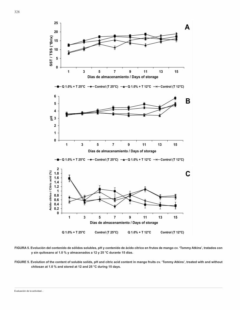
En las Figuras 4 y 5 se muestra el desarrollo de la ma-duración de los mangos tratados con quitosano y sin este, y posteriormente almacenados a 12 y 25 °C. En relación con la pérdida de peso, se observó que el menor porcentaje (aproximadamente de 3 % al término de 15 días de almace-namiento) correspondió a los frutos tratados con quitosano y almacenados a 12 °C (Figura 4A). Los mangos tratados con quitosano y almacenados a 25 °C tuvieron la mayor pérdida de peso (aproximadamente 16 %) en comparación con los tratamientos restantes. La firmeza de los frutos cayó en un rango de 30 a 10 N en los frutos con quitosano y sin este, respectivamente, independientemente de la temperatura de almacenamiento (Figura 4B). Los valores de °Brix y pH va-riaron de 14 a 18 %, y de 4 a 5.5 respectivamente (Figuras 5A y B). El contenido de SST más alto correspondió a los frutos provenientes del control, almacenados a 25 °C, en tanto que el pH mayor fue en los frutos tratados con quito-sano y almacenados a esta temperatura. En relación con el contenido de ácido cítrico (Figura 5C), el porcentaje mayor (0.8 %) correspondió a los mangos tratados y sin tratar con quitosano cuando se almacenaron a 12 °C.
La respuesta fisiológica de los frutos tratados con quito-sano en este trabajo concuerda con investigaciones previas en varios productos hortofrutícolas donde se reporta sobre su acción como una cubierta semipermeable, que regula el intercambio gaseoso y por lo tanto disminuye la transpiración (Bautista-Baños et al., 2006). En general, en estudios lleva-dos a cabo sobre el efecto de la aplicación de quitosano en frutos como el litchi, el plátano y el pimiento morrón, se ob-servó menor pérdida de peso que en productos no tratados, al ser almacenados a temperaturas menores de los 15 °C (Kittur et al., 1998; Du et al., 1997; Ghaouth et al., 1991b). En relación a las otras variables analizadas, no se encontró un efecto significativo por la aplicación del quitosano, y se observó que durante el almacenamiento los jitomates trata-dos con quitosano y sin él siguieron una maduración normal
CUADRO 2. Efecto del quitosano y temperatura de almacenamiento en la incidencia y severidad de Alternaria alternata en frutos de mango cv. ‘Tommy Atkins’.
TABLE 2. Effect of chitosan and storage temperature on the incidence and severity of Alternaria alternata in ‘Tommy Atkins’ cv. mango fruits.
Tratamientos / TreatmentsIncidencia / Incidence
(%)*
Severidad / Severity
(%)*
Quitosano / Chitosan 1 %
Temperatura / Temperature 12 °C100 58
Quitosano / Chitosan 1 %
Temperatura / Temperature 25 °C100 90
Control 100 100
*12 días de almacenamiento.*12 days of storage.
of TSS corresponded to the fruits from the control, stored at 25 °C, while the highest pH was in the fruits treated with chitosan and stored at this temperature. With respect to the content of citric acid (Figure 5C), the highest percentage (0.8 %) corresponded to the mangos treated and untreated with chitosan when they were stored at 12 °C.
The physiological response of the fruits treated with chitosan in this work agrees with previous investigations in various fruit products where its action as a semi-per-meable coating is reported, which regulates the gas ex-change and therefore diminished transpiration (Bautista-Baños et al., 2006). In general, in studies made on the effect of the application of chitosan on fruits such as litchi, banana and red bell pepper, lower weight loss was ob-served than in untreated products, when stored at temper-atures lower than 15 °C (Kittur et al., 1998; Du et al., 1997; Ghaouth et al., 1991b). With respect to the other variables analyzed, no significant effect was found from the applica-tion of chitosan, and it was observed that during storage the tomatoes treated with chitosan and without treatment followed a normal maturation (Sánchez-Domínguez et al., 2011). However, different authors demonstrate that tem-perature was the factor that most influenced the final val-ues of most of the physio-chemical variables evaluated (Zhang and Quantick, 1998; Ramos-García et al., 2010; Zhu et al., 2008).
Chitosan is a compound that presents bio-functional characteristics, given that it can also be used without prob-lems to elaborate edible coatings. Although in this investiga-tion the fungicidal activity of chitosan was not demonstrated when it was applied in situ, it is considered convenient to experiment in future investigations with this compound as an edible coating added with other natural compounds such as botanical extracts and essential oils.
CONCLUSIONS
Mycelial growth, germination and sporulation of A. alternata showed significant inhibition in the presence of chitosan at 1.0 %.

Revista Chapingo Serie Horticultura 19(3): 315-331, 2013
327
FIGURA 4. Evolución de la pérdida de peso y firmeza en frutos de mango cv. ‘Tommy Atkins’, tratados con y sin quitosano al 1.0 % y alma-cenados a 12 y 25 °C durante 15 días.
FIGURE 4. Evolution of weight loss and firmness in mango fruits cv. ‘Tommy Atkins’, treated with and without chitosan at 1.0 % and stored at 12 and 25 °C during 15 days.
Evaluación de la actividad...
328
FIGURA 5. Evolución del contenido de sólidos solubles, pH y contenido de ácido cítrico en frutos de mango cv. ‘Tommy Atkins’, tratados con y sin quitosano al 1.0 % y almacenados a 12 y 25 °C durante 15 días.
FIGURE 5. Evolution of the content of soluble solids, pH and citric acid content in mango fruits cv. ‘Tommy Atkins’, treated with and without chitosan at 1.0 % and stored at 12 and 25 °C during 15 days.
Revista Chapingo Serie Horticultura 19(3): 315-331, 2013
329
(Sánchez-Domínguez et al., 2011). Sin embargo, distintos autores evidenciaron que la temperatura fue el factor que más influyó en los valores finales de la mayoría de las va-riables físico-químicas evaluadas (Zhang y Quantick, 1998; Ramos-García et al., 2010; Zhu et al., 2008).
El quitosano es un compuesto que presenta caracte-rísticas biofuncionales, ya que puede utilizarse también sin problemas para elaborar recubrimientos comestibles, Aun-que en esta investigación no se demostró la actividad fun-gicida del quitosano cuando se aplicó in situ, se considera conveniente experimentar en futuras investigaciones con este compuesto como un recubrimiento comestible adicio-nado con otros compuestos naturales tales como extractos botánicos y aceites esenciales.
CONCLUSIONES
El crecimiento micelial, germinación y esporulación de A. alternata mostraron una inhibición significativa en presencia del quitosano al 1.0 %.
La incubación de A. alternata en presencia del quito-sano al 1 % ocasionó una intensa y amplia vacuolización del micelio y esporas, la salida de material citoplásmico y la formación de material fibrilar alrededor de las células.
El quitosano aplicado en los frutos de mango no con-troló la mancha negra del fruto pero sí inhibió la extensión de la enfermedad.
El proceso de maduración no se vio afectado por la combinación del quitosano y la temperatura.
La temperatura fue el principal factor que afectó los cambios en el contenido de SST (°Brix), la firmeza y el con-tenido de ácido cítrico en los frutos de mango.
LITERATURA CITADAANÓNIMO. 2005. Pérdidas en la manipulación después de la
cosecha. Organización de las Naciones Unidas para la Agricultura y la Alimentación. Guayaquil, Ecuador. 8 p. ftp://ftp.fao.org/docrep/fao/meeting/009/j5778s.pdf
ANÓNIMO. 2010. La imagen agropecuaria. Secretaría de Agricultura, Gadanería, Desarrollo Rural, Pesca y Alimentación. Boletín Agropecuario 1: 1-3.
BAUTISTA-BAÑOS, S.; DÍAZ-PÉREZ, J. C.; VILLANUEVA-ARCE, R.; EVANGELISTA- LOZANO, S. 1998. Enfermedades postcosecha en frutas y hortalizas. Alternativas de control, pp. 60-63. In: Calidad y Manejo en Postcosecha de Frutas y Hortalizas. DÍAZ-PÉREZ, J. C.; LÓPEZ-GÓMEZ, R.; RODRÍGUEZ- AMBRÍZ, S.L.; ARAU-ROFFIELY, L. A.; CANO-OCHOA, C. F. (eds.). Centro de Desarrollo de Productos Bióticos-Instituto Politécnico Nacional, Instituto Tecnológico de Zacatepec. Morelos, México.
The incubation of A. alternata in the presence of chi-tosan at 1 % caused an intense and ample vacuolization of mycelia and spores, the output of cytoplasmic material and the formation of fibrilar material around the cells.
Chitosan applied to mango fruits did not control black spot of the fruit, but it did inhibit the extension of the disease.
The ripening process was not affected by the combi-nation of chitosan and temperature.
Temperature was the principal factor that affected the changes in the content of TSS (°Brix) firmness and the citric acid content in the mango fruits.
End of English Version
BAUTISTA-BAÑOS, S.; HERNÁNDEZ-LÓPEZ, M.; BOSQUEZ-MOLINA, E.; WILSON, C. L. 2003. Effects of chitosan and plant extracts on growth of Colletotrichum gloeosporioides, anthracnose levels and quality of papaya fruit. Crop Protection 22(9): 1087-1092. doi: 10.1016/S0261-2194(03)00117-0
BAUTISTA-BAÑOS, S.; HERNÁNDEZ-LÓPEZ, M.; BOSQUEZ-MOLINA, E. 2004. Growth inhibition of selected fungi by chitosan and plant extracts. Revista Mexicana de Fitopatología 22(2): 178-186. http://sociedadmexicanadefitopatologia.org/archives/61222204.pdf
BAUTISTA-BAÑOS, S.; HERNÁNDEZ-LAUZARDO, A. N.; VELÁZQUEZ-VALLE, M. G.; HERNÁNDEZ-LÓPEZ, M.; AIT-BARKA, E.; BOSQUEZ-MOLINA, E.; WILSON, C. L. 2006. Chitosan as a potential natural compound to control pre-and postharvest diseases of horticultural commodities. Crop Protection 25(2): 108-118. doi: 10.1016/j.cropro.2005.03.010
BENHAMOU, N. 1992. Ultrastructural and cytochemical aspects of chitosan on Fusarium oxysporum f. sp. radicis lycopersici, agent of tomato crown and root rot. Phytopathology 82(10):1185-1193.http://www.apsnet.org/publications/phytopathology/backissues/Documents/1992Articles/phyto82n10_1185.pdf
BEN-SHALOM, N.; ARDI, R.; PINTO, R.; AKI, C.; FALLIK, E. 2003. Controlling gray mould caused by Botrytis cinerea in cucumber plants by means of chitosan. Crop Protection 22(2): 285-290. doi. 10.1016/S0261-2194(02)00149-7
DU, J.; GEMMA, H.; IWAHORI, S. 1997. Effects of chitosan coating on the storage of peach, japanese pear, and kiwifruit. Journal of the Japanese Society for Horticultural Science 66(1): 15-22. https://www.jstage.jst.go.jp/article/jjshs1925/66/1/66_1_15/_pdf
ERYANI-RAQEEB, A. A.; MAHMUD, T. M. M.; SYED O., S. R.; MOHAMED Z., A. R.; ERYANI, A. R. 2009. Effects of

Evaluación de la actividad...
330
calcium and chitosan treatments on controlling anthracnose and postharvest quality of papaya (Carica papaya L.). International Journal of Agricultural Research 4(2): 53-68. doi: 10.3923/ijar.2009.53.68
GARCÍA-RINCÓN, J., VEGA-PÉREZ, J., GUERRA-SÁNCHEZ, M.G., HERNÁNDEZ-LAUZARDO, A.N., PEÑA-DÍAZ, A., VELÁZQUEZ-DEL VALLE, M.G. 2010. Effect of chitosan on growth and plasma membrane properties of Rhizopus stolonifer (Ehrnb.:Fr.) Vuill. Pesticide Biochemistry and Physiology 97(3): 275-278. doi: 10.1016/j.pestbp.2010.03.008
GHAOUTH, A.; ARUL, J.; PONNAMPALAM, R.; BOULET, M. 1991a. Chitosan coating effect on storability and quality of fresh strawberries. Journal of Food Science 56(6): 1618-1620. doi: 10.1111/j.1365-2621.1991.tb08655.x
GHAOUTH, A.; ARUL, J.; PONNAMPALAM, R.; BOULET, M. 1991b. Use of chitosan coating to reduce water loss and maintain quality of cucumber and bell pepper fruits. Journal of Food Processing and. Preservation 15(5): 359-368. doi: 10.1111/j.1745-4549.1991.tb00178.x
GHAOUTH, A.; ARUL, J.; ASSELIN, A.; BENHAMOU, N. 1992a. Antifungal activity of chitosan on postharvest pathogens: Induction of morphological and cytological alterations in Rhizopus stolonifer. Mycological Research 96(9): 769-779. doi: 10.1016/S0953-7562(09)80447-4
GHAOUTH, A.; ARUL, J., GRENIER, J.; ASSELIN, A. 1992b. Antifungal activity of chitosan on two postharvest pathogens of strawberry fruits. Phytopathology 82(4): 398-402. doi: 10.1094/Phyto-82-398
GHAOUTH, A.; ARUL, J.; GRENIER, J.; BENHAMOU, N.; ASSELIN, A.; BÉLANGER, R. 1994. Effect of chitosan on cucumber plants: Suppresion of Phythium aphanidermatum and induction of defence reactions. Phytopathology 84(3): 313-320. doi: 10.1094/Phyto-84-313
HIRANO, A.; NAGAO, N. 1989. Effects of chitosan, pectic acid, lysozyme, and chitinase on the growth of several phytopathogens. Agricultural and Biological Chemistry 53(11): 3065-3066. http://ci.nii.ac.jp/lognavi?name=nels&lang=en&type=pdf&id=ART0008331431
KITTUR, F. S.; KUMAR, K. R.; THARANATHAN, R. N. 1998. Functional packaging properties of chitosan films. Zeitschrift für Tebensmittelunterssuchung und-forshung A. 206(1): 44-47. doi: 10.1007/s002170050211
PALMA-GUERRERO, J.; JANSSON, H.-B.; SALINA, J.; LÓPEZ-LLORCA, L. V. 2008. Effect of chitosan on hyphal growth and spore germination of plant pathogenic and biocontrol fungi. Journal of Applied Microbiology 104(2): 541-553. doi: 10.1111/j.1365-2672.2007.03567.x
PALMA-GUERRERO, J.; HUANG, I.-C.; JANSSON, H.-B.; SALINAS, J.; LOPEZ-LLORCA, L. V.; READ, N. D. 2009. Chitosan permeabilizes the plasma membrane and kills cells of Neurospora crassa in an energy dependent manner. Fungal Genetics and Biology 46(8): 585-594. doi:10.1016/j.fgb.2009.02.010
PALMA-GUERRERO, J.; LÓPEZ-JIMÉNEZ, J. A.; PÉREZ-BERNÁ, A. J.; HUANG, C.-I.; JANSSON, H.-B.; SALINAS, J.; VILLALAÍN, J.; READ, N. D.; LÓPEZ-LORCA, L. V. 2010.
Membrane fluidity determines sensitivity of filamentous fungi to chitosan. Molecular Microbiology 75(4): 1021-1032. doi: 10.1111/j.1365-2958.2009.07039.x
PLOETZ, R. C.; ZENTMYER, G. A.; NISHIJIMA, W. T.; ROHRBACH, K. G.; OHR, H. D. 1998. Compendium of Tropical Fruit Diseases. The American Phytopathological Society. St. Paul Minnesota, USA. 88 p.
RAMOS-GARCÍA, M.; BAUTISTA-BAÑOS, S.; TRONCOSO-ROJAS, R.; BOSQUEZ-MOLINA, E.; ALÍA-TEJACAL, I.; GUILLÉN-SÁNCHEZ, D.; GUTIÉRREZ-MARTÍNEZ, P. 2010. Papaya postharvest handling in México: use of chitosan and isothiocyanates to control postharvest diseases. Fresh Produce 4(ESP1): 21-28. http://www.researchgate.net/publication/244992191_Papaya_Postharvest_Handling_in_Mexico_Use_of_Chitosan_and_Isothiocyanates_to_Control_Postharvest_Diseases/file/9c96051d50caf9f395.pdf
REDDY, M. V. B.; ARAUL, J.; AIT-BARKA, E.; ANGERS, P.; RICHARD, C.; CASTAIGNE, F. 1998. Effect of chitosan on growth and toxin production by Alternaria alternata f. sp. lycopersici. Biocontrol Science and Technology 8(1): 33-43. doi: 10.1080/09583159830414
SÁNCHEZ-DOMÍNGUEZ, D.; BAUTISTA-BAÑOS, S.; CASTILLO O., P. 2007. Efecto del quitosano en el desarrollo y morfología de Alternaria alternata (Fr..) Keissl. Anales de Biología 29: 23-32. https://www.um.es/analesdebiologia/numeros/29/PDF/03-EFECTO.pdf
SÁNCHEZ-DOMÍNGUEZ, D.; RÍOS, M. Y.; CASTILLO-OCAMPO, P.; ZAVALA-PADILLA, G.; RAMOS-GARCÍA, M.; BAUTISTA-BAÑOS, S. 2011. Cytological and biochemical changes induced by chitosan in the pathosystem Alternaria alternata-tomato. Pesticide Biochemistry and Physiology 99(3): 250-255. doi: 10.1016/j.pestbp.2011.01.003
SANDFORD, P. 1989. Chitosan: commercial uses and potential applications, pp. 51-69. In: Chitin and Chitosan: Sources, Chemistry, Biochemistry. Physical Properties and Applications. SKJAK-BRAEK, G.; ANTHOSEN, T.; STANDFORD, P. (eds). Elsevier Applied Science. New York, USA.
SIMMONS, E. G. 1992. Alternaria taxonomy: current status, viewpoint, challenge, pp. 1-35. In: Alternaria biology, plant diseases and metabolites. CHELKOWSKI, J.; VISCONTI, A. (eds). Elsevier Science Publishers. Amsterdam.
SONG, Y.; BABIKER, E.E.; USUI, M.; SAITO, A.; KATO, A. 2002. Emulsifying properties and bactericidal action of chitosan-lysozyme conjugates. Food Research International 35(5): 459-466. doi: 10.1016/S0963-9969(01)00144-2
TERRY, L. A.; JOYCE, D. C. 2004. Elicitors of induced disease resistance in postharvest horticultural crops: a brief review. Postharvest Biology and Technology 32(1): 1-13. doi: 10.1016/j.postharvbio.2003.09.016
VERO M., S.; MONDINO, P. 1999. Medidas para conservar frutas y hortalizas. Control biológico postcosecha. Horticultura Internacional 7: 29-36. http://www.magrama.gob.es/ministerio/pags/biblioteca/revistas/pdf_hortint/hortint_1999_26_29_36.pdf
WILSON, C. L.; GHAOUTH, A.; CHALUTS, E.; DROBY, S.;

Revista Chapingo Serie Horticultura 19(3): 315-331, 2013
331
STEVENS, C.; LU, J. L.; KHAN, V.; ARUL, J. 1994. Potential of induced resistance to control postharvest diseases of fruits and vegetables. Plant Disease 78: 837-844. doi: 10.1094/PD-78-0837
ZHANG, D.; QUANTICK, P. C. 1998. Antifungal effects of chitosan coating on fresh strawberries and raspberries during storage. Journal of Horticultural Science & Biotechnology 73(6): 763-767. http://www.jhortscib.org/Vol73/73_6/7.htm
ZHU, X.; WANG, Q.; CAO, J.; JIANG, W. 2008. Effects of chitosan coating on postharvest quality of mango (Mangifera indica L. cv. Tainong) fruits. Journal of Food Processing and Preservation 32(5): 770-784. doi: 10.1111/j.1745-4549.2008.00213.x