Embed Size (px)
Citation preview
1
Evaluating Knee Pain
Matthew T. Boes, M.D.Raleigh Orthopaedic Clinic
September 24, 2011
Introduction
• Approach to patient with knee pain / injury– History– Examination– Radiographs– Guidelines for additional
imaging
2
History
• Age• History of specific injury / traumatic event?• Location of pain• Nature of pain• Are there mechanical symptoms?• Aggravating / precipitating factors
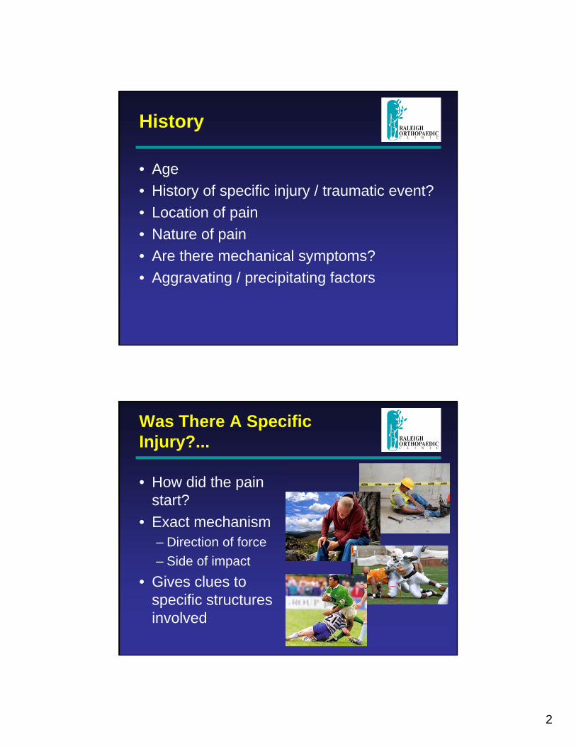
Was There A Specific Injury?...
• How did the pain start?
• Exact mechanism– Direction of force– Side of impact
• Gives clues to specific structures involved
3
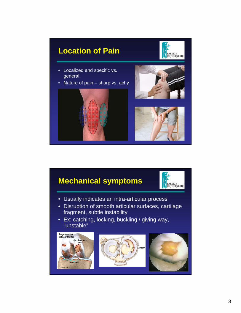
Location of Pain
• Localized and specific vs. general
• Nature of pain – sharp vs. achy
Mechanical symptoms
• Usually indicates an intra-articular process• Disruption of smooth articular surfaces, cartilage
fragment, subtle instability• Ex: catching, locking, buckling / giving way,
“unstable”
4
Aggravating / Precipitating Factors
• Stairs • Start up• Prolonged sitting
– Driving• Twisting / squatting• Side-to-side activity• Impact activity
Examination
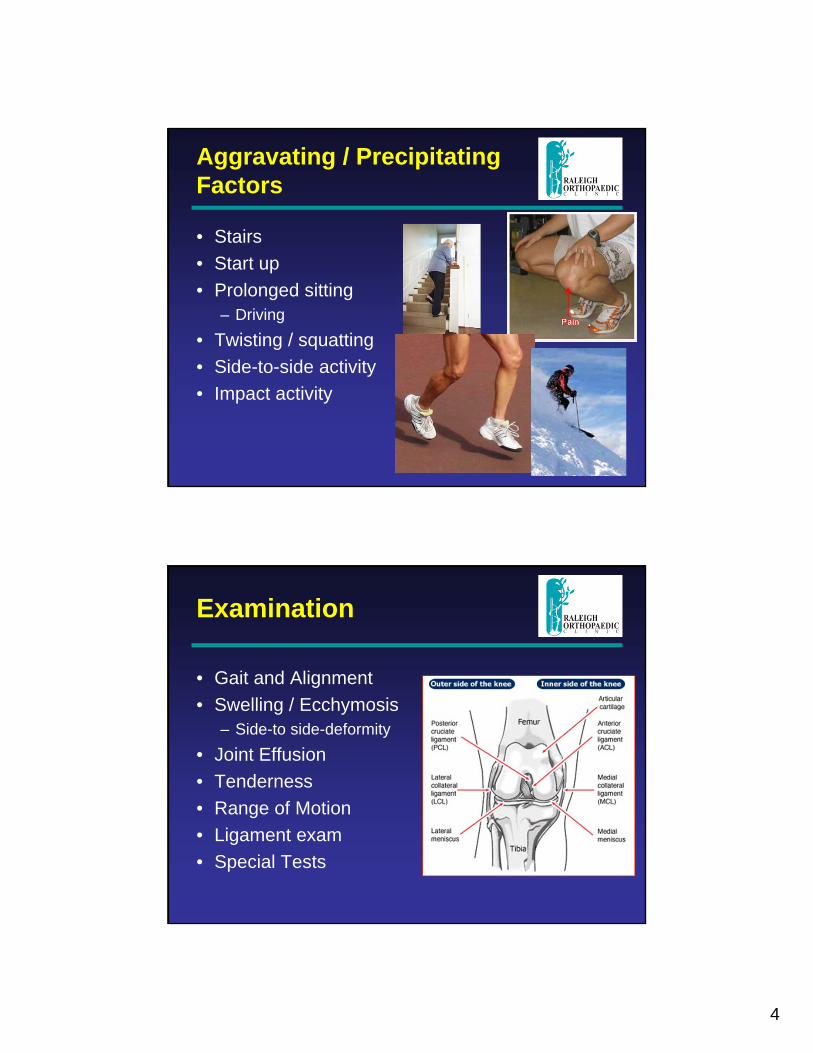
• Gait and Alignment• Swelling / Ecchymosis
– Side-to side-deformity• Joint Effusion• Tenderness• Range of Motion• Ligament exam• Special Tests
5
Gait and Alignment
• Painful weight-bearing• Guarding with certain
motions• Varus or valgus alignment
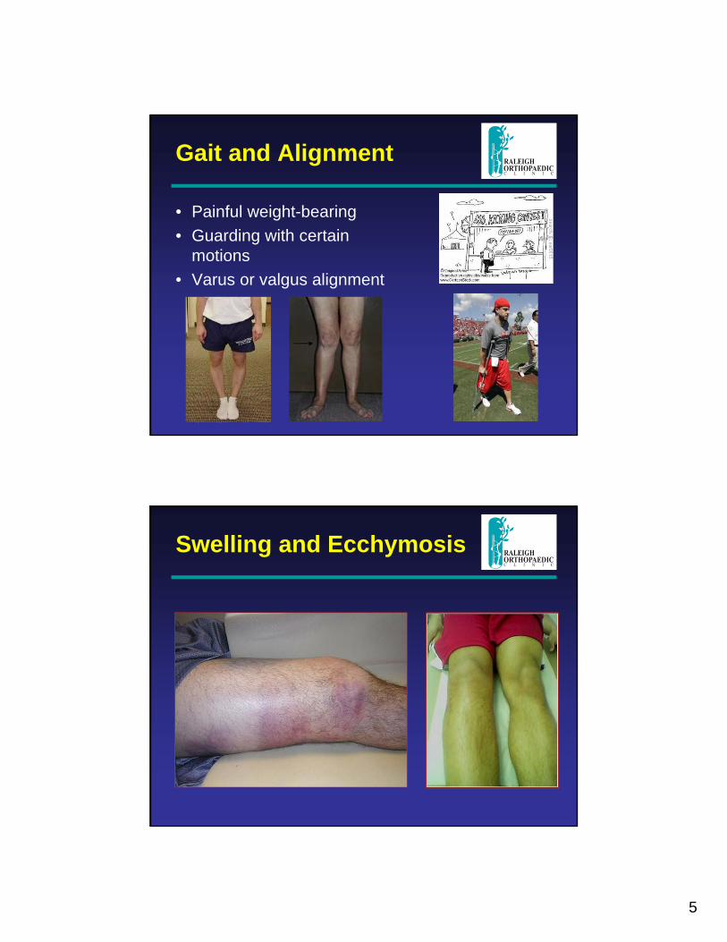
Swelling and Ecchymosis
6
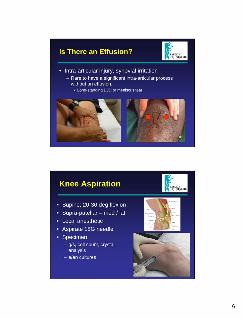
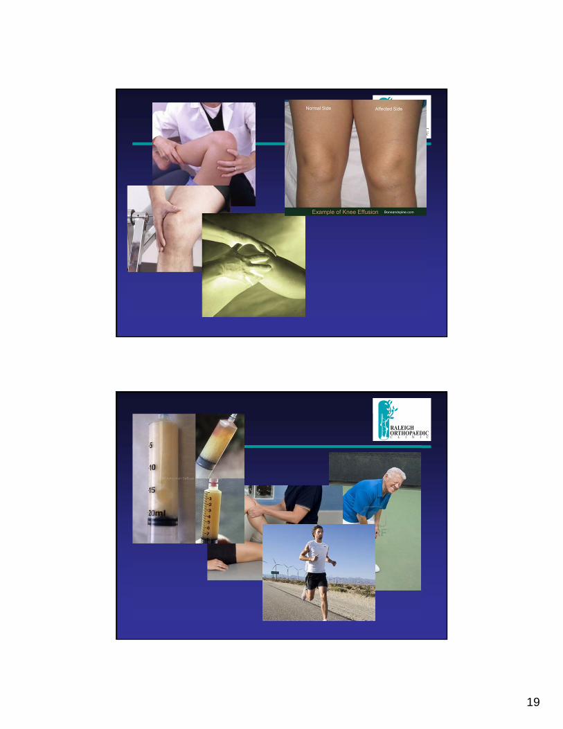
Is There an Effusion?
• Intra-articular injury, synovial irritation– Rare to have a significant intra-articular process
without an effusion.• Long-standing DJD or meniscus tear
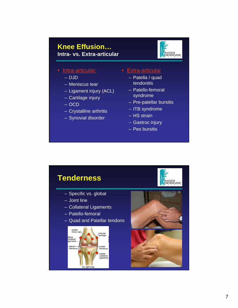
Knee Aspiration
• Supine; 20-30 deg flexion• Supra-patellar – med / lat• Local anesthetic• Aspirate 18G needle• Specimen
– g/s, cell count, crystal analysis
– a/an cultures
7
Knee Effusion…Intra- vs. Extra-articular
• Intra-articular:– DJD– Meniscus tear– Ligament injury (ACL)– Cartilage injury– OCD– Crystalline arthritis– Synovial disorder
• Extra-articular– Patella / quad
tendonitis– Patello-femoral
syndrome– Pre-patellar bursitis– ITB syndrome– HS strain– Gastroc injury– Pes bursitis
Tenderness
– Specific vs. global– Joint line– Collateral Ligaments– Patello-femoral– Quad and Patellar tendons
8
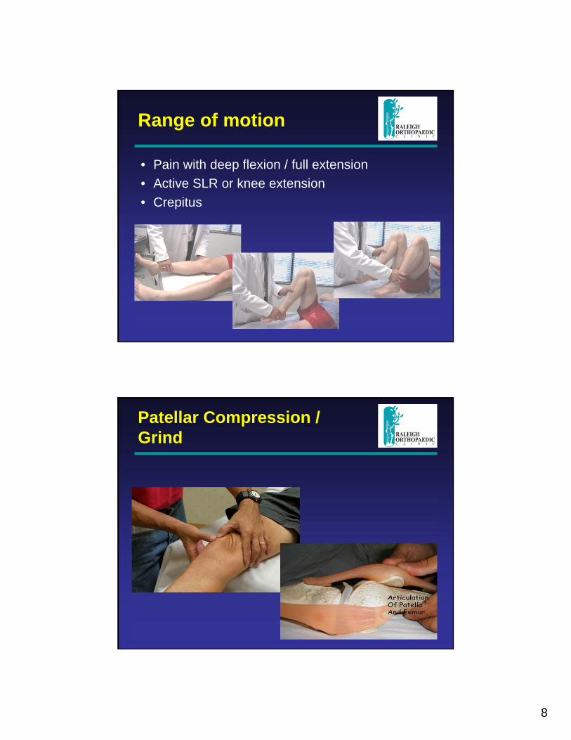
Range of motion
• Pain with deep flexion / full extension• Active SLR or knee extension• Crepitus
Patellar Compression / Grind
9
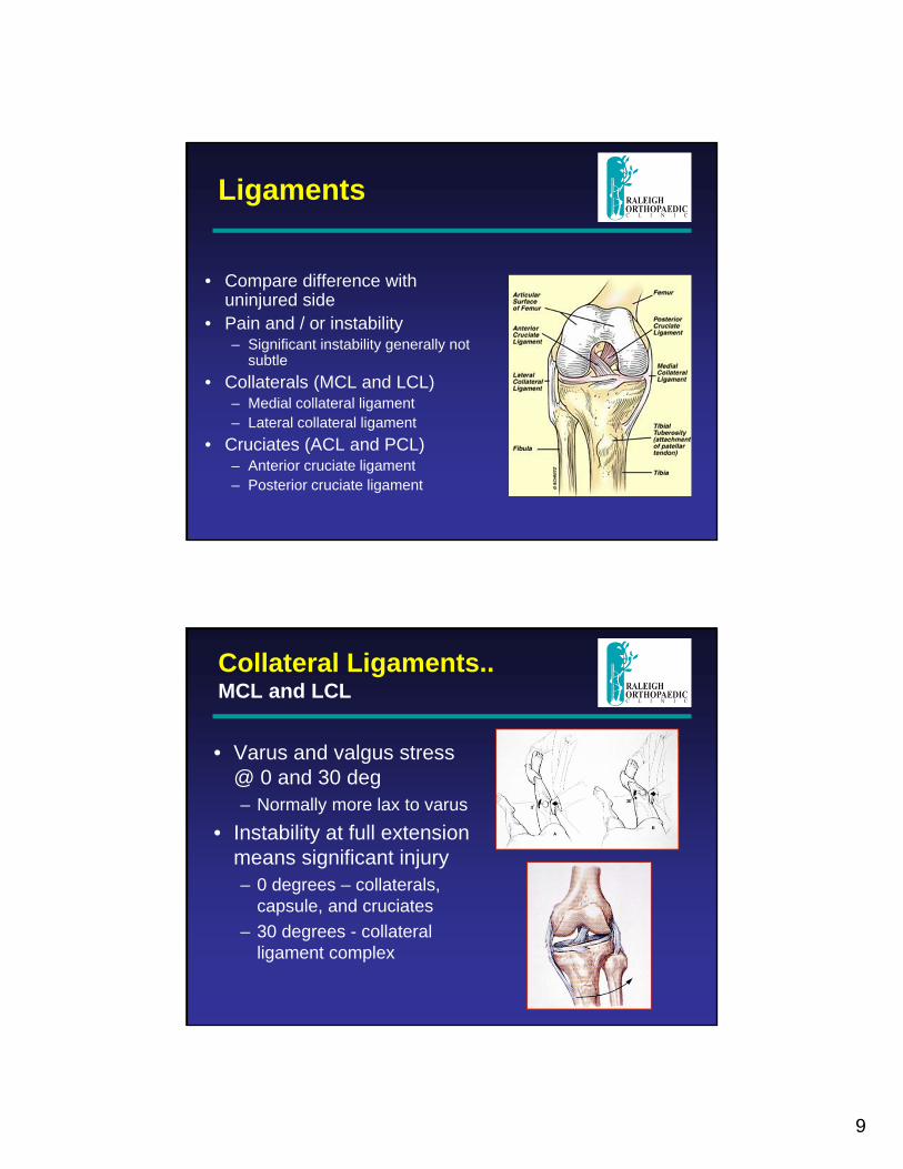
Ligaments
• Compare difference with uninjured side
• Pain and / or instability– Significant instability generally not
subtle• Collaterals (MCL and LCL)
– Medial collateral ligament – Lateral collateral ligament
• Cruciates (ACL and PCL)– Anterior cruciate ligament– Posterior cruciate ligament
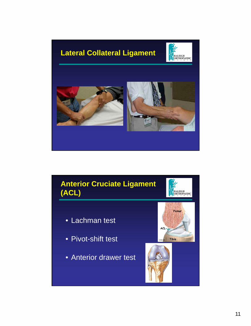
Collateral Ligaments..MCL and LCL
• Varus and valgus stress @ 0 and 30 deg– Normally more lax to varus
• Instability at full extension means significant injury– 0 degrees – collaterals,
capsule, and cruciates– 30 degrees - collateral
ligament complex
10
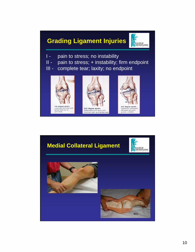
Grading Ligament Injuries
I - pain to stress; no instabilityII - pain to stress; + instability; firm endpointIII - complete tear; laxity; no endpoint
Medial Collateral Ligament
11
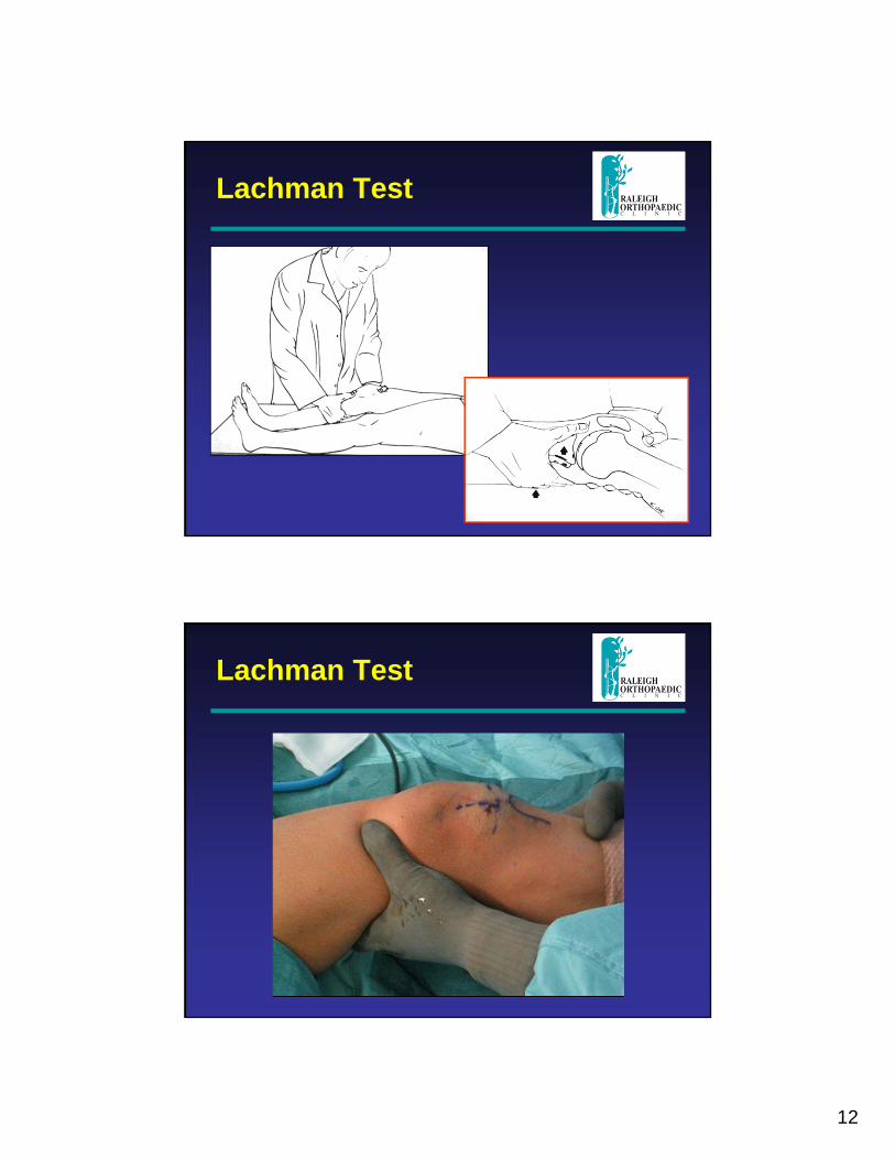
Lateral Collateral Ligament
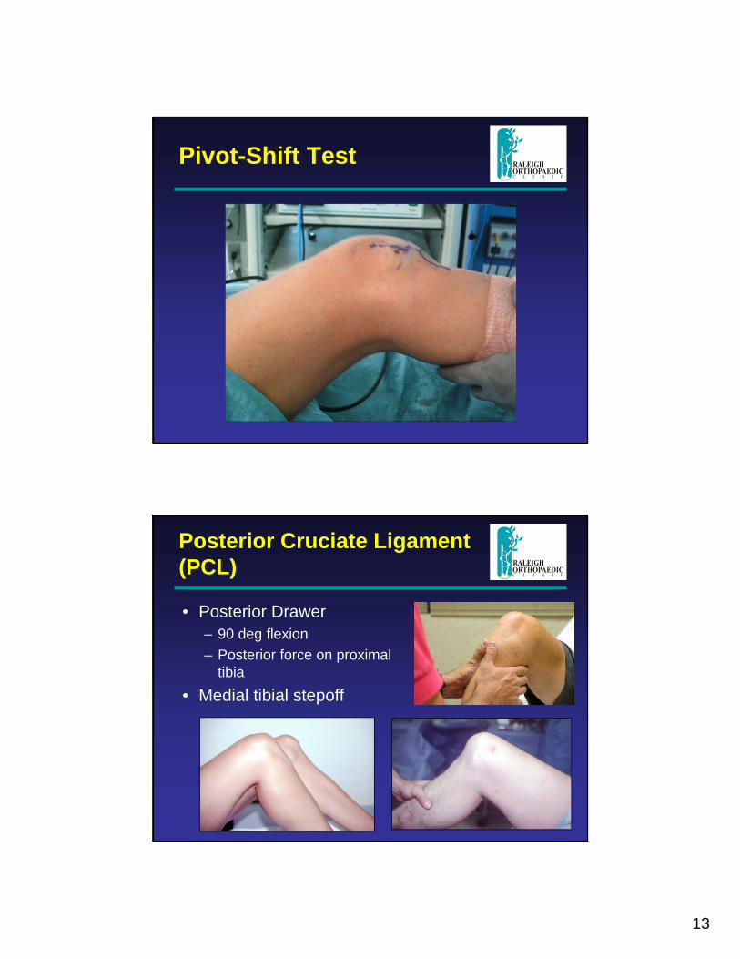
Anterior Cruciate Ligament (ACL)
• Lachman test
• Pivot-shift test
• Anterior drawer test
12
Lachman Test
Lachman Test
13
Pivot-Shift Test
Posterior Cruciate Ligament (PCL)
• Posterior Drawer– 90 deg flexion– Posterior force on proximal
tibia• Medial tibial stepoff
14
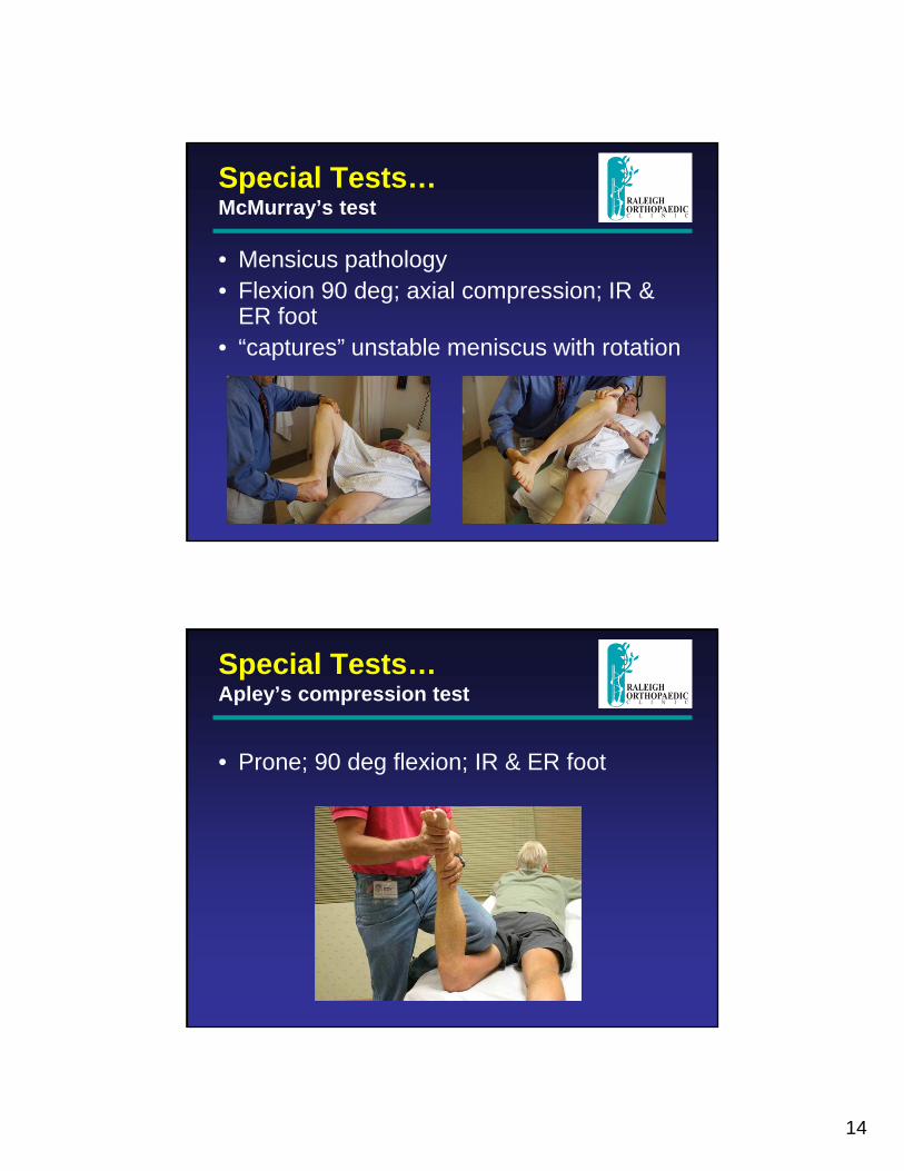
Special Tests…McMurray’s test
• Mensicus pathology• Flexion 90 deg; axial compression; IR &
ER foot• “captures” unstable meniscus with rotation
Special Tests…Apley’s compression test
• Prone; 90 deg flexion; IR & ER foot
15
DX: meniscus tear
• Joint line tenderness• Effusion• Pain with forced flexion• Block to full extension• (+) McMurray’s or Appley’s test
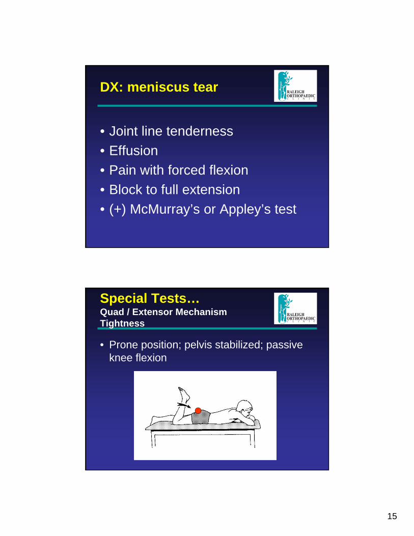
Special Tests…Quad / Extensor Mechanism Tightness
• Prone position; pelvis stabilized; passive knee flexion
16
Special Tests…Quad / Extensor Mechanism Tightness
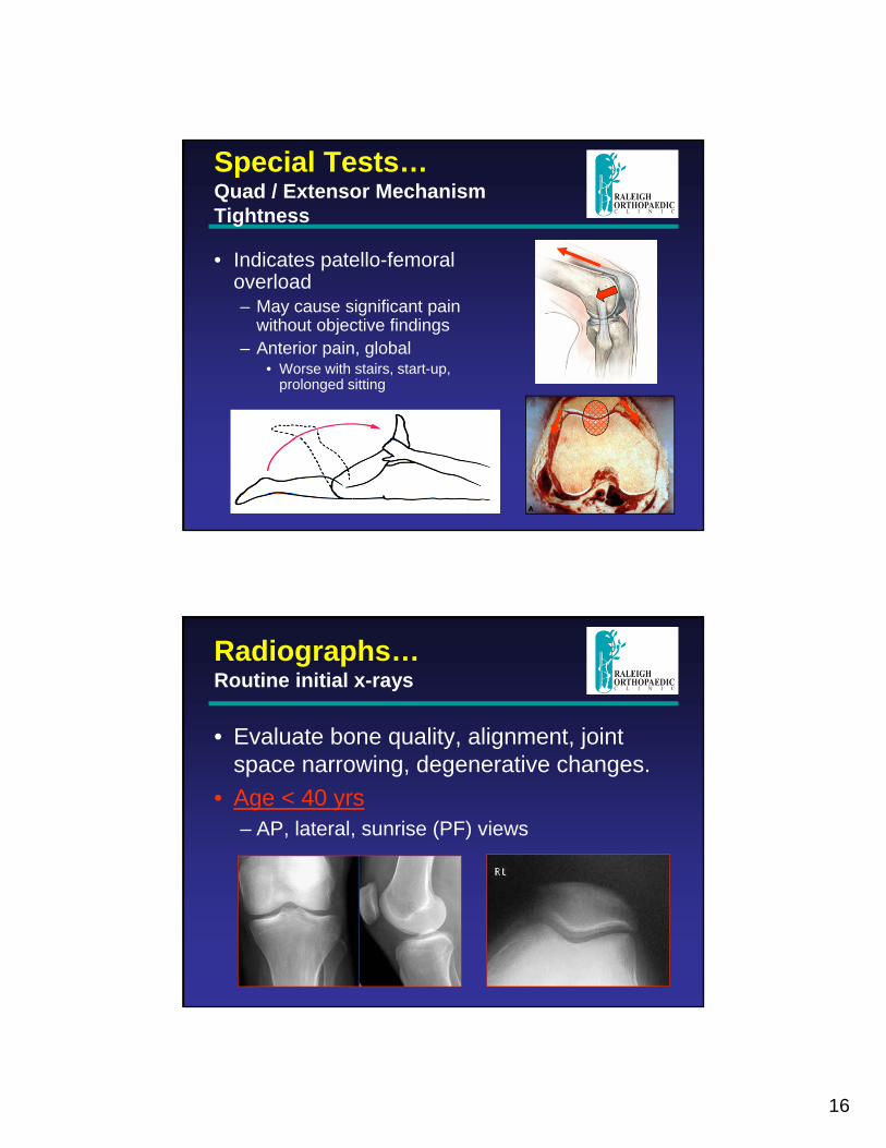
• Indicates patello-femoral overload– May cause significant pain
without objective findings– Anterior pain, global
• Worse with stairs, start-up, prolonged sitting
Radiographs…Routine initial x-rays
• Evaluate bone quality, alignment, joint space narrowing, degenerative changes.
• Age < 40 yrs– AP, lateral, sunrise (PF) views
17
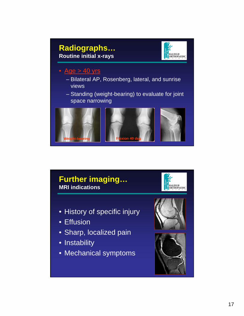
Radiographs…Routine initial x-rays
• Age > 40 yrs– Bilateral AP, Rosenberg, lateral, and sunrise
views– Standing (weight-bearing) to evaluate for joint
space narrowing
Flexion 40 degWeight-bearing
Further imaging…MRI indications
• History of specific injury• Effusion• Sharp, localized pain• Instability• Mechanical symptoms
18
Thank You
Madeline
• C c bv b v cxc,ll
• Ybgtb
• ‘’’’;;l,nm
19