Embed Size (px)
Citation preview

ORIGINAL PAPER
Evaluating the bone regeneration in calvarial defect
using osteoblasts differentiated from adipose-derived
mesenchymal stem cells on three different scaffolds:
an animal study
Hassan Semyari . Mahmood Rajipour .
Shabnam Sabetkish . Nastaran Sabetkish .
Fatemeh Mashhadi Abbas . Abdol-Mohammad Kajbafzadeh
Received: 30 November 2014 / Accepted: 19 June 2015 / Published online: 25 June 2015
� Springer Science+Business Media Dordrecht 2015
Abstract The aim of this study was to investigate
the effect of three different scaffolds on the viability
and differentiation of adipose-derived mesenchymal
stem cells (ADMSCs) to osteoblast for bone regener-
ation of calvarial defect in rabbit model. Adipose was
harvested from the nape of 12 rabbits by direct surgery
or hollow-tip cannula. Two standardized circular
calvarial defects (case and control), 8 mm in diameter
each, were created in all the animals. The animals
were divided into 3 different groups. In group 1 (G1),
the defect was filled with polyamide ? ADMSC. In
group 2, poly lactic-co-glycolic acid ? ADMSC was
used. In group 3, decellularized amniotic mem-
brane ? ADMSC was applied. In the control defect,
the non-seeded scaffolds were applied for filling the
defect. Decellularized pericardial scaffolds were used
as a membrane on the scaffolds. The animals were
euthanized 2, 4, and 8 weeks of operation and new
bone formation was assessed by different analyses.
Immunohistochemical (IHC) staining with osteopon-
tin and osteocalcin antibodies was also performed.
After 2 weeks of wound healing, minimal bone
regeneration was detected in all groups. Almost
complete defect closure was observed in all experi-
mental groups after 8 weeks of operation, with the
greatest defect closure in the animals treated with
polyamide scaffolds as compared to biopsies obtained
from control defects and other experimental groups.
The maximal tensile load was higher in G1, 4 and
8 weeks postoperatively, suggesting the usefulness of
polyamide ? ADMSC for bone regeneration in cal-
varial defects. Results of the IHC staining demon-
strated a significant difference between seeded and
non-seeded scaffold in both short- and long-term
follow-ups (P\ 0.05). In addition, a significant
difference was observed in enhancement of IHC
staining of both markers in polyamide group (seeded
or non-seeded) 4 and 8 weeks postoperatively in
comparison with other scaffolds. It was concluded that
bone regeneration in critical calvarial defect was more
successful in seeded polyamide.
Keywords Adipose � Mesenchymal stem cells �
Calvaria � Bone regeneration � Scaffold
H. Semyari � M. Rajipour
Department of Periodontics, Faculty of Dentistry, Shahed
University, Tehran, Islamic Republic of Iran
S. Sabetkish � N. Sabetkish � A.-M. Kajbafzadeh (&)
Pediatric Urology Research Center, Section of Tissue
Engineering and Stem Cells Therapy, Children’s Hospital
Medical Center, Tehran University of Medical Sciences,
No. 62, Dr. Qarib’s Street, Keshavarz Boulevard,
1419433151 Tehran, Islamic Republic of Iran
e-mail: [email protected]
F. M. Abbas
Department of Oral and Maxillofacial Pathology, School
of Dentistry, Shahid Beheshti University of Medical
Sciences, Tehran, Islamic Republic of Iran
123
Cell Tissue Bank (2016) 17:69–83
DOI 10.1007/s10561-015-9518-5

Introduction
Developments in the field of dentistry were dramatic
during the last few years. Meanwhile, the application
of biodegradable scaffolds, stem cells (SCs) and
specific growth biomarkers like VEGF, FGF, BMP,
and Emdogain has been significantly increased (Rak-
manee et al. 2008). Bone regeneration in calvarial
defects following trauma or congenital deformities is
still a challenge for surgeons. Over the past years, the
beneficial effects of tissue engineering methods on
bone regeneration have been verified. It seems that the
most extensive application of tissue engineering is in
the area of mineralized tissue repair (Goldberg and
Smith 2004).
The capability of self renewal and generating a
wide range of specialized cell types are two major
properties of SCs (Anderson et al. 2001). SCs have
the ability to produce and differentiate to mature
progeny cells which influence the cell-base therapy
approaches. Mesenchymal stem cells (MSCs) that
possess the capacity to differentiate into osteoblasts
have been applied to evaluate bone regeneration in
calvarial defects (Koob et al. 2010). Adipose tissue
has been considered as a more practical and easy
source for deriving an abundant supply of MSCs as
compared with bone-marrow (Kim et al. 2014). In
the research for alternative sources of MSCs,
adipose tissues have been evaluated for their
potency to repair calvarial defects. Zuk et al.
(2001) described the properties of human adipose-
derived mesenchymal stem cells (ADMSCs) for the
first time. They also confirmed that ADMSCs are
able to differentiate into adipogenic, osteogenic,
chondrogenic, and myogenic lineages.
In addition, a seeded three-dimensional (3D)
scaffold which is needed for appropriate cell inter-
actions is of high importance for bone or tissue
regeneration (Mendes et al. 2002). ADMSCs in
combination with prefabricated scaffolds can be
expeditiously applied for numerous regenerative
therapies (Ge et al. 2012).
In the current study, we evaluated the behavior of
ADMSCs seeded on three different seeded scaffolds
including poly lactic-co-glycolic acid (PLGA),
decellularized amniotic membrane (DAM), and
polyamide and compared the in vivo effect of pre-
seeded and non-seeded scaffolds on repair of
calvarial defect.
Materials and methods
Animals
Twelve rabbits, aged 9–12 months, weighting approx-
imately 2–3 kg were selected for this study. All the
animals had healthy calvarium in clinical exam.
Animal selection, management and calvarial defect
preparation were conducted in accordance with the
Institutional Animal Care of Tehran University of
Medical Sciences. Rabbits were kept in separated
cages and monitored preciously for their healthy
condition. All the experimental animals were under
standard laboratory diet during the study.
Decellularization procedure
Pericardium
Rabbit pericardial tissue was decellularized with 1 %
Trypsin and 0.02 % ethylenediaminetetraacetic acid
(EDTA) at 35 �C for 12 h. The procedure was
followed by sodium dodecyl sulphate (SDS) 0.5 %
for 6 h. The previous steps were conducted under
continuous shaking. Finally, decellularized peri-
cardium was washed four times for periods of
10 min with phosphate-buffered saline (PBS) in order
to remove residual substances.
Amniotic membrane
Rabbit amniotic membrane was placed in distilled
water at 4 �C for 2 h. Then, 0.2 % SDS was applied
for the next 5 h. In the next stage, DAM was
maintained in PBS containing a cocktail of antibodies.
ADMSCs isolation and culture
Fatty tissue (500 mg) was obtained from the nape of
12 rabbits under sterile condition and washed with
sterile PBS. After mincing the tissues finely with a
scalpel, they were placed in collagenase type 1
solution (0.1 mg/ml) for 1 h at 37 �C in a shaking
water bath for digestion. In the next step, a 70-lm
mesh filter (falcon) was applied for filtering the
solution. Afterwards, cells were centrifuged and
cultured in Dulbecco’s modified Eagle’s medium
(DMEM), containing L-glutamine and penicillin/strep-
tomycin. Subsequently, For the purpose of cell
70 Cell Tissue Bank (2016) 17:69–83
123

attachment, the outgrown cells were incubated at
37 �C in a humidified atmosphere of 5 % CO2 and
95 % air for 36 h containing 10 % AS, fetal bovine
serum (FBS) or serum-free medium ? bFGF (10 ng/
ml). Non-adherent cells were washed away and
ADMSCs were isolated after culturing of plastic-
adherent cells (ADMSCs) on the third day. The
medium was changed with 3 days intervals. After
the first culture, cells were placed in 0.025 % Trypsin/
EDTA for 2 min in the incubator. Bone morpho-
genetic proteins (BMPs) consisting of osteocalsin and
osteopontin were added to the subcultured cells in the
third passage for better differentiation to osteoblast.
Identity of isolated stem cells
Flow cytometry analysis
Harvested cells of the third passage were incubated
with CD44, CD29, CD73, CD45, CD90, and CD105
antibodies as MCSs markers. Then, the labeled cells
were analyzed by flow cytometry.
Differentiation potential of ADMSCs
In order to investigate the differentiation potential of
cultured cells toward osteogenic cell lineage,
ADMSCs from the third passage were placed in
DMEM consisting of 50 mg/ml ascorbic 2-phosphate,
10 nM Dexamethason, and 10 mM b-glycerol phos-
phate. At the end of this period, alizarin red staining
was used to show the deposition of mineralization
matrix. For staining, the cultures were first fixed by
methanol for 10 min and then subjected to alizarin red
solution (2 g in 100 ml water) for 2 min.
Providing seeded scaffolds
The in vitro cell-seeding process was performed in 3
passages. Cells were washed and harvested using a
0.05 % Trypsin solution. All scaffolds were cut into
1.5 9 1.5 cm2 samples for cell culture. The scaffolds
were sterilized using 75 % ethanol for 2 h in a 24-well
plate and rinsed once in PBS and then in Hanks’
buffered saline solution (HBSS) for 60 min before the
cell seeding process. Sterilized scaffolds were trans-
ferred into 24-well culture plates and immersed in a-
MEM overnight. At a density of 1 9 105 cells per cm2,
ADMSCs were seeded on PLGA, polyamide, and
DAM using a dynamic seeding method on a rotating
shaker for 6 h in 5 % carbon dioxide at 37 �C at 95 %
humidity. Finally, the scaffolds were placed in a
6-well culture dish flask with culture media to allow
free contact of the media with the samples.
DNA quantification
In this procedure, the cell walls/membranes are disrupted
in order to isolate DNA. Lipids, proteins, and sugars are
also separated from nucleic acid. This method was
conducted according to the method described by Laird
et al. (1991). ForDNAquantification, 1 mgsamples from
decellularized pericardium and DAMwere obtained and
homogenized in a solution containing 0.25 % trypsin and
1 mM EDTA in deionized water. The homogenate was
incubated with invariable stirring for 3 h at 37 �C. In the
next step, the cell lysis was continued with a solution
containing 2 % SDS, 5 mM EDTA, 200 mMNaCl, and
100 mMTRIS–HCl, pH 8.5 for 24 h at 55 �C. TheDNA
extraction was performed in isopropanol and later
dissolved in a solution of 10 mM TrisHCl, 0.1 mM
EDTA, pH 7.5. Spectrophotometrically at 260 nm was
applied for determining the amount of DNA.
Scanning electron microscopy (SEM)
Several images with different magnifications were
taken from PLGA, polyamide, DAM, and all the pre-
seeded scaffolds after 8 weeks of operation and
analyzed using SEM (S3500N; Hitachi High Tech-
nologies America) at voltage of 5 kV in order to
determine the efficacy of our decellularization proce-
dure in cell removal and preservation of extra cellular
matrix (ECM). In order to determine the efficiency of
in vitro cell seeding, pre-seeded scaffolds were placed
in 2.5 % Glutaraldehyde at 4 �C for 1.5 h. Afterward,
they were washed with PBS three times for periods of
30 min at 4 �C. They were then dehydrated in ethanol
with concentrations of 30, 50, 70, and 90 % and
processed under a critical point dryer (Autosamdri-
814; Tousimis) for 15 min. In the next step, the
samples were coated with gold (2 nm thick approx-
imately) by the application of a Gatan ion beam coater.
Surgical procedure and defect treatment
Food was withheld the night before the surgery. A
prophylactic antibiotic (cefazolin; 22.0 mg/kg) was
Cell Tissue Bank (2016) 17:69–83 71
123

administered pre-operatively. The animals were anes-
thetized by intravenous injection of diazepam
(0.2 mg/kg) and ketamine (6.0 mg/kg). Calvarial
infiltration anesthesia was applied at the surgical sites.
The animals were randomly divided into three groups.
Two holes of 8 mm were made in calvarium by the
application of trephine dental drills in all the animals.
One of the standardized circular calvarial defects was
considered as control in which the non-seeded scaf-
folds were applied for filling the defect. The other
standardized calvarial defect was repaired using three
separated methods in each group. ADMSCs (at a
density of 1 9 105 cells per cm2) were seeded on three
different scaffolds. In group 1 (G1), the area of the
defect was repaired with polyamide ? ADMSC. In
group 2 (G2), PLGA ? ADMSC was used at the site
of defect and in group 3 (G3), DAM ? ADMSC was
applied. Decellularized pericardial scaffolds were also
used as a membrane on all the scaffolds (Fig. 1).
Animals received standard laboratory diet 2 weeks
postoperatively. Daily intramuscular injection of
Enrofloxacin 2.5 mg/kg was performed within the
first 2 weeks to prevent infection. Biopsies were taken
at 2, 4, and 8 weeks after the operation for investigat-
ing the regenerative capacity of the seeded and non-
seeded scaffolds via further histological observations
and biomechanical analysis.
Histological processing
To evaluate the effectiveness of ADMSCs in healing
process, histological examination was accomplished
by a pathologist who was totally blind to the current
strategy. In order to achieve this objective, a specimen
of 4 mm2 of all the experimental and control samples
was fixed in 10 % buffered formalin for 2–4 days,
decalcified in 5 % formic acid for 6–8 weeks and
dehydrated in graded ethanol. Then, the tissue blocks
were embedded in paraffin for further staining with
haematoxylin and eosin (H&E). For image analysis,
photoshop CS3 software (Adobe Systems, Inc.,
Mountain View, CA, USA) and Image Pro (Image
Pro Inc., Boston, MA, USA) were applied. IHMM,
Ver.1, sbmu, Irani software was used for evaluation of
regenerated bone and inflammation.
For determining the efficacy of different seeded and
non-seeded scaffolds and identifying the regeneration
of bone elements on different scaffolds, immunohis-
tochemical (IHC) staining with osteopontin and
osteocalcin antibodies was performed (Dako, Trappes,
France). Photoshop 10.0 software (Adobe Systems,
Inc., Mountain View, CA, USA), and Image Pro
(Image Pro Inc., Boston, MA, USA) were applied for
analyzing the images at each time point. SPSS 16
(Chicago, IL, USA) was used for data analysis.
Mechanical properties
Scaffolds must be able to endure intense mechanical
loads in native organs. To evaluate the tensile strength,
pre-seeded scaffolds of all groups were tested 4 and
8 weeks postoperatively via a tensile-test device
(Zwick/Roell, Model: Hct 400/25, Germany). A
constant elongation rate of 0.1 mm/s (6 mm/min)
was used after clamping the samples in sample
holders. Each sample was subjected to mounting
uniaxial tensile testing until the emergence of tear in
the tissue and disappearance of load demonstrated by
the device. Parameters such as stiffness (N/m), elastic
modulus and maximum force (N) were documented by
the device. Eventually, the curve of strength–stress
was drawn by the system in which the maximal point
indicates the maximum pressure tolerance. Four
samples from each group were tested, and the results
were averaged.
Statistical analysis
The statistical analysis was performed using SPSS�,
version 19. One-way analysis of variance (ANOVA)
and independent sample t test were used for comparing
graft particles retention and group means. Inflamma-
tory infiltrate scores were compared between treat-
ment and control groups by Wilcoxon’s signed-rank
test. P values less than 0.05 were considered as
statistical significance.
Results
Identity of isolated stem cells
Single cell suspension of primary cultured ADMSCs
was plated in 25-cm2 culture flasks under specific
culture condition to confirm the characterization of the
cultured cells. The formation of approximately 25
single colonies with spindle-shaped cells authenti-
cated the ability of ADMSCs to form adherent
72 Cell Tissue Bank (2016) 17:69–83
123

clonogenic cell clusters of Mesenchymal-like cells.
Microscopic evaluation of primary ADMSCs demon-
strated large flattened, long spindle-shaped, and short-
shaped cells after 7 days of the initial plating. Flow
cytometry outcomes demonstrated that the expression
of CD44, CD29, CD73, CD45, CD90, and CD105 was
Fig. 1 Surgical technique: Adipose harvest with hollow Tip
Cannula (a) Adipose harvest with direct surgery (b) Creation of
two calvarial defects (8 mm) for case and control samples
(c) The defects were repaired with different seeded and non-
seeded scaffolds and covered with decellularized pericardial
tissue (d) biopsies are taken after 2, 4, and 8 weeks of bone
regeneration (e, f)
Cell Tissue Bank (2016) 17:69–83 73
123

96.7, 99.5, 65.7, 99.7, 99.4, and 99.8 % respectively.
The results confirmed the exhibition of MSCs prop-
erties in the majority of ADMSCs (Table 1; Fig. 2).
Alizarin red staining was applied to analyze ADMSCs
ability to differentiate into osteocytes that revealed
mineralization in their matrix. Alizarin red staining
was associated with distribution of mineralized
deposits throughout the tissue culture wells (Fig. 3).
Histopathological examination
By the end of decellularization process, both pericar-
dial tissue and amniotic membrane became whitish
and translucent in appearance. The gross appearance
of both tissues was maintained while DAPI staining
revealed that complete cell removal was achieved
(Fig. 4).
All rabbits survived the whole period of our study.
Grafted scaffolds persisted at the same position
without significant shrinkage, reactive or infectious
changes in macroscopic view at the time of biopsy.
However, microscopic evaluation of grafts revealed
variable grades of regeneration in implanted scaffolds.
After 2 weeks of operation,minimal bone regeneration
was detected in all groups which were negligible and
increased throughout the study period. Significant
enhancement in bone regeneration was observed in all
groups as compared to control specimens 4 weeks post
operatively (P\ 0.05). At this time, all groups formed
bony bridging in areas of the defect which was more
significant in polyamide ? ADMSC group (P =
0.03). According to histopathological evaluations,
new bone formation was localized to the area of the
scaffold which was contiguous with the dura mater by
4 weeks of healing. This result can verify probable
signaling between the underlying dura mater and
ADMSCs within the scaffolds. After 8 weeks of
operation, new bone was expanded into both pre-
seeded and non-seeded scaffolds. In spite of the fact
that bone regeneration was better in case specimens in
all groups 2 months after surgery, no significant
difference was detected between case and control
samples. Bone regeneration was significantly
enhanced in all groups in biopsies obtained 8 weeks
after surgery in comparison to those taken at 4 weeks
(P\ 0.05) (Table 2) (Fig. 5). Inflammation percent-
age is also demonstrated in Table 3. Accordingly,
inflammation was 10–30 % in all groups after 4 weeks
of implantation. However, it was[10 % in all groups
after 2 months. Moreover, blood vessel infiltration
could be seen in the experimental bone repair area of all
groups.
IHC staining was also performed by the application
of antibodies against osteopontin and osteocalcin. IHC
staining of both markers revealed a significant differ-
ence between seeded and non-seeded scaffold in both
short- and long-term follow-ups (P\ 0.05). Never-
theless, No significant difference was observed in
biopsies taken after 4 and 8 weeks in none of the
groups (P[ 0.05). However, a significant difference
was observed in enhancement of IHC staining of both
markers in polyamide group (with or without
ADMSCs) after 4 and 8 weeks of operation in
comparison with other scaffolds (Figs. 6, 7; Table 3).
Table 1 Data are means (% of normal bone tissue)
P valuea 8 weeks 4 weeks Group
Control Case Control Case
Bone regeneration (%)
0.01 42.50 62.50 9.35 21.75 Polyamide ? ADMSCs
0.03 29.84 44.68 4.65 11.64 Polyglycolic acid ? ADMSC
0.04 17.31 22.54 2.47 5.42 Amniotic membrane ? ADMSC
P\ 0.05 is considered statistically significant. Bone regeneration was significantly enhanced in all groups as compared to control
specimens 4 and 8 weeks post operatively
Bone regeneration was better in case specimens in all groups, 2 months after surgery. However, no significant difference was
detected (P[ 0.05)a Bone regeneration was significantly enhanced in all groups in biopsies obtained 8 weeks after surgery in comparison to those taken
at 4 weeks
74 Cell Tissue Bank (2016) 17:69–83
123

Fig. 2 Flow cytometry: Expression of CD44, CD29, CD73, CD45, CD90, and CD105 were evaluated to show that cultured cells are
ADMSCs
Cell Tissue Bank (2016) 17:69–83 75
123
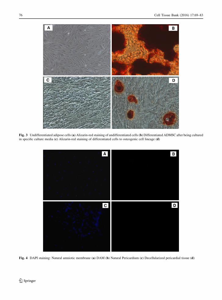
Fig. 3 Undifferentiated adipose cells (a) Alizarin-red staining of undifferentiated cells (b) Differentiated ADMSC after being cultured
in specific culture media (c) Alizarin-red staining of differentiated cells to osteogenic cell lineage (d)
Fig. 4 DAPI staining: Natural amniotic membrane (a) DAM (b) Natural Pericardium (c) Decellularized pericardial tissue (d)
76 Cell Tissue Bank (2016) 17:69–83
123

Scanning electron microscopy
SEM revealed that cells were entirely removed from
DAM without any detectable disarrangement of the
matrix or collagen degradation. PLGA and polyamide
were examined by SEM the result of which revealed
well-organized porous that were satisfactory for cell
seeding. The results of SEM after 8 weeks of implan-
tation showed well-organized cell-seeded scaffolds
with more seeded cells (Fig. 8).
Mechanical properties
No significant difference was detected in the mechan-
ical properties of the matrices in G1 compared with the
properties of the native samples after 8 weeks of
operation. In fact, the maximal load in both native and
experimental samples of G1 was nearly the same,
suggesting its usefulness for bone regeneration in
calvarial defects. The cell-seeded scaffolds in G2
showed biophysical properties that were similar to
Fig. 5 Histopathological examination: H&E staining of differ-
ent seeded scaffolds after 4 weeks of implantation (a) case
samples of different scaffolds 8 weeks postoperatively (b) H&E
staining of different non-seeded scaffolds 1 month postopera-
tively (c) control specimens of different scaffolds 2 months after
surgery (d)
Table 2 Inflammation was similar in all case and control samples in short-term follow-up (10–30 %)
8 weeks 4 weeks Group
Control Case Control Case
Inflammation (%)
\10 \10 10–30 10–30 Polyamide ? ADMSC
\10 \10 10–30 10–30 Polyglycolic acid ? ADMSC
\10 \10 10–30 10–30 Amniotic membrane ? ADMSC
The percentage of inflammation decreased to 10 % in all experimental groups without any significant difference
Cell Tissue Bank (2016) 17:69–83 77
123

those of native tissues but the tensile strength of these
matrices were not as well as samples in G1. The case
samples in G3 had biophysical properties that were the
least similar to those of the native samples. However,
all the samples demonstrated improvement in mechan-
ical properties after 8 weeks of operation, which was
more significant in G1 compared to normal sample
(Fig. 9).
Fig. 6 IHC staining with osteocalcin: Different seeded scaf-
folds after 4 weeks of implantation (a) case samples of different
scaffolds 8 weeks postoperatively (b) different non-seeded
scaffolds 1 month postoperatively (c) control specimens of
different scaffolds 2 months after surgery (d)
Table 3 Data are means (% of normal calvarial bone tissue)
Osteocalcin Osteopontin
4 Weeks 8 Weeks 4 Weeks 8 Weeks
PLGA Seeded 115 121.25 132.25 140
Non-seeded 54.5 61.75 70.5 81.75
Polyamide Seeded 148.5 173.75 162.25 174.75
Non-Seeded 68.25 76 96.75 104.5
AM Seeded 104.25 113.5 124.5 131
Non-Seeded 38.75 42.75 53.25 62.5
P\ 0.05 is considered statistically significant. In the seeded groups, all markers revealed significant enhancement in biopsies
obtained 4 and 8 weeks after surgery in comparison to the non-seeded groups (P\ 0.05). A significant difference was also observed
between the polyamide and other scaffolds (PLGA and AM) between 4 and 8 weeks of operation (P\ 0.05)
78 Cell Tissue Bank (2016) 17:69–83
123

DNA quantification
DNA quantification was less than 5 % in decellular-
ized pericardium and DAM compared to control
samples. DNA quantification demonstrated 1.4 and
1.2 ng dsDNA per mg dry weight of ECM, in
decellularized pericardium and DAM, respectively.
These findings demonstrated the efficacy of decellu-
larization process in removal of cell components as
well as ECM preservation that warranty its potentials
in effective in vivo studies with minimal adverse host
reaction.
Discussion
The aim of the current study was to evaluate the
potential of three different scaffolds seeded with
ADMSCs to repair calvarial defects and assess the
wound healing process and bone regeneration in an
animal model. In this experimental study, all the
seeded and non-seeded scaffolds had the capability of
bone regeneration in the calvarial defect after 8 weeks
of follow-up. However, new bone formation was more
significant in polyamide ? ADMSC group.
Developmental anomalies, acute and chronic
pathological processes, and trauma may cause patients
to lose their natural calvarial bone. In spite of the fact
that no intervention is needed in small bone defects
and they can repair spontaneously, reconstructive
surgery is necessary in extensive bone damages. The
success in bone regeneration depends on the morphol-
ogy of the defect and the graft material (Lupovici
2009). In the wake of such phenomena, we tried to
determine the best method to overcome this noticeable
complication and recreate natural calvarial bone
Fig. 7 IHC staining with osteopontin: Different seeded scaf-
folds after 4 weeks of implantation (a) case samples of different
scaffolds 8 weeks postoperatively (b) different non-seeded
scaffolds 1 month postoperatively (c) control specimens of
different scaffolds 2 months after surgery (d)
Cell Tissue Bank (2016) 17:69–83 79
123

function. Regenerative procedures for obtaining a
feasible and newly formed calvarial bone still remain
challenging. However, the application of pre-seeded
or even non pre-seeded scaffolds may be the safest
strategy for bone engineering and tissue regeneration.
Considering the fact that a critical size defect model is
crucial for evaluating biomaterials in bone defect, a
standardized rabbit calvarial defect was selected for
this experiment.
The therapeutic advantages of delivering cells in
biodegradable scaffolds have been demonstrated in
different aspects of tissue engineering researches (Lee
Fig. 8 Scanning electron micrograph: PLGA (a), polyamide (b) and DAM (c) after 4 weeks of transplantation. PLGA (d), polyamide
(e), and DAM after 8 weeks of transplantation
Fig. 9 Biomechanical properties of natural tissue and different scaffolds 4 and 8 weeks postoperatively
80 Cell Tissue Bank (2016) 17:69–83
123

et al. 2001; Cowan et al. 2004; Kajbafzadeh et al.
2014a, b). As MSCs are capable in tissue regeneration
and immunosuppressive properties, they have been
considered as an excellent treatment for chronic
inflammation and tissue defects (Parekkadan and
Milwid 2010). Due to the fact that patients with
calvarial defect may confront with several complica-
tions associated with gene therapy approaches (Lee
et al. 2001; Cowan et al. 2004), using pre-seeded
scaffolds with accessible source of osteogenic cells
that do not need genetic manipulation may pave the
road to repair calvarial defects. So, in the current study
ADMSCs were avoided form genetic manipulation. In
addition, in the current study the use of autogenous
ADMSCs prevented many of the current limitations
associated with bone grafts. Unavailability and risk of
pathogen transmission are among the limitations of the
autologous and allogenic bone grafts, respectively
(Zomorodian and Baghaban Eslaminejad 2012).
MSCs can be considered as substitute for the men-
tioned grafts. ADMSCs have the ability to differen-
tiate into numerous functional SMCs and osteogenic
cells which may be useful in calvarial bone regener-
ative medicine. ADMSCs have the ability to prolifer-
ate rapidly in vitro in comparison with bone-marrow
stem cells which is important for clinical application.
One of the benefits of the current study is the
application of ADMSCs with better immuno-compat-
ibility, isolation, and expansion compared to bone
marrow-derived mesenchymal stem cells (Bourin
et al. 2013; Kim et al. 2014). So, we tried to investigate
the compatibility of ADMSCs as a cell source in
treating calvarial defects.
It has been also demonstrated that bone regenera-
tion and cell proliferation in lost bone tissue can be
enhanced by the application of tissue engineered
scaffolds (Yun et al. 2012). PLGA has been considered
as a popular polymer in the tissue engineering field.
Tissue compatibility and reproducible mechanical
properties are among the noticeable properties of
PLGA which are necessary especially for bone
regeneration (Boland et al. 2001). Architectural sup-
port, ease of cell inoculation without leakage and
stratification of the MSCs are other characteristics of
the PLGA (Uematsu et al. 2005). Having the top and
bottom pores is one of the characteristic of PLGA. It
should be also mentioned that top and bottom pores are
efficient for raising the efficiency of cell seeding and
holding the seeded cells in this scaffold, respectively.
In one study, three-dimensional PLGA scaffolds were
seeded with MSCs and transplanted in the created
defects of the knee joints of rabbits. The histological
results after 12 weeks of transplantation demonstrated
hyaline-like cartilage in the defects. In accordance
with the results of our study, they concluded that the
structure of the PLGA scaffold provided architectural
support and satisfactory induction of chondrogenesis
in animal model (Uematsu et al. 2005). In another
study, the effect of PLGA alone, PLGA seeded with
differentiated ADMSCs, and PLGA seeded with
undifferentiated ADMSCs was assessed in bone
regeneration by implanting the scaffolds in a critical
nude rat calvarial defect. The results demonstrated that
PLGA scaffolds seeded with osteogenically differen-
tiated ADMSCs obtained better results regarding bone
regeneration (Yoon et al. 2007).
Polyamide has a composition and structure very
close to natural bone collagenous proteins and there-
fore has been considered as an ideal scaffold for repair
of bone defects and biomaterial application (Wang
et al. 2007). The outcome of the current study
demonstrated that in the calvarial defect treated with
polyamide ? ADMSCs, the evidence of osteogenesis
and enhanced biological activity were observed. This
scaffold was superior to other applied scaffolds in both
long and short-term follow-up.
However, other natural decellularized matrices
which have an appropriate histological and biophys-
ical property can be applied for the purpose of
calvarial defect regeneration. Amniotic membrane
contains collagen type I and this instinctive property
makes it ideal for tissue regeneration procedures. It
should be also mentioned that the tensile strength and
formation of the bone is due to this type of collagen.
To the best of our knowledge this is the first study in
which DAM has been used to regenerate calvarial
defect.
Due to desirable mechanical integrity, biocompat-
ibility, and osteogenesis characteristics of PLGA,
polyamide, and DAM they may have the potential to
be applied in reconstructive and maxillofacial surgery.
By the fixation of the applied scaffolds onto the
defects, we tried to optimize architectural
compatibility.
The use of a barrier membrane is crucial for
satisfactory osteoinductive regeneration. Pericardium
is considered as a barrier membrane with excellent
biocompatibility which has been widely applied for
Cell Tissue Bank (2016) 17:69–83 81
123

reconstructing the bone defect adjacent to the
implants. However, few clinical studies have been
performed regarding this procedure (Ahn et al. 2012).
In one study, it has been demonstrated that bone graft
with pericardium membrane can facilitate bone
regeneration by inhibiting connective tissue invasion
(Ahn et al. 2012). The results of the current study also
demonstrated that decellularized pericardial tissue
used as a membrane on the scaffolds, plays a crucial
role in enhancing bone regeneration.
Defect type plays a crucial role in avoiding the
epithelium migration and holding the implanted
scaffolds in place of defect (Suaid et al. 2011). In the
study of Caplanis et al., no histological effects on bone
activation or regeneration attachment were found
following the application of demineralized freeze-
dried bone allografts in maxillary canine defects after
4 weeks. Additionally, no inflammatory cells or
resorption of the implants were noticed (Caplanis
et al. 1998). Findings of the current study demon-
strated that by the application of polyami-
de ? ADMSCs in area of defect, a better outcome
can be achieved regarding the extension of calvarial
bone regeneration. In comparison, the cell-engineered
scaffolds of all groups demonstrated satisfactory
biocompatibility, faster and more effective osteogen-
esis at the defect area compared to pure scaffolds.
However, in the amniotic membrane ? ADMSC
group, mild inflammatory reaction was recog-
nized. The results of this study showed that polyamide
had more satisfactory results on bone generation of
calvarial defect.
In the current study, we rendered an animal model
to evaluate the potential of bone regeneration by the
creation of calvarial bone defects. One the benefits of
this study are the application of a stable carrier
scaffold which is necessary for cell attachment and
suitable for clinical settings. This scaffold did not
show any remarkable inflammation during the healing
period. However, some potential limitations should be
underlined in the current study. It would be better to
track ADMSCs with staining protocols before seeding
the cells on the scaffolds to evaluate the role of seeded
cells in repairing the calvarial defect. Moreover, more
functional evaluation should be performed to ensure
adequate functional property of the regenerated bone.
Additionally, more long-term investigations are
required to assess the feasibility and safety of this
procedure, enrich our understanding of pre-seeded
ADMSCs on scaffolds, and set up foundations for
utilization of ADMSCs in repair of calvarial defect,
before clinical use.
Conclusion
The outcomes of this preliminary report demonstrated
that polyamide Nano Scaffold seeded with ADMSCs
can be successfully used in repairing calvarial defects.
The histological findings of this type of repair revealed
the development of well-vascularized and newly
formed bone. While the osteoinductivity of ADMSCs
has been authenticated via in vitro examinations,
further trials are recommended to support the clinical
relevance.
Acknowledgments We highly appreciate Dr. Laleh
Nikfarjam at the Pediatric Urology Research Center for
collaborating in cell culture section of this experimental study.
Special thanks to associated professor Masoud Soleimani at the
Department of Hematology, School of Medical Science, Tarbiat
Modares University for performing the IHC staining of the
current study.
Compliance with Ethical Standards
Conflict of interest None of the authors has direct or indirect
commercial financial incentive associating with publishing the
article and does not have any conflict of interest, and will sign
the Disclosing Form.
References
Ahn Y-S, Kim S-G et al (2012) Effect of guided bone regen-
eration with or without pericardium bioabsorbable mem-
brane on bone formation. Oral Surg Oral Med Oral Pathol
Oral Radiol 114(5):S126–S131
Anderson DJ, Gage FH et al (2001) Can stem cells cross lineage
boundaries? Nat Med 7(4):393–395
Boland ED, Wnek GE et al (2001) Tailoring tissue engineering
scaffolds using electrostatic processing techniques: a study
of poly (glycolic acid) electrospinning. J Macromol Sci A
38(12):1231–1243
Bourin P, Bunnell BA et al (2013) Stromal cells from the adi-
pose tissue-derived stromal vascular fraction and culture
expanded adipose tissue-derived stromal/stem cells: a joint
statement of the International Federation for Adipose
Therapeutics and Science (IFATS) and the International
Society for Cellular Therapy (ISCT). Cytotherapy
15(6):641–648
Caplanis N, Lee MB et al (1998) Effect of allogeneic freeze-
dried demineralized bone matrix on regeneration of alve-
olar bone and periodontal attachment in dogs. J Clin
Periodontol 25(10):801–806
82 Cell Tissue Bank (2016) 17:69–83
123

Cowan CM, Shi Y-Y et al (2004) Adipose-derived adult stromal
cells heal critical-size mouse calvarial defects. Nat
Biotechnol 22(5):560–567
Ge S, Zhao N et al (2012) Bone repair by periodontal ligament
stem cellseeded nanohydroxyapatite-chitosan scaffold. Int
J Nanomed 7:5405
Goldberg M, Smith AJ (2004) Cells and extracellular matrices
of dentin and pulp: a biological basis for repair and tissue
engineering. Crit Rev Oral Biol Med 15(1):13–27
Kajbafzadeh A-M, Sabetkish S et al (2014a) Tissue-engineered
cholecyst-derived extracellular matrix: a biomaterial for
in vivo autologous bladder muscular wall regeneration.
Pediatr Surg Inter 30(4):371–380
Kajbafzadeh A-M, Tourchi A et al (2014b) Bladder muscular
wall regeneration with autologous adipose mesenchymal
stem cells on three-dimensional collagen-based tissue-
engineered prepuce and biocompatible nanofibrillar scaf-
fold. J Pediatr Urol 10(6):1051–1058
Kim DS, Lee MW et al (2014) Gene expression profiles of
human adipose tissue-derived mesenchymal stem cells are
modified by cell culture density. PLoS One 9(1):e83363
Koob S, Torio-Padron N et al (2010) Bone formation and neo-
vascularization mediated by mesenchymal stem cells and
endothelial cells in critical-sized calvarial defects. Tissue
Eng A 17(3–4):311–321
Laird PW, Zijderveld A et al (1991) Simplified mammalian
DNA isolation procedure. Nucleic Acids Res 19(15):4293
Lee JY, Musgrave D et al (2001) Effect of bone morphogenetic
protein-2-expressing muscle-derived cells on healing of
critical-sized bone defects in mice. J Bone Joint Surg
83(7):1032–1039
Lupovici JA (2009) Histological and clinical results of DFDBA
with lecithin carrier used in dental implant application:
three case reports. Pract Proced Aesthet Dent
21(4):223–230
Mendes S, Tibbe J et al (2002) Bone tissue-engineered implants
using human bone marrow stromal cells: effect of culture
conditions and donor age. Tissue Eng 8(6):911–920
Parekkadan B, Milwid JM (2010) Mesenchymal stem cells as
therapeutics. Annu Rev Biomed Eng 12:87–117
Rakmanee T, Donos N et al (2008) Effects of Emdogain on
growth factor profiles of mesenchymal stem-cells. J Dent
Res 1–1
Suaid FF, Ribeiro FV et al (2011) Autologous periodontal
ligament cells in the treatment of class II furcation defects:
a study in dogs. J Clin Periodontol 38(5):491–498
Uematsu K, Hattori K et al (2005) Cartilage regeneration using
mesenchymal stem cells and a three-dimensional poly-
lactic-glycolic acid (PLGA) scaffold. Biomater 26(20):
4273–4279
Wang H, Li Y et al (2007) Biocompatibility and osteogenesis of
biomimetic nano-hydroxyapatite/polyamide composite
scaffolds for bone tissue engineering. Biomater 28(22):
3338–3348
Yoon E, Dhar S et al (2007) In vivo osteogenic potential of
human adipose-derived stem cells/poly lactide-co-glycolic
acid constructs for bone regeneration in a rat critical-sized
calvarial defect model. Tissue Eng 13(3):619–627
Yun J-H, Yoo J-H et al (2012) Synergistic effect of bone mar-
row-derived mesenchymal stem cells and platelet-rich
plasma on bone regeneration of calvarial defects in rabbits.
Tissue Eng Regen Med 9(1):17–23
Zomorodian E, Baghaban Eslaminejad M (2012) Mesenchymal
stem cells as a potent cell source for bone regeneration.
Stem Cells Int 2012(2012):980353. doi:10.1155/2012/
980353
Zuk PA, Zhu M et al (2001) Multilineage cells from human
adipose tissue: implications for cell-based therapies. Tissue
Eng 7(2):211–228
Cell Tissue Bank (2016) 17:69–83 83
123