Embed Size (px)
Citation preview

4555TECHNICAL PAPER RESEARCH ARTICLE
INTRODUCTIONThe attributes of the zebrafish have established it as a powerfulmodel for the study of vertebrate development. The accessibility ofits externally fertilized embryos allows numerous manipulations,and the transparency of the zebrafish embryo permits direct serialvisualization of biological processes in vivo (Beis and Stainier,2006). These characteristics have enabled forward genetic screensfor mutants affecting a wide range of developmental processes(Patton and Zon, 2001). However, the size of the zebrafish genome,the relatively long generation time, and the expense of maintaininglarge populations preclude forward genetic screening to saturation.Consequently, mutants exist for only a small fraction of zebrafishgenes. Although modified antisense oligonucleotides can achievetargeted gene knockdown during embryogenesis (Nasevicius andEkker, 2000), this technology is problematic with regard topersistence, specificity and penetrance (Robu et al., 2007; Eisen
and Smith, 2008). Thus, definitive reverse genetic strategies arerequired, especially for the study of later developmental processes,analysis of adult physiology, and for the establishment of diseasemodels.
Zinc-finger nucleases (ZFNs) are chimeric fusions between azinc-finger protein (ZFP) and the nuclease domain of FokI (Urnovet al., 2010). ZFNs have been employed to achieve heritabletargeted gene disruption in the genomes of numerous plant andanimal species, including zebrafish (Bibikova et al., 2002; Beumeret al., 2008; Doyon et al., 2008; Meng et al., 2008; Foley et al.,2009; Geurts et al., 2009; Cui et al., 2010; Mashimo et al., 2010;Meyer et al., 2010; Takasu et al., 2010). Gene disruption isachieved through imprecise repair of a ZFN-induced double-strandbreak within the coding sequence of a target gene (Urnov et al.,2010). ZFNs have also been utilized to achieve tailor-madegenomic alterations in animal genomes through stimulation ofhomology-directed repair from an exogenous donor DNA (Beumeret al., 2008; Cui et al., 2010; Meyer et al., 2010). Although off-target lesions in the genome can result from ZFN treatment, theirfrequency is significantly lower than at the target site and theirlocation can be predicted based on ZFN specificity (Meng et al.,2008; Perez et al., 2008; Gupta et al., 2011). Together, thesecharacteristics make ZFNs an ideal tool to facilitate reverse geneticstudies in zebrafish and other organisms.
Despite their utility, the widespread implementation of ZFNs ishindered by the difficulty in creating highly specific ZFPs. Twogeneral approaches have been employed for this purpose: selection-based methods, which identify ZFPs with a desired specificity fromrandomized libraries, and assembly-based methods, which utilizearchives of predefined zinc fingers to construct ZFPs with a desiredspecificity. Each approach is hampered by limitations. Selection-based approaches (Greisman and Pabo, 1997; Isalan et al., 1998;
Development 138, 4555-4564 (2011) doi:10.1242/dev.066779© 2011. Published by The Company of Biologists Ltd
1Program in Gene Function and Expression, University of Massachusetts MedicalSchool, Worcester, MA 01605, USA. 2Department of Pediatric Oncology, Dana-Farber Cancer Institute, Harvard Medical School, Boston, MA 02115, USA.3Department of Cell and Developmental Biology, Perelman School of Medicine atthe University of Pennsylvania, Philadelphia, PA 19104-6058, USA. 4Department ofGenetics, Yale University School of Medicine, New Haven, CT 06510, USA.5Department of Biochemistry and Molecular Pharmacology, University ofMassachusetts Medical School, Worcester, MA 01605, USA.
*These authors contributed equally to this work†Present address: Max-Planck Institute for Molecular Biomedicine, Laboratory forCardiovascular Patterning, Roentgenstr. 20, 48149 Muenster, Germany‡Present address: Bio-Rad Laboratories, Gene Expression Division, 2000 Alfred NobleDrive, Hercules, CA 94547, USA§Authors for correspondence ([email protected];[email protected])
Accepted 3 August 2011
SUMMARYZinc-finger nucleases (ZFNs) allow targeted gene inactivation in a wide range of model organisms. However, construction oftarget-specific ZFNs is technically challenging. Here, we evaluate a straightforward modular assembly-based approach for ZFNconstruction and gene inactivation in zebrafish. From an archive of 27 different zinc-finger modules, we assembled more than 70different zinc-finger cassettes and evaluated their specificity using a bacterial one-hybrid assay. In parallel, we constructed ZFNsfrom these cassettes and tested their ability to induce lesions in zebrafish embryos. We found that the majority of zinc-fingerproteins assembled from these modules have favorable specificities and nearly one-third of modular ZFNs generated lesions attheir targets in the zebrafish genome. To facilitate the application of ZFNs within the zebrafish community we constructed apublic database of sites in the zebrafish genome that can be targeted using this archive. Importantly, we generated new germlinemutations in eight different genes, confirming that this is a viable platform for heritable gene inactivation in vertebrates.Characterization of one of these mutants, gata2a, revealed an unexpected role for this transcription factor in vasculardevelopment. This work provides a resource to allow targeted germline gene inactivation in zebrafish and highlights the benefitof a definitive reverse genetic strategy to reveal gene function.
KEY WORDS: gata2, Vascular development, Zebrafish, Zinc-finger nuclease
Evaluation and application of modularly assembled zinc-finger nucleases in zebrafishCong Zhu1,*, Tom Smith1,*, Joseph McNulty1, Amy L. Rayla1, Abirami Lakshmanan1, Arndt F. Siekmann1,†,Matthew Buffardi1, Xiangdong Meng1,‡, Jimann Shin2, Arun Padmanabhan3, Daniel Cifuentes4, Antonio J. Giraldez4, A. Thomas Look2, Jonathan A. Epstein3, Nathan D. Lawson1,§ and Scot A. Wolfe1,5,§
DEVELO
PMENT

4556
Maeder et al., 2008; Meng et al., 2008), although effective ingenerating specific ZFPs, can be time-consuming and technicallychallenging (Kim et al., 2010). Assembly-based approaches aremore straightforward (Carroll et al., 2006; Kim et al., 2009) but aredependent on the quality of the available zinc-finger archive andthe difficulty in predicting context-dependent effects betweenneighboring zinc fingers (Desjarlais and Berg, 1993; Greisman andPabo, 1997; Wolfe et al., 1999). Although assembly-based, ormodular, ZFNs have suffered from relatively low success rates(Ramirez et al., 2008), continued characterization of availablearchives and investigation of context dependence (Carroll et al.,2006; Kim et al., 2009; Sander et al., 2009), along with thegeneration of new archives with expanded specificity, will makethis a more viable approach.
In this study we evaluated an assembly-based approach forcreating ZFNs for targeted gene disruption in the zebrafish genome.We constructed an archive of ZFP modules recognizing 27 differenttriplet sequences and characterized the DNA-binding specificity forthree-finger ZFP cassettes assembled from this archive. In parallel,we tested ZFNs constructed from these ZFPs for their ability toinduce both somatic and germline lesions in zebrafish embryos. Tofacilitate the application of this technology in the zebrafishcommunity, we developed a publicly accessible database thatassists users in modular ZFN construction using our archive.Finally, we characterized the phenotypes in zebrafish embryosbearing a ZFN-induced deletion in the gata2a gene, revealing itsrole in vascular morphogenesis.
MATERIALS AND METHODSFish linesZebrafish were handled according to established protocols (Westerfield,1993) and in accordance with Institutional Animal Care and UseCommittee (IACUC) guidelines of participating institutions (University ofMassachusetts Medical School, University of Pennsylvania, Dana FarberCancer Institute and Yale University). The Tg(kdrl:egfp)la116 line has beendescribed elsewhere (Choi et al., 2007).
Construction of a modular zinc-finger archiveAn archive of zinc-finger modules originating from multiple sources (seeTable S1 in the supplementary material and Results) was constructed bysubcloning ZFPs into either a pBluescript or pCS2 vector using standardmethods. Clones were sequence verified and arrayed into a 96-well plateto generate an address for each module. Plasmids from the archive areavailable through addgene.org.
Modular assembly of ZFNsTo assemble three-finger ZFPs we first separately amplified modules for eachof the three finger positions from plasmid templates by PCR. Distinct primerpairs were used to amplify modules for each position (fingers 1, 2 and 3) (seeTable S2 in the supplementary material). PCR was performed usingAdvantage HF2 polymerase (Clontech) and 20 ng plasmid template. Finger1 modules were amplified as follows: 94°C for 2 minutes, followed by 20-30 cycles of 94°C for 30 seconds, 60°C for 30 seconds and 68°C for 20seconds, with a final extension of 68°C for 5 minutes. For finger 2 or 3modules: 94°C for 2 minutes, then 20-30 cycles of 94°C for 30 seconds,57°C for 30 seconds and 68°C for 20 seconds, with a final extension of 68°Cfor 5 minutes. All initial PCR products were gel purified. Three-fingercassettes were constructed by overlapping PCR using 30 ng of each fingermodule in a 50 ml reaction with Advantage HF2 polymerase. The first fivecycles were performed without primers: 94°C for 2 minutes, followed by fivecycles of 94°C for 30 seconds, 55°C for 30 seconds and 68°C for 1 minute.We added 2 ml of 10 mM F1 forward and F3 reverse primer and performedthe following steps: 24 cycles of 94°C for 15 seconds, 60°C for 30 seconds,68°C for 1 minute, with a final extension of 68°C for 5 minutes. Final PCRproducts were gel purified, digested with Acc65I and BamHI and cloned into
the pCS2 nuclease backbone in frame with the EL/KK heterodimeric variantsof FokI (Miller et al., 2007), such that the 5� ZFP was fused to the ELnuclease and the 3� ZFP was fused to the KK nuclease. In several cases,DD/RR nuclease variants (Miller et al., 2007; Szczepek et al., 2007) wereused. All constructs were sequence verified.
ZFN injections into zebrafish embryosSynthesis and injection of ZFN mRNAs, dose optimization (see Table S3in the supplementary material) and establishment and identification ofgermline lesions were performed as previously described (Meng et al.,2008; Gupta et al., 2011).
Bacterial one-hybrid binding site selectionsSequence-verified ZFP cassettes were cloned into the 1352-UV2expression vector using Acc65I and BamHI. Bacterial one-hybrid (B1H)binding site selections were typically carried out at 5 mM 3-amino-1,2,4-triazole (3-AT), 10 mM IPTG and in the absence of uracil as previouslydescribed (Noyes et al., 2008; Gupta et al., 2011). For some ZFPs, lowerstringency was required to achieve sufficient enrichment of recognitionsequences above background.
Illumina library preparation of B1H-selected binding sitesPreparation of selected binding sites for deep sequencing was performedas described (Gupta et al., 2011), except that amplicons were digested witheither EcoRI or NotI prior to ligation of bar-coded adapters for theidentification of sequences associated with each ZFP (see Table S2 in thesupplementary material).
Evaluation of the DNA-binding specificity of each ZFP andindividual zinc-finger modulesRecognition motifs within the population of unique 28 bp sequencesrecovered from each binding site selection were identified using MEME(Bailey and Elkan, 1994). Position weight matrices (PWMs) weregenerated using the Log-Odds method (Hertz and Stormo, 1999) from thealigned sequences within the most statistically significant motif, weightingeach sequence based on the number of counts within the Illumina datasetusing the formula:
where S is the PWM score over the 9 bp target site s, b,i is the identityof base b at position i, fb,i is the normalized frequency of occurrence ofbase b at position i, and pb,i is the probability of observing the same baseat the same position in a background model, which is assumed to be anequal distribution. Each ZFP was evaluated by calculating the score forits target site from its derived PWM. Each individual finger module wasevaluated by calculating the score for its corresponding 3 bp recognitionelement (assuming canonical recognition) from this PWM. When datawere available for multiple identical modules at identical positions, thescores for these modules were averaged for an overall assessment of thequality of the finger module. Modules with (average) scores that weretwo standard deviations below the average score of all modules at thatposition (i.e. finger 1, finger 2 or finger 3) were considered‘questionable’ and flagged.
Analysis of somatic lesion frequencySomatic lesion frequency was determined by Illumina sequencing aspreviously described (run #1) (Meng et al., 2008; Gupta et al., 2011). Asecond round of sequencing was performed for a subset of ZFNs froman independent set of injections (run #2). To avoid cross-contaminationwith samples from the first analysis, a unique pair of PCR primers wasdesigned for each target site that would not amplify the PCR productsfrom the first trial. These primers encoded the Illumina P1 and P2sequences within each primer pair (see Table S2 in the supplementarymaterial). In this case, PCR products for each ZFN target site were gelpurified and amplified using Illumina genomic DNA primers (1.1 and2.1). The PCR products for each target were then pooled at equal molarratio, sequenced at 7 pM on a HiSeq 2000 (Illumina), and analyzed in amanner identical to run #1.
S= log2fb,i
pb,i1
9
∑ (1) ,
RESEARCH ARTICLE Development 138 (20)
DEVELO
PMENT
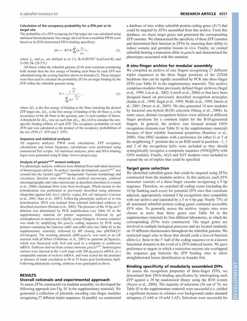
Calculation of the occupancy probability for a ZFN pair at itstarget siteThe probability of a ZFP occupying its 9 bp target site was calculated usingstatistical thermodynamic free energy derived from a modified PWM scorebased on its B1H-determined DNA-binding specificity:
where fb,i and pb,i are defined as in (1), R0.001987 kcal/(mol.K) andT301.5K (28.5°C).
All 9mers within the zebrafish genome (Zv8) were extracted consideringboth strands then the free energy of binding each 9mer by each ZFP wascalculated using the scoring function shown in formula (2). These energieswere then used to calculate the probability (P) for on-target binding by theZFP within the zebrafish genome using:
where DGs is the free energy of binding to the 9mer matching the desiredZFP target site, DGs,i is the free energy of binding of the ith 9mer, ni is theoccurrence of the ith 9mer in the genome, and l is total number of 9mers.A threshold for DGs,i was set such that DGs,i–DGs≤4.0 to simulate the non-specific binding affinity of each ZFP. The occupancy probability of eachZFN pair was calculated as the product of the occupancy probabilities ofthe two ZFPs (3� ZFP and 5� ZFP).
Sequence and statistical analysesAll sequence analyses, PWM score calculations, ZFP occupancycalculations and lesion frequency calculations were performed usingcustomized Perl scripts. All statistical analyses, plots and DNA-bindinglogos were generated using R (http://www.r-project.org).
Analysis of gata2aum27 mutant embryosFor phenotypic analysis, embryos were obtained from individual incrossesof heterozygous carriers. To analyze vascular development, gata2aum27 wascrossed into the Tg(kdrl:egfp)la116 background. Vascular morphology andcirculatory function were observed by confocal microscopy, confocalmicroangiography and video microscopy as described elsewhere (Covassinet al., 2006); Quantum Dots were from Invitrogen. Whole-mount in situhybridization was performed as previously described using antisenseriboprobes against kdrl, hey2, efnb2a, vegfaa, flt4, tal1 and gata1 (Lawsonet al., 2001; Hart et al., 2007). Following phenotypic analysis or in situhybridization, DNA was isolated from selected individual embryos asdescribed elsewhere (Roman et al., 2002). The presence of the 10 bp um27deletion was determined by PCR amplification (see Table S2 in thesupplementary material for primer sequences), followed by gelelectrophoresis or analysis on a QiaXL system (Qiagen). A rescue constructwas made by amplifying the gata2a coding sequence by PCR usingprimers containing the Gateway attB1 and attB2 sites (see Table S2 in thesupplementary material), followed by BP cloning into pDONR221(Invitrogen). The resulting plasmid, pME-gata2a, was used in an LRreaction with pCSDest (Villefranc et al., 2007) to generate pCSgata2a,which was linearized with NotI and used as a template to synthesizemRNA. Embryos derived from crosses between gata2aum27 heterozygouscarriers were injected at the 1-cell stage with 200 pg gata2a mRNA, or acomparable amount of mcherry mRNA, and were scored for the presenceor absence of trunk circulation at 48 to 55 hours post-fertilization (hpf).Following phenotypic scoring, embryos were genotyped as above.
RESULTSOverall rationale and experimental approachTo assess ZFNs constructed via modular assembly, we developed thefollowing approach (see Fig. S1 in the supplementary material). Wegenerated a collection of plasmids encoding zinc-finger modulesrecognizing 27 different triplet sequences. In parallel, we constructed
P =e
–ΔGsRT
ni e–
ΔGs ,i
RT
1
l
∑ (3) ,
ΔG = –RT • ( ln
1
9
∑ fb,i
pb,i
) (2) ,
a database of sites within zebrafish protein-coding genes (Zv7) thatcould be targeted by ZFNs assembled from this archive. From thisdatabase, we chose target genes and generated the correspondingZFP cassettes. We characterized the specificity of these ZFP cassettesand determined their function in ZFNs by assessing their ability toinduce somatic and germline lesions in vivo. Finally, we createdzebrafish bearing a truncation allele in gata2a and characterized thephenotypes associated with this mutation.
A zinc-finger archive for modular assemblyWe compiled an archive of zinc fingers recognizing 27 differenttriplet sequences in the three finger positions of the Zif268backbone that can be rapidly assembled by PCR into three-fingerZFPs (see Table S1 in the supplementary material). This archivecomprises modules from previously defined finger archives (Segalet al., 1999; Liu et al., 2002; Carroll et al., 2006) or that have beendesigned based on previously described recognition principles(Isalan et al., 1998; Segal et al., 1999; Wolfe et al., 1999; Dreier etal., 2001; Dreier et al., 2005). We also generated 14 new modulesby bacterial one-hybrid (B1H) selection (Meng et al., 2008). Insome cases, distinct recognition helices were utilized at differentfinger positions for a common triplet for the B1H-generatedfingers. In general, the archive was focused around GNNrecognition elements (see Table S1 in the supplementary material)because of their reliable functional properties (Ramirez et al.,2008). Nine HNG modules with a preference for either G or T atthe neighboring 3� position due to an RSD motif at positions –1, 1and 2 of the recognition helix were included as they shouldsynergistically recognize a composite sequence (NNGGNN) withGNN modules. Finally, AGA and TGT modules were included toexpand the set of triplets that could be specified.
Target gene selectionWe identified zebrafish genes that could be targeted using ZFNsconstructed from the modular archive. In this analysis, each ZFNmonomer consists of a three-finger cassette recognizing a 9 bpsequence. Therefore, we searched all coding exons (including the10 bp flanking each exon) for potential ZFN sites that constituteadjacent, appropriately oriented 9 bp recognition sites compatiblewith our archive and separated by a 5 or 6 bp gap. Nearly 75% ofall annotated zebrafish protein coding genes contained accessibleZFN sites. To generally assess the archive, targets sites werechosen in more than thirty genes (see Table S4 in thesupplementary material) by four different laboratories, in which thecorresponding ZFNs were constructed. The target genes areinvolved in multiple biological processes and are located randomlyon 19 different chromosomes throughout the zebrafish genome. Werestricted target sites to those that should yield a loss-of-functionallele (i.e. those in the 5� half of the coding sequence or in a knownfunctional domain) in the event of a ZFN-induced lesion. We gavepreference to targets in which a restriction enzyme site overlappedthe sequence gap between the ZFP binding sites to allowstraightforward lesion identification in founder fish.
Binding specificity of modularly assembled ZFPsTo assess the recognition properties of three-finger ZFPs, wedetermined their DNA-binding specificities by interrogating eachZFP against a 28 bp randomized library using the B1H system(Noyes et al., 2008). The majority of selections (56 out of 76; seeTable S5 in the supplementary material) were successful (i.e. yieldeda significant increase in colonies over background) under standardstringency (5 mM or 10 mM 3-AT). Selections were successful for
4557RESEARCH ARTICLEA zebrafish knockout kit
DEVELO
PMENT

4558
19 of the remaining 20 ZFPs at lower stringency (≤2.5 mM 3-AT).For each ZFP a recognition motif (sequence logo) (Schneider andStephens, 1990) and position weight matrix (PWM) were generatedbased on binding sites recovered following selection. In general,determined recognition motifs resembled the desired target motif,although there was variability in the overall quality of the PWMscore of the target site across all ZFPs (see Fig. S2 in thesupplementary material). For example, the 5p ZFP targeting gata2aproperly specified all 9 bp and displayed among the highest PWMscores, suggesting excellent specificity for its target (Fig. 1A). MostZFPs exhibited more modest specificity for their target, similar to thedrosha (rnasen – Zebrafish Information Network) 3p ZFP (Fig. 1B),whereas several displayed weaker preference, such as the nf1b 5p(Fig. 1C). Overall, the distribution of PWM scores for the modularZFP cassettes was similar to the engineered ZFPs successfully usedto target the zebrafish kdrl locus (Fig. 1D) (Meng et al., 2008; Guptaet al., 2011), indicating that the ZFPs constructed using this archivegenerally display good recognition properties.
We further analyzed the B1H binding site selection data to assessthe quality of individual modules, allowing us to identify thosewith poor or context-dependent specificity. Overall, most modulesdisplayed a strong preference for their recognition triplet based onindividually derived PWM scores (Fig. 2A), although modules atfingers 1 and 3 display lower median scores than those at finger 2.
This is likely to be due to fraying effects at the edges of the protein-DNA complex that reduce the specificity of the determinants atthese positions (Choo, 1998). Comparison of PWM values acrossall individual modules revealed that those targeting purine-richtriplets were among the most robust, regardless of their fingerposition. For example, the GAG module consistently displayedexcellent specificity in multiple finger positions in most ZFPs (Fig.2B), as did modules targeting GAT, GGG and GGT (see Tables S5and S6 in the supplementary material). We also identified fourmodules within the archive that displayed poor specificity at aspecific position or in a context-dependent manner. For example,the TTG module displays context-dependent alterations inspecificity. We originally utilized the TTG module at finger 3 inZFPs targeting the zebrafish kdrl locus, where it displays moderatepreference (NtG) for its triplet (Meng et al., 2008; Gupta et al.,2011), similar to the efnb2a 5p ZFP in this study (Fig. 2C).However, when it is placed at the finger 2 position adjacent to a C-terminal module bearing an RSD motif at positions –1, 1 and 2, thespecificity shifts to a strong preference for a GNG binding site [Fig.2C, zgc:66439 (clec14a – Zebrafish Information Network), 3pZFP]. This dramatic alteration in specificity is not observed whenthe neighboring C-terminal module lacks an RSD motif (Fig. 2C,kif1b, 5p ZFP). Overall, the majority of analyzed modules havefavorable recognition properties in multiple contexts, althoughseveral might specify incorrect triplets in particular contexts.
In vivo activity of modular ZFNsWe evaluated the ability of ZFNs constructed from our modulararchive to induce lesions at a target site within the zebrafish genome.Twenty-nine pairs of ZFNs targeting 28 genes (see Table S7 in thesupplementary material) were constructed from 76 ZFPs describedabove (see Table S5 in the supplementary material) by fusing themto engineered heterodimeric FokI nuclease domains (Miller et al.,2007; Szczepek et al., 2007). We assessed lesion frequency by deepsequencing the target region in normal embryos at 24 hpf followinginjection of mRNAs encoding ZFNs at an optimal dose (see TableS3 in the supplementary material). Eight of the 29 ZFNs displayedlesions frequencies of 1% or more in at least one set of injections(see Table S7 in the supplementary material). The previouslycharacterized kdrl ZFNs were injected as a positive control andgenerated an in vivo lesion frequency of ~7%, similar to our previousobservations using Illumina sequencing (Gupta et al., 2011).
Germline transmission of ZFN-induced mutantallelesDeep sequencing is likely to underestimate the frequency of ZFN-induced lesions because of short read lengths, and assessment ofsomatic lesions in embryos may not reflect the frequency of
RESEARCH ARTICLE Development 138 (20)
Fig. 1. DNA-binding specificity of modular zinc-finger proteins(ZFPs). (A-C)Sequence logos and position weight matrix (PWM) scoresdetermined for 5� (5p) or 3� (3p) target sites for zebrafish (A) gata2a,(B) drosha and (C) nf1b. (D)Box plot depicting meta-analysis of PWMscores for each target site across all ZFPs for which bacterial one-hybrid(B1H) selections were successful. PWM scores for the previouslydescribed kdrl ZFPs are shown for reference. Whiskers indicate thelargest (smallest) datum still within 1.5 interquartile range (IQR) of theupper (lower) quartile, where outliers are indicated as open circles.
Fig. 2. Assessing the quality and behavior ofindividual ZFP modules. (A)Box plot depicting meta-analysis of PWM values for individual ZFPmodules in each of the three positions in the Zif268backbone. 5p (red diamonds) and 3p (blue circles)scores for fingers in the kdrl ZFPs are shown forreference. Whiskers indicate the largest (smallest)datum still within 1.5 interquartile range (IQR) of theupper (lower) quartile, where outliers are indicated asopen circles. (B) Sequence logos for ZFP monomersrecognizing GAG in each finger position. (C)Sequencelogos for ZFPs containing TTG modules at the indicatedfinger position. D
EVELO
PMENT

germline transmission. Therefore, we determined the ability of 12ZFNs targeting 11 genes to generate founder fish bearing germlinelesions at the desired target site (Table 1). For most ZFNs thatinduced appreciable somatic lesion frequencies, we identifiedfounder fish that transmitted mutant alleles through their germlineand did so at a higher frequency than the somatic lesion rate (Table1). Overall, there was a moderate correlation between the somaticlesion frequency and the founder rate (R20.71). Notably, for twodifferent ZFNs [ago2 (eif2c2 – Zebrafish Information Network)and nf1a], we were able to obtain founders despite lesion ratesbelow 1% in somatic cells. Conversely, embryos injected withZFNs targeting braf failed to yield founders even with a somaticlesion frequency greater than 1% (Table 1, see Table S7 in thesupplementary material).
Correlations between ZFP specificity and ZFNactivityWe next investigated potential correlations between the recognitionproperties of the ZFPs and the activity of the ZFNs in vivo. Thisanalysis could provide a basis for estimating the likelihood ofsuccess of ZFNs generated from this archive. Surprisingly, we didnot observe a significant correlation between DNA-bindingspecificity of the ZFPs and the activity of the respective ZFNs invivo (Fig. 3A). Specificity alone may not be predictive of activitybecause genomic sequence is not random and thus activity mightdepend on the number of favorable binding sites for each ZFPwithin the genome. Therefore, we estimated the fractionaloccupancy of each ZFP monomer at its target site based on itsDNA-binding specificity and the number of alternative high-affinity binding sites within the zebrafish genome (see Table S7 inthe supplementary material). Again, there was no significantdifference between the predicted target site occupancy for theactive and inactive groups (Fig. 3B).
Although DNA-binding specificity did not appear to definitivelypredict in vivo ZFN activity, we did observe a strong correlation(P0.001) between the number of GNN subsites within therecognition sequences and active ZFNs (Fig. 3C), consistent withprevious observations (Ramirez et al., 2008). One additionalprominent element of our module archive is the inclusion of NNGfingers. There are, on average, more NNG fingers in active ZFNsthan inactive ZFNs (Fig. 3D), but the difference is not significant(P0.36). Combined, a significant difference (P0.019) existsbetween the average total number of GNN and NNG fingers in the
active versus inactive groups of ZFNs, but the majority of thepredictive value of this combination is derived from the GNNfingers.
A zebrafish database for modular ZFNsTo facilitate ZFN construction by the zebrafish community, wedesigned a web-accessible database (pgfe.umassmed.edu/ZFNV1/)that can be used in a standard workflow (see Fig. S3 in thesupplementary material), allowing researchers to easily constructZFNs, generate founder zebrafish bearing targeted lesions, andidentify associated phenotypes within 7-12 months. A user canidentify ZFN target sites in a gene of interest by entering oruploading an ENSEMBL gene number or RefSeq ID. The resultingoutput provides a count of ZFN target sites, a gene description andabbreviation, and a link to the locus on the UCSC genome browser(Fig. 4A). The user can view sites within one particular gene or alltargets using a sortable list (Fig. 4B) that aids identification ofoptimal sites based on position (exon rank), presence of arestriction enzyme site in the spacer sequence, or a revised efficacyscore. The efficacy score has a maximum of 12 points, where onepoint is assigned for each guanine at the edge of each recognitiontriplet (i.e. GNN and NNG fingers), as active ZFNs are enrichedfor these contacts (see above). The database also notes single-nucleotide polymorphisms in the target site in the referenceTuebingen (Tu) and AB wild-type zebrafish strains that have been
4559RESEARCH ARTICLEA zebrafish knockout kit
Table 1. Somatic lesion frequency and germline transmissionrate of ZFN-induced mutagenic alleles
Somatic lesion Frequency of ZFN ID Gene frequency (%) founders (%)
ZFNv1_68779 chd5 <0.5 0ZFNv1_53751 kif1b <0.5 0ZFNv1_27773 braf >1 0ZFNv1_5254 nf1b ND 1.3ZFNv1_5253 nf1b ~1 3ZFNv1_87654 gata2a ~1 4ZFNv1_19302 nf1a <0.5 5.5ZFNv1_94061 ago2 <0.5 6.5ZFNv1_83611 cxcr4a ~1 10ZFNv1_37185 numb-like ~1 13ZFNv1_53765 kif1b 1-2 18ZFNv1_25601 gata3 >1 25
ND, not determined.
Fig. 3. Correlating ZFP characteristics with in vivo zinc-fingernuclease (ZFN) activity. Box plots depicting (A) PWM score, (B)estimated target site occupancy, (C) number of GNN modules in a ZFNpair and (D) number of NNG modules in a ZFN pair in both active andinactive ZFNs, where red dots indicate the mean of each population.Active ZFNs are those in which somatic lesion frequencies were greaterthan 0.5% or that successfully generated founders for mutant alleles.Whiskers indicate the largest (smallest) datum still within 1.5interquartile range (IQR) of the upper (lower) quartile, where outliersare indicated as open circles.
DEVELO
PMENT

4560
sequenced by the Sanger Center (D. Stemple, personalcommunication), although we recommend sequencing the targetsite in wild-type strains other than the hybrid Tu/AB (SAT)reference line.
Once a target site is chosen, the user can click on the ZFN entry(QueryID; see Fig. 4B) for details about each construct (Fig. 4C),such as the target site sequence, the amino acid sequence of therecognition helices for the assembled ZFNs, and clone IDs for eachplasmid in the archive (available at addgene.org) to facilitateoverlapping PCR of the appropriate modules (see Table S1 in thesupplementary material). Alternatively, the DNA sequence of theZFP cassette is included for direct synthesis. In either case, theinclusion of the amino acid sequence for each entry (Fig. 4C) aidsin sequence validation of ZFN-containing plasmids. ZFNscontaining modules with poor or context-dependent specificity (seeabove) are flagged with an asterisk. For lesion detection, we alsoinclude restriction sites, when present, within the spacer regionseparating the ZFP binding sites, along with sequences of flankingprimers for PCR amplification. The primer and restriction enzymeinformation also facilitates easy genotypic analysis of founders andmutant embryos. Data from these pages can be exported into Excel(Microsoft) for easy reference. A query page(http://pgfe.umassmed.edu/ZFPmodularsearch.html) is available toidentify ZFN sites in sequences not contained within our database.
A novel role for gata2a in vascular developmentTo demonstrate the application of our archive to interrogate genefunction and to provide an example of the workflow shown in Fig.S3 in the supplementary material, we investigated defectsassociated with a ZFN-induced mutation in the gata2a gene. Gata2is a zinc-finger transcription factor that is essential for definitivehematopoiesis and maintenance of stem cell progenitors in bothembryos and adults (Tsai et al., 1994; Rodrigues et al., 2005).Gata2 has also been implicated in angiogenesis and is associatedwith early onset coronary artery disease in humans (Connelly et al.,2006; Mammoto et al., 2009), but its role in vascular development
is unknown. As described above, we constructed ZFNs targetingthe fourth exon of gata2a. The target sequence is upstream of twozinc fingers required for function of the mammalian Gata2ortholog (Minegishi et al., 2003) and, therefore, truncation in thisregion would be expected to generate a null allele (Fig. 5A).Following injection of ZFNs targeting this site, we identified afounder fish bearing a 10 bp deletion allele, referred to asgata2aum27, which causes a frameshift to a premature stop codon(Fig. 5A,B).
To identify phenotypes caused by gata2a deficiency weobserved embryos derived from an incross of gata2aum27
heterozygous carriers derived from the original founder. At 24 and48 hpf, all embryos appeared morphologically normal, includingnormal development of neural tube, notochord and somites (seeFig. S4 in the supplementary material; data not shown). However,~25% of embryos failed to display trunk blood vessel circulationby 48 hpf (see Movie 1 in the supplementary material, Table 2)whereas the remaining siblings were normal (see Movie 2 in thesupplementary material). Genotypic analysis demonstrated thatembryos with defective trunk circulation were homozygous for theum27 deletion, whereas normal embryos were heterozygous orhomozygous for the wild-type allele (Fig. 5C, Table 2).Importantly, injection of mRNA encoding wild-type Gata2a couldrescue trunk circulation in gata2aum27 mutant embryos (16 out of26 mutant embryos in two separate experiments) (see Movie 3 inthe supplementary material, Fig. 5D,E), whereas injection withmcherry mRNA did not (0 out of 13 mutant embryos in twoseparate experiments) (see Movie 4 in the supplementary material,Fig. 5D,E). Closer inspection of gata2aum27 mutant embryosrevealed the occurrence of pulsating blood cells trapped in thetrunk blood vessels and abnormal circulatory connections, orshunts, between the dorsal aorta and posterior cardinal vein (seeMovies 1 and 5 in the supplementary material). Despite thesedefects, mutant embryos displayed a beating heart, the presence ofblood cells and circulation through cranial blood vessels (seeMovie 6 in the supplementary material), similar to sibling embryos
RESEARCH ARTICLE Development 138 (20)
Fig. 4. Screenshots from amodular ZFP/ZFN database.(A)Example output from asearch for ZFN target sites. Circleindicates the link to the pageshown in B. (B)List of targets inthe fgf24 gene with associatedinformation. Circled numberindicates the link to individualtarget site and ZFN informationshown in C. (C)Example outputpage with individual target siteinformation.
DEVELO
PMENT

with trunk circulation (see Movie 7 in the supplementary material).Together, these observations demonstrate that gata2a deficiencyleads to a specific defect in circulatory function in embryoniczebrafish.
To further characterize the phenotype caused by loss of gata2a,we performed confocal microangiography on wild-type andgata2aum27 mutant siblings bearing the Tg(kdrl:egfp)la116 transgeneat 50 hpf. Consistent with normal head circulation in gata2aum27
mutant embryos, we did not note any overt defects in vascularmorphology of the cranial vessels when compared with wild-typesiblings (Fig. 6A,B). We observed normal formation of arteries andveins within the head, which are fully perfused followingangiography (Fig. 6A,B). By contrast, the lateral dorsal aortae ingata2aum27 mutant embryos are more dilated than in wild-typesiblings, although these vessels are perfused, suggesting thatlumenization is not affected (Fig. 6C,D). Within the trunk, we
observed specific defects in vascular morphology. Whereasintersegmental vessels appeared largely normal in gata2aum27
mutant embryos, the dorsal aorta was discontinuous (Fig. 6E).Accordingly, gata2aum27 mutant trunk vessels were poorly perfusedfollowing angiography when compared with their wild-typesiblings (Fig. 6E,F). These analyses suggest a specific defect in themorphogenesis of the dorsal aorta in gata2aum27 mutant embryos,which would be the likely cause of the arteriovenous shunts notedabove.
We have previously observed aorta morphogenesis defects andarteriovenous shunts in zebrafish embryos deficient for Notch orVegf signaling (Lawson et al., 2001; Covassin et al., 2006). In thesecases, these defects were associated with loss of arterial endothelialcell identity. However, gata2aum27 mutant embryos did not displayloss of artery markers, such as efnb2a, and exhibited normal levelsof vegfaa expression (data not shown). Similarly, the hey2 gene,which is also required for proper dorsal aorta morphogenesis(Zhong et al., 2000), was expressed at normal levels in gata2aum27
mutants (Fig. 7A,B). We also did not observe any significantchanges in the expression of blood markers such as gata1 and tal1(Fig. 7C,D; data not shown). However, gata2aum27 mutant embryosdisplayed a subtle, but consistent, downregulation of kdrl, thefunctional ortholog of Vegfr2, when compared with wild-typesiblings (Fig. 7E,F). Taken together, our results demonstrate thatgata2a function is essential for the proper morphogenesis of thedorsal aorta, but may be dispensable for arterial endothelialdifferentiation.
DISCUSSIONWith the increasing characterization of the genomes of model andnon-model organisms, there is a broad need for reverse geneticapproaches to assess gene function in multiple species. ZFNs allowthe direct introduction of targeted germline lesions in vivo, withoutthe need for species-matched embryonic stem cell lines, thusproviding a general means to determine gene function. However,the primary limitation hindering the widespread employment ofthis technology has been the lack of an affordable, simple methodfor constructing ZFNs that are active in vivo. Modularly assembledZFNs provide a rapid and effective method for introducing targetedlesions into the genomes of animals. The specificity analysis of ourassembled ZFPs indicates that the majority have similar recognitionpotential to the selected ZFPs incorporated into our kdrl ZFNs(Meng et al., 2008; Gupta et al., 2011). Consequently, a sizablefraction (29%) of ZFNs constructed from these modules displayedsignificant activity (≥1%) for the generation of somatic lesions,
4561RESEARCH ARTICLEA zebrafish knockout kit
Fig. 5. The ZFN-induced um27 lesion is a truncation allele ofgata2a. (A)Location of frameshift caused by the um27 deletion in thezebrafish gata2a gene. ZF, zinc-finger domain. (B)gata2a coding andamino acid sequence in the region of the um27 deletion. ZFNrecognition sequences are boxed. (C)Genotype of embryos derivedfrom an incross of gata2aum27 heterozygous carriers. (D)Genotype ofembryos with normal circulation derived from a gata2aum27 incrossinjected with 200 pg gata2a mRNA. Asterisks denote mutant embryoswith normal trunk circulation. (E)Percentage of gata2aum27 mutantembryos displaying the indicated circulation phenotypes.
Table 2. Linkage of gata2aum27 to circulatory defects in mutant embryosgata2aum27 genotype
Parents Circulation phenotype No. embryos Proportion +/+ +/– –/–
A2�A5 Normal 116 0.77 1 10 0Head but no trunk 34 0.23 0 0 11
Normal 59 0.81 3 7 2Head but no trunk 14 0.19 1 0 11
A3�A6 Normal 42 0.79 4 7 1Head but no trunk 11 0.21 0 0 12
Normal 67 0.79 3 8 0Head but no trunk 18 0.21 0 0 11
Total Normal 284 0.79 11 32 3Head but no trunk 77 0.21 1 0 45
Results are shown for two separate clutches (different days) from each set of parents. DEVELO
PMENT

4562
establishing the utility of this archive for targeted genomemanipulation in complex genomes. This success rate comparesfavorably with that of other single-finger modular assemblyarchives for constructing ZFNs (Ramirez et al., 2008; Kim et al.,2009; Kim et al., 2011). Moreover, characterization of DNA-binding specificity provides additional information about theperformance of individual modules within this archive and willfacilitate its subsequent improvement.
Surprisingly, we did not find a strong correlation between ZFPspecificity and in vivo ZFN activity. Previous studies have shownthat improvements in DNA-binding specificity can lead toimproved ZFN activity (Cornu et al., 2008) and precision (Guptaet al., 2011). It is likely that other factors, such as ZFP affinity, alsosignificantly influence ZFN activity. Whereas the recognitionmotifs generated using the B1H system provide an estimate of ZFPspecificity, our analysis utilized varying stringencies to achieveenrichment of binding sites, possibly bypassing variations thatcould reflect differences in affinity. That said, seven of eight ZFNsthat contained at least one ZFP that displayed low activity in theB1H system (i.e. binding site selections required a stringencybelow 5 mM 3-AT) displayed low somatic lesion rates (<0.5%).This correlation is consistent with studies utilizing the bacterialtwo-hybrid system, in which highly active ZFPs often perform wellin ZFNs (Ramirez et al., 2008; Lam et al., 2011). Thus, a more
refined analysis of ZFP affinity might provide additional insightsinto the behavior of our archive. Additionally, ZFN activity couldbe affected by properties of the endogenous target sequence (e.g.chromatin architecture or DNA methylation status) that couldinterfere with nuclease recognition or function.
We did observe significantly higher rates of ZFN activity as afunction of the number of GNN fingers, consistent with previousobservations (Ramirez et al., 2008), and a slight increase in theaverage number of NNG fingers in the active versus inactive ZFNpopulation. In both sets of modules, arginine-guanine interactions,which can contribute dramatically to binding affinity andspecificity (Elrod-Erickson and Pabo, 1999), appear to be criticallinchpins for the formation of active ZFN complexes.Consequently, this information is incorporated into a simple scoringfunction for evaluating our ZFNs that notes the number of thesecontacts. Consistent with the importance of arginine-guanineinteractions, their preservation is a defining feature of active off-target sites for kdrl ZFNs in zebrafish (Gupta et al., 2011). Clearly,the presence of a large number of arginine-guanine interactions isnot required for the construction of active ZFNs (Hockemeyer etal., 2009), but these particular examples employed larger numbersof fingers, which might compensate for the absence of thesefavorable interactions.
A limitation of the modular assembly approach highlighted byour analysis is position or context-dependent specificity ofindividual modules. In other studies, this problem has beenpartially mitigated by the creation of extensive archives of selectedtwo-finger modules that allow the assembly of multiple ZFPsagainst a single target site. For example, Sangamo BioSciencesemploys a proprietary archive of two-finger modules with originsin context-dependent selection for ZFN assembly (Isalan and Choo,2001). Recently, the Zinc Finger Consortium reported the selectionand construction of an archive of two-finger modules, in whichthree-finger proteins are created from the assembly of overlapping
RESEARCH ARTICLE Development 138 (20)
Fig. 6. gata2aum27 mutant embryos display defects in aortamorphogenesis. Confocal microangiography using Quantum Dots inTg(kdrl:egfp)la116 transgenic zebrafish embryos. Endothelial cells aregreen and vessel perfusion is red. (A,B)Cranial blood vessels ingata2aum27 mutant (A) and wild-type (B) embryos. Lateral views,anterior to the left, dorsal is up. (C,D)Lateral dorsal aortae (arrows) ingata2aum27 mutant (C) and wild-type (D) embryos. Dorsal views,anterior is up. (E,F)Trunk blood vessels in gata2aum27 mutant (E) andwild-type (F) embryos. Lateral views, anterior to the left, dorsal is up.Segmental vessels are indicated by arrowheads and the dorsal aorta byan arrow; the asterisk indicates a region of aorta that failed to form.
Fig. 7. gata2aum27 mutant embryos show reduced kdrl expressionbut normal artery and blood differentiation. (A-F)Whole-mount insitu hybridization at 24 hpf using antisense riboprobes against hey2(A,B), tal1 (C,D) and kdrl (E,F) in genotypically wild-type zebrafishembryos (A,C,E) and those homozygous for the um27 deletion (B,D,F).Lateral views, anterior to the left, dorsal is up.
DEVELO
PMENT

two-finger modules (CoDA) (Sander et al., 2011). By reducingcontext-dependent effects both methods are anticipated to improvethe success rate for the assembly of functional ZFPs over thoseassembled from single-finger archives. For the Zinc FingerConsortium-based CoDA ZFPs this has been directly demonstratedon a large scale, in which ~50% of ZFNs were active in vivo(Sander et al., 2011), although only ZFPs that performed well in abacterial two-hybrid assay were used for in vivo testing in this case.Taking this prefiltering into account, the in vivo efficacy of ZFNsfrom our archive compares favorably to the CoDA approach.Furthermore, our archive is capable of targeting genomic sequencesthat are not accessible using CoDA because of differences in therecognition sequences of modules between these archives. Finally,the detailed characterization of ZFPs constructed from our archiveallowed us to identify context-dependent or otherwise poorlyperforming modules, facilitating future improvements to ourcollection. Taken together, we believe that our archive and theassociated computational resources will provide a viable andaccessible platform for the generation of gene knockouts inzebrafish, as well as in other organisms.
A number of studies have now described ZFNs that are capableof producing somatic lesions in zebrafish embryos. However, onlya handful of new mutants have been generated and there is apaucity of data concerning germline transmission of ZFN-inducedalleles. To date, the modular archive described here has been usedto generate eight new zebrafish mutants. Significantly, several ofthese new lines have been used to reveal novel aspects ofmicroRNA biogenesis and vascular development (Siekmann et al.,2009; Cifuentes et al., 2010), whereas others, such as nf1a andnf1b, will serve as important disease models (Padmanabhan et al.,2009; Lee et al., 2010). Equally importantly, our archive has beenused to independently generate these mutant lines in four separatelaboratories, suggesting that this approach can be easilyimplemented within the research community at large to createzebrafish lines bearing mutations in genes of interest.
Among the new mutants that we have generated is a truncationin the gata2a gene. We observed that gata2a-deficient embryosdisplayed defects in the formation of the dorsal aorta, yet theremainder of the vascular system is remarkably unaffected. Thisphenotype is reminiscent of embryos lacking Vegf or Notchsignaling components and is often attributed to the loss of properlyspecified artery and vein endothelial identity (Lawson et al., 2001;Covassin et al., 2006). However, we did not observe obvious lossof artery marker gene expression. These results are somewhatsurprising and suggest that vascular morphogenesis and endothelialdifferentiation might be uncoupled at some point downstream ofVegf. Alternatively, the phenotype of gata2aum27 mutant embryosmight reflect a defect in endothelial mechanosensation that affectsmorphogenesis independently of differentiation. Indeed, we do notobserve any major changes in vascular morphology until the onsetof circulation, although kdrl expression is slightly downregulatedat 24 hpf, prior to blood flow. In addition, GATA2 has recentlybeen implicated in the induction of VEGFR2 in human endothelialcells in response to increased extracellular matrix stiffening(Mammoto et al., 2009), suggesting a role for this transcriptionfactor in mediating endothelial mechanosensation. In any case, ourobservations reveal an unexpected and previously undescribed rolefor gata2a in artery morphogenesis. Importantly, the gata2aum27
mutant generated using modular ZFNs now provides an importanttool to further characterize the observed vascular phenotype and toidentify relevant target genes that are required for vascularmorphogenesis or function.
AcknowledgementsWe thank Craig Ceol, Stefania Nicoli and Fatma Kok for critical reading of themanuscript, John Polli for excellent fish care, and Derek Stemple for providingthe Tu and AB genome sequences.
FundingThis work was funded by grants from the NIH National Heart, Lung, and BloodInstitute to S.A.W. and N.D.L. (R01 HL093766) and to N.D.L. (R01 HL079266)and from the Department of Defense to A.T.L. and J.A.E. (DOD, NF050175),and from the National Institute of General Medical Sciences to A.J.G. (R01GM081602). Deposited in PMC for release after 12 months.
Competing interests statementThe authors declare no competing financial interests.
Supplementary materialSupplementary material for this article is available athttp://dev.biologists.org/lookup/suppl/doi:10.1242/dev.066779/-/DC1
ReferencesBailey, T. L. and Elkan, C. (1994). Fitting a mixture model by expectation
maximization to discover motifs in biopolymers. Proc. Int. Conf. Intell. Syst. Mol.Biol. 2, 28-36.
Beis, D. and Stainier, D. Y. (2006). In vivo cell biology: following the zebrafishtrend. Trends Cell Biol. 16, 105-112.
Beumer, K. J., Trautman, J. K., Bozas, A., Liu, J. L., Rutter, J., Gall, J. G. andCarroll, D. (2008). Efficient gene targeting in Drosophila by direct embryoinjection with zinc-finger nucleases. Proc. Natl. Acad. Sci. USA 105, 19821-19826.
Bibikova, M., Golic, M., Golic, K. G. and Carroll, D. (2002). Targetedchromosomal cleavage and mutagenesis in Drosophila using zinc-fingernucleases. Genetics 161, 1169-1175.
Carroll, D., Morton, J. J., Beumer, K. J. and Segal, D. J. (2006). Design,construction and in vitro testing of zinc finger nucleases. Nat. Protoc. 1, 1329-1341.
Choi, J., Dong, L., Ahn, J., Dao, D., Hammerschmidt, M. and Chen, J. N.(2007). FoxH1 negatively modulates flk1 gene expression and vascular formationin zebrafish. Dev. Biol. 304, 735-744.
Choo, Y. (1998). End effects in DNA recognition by zinc finger arrays. NucleicAcids Res. 26, 554-557.
Cifuentes, D., Xue, H., Taylor, D. W., Patnode, H., Mishima, Y., Cheloufi, S.,Ma, E., Mane, S., Hannon, G. J., Lawson, N. D. et al. (2010). A novel miRNAprocessing pathway independent of Dicer requires Argonaute2 catalytic activity.Science 328, 1694-1698.
Connelly, J. J., Wang, T., Cox, J. E., Haynes, C., Wang, L., Shah, S. H.,Crosslin, D. R., Hale, A. B., Nelson, S., Crossman, D. C. et al. (2006). GATA2is associated with familial early-onset coronary artery disease. PLoS Genet. 2,e139.
Cornu, T. I., Thibodeau-Beganny, S., Guhl, E., Alwin, S., Eichtinger, M.,Joung, J. K. and Cathomen, T. (2008). DNA-binding specificity is a majordeterminant of the activity and toxicity of zinc-finger nucleases. Mol. Ther. 16,352-358.
Covassin, L. D., Villefranc, J. A., Kacergis, M. C., Weinstein, B. M. andLawson, N. D. (2006). Distinct genetic interactions between multiple Vegfreceptors are required for development of different blood vessel types inzebrafish. Proc. Natl. Acad. Sci. USA 103, 6554-6559.
Cui, X., Ji, D., Fisher, D. A., Wu, Y., Briner, D. M. and Weinstein, E. J. (2010).Targeted integration in rat and mouse embryos with zinc-finger nucleases. Nat.Biotechnol. 29, 64-67.
Desjarlais, J. R. and Berg, J. M. (1993). Use of a zinc-finger consensus sequenceframework and specificity rules to design specific DNA binding proteins. Proc.Natl. Acad. Sci. USA 90, 2256-2260.
Doyon, Y., McCammon, J. M., Miller, J. C., Faraji, F., Ngo, C., Katibah, G. E.,Amora, R., Hocking, T. D., Zhang, L., Rebar, E. J. et al. (2008). Heritabletargeted gene disruption in zebrafish using designed zinc-finger nucleases. Nat.Biotechnol. 26, 702-708.
Dreier, B., Beerli, R. R., Segal, D. J., Flippin, J. D. and Barbas, C. F., 3rd (2001).Development of zinc finger domains for recognition of the 5�-ANN-3� family ofDNA sequences and their use in the construction of artificial transcription factor.J. Biol. Chem. 276, 29466-29478.
Dreier, B., Fuller, R. P., Segal, D. J., Lund, C. V., Blancafort, P., Huber, A.,Koksch, B. and Barbas, C. F., 3rd (2005). Development of zinc finger domainsfor recognition of the 5�-CNN-3� family DNA sequences and their use in theconstruction of artificial transcription factors. J. Biol. Chem. 280, 35588-35597.
Eisen, J. S. and Smith, J. C. (2008). Controlling morpholino experiments: don’tstop making antisense. Development 135, 1735-1743.
Elrod-Erickson, M. and Pabo, C. O. (1999). Binding studies with mutants ofZif268. Contribution of individual side chains to binding affinity and specificity inthe Zif268 zinc finger-DNA complex. J. Biol. Chem. 274, 19281-19285.
4563RESEARCH ARTICLEA zebrafish knockout kit
DEVELO
PMENT

4564
Foley, J. E., Yeh, J. R., Maeder, M. L., Reyon, D., Sander, J. D., Peterson, R. T.and Joung, J. K. (2009). Rapid mutation of endogenous zebrafish genes usingzinc finger nucleases made by Oligomerized Pool ENgineering (OPEN). PLoS ONE4, e4348.
Geurts, A. M., Cost, G. J., Freyvert, Y., Zeitler, B., Miller, J. C., Choi, V. M.,Jenkins, S. S., Wood, A., Cui, X., Meng, X. et al. (2009). Knockout rats viaembryo microinjection of zinc-finger nucleases. Science 325, 433.
Greisman, H. A. and Pabo, C. O. (1997). A general strategy for selecting high-affinity zinc finger proteins for diverse DNA target sites. Science 275, 657-661.
Gupta, A., Meng, X., Zhu, L. J., Lawson, N. D. and Wolfe, S. A. (2011). Zincfinger protein-dependent and -independent contributions to the in vivo off-target activity of zinc finger nucleases. Nucleic Acids Res. 39, 381-392.
Hart, D. O., Raha, T., Lawson, N. D. and Green, M. R. (2007). Initiation ofzebrafish haematopoiesis by the TATA-box-binding protein-related factor Trf3�.Nature 450, 1082-1085.
Hertz, G. Z. and Stormo, G. D. (1999). Identifying DNA and protein patterns withstatistically significant alignments of multiple sequences. Bioinformatics 15, 563-577.
Hockemeyer, D., Soldner, F., Beard, C., Gao, Q., Mitalipova, M., DeKelver, R.C., Katibah, G. E., Amora, R., Boydston, E. A., Zeitler, B. et al. (2009).Efficient targeting of expressed and silent genes in human ESCs and iPSCs usingzinc-finger nucleases. Nat. Biotechnol. 27, 851-857.
Isalan, M. and Choo, Y. (2001). Rapid, high-throughput engineering of sequence-specific zinc finger DNA-binding proteins. Methods Enzymol. 340, 593-609.
Isalan, M., Klug, A. and Choo, Y. (1998). Comprehensive DNA recognitionthrough concerted interactions from adjacent zinc fingers. Biochemistry 37,12026-12033.
Kim, H. J., Lee, H. J., Kim, H., Cho, S. W. and Kim, J. S. (2009). Targetedgenome editing in human cells with zinc finger nucleases constructed viamodular assembly. Genome Res. 19, 1279-1288.
Kim, S., Kim, E. J. and Kim, J. S. (2010). Construction of combinatorial librariesthat encode zinc finger-based transcription factors. Methods Mol. Biol. 649,133-147.
Kim, S., Lee, M. J., Kim, H., Kang, M. and Kim, J.-S. (2011). Preassembled zinc-finger arrays for rapid construction of ZFNs. Nat. Methods 8, 7-7.
Lam, K. N., van Bakel, H., Cote, A. G., van der Ven, A. and Hughes, T. R.(2011). Sequence specificity is obtained from the majority of modular C2H2zinc-finger arrays. Nucleic Acids Res. 39, 4680-4690.
Lawson, N. D., Scheer, N., Pham, V. N., Kim, C. H., Chitnis, A. B., Campos-Ortega, J. A. and Weinstein, B. M. (2001). Notch signaling is required forarterial-venous differentiation during embryonic vascular development.Development 128, 3675-3683.
Lee, J. S., Padmanabhan, A., Shin, J., Zhu, S., Guo, F., Kanki, J. P., Epstein, J.A. and Look, A. T. (2010). Oligodendrocyte progenitor cell numbers andmigration are regulated by the zebrafish orthologs of the NF1 tumor suppressorgene. Hum. Mol. Genet. 19, 4643-4653.
Liu, Q., Xia, Z., Zhong, X. and Case, C. C. (2002). Validated zinc finger proteindesigns for all 16 GNN DNA triplet targets. J. Biol. Chem. 277, 3850-3856.
Maeder, M. L., Thibodeau-Beganny, S., Osiak, A., Wright, D. A., Anthony, R.M., Eichtinger, M., Jiang, T., Foley, J. E., Winfrey, R. J., Townsend, J. A. etal. (2008). Rapid ‘open-source’ engineering of customized zinc-finger nucleasesfor highly efficient gene modification. Mol. Cell 31, 294-301.
Mammoto, A., Connor, K. M., Mammoto, T., Yung, C. W., Huh, D., Aderman,C. M., Mostoslavsky, G., Smith, L. E. and Ingber, D. E. (2009). Amechanosensitive transcriptional mechanism that controls angiogenesis. Nature457, 1103-1108.
Mashimo, T., Takizawa, A., Voigt, B., Yoshimi, K., Hiai, H., Kuramoto, T. andSerikawa, T. (2010). Generation of knockout rats with X-linked severecombined immunodeficiency (X-SCID) using zinc-finger nucleases. PLoS ONE 5,e8870.
Meng, X., Noyes, M. B., Zhu, L. J., Lawson, N. D. and Wolfe, S. A. (2008).Targeted gene inactivation in zebrafish using engineered zinc-finger nucleases.Nat. Biotechnol. 26, 695-701.
Meyer, M., de Angelis, M. H., Wurst, W. and Kuhn, R. (2010). Gene targetingby homologous recombination in mouse zygotes mediated by zinc-fingernucleases. Proc. Natl. Acad. Sci. USA 107, 15022-15026.
Miller, J. C., Holmes, M. C., Wang, J., Guschin, D. Y., Lee, Y. L., Rupniewski,I., Beausejour, C. M., Waite, A. J., Wang, N. S., Kim, K. A. et al. (2007). Animproved zinc-finger nuclease architecture for highly specific genome editing.Nat. Biotechnol. 25, 778-785.
Minegishi, N., Suzuki, N., Yokomizo, T., Pan, X., Fujimoto, T., Takahashi, S.,Hara, T., Miyajima, A., Nishikawa, S. and Yamamoto, M. (2003). Expressionand domain-specific function of GATA-2 during differentiation of the
hematopoietic precursor cells in midgestation mouse embryos. Blood 102, 896-905.
Nasevicius, A. and Ekker, S. C. (2000). Effective targeted gene ‘knockdown’ inzebrafish. Nat. Genet. 26, 216-220.
Noyes, M. B., Meng, X., Wakabayashi, A., Sinha, S., Brodsky, M. H. andWolfe, S. A. (2008). A systematic characterization of factors that regulateDrosophila segmentation via a bacterial one-hybrid system. Nucleic Acids Res.36, 2547-2560.
Padmanabhan, A., Lee, J. S., Ismat, F. A., Lu, M. M., Lawson, N. D., Kanki, J.P., Look, A. T. and Epstein, J. A. (2009). Cardiac and vascular functions of thezebrafish orthologues of the type I neurofibromatosis gene NFI. Proc. Natl. Acad.Sci. USA 106, 22305-22310.
Patton, E. E. and Zon, L. I. (2001). The art and design of genetic screens:zebrafish. Nat. Rev. Genet. 2, 956-966.
Perez, E. E., Wang, J., Miller, J. C., Jouvenot, Y., Kim, K. A., Liu, O., Wang, N.,Lee, G., Bartsevich, V. V., Lee, Y. L. et al. (2008). Establishment of HIV-1resistance in CD4+ T cells by genome editing using zinc-finger nucleases. Nat.Biotechnol. 26, 808-816.
Ramirez, C. L., Foley, J. E., Wright, D. A., Muller-Lerch, F., Rahman, S. H.,Cornu, T. I., Winfrey, R. J., Sander, J. D., Fu, F., Townsend, J. A. et al.(2008). Unexpected failure rates for modular assembly of engineered zincfingers. Nat. Methods 5, 374-375.
Robu, M. E., Larson, J. D., Nasevicius, A., Beiraghi, S., Brenner, C., Farber, S.A. and Ekker, S. C. (2007). p53 activation by knockdown technologies. PLoSGenet. 3, e78.
Rodrigues, N. P., Janzen, V., Forkert, R., Dombkowski, D. M., Boyd, A. S.,Orkin, S. H., Enver, T., Vyas, P. and Scadden, D. T. (2005). Haploinsufficiencyof GATA-2 perturbs adult hematopoietic stem-cell homeostasis. Blood 106, 477-484.
Roman, B. L., Pham, V. N., Lawson, N. D., Kulik, M., Childs, S., Lekven, A. C.,Garrity, D. M., Moon, R. T., Fishman, M. C., Lechleider, R. J. et al. (2002).Disruption of acvrl1 increases endothelial cell number in zebrafish cranial vessels.Development 129, 3009-3019.
Sander, J. D., Zaback, P., Joung, J. K., Voytas, D. F. and Dobbs, D. (2009). Anaffinity-based scoring scheme for predicting DNA-binding activities of modularlyassembled zinc-finger proteins. Nucleic Acids Res. 37, 506-515.
Sander, J. D., Dahlborg, E. J., Goodwin, M. J., Cade, L., Zhang, F., Cifuentes,D., Curtin, S. J., Blackburn, J. S., Thibodeau-Beganny, S., Qi, Y. et al.(2011). Selection-free zinc-finger-nuclease engineering by context-dependentassembly (CoDA). Nat. Methods 8, 67-69.
Schneider, T. D. and Stephens, R. M. (1990). Sequence logos: a new way todisplay consensus sequences. Nucleic Acids Res. 18, 6097-6100.
Segal, D. J., Dreier, B., Beerli, R. R. and Barbas, C. F., 3rd (1999). Towardcontrolling gene expression at will: selection and design of zinc finger domainsrecognizing each of the 5�-GNN-3� DNA target sequences. Proc. Natl. Acad. Sci.USA 96, 2758-2763.
Siekmann, A. F., Standley, C., Fogarty, K. E., Wolfe, S. A. and Lawson, N. D.(2009). Chemokine signaling guides regional patterning of the first embryonicartery. Genes Dev. 23, 2272-2277.
Szczepek, M., Brondani, V., Buchel, J., Serrano, L., Segal, D. J. andCathomen, T. (2007). Structure-based redesign of the dimerization interfacereduces the toxicity of zinc-finger nucleases. Nat. Biotechnol. 25, 786-793.
Takasu, Y., Kobayashi, I., Beumer, K., Uchino, K., Sezutsu, H., Sajwan, S.,Carroll, D., Tamura, T. and Zurovec, M. (2010). Targeted mutagenesis in thesilkworm Bombyx mori using zinc finger nuclease mRNA injection. InsectBiochem. Mol. Biol. 40, 759-765.
Tsai, F. Y., Keller, G., Kuo, F. C., Weiss, M., Chen, J., Rosenblatt, M., Alt, F. W.and Orkin, S. H. (1994). An early haematopoietic defect in mice lacking thetranscription factor GATA-2. Nature 371, 221-226.
Urnov, F. D., Rebar, E. J., Holmes, M. C., Zhang, H. S. and Gregory, P. D.(2010). Genome editing with engineered zinc finger nucleases. Nat. Rev. Genet.11, 636-646.
Villefranc, J. A., Amigo, J. and Lawson, N. D. (2007). Gateway compatiblevectors for analysis of gene function in the zebrafish. Dev. Dyn. 236, 3077-3087.
Westerfield, M. (1993). The Zebrafish Book. Eugene, OR: University of OregonPress.
Wolfe, S. A., Greisman, H. A., Ramm, E. I. and Pabo, C. O. (1999). Analysis ofzinc fingers optimized via phage display: evaluating the utility of a recognitioncode. J. Mol. Biol. 285, 1917-1934.
Zhong, T. P., Rosenberg, M., Mohideen, M. A., Weinstein, B. and Fishman,M. C. (2000). gridlock, an HLH gene required for assembly of the aorta inzebrafish. Science 287, 1820-1824.
RESEARCH ARTICLE Development 138 (20)
DEVELO
PMENT