Embed Size (px)
Citation preview
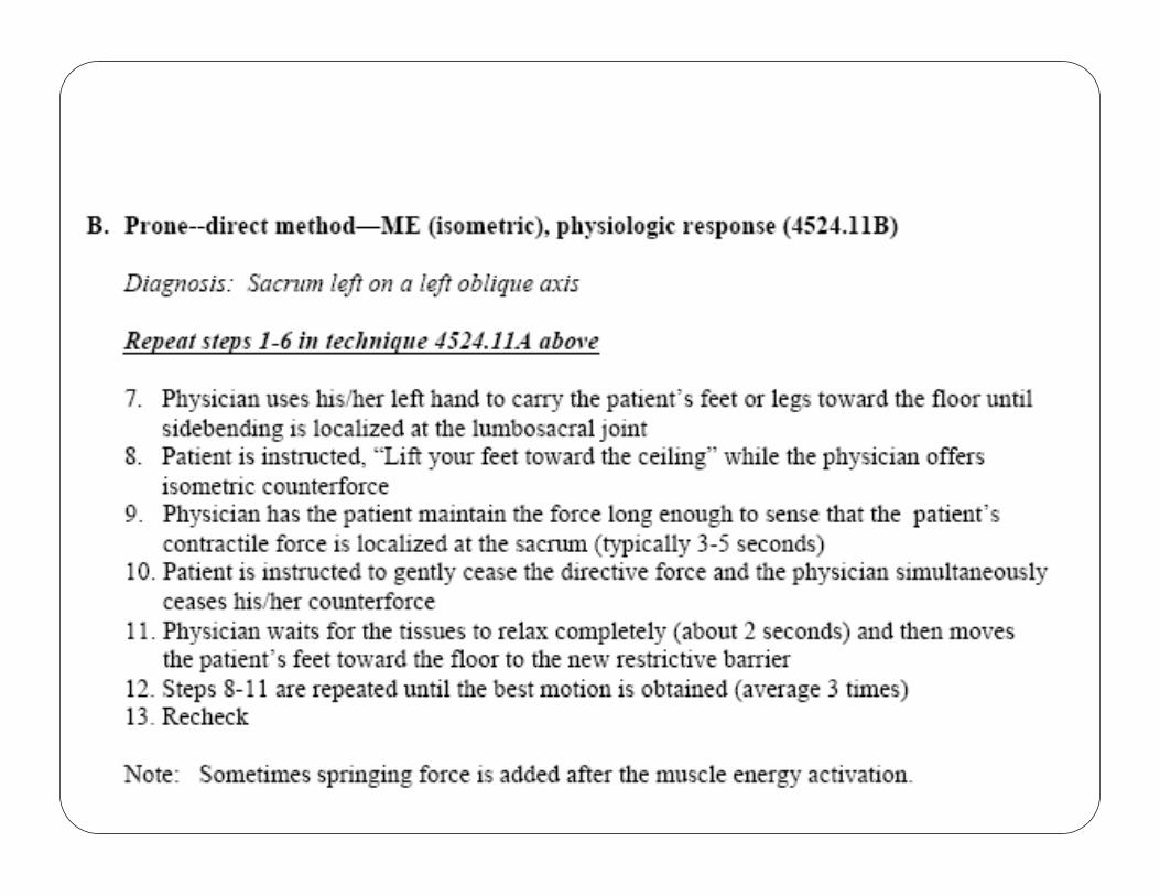
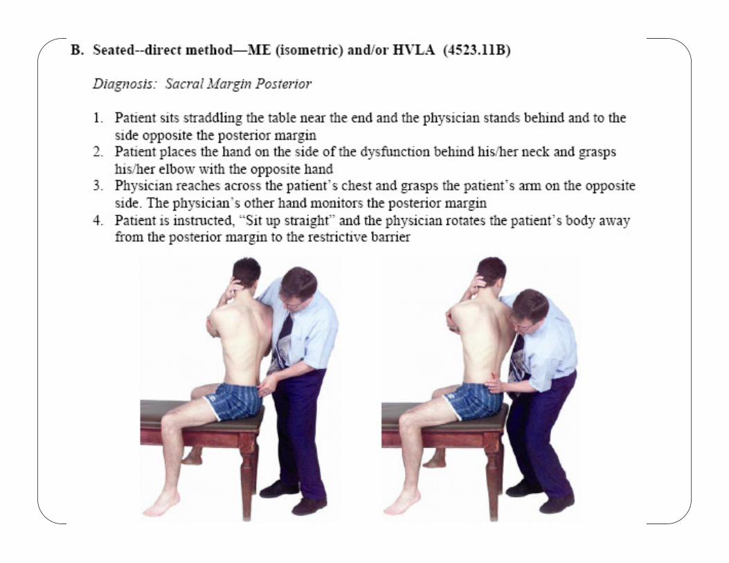
Diagnosis and Treatment of Sacral Somatic Dysfunction, with Indirect,Direct and HVLA
Techniques
(Counterstrain and Muscle Energy)
Evaluation and Treatment of Sacral
Somatic Dysfunction
F. P Wedel, D.O.Associate Adjunct Professor in Osteopathic Principles and PracticeA.T. Still University School of Osteopathic Medicine in Arizona
Learning Objectives� Review the following diagnostic and treatment techniques related to sacral somatic dysfunction:� Lumbosacral spring test
� Sacral palpation
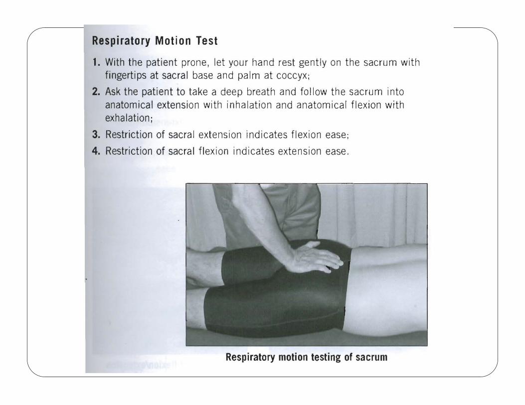
� Respiratory motion test
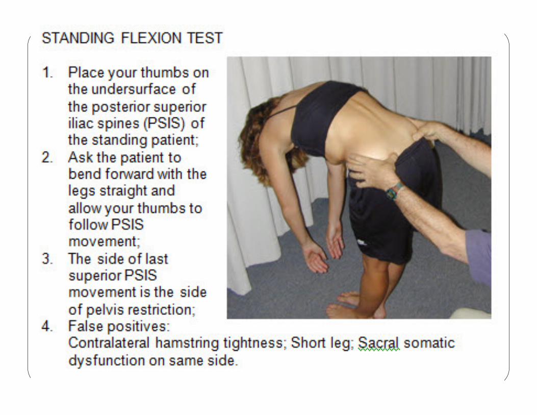
� Seated flexion test
� Sacral somatic dysfunctions – see table
� Clinical presentations applicable to sacral diagnosis and treatment
� Techniques for sacral somatic dysfunction
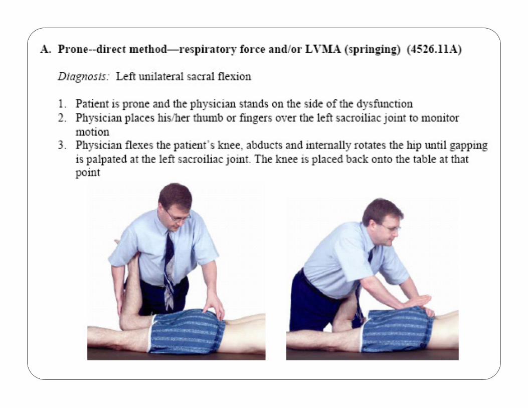
Sacral Techniques Covered:1. Supine, indirect, respiratory cooperation, for bilateral flexion -2. Supine, direct, muscle energy, for bilateral flexion -3. Prone, direct, respiratory cooperation, for bilateral extension - Supine, indirect, respiratory cooperation, for bilateral extension- Prone, direct, LVMA, for sacral rotation on same axis (anterior torsions)-
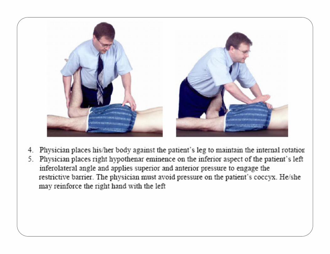
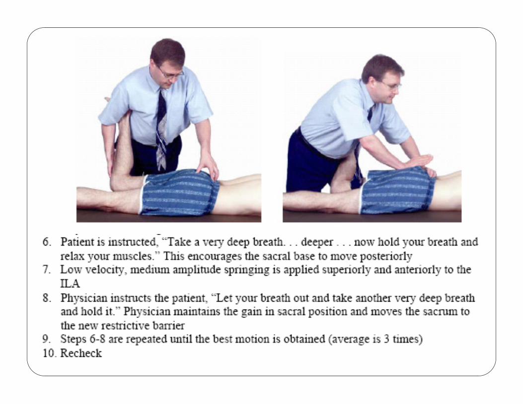
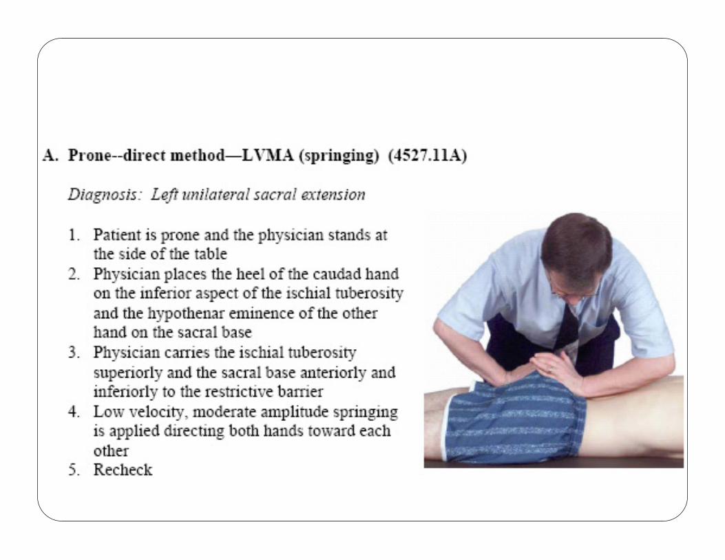
4. Prone, direct, muscle energy, for sacral rotation on same axis (anterior torsions)-Prone, direct, LVMA, for unilateral flexion (shear) - Prone, direct, LVMA, for unilateral extension (shear) –
5. HVLA for Anterior and Posterior sacral torsions
Sacral Clinical Presentations� Presentations commonly associated with sacral somatic dysfunction and/or benefiting from correction of that dysfunction:� Low back pain – traumatic history
� Status Post Labor – History of difficult labor
� Constipation
� Menstrual cramps / dysfunction
� Prostate dysfunction
BACKGROUND
SACRAL STRUCTURE,LIGAMENTS AND
MUSCLES
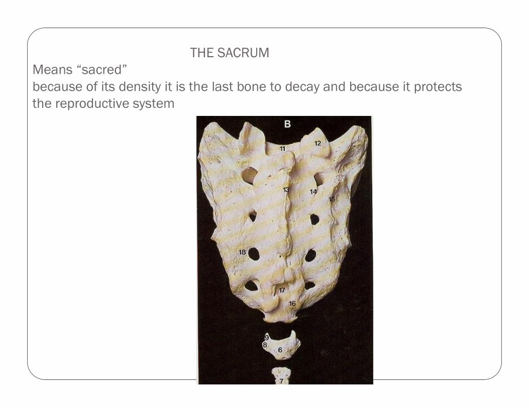
THE SACRUM
Means “sacred”
because of its density it is the last bone to decay and because it protects
the reproductive system
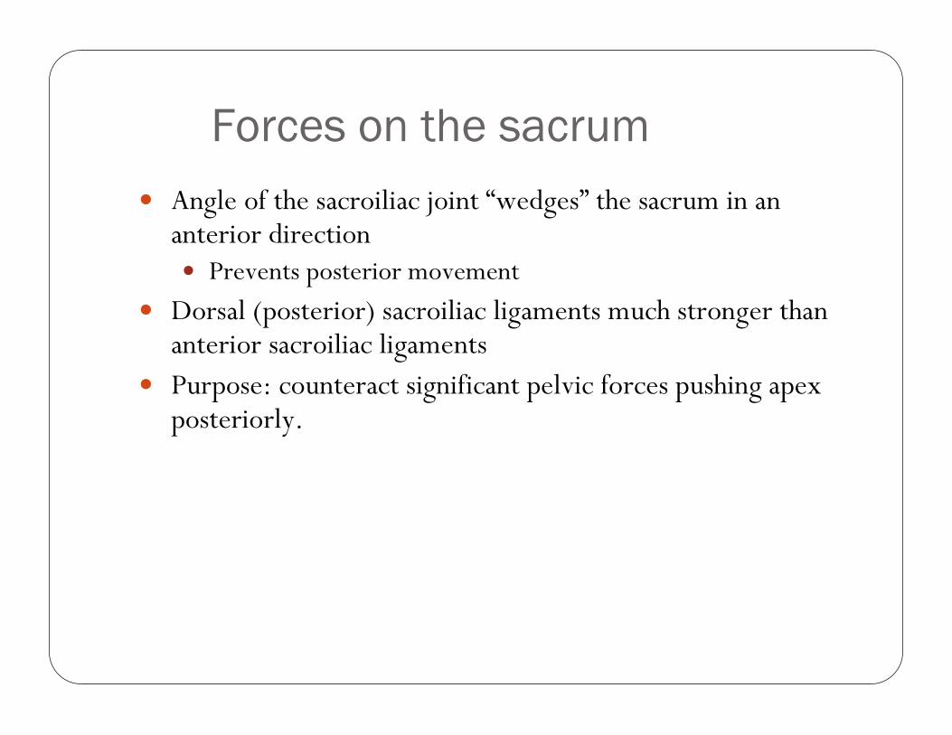
Forces on the sacrum
� Angle of the sacroiliac joint “wedges” the sacrum in an anterior direction� Prevents posterior movement
� Dorsal (posterior) sacroiliac ligaments much stronger than anterior sacroiliac ligaments
� Purpose: counteract significant pelvic forces pushing apex posteriorly.
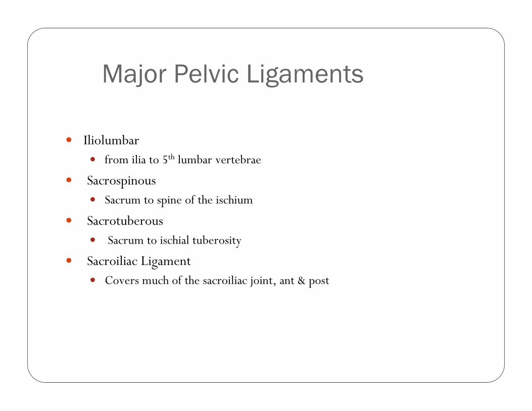
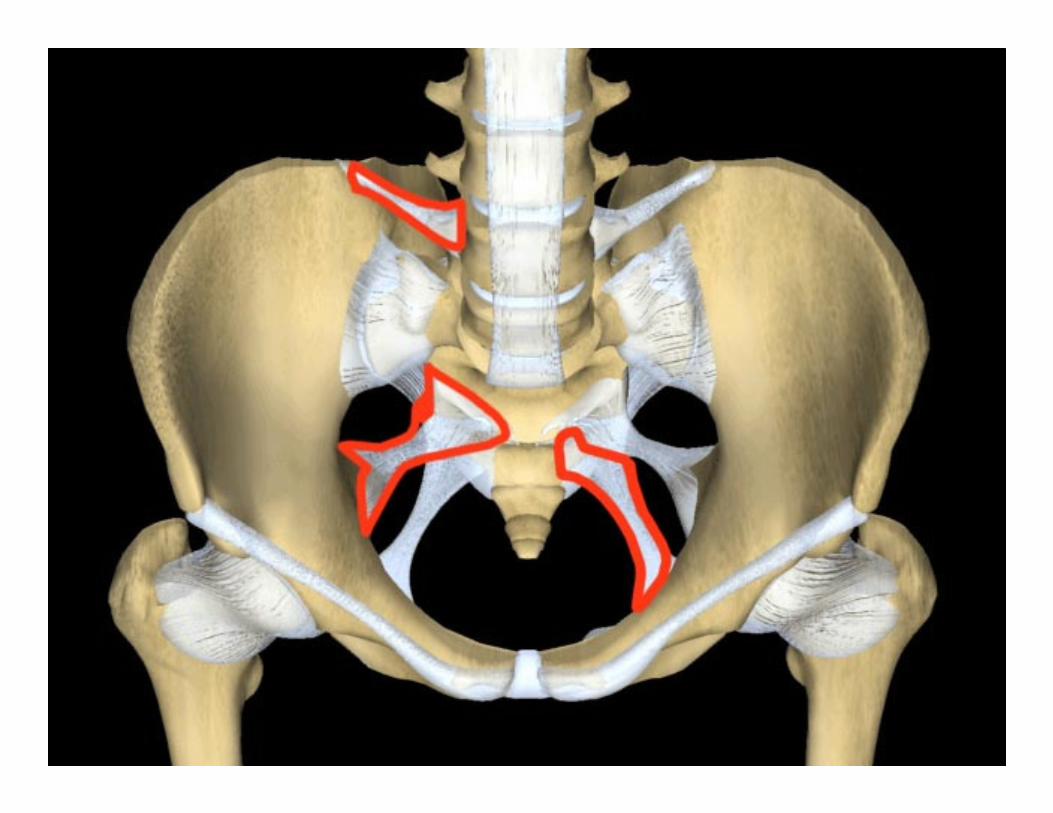
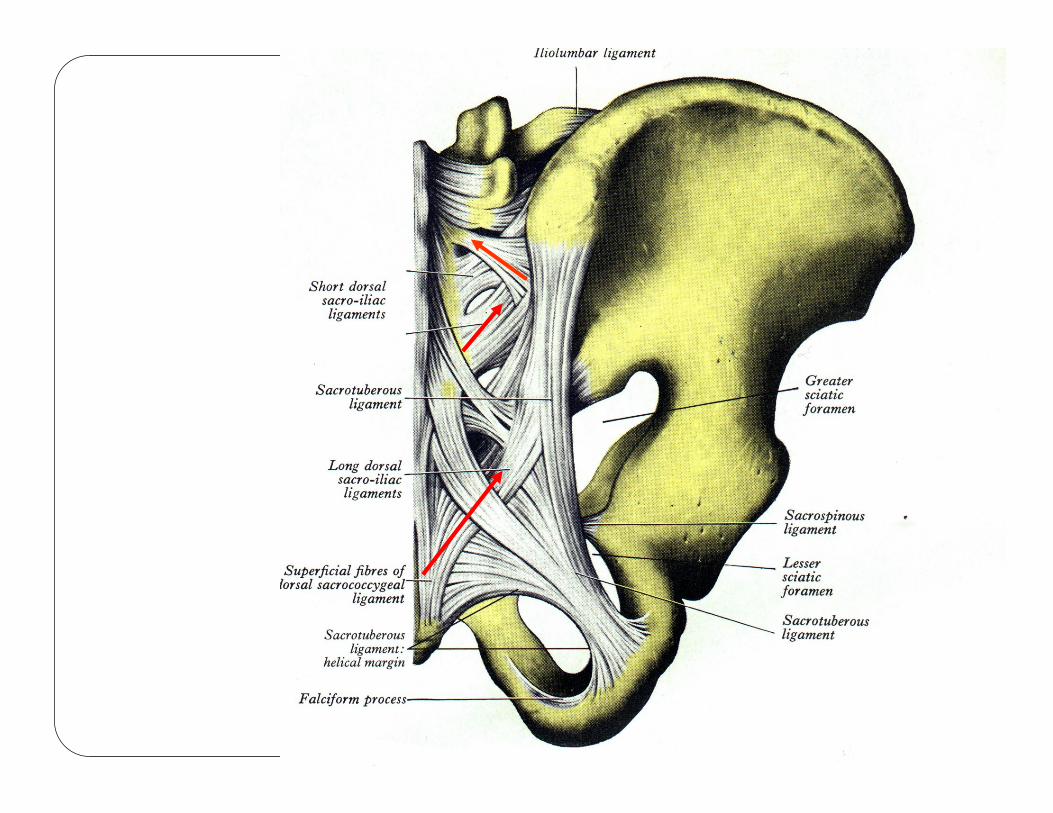
Major Pelvic Ligaments
� Iliolumbar� from ilia to 5th lumbar vertebrae
� Sacrospinous� Sacrum to spine of the ischium
� Sacrotuberous� Sacrum to ischial tuberosity
� Sacroiliac Ligament� Covers much of the sacroiliac joint, ant & post
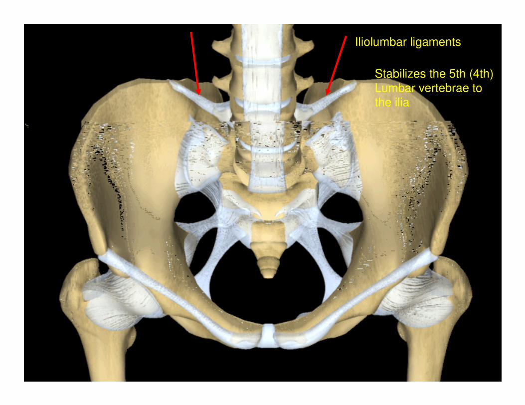
Iliolumbar ligaments
Stabilizes the 5th (4th)
Lumbar vertebrae to
the ilia
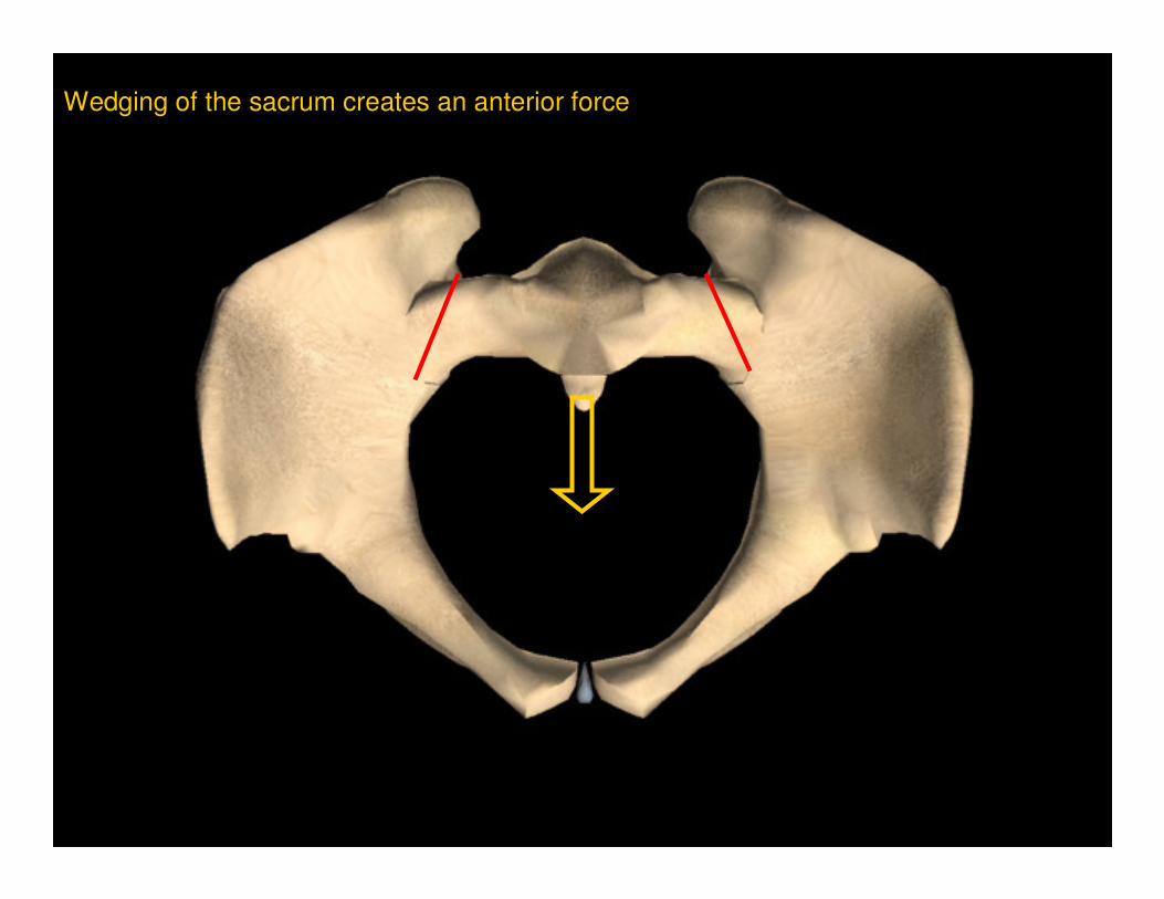
Wedging of the sacrum creates an anterior force
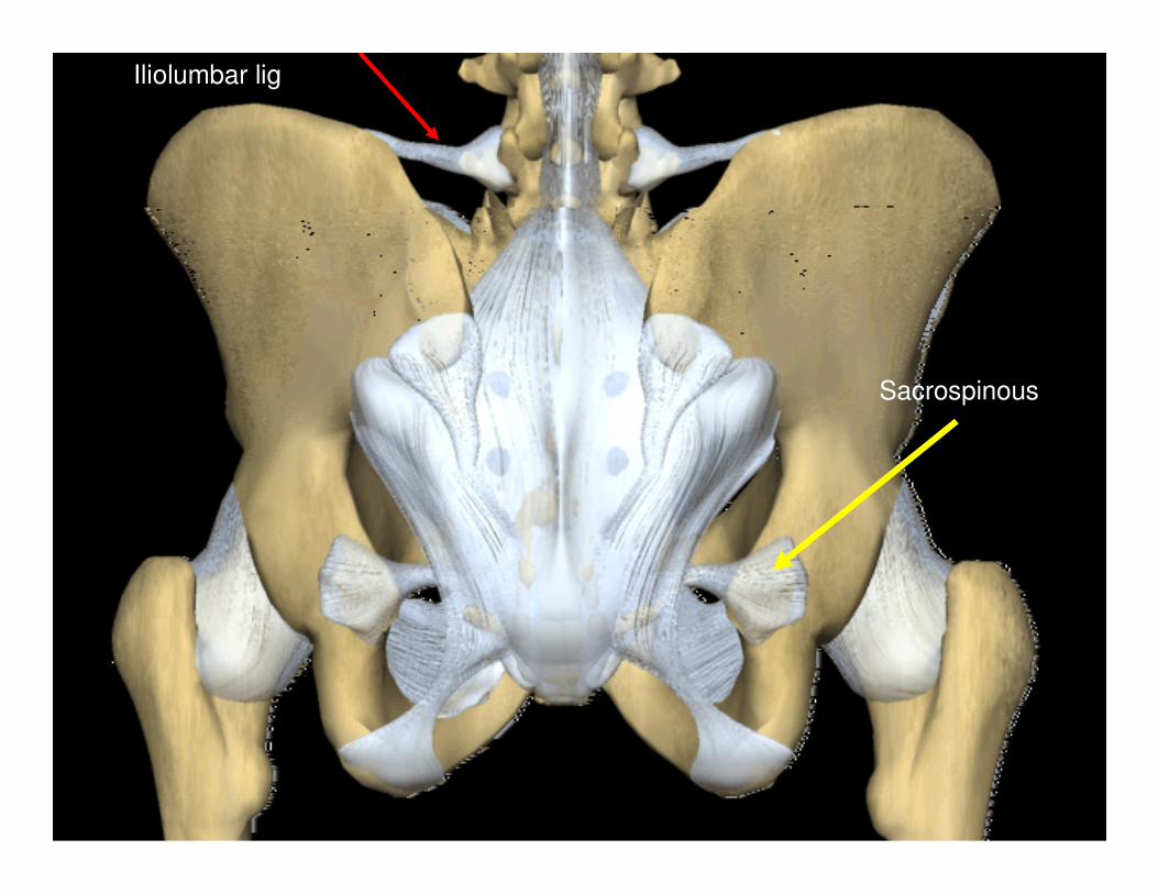
Iliolumbar lig
Sacrospinous
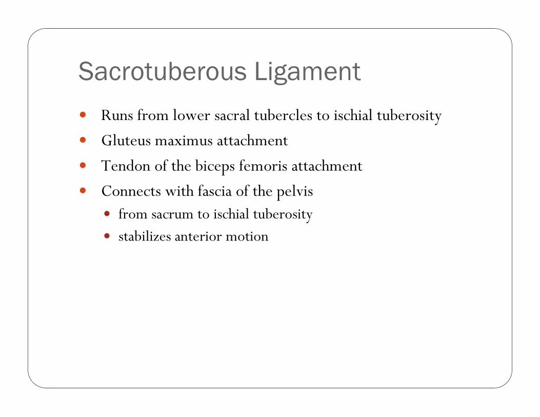
Sacrotuberous Ligament
� Runs from lower sacral tubercles to ischial tuberosity
� Gluteus maximus attachment
� Tendon of the biceps femoris attachment
� Connects with fascia of the pelvis� from sacrum to ischial tuberosity
� stabilizes anterior motion
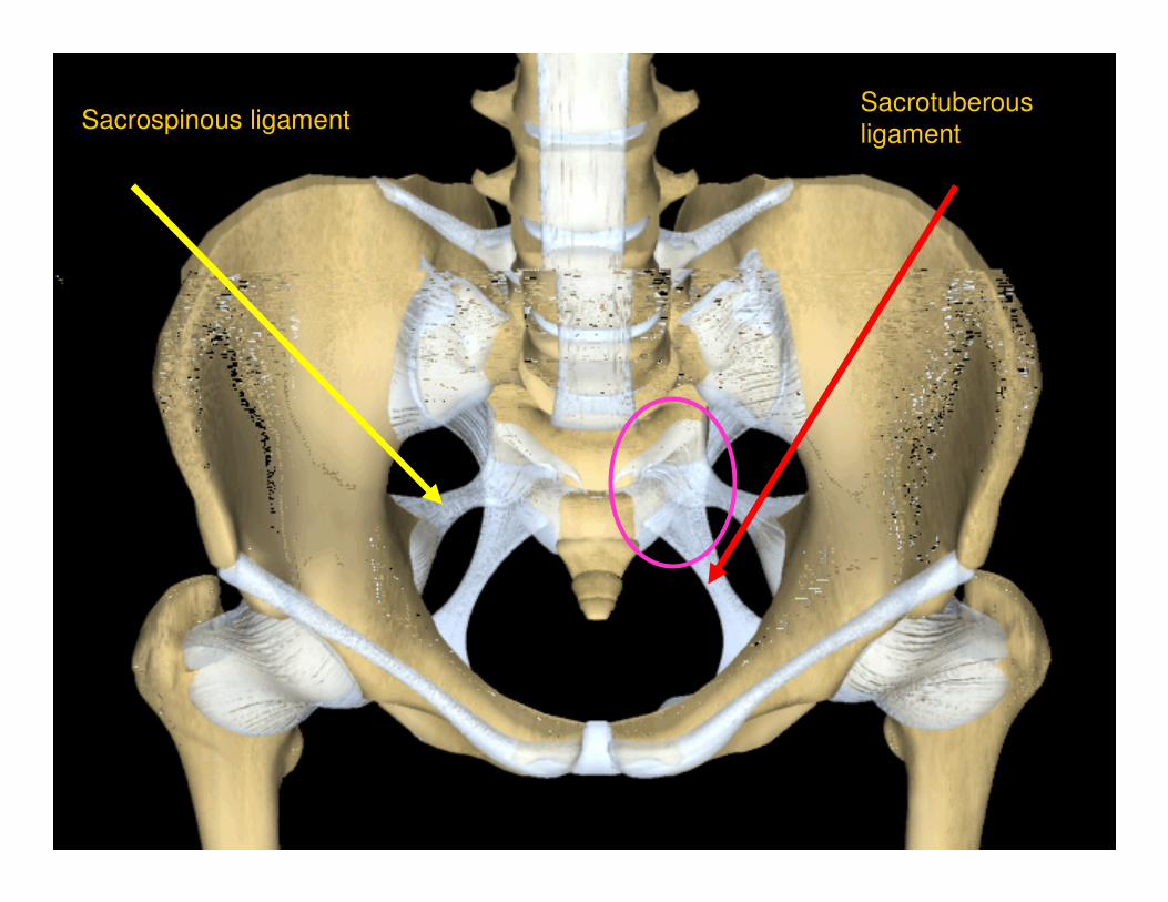
Sacrospinous ligamentSacrotuberous
ligament
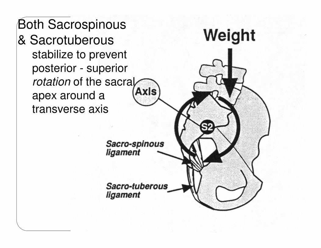
Both Sacrospinous
& Sacrotuberousstabilize to prevent posterior - superior rotation of the sacral apex around a transverse axis
Sacroiliac Ligament
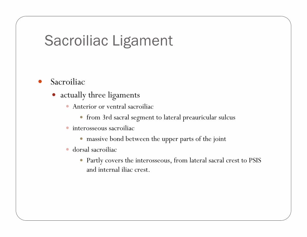
� Sacroiliac� actually three ligaments
� Anterior or ventral sacroiliac
� from 3rd sacral segment to lateral preauricular sulcus
� interosseous sacroiliac
� massive bond between the upper parts of the joint
� dorsal sacroiliac
� Partly covers the interosseous, from lateral sacral crest to PSIS and internal iliac crest.
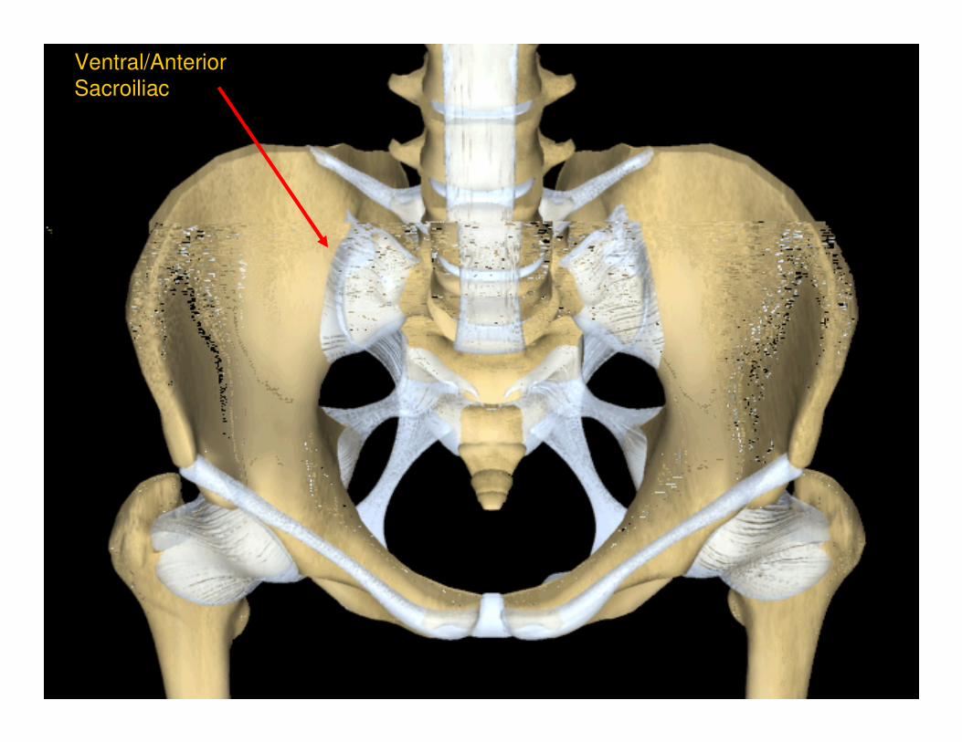
Ventral/Anterior
Sacroiliac
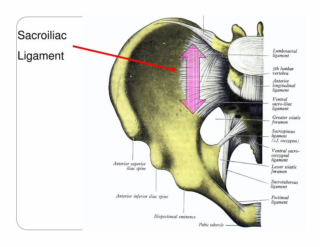
Sacroiliac
Ligament
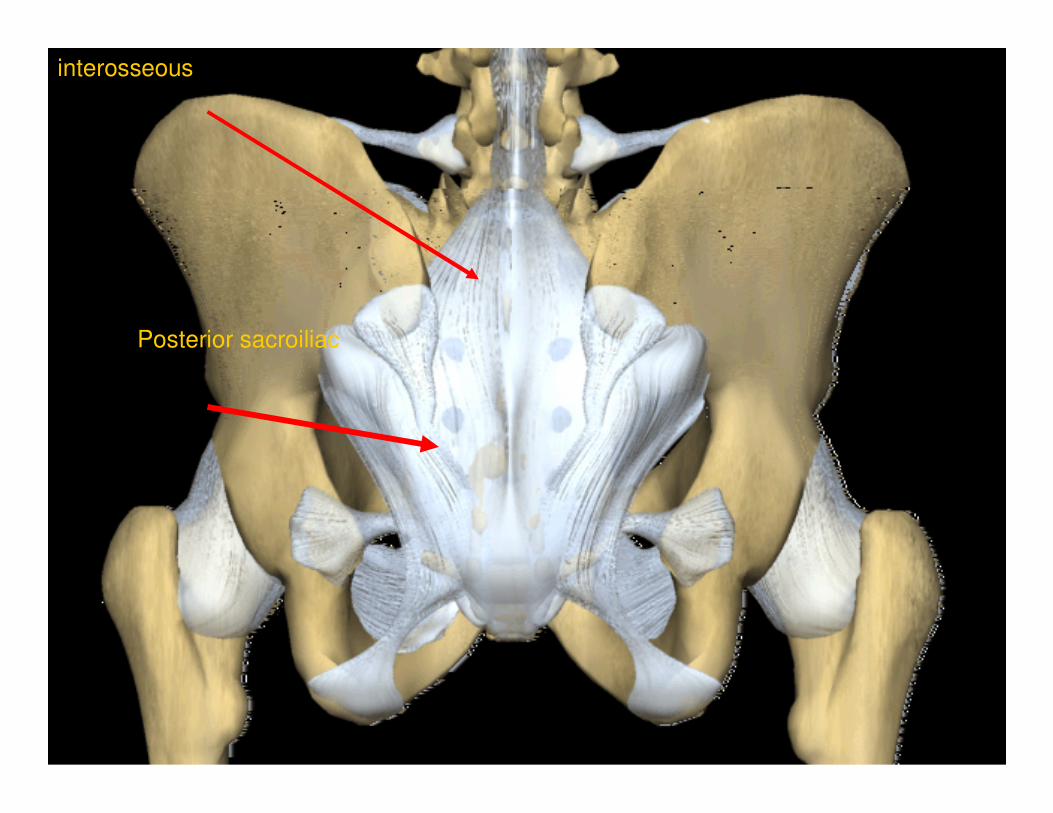
interosseous
Posterior sacroiliac
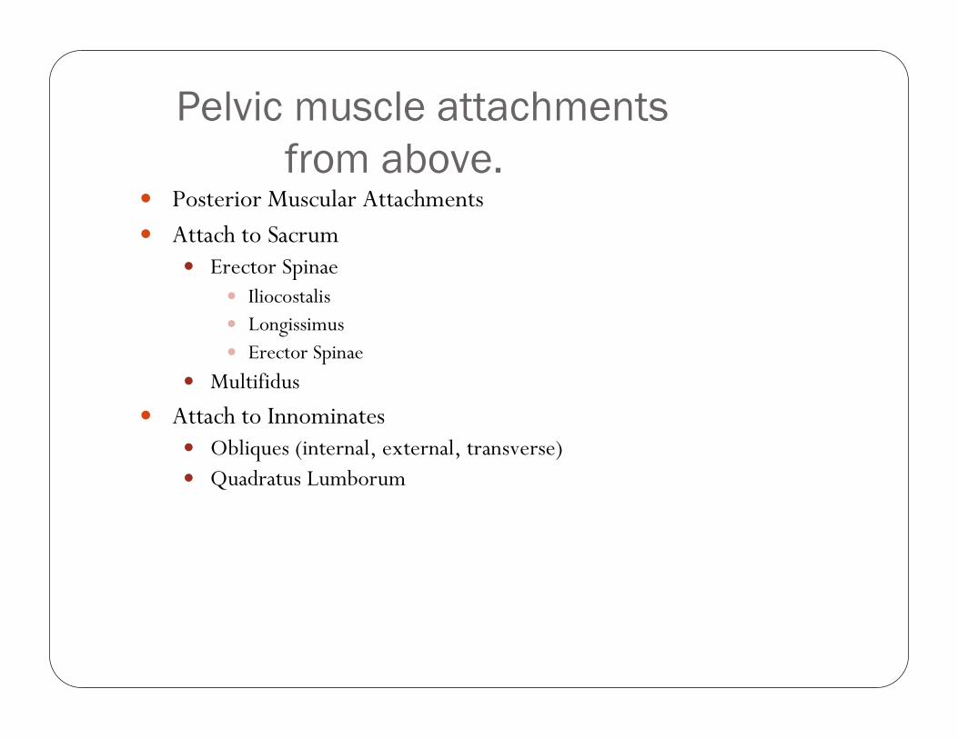
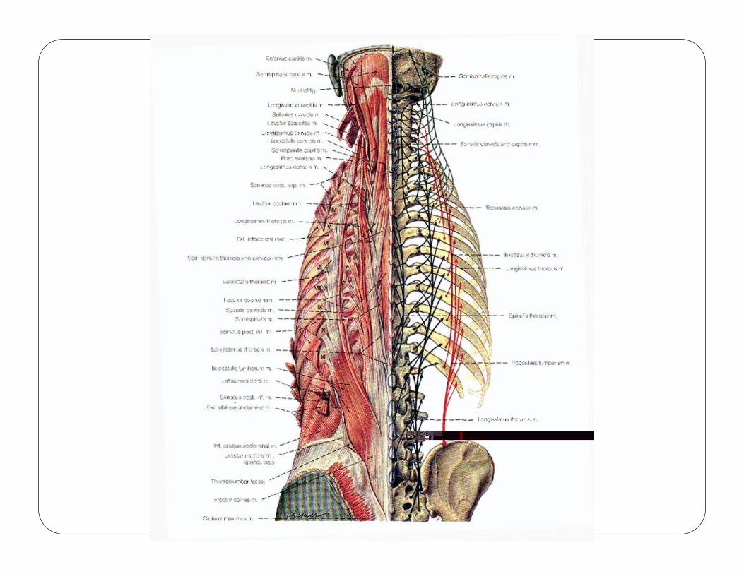
Pelvic muscle attachments
from above.� Posterior Muscular Attachments
� Attach to Sacrum� Erector Spinae
� Iliocostalis� Longissimus� Erector Spinae
� Multifidus
� Attach to Innominates� Obliques (internal, external, transverse)� Quadratus Lumborum
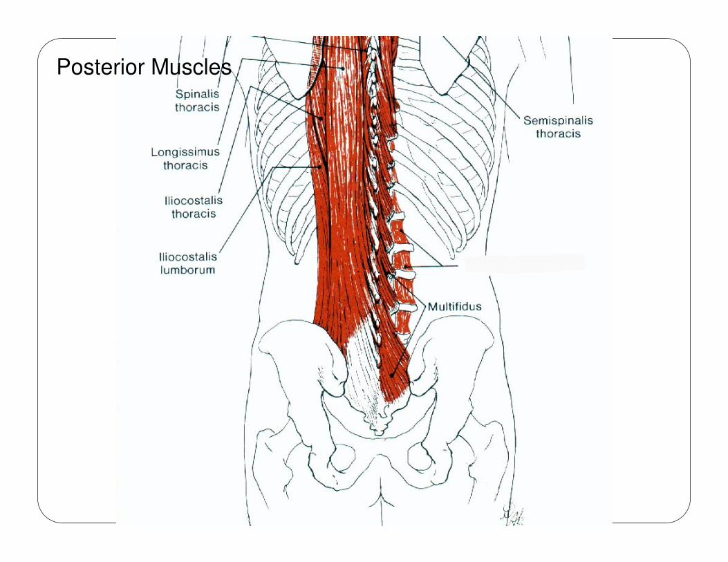
Posterior Muscles
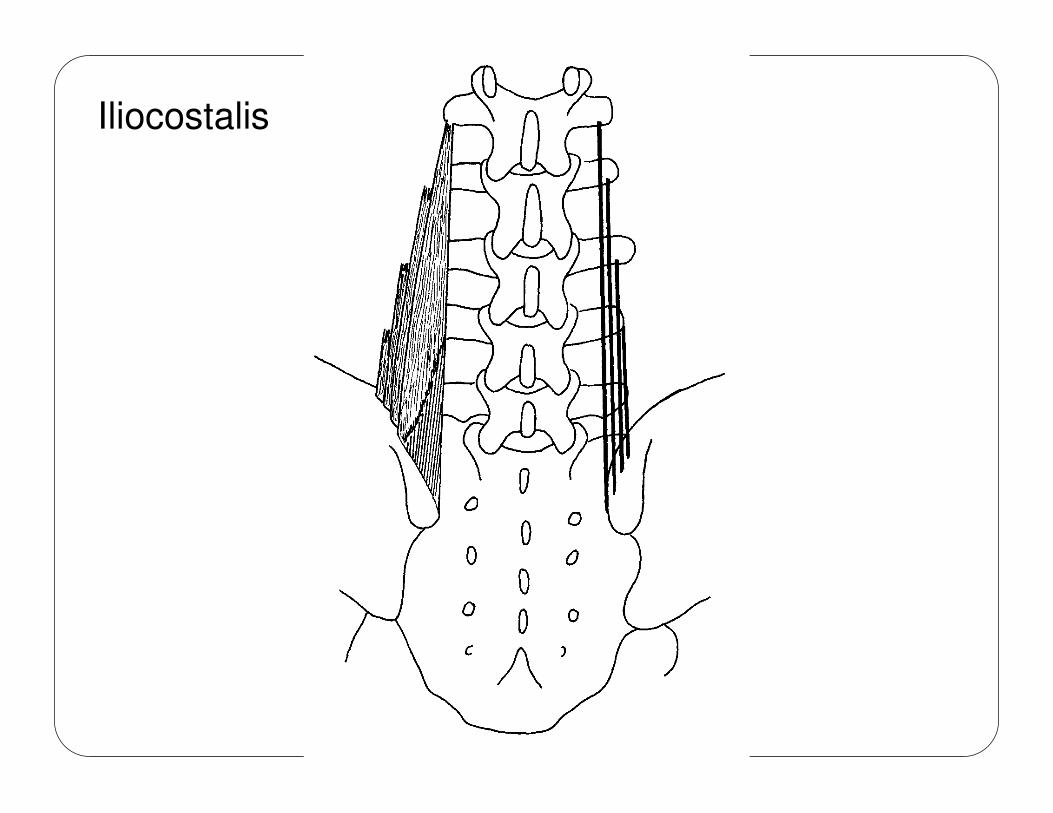
Iliocostalis
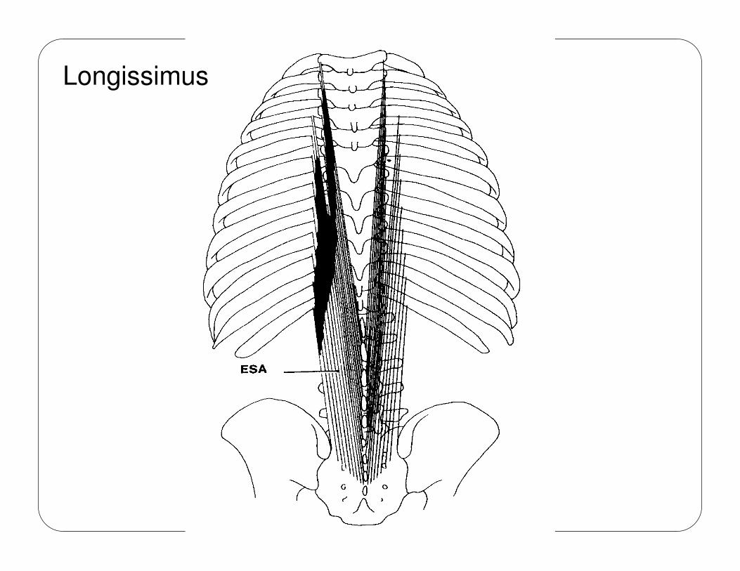
Longissimus
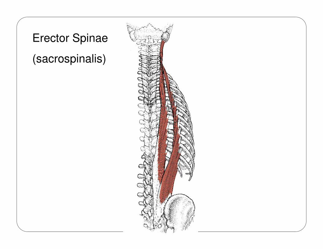
Erector Spinae
(sacrospinalis)
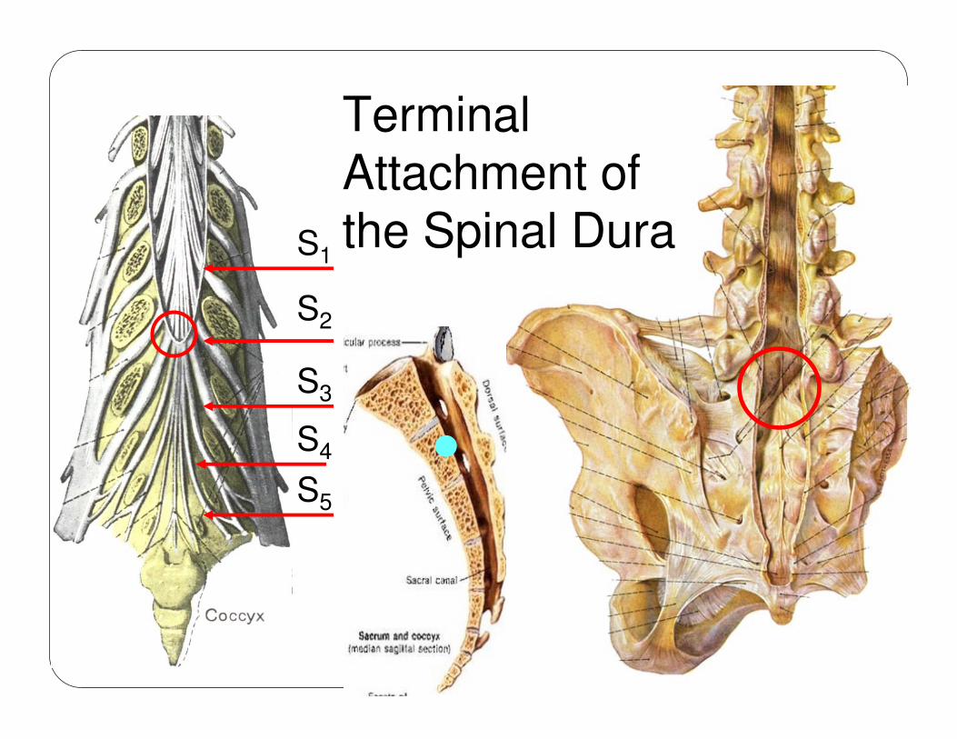
Terminal Attachment of the Spinal DuraS1
S2
S3
S4
S5
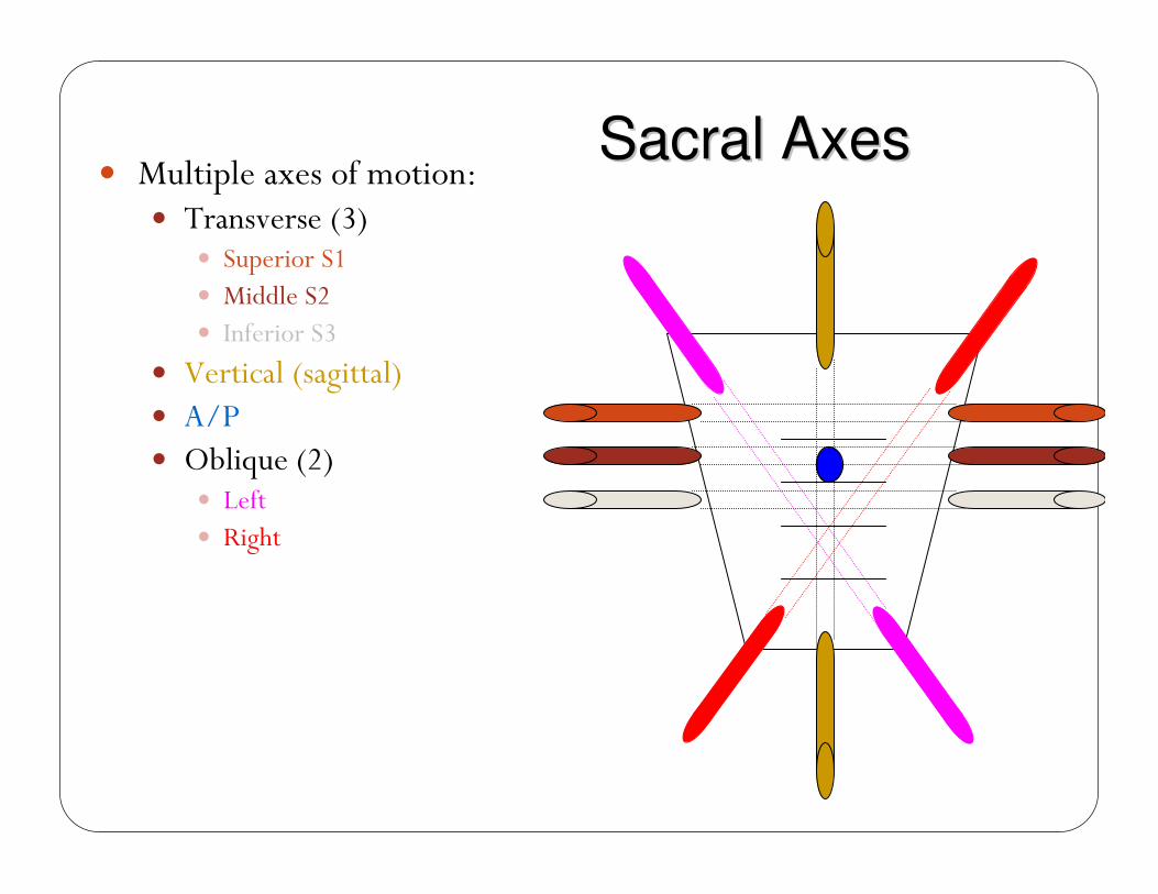
� Multiple axes of motion:� Transverse (3)
� Superior S1� Middle S2� Inferior S3
� Vertical (sagittal)� A/P� Oblique (2)
� Left� Right
Sacral AxesSacral Axes
SACRAL ANATOMICAL AXISSACRAL ANATOMICAL AXISSACRAL ANATOMICAL AXISSACRAL ANATOMICAL AXIS
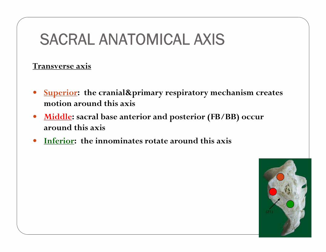
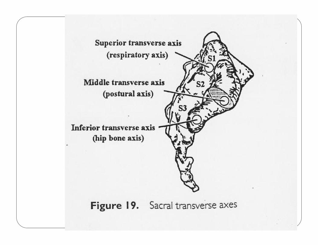
Transverse axis
� Superior: the cranial&primary respiratory mechanism creates motion around this axis
� Middle: sacral base anterior and posterior (FB/BB) occur around this axis
� Inferior: the innominates rotate around this axis
SACRAL PHYSIOLOGIC AXISSACRAL PHYSIOLOGIC AXISSACRAL PHYSIOLOGIC AXISSACRAL PHYSIOLOGIC AXIS
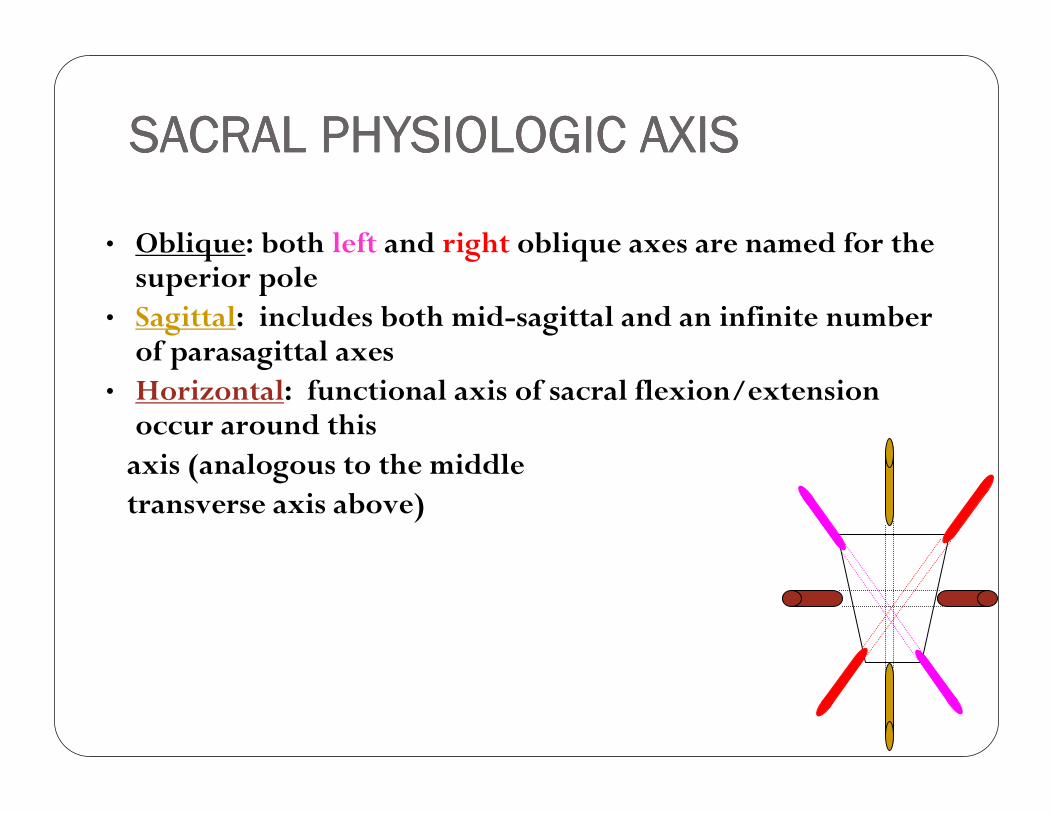
• Oblique: both left and right oblique axes are named for the superior pole
• Sagittal: includes both mid-sagittal and an infinite number of parasagittal axes
• Horizontal: functional axis of sacral flexion/extension occur around this axis (analogous to the middle transverse axis above)
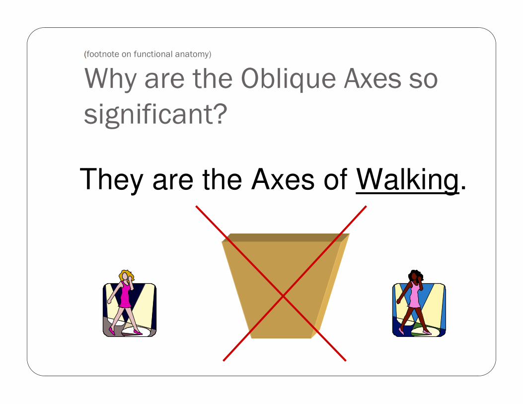
(footnote on functional anatomy)
Why are the Oblique Axes so
significant?
They are the Axes of Walking.
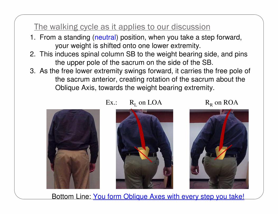
The walking cycle as it applies to our discussion1. From a standing (neutral) position, when you take a step forward,
your weight is shifted onto one lower extremity.
2. This induces spinal column SB to the weight bearing side, and pins
the upper pole of the sacrum on the side of the SB.
3. As the free lower extremity swings forward, it carries the free pole of
the sacrum anterior, creating rotation of the sacrum about the
Oblique Axis, towards the weight bearing extremity.
Bottom Line: You form Oblique Axes with every step you take!
Ex.: RL on LOA RR on ROA
TESTS
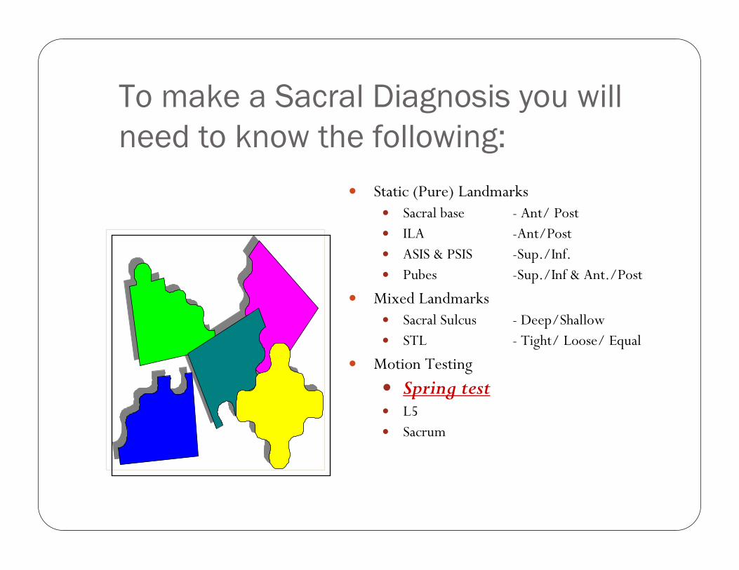
To make a Sacral Diagnosis you will
need to know the following:
� Static (Pure) Landmarks� Sacral base - Ant/ Post� ILA -Ant/Post� ASIS & PSIS -Sup./Inf.� Pubes -Sup./Inf & Ant./Post
� Mixed Landmarks� Sacral Sulcus - Deep/Shallow� STL - Tight/ Loose/ Equal
� Motion Testing
� Spring test� L5� Sacrum
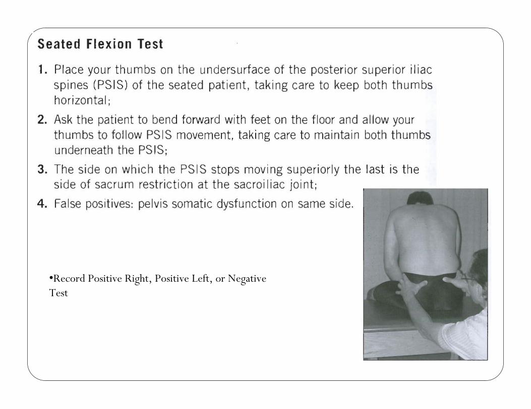
•Record Positive Right, Positive Left, or Negative Test
Most of those pieces we have discussed,
except...
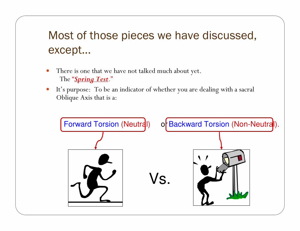
� There is one that we have not talked much about yet.The “Spring Test.”
� It’s purpose: To be an indicator of whether you are dealing with a sacral Oblique Axis that is a:
Vs.
Forward Torsion (Neutral) or Backward Torsion (Non-Neutral).
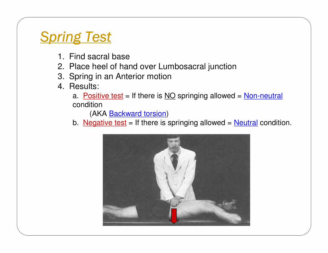
Spring TestSpring TestSpring TestSpring Test
1. Find sacral base
2. Place heel of hand over Lumbosacral junction
3. Spring in an Anterior motion
4. Results:a. Positive test = If there is NO springing allowed = Non-neutral
condition
(AKA Backward torsion)
b. Negative test = If there is springing allowed = Neutral condition.

Prone Landmarks
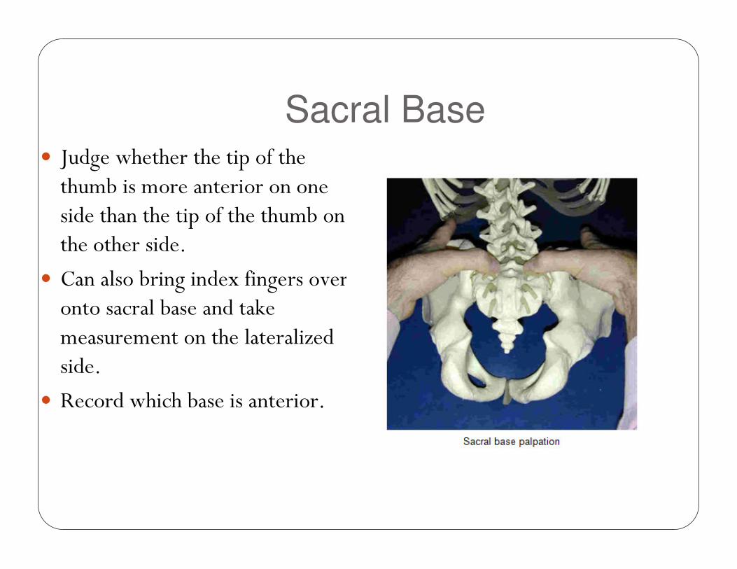
Sacral Base� Judge whether the tip of the thumb is more anterior on one side than the tip of the thumb on the other side.
� Can also bring index fingers over onto sacral base and take measurement on the lateralized side.
� Record which base is anterior.
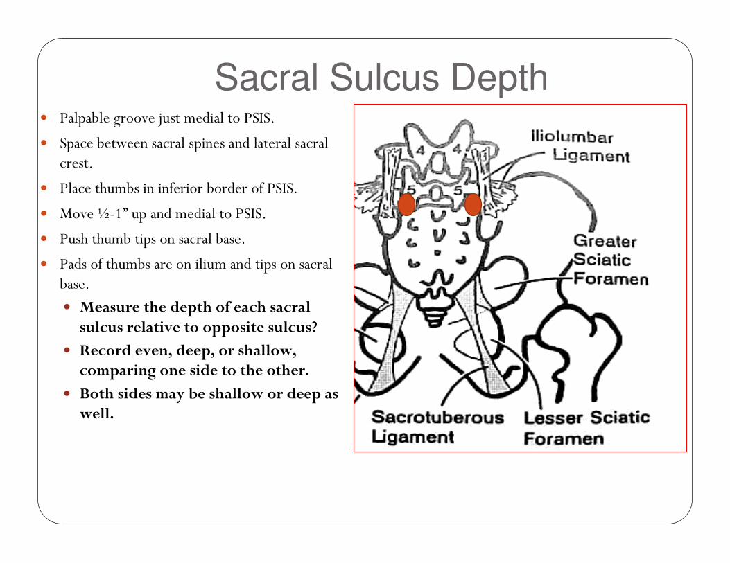
Sacral Sulcus Depth� Palpable groove just medial to PSIS.
� Space between sacral spines and lateral sacral crest.
� Place thumbs in inferior border of PSIS.
� Move ½-1” up and medial to PSIS.
� Push thumb tips on sacral base.
� Pads of thumbs are on ilium and tips on sacral base.
� Measure the depth of each sacral sulcus relative to opposite sulcus?
� Record even, deep, or shallow, comparing one side to the other.
� Both sides may be shallow or deep as well.
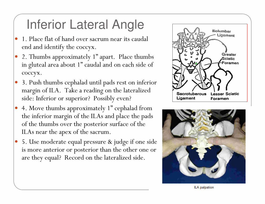
Inferior Lateral Angle� 1. Place flat of hand over sacrum near its caudal end and identify the coccyx.
� 2. Thumbs approximately 1” apart. Place thumbs in gluteal area about 1” caudal and on each side of coccyx.
� 3. Push thumbs cephalad until pads rest on inferior margin of ILA. Take a reading on the lateralized side: Inferior or superior? Possibly even?
� 4. Move thumbs approximately 1” cephalad from the inferior margin of the ILAs and place the pads of the thumbs over the posterior surface of the ILAs near the apex of the sacrum.
� 5. Use moderate equal pressure & judge if one side is more anterior or posterior than the other one or are they equal? Record on the lateralized side.
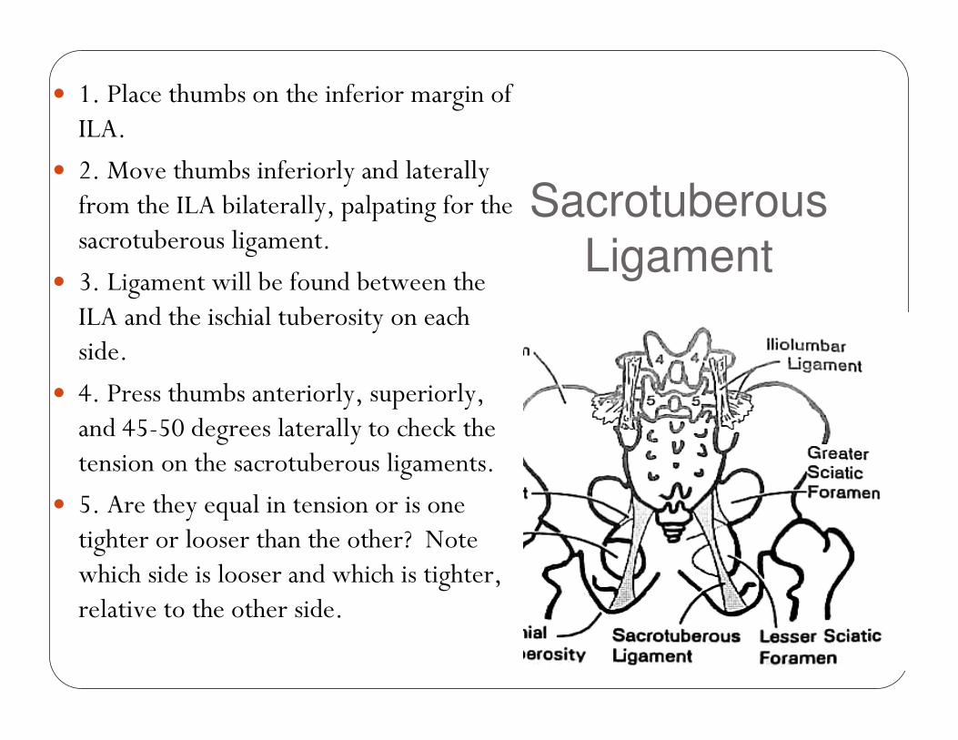
Sacrotuberous
Ligament
� 1. Place thumbs on the inferior margin of ILA.
� 2. Move thumbs inferiorly and laterally from the ILA bilaterally, palpating for the sacrotuberous ligament.
� 3. Ligament will be found between the ILA and the ischial tuberosity on each side.
� 4. Press thumbs anteriorly, superiorly, and 45-50 degrees laterally to check the tension on the sacrotuberous ligaments.
� 5. Are they equal in tension or is one tighter or looser than the other? Note which side is looser and which is tighter, relative to the other side.
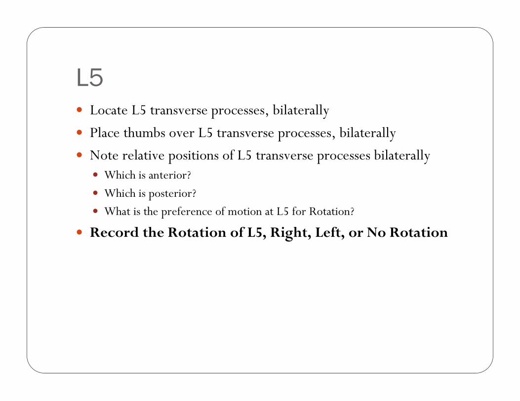
L5� Locate L5 transverse processes, bilaterally
� Place thumbs over L5 transverse processes, bilaterally
� Note relative positions of L5 transverse processes bilaterally� Which is anterior?
� Which is posterior?
� What is the preference of motion at L5 for Rotation?
� Record the Rotation of L5, Right, Left, or No Rotation
Motion Tests for Sacral Diagnosis
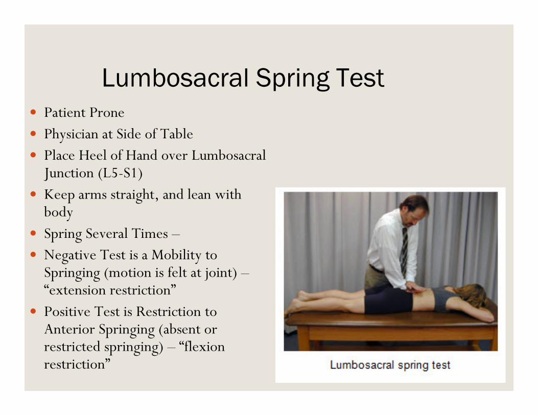
Lumbosacral Spring Test� Patient Prone
� Physician at Side of Table� Place Heel of Hand over LumbosacralJunction (L5-S1)
� Keep arms straight, and lean with body
� Spring Several Times –� Negative Test is a Mobility to Springing (motion is felt at joint) –“extension restriction”
� Positive Test is Restriction to Anterior Springing (absent or restricted springing) – “flexion restriction”
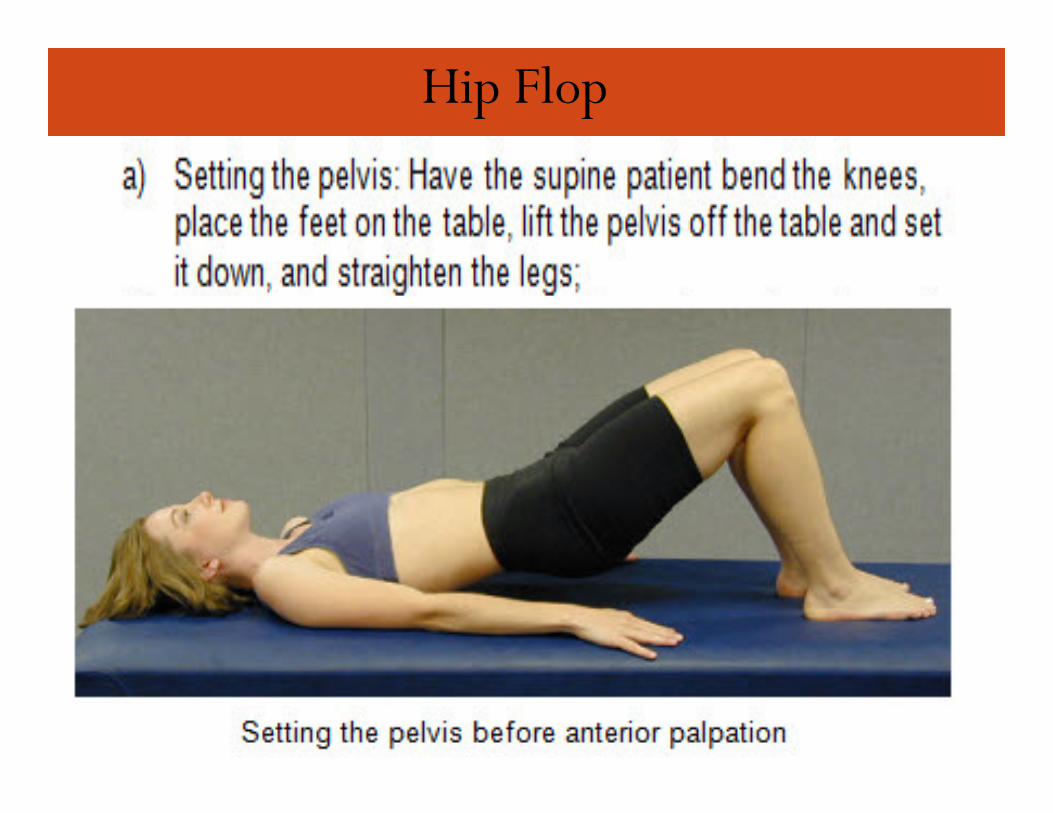
Hip Flop
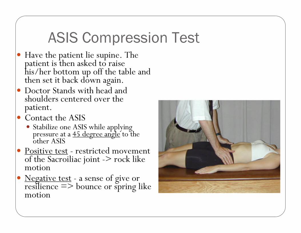
ASIS Compression Test� Have the patient lie supine. The patient is then asked to raise his/her bottom up off the table and then set it back down again.
� Doctor Stands with head and shoulders centered over the patient.
� Contact the ASIS � Stabilize one ASIS while applying pressure at a 45 degree angle to the other ASIS
� Positive test - restricted movement of the Sacroiliac joint -> rock like motion
� Negative test - a sense of give or resilience => bounce or spring like motion
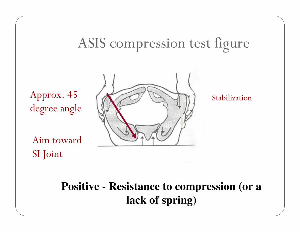
ASIS compression test figure
Positive - Resistance to compression (or a
lack of spring)
Approx. 45 degree angle
Stabilization
Aim toward SI Joint
DIAGNOSIS AND
TREATMENT
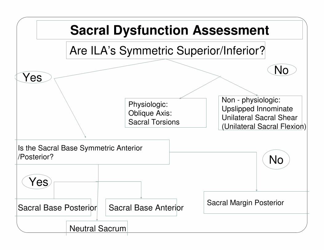
Sacral Dysfunction Assessment
Is the Sacral Base Symmetric Anterior
/Posterior?
Are ILA’s Symmetric Superior/Inferior?
YesNo
Physiologic:
Oblique Axis:
Sacral Torsions
Yes
Non - physiologic:
Upslipped Innominate
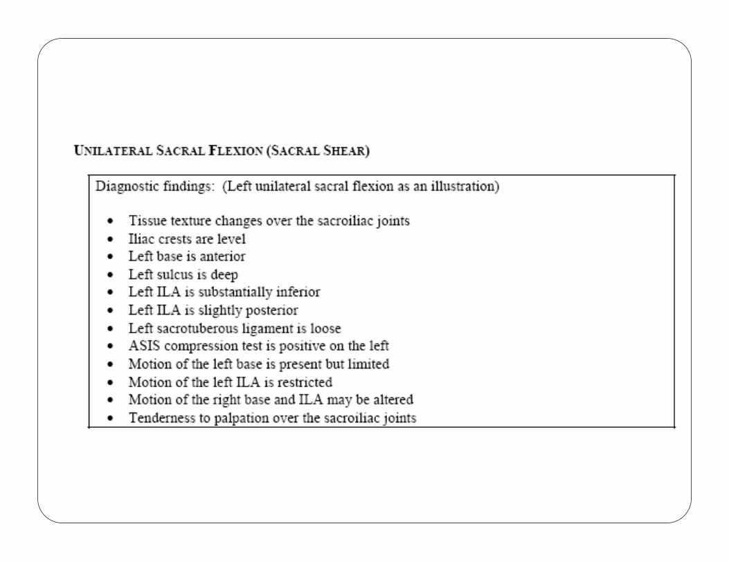
Unilateral Sacral Shear
(Unilateral Sacral Flexion)
Sacral Base Anterior
Neutral Sacrum
Sacral Margin Posterior
No
Sacral Base Posterior
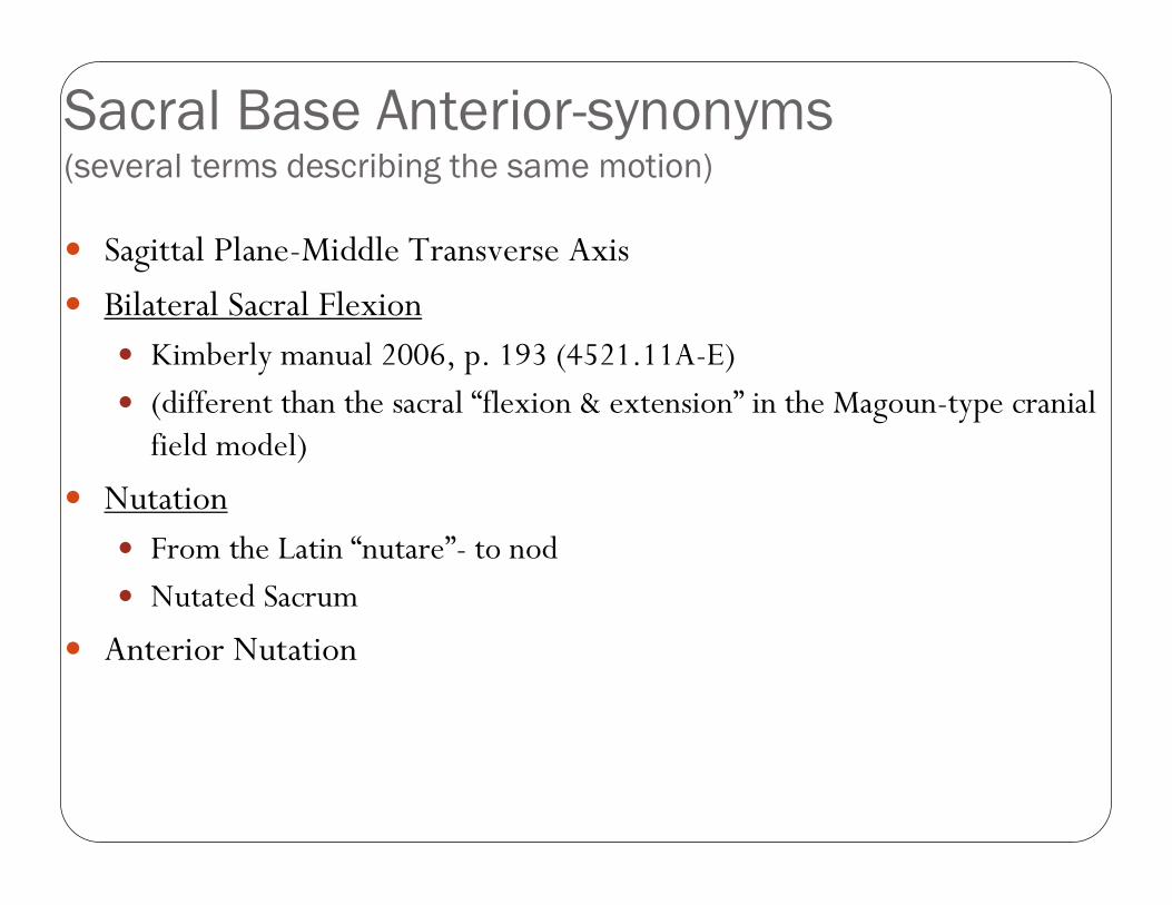
Sacral Base Anterior-synonyms(several terms describing the same motion)
� Sagittal Plane-Middle Transverse Axis
� Bilateral Sacral Flexion� Kimberly manual 2006, p. 193 (4521.11A-E)
� (different than the sacral “flexion & extension” in the Magoun-type cranial field model)
� Nutation� From the Latin “nutare”- to nod
� Nutated Sacrum
� Anterior Nutation
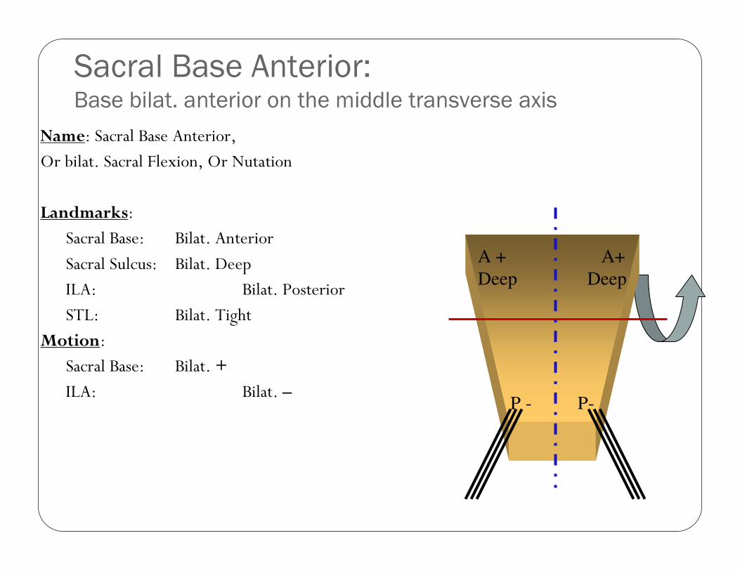
Sacral Base Anterior:Base bilat. anterior on the middle transverse axis
A + A+
Deep Deep
P - P-
Name: Sacral Base Anterior,
Or bilat. Sacral Flexion, Or Nutation
Landmarks:
Sacral Base: Bilat. Anterior
Sacral Sulcus: Bilat. Deep
ILA: Bilat. Posterior
STL: Bilat. Tight
Motion:
Sacral Base: Bilat. +
ILA: Bilat. –
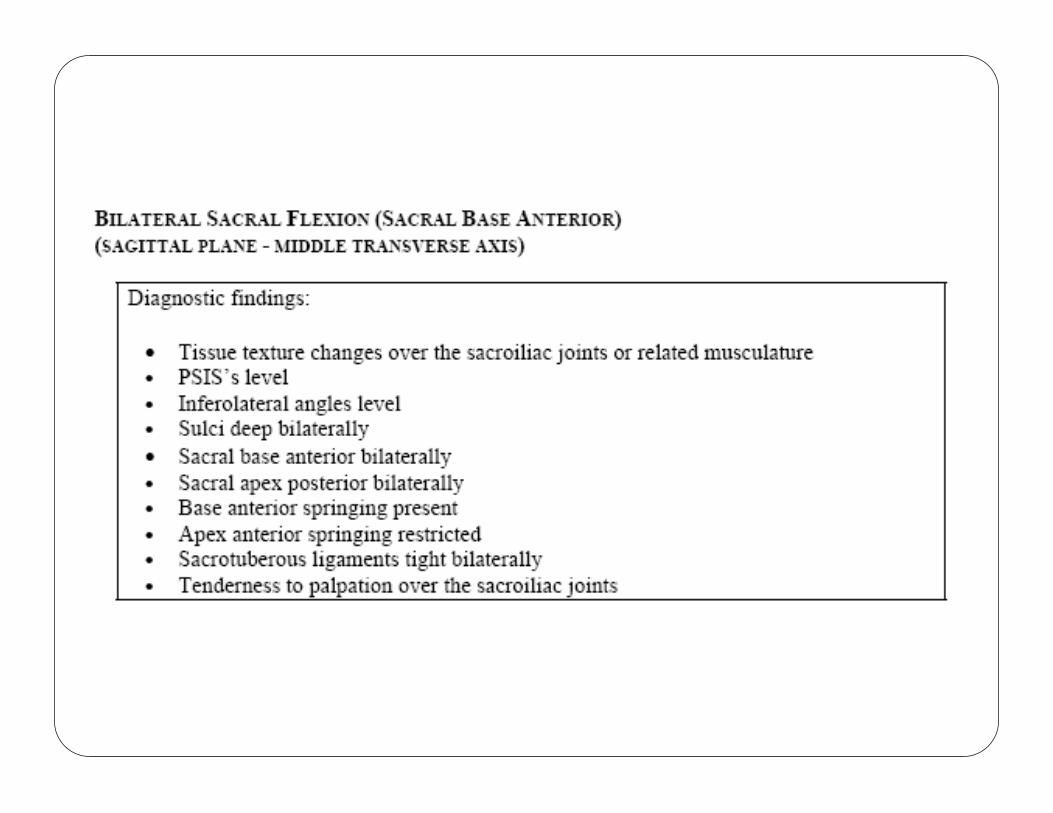
Sacral Base Anterior
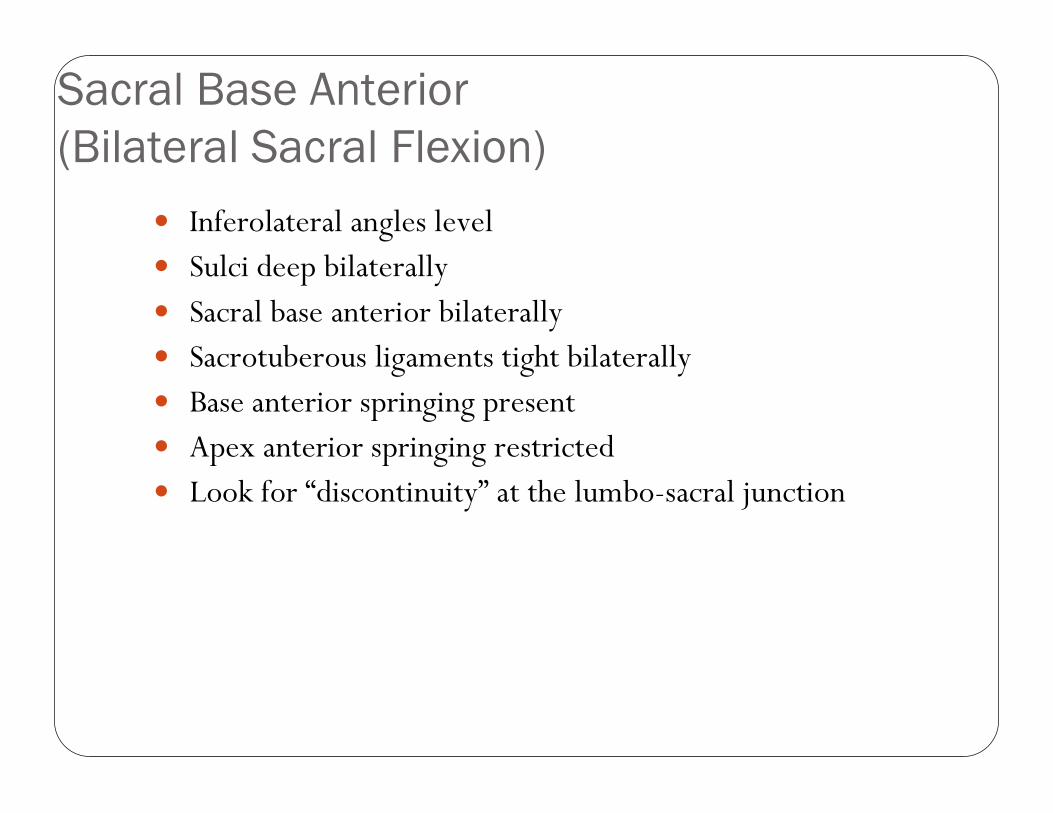
(Bilateral Sacral Flexion)
� Inferolateral angles level� Sulci deep bilaterally� Sacral base anterior bilaterally� Sacrotuberous ligaments tight bilaterally� Base anterior springing present� Apex anterior springing restricted� Look for “discontinuity” at the lumbo-sacral junction
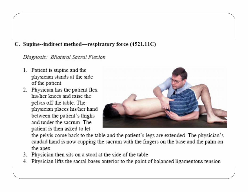
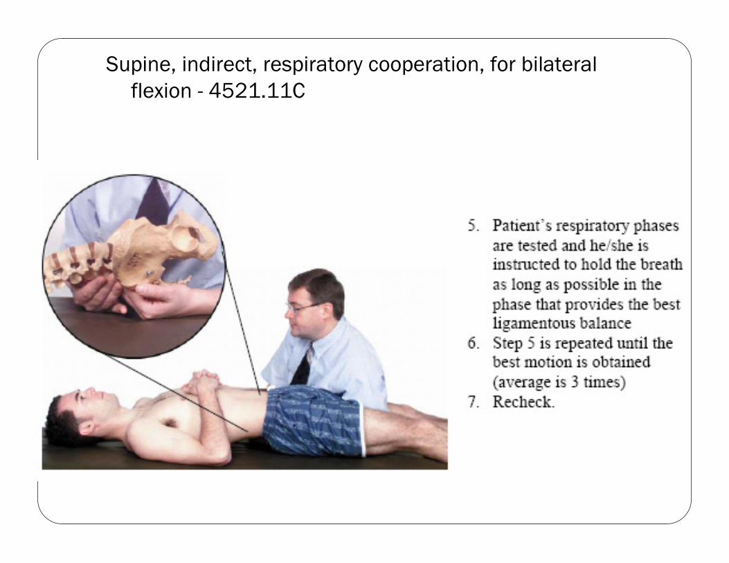
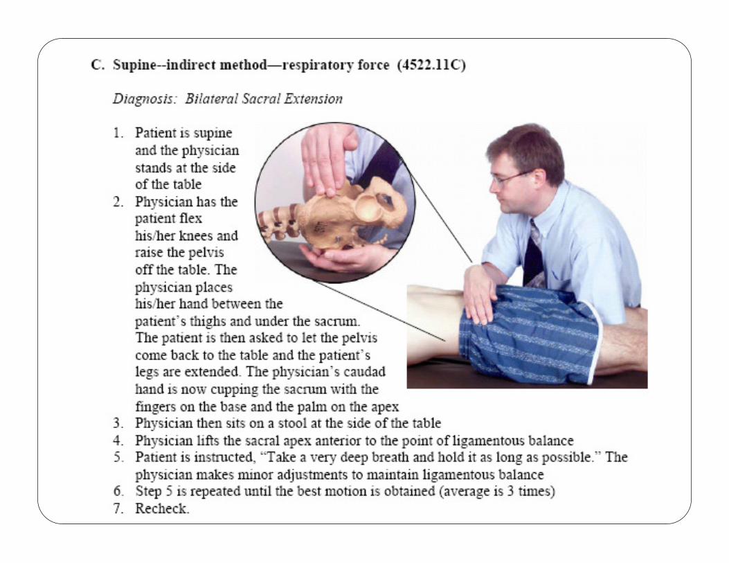
Supine, indirect, respiratory cooperation, for bilateral
flexion - 4521.11C
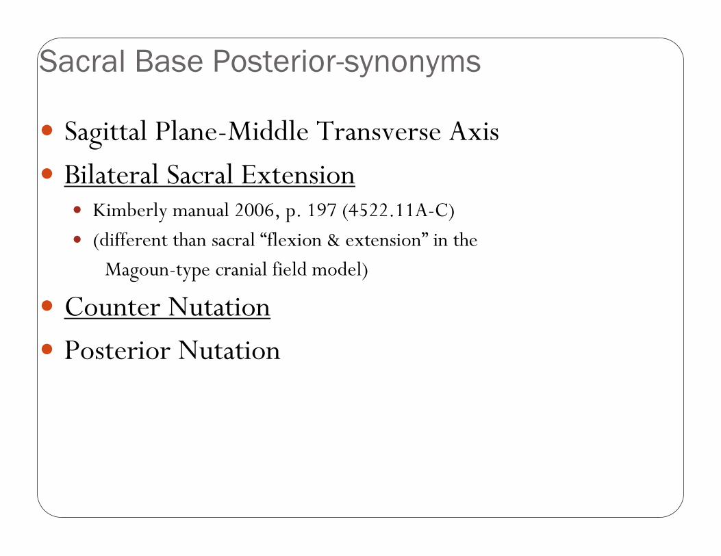
Sacral Base Posterior-synonyms
� Sagittal Plane-Middle Transverse Axis
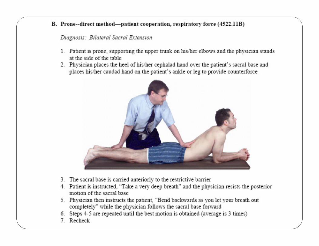
� Bilateral Sacral Extension� Kimberly manual 2006, p. 197 (4522.11A-C)
� (different than sacral “flexion & extension” in the
Magoun-type cranial field model)
� Counter Nutation
� Posterior Nutation
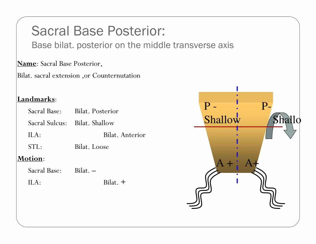
Sacral Base Posterior:Base bilat. posterior on the middle transverse axis
P - P-
Shallow Shallow
A + A+
Name: Sacral Base Posterior,
Bilat. sacral extension ,or Counternutation
Landmarks:
Sacral Base: Bilat. Posterior
Sacral Sulcus: Bilat. Shallow
ILA: Bilat. Anterior
STL: Bilat. Loose
Motion:
Sacral Base: Bilat. –
ILA: Bilat. +
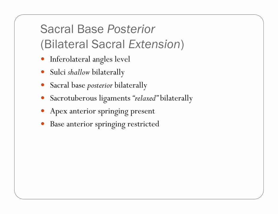
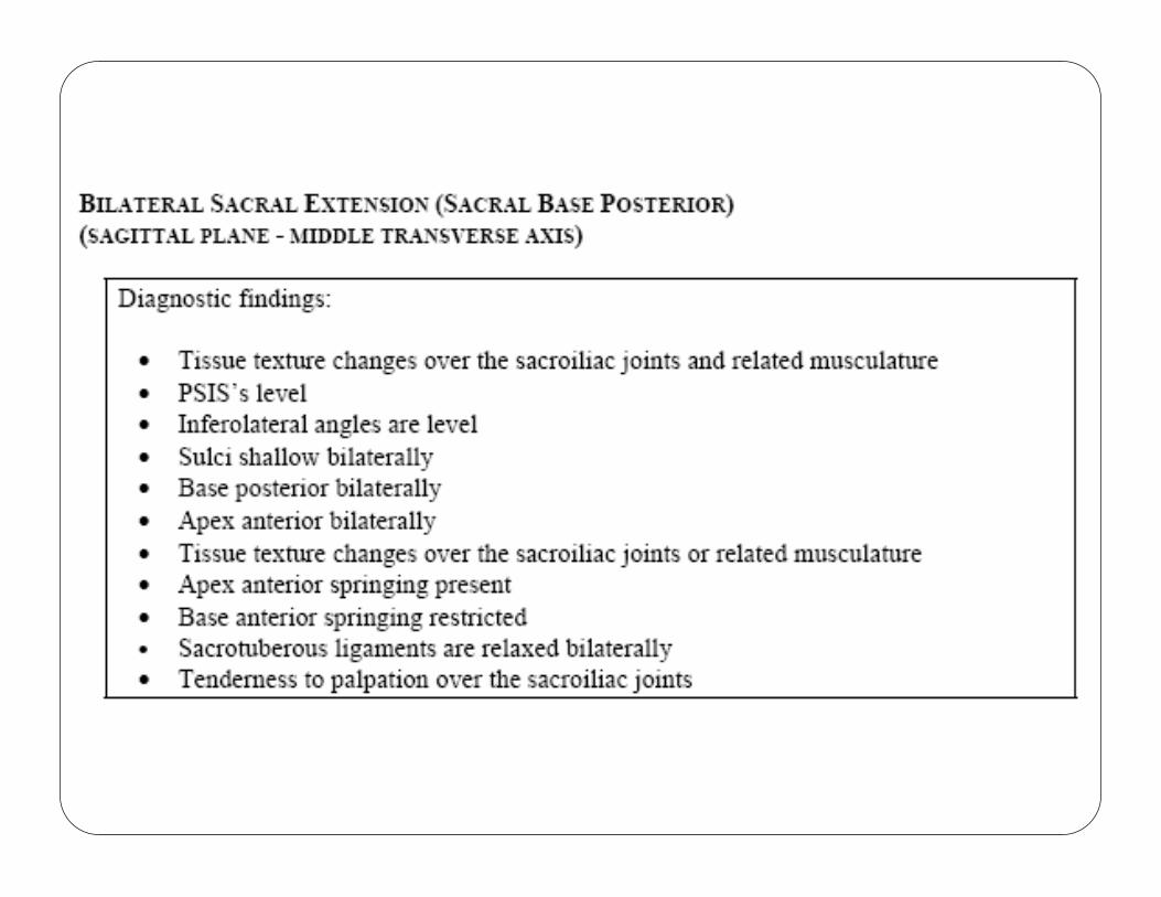
Sacral Base Posterior
(Bilateral Sacral Extension)� Inferolateral angles level
� Sulci shallow bilaterally
� Sacral base posterior bilaterally
� Sacrotuberous ligaments “relaxed” bilaterally
� Apex anterior springing present
� Base anterior springing restricted
SACRAL MECHANICSSACRAL MECHANICSSACRAL MECHANICSSACRAL MECHANICS
� Physiologic diagnoses of the sacrum occur inneutral and non-neutral mechanics:
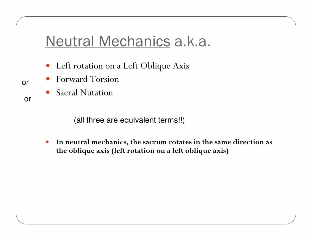
Neutral Mechanics a.k.a.
� Left rotation on a Left Oblique Axis� Forward Torsion� Sacral Nutation
� In neutral mechanics, the sacrum rotates in the same direction as the oblique axis (left rotation on a left oblique axis)
(all three are equivalent terms!!)
or
or
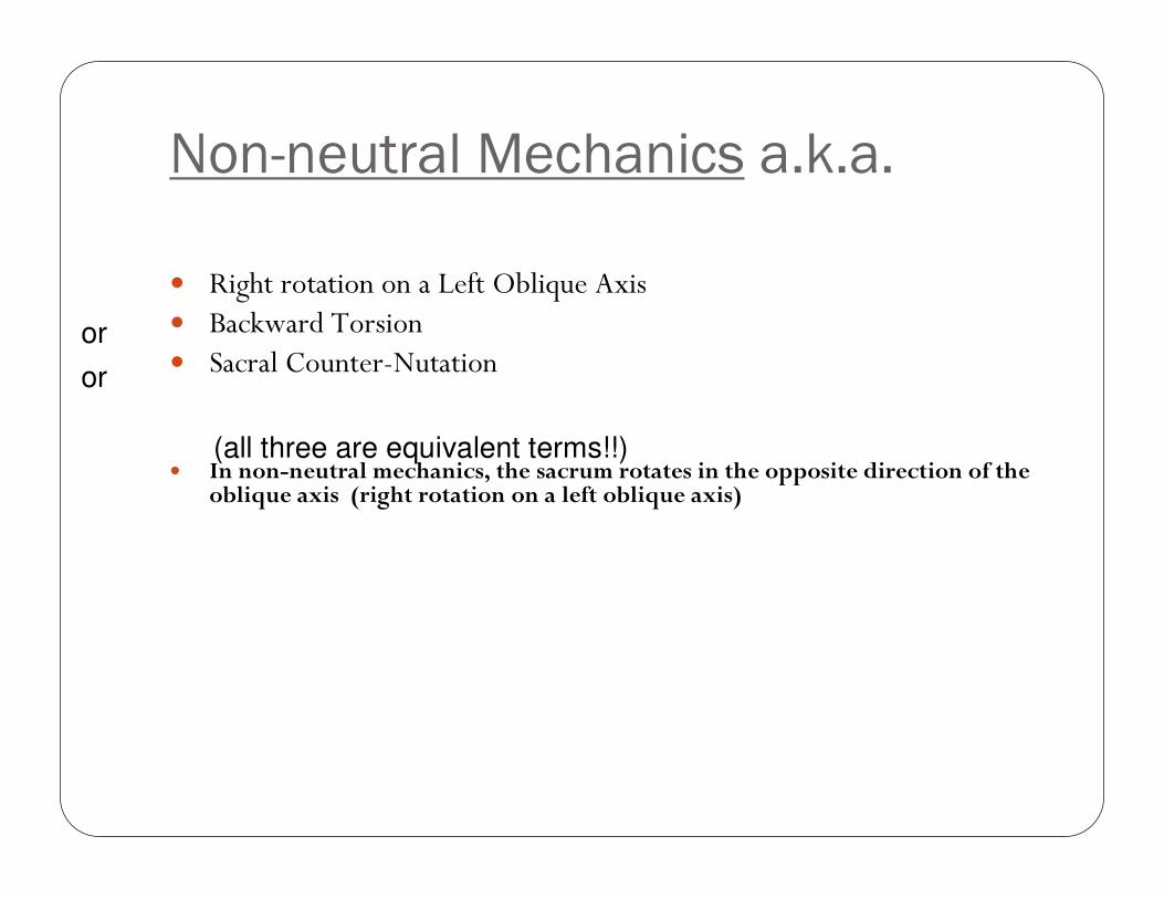
Non-neutral Mechanics a.k.a.
� Right rotation on a Left Oblique Axis� Backward Torsion� Sacral Counter-Nutation
� In non-neutral mechanics, the sacrum rotates in the opposite direction of the oblique axis (right rotation on a left oblique axis)
(all three are equivalent terms!!)
or
or
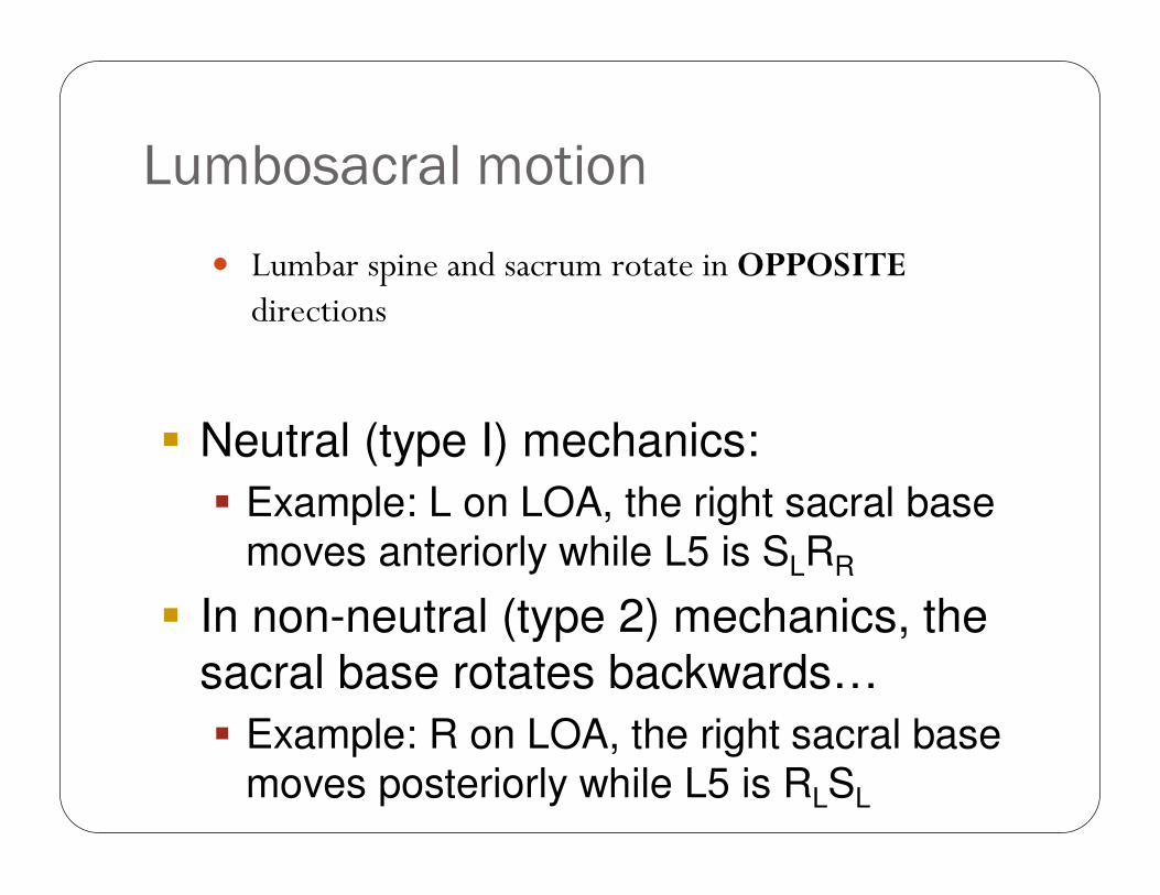
Lumbosacral motion
� Lumbar spine and sacrum rotate in OPPOSITEdirections
� Neutral (type I) mechanics:
� Example: L on LOA, the right sacral base moves anteriorly while L5 is SLRR
� In non-neutral (type 2) mechanics, the
sacral base rotates backwards…
� Example: R on LOA, the right sacral base moves posteriorly while L5 is RLSL
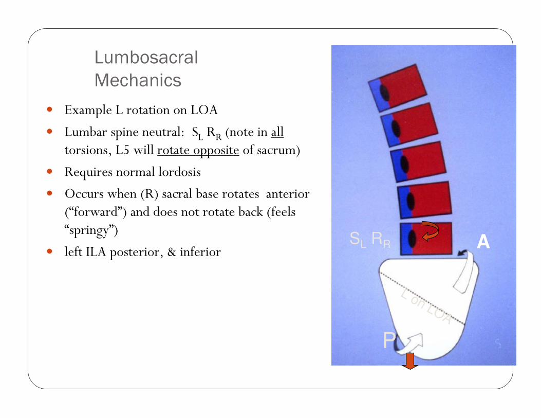
Lumbosacral
Mechanics
� Example L rotation on LOA
� Lumbar spine neutral: SL RR (note in alltorsions, L5 will rotate opposite of sacrum)
� Requires normal lordosis
� Occurs when (R) sacral base rotates anterior (“forward”) and does not rotate back (feels “springy”)
� left ILA posterior, & inferior A
P
SL RR
L on LOA
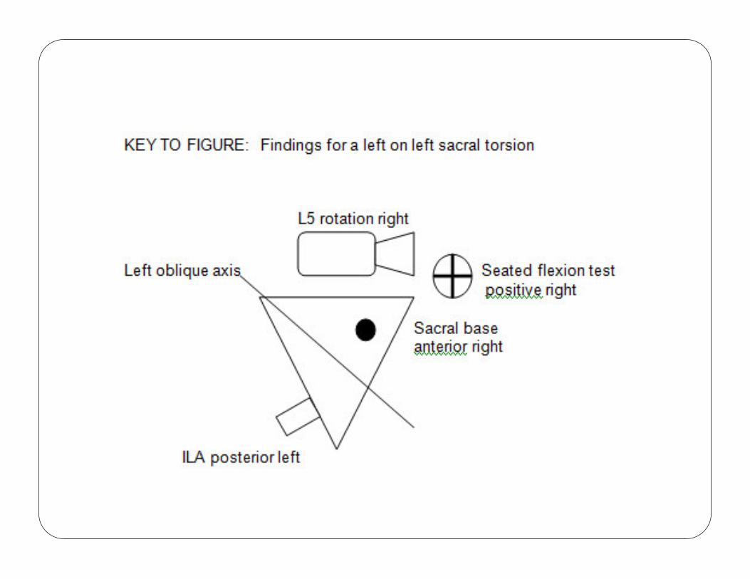
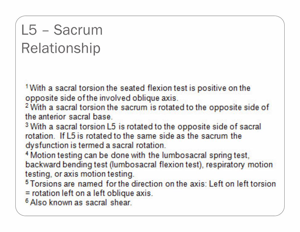
L5 – Sacrum
Relationship
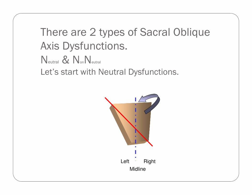
There are 2 types of Sacral Oblique
Axis Dysfunctions.
Neutral & NonNeutral
Let’s start with Neutral Dysfunctions.
Left Right
Midline
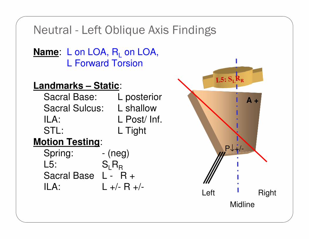
Neutral - Left Oblique Axis Findings
Name: L on LOA, RL on LOA,
L Forward Torsion
Landmarks – Static:
Sacral Base: L posterior
Sacral Sulcus: L shallow
ILA: L Post/ Inf.STL: L Tight
Motion Testing:
Spring: - (neg)
L5: SLRR
Sacral Base L - R +ILA: L +/- R +/-
Left Right
Midline
L5: SLRR
A +
P↓+/-
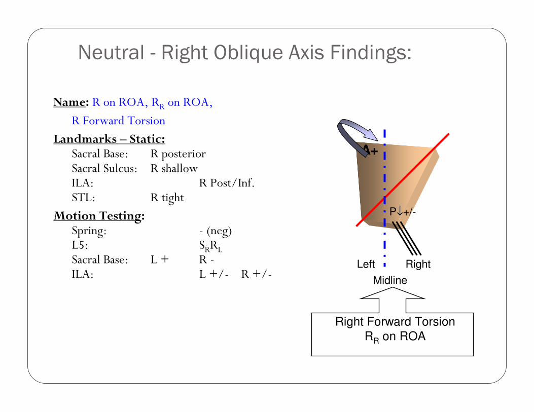
Neutral - Right Oblique Axis Findings:
Name: R on ROA, RR on ROA,
R Forward Torsion
Landmarks – Static: Sacral Base: R posteriorSacral Sulcus: R shallowILA: R Post/Inf.STL: R tight
Motion Testing:Spring: - (neg)L5: SRRLSacral Base: L + R -ILA: L +/- R +/-
Right Forward Torsion
RR on ROA
Left Right
Midline
P↓+/-
A+
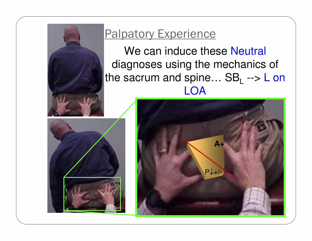
Palpatory Experience
We can induce these Neutraldiagnoses using the mechanics of
the sacrum and spine… SBL --> L on
LOA
A+
P↓+/-
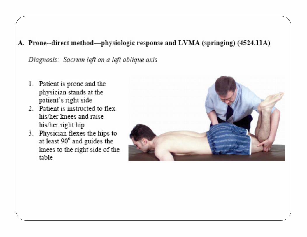

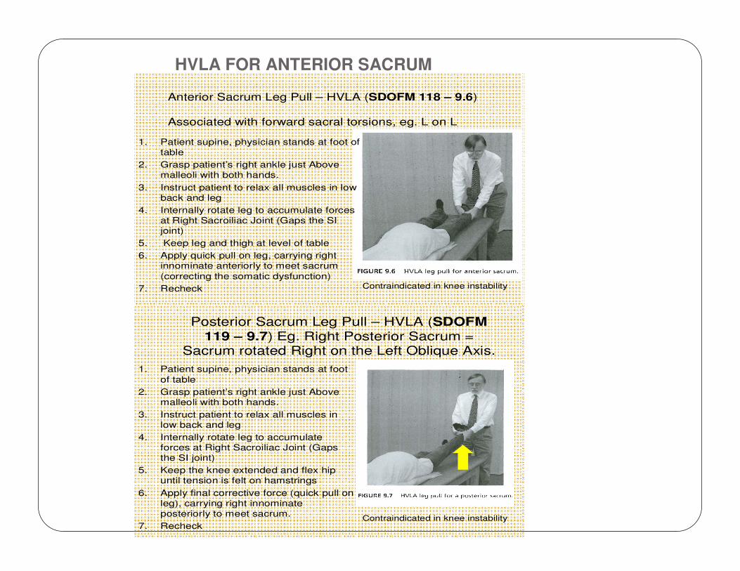
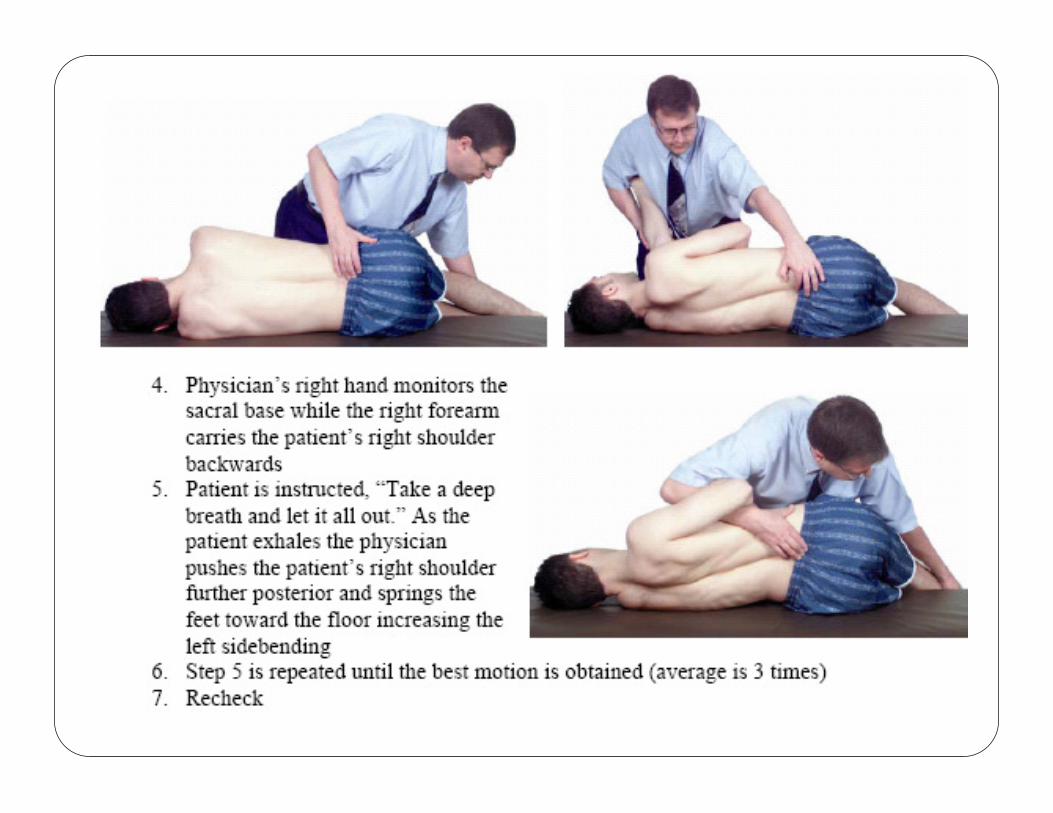
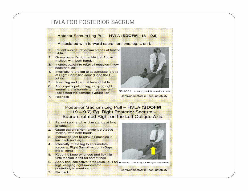
Anterior Sacrum Leg Pull – HVLA (SDOFM 118 – 9.6)
Associated with forward sacral torsions, eg. L on L
1. Patient supine, physician stands at foot of
table
2. Grasp patient’s right ankle just Above
malleoli with both hands.
3. Instruct patient to relax all muscles in low
back and leg
4. Internally rotate leg to accumulate forces
at Right Sacroiliac Joint (Gaps the SI
joint)
5. Keep leg and thigh at level of table
6. Apply quick pull on leg, carrying right
innominate anteriorly to meet sacrum
(correcting the somatic dysfunction)
7. Recheck Contraindicated in knee instability
Posterior Sacrum Leg Pull – HVLA (SDOFM
119 – 9.7) Eg. Right Posterior Sacrum = Sacrum rotated Right on the Left Oblique Axis.
1. Patient supine, physician stands at foot
of table
2. Grasp patient’s right ankle just Above
malleoli with both hands.
3. Instruct patient to relax all muscles in
low back and leg
4. Internally rotate leg to accumulate
forces at Right Sacroiliac Joint (Gaps
the SI joint)
5. Keep the knee extended and flex hip
until tension is felt on hamstrings
6. Apply final corrective force (quick pull on
leg), carrying right innominate
posteriorly to meet sacrum.
7. RecheckContraindicated in knee instability
HVLA FOR ANTERIOR SACRUM
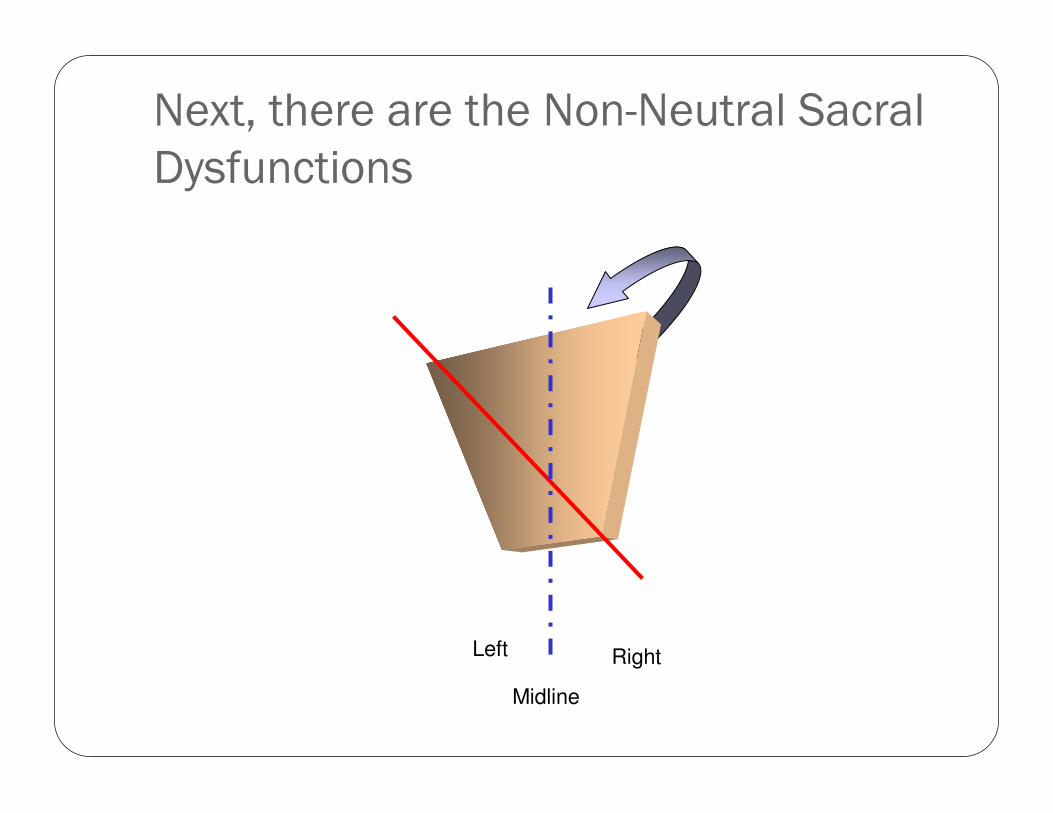
Next, there are the Non-Neutral Sacral
Dysfunctions
Left Right
Midline
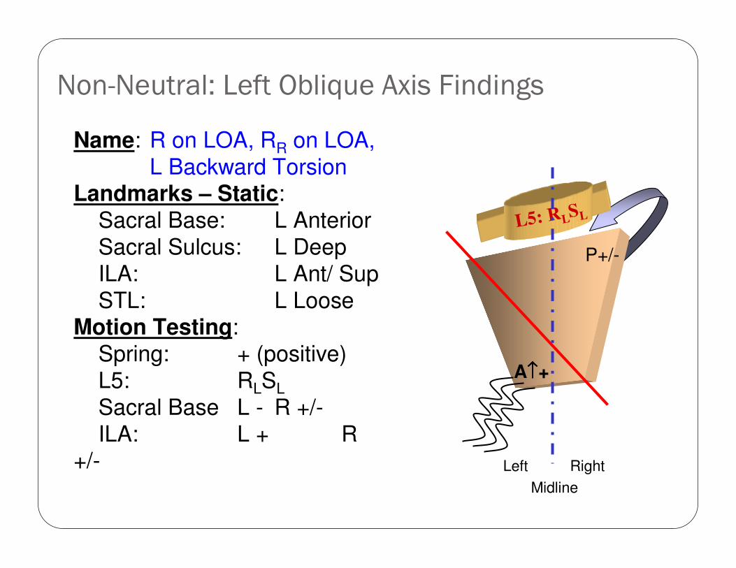
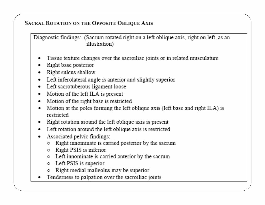
Non-Neutral: Left Oblique Axis Findings
Name: R on LOA, RR on LOA,
L Backward TorsionLandmarks – Static:
Sacral Base: L Anterior
Sacral Sulcus: L Deep
ILA: L Ant/ Sup
STL: L LooseMotion Testing:
Spring: + (positive)
L5: RLSL
Sacral Base L - R +/-
ILA: L + R +/-
L5: RLSL
Left Right
Midline
P+/-
A↑↑↑↑+
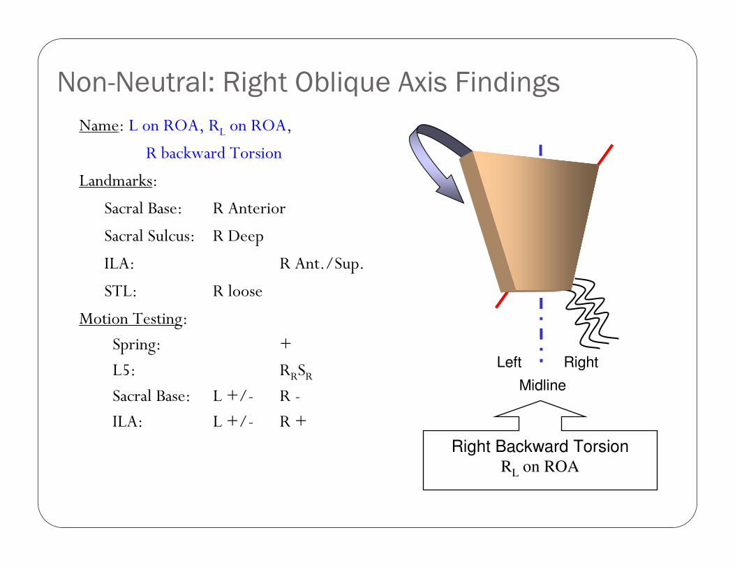
Non-Neutral: Right Oblique Axis Findings
Name: L on ROA, RL on ROA,
R backward Torsion
Landmarks:
Sacral Base: R Anterior
Sacral Sulcus: R Deep
ILA: R Ant./Sup.
STL: R loose
Motion Testing:
Spring: +
L5: RRSRSacral Base: L +/- R -
ILA: L +/- R +
A↑↑↑↑+
P+/-
Left Right
Midline
Right Backward TorsionRL on ROA
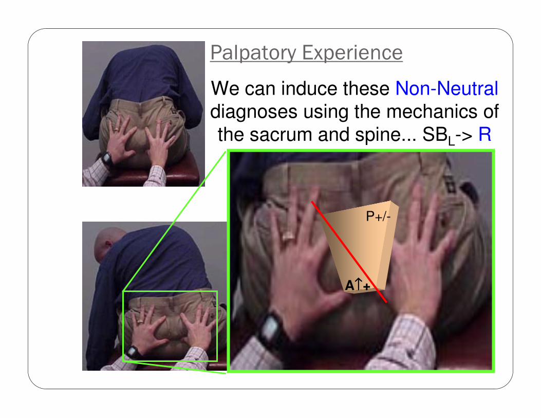
Palpatory Experience
We can induce these Non-Neutraldiagnoses using the mechanics of the sacrum and spine... SBL-> R
on LOA
P+/-
A↑↑↑↑+
HVLA FOR POSTERIOR SACRUM
Anterior Sacrum Leg Pull – HVLA (SDOFM 118 – 9.6)
Associated with forward sacral torsions, eg. L on L
1. Patient supine, physician stands at foot of
table
2. Grasp patient’s right ankle just Above
malleoli with both hands.
3. Instruct patient to relax all muscles in low
back and leg
4. Internally rotate leg to accumulate forces
at Right Sacroiliac Joint (Gaps the SI
joint)
5. Keep leg and thigh at level of table
6. Apply quick pull on leg, carrying right
innominate anteriorly to meet sacrum
(correcting the somatic dysfunction)
7. Recheck Contraindicated in knee instability
Posterior Sacrum Leg Pull – HVLA (SDOFM 119 – 9.7) Eg. Right Posterior Sacrum =
Sacrum rotated Right on the Left Oblique Axis.
1. Patient supine, physician stands at foot
of table
2. Grasp patient’s right ankle just Above malleoli with both hands.
3. Instruct patient to relax all muscles in
low back and leg
4. Internally rotate leg to accumulate
forces at Right Sacroiliac Joint (Gaps
the SI joint)
5. Keep the knee extended and flex hip
until tension is felt on hamstrings
6. Apply final corrective force (quick pull on leg), carrying right innominate
posteriorly to meet sacrum.
7. RecheckContraindicated in knee instability
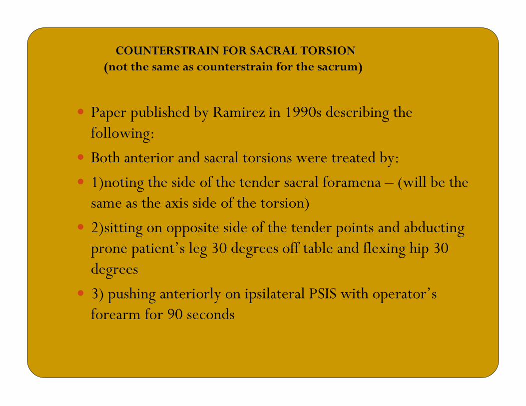
COUNTERSTRAIN FOR SACRAL TORSION (not the same as counterstrain for the sacrum)
� Paper published by Ramirez in 1990s describing the following:
� Both anterior and sacral torsions were treated by:
� 1)noting the side of the tender sacral foramena – (will be the same as the axis side of the torsion)
� 2)sitting on opposite side of the tender points and abducting prone patient’s leg 30 degrees off table and flexing hip 30 degrees
� 3) pushing anteriorly on ipsilateral PSIS with operator’s forearm for 90 seconds
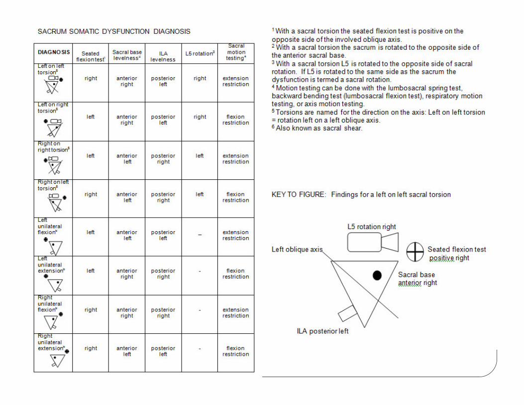
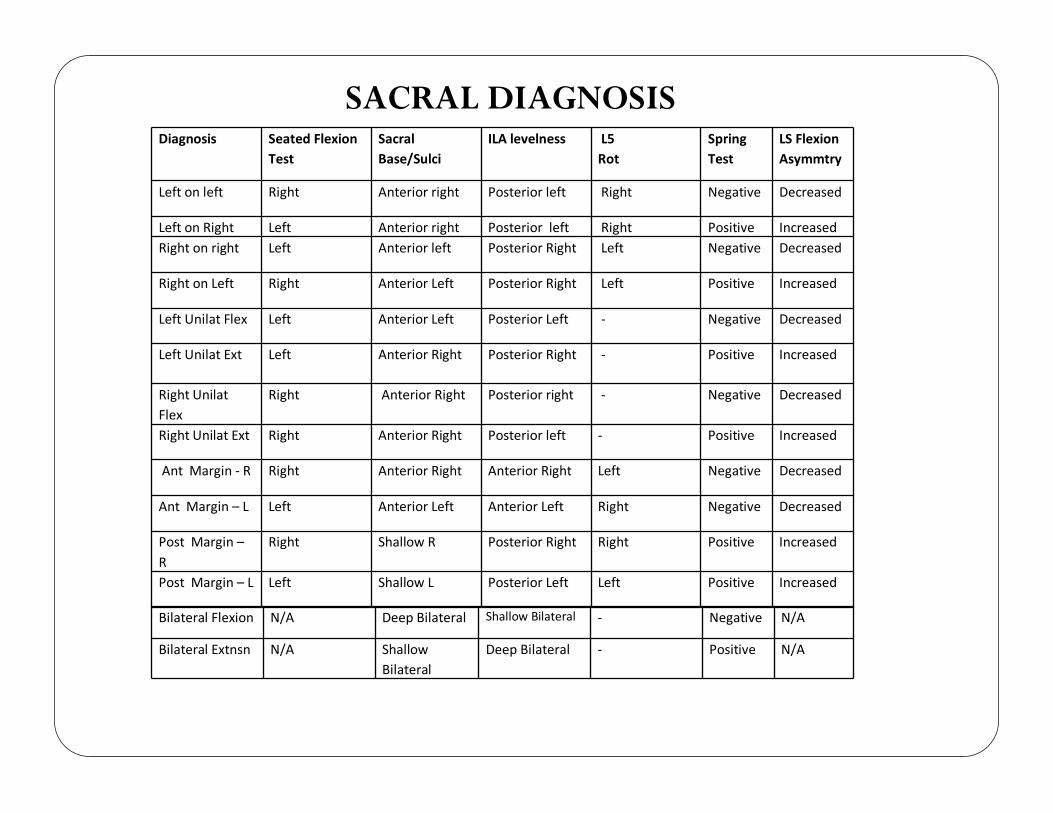
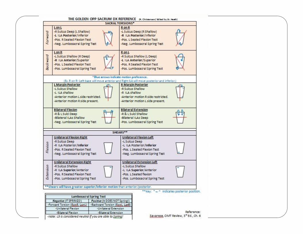
Diagnosis Seated Flexion
Test
Sacral
Base/Sulci
ILA levelness L5
Rot
Spring
Test
LS Flexion
Asymmtry
Left on left Right Anterior right Posterior left Right Negative Decreased
Left on Right Left Anterior right Posterior left Right Positive Increased
Right on right Left Anterior left Posterior Right Left Negative Decreased
Right on Left Right Anterior Left Posterior Right Left Positive Increased
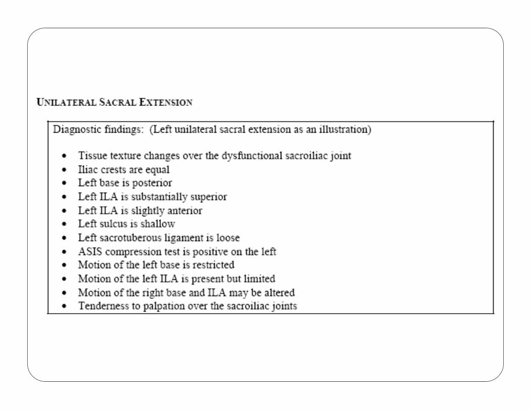
Left Unilat Flex Left Anterior Left Posterior Left - Negative Decreased
Left Unilat Ext Left Anterior Right Posterior Right - Positive Increased
Right Unilat
Flex
Right Anterior Right Posterior right - Negative Decreased
Right Unilat Ext Right Anterior Right Posterior left - Positive Increased
Ant Margin - R Right Anterior Right Anterior Right Left Negative Decreased
Ant Margin – L Left Anterior Left Anterior Left Right Negative Decreased
Post Margin –
R
Right Shallow R Posterior Right Right Positive Increased
Post Margin – L Left Shallow L Posterior Left Left Positive Increased
Bilateral Flexion N/A Deep Bilateral Shallow Bilateral - Negative N/A
Bilateral Extnsn N/A Shallow
Bilateral
Deep Bilateral - Positive N/A
SACRAL DIAGNOSIS
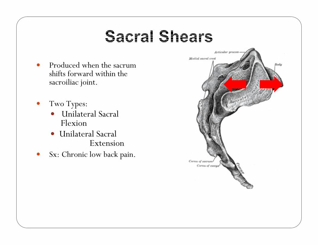
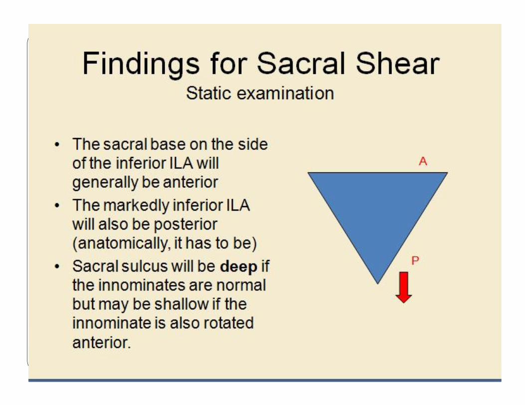
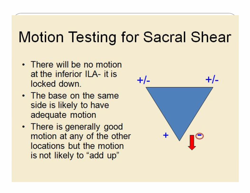
� Produced when the sacrum shifts forward within the sacroiliac joint.
� Two Types:� Unilateral Sacral Flexion
� Unilateral Sacral Extension
� Sx: Chronic low back pain.
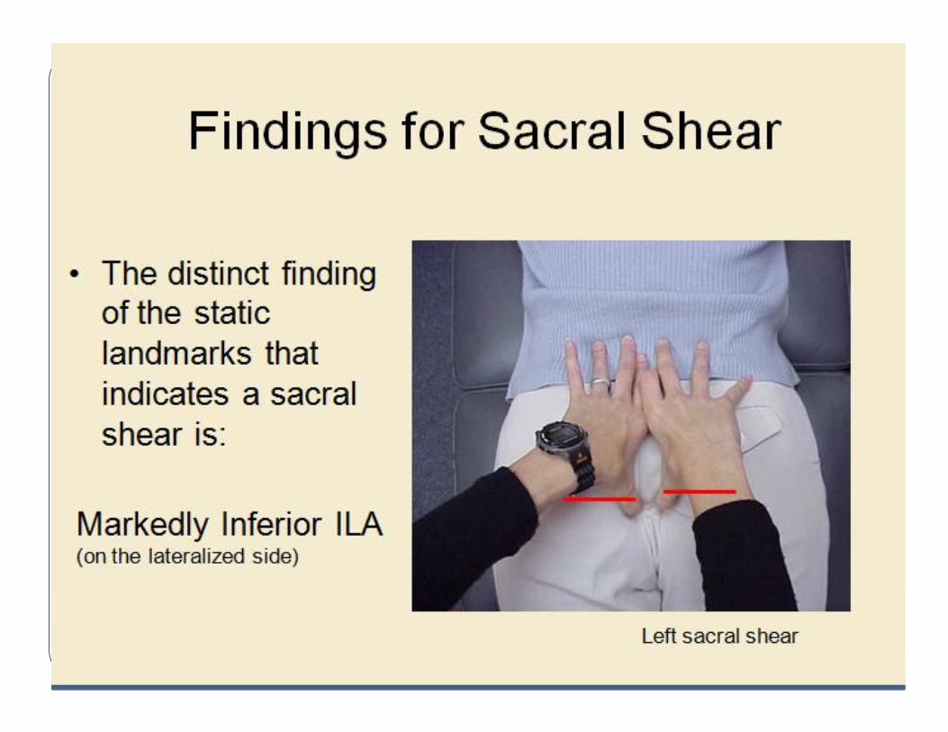
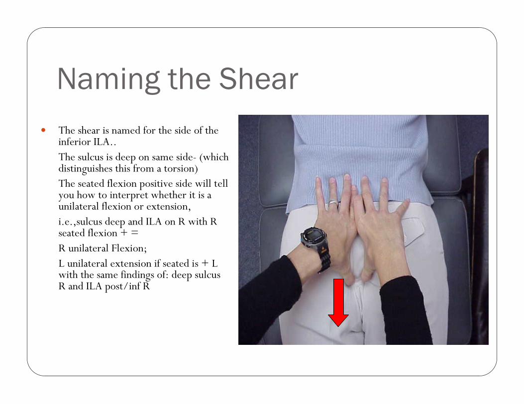
Naming the Shear
� The shear is named for the side of the inferior ILA..The sulcus is deep on same side- (which distinguishes this from a torsion)The seated flexion positive side will tell you how to interpret whether it is a unilateral flexion or extension,i.e.,sulcus deep and ILA on R with R seated flexion + = R unilateral Flexion; L unilateral extension if seated is + L with the same findings of: deep sulcusR and ILA post/inf R

THANK YOU


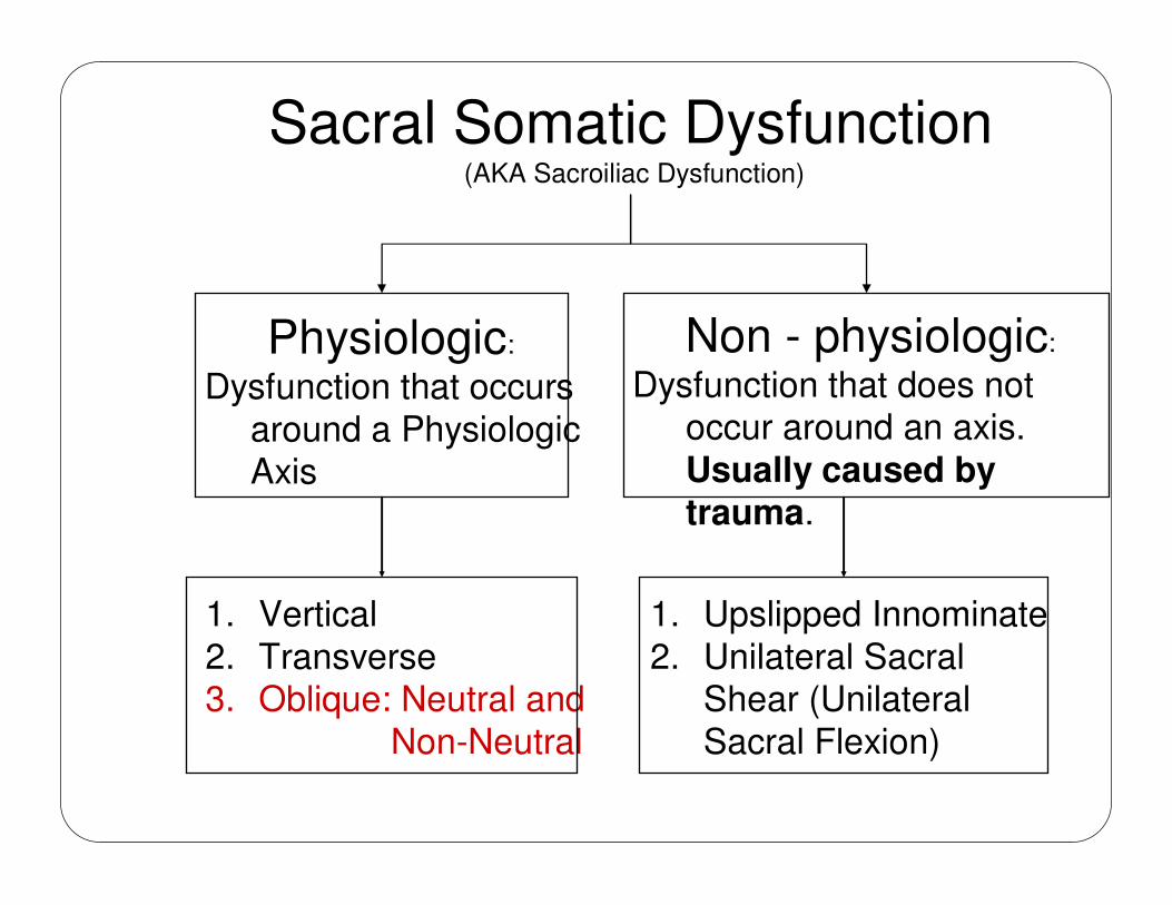
Sacral Somatic Dysfunction(AKA Sacroiliac Dysfunction)
Physiologic:
Dysfunction that occurs
around a Physiologic Axis
Non - physiologic:
Dysfunction that does not occur around an axis.
Usually caused by trauma.
1. Vertical
2. Transverse3. Oblique: Neutral and
Non-Neutral
1. Upslipped Innominate
2. Unilateral Sacral Shear (Unilateral
Sacral Flexion)
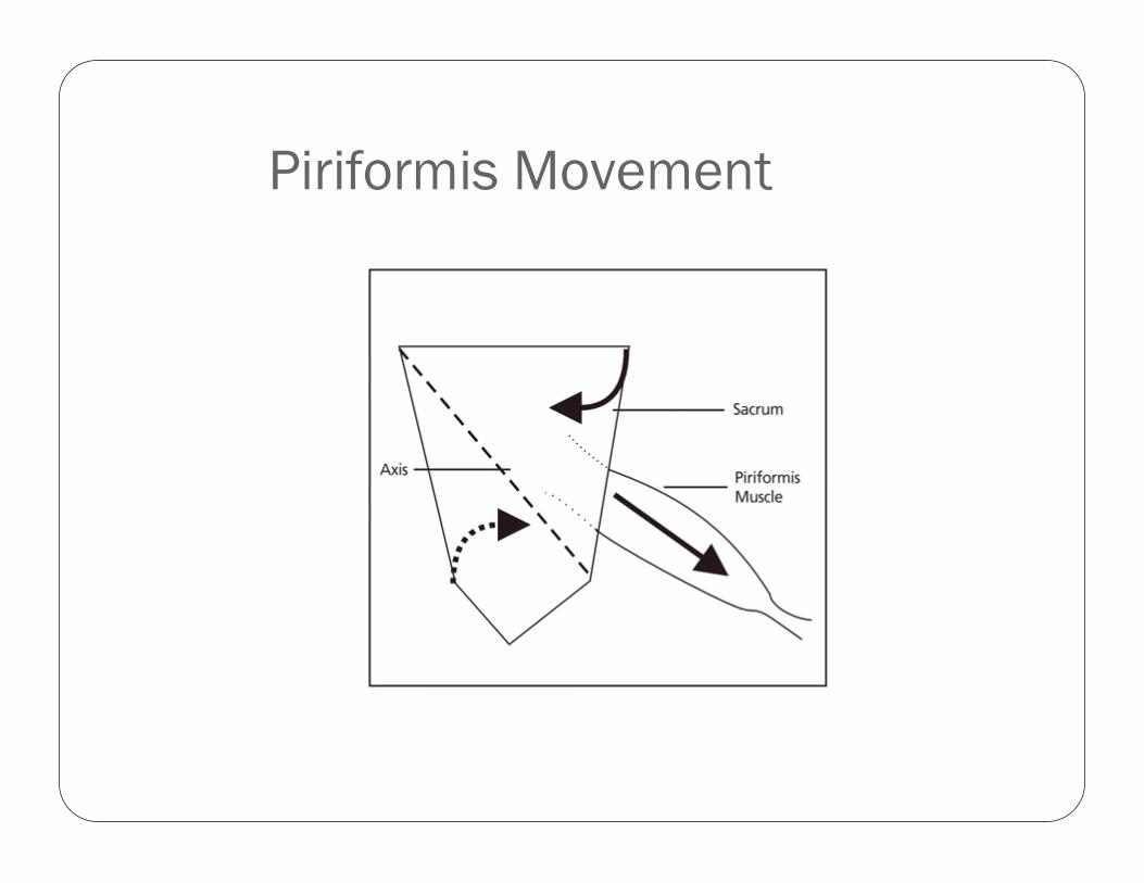
Piriformis Movement
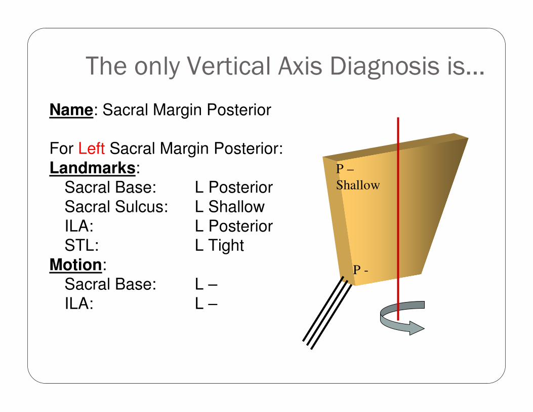
The only Vertical Axis Diagnosis is…
Name: Sacral Margin Posterior
For Left Sacral Margin Posterior:
Landmarks:
Sacral Base: L Posterior
Sacral Sulcus: L Shallow
ILA: L PosteriorSTL: L Tight
Motion:
Sacral Base: L –
ILA: L –
P –
Shallow
P -
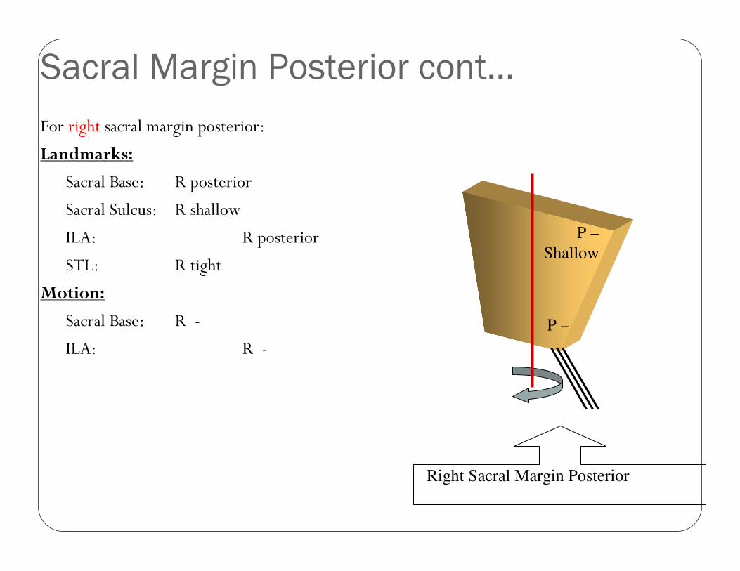
Sacral Margin Posterior cont...
For right sacral margin posterior:
Landmarks:
Sacral Base: R posterior
Sacral Sulcus: R shallow
ILA: R posterior
STL: R tight
Motion:
Sacral Base: R -
ILA: R -
Right Sacral Margin Posterior
P –
Shallow
P –
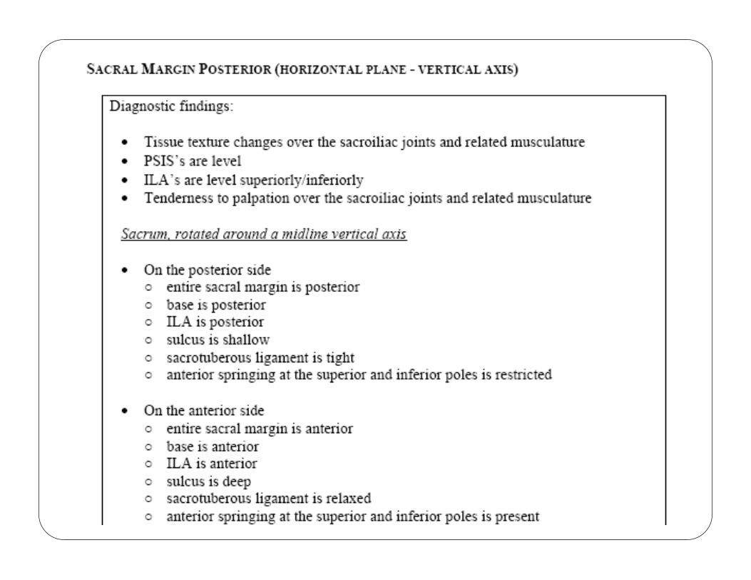
Sacral Margin Posterior:(ILA’s are level superiorly/inferiorly)
On the posterior side:
� Entire sacral margin is posterior� Base is posterior
� ILA is posterior
� Sulcus is shallow
� Sacrotuberous ligament is tight
� Anterior springing at the superior and inferior poles is restricted
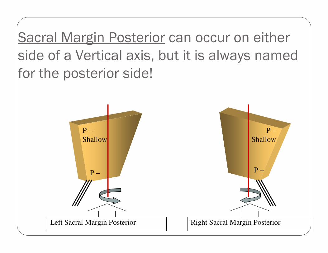
Sacral Margin Posterior can occur on either
side of a Vertical axis, but it is always named
for the posterior side!
Left Sacral Margin Posterior Right Sacral Margin Posterior
P –
Shallow
P –
P –
Shallow
P –


![Three-dimensional Nuclear Telomere Architecture Is ... · Telomere dysfunction is known to promote chromosomal instability (CIN) and carcinogenesis [16]. In most human somatic cells,](https://img.pdfslide.net/doc/110x75/5f2629fd310cc83259516f0d/three-dimensional-nuclear-telomere-architecture-is-telomere-dysfunction-is-known.jpg)