Embed Size (px)
Citation preview

Evaluation of Cerebrovascular Reserve in Patients Undergoing Carotid Artery
Stenting and its Usefulness in Predicting Significant Hemodynamic Changes
during Temporary Carotid Occlusion
Miloslav Spacek, MD, Cyril Stechovsky, MD, Martin Horvath, MD, Petr Hajek, MD,
PhD, Petra Zimolova, MD and Josef Veselka MD, PhD, FESC, FSCAI, FICA.
Department of Cardiology, Motol University Hospital, 2nd Medical School, Charles
University, Prague, Czech Republic
Short title: cerebrovascular reserve & carotid occlusion tolerance
Corresponding author:
Miloslav Spacek, MD. Department of Cardiology, Motol University Hospital, 2nd
Medical School, Charles University, V Úvalu 84, 150 06, Prague 5, Czech Republic.
Phone: +420224434901, Fax: +420224434920, E-mail: [email protected].

SUMMARY
We investigated the usefulness of cerebrovascular reserve (CVR) testing to predict
severe hemodynamic changes during proximally protected carotid artery stenting. Of
90 patients referred, 63 eligible underwent complete evaluation of the extent of
carotid artery disease and transcranial doppler ultrasound (TCD) assessment of CVR
by means of a breath-holding test and ophthalmic artery flow pattern evaluation.
Periprocedural TCD monitoring of the ipsilateral middle cerebral artery flow was
performed in 24 patients undergoing proximally protected procedure (requiring
induction of flow arrest within internal carotid artery). Abnormal CVR was significantly
less common in patients with unilateral compared to bilateral carotid artery disease
(26.3% vs. 76.9%, p=0.02), while ophthalmic artery flow reversal was rare in patients
with unilateral carotid artery disease (2.5% vs. 42.9%, p<0.01). During the induction
of carotid flow arrest, the average mean flow velocity drop following external carotid
artery occlusion was low (3.5%, p=0.67) compared to the induction of complete flow
arrest (32.8%, p<0.01). Six patients had a total mean flow velocity drop >50%,
including 2 patients with normal pre-procedural CVR. Our results suggest that TCD
evaluation of CVR is not a reliable predictor of hemodynamic changes induced during
proximally protected carotid artery stenting in patients with unilateral carotid artery
disease.
Keywords: cerebrovascular reserve, carotid artery occlusion, carotid artery stenting,
transcranial Doppler ultrasound, cerebral collateral circulation

INTRODUCTION
Carotid artery disease is an important risk factor for a stroke development and it has
been estimated that stenosis of the internal carotid artery is responsible for up to 15-
20% of the occurrence of all strokes or transient ischemic attacks (Sacco et al. 1995).
The prevalence of carotid artery disease increases with age and its wide variability of
clinical manifestation ranges from incidental asymptomatic findings to devastating or
fatal stroke, with cerebral collateral circulation being considered one of the major
modifying factors (Liebeskind 2003). In order to decrease the stroke burden, carotid
endarterectomy has been used extensively to eliminate both hemodynamically
significant stenosis as well as carotid artery plaques as a source of cerebral
atheroemboli. Over time, carotid artery stenting has evolved into a reliable method so
that it may now be offered as a first-line treatment to selected patients (Spacek and
Veselka 2013). Nevertheless, because of the improvement in medical therapy, there
is a concern regarding the indication for invasive treatment in asymptomatic patients
with severe unilateral carotid stenosis. Therefore, the safety of the procedure is of a
paramount importance in order to evaluate the true benefit. With an increasing use of
proximal protection systems resembling a surgical clamp (inducing flow stagnation
within internal carotid artery by sequential balloon occlusion of external and common
carotid artery), it is necessary to understand collateral cerebral circulation in order to
protect patients from peri-procedural hypoperfusion, which has been proven to
increase the risk of peri-procedural cerebral events (Ackerstaff et al. 2005, Caplan
and Hennerici 1998).
Transcranial Doppler ultrasound (TCD) is a useful tool allowing testing of
cerebrovascular reserve (CVR) and monitoring in real time of a patient’s
hemodynamic status during procedure (Spacek et al. in press). Its usefulness in

predicting severe hemodynamic changes during the period of carotid flow arrest has,
however, been poorly defined. In our study, we prospectively evaluated CVR in
asymptomatic patients referred for carotid artery stenting and focused on the ability of
a CVR examination to predict severe hemodynamic changes during proximally
protected procedures in patients with unilateral carotid artery disease.
PATIENTS AND METHODS
During the study period between October 2012 and December 2014, we evaluated
90 asymptomatic patients considered high-risk for carotid endarterectomy (either
multiple comorbidities and/or “hostile neck” with difficult approach to bifurcation
including restenosis after endarterectomy), who had been referred to our institution
for endovascular treatment with a presumed internal carotid artery stenosis of > 70%
based on an outpatient non-invasive examination. Prior to carotid angiography, the
majority of patients underwent a complete ultrasound reevaluation of the cervical
arteries, performed by an experienced sonographer, using color Doppler flow
imaging and pulse-Doppler examination (Toshiba Nemio XG, Toshiba Corporation,
Tokyo, Japan). Apart from that, the extent of carotid and vertebral artery disease was
reassessed during conventional cerebral angiography with severe carotid artery
disease defined as >70% internal carotid artery stenosis according to the NASCET
criteria (North American Symptomatic Carotid Endarterectomy Trial Collaborators
1991). Patients with significant external carotid artery disease (>70% stenosis) as
well as with severe steno-occlusive disease of the vertebral arteries (abnormal flow
pattern or retrograde flow) were excluded from the study (6 and 3 patients,
respectively, predominantly with extensive carotid artery disease). Prior to TCD
evaluation, all patients gave written informed consent for participation in the study,

the study was performed in accordance with the Declaration of Helsinki of the World
Medical Association, and the study protocol was approved by the local ethics
committee.
Cerebrovascular reserve was assessed using Multi-Dop T digital transcranial Doppler
ultrasound system (DWL Elektronische Systeme, Sipplingen, Germany) by means of
a breath-holding test (Markus and Harrison 1992) with evaluation of the ophthalmic
artery flow pattern by a sonographer unaware either of the target internal carotid
artery stenosis severity, or the complexity of carotid artery disease. During breath-
holding test, two 2.5-MHz transducers fitted on a headband and placed on the
temporal bone windows, were used to obtain a continuous measurement of the mean
flow velocities (MFV) in the middle cerebral arteries. The depth of insonation ranged
45-55mm. Patients with an insufficient insonation window ipsilateral to the target
artery were excluded from the study (18 patients)
The breath-holding index (BHI) was calculated by dividing the percent increase of
MFV occurring during breath holding by the time in seconds that subjects held their
breath. A breath-holding period of at least 24 seconds (ranging 25-30 seconds) was
considered sufficient based on previous reports (Alexandrov 2005) in association
with careful attention to normal inspiration in order to avoid a Valsalva maneuver (Fig.
1). The study was performed after at least 5 minutes of initial rest period in a quiet
room, with patients lying in a comfortable supine position without any visual or
auditory stimulation. Maximal MFV was recorded 4 seconds after termination of the
apnea interval. Patients were trained to perform the procedure correctly before
proceeding to a definitive recording, which was performed 3 times with at least a 2-
minute interval between each test, to allow the cerebrovascular circulation to
normalize. The average of the 3 measurements was calculated as the final value.

Following breath-holding test, the direction of flow in both ophthalmic arteries at an
insonation depth of 50 ± 5mm was obtained, with flow direction away from the probe
(flow reversal) considered abnormal and suggestive of depleted CVR. With regard to
the previously published values defining normal BHI range (Zavoreo and Demarin
2004, Jimenez-Caballero and Segura 2006, Bago-Rozankovic et al. 2009, Barret et
al. 2001) and in accordance with our observation derived from the control group, we
arbitrarily defined the cut-off values for impaired BHI as <0.95 and as completely
depleted in the case of BHI <0.69 as reported by Vernieri et al. (1999).
Carotid artery stenting was performed according to standardized protocols by an
experienced operator, who had performed more than 700 CAS procedures before the
beginning of the study. The operator was unaware of the results of CVR testing and
the choice of an optimal device was based on personal experience. Use of protection
systems was mandatory in all patients, while filter protection systems were used in
cases involving more difficult carotid artery access. In all study patients, continuous
monitoring of the middle cerebral artery MFV ipsilateral to the target internal carotid
artery was performed during procedure. For a proximally protected procedure, a
MO.MA Cerebral Protection Device (Medtronic, Minneapolis, MN) was used. This
system induces flow arrest within internal carotid artery by sequential balloon
occlusion of the ipsilateral external and common carotid artery, thereby resembling a
surgical (external) clamp. The completeness of flow arrest was confirmed by contrast
agent stagnation within the internal carotid artery together with the absence of
contrast agent induced high-intensity transient signals detected by TCD in the
ipsilateral middle cerebral artery. As a cut-off for significant hypoperfusion during the
period of flow arrest, we used a previously recommended >50% MFV drop from
baseline values, a threshold considered to increase the risk of neurologic
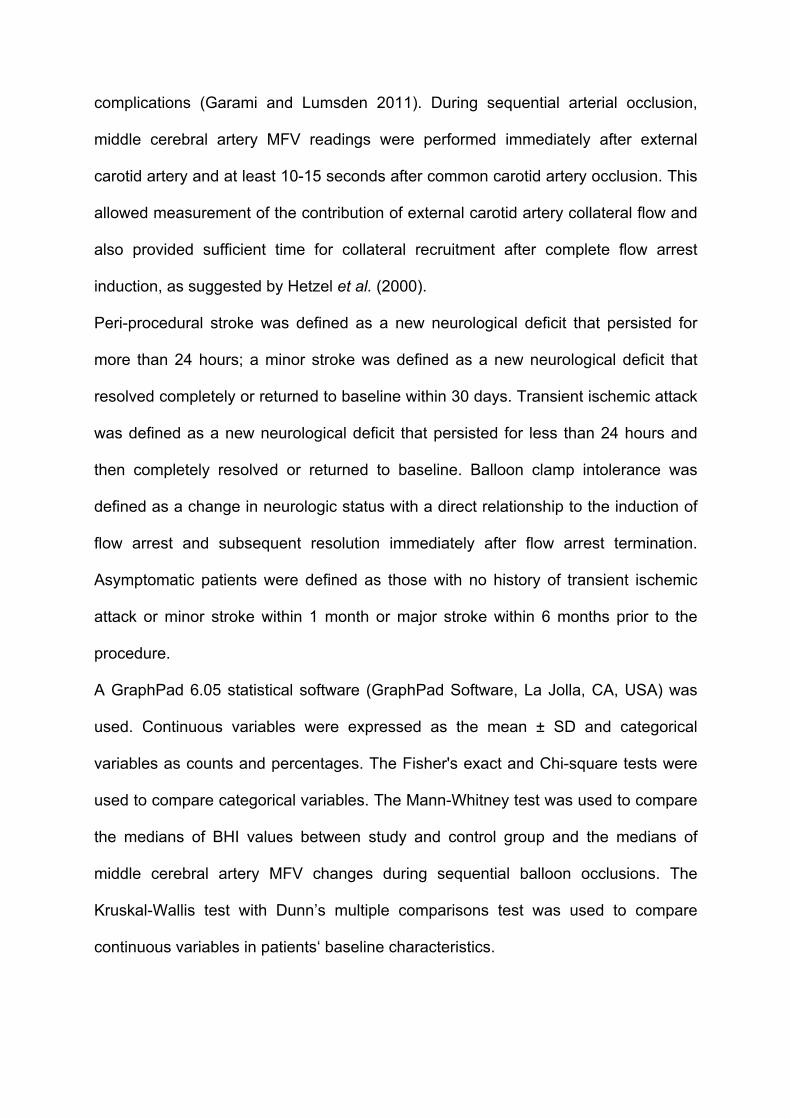
complications (Garami and Lumsden 2011). During sequential arterial occlusion,
middle cerebral artery MFV readings were performed immediately after external
carotid artery and at least 10-15 seconds after common carotid artery occlusion. This
allowed measurement of the contribution of external carotid artery collateral flow and
also provided sufficient time for collateral recruitment after complete flow arrest
induction, as suggested by Hetzel et al. (2000).
Peri-procedural stroke was defined as a new neurological deficit that persisted for
more than 24 hours; a minor stroke was defined as a new neurological deficit that
resolved completely or returned to baseline within 30 days. Transient ischemic attack
was defined as a new neurological deficit that persisted for less than 24 hours and
then completely resolved or returned to baseline. Balloon clamp intolerance was
defined as a change in neurologic status with a direct relationship to the induction of
flow arrest and subsequent resolution immediately after flow arrest termination.
Asymptomatic patients were defined as those with no history of transient ischemic
attack or minor stroke within 1 month or major stroke within 6 months prior to the
procedure.
A GraphPad 6.05 statistical software (GraphPad Software, La Jolla, CA, USA) was
used. Continuous variables were expressed as the mean ± SD and categorical
variables as counts and percentages. The Fisher's exact and Chi-square tests were
used to compare categorical variables. The Mann-Whitney test was used to compare
the medians of BHI values between study and control group and the medians of
middle cerebral artery MFV changes during sequential balloon occlusions. The
Kruskal-Wallis test with Dunn’s multiple comparisons test was used to compare
continuous variables in patients‘ baseline characteristics.

RESULTS
Patients’ baseline characteristics are summarized in Table 1. The study group
consisted of 39 patients (67 ± 7yrs) with unilateral carotid stenosis of an average
severity of 84 ± 9%. During carotid artery stenting, technical success was attained in
all but 1 patient and this was because of severe vascular tortuosity. That patient
subsequently opted for surgical treatment. Two minor strokes occurred: 1 patient
developed a prolonged vagal reaction during postdilatation requiring repeated doses
of atropine and high volumes of fluids, and the other patient problem occurred due to
silent hypoperfusion during the period of flow arrest. In this patient, clinical
intolerance developed during the final period of blood aspiration, requiring pre-term
flow arrest termination, which was followed by several showers of microembolic
signals that were detected by TCD. Both patients had near-complete neurological
resolution by the time of hospital discharge and were symptom free at the 30-day
follow-up visit.
Ophthalmic artery flow reversal ipsilateral to target internal carotid artery stenosis
was detected in only 1 patient and, as expected, was normal on the non-stenotic side
in all patients. The average BHI values obtained ipsilateral and contralateral to the
target internal carotid artery were 1.262 ± 0.427 and 1.432 ± 0.397 (p=0.10),
respectively. When compared to the subjects with excluded severe carotid artery
disease (72 ± 4 yrs, average BHI 1.608 ± 0.308, normal bilateral ophthalmic artery
flow in all patients), the average BHI values were significantly lower ipsilateral to the
target artery (p<0.01). Fig. 2 summarizes BHI values obtained from patients with
unilateral and from subjects with excluded carotid artery disease.
Using the combined approach of abnormal BHI values and/or reversed ophthalmic
artery flow, we were able to identify 10 patients (26.3%) as having abnormal CVR, of
which 6 (15.8%) were categorized as having impaired and 4 (10.5%) as having
depleted CVR (3 based on BHI values and 1 with ophthalmic artery flow reversal).
None of the patients with normal BHI values had impaired BHI on the non-stenotic
side, while 2 of the patients with depleted CVR had impaired contralateral BHI
values.
Fourteen patients (68 ± 5 yrs) had severe bilateral carotid artery disease. Tables 2 &
3 (patients with severe stenosis or occlusion contralateral to the target internal carotid
artery, respectively) summarize their ophthalmic artery flow patterns and BHI values.
Compared to the study group, ophthalmic artery flow reversal was identified in a
significantly larger proportion of patients (42.9% vs. 2.5%, p<0.01). When combined
with low BHI values, abnormal CVR in at least one territory was detected in a
significantly larger percentage as well (76.9% vs. 26.3%, p=0.02).
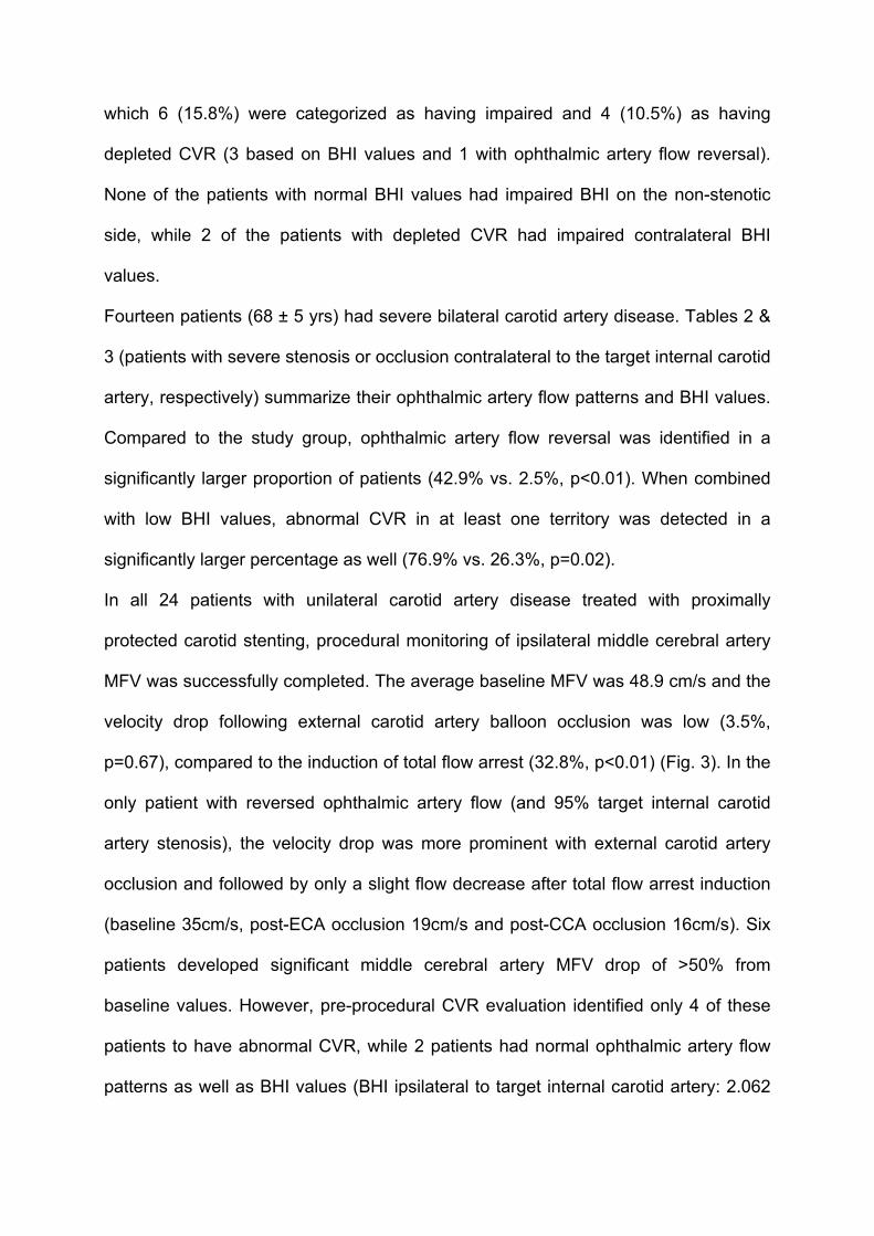
In all 24 patients with unilateral carotid artery disease treated with proximally
protected carotid stenting, procedural monitoring of ipsilateral middle cerebral artery
MFV was successfully completed. The average baseline MFV was 48.9 cm/s and the
velocity drop following external carotid artery balloon occlusion was low (3.5%,
p=0.67), compared to the induction of total flow arrest (32.8%, p<0.01) (Fig. 3). In the
only patient with reversed ophthalmic artery flow (and 95% target internal carotid
artery stenosis), the velocity drop was more prominent with external carotid artery
occlusion and followed by only a slight flow decrease after total flow arrest induction
(baseline 35cm/s, post-ECA occlusion 19cm/s and post-CCA occlusion 16cm/s). Six
patients developed significant middle cerebral artery MFV drop of >50% from
baseline values. However, pre-procedural CVR evaluation identified only 4 of these
patients to have abnormal CVR, while 2 patients had normal ophthalmic artery flow
patterns as well as BHI values (BHI ipsilateral to target internal carotid artery: 2.062

and 1.489, internal carotid artery stenosis 80% and 70%, respectively). In the patient
with a pre-procedural BHI value of 1.489, clinical intolerance developed immediately
after sequential balloon occlusion with near-complete MCA flow arrest (Fig. 4),
suggesting insufficient collateral support. Immediate flow arrest termination led to a
prompt and complete resolution of neurological symptoms and the procedure was
successfully completed using filter-type protection.
DISCUSSION
In our study, we present our single-center experience with consecutive patients
referred for carotid artery stenting in whom CVR was prospectively evaluated by
means of TCD. Moreover, we used the induction of flow arrest within internal carotid
artery during proximally protected procedures as a test to assess the ability of CVR to
predict severe hemodynamic changes during total carotid artery occlusion. Based on
our results, we suggest that pre-procedural TCD testing is not a reliable predictor of
severe hemodynamic changes in unselected patients with unilateral carotid stenosis
undergoing proximally protected carotid artery stenting.
Proximal protection systems currently represent one of the most promising
improvements of the carotid stenting procedure. These systems essentially
reproduce the hemodynamic condition brought about by surgical (external) clamping,
but do not have the ability to shunt the lesion. Despite being effective in reducing the
amount of emboli particles released during the period of flow arrest (Montorsi et al.
2011), the trade-off is that the ipsilateral hemisphere is totally dependent on the
collateral cerebral circulation and may suffer from “silent hypoperfusion” as well as
may be more susceptible to residual embolic load commonly observed during balloon
deflations (Spacek and Veselka 2014). This may partly explain why inconsistent

results have been reported regarding the superior safety of these systems (Bijuklic et
al. 2012, de Castro-Afonso et al. 2013). The ability to assess in advance the risk of
hypoperfusion induced during the period of flow arrest might help in identifying those
for whom such an approach would be inappropriate, thereby potentially increasing
the safety of the procedure.
Cerebral collateral circulation plays an important role in stroke development and is
responsible for hemodynamic compensation in patients undergoing temporary carotid
occlusion. While the circle of Willis (particularly the anterior communicating artery) is
considered the most important (primary) collateral source immediately able to
support/restore middle cerebral artery flow, the ophthalmic artery is considered to be
a secondary collateral pathway requiring time to come into play (Spacek et al. in
press, Hofmeijer et al. 2002). It has been reported that large inter-individual variability
can be found regarding the completeness of the circle of Willis with up to 10% of
individuals having a hypoplastic anterior communicating artery and with almost every
fourth person having hypoplasty of the posterior collateral circulation (Iqbal 2013).
Several authors have tried to estimate the role of pre-procedural TCD to detect
severe hemodynamic changes during temporary carotid occlusion. Interestingly, it
has been suggested that a pre-procedural carotid compression test might be useful
in determining the patients who would tolerate carotid occlusion well (Anzola et al.
2008). This test, however, carries a risk of stroke due to mobilization of plaque
content. Conversely, little is known about the use of pre-procedural CVR evaluation
and most of the data come from studies with patients undergoing surgical carotid
endarterectomy, resulting in ambiguous conclusions (Telman et al. 2007, Visser et al.
2000). The drawback of these studies were unselected inclusion of patients with both
unilateral and bilateral carotid artery disease, lack of evaluation of ophthalmic artery

flow, and the use of a shunt requirement as an endpoint, all of which make
interpretation difficult and the results not generalizable to patients undergoing a
proximally protected carotid artery stenting.
In our study, we first defined the CVR patterns in unselected consecutive patients
referred for carotid artery stenting with emphasis on ophthalmic artery flow
assessment. Interestingly, Fearn et al. (2000) evaluated the contribution of external
carotid artery flow to the cerebral circulation in unselected patients (including both
those with unilateral and bilateral carotid artery disease) undergoing surgical carotid
endarterectomy and concluded that external carotid artery may significantly
contribute to the ipsilateral middle cerebral artery flow. However, Rutgers et al.
(2000) observed in their large series that ophthalmic artery flow reversal was never
present in patients with internal carotid artery stenosis of 80% or less. We observed
that ophthalmic artery flow reversal was dependent on the severity of carotid artery
disease and prevailed in the group of patients with the most extensive bilateral
disease, but was rare in patients with unilateral stenosis. Therefore, the finding of
abnormal CVR in the study group more commonly represented by impaired BHI
values underscores the importance of the ophthalmic artery as the last-option
collateral that becomes activated when primary collaterals fail. In accordance with
this finding, a relatively minor contribution of external carotid artery to middle cerebral
artery flow was observed during selective external carotid artery occlusion as
compared to total flow arrest.
It is well known that carotid artery stenting produces larger amounts of cerebral
embolism compared to carotid endarterectomy, in which a hemodynamic stroke may
contribute to the risk of a peri-procedural neurologic complication (Jordan et al.
1999). While shunt insertion may improve the hemodynamic situation in patients

undergoing surgical procedure, no such intervention is possible during proximally
protected carotid artery stenting. Interestingly, Stabile et al. (2010) in their large
experience with 1300 patients (87.5% with unilateral carotid artery disease) who were
managed with a proximally protected procedure, reported that, despite clinical
tolerance by most of the patients of the flow arrest, up to every fifth patient developed
signs of clinical intolerance during the period of active blood aspiration, suggesting
that a large proportion of patients had borderline or compromised cerebral blood flow
during the period of flow arrest. Indeed, we observed in our study that 6 of the 24
patients treated with a proximally protected procedure had a middle cerebral artery
MFV drop of more than 50% form baseline values, a cut-off considered to increase
the risk of procedural complications. In that case, we apply a slow TCD-guided blood
aspiration technique to prevent the development of clinical intolerance requiring pre-
term balloon deflations. However, we observed that such a situation might develop
despite normal pre-procedural CVR values, suggesting that residual flow through the
target internal carotid artery may be sufficient to mask compromised cerebral
collateralization, thereby making pre-procedural CVR evaluation unreliable in the
prediction of hemodynamic changes during balloon clamping.
LIMITATIONS
The limitation of our study is a relatively low patient population. A significantly larger
number of patients will be needed to confirm whether the results are generalizable to
patients with both high-grade (90-99%) and low-grade (70-89%) internal carotid
artery stenosis. However, with the decreasing numbers of asymptomatic patients
referred for invasive treatment, it is currently unlikely that such a study will be
performed without a multi-center design.

CONCLUSION
The majority of asymptomatic patients with severe unilateral carotid artery stenosis
show good tolerance of proximally protected carotid artery stenting with temporary
carotid occlusion. Despite that, cerebral blood flow is significantly decreased in a
subgroup of patients, exposing them to the potential risk of silent hypoperfusion, with
a resultant increase in susceptibility to neurologic complications. Transcranial
Doppler ultrasound, despite its usefulness in procedural monitoring, is not a reliable
tool for prediction of severe hemodynamic changes in patients with unilateral carotid
stenosis who are undergoing proximally protected carotid artery stenting.
ACKNOWLEDGEMENTS
The research was supported by project of the Ministry of Health of the Czech
Republic: conceptual development of research organization 00064203.

REFERENCES
ACKERSTAFF RG, SUTTORP MJ, van den BERG JC, OVERTOOM TT, VOS JA,
BAL ET, ZANEN P: Prediction of early cerebral outcome by transcranial Doppler
monitoring in carotid bifurcation angioplasty and stenting. J Vasc Surg 41: 618-624,
2005.
ALEXANDROV AV: The role of ultrasound in the management of cerebrovascular
disease. In: Introduction to vascular ultrasonography, Fifth edition. WJ ZWIEBEL, JS
PELLERITO (eds.), Elsevier Sounders, Philadelphia, 2005, pp 107-132.
ANZOLA GP, LIMONI P, CAVRINI G: Predictors of carotid clamping intolerance
during endarterectomy that would be wise to apply to stenting procedures.
Cerebrovasc Dis 26: 494-501, 2008.
BAGO-ROZANKOVIC P, LOVRENCIC-HUZJAN A, STRINEKA M, BASIC S,
DEMARIN V: Assessment of breath holding index during orthostasis. Acta Clin Croat
48: 299-304, 2009.
BARRET KM, ACKERMAN RH, GAHN G, ROMERO JM, CANDIA M: Basilar and
middle cerebral artery reserve: a comparative study using transcranial doppler and
breath-holding techniques. Stroke 32: 2793-2796, 2001.
BIJUKLIC K, WANDLER A, HAZIZI F, SCHOFER J: The PROFI Study (Prevention of
Cerebral Embolization by Proximal Balloon Occlusion Compared to Filter Protection
During Carotid Arty Stenting): a prospective randomized trial. J Am Coll Cardiol 59:
1383-1389, 2012.
CAPLAN LR, HENNERICI M: Impaired clearance of emboli (washout) is an important
link between hypoperfusion, embolism, and ischemic stroke. Arch Neurol 55: 1475-
1482, 1998.

de CASTRO-AFONSO LH, ABUD LG, ROLO JG, dos SANTOS AC, de OLIVEIRA L,
BARRIERA CM, VELASCO TR, PONTES-NETO OM, ABUD DG: Flow reversal
versus filter protection: a pilot carotid artery stenting randomized trial. Circ
Cardiovasc Interv 6: 552–559, 2013.
FEARN SJ, PICTON AJ, MORTIMER AJ, PARRY AD, McCOLLUMN CN: The
contribution of the external carotid artery to cerebral perfusion in carotid disease. J
Vasc Surg 31: 989-993, 2000.
GARAMI Z, LUMSDEN AB: Intra-operative TCD Monitoring. In: Cerebrovascular
ultrasound in stroke prevention and treatment, Second Edition. AV ALEXANDROV
(ed.), Wiley-Blackwell Publishing, Oxford, UK, 2011, pp 214-227.
HETZEL A, von REUTERN G, WERNZ MG, DROSTE DW, SCHUMACHER M: The
carotid compression test for therapeutic occlusion of the internal carotid artery.
Comparison of angiography with transcranial Doppler sonography. Cerebrovasc Dis
10: 194-199, 2000.
HOFMEIJER J, KLIJN CJ, KAPPELLE LJ, Van HUFFELEN AC, Van GIJN J:
Collateral circulation via the opthalmic artery or leptomeningeal vessels is associated
with impaired cerebral vasoreactivity in patients with symptomatic carotid artery
occlusion. Cerebrovasc Dis 14: 22-26, 2002.
IQBAL S: A comprehensive study of the anatomical variations of the circle of willis in
adult brains. J Clin Diagn Res 7: 2423-2427, 2013.
JIMENEZ-CABALLERO PE, SEGURA T: Normal values of cerebral vasomotor
reactivity using the breath-holding test. Rev Neurol 43: 598-602, 2006.
JORDAN WD Jr, VOELLINGER DC, DOBBLAR DD, PLYUSHCHEVA NP, FISHER
WS, McDOWELL HA: Microemboli detected by transcranial Doppler monitoring in

patients during carotid angioplasty versus carotid endarterectomy. Cardiovasc Surg
7: 33-38, 1999.
LIEBESKIND DS: Collateral circulation. Stroke 34: 2279-2284, 2003.
MARKUS HS, HARRISON MJ: Estimation of cerebrovascular reactivity using
transcranial Doppler, including the use of breath-holding as the vasodilatory stimulus.
Stroke 23: 668-673, 1992.
MONTORSI P, CAPUTI L, GALLI S, CICERI E, BALLERINI G, AGRIFOGLIO M,
RAVAGNANI P, TRABATTONI D, PONTONE G, FABBIOCCHI F, LOALDI A,
PARATI E, ANDREINI D, VEGLIA F, BAARTORELLI AL: Microembolization during
carotid artery stenting in patients with high-risk, lipid-rich plaque. A randomized trial
of proximal versus distal cerebral protection. J Am Coll Cardiol 58: 1656-1663, 2011.
NORTH AMERICAN SYMPTOMATIC CAROTID ENDARTERECTOMY TRIAL
COLLABORATORS: Beneficial effect of carotid endarterectomy in symptomatic
patients with high-grade carotid stenosis. N Engl J Med 325: 445-453, 1991.
RUTGERS DR, KLIJN CJ, KAPELLE LJ, van HUFFELEN AC, van der GROND J: A
longitudinal study of collateral flow patterns in the circle of Willis and the ophthalmic
artery in patients with a symptomatic internal carotid artery occlusion. Stroke 31:
1913-1920, 2000.
SACCO RL, KARGMAN DE, GU Q, ZAMANILLO MC: Race-ethnicity and
determinants of intracranial atherosclerotic cerebral infarction. The Northern
Manhattan Stroke Study. Stroke 226: 14-20, 1995.
SPACEK M, SORRELL VL, VESELKA J: Transcranial doppler ultrasound in the
current era of carotid artery stenting. Accepted by Ultraschall Med, 2014.

SPACEK M, TESAR D, VESELKA J: The Paramount Role of the Anterior
Communicating Artery in the Collateral Cerebral Circulation. Accepted by Int J Angiol,
2014.
SPACEK M, VESELKA J: Microembolization following balloon deflation during
proximally protected carotid artery stenting - a potential focus of procedure
improvement? Catheter Cardiovasc Interv 83: 1185-1186, 2014.
SPACEK M, VESELKA J: Carotid artery stenting - current status of the procedure.
Arch Med Sci 9: 1028-1034, 2013.
STABILE E, SALEMME L, SORROPAGO G, TESORIO T, NAMMAS W, MIRANDA
M, POPUSOI G, CIOPPA A, AMBROSINI V, COTA L, PETRONI G, DELLA PIETRA
G, AUSANIA A, FONTANELLI A, BIAMINO G, RUBINO P: Proximal endovascular
occlusion for carotid artery stenting: results from a prospective registry of 1,300
patients. J Am Coll Cardiol 55: 1661-1667, 2010.
TELMAN G, KOUPERBERG E, NITECKI S, KARRAM T, SCHWARZ HA,
SPRECHER E, HOFFMAN A, YARNITSKY D: Preoperative cerebral hemodynamics
and shunting during carotid endarterectomy in patients with severe unilateral carotid
stenosis. J Clin Ultrasound 35: 498-503, 2007.
VERNIERI F, PASQUALETTI P, PASSARELLI F, ROSSINI PM, SILVESTRINI M:
Outcome of carotid artery occlusion is predicted by cerebrovascular reactivity. Stroke
30: 593-598, 1999.
VISSER GH, WIENEKE GH, van HUFFELEN AC, EIKELBOOM BC: The use of
preoperative transcranial doppler variables to predict which patients do not need a
shunt during carotid endarterectomy. Eur J Vasc Endovasc Surg 19: 226-232, 2000.
ZAVOREO I, DEMARIN V. Breath holding index in the evaluation of cerebral
vasoreactivity. Acta Clin Croat 43: 15-19, 2004.

TABLES
Table 1: Comparison of the patients‘ baseline characteristics.
Unilateral
Group
N=39
Bilateral
Group
N=14
Control
Group
N=10
p-
value
Sex (men) 29 (74%) 11 (79%) 4 (40%) 0.08
Cholesterol (median; IQR) 4.2 (3.7-5.1) 4 (3.4-4.4) 5.0 (4.1-5.6) 0.12
Triglycerides (median; IQR) 1.5 (1.2-2.3) 1.7 (1.3-2.0) 1.3 (0.9-2.6) 0.76
Arterial hypertension 38 (97%) 11 (79%) 9 (90%) 0.08
Diabetes mellitus 12 (31%) 7 (50%) 6 (60%) 0.16
Peripheral arterial disease 11 (28%) 6 (43%) 3 (30%) 0.60
Current smoking 18 (46%) 6 (43%) 3 (30%) 0.65
Left ventricular ejection fraction <40% 3 (8%) 3 (21%) 0 (0%) 0.25
History of myocardial infarction 14 (36%) 4 (29%) 3 (30%) 0.86
Severe renal insufficieny 5 (13%) 5 (36%) 2 (20%) 0.17
Statin (pre-procedural use) 30 (77%) 9 (64%) 6 (60%) 0.46
ACE Inhibitor / Angiotensin receptor
blocker (pre-procedural use)
31 (79%) 12 (86%) 7 (70%) 0.65
Betablocker (pre-procedural use) 29 (74%) 8 (57%) 7 (70%) 0.48
Clopidogrel (pre-procedural use) 18 (46%) 5 (36%) 2 (20%) 0.30
Acetylsalicylic acid (pre-procedural use) 34 (87%) 13 (93%) 8 (80%) 0.65

Table 2: BHI values and ophthalmic artery flow patterns in patients with bilateral
carotid artery disease without carotid artery occlusion.
Patient Left internal
carotid artery
stenosis (%)
BHI left Ophthalmic
artery flow
reversal
Right internal
carotid artery
stenosis (%)
BHI right Ophthalmic
artery flow
reversal
1 80 0.495 - 80 0.789 -
2 70 1.576 - 80 1.968 -
3 99 0.545 - 80 1.375 -
4 70 2.008 - 95 1.624 -
5 75 0.176 - 95 0.431 +
6 70 NA - 85 1.333 -
7 70 1.061 - 75 1.010 -

Table 3: BHI values and ophthalmic artery flow patterns in patients with bilateral
carotid artery disease including carotid artery occlusion contralateral to target internal
carotid artery.
Patient BHI occluded
side
Ophthalmic
artery flow
reversal
Internal carotid
artery stenosis
severity (%)
BHI stenotic
side
Ophthalmic
artery flow
reversal
1 0.178 - 95 0.121 -
2 NA + 70 1.199 -
3 1.040 + 75 1.062 -
4 0.880 + 90 1.511 -
5 0.250 + 90 1.263 -
6 0.600 + 90 0.705 -
7 0.588 - 70 0.769 -

FIGURES and LEGENDS
Fig. 1: Example of hemodynamic changes detected by TCD during a breath-holding
test (patient from control group).
Legend: Bottom panel: continuous middle cerebral artery MFV monitoring with
breath-holding period marked by horizontal arrow. Top left panel: spectral Doppler
trace obtained at baseline. Top right panel: spectral Doppler trace obtained 4
seconds after termination of apnea period. Both scales in cm/s.

Fig. 2: Comparison of BHI values in patients with unilateral carotid artery disease
(left and middle column) and in control group.
Legend: Breath-holding test not completed in 1 patient unable to hold his breath for
sufficient time period. BHI not available due to insufficient TCD window in 3 patients
contralaterally to the target internal carotid artery and unilaterally in 1 control subject.

Fig. 3: Middle cerebral artery MFV changes during flow arrest.
Legend: ECA - external carotid artery

Fig. 4: Spectral Doppler trace of middle cerebral artery flow ipsilateral to target artery
during the period of flow arrest induction in the patient with clinical balloon clamp
intolerance.
Legend: ECA - external carotid artery, CCA - common carotid artery. Scale in cm/s.