Embed Size (px)
Citation preview

EVALUATION OF ETHANOLIC EXTRACT OF OTTELIA
ALISMOIDES (L.) PERS ON THE PAIN THRESHOLD RESPONSE IN
STZ INDUCED DIABETIC NEUROPATHIC PAIN MODEL IN RATS
Dissertation Submitted to
THE TAMILNADU Dr. M.G.R. MEDICAL UNIVERSITY,
CHENNAI- 32.
In partial fulfilment for the requirements for the award of the degree of
MASTER OF PHARMACY
IN
BRANCH – IV- PHARMACOLOGY
Submitted by
KAVYA. V
REGISTER NO: 261525503
Under the guidance of
Mrs. G.SUMITHIRA, M. Pharm.,
Assistant Professor, Dept. of Pharmacology
THE ERODE COLLEGE OF PHARMACY AND RESEARCH INSTITUTE,
ERODE- 638112.
October - 2017

EVALUATION CERTIFICATE
This is to certify that the dissertation work entitled “EVALUATION OF
ETHANOLIC EXTRACT OF OTTELIA ALISMOIDES (L.) PERS ON
THE PAIN THRESHOLD RESPONSE IN STZ INDUCED DIABETIC
NEUROPATHIC PAIN MODEL IN RATS” submitted by Register No:
261525503 to The Tamil Nadu Dr. M.G.R Medical University, Chennai, in
partial fulfilment for the degree of Master of Pharmacy in Pharmacology is
the bonafide work carried out under guidance and direct supervision of
Mrs.G.SUMITHIRA, M. Pharm., Assistant Professor at the Department of
Pharmacology, The Erode College of Pharmacy and Research Institute,
Erode-638112 and was evaluated by us during the academic year 2016-
2017.
1. INTERNAL EXAMINER 2.EXTERNAL EXAMINER
3. CONVENER OF EXAMINATION
Examination Centre: The Erode College of Pharmacy and Research
Institute.
Date:

The Erode College of Pharmacy and Research Institute
Dr. V. Ganesan, M.Pharm., Ph.D.,
Principal,
Professor and Head, Department of Pharmaceutics,
The Erode College of Pharmacy and Research Institute,
Erode - 638112.
CERTIFICATE
This is to certify that the dissertation work entitled “EVALUATION OF
ETHANOLIC EXTRACT OF OTTELIA ALISMOIDES (L.) PERS ON
THE PAIN THRESHOLD RESPONSE IN STZ INDUCED DIABETIC
NEUROPATHIC PAIN MODEL IN RATS” submitted by Register No:
261525503 to The Tamil Nadu Dr. M.G.R Medical University, Chennai, in
partial fulfilment for the degree of Master of Pharmacy in Pharmacology is
the bonafide work carried out under the guidance and direct supervision of
Mrs.G.SUMITHIRA, M. Pharm., Assistant Professor at the Department of
Pharmacology, The Erode College of Pharmacy and Research Institute,
Erode- 638112, during the academic year 2016-2017.
Place : Erode Dr. V. Ganesan, M.Pharm., Ph.D.,
Date : Principal

The Erode College of Pharmacy and Research Institute
Dr. V. Rajesh, M.Pharm., Ph.D.,
Professor and Head,
Department of Pharmacology,
The Erode College of Pharmacy and Research Institute,
Erode - 638112.
CERTIFICATE
This is to certify that the dissertation work entitled “EVALUATION OF
ETHANOLIC EXTRACT OF OTTELIA ALISMOIDES (L.) PERS ON
THE PAIN THRESHOLD RESPONSE IN STZ INDUCED DIABETIC
NEUROPATHIC PAIN MODEL IN RATS” submitted by Register No:
261525503 to The Tamil Nadu Dr. M.G.R Medical University, Chennai, in
partial fulfilment for the degree of Master of Pharmacy in Pharmacology is
the bonafide work carried out under the guidance and direct supervision of
Mrs.G.Sumithira, M.Pharm., Assistant Professor at the Department of
Pharmacology, The Erode College of Pharmacy and Research Institute,
Erode- 638112, during the academic year 2016-2017.
PLACE: ERODE Dr. V. Rajesh, M.Pharm., Ph.D.,
DATE: HOD

The Erode College of Pharmacy and Research Institute
Mrs. G.SUMITHIRA, M. Pharm.,
Assistant Professor,
Department of Pharmacology,
The Erode College of Pharmacy and Research Institute,
Erode - 638112.
CERTIFICATE
This is to certify that the dissertation work entitled “Evaluation of
Neuroprotective Effect Plecospermum Spinosum in Experimentally
induced Diabetic neuropathic pain in rats” submitted by Register No:
261525502 to The Tamil Nadu Dr. M.G.R Medical University, Chennai, in
partial fulfilment for the degree of Master of Pharmacy in Pharmacology is
the bonafide work carried out under my guidance and direct supervision at the
Department of Pharmacology, The Erode College of Pharmacy and Research
Institute, Erode-638112, during the academic year 2016-2017.
Place : Erode Mrs. G.Sumithira, M.Pharm.,
Date : Assistant professor

DECLARATION
I do hereby declare that the dissertation work entitled “EVALUATION
OF ETHANOLIC EXTRACT OF OTTELIA ALISMOIDES (L.) PERS ON
THE PAIN THRESHOLD RESPONSE IN STZ INDUCED DIABETIC
NEUROPATHIC PAIN MODEL IN RATS” submitted to The Tamil Nadu
Dr. M.G.R Medical University, Chennai, in the partial fulfilment for the Degree
of Master of Pharmacy in Pharmacology, was carried out by myself under
the guidance and direct supervision of Mrs. G.SUMITHIRA, M. Pharm.,
Assistant Professor, at the Department of Pharmacology, The Erode
College of Pharmacy and Research Institute, Erode-638112, during the
academic year 2016-2017.
This work is original and has not been submitted in part or full for the
award of any other Degree or Diploma of this or any other University.
Place: Erode Register No: 261525503
Date:

ACKNOWLEDGEMENTS
The secret of success is undaunted ardor, motivation, dedication,
confidence on self and above all the blessing of god. I bow in reverence to the
almighty for bestowing upon me all his kindness that has helped me
throughout the journey of my life. Success is an outcome of collaborated
efforts aimed that achieving different goals. I hereby take this opportunity to
acknowledge all those who have helped me in the completion of this
dissertation work.
It gives me an immense pleasure to express my deepest than heartfelt,
indebtedness and regards to my respected guide Mrs.G.Sumithira,
M.Pharm., Asst. Professor, Department of Pharmacology for her inspiring
nature, constant encouragement, valuable guidance and support to me
throughout the course of this work.
I express my sincere thank and respectful regards to the President
Dr.K.R. Paramasivam M.sc., Ph.D., and the Secretary & Correspondent
Mr. A. Natarajan, B.A., H.D.C., for providing necessary facilities to carry out
this dissertation work successfully.I express my deep sense of gratitude to
honourable Principal & Prof.Dr. V. Ganesan, M.Pharm., Ph.D., and HOD,
Dept of Pharmaceutics, The Erode college of Pharmacy and Research
Institute, for providing necessary facilities to carry out this dissertation work
successfully.

I now take this opportunity to express my sincere thanks to
Prof. Dr. M. Periasamy M.Pharm., Ph.D., for giving his valuable guidance
and constant encouragement throughout the project work.
I express my heartful thank to Vice- Principal &
Prof. Dr. V.S. Saravanan, M.Pharm., Ph.D., and HOD, Dept of
Pharmaceutical Analysis, for providing necessary facilities to carry out this
dissertation work successfully.
I express my sincere thanks to Mr. P. Royal Frank M.Pharm.,
Mrs. Rajamathanky M.Pharm., and Mrs. Rajeswari M.Pharm., Dept of
Pharmacology, for their support and encouragement throughout the study.
I express my great thanks to Mrs. Uma Maheswari, M.Com, Lab
attender, (Department of Pharmacology), for her sincere help and technical
support during the extraction process.
I express my heartful thanks to Mrs.Chithra, D.Pharm, (Store
keeper), Mr.Velmurugan, D.Pharm, Mr.Kannan, D.Pharm and
Mrs.Kanimozhi for their help during plant extraction process and
phytochemical analysis.
I express my sincere thanks to Mr. Varatharajan Librarian who helped
me to take reference for carryout my project work.
I also thank to my friend A. Ashma, Mr. Kavin Kumar, Mr. Aamin. SB
Mrs. Porselvi udhayan, Mr.Ragupathi, Mr. Subhash Chandra Bose,

Mrs.Jency Abraham, Mr.Rajamanikandan, Ms.Muhazeena Mr. Parthiban,
Ms.Gomadhi, Ms. Sona preethi and all others from the Department of
Pharmacology for spending their valuable time during various stages of my
project work.
Last but not least I express my warmest and warm and most important
acknowledgement to my parents Mr.G.Varadharaj, Mrs.V.Palaniammal and
my Brother Mr.V.Prasanth kumar, with deep appreciation and moral support
encouragement and everlasting love that served as a source of my
inspiration, strength, determination and enthusiasm at each and every front of
my life, to transfer my dreams in to reality.
With Thanks
Reg.No:261525503

LIST OF ABBREVATIONS
ADA : American Diabetes Association
AEGs : Advanced Glycosylation products
AI : Atherogenic index
ANOVA : Analysis of variance
ATP : Adenosine Triphosphate
AC : Action Potential
CVD : Cardiovascular Disease
CNS : Central nervous system
CTS : Carpel tunnel syndrome
DM : Diabetes Mellitus
DNA : Deoxyribonucleic Acid
DN : Diabetic Neuropathy
EEOA : Ethanolic extract of Ottelia alismoides
EAATS : Excitatory amino acid transporters
FBG : Fasting Blood Glucose
GAD : Glutamic acid Decarboxylase
GLP : Glucogon like peptide
GTP : Guanosine Triphosphate
GLUT : Glucose transporter
GDM : Gestational diabetes mellitus
HNF : Hepatic Nuclear Factor
HLA : Human Leukocyte Antigen
IDDM : Insulin Dependent Diabetes Mellitus
IGT : Impaired Glucose Tolerance

IL : Interleukin
IFN : Interferon
IAPP : Islet Amyloid polypeptide
ICA : Islet Cell Antibodies
IGF : Insulin like Growth Factor
IFC : Interferential current
LADA : Latent Autoimmune Diabetes in Adults
LD50 : Median Lethal Dose
MODY : Maturity onset of Diabetes in young
MAPK : Mitogen Activated Protein Kinase
MHC : Major Histocompatibility Complex
MIRE : Monochromatic infrared photo energy (MIRE)
NIDDM : Non Insulin Dependent Diabetes mellitus
NADH : Nicotinamide Adenine Di nucleotide
NADPH : Nicotinamide Adenine Di nucleotide Phosphate
NMDA : N- methyl Daspartate
NGA : Nerve growth factor
OGTT : Oral Glucose Tolerance Test
OECD : Organisation of Economic Co-operation and Development
PAG : Periaqueductal gray (PAG)
SEM : standard error mean
SGOT : Serum Glutamate Oxaloacetate Transaminase
SGPT : Serum Glutamate Pyruvate Transaminase
STT : Spinothalamic tract
SRT : Spinoreticular tract
SMT : Spinomesencephalic tract

SHT : Spinohypothalamic tract
TG : Triglycerides
TNF : Tumour Necrosis Factor
TENS : Transcutaneous electrical nerve stimulation
WHO : World Health Organisation
Fig : Figure
Cm : Centimetre
dL : Decilitre
i.p. : intra peritoneal
Kg : Kilogram
Min : Minute
Mg : Milligram
Ml : Millilitre
mmol/L : millimoles per litre
Nm : nano meter
p.o. : per oral
b.w. : body weight
qs : quantity sufficient
Sec : Seconds
◦C : degree Celsius
µL : micro litre
%PT : Percentage protection
GSH : Reduced Glutathione
LPO : Lipid Peroxidation
MDA : Malondialdehyde

SOD : superoxide dismutase
CAT : Catalase
GPx : Glutathione peroxidase

CONTENTS
CHAPTER
NO. TITLE PAGE NO.
1. Introduction 1
2. Review of Literature 5
3. Plant Description 56
4. Scope of the Present Study 64
5. Aim and Objectives 65
6. Plan of Work 67
7. Materials and Methods 68
8. Results 99
9. Discussion 127
10. Summary and Conclusion 134
11. Future Prospectives 136
12. Bibliography 137

LIST OF TABLES
TABLE
NO TITLE
PAGE
NO
1. Ethanobotanical and Medicinal uses of EEOA 62
2. Appearance and percentage yield of EEOA 99
3. Preliminary phytochemical constituents present in EEOA
100
4. Effect of EEOA on Blood Glucose level in Experimentally
induced Diabetic rat model
101
5. Effect of EEOA on Body Weight and Organ Weight in
Experimentally induced Diabetic rat model
102
6. Effect of EEOA on Antioxidant level (SOD, GSH, CAT, GPx) in
Experimentally induced Diabetic rat model
104
7. Effect of EEOA on Malondialdehyde in Experimentally induced
Diabetic rat model
108
8. Effect of EEOA on Na+ K+ ATPase activity in Experimentally
induced Diabetic rat model.
110
9. Effect of EEPS on Hot Plate Test (Thermal hyperalgesia) in
Experimentally induced Diabetic rat model. 112

10. Effect of EEOA on hot plate test in Experimentally induced
Diabetic rat model 112
11. Effect of EEOA on Formalin Test (Thermal hyperalgesia) in
Experimentally induced Diabetic rat model 114
12.
Effect of EEOA on Hot water tail tail immerssion (Thermal
hyperalgesia) in Experimentally induced Diabetic rat model .
118
13.
Effect of EEOA on cold water tail immerssion (Thermal
hyperalgesia) in Experimentally induced Diabetic .
120
14
Effect of EEOA on cold plate test in Experimentally induced
Diabetic in diabetic rat model.
122
15
Effect of EEOA on Tail clip method in Experimentally induced
Diabetic in diabetic rat model.
124

LIST OF FIGURES
TABLE
NO TITLE
PAGE
NO
1.
Complication of Diabetes 5
2. Difference between healthy nerve and damage nerve 9
3. Pathogenesis of Diabetic Neuropathy 22
4. Classification of pain 24
5. Pain process 26
6. Ascending and descending pathway 31
7 Structure of streptozocin 39
8 Structure of pregabalin 43
9 Habit of Ottelia alismoides (L.) Pers 58
10 Leaves Ottelia alismoides (L.) Pers 58
11 Flowers of Ottelia alismoides (L.) Pers 58
12 Fruit of Ottelia alismoides (L.) Pers 58
13 Seed of Ottelia alismoides (L.) Pers 58
14 Root of Ottelia alismoides (L.) Pers 58
15 Effect of EEOA on blood glucose level in Experimentally Induced
diabetic rat model 103
16 Effect of EEOA on antioxidant level(SOD,CAT and GSH) in
Experimentally Induced diabetic rat model 106
17 Effect of EEOA on Liver Malondialdehyde(MDA) level in
Experimentally Induced diabetic rat model 109
18 Effect of EEOA on Na+ K ATP+ ase level in Experimentally
Induced diabetic rat model 111
19 Effect of EEOA on Hot Plate test in Experimentally Induced
diabetic rat model 113
20 Effect of EEOA on Formalin test in Experimentally Induced
diabetic rat model 115
21 Effect of EEOA on Tail Flick test (Thermal hyperalgesia) in
Experimentally Induced diabetic rat model 117
22 Effect of EEOA on Hot water Tail Immerssion test in
Experimentally Induced diabetic rat model 119
23 Effect of EEOA on Cold water Tail Immerssion test in
Experimentally Induced diabetic rat model 121
24 Effect of EEOA on Cold plate test in Experimentally Induced
diabetic rat model 123
25 Effect of EEOA on Tail clip test in Experimentally Induced diabetic
rat model 125
26 Histopathology of Sciatic nerve 126

Dept. of Pharmacology, The Erode College of Pharmacy & Research Institute 1
1.INTRODUCTION
Diabetes is a serious, chronic disease that occurs either when the
pancreas does not produce enough insulin (a hormone that regulates blood
glucose), or when the body cannot effectively use the insulin it produces.
Raised blood glucose, a common effect of uncontrolled diabetes, may, over
time, lead to serious damage to the heart, blood vessels, eyes, kidneys and
nerves. More than 400 million people live with diabetes. There are four types
of diabetes mellitus, theyareType-1diabetes (previously known as insulin-
dependent, juvenile or childhood-onset diabetes), Type2 diabetes (formerly
called non-insulin-dependent or adultonset diabetes), Gestational diabetes
(GDM) is a temporary condition that occurs in pregnancy and carries longterm
risk of type 2 diabetes and MODY (Maturity onset Diabetes of the young)1.
Globally, an estimated 422 million adults are living with diabetes
mellitus, according to the latest 2016 data from the World Health Organization
(WHO)2. Diabetes prevalence is increasing rapidly; previous 2013 estimates
from the International Diabetes Federation put the number at 381 million
people having diabetes3. The number is projected to almost double by
2030.Type 2 diabetes makes up about 85-90% of all cases. Increases in the
overall diabetes prevalence rates largely reflect an increase in risk factors for
type 2, notably greater longevity and being overweight or obese2.
Long standing diabetes mellitus leads to multiple organ damage and it
is associated with an increased prevalence of microvascular disease
(Nephropathy, Neuropathy& Retinopathy) and macro vascular diseases

Dept. of Pharmacology, The Erode College of Pharmacy & Research Institute 2
(Peripheral vascular disease, Ischemic heart disease& Stroke). Poor glycemic
control, a factor that has been observed in the Indian population with diabetes
put them at risk of complication including neuropathy-24.6%, cardiovascular
disease-23.6%, kidney problem-21.1%, retinopathy- 16.6% and foot ulcer-
5.5%4.
Globally diabetic neuropathy affects approximately 132 million people
as of 2010 (1.9% of the population).Diabetes is the leading known cause of
neuropathy in developed countries, and neuropathy is the most common
complication and greatest source of morbidity and mortality in diabetes. It is
estimated that neuropathy affects 25% of people with diabetes. Diabetic
neuropathy is implicated in 50–75% of nontraumatic amputations.
The main risk factor for diabetic neuropathy is hyperglycemia. In the
DCCT (Diabetes Control and Complications Trial, 1995) study, the annual
incidence of neuropathy was 2% per year but dropped to 0.56% with intensive
treatment of Type 1 diabetics. The progression of neuropathy is dependent on
the degree of glycemic control in both Type1 and Type2 diabetes. Duration of
diabetes, age, cigarette smoking, hypertension, height, and hyperlipidemia
are also risk factors for diabetic neuropathy5. Diabetic neuropathy is a
complication of diabetes in which nerves are damaged due to longterm high
levels of blood sugar or hyperglycemia. Diabetic neuropathy can affect many
parts of the body including the legs, feet, bladder, heart, gastrointestinal
system, and reproductive system. Diabetic neuropathy generally develops
slowly over a period of months as ongoing high blood sugar levels damage
the nerves of the body. Symptoms of diabetic neuropathy can include a

Dept. of Pharmacology, The Erode College of Pharmacy & Research Institute 3
sensation of pain, numbness, tingling, or prickling that begins in the feet. In
later stages of diabetic neuropathy, the hands can be affected as well.
In some cases of diabetic neuropathy, the abnormal sensations can extend to
the arm, legs and trunk6.
Several medications are used to relieve nerve pain, but they don't work
for everyone and most have side effects that must be weighed against the
benefits they offer. There are also a number of alternative therapies, such as
capsaicin cream (made from chili peppers), physical therapy or acupuncture,
that may help with pain relief medications. Antiseizure medications drugs
such as gabapentin (Gralise, Neurontin), pregabalin (Lyrica) and
carbamazepine (Carbatrol, Tegretol) Tricyclic antidepressant drugs such as
amitriptyline, desipramine (Norpramin) and imipramine (Tofranil), serotonin
and norepinephrine reuptake inhibitors (SNRIs), such as duloxetine
(Cymbalta), can relieve pain with fewer side effects7.
NATURALHERBES FOR THE TREATMENT OF DIABETIC NEUROPATHY
Various herbal remedies for diabetic neuropathy described in the
ancient healthcare system of India. These herbs are effective in restoring the
sensation in feet, healing the ulcers and keeping the sugar levels under
control without causing any side effects. The herbs act together to keep
nourishing the nerves damaged by diabetic neuropathy. These herbal
remedies are combination of various herbal supplements which are otherwise
useful in many other ailments like sexual weakness, erectile dysfunction, lack
of stamina and strength, ageing related problems. The herbs can also be
used by females to restore libido, fatigue, general weakness and pain in the

Dept. of Pharmacology, The Erode College of Pharmacy & Research Institute 4
calf muscles due to diabetic neuropathy as well as high sugar levels8. The
herbs are totally natural without any preservatives or chemicals. The herbs
like Plecospermum spinosum9 Ashwagandha, Chandraprabha Vati, Shilajit,
Indian gooseberry, Turmeric, Flaxseed oil, Ginger, Fennel seed, Castorl
oil,Holy basil, Jamun and Fenugreek, Bitter melon, Ginseng, Cayenne
Pepper, cinnamon and Bilberry leaves. Above herbs for diabetic neuropathy
may be alleviate or decrease the symptoms of neuropathy.

Dept. of Pharmacology, The Erode College of Pharmacy & Research Institute 5
2. REVIEW OF LITERATURE
2.1 DIABETES:
Diabetes is a disease that affects body ability to produce or use insulin.
Insulin is a hormone. When body turns the food into energy (also called sugar
or glucose), insulin is released to help transport this energy to the cells.
Insulin acts as a “key”. Its chemical message tells the cell to open and
receive glucose. If body produce little or no insulin, or are insulin resistant, too
much sugar remains in blood. Blood glucose levels are higher than normal for
individuals with diabetes10. All types of diabetic patients insulin dependent
diabetes mellitus (IDDM), non-insulin dependent diabetes mellitus (NIDDM)
and secondary diabetes patients can develop neuropathy11.
2.1.1 COMPLICATINS OF DIABETES:
Long term complication of diabetes develop gradually. The longer have
diabetes and less controlled blood sugar the higher the risk of complications.
Eventually diabetic complications may be disabeling or even life threatening.
Fifgure No: 1 : Complications of diabetes.

Dept. of Pharmacology, The Erode College of Pharmacy & Research Institute 6
The most common long term complication diabetes divided into
macrovascular and microvascular Macrovascular complications include
damage to the large blood vessels of heart, brain and legs. Microvacular
complications include damage to the small blood vessels causing problem in
the eyes, kidneys, feet and nerves.
Other parts of body can also be affected by diabetes including the
digestive system, the skin, sexual organs, teeth and gums and the immune
system12.
2.2 MACROVASCULAR DISEASE:
The common macrovascular disease are cardiovascular disease (in the
heart). cerebrovascular disease (in the brain), and peripheral vascular
disease (in the limbs)13.
2.2.1 Cardiovascular disease:
Diabetes dramatically increases the risk of various cardiovascular
problems including coronary artery with chest pain(angina), heart attack,
stroke and narrowing of arteries(atherosclerosis), blood vessel diseases. The
risk is greater for people with diabetes, who often have increased cholesterol,
blood pressure levels. Smoking having a family history of cardiovascular
disease being inactive also increase the risk14.

Dept. of Pharmacology, The Erode College of Pharmacy & Research Institute 7
2.2.2 Cerebrovascular disease:
Cerebrovascular disease is a vascular disease of the cerebral
circulation. Arteries supplying oxygen to the brain are affected resulting in one
of a number of cerebrovascular diseases15. It occurs when high cholesterol
level, together with inflammation in the arteries of the brain. cause cholesterol
buildup in the vessel are a thick , waxy plaque that can narrow and block
blood flow in the arteries16. Cerebrovascular transient ischemia attacks(TIAS),
which often herald a completed stroke, are also more common among
patients with diabetes17.
2.2.3 Peripheral vascular disease:
A condition in which the arteries in the legs, and sometimes the
arms18. Peripheral vascular disease 20 times more common in people with
diabetes than in the general population. Along with diabetes, other risk factor
for peripheral vascular disease are smoking, inactivity and high blood lipid
levels (cholesterol and triglyceroids). In people with diabetes chronic high
blood glucose raises the risk of developing peripheral vascular disease.
2.3 MICROVASCULAR DISEASE:
Microvascular complications include damage to eyes (retinopathy)
leading to blindness, to kidneys (nephropathy) leading to renal failure and to
nerves (neuropathy) leading to impotence and diabetic foot disorders (which
include severe infections leading to amputation)19.

Dept. of Pharmacology, The Erode College of Pharmacy & Research Institute 8
2.3.1 Diabetic retinopathy:
Diabetic retinopathy is a leading cause of blindness and visual
disability. It is caused by small blood vessel damage to the back layer of the
eye, the retina, leading to progressive loss of vision, even blindness.
2.3.2 Diabetic nephropathy:
Diabetic kidney disease is also caused by damage to small blood
vessels in the kidneys. This can cause kidney failure, and eventually lead to
death. In developed countries, this is a leading cause of dialysis and kidney
transplant.
2.3.3 Diabetic neuropathy:
Diabetes causes nerve damage through different mechanisms,
including direct damage by the hyperglycemia and decreased blood flow to
nerves by damaging small blood vessels. This nerve damage can lead to
sensory loss, damage to limbs, and impotence in diabetic men. It is the most
common complication of diabetes.
2.4 NEUROPATHY:
Neuropathy the medical term for a condition in which there are
problems with nerves in the body either they have been damaged or are
affected by a disease. Usually, neuropathy affects the peripheral nervous
system rather than the central nervous system (brain and spine)20.

Dept. of Pharmacology, The Erode College of Pharmacy & Research Institute 9
Fig.No.2 Difference between healthy nerve cell and damaged cell
In the peripheral nervous system, there are three primary types of nerves:
Sensory nerves control the senses and the body's interpretation of
different sensations.
Motor nerves control muscle movement and power.
Autonomic nerves control bodily systems like the gastrointestinal and
urinary systems.

Dept. of Pharmacology, The Erode College of Pharmacy & Research Institute 10
2.4.1 Complication of Diabetic Neuropathy:
Diabetic neuropathy can cause a number of serious complications, including:
Loss of a limb.
Charcot joint.
Urinary tract infections and urinary incontinence.
Hypoglycemia unawareness.
Low blood pressure.
Digestive problems.
Sexual dysfunction.
Increased or decreased sweating
2.4.2 Types of Neuropathy:
Diabetic neuropathy can be broken into several types. This is because
we have different kinds of nerves in our bodies that serve different
functions. The symptoms and treatments depend on which type of diabetic
neuropathy21.
There are four types of diabetic neuropathy:
Peripheral neuropathy (also called diabetic nerve pain and distal
polyneuropathy)
Proximal neuropathy (also called diabetic amyotrophy)
Autonomic neuropathy
Focal neuropathy (also called mononeuropathy)

Dept. of Pharmacology, The Erode College of Pharmacy & Research Institute 11
Peripheral Neuropathy:
Peripheral neuropathy refers to the conditions that result when nerves
that carry messages to and from the brain and spinal cord from and to the rest
of the body are damaged or diseased22. Peripheral diabetic neuropathy goes
by various names peripheral diabetic nerve pain and distal polyneuropathy.
Peripheral neuropathy can affect one nerve (mononeuropathy), two or more
nerves in different areas (multiple mononeuropathy) or many nerves
(polyneuropathy)23. It may be chronic (a long-term condition where symptoms
begin subtly and progress slowly) or acute (sudden onset, rapid progress, and
slow resolution)24-26. The peripheral nerves make up an intricate network that
connects the brain and spinal cord to the muscles, skin, and internal organs.
Peripheral nerves come out of the spinal cord and are arranged along lines in
the body called dermatomes. Typically, damage to a nerve will affect one or
more dermatomes, which can be tracked to specific areas of the body. Damage
to these nerves interrupts communication between the brain and other parts of
the body and can impair muscle movement, prevent normal sensation in the
arms and legs, and cause pain22.
Proximal Neuropathy:
Proximal neuropathy can also be called diabetic amyotrophy. That myo
in the word means muscle, so this is a form of neuropathy that can cause
muscle weakness. It specifically affects the muscles in the upper part of your
leg(s), buttocks, and hips27.

Dept. of Pharmacology, The Erode College of Pharmacy & Research Institute 12
Sometimes, proximal neuropathy can also involve nerve pain,
especially pain that shoots from the low back and down the leg28. The
technical medical term for that is radiculopathy, although most people refer to
it as sciatica. If there’s also shooting nerve pain involved, this form of
neuropathy can also be called polyradiculopathy-diabetic amyotrophy.
Proximal neuropathy is the second most common type of diabetic neuropathy
(second only to peripheral diabetic neuropathy). It usually affects elderly
people with diabetes; as opposed to peripheral neuropathy, it usually resolves
with time or treatment21.
AUTONOMIC NEUROPATHY:
Autonomic neuropathy is a group of symptoms that occur when there is
damage to the nerves that manage every day body functions such as blood
pressure, heart rate, sweating, bowel and bladder emptying, and digestion29.
Autonomic neuropathy may be seen with Alcohol abuse, diabetes (diabetic
neuropathy), disorders involving scarring of tissues around the nerves,
Parkinson disease, Spinal cord injury, Surgery or injury involving the nerves30.
The first objective of management of a patient with autonomic neuropathy is
to administer specific treatment for treatable conditions. For example, if an
autoimmune neuropathy is present, attempted management with
immunomodulatory therapies should be considered31.
Focal Neruopathy:
Focal neuropathy by contrast, affects one specific nerve It’s focused
neuropathy and also called mononeuropathy. Focal neuropathy which comes

Dept. of Pharmacology, The Erode College of Pharmacy & Research Institute 13
on suddenly, most often affects nerves in the head (especially ones that go to
the eyes). It can also affect the torso and legs. The diabetic patients are also
susceptible to a variety of asymmetric and focal neuropathies32.
Types of focal neuropathy:
a. Cranial neuropathy
b. Truncal neuropathy
c. Entrapment neuropathy
a. Cranial Neuropathy :
When nervous in the brain (or) brain stem affected areas like face and
eyes. It is called cranial neuropathy33. The cranial nerve control such a
function as vision, hearing, facial movement and the actions of some of the
organs in the head chest and abdomen33. Third, fourth, and sixth cranial
nerves are commonly involved. Elderly patients are the most affected. Two
specific types of cranial neuropathy are optic neuropathy and auditory
neuropathy. Optic neuropathy refers to damage or disease of the optic nerve
that transmits visual signals from the retina of the eye to the brain. Auditory
neuropathy involves the nerve that carries signals from the inner ear to the
brain and is responsible for hearing34.
b. Truncal Neuropathy :
Symptomatic truncal polyneuropathy though less common, tends to
occur in the setting of long standing diabetes with other microvascular

Dept. of Pharmacology, The Erode College of Pharmacy & Research Institute 14
complications especially peripheral neuropathy35. Truncal neuropathy is an
important cause of chest and abdominal pain36. It is also called as
thoracoabdominal neuropathy, thoracia poly rediculopathy, truncal
mononeuropathy. On examination, hypoaesthesia or hyperaesthesia may be
present in the appropriate thoracic segment and abdominal muscle weakness
leading to abdominal swelling37.
C. Entrapment neuropathy :
Nerve compression syndrome or compression neuropathy, also known
as entrapment neuropathy, is a medical condition caused by direct pressure
on a nerve.38 It is known colloquially as a trapped nerve, though this may also
refer to nerve root compression (by a herniated disc, for example). Its
symptoms include pain, tingling, numbness and muscle weakness. The
symptoms affect just one particular part of the body, depending on which
nerve is affected39.
2.4.3 CAUSES AND PATHOGENESIS OF DIABETIC NEUROPATHY:
Causes of diabetic neuropathy:
Diabetes: Diabetes is the condition most commonly associated with
neuropathy. The characteristic symptoms of peripheral neuropathy often seen
in people with diabetes are sometimes referred to as diabetic neuropathy. The
risk of having diabetic neuropathy rises with age and duration of diabetes.
Neuropathy is most common in people who have had diabetes for decades
and is generally more severe in those who have had difficulty controlling their

Dept. of Pharmacology, The Erode College of Pharmacy & Research Institute 15
diabetes, or those who are overweight or have elevated blood lipids and high
blood pressure40.
Vitamin deficiencies: Deficiencies of the vitamins B12 and folate as
well as other B vitamins can cause damage to the nerves.
Autoimmune neuropathy: Autoimmune diseases such as rheumatoid
arthritis, systemic lupus, and Guillain-Barre syndrome can cause
neuropathies.
Infection: Some infections, including HIV/AIDS, Lyme disease,
leprosy, and syphilis, can damage nerves.
Post-herpetic neuralgia: Post-herpetic neuralgia, a complication
of shingles (varicella-zoster virus infection) is a form of neuropathy.
Alcoholic neuropathy: Alcoholism is often associated with peripheral
neuropathy. Although the exact reasons for the nerve damage are unclear, it
probably arises from a combination of damage to the nerves by alcohol itself
along with the poor nutrition and associated vitamin deficiencies that are
common in alcoholics.
Genetic or inherited disorders: Genetic or inherited disorders can
affect the nerves and are responsible for some cases of neuropathy.
Examples include Friedreich's ataxia and Charcot-Marie-Tooth disease.

Dept. of Pharmacology, The Erode College of Pharmacy & Research Institute 16
Amyloidosis: Amyloidosis is a condition in which abnormal protein
fibers are deposited in tissues and organs. These protein deposits can lead to
varying degrees of organ damage and may be a cause of neuropathy.
Uremia: Uremia (a high concentration of waste products in the blood
due to kidney failure) can lead to neuropathy.
Toxins and poisons can damage nerves. Examples include, gold
compounds, lead, arsenic, mercury, some industrial solvents, nitrous oxide,
and organophosphate pesticides.
Drugs or medication: Certain drugs and medications can cause nerve
damage. Examples include cancer therapy drugs such as vincristine
(Oncovin, Vincasar), and antibiotics such as metronidazole (Flagyl),
and isoniazid (Nydrazid, Laniazid).
Trauma/Injury: Trauma or injury to nerves, including prolonged
pressure on a nerve or group of nerves, is a common cause of neuropathy.
Decreased blood flow (ischemia) to the nerves can also lead to long-term
damage.
Tumors: Benign or malignant tumors of the nerves or nearby
structures may damage the nerves directly, by invading the nerves, or cause
neuropathy due to pressure on the nerves.
Idiopathic: Idiopathic neuropathy is neuropathy for which no cause
has been established. The term idiopathic is used in medicine to denote the
fact that no cause is known.

Dept. of Pharmacology, The Erode College of Pharmacy & Research Institute 17
2.4.2 Pathogenesis of diabetic neuropathy:
The cause of diabetic Neuropathy remains unknown but ischemic and
metabolic complication are implicated. The following mechanisms seem to be
involved:
1. Increased flux through the polyol pathway, mediated by aldose
reductase and sorbitol dehydrogenase, leading to accumulation of
sorbitol and depletion of myo-inositol. The latter reduction is associated
with reduced Na+-K+-ATPase activity41.
Fig. No.3 Pathogenesis of diabetic neuropathy
a. Endoneurial microvascular damage and hypoxia due to nitric oxide
inactivation42.
b. Accumulation of advanced glycation end products (AGEs) that exert
their damaging effects by binding to specific receptors on the surface of
neurons. Binding of AGEs to their receptors causes oxidative stress
and activates nuclear factor-κB (NF-κB). There is increasing evidence

Dept. of Pharmacology, The Erode College of Pharmacy & Research Institute 18
that the diverse agents able to activate NF-κB elevate levels of reactive
oxygen species (ROS). Also, chemically distinct antioxidants and over
expression of antioxidant enzymes can inhibit NF-κB activation41-44.
c. Increased nerve lipid peroxidation in vivo. The most reliable index of
increased oxidative stress is reduction in GSH42.
d. Activation of protein kinase C (PKC) by increased release of
intracellular diacylglycerol (DAG) due to glycolysis. Hyperglycemia
activates PKC, especially its βII isoform through increased de novo
synthesis of DAG. The increased activity of PKCβ may impair
endoneurial blood flow. Recently, hyperglycemia has been associated
with activation of PKC and increase in Na1.7 tetrodotoxin-sensitive
voltage-gated sodium channel isoform; both of which play a critical role
in the perception of pain45-48.
e. Alterations in mitogen-activated protein kinases (MAPKs) result in a
signaling cascade involved in the pathogenesis of peripheral diabetic
neuropathy49.
f. Abnormal Ca2+ homeostasis and signaling50.
2.4.5 Diagnosis of diabetic neuropathy:
Diagnose of neuropathy on the basis of symptoms and a physical
exam51. The diagnosis of DN in time is very important because effective
intervention will be possible only during the subclinical or early phase of
dysfunction52. During the exam, check blood pressure, heart rate, muscle

Dept. of Pharmacology, The Erode College of Pharmacy & Research Institute 19
strength, reflexes, and sensitivity to position changes, vibration, temperature,
or light touch.
Morphological testing:
Measures such as sural nerve biopsies provide an opportunity to study
the biochemical and morphometric parameters of myelinated and
unmyelinated fiber populations using the light microscope. Analysis includes
examination of the myelinated nerve fibre size and distribution, myelinated
nerve fiber density, index of circularity, and a measure of focal fiber loss53
Superficial Pain Testing:
Pain sensation can be tested with a sterile safety pin. The site of
testing varies with the specific algorithm but may include the dorsum of the
great toe or the plantar aspect of the distal first, third and fifth toe of each foot.
Most commonly, the stimulus is applied once per site. Results are scored
accordingly54-55.
Vibration perception thresholds:
Vibration thresholds is performed using a handheld device (Bio-
Thesiometer). This instrument quantitatively tests vibratory sensation with a
specialized probr set at 100-1Hz and hasan adjustable amplitude ranging
from 0-50 volts56. As an easy and traditional way to test vibratory sensation,
the128Hz standard (non-graduated) tuning forkis a tool of screening for
diabetic neuropathy. The risk of foot ulceration is increased 3-4 fold if the
vibration perception threshold exceeds 25 volts. Vibrameter is also based on

Dept. of Pharmacology, The Erode College of Pharmacy & Research Institute 20
the principle of biosthesiometer but results are given directly in mm of probe
displacement52.
Light touch sensation:
Light touch perception can be evaluated by using a number of methods
from a finger, to cotton, to specifically calibrated devices. The best known of
the calibrated devices is the Semmes- Weinstein 10-g monofilament, a nylon
filament embedded in a plastic handle57. A series of increasingly thick
filaments are tested, and the threshold at which the first one can be felt when
buckling is noted. The inability to feel the 10 gm filament indicate that patient
is prone to foot ulceration52.
Thermal thresholds:
The Thermal threshold parameters studied were: (1) sensory
thresholds to cold and heat and (2) pain thresholds induced by cold and heat
on both palms and on the dorsum and soles of both feet50. The equipment
used for thermal threshold assessment are expensive and mostly used for
research purposes. Pain threshold can be determined either by application of
high or low temperature or by using the “Pinchometer” or a series of weighted
needles52.
Electrophysiology:
All electrophysiological studies were performed on a multiple channel
EMG (Macro electromyography) apparatus56. Often performed along with

Dept. of Pharmacology, The Erode College of Pharmacy & Research Institute 21
nerve conduction studies, electromyography measures the electrical
discharges produced in the muscles.
Nerve conduction studies. These measure the ability of peripheral
nerves to conduct electrical impulses and are abnormal when pathological
changes are present in myelinated nodes of Ranvier and axons51.
Autonomic testing
Symptoms of autonomic neuropathy, may request special tests to look
the blood pressure in different positions and assess the ability to sweat51.
A check of heart rate variability
It shows how the heart responds to deep breathing and to changes in
blood pressure and posture.
Ultrasound:
It uses sound waves to produce an image of internal organs. An
ultrasound of the bladder and other parts of the urinary tract, for example, can
be used to assess the structure of these organs and show whether the
bladder empties completely after urination.
Foot Exams:
Experts recommend that people with diabetes have a comprehensive
foot exam each year to check for peripheral neuropathy. People diagnosed
with peripheral neuropathy need more frequent foot exams51.

Dept. of Pharmacology, The Erode College of Pharmacy & Research Institute 22
2.5 PAIN:
Pain has been defined as “an unpleasant sensory or emotional
experience associated with actual or potential tissue damage”60.
Classification of pain61:
Fig. No. 4 Classification of pain
2.5.1 NOCICEPTOR:
A nociceptor is a type of receptor at the end of a sensory
neuron's axon that responds to damaging or potentially damaging stimuli by
sending pain signals to the spinal cord and the brain. This process is
called nociception62.
The peripheral terminal of the mature nociceptor is where the noxious
stimuli are detected and transduced into electrical energy. When the electrical
energy reaches a threshold value, an action potential is induced and driven
towards the central nervous system (CNS). The sensory specificity of

Dept. of Pharmacology, The Erode College of Pharmacy & Research Institute 23
nociceptors is established by the high threshold only to particular features of
stimuli. Only when the high threshold has been reached by either chemical,
thermal, or mechanical environments are the nociceptors triggered. The
majority of nociceptors are classified by which of the environmental modalities
they respond to. Some nociceptors respond to more than one of these
modalities and are consequently designated polymodal. Other nociceptors
respond to none of these modalities (although they may respond to
stimulation under conditions of inflammation) and are referred to as sleeping
or silent63.
Nociceptors have two different types of axons. The first are the Aδ
fiber axons. They are myelinated and can allow an action potential to travel at
a rate of about 20 meters/second towards the CNS. The other type is the
more slowly conducting C fiber axons. These only conduct at speeds of
around 2 meters/second. This is due to the light or non-myelination of the
axon. As a result, pain comes in two phases. The first phase is mediated by
the fast-conducting Aδ fibers and the second part due to (Polymodal) C fibers.
The pain associated with the Aδ fibers can be associated to an initial
extremely sharp pain. The second phase is a more prolonged and slightly less
intense feeling of pain as a result of the acute damage. If there is massive or
prolonged input to a C fiber, there is a progressive build up in the spinal cord
dorsal horn; this phenomenon is similar to tetanus in muscles but is
called wind-up. If wind-up occurs there is a probability of increased sensitivity
to pain64-65.

Dept. of Pharmacology, The Erode College of Pharmacy & Research Institute 24
Inflammatory mediators (e g bradykinin, serotonin, prostaglandins, cytokines,
and H+) are released from damaged tissue and can stimulate nociceptors
directly. They can also act to reduce the activation threshold of nociceptors so
that the stimulation required to cause activation is less. This process is called
primary sensitisation66.
2.6 NEUROPATHIC PAIN:
Neuropathic pain is pain caused by damage or disease affecting
the somatosensory nervous system. Neuropathic pain may be associated with
abnormal sensations called dysesthesia or pain from normally non-painful
stimuli (allodynia). It may have continuous and/or episodic (paroxysmal)
components. The latter resemble stabbings or electric shocks. Common
qualities include burning or coldness, "pins and needles" sensations,
numbness and itching67.
2.7 PAIN PROCESS:
There are four steps involved in how our bodies process pain11.
Transduction
Transmission
Perception
Modulation
Dept. of Pharmacology, The Erode College of Pharmacy & Research Institute 25
Transduction:
Transduction is the conversion of a noxious stimulus (mechanical,
chemical or thermal) into electrical energy by a peripheral nociceptor (free
afferent nerve ending)11.
This process begins when peripheral nerve terminals of nociceptor C
fibres and A-delta (Aδ) fibres are depolarised by noxious mechanical, thermal
or chemical energy. The membranes of these terminal contains protein and
voltage-gated ion channels that convert thermal, mechanical or chemical
energy into action potencial (AP). Nociceptor terminals spread densely
throughout the skin68. Nociceptors are exposed to noxious stimuli when tissue
damage and inflammation occurs as a result of, for example trauma, surgery,
inflammation, infection, and ischemia.
The nociceptors are distributed in the:
Somatic structures (skin, muscle, connective tissue, bones, joints)
Visceral structures (Visceral organs such as liver, gastro-intestinal
tract)
The C fibre and A-delta fibres are associated with different qualities of
pain.
The chemical mediators like Prostaglandin, bradykinin, serotonin,
suptance P, Potassium, Histamine are activate and / or sensities the
nociceptor to the noxious stimuli. In order for a pain impulse to be generated
an exchange of sodium and potassium ions (depolorisation and re-

Dept. of Pharmacology, The Erode College of Pharmacy & Research Institute 26
polarisation) occurs at the cell membranes. This results in an action potential
and generation of a pain impulse68.
Fig. No. 5 Pain Process
2.7.2 TRANSMISSION:
The transmission process in three stage. The pain impulse is
transmitted:
1. From the site of transduction along the nociceptor fibres to the
dorsal horn in the spinal cord.
2. From the spinal cord to the brain stem.
3. Through connections between the thalamus, cortex and higher
levels of the brain.
The C fibre A-delta fibres terminate in the dorsal horn of the spinal cord.
There is a synaptic cleft between the terminal ends of the C fibre and A-delta
fibres and the nociceptive dorsal horn neurons (NDHN). In order for the pain

Dept. of Pharmacology, The Erode College of Pharmacy & Research Institute 27
impulses to be transmitted across the synaptic cleft to the NDHN. Excitatory
neurotransmitters are released, which bind to specific receptors in the NDHN.
These neurotransmitters are:
Adenosine triphosphate
Glutamate
Calcitonin gene-related peptide
Bradykinin
Nitric oxide
Substance P
The pain impulse is then transmitted from the spinal cord to the brain
stem and thalamus via two main nociceptive ascending pathways. These are
the spinothalamic pathway and the spinoparabrachial pathway.
The brain does not have a discrete pain centre, so when impulses
arrive in the thalamus they are directed to multiple areas in the brain where
they are prosessed68.
2.7.3 MODULATION:
Modulation of nociceptive transmission is an adaptive process involving
both excitory and inhibitory mechanisms. Peripheral modulation can be
accomplished by69:
Inhibiting the sensitization of nociceptor terminals
Inhibiting depolarization and repolarization of the axonal membrane by
preventing the generation or conduction of an action potential by

Dept. of Pharmacology, The Erode College of Pharmacy & Research Institute 28
blocking the influx of sodium through voltagegated sodium channels
located along first and second order afferents.
Inhibiting the inflammatory responses to trauma with hydrocortisone.
Stimulating the large fast Aβ fibers in the area of injury can induce
interneurons in the dorsal horn to release GABA and glycine which
inhibit the release of glutamate from the primary afferent terminal, there
by preventing depolarization of the second order neuron. Mechanical
stimulation and transcutaneous electrical nerve stimulation (TENS) are
believed to reduce the perception of pain by activating fast Aβ fibersR.
2.7.4 PERCEPTION:
Perception of nociceptive pain is dependant upon neural processing in
the spinal cord and several brain regions. Pain becomes more than a pattern
of nociceptive action potentials when they reach the brain. Action potentials
ascending the spinothalamic tract are decoded by the thalamus, sensorimotor
cortex, insular cortex and the anterior cingulate to be perceived as an
unpleasant sensation that can be localized to a specific region of the body.
Action potential ascending the spinobulbar tract are decoded by the amygdala
and hypothalamus to generate a sense of urgency and intensity. It is the
intergration of sensations, emotions and cognition that result in our perception
of pain69.
2.8 PAIN PATHWAY:
There are two pain pathways they are ascending pathway and
descending pain pathway.

Dept. of Pharmacology, The Erode College of Pharmacy & Research Institute 29
2.8.1 Ascending Pathway:
The central processes of the afferent neurons enter the brain or spinal
cord and synapse upon interneurons there. The central processes diverge to
terminate on several, or many, interneurons and converge so that the
processes of many afferent neurons terminate upon a single interneuron. The
interneurons upon which the afferent neurons synapse are termed second-
order neurons, and these in turn synapse with third-order neurons, and so on,
until the information (coded action potentials) reaches the cerebral cortex70.
Various sensory signals take two different paths to reach the brain,
both of which start in a given part of the body and end in the brain’s
somatosensory cortex15. Each of these paths consists of a chain of three
neurons that pass the nerve impulses from one to the next. Where these two
paths differ is in the location where they cross the midline in the spinal cord.
The nerves responsible for sensory inputs, as well as those responsible for
motor control, are crossed. In other words, the neural pathways from the left
side of the body terminate in the right hemisphere of the brain, and vice versa.
Hence, at some point in the body, these pathways must cross the body’s
midline (in scientific terminology, they must “decussate”).
Any incoming sensory impulse-whether for touch, pain, heat, or
proprioception-follows from the spinal cord to the brain. Regardless of the
sensory modality, the three neurons in the form of a chain running from one
side of the spinal cord to the other, and the cell body of the first neuron in this
chain is always located in a spinal (dorsal root) ganglion. This neuron is said

Dept. of Pharmacology, The Erode College of Pharmacy & Research Institute 30
to be T-shaped, because its axon emerges as a short extension from its cell
body and then soon divides into two branches going in opposite directions:
one goes to the part of the body that is innervated by this spinal nerve, while
the other immediately enters the dorsal root of the spinal cord (an essentially
sensory part of the spinal cord, as opposed to the ventral root, which is a
motor area). It is from this point on that the two pathways differ.
The pathway responsible for touch and proprioception is called the
Lemniscal pathway. The first axon in this pathway runs along the dorsal root
of the spinal nerve and up the dorsal column of the spinal cord. (Along the
way, this axon also sends out collaterals: branches in the dorsal root that play
a valuable role in the local inhibition of pain, among other functions.)
The primary axon, however, remains on the same side of the spinal
cord as the side of the body that it innervates (the “ipsilateral” side) until it
connects with the second neuron in the chain, which in the case of the
lemniscal pathway is located in the medulla. The axon of this second neuron
crosses the midline immediately. It then travels up through the medial
lemniscus to the ventral poster lateral (VPL) nucleus of the thalamus, where it
connects with the third neuron in the chain.
The pathway that carries information about pain and non-painful
temperatures is called the neo spinothalamic pathway (or often simply the
spinothalamic pathway). The first neuron in this pathway connects to the
second neuron not in the medulla, but in the dorsal horn of the spinal cord, on
the same side that the nerve impulse comes from. This second neuron has a

Dept. of Pharmacology, The Erode College of Pharmacy & Research Institute 31
single axon, which immediately crosses the midline to the other (contralateral)
side of the spinal cord and goes up to the brain along with the other axons
forming the lateral spinothalamic tract. This part of the pathway is described
as contralateral, meaning that it runs along the side of the body opposite to
the area that its axons innervate.
The axon of the second neuron connects to the third and final neuron
of this ascending pathway in the ventral posterolateral (VPL) nucleus of the
thalamus. In both of these pathways, the third neuron sends its axon to the
somatosensory cortex, the part of the brain that determines exactly where the
original stimulus occurred in the body71.
Fig. No. 6 Pain Processing Pathways
2.8.2 DESCENDING PATHWAYS:
A nerve pathway that go down the spinal cord and allow the brain to
control movement of the body below the head. Descending pathways begin in
the periaqueductal gray (PAG).Stimulation of the PAG has been shown to

Dept. of Pharmacology, The Erode College of Pharmacy & Research Institute 32
produce analgesia, but no change in the ability to detect temperature,
pressure, or touch. The neurons beginning in the PAG end on cells in the
medulla, including the serotonergic cell bodies of the raphe nuclei. The
serotonergic neurons then descend into the spinal cord to inhibit cell firing.
Other cells in the PAG terminate close to the locuscoeruleus in the brainstem.
Thus, there are at least two major pathways that descend to the spinal cord to
inhibit the projection of pain72.
2.9 TREATMENT OF DIABETIC NEUROPATHY:
The treatment of diabetic neuropathy can be broadly divided into two
major groups51,73-75
1. Symptomatic treatment.
2. Treatment for nerve regeneration.
Symptomatic Treatment:
Pain is the most common symptoms which could be superficial, deep
or aching. Simple reassurance that the pain is not permanent, does produce
great relief from pain, pain related anxiety or depression. However, following
measures can be take in order of preference for pain relief:
1. Tricyclic anti-depressants:
Imipramine
Amitriptyline
Desipramine
Nortriptyline
Clomipramine

Dept. of Pharmacology, The Erode College of Pharmacy & Research Institute 33
2.Anti-convulsants:
Carbamazepine
Clonazepam
Phenytoin
Gabapentin
Pregabalin
3.Serotonin- Norepinephrine reuptake inhibitors:
Duloxetine
Venlafaxine
4.Selective serotonin reuptake inhibitors:
Fluoxetine
Paroxetine
Sertraline
Citalopram
5.Antioxidants:
Alpha-lipoic acid
6.Opioids:
a. Typical opioid:
Oxycodone
b.Atypical opioids:
Tramadol
Tapentadol

Dept. of Pharmacology, The Erode College of Pharmacy & Research Institute 34
Topical agents:
Treatments that are applied to the skin typically to the feet. sprays or
patches for the feet may relieve pain. Oil are suggest they may help relieve
symptoms and improve nerve function in some patients.
Capsaicin
Clonidine
Lidocaine patch
Nitrate sprays or patches
Primrose oil
Miscellaneous:
Bed cradle can keep sheets and blankets from touching sensitive feet
and legs.
Acupuncture
Biofeedback or physical therapy()
Electrical nerve stimulation
Magnetic therapy
Laser or light therapy
Hot wax
Monochromatic infrared photo energy(MIRE)
Interferential current(IFC)
Therapeutic ultrasound

Dept. of Pharmacology, The Erode College of Pharmacy & Research Institute 35
Treatment for nerve regeneration:
The pathologic basis for the treatment of diabetic neuropathy, it is
important to enhance nerve regeneration as well as prevent nerve
degeneration76. Nerve regeneration or sprouting in diabetes may occur not
only in the nerve trunk but also in the dermis and around dorsal root ganglion
neurons, thereby being implicated in the generation of pain sensation. Nerve
regenerative capacity has been shown to be decreased in diabetic patients as
well as in diabetic animals. Rationally accepted replacement therapy with
neurotrophic factors has not provided any success in treating diabetic
neuropathy. Aside from adverse effects of those factors, more rigorous
consideration for their delivery system may be needed for any possible
success. Although conventional therapeutic drugs like aldose reductase (AR)
inhibitors and vasodilators have been shown to enhance nerve regeneration,
their efficacy should be strictly evaluated with respect to nerve regenerative
capacity.
2.10 TREATMENT DESIGNED TO MODIFY THE COURSE OF DIABETES:
The treatment of diabetic neuropathy is aimed at preventing the
progression of neuropathy and providing symptomatic relief.
Glycaemia control:
Regulation and maintenance blood glucose levels within normal
ranges; aim of the treatment of diabetes mellitus (by diet, oral hypoglycaemic
agents or parenteral insulin), longterm glycaemic control reduces later
Dept. of Pharmacology, The Erode College of Pharmacy & Research Institute 36
incidence of secondary diabetic complications77. Tight glycemic control is the
only strategy convincingly shown to prevent or delay the development of
neuropathy in patients with type 1 diabetes and to slow the progression of
neuropathy in some patients with type 2 diabetes78. Studies have shown that
good control can prevent or delay the onset of diabetic peripheral neuropathy.
The effectiveness of normoglycaemia in improving damaged nerves has been
documented in some patients who have undergone combined pancreatic and
renal transplantation79.
Aldose reductase inhibitors:
Aldose reductase is an enzyme that is normally present in many other
parts of the body, and catalyzes one of the steps in the sorbitol(polyol)
pathway that is responsible for fructose formation from glucose. Aldose
reductase activity increases as the glucose concentration rises in diabetes in
those tissues that are not insulin sensitive, which include the lenses,
peripheral nerves and glomerulus. Sorbitol does not diffuse through cell
membranes easily and therefore accumulates, causing osmotic damage
which leads to retinopathy and neuropathy. The aldose reductase inhibitors
prevent conversion of glucose to sorbitol in presence of hyperglycaemia,
Therefore, it prevents the polyol pathway cascade. ARIs are alreastat,
tolerestat, epalrestat, sorbinil, and zopolrestat. There is great controversy
about the mechanisms of action of the ARIs, and suggestions range from
altered phosphoinositide metabolism and Na+ - K+ adenosine triphosphate
activity, through reduced glutathione levels, to vasodilation and improved
blood flow to nerve80-81.

Dept. of Pharmacology, The Erode College of Pharmacy & Research Institute 37
Alpha-Lipoic acid:
The antioxidant alpha-lipoic acid (or ALA), taken in pill form, lessened
pain in people with diabetic neuropathy or nerve damage resulting from
diabetes. ALA is a sulfur-containing compound that is made in small amounts
in the body but is not found in food82-83. It’s potent antioxidant, prevents or
improves nerve conduction attributes, endoneurial blood flow, and nerve
Na+ K+ ATPase activity in experimental diabetes and in humans and may
improve positive neuropathic sensory symptoms.
Carnitine:
Acetyl-L-carnitine (ALC) is deficient in diabetes83. Acetyl-L-carnitine
(ALC), a constructive molecule in fatty acid metabolism, is an agent potentially
effective for treating neuropathic pain (NP). It is also called as nerve productor
or nervepredicting agent Acetyl L-Carnitine which involved in the energy
production of human body. Carnitine made in liver and kidneys from the
aminoacids. Acetyl L-Carnitine improves the cells ability to produce energy,
this allows the cells to do their jobs. But more energy creates more free-
radicals. Adding Alpha Lipoic Acid to the Aceytl L-Carnitine handles these free
radicals. The cell producing better, can now use cholesterol and B vitamins to
repair the nerve cells.
2.11 DIABETES INDUCING AGENTS:
At present time best and quickest way to induce diabetes is with use of
chemicals (alloxan, streptozotocin, dithizone, monosodium glutamates etc.),

Dept. of Pharmacology, The Erode College of Pharmacy & Research Institute 38
viruses and genetically diabetic rats. In recent years, scientists and
technologists have worked toward refining techniques that have led to the
discovery of chemical agents that physiologically alter the function of the
pancreas. The main advantage of using such chemicals is that body changes
during and after the induction of diabetes can be observed. The five major
diabetogenic agents are chemicals, biological agents, peptides, potentiators,
and steroids but most commonly used chemical agents are alloxon and
streptozotozine84
Alloxan:
Alloxan (2, 4, 5, 6-tetra oxy pyrimidine; 2, 4, 5, 6- pyrimidinetetrone) is
an oxygenated pyrimidine derivative which is present as alloxan hydrate in
aqueous solution. It is most prominent chemical compound used in
diabetogenic research. In research it is used for induction of Type 1 diabetes.
Alloxan is a urea derivative which causes selective necrosis of the β- cells of
pancreatic islets It has been widely used to induce experimental diabetes in
animals such as rabbits, rats, mice and dogs with different grates severity by
varying the dose of alloxan used85-86.
Mechanism action of Alloxan:
Alloxan are toxic glucose analogues that preferentially accumulate in
pancreatic beta cells via the GLUT2 glucose transporter. In the presence of
intracellular thiols, especially glutathione, alloxan generates reactive oxygen
species (ROS) in a cyclic redox reaction with its reduction product, dialuric
acid. Autoxidation of dialuric acid generates superoxide radicals, hydrogen

Dept. of Pharmacology, The Erode College of Pharmacy & Research Institute 39
peroxide and, in a final iron-catalysed reaction step, hydroxyl radicals. These
hydroxyl radicals are ultimately responsible for the death of the beta cells,
which have a particularly low antioxidative defence capacity, and the ensuing
state of insulin-dependent 'alloxan diabetes'. As a thiol reagent, alloxan also
selectively inhibits glucose-induced insulin secretion through its ability to
inhibit the beta cell glucose sensor glucokinase.
Streptozotocin:
Streptozotocin is naturally occurring chemical; used to produce Type- 1
diabetes in animal model and Type- 2 diabetes with multiple low doses. It is
also used in medicine for treating metastatic cancer of islets of Langerhans87.
Streptozotocin was originally identified in the late 1950s as an
antibiotic. The drug was discovered in a strain of the soil microbe
Streptomyces achromogenes. The soil sample in which the microbe turned up
had been taken from Blue Rapids, Kansas, which can therefore be considered
the birthplace of streptozotocin.
Fig. No.8 2-deoxy-2-{[methyl (nitroso) amino] carbonyl} amino)--D-glucopyranose

Dept. of Pharmacology, The Erode College of Pharmacy & Research Institute 40
MECHANISAM OF STREPTOZOTOCIN:
STZ is a broad-spectrum antibiotic that is toxic to the insulin
producing β cells of pancreatic islets. It is currently used clinically for the
treatment of metastatic islet cell carcinoma of the pancreas and has been
used investigationally in a wide variety of large and small animal species. The
method of STZ action in β cell depletion has been studied extensively over the
years. Streptozotocin prevents DNA (Deoxyribonucleic acid) synthesis in
mammalian and bacterial cells, in the bacterial cells; it renders special
reaction with cytosine groups, resulting in degeneration and destruction of
DNA. The streptozotocin enters the pancreatic cell via a glucose transporter-
GLUT2 (Glucose transporter 2) and causes alkylation of DNA. Further STZ
induces activation of poly adenosine diphoshate ribosylation and nitric oxide
release, as a result of STZ action, pancreatic -cells are destroyed by necrosis
and finally induced insulin dependent diabetes88-89.
Dithizone:
Dithizone induced the symptoms of diabetes in cats, rabbits, golden
hamsters and in mice. In dithizonised diabetic animals, the levels of serum
zinc, iron, and potassium were found to be higher than normal but copper and
magnesium levels were unchanged. After treatment with insulin, most of these
serum levels were normal, except for serum potassium and magnesium90.

Dept. of Pharmacology, The Erode College of Pharmacy & Research Institute 41
Mehanism of Dithizone:
Zinc-chelating agent such as dithizone is causes diabetes in laboratory
animals. Dithizone has abilities to permeate membranes and to complex zinc
inside liposomes with the release of protons, that can enhance
diabetogenicity. When such complexing agents are added to lipid vesicles at
pH 6 containing entrapped zinc ions, they acidify the contents of these
vesicles. Such proton release occurs within the zinc-containing insulin storage
granules of pancreatic beta-cells; solubilisation of insulin would be induced
which leads to osmotic stress and eventually the granule rupture and finally
diabetes is induced91.
Gold thioglucose:
Gold thioglucose is diabetogenic compound, which is induced
hyperphagia and severe obesity induced Type -2 diabetes.
Mechanism of Action Gold thioglucose:
Developed obesity induces diabetes in genetically normal mouse
strains. Gold thioglucose treated DBA/2 (Dilute Brown Non- Agouti),
C57BLKs, and BDF1 mice gained weight rapidly and significantly increase
non fasting plasma glucose level within 812 weeks. These mice showed
impaired insulin secretion, mainly in early phase after glucose load and
reduced insulin content in pancreatic islets92.

Dept. of Pharmacology, The Erode College of Pharmacy & Research Institute 42
Monosodium glutamate:
Monosodium glutamate induces Type -2 diabetes without polyphagia.
Mechanism of Action:
Monosodium glutamate causes a very large insulin response after
ingestion. It is developed glycosuria in both male and female mice but not
induced polyphagia. Within 29 weeks level of glucose concentration in blood,
total cholesterol and triglyceride were higher93.
Other diabetogenic agents:
1. Dehydroascorbic acid 650/kg for three days in rat
2. Dehydroascorbic acid 1.5 mg/kg in rat
3. Dehydroglucoascarbic acid 3.5-3.9 gm/kg in rat
4. Methyl alloxan 53mg/kg in rat
5. Ethyl alloxan 53-130 mg/kg in rat
6. Oxime and dithizone 53 mg/kg in rabbit
7. Sodium diethyldithiocarbonate 0.5-1 gm/kg in rabbit
8. Pottasium xanthate 200-350 mg/kg in rabbit
9. Uric acid 1 gm/kg in rabbit

Dept. of Pharmacology, The Erode College of Pharmacy & Research Institute 43
2.12 PREGABALINE:
Fig. No. 9 Pregabalin,(S)-3-(aminomethyl)-5-methylhexanoic acid,
is a pharmacologically active S-enantiomer of a racemic 3- isobutyl gamma
amino butyric acid analogue. It is a well-established anticonvulsant and
analgesic agent. In fact Pregabalin is the first drug to receive an approved
labelling from Food and Drug Association (FDA) for the treatment of diabetic
neuropathy and post herpatic neuralgeia. The major advantage of pregabalin
is its relative reliability, easy use and high tolerance in patients with
neuropathic pain. Pregabalin is structurally related to the antiepileptic drug
gabapentin and the site of action of both drugs is similar, the alpha2delta
(alpha2delta) protein, an auxiliary subunit of voltage gated calcium channels.
Pregabalin, marketed under the brand name Lyrica among others, is a
medication used to treat epilepsy, neuropathic pain, fibromyalgia, and
generalized anxiety disorder. Its use for epilepsy is as an addon therapy for
partial seizures with or without secondary generalization in adults. Some off
Label uses of pregabalin include restless leg syndrome, prevention of
migraines, social anxiety disorder, and alcohol withdrawal. When used before
surgery it does not appear to affect pain after surgery but may decrease the
use of opioids.

Dept. of Pharmacology, The Erode College of Pharmacy & Research Institute 44
MECHANISM OF PREGABALIN:
Pregabalin blocks the VGCC and hence decrease glutamate and
sensory neuropeptides (substance P and CGRP) release at synapse by
decreasing Ca2+ influx. EAATs (excitatory amino acid transporters) activity is
increased by pregabalin which caused more decrease in synaptic availability
of glutamate. Decreased glutamate levels further inhibited the activation of
NMDA and decreased the neuronal firing. Additionally pregabalin also
activates the KATP channels, which also contributes to inhibition of neuronal
excitation. Pregabalin through all these pathways ultimately provides
significant pain relief in various neuropathic pain states94.
LITERATURE REVIEW OF DIABETIC NEUROPATHY:
95. Faisal Mohd et al., reported “The Pharmacological Evaluation of
Epigallocatechin-3-Gallate (EGCG) Against Diabetic Neuropathy in Wistar
rats”. The observation of this indicates that hyperglycemia and oxidative
stress is responsible for the development of diabetic neuropathy. Protective
effect of EGCG against diabetic neuropathy could be due to controlling
hyperglycemia and reducing oxidative stress. This could provide a rational for
the use of Epigallocatechin-3-gallate in fever folk medicine.
96. “Evaluation of Neuroprotective Effect of Ficus benghalensis against
alloxan induced diabetic neuropathy in rats”. This study has revealed that oral
administration of Methanolic extract of Ficusbenghalensis leaves not only
attenuated the alloxan induced diabetic condition but also reversed the
neuropathic pain. The probable mechanism could be via enhancing insulin

Dept. of Pharmacology, The Erode College of Pharmacy & Research Institute 45
production and decreasing the glucagon production. The onset of neuropathic
complications could be prevented by early gly cemiccontrol and it was
reported by Stalin et al.,
97. Alsharari et al., revealed the “Ameliorative potential of morin in
streptozotocin induced neuropathic pain in rats”. The present study is
aboutmorin ameliorative hyperglycemia induced mechanical or thermal
hyperalgesia and lowers neuropathic pain by reducing oxidative stress in the
nerve of diabetic rats by virtue of its antioxidative and antiinflammatory
properties. Morphological assessments show that the sciatic nerve is also
markedly reduced by the administration of morin. These findings suggest that
morintraetment might be beneficial in chronic diabetics exhibiting neuropathy.
98. “Effects of Ethyl Acetate Fraction of Ziziphus mauritianaLam.
Leaves on STZ induced diabetes and diabetic neuropathy in mice”. The study
indicates that STZ induced diabetic mice shows elevated blood glucose level
and reduced pain threshold in thermal hyperalgesia and cold allodynia, and
also reduced motorinco-cordination. Ethyl Aceteate fraction of Methanolic
extract of Ziziphusmaurititana Lam. leaves has Antidiabetic activity and also
protective against diabetic neuropathy in STZ induced diabetic mice was
studied by Mohammed Mubashir et al.,
99. Amit Kumar Gangwar et al., reported “Investigation of
Neuroprotective Effect of Rasagiline in diabetic neuropathy in Streptozotocin
induced Type-2 diabetic rats”.This study indicates Rasagline combine with

Dept. of Pharmacology, The Erode College of Pharmacy & Research Institute 46
glimiperide significant reduce in symptoms of diabetic neuropathy in a dose
dependent amnner.
100. Parkar N &Addepalli V et al., studiedNobiletin on Diabetic
Neuropathy in Experimental rats. This study concluded t hat treatment with
nobiletin ameliorated diabetic neuropathy in STZ rats which was evident by
improved nociceptive latency and nerve conduction velocity. Thus the results
of the study lead to suggest the protective role of nobiletin in STZ induced
diabetic neuropathy.
101. Neuroprotective effects of Gymnema sylvestreonstreptozotocin-
induced diabetic neuropathy in rats. The present study demonstrate the
ameliorative properties of GSextract against chemically-induced DN in rats via
its antihyperglycemic, antioxidative and anti-inflammatory properties.
Furthermore, morphological examinations indicate that the damage caused to
the sciatic nerve by STZ was markedly reduced following the administration of
GS. Therefore, GS extract application may be useful for the treatment of
neuropathy in patients with chronic diabetes was revealed by Amal Jamil
Fatani et al.,
102. “Effect Of CocciniaIndica Leaf Extract On Diabetic Neuropathy
Pain In Rats”. This study indicates that freshly prepared Coccinia indica
attenuates hyperglycemia and diabetic neuropathy in STZ-induced diabetic
rat, due to antihyperglycemic and analgesic activity. We also showed that
higher dose of Coccinia indica plant extracts (500 mg/kg/day) have more

Dept. of Pharmacology, The Erode College of Pharmacy & Research Institute 47
blood glucose lowering effects when treatment started one week after the STZ
injection, It was proved by Ravikant et al.,
103. Sandeeep Kumar K et al., Experimentally studied the
“Neuroprotective effect of ethanolic root extract of Boerhaavia diffusa (Linn.)
against Streptozotocin induced Diabetic neuropathy in animal model”. The
present study has shown that, the ethanol extract of B. diffusa root have
attenuated the STZ induced diabetic neuropathy in rats. These effects may be
attributed to its potential antidiabetic, anti-oxidant and calcium channel
blocking properties.
104. Pratibha D Nadig et al., reported “Effect of Tinospora cordifolia on
experimental diabetic neuropathy”. The invitro effect of TC on aldose
reductase inhibition was observed. It showed an inhibitory effect although the
IC50 was higher as compared to the standard (quercetin). Thus, the beneficial
effect of TC on diabetic neuropathy appears to be due to its analgesic effect
and unrelated to its antihyperglycemic effect. Aldose reductase inhibition may
contribute to this action. An antioxidant role of TC cannot be ruled out as
oxidative damage contributes to the causation of diabetic neuropathy and TC
is proven to be an antioxidant in experimental models.
105. Ravichandran Ranjithkumar et al., “Standardized Aqueous
Tribulus terristris (Nerunjil) extract attenuates hyperalgesia in experimentally
induced diabetic neuropathic pain model: Role of oxidative stress and
inflammatory mediators” this study confessed that nerunjil attenuated the
neuropathic pain through modulation of oxidative stress and inflammatory

Dept. of Pharmacology, The Erode College of Pharmacy & Research Institute 48
cytokine release in diabetic states. These findings suggest that neunjil may
potentially have clinical applications to treat neuropathic pain in diabetic
animals.
106. Ramdas B. PandhareI, et al., revealed that “Attenuating effect of
seeds of Adenanthera pavonina aqueous extract in neuropathic pain in
streptozotocininduced diabetic rats: an evidence of neuroprotective effects”.
pregabalin is well documented to exert its beneficial effect in neuropathic pain
via inhibition of voltage gated calcium [Cav 2.2 (a2δ subunit)] channels and
therefore, it is proposed that potential antioxidative and neuroprotective
actions of Adenanthera pavonina may be an important factor in attenuating
STZinduced diabetic peripheral neuropathic pain.
107. Gowhar Ali et al., revealed that “A streptozotocin induced diabetic
neuropathic pain model for static or dynamic mechanical allodynia and
vulvodynia: validation using topical and systemic gabapentin”. This study
confessed that outcomes of topical and systemic gabapentin in the diabetic
neuropathy model of vulvodynia tend to validate it as a useful non-clinical
paradigm. The complex nature of the diabetic neuropathic pain syndrome and
the unpredictable rate of absorption of gabapentin from the gel formulation
warrent further research to correlate any antivulvodynia activity of gabapentin
gel with gabapentin penetration after topical appilcation.
108. “Neuroprotective effect of Cerebrolysin on Diabetic neuropathy: A
study on male rats” by Naseer Zangiabadi et al., In the present study it was
observed that intraperitonial injection of cerebrolysin is effective in the

Dept. of Pharmacology, The Erode College of Pharmacy & Research Institute 49
treatment of diabetic neuropathy and can improve the function of peripheral
nerves.
109. Jurairat Khongrum et al., reported “Moringa Oleifera leaves
extract attenuates neuropathic pain induced by chronic constriction injury”.
The possible mechanism underlying these studies may be M.oleifera exert the
antioxidant effect. However, these, the precise underlying mechanism still
require further investigation.
110. Saeed Esmaeili-Mahani et al., “Olive (Oleaeuropaea L.)Leaf
extract attenuates early diabetic neuropathic pain through prevention of high
glucose-induced apoptosis: In vitro and in vivo studies”. The study suggest
that olive leaf extract rescues PC12 cells against high glucose-induced neural
damage and attenuates thermal hyperalgesia in diabetic rats. The
mechanisms of these effects may be due, at least in part, to reduce neuronal
apoptosis. Therefore, the data suggest therapeutic potential of o live leaf
extract in attenuation of diabetic neuropathic pain.
111. Roman adhikari, jyothi yet al., “Combined effect of aqueous
extract of curcuma longa Linn With metformin in diabetes induced neuroptahic
pain in rats “This study reveals Uncontrolled blood glucose level leading to
hypoglycemia, oxidative stress, and end-organ complications can ultimately
become fatal. A number of commercially available OHAs though are effective
in controlling the blood glucose, long-term use of the higher doses reports
severe side effects. Hence, herbal drugs possessing anti-diabetic and anti-
oxidant properties are desired for long-term treatment in order to reduce the

Dept. of Pharmacology, The Erode College of Pharmacy & Research Institute 50
dose of commonly used OHA and avoid their side effects. The basic
pathophysiology of diabetes involves the destruction of β-cell or its improper
functioning in secreting insulin due to unwanted metabolic changes, hence β
cells become more compromised to the stimuli that demands excess insulin
for the body. CUR has a property of protecting the islets β-cell, decrease the
insulin resistance and decrease the oxidative stress. Hence, the use of CUR
along with the OHA could be useful in preventing the complications
associated with Diabetes when subjected to prolonged treatment.
112. N Premkumar et al., reported Antinociceptive property of Emblica
Officinalis garten in high fat diet fed/low dose Streptozotocin induced diabetic
neuropathy in rats. This study reveals that E.officinalis fruit extract induced
reduction in oxidative stress secondary to decrease in blood glucose level
may be responsible for ameliorting axonal degenration and neuropathy in
sciatic nerve EOE has produced a pronounced antinociceptive activity from
3week onwards but not quercetin and thus it can prevent as well as cure
diabetic neuropathy. The curative and preventive property of EOE in diabetic
neuropathy may be due to its improvement in glucose in tolereance and
antioxidant property.
113. Archana jorige et al., “Neuroprodective and antioxidant role of
pregabalin in sterptozotocine induced neurotoxicity” the present study
revealed that 15mg/kg pregabalin significantly prevented the amelioration of
defensive antioxidant enzyme levels and reduced the lipid peroxidation in
sciatic nerves of STZ induced diabetic rat model. It also prevented the
abnormal structural changes of sciatic nerve in diabetic rats. There by the

Dept. of Pharmacology, The Erode College of Pharmacy & Research Institute 51
present study uncovered the neuroprotective action of pregabalin in STZ
induced neurotoxicity. The anti-neuropathic effect of pregabailn was not only
mediated through its action on voltage gated Ca+2 but also through its
antioxidant action by the restoration of endogenous antioxidant enzyme
levels.
114. Sameer Nasikkar et al., “Protective effect of Lagerstroemia
speciosa leaves extract against streptozotocin induced diabetic neuropathy in
experimental rats”. The present data conclude that the repeated dose
treatment of Lagerstroemia Speciosa leaves extract for four weeks in diabetic
rats not only attenuate the hyperglycemic condition but also reversed DPN
symptoms. It is a potent anti-diabetic and antioxidant, which has observed as
its ameliorative effects.
115. Solanki nilay D et al., “Experimental study on operculina
turpethum in STZ induced diabetic neuropathy, neurodegeneration and
cardiovascular complications”. These studies have concluded that there is
definite role of operculina turpethum in the reversal of major diabetic
complications especially neuropathy and cardiovascular complication. In this
study number of animals utilized was limited as per ethical apporuval, so
further extrapolation of this study need to he done by using other species of
animals.
116.Nasser Zangiabadi et al., “The Effect of Angipars on Diabetic
Neuropathy in STZ Induced Diabetic Male Rats”. In this study on Behavioural,
Electrophysiological, Sciatic Histological and Ultra structural Indices.

Dept. of Pharmacology, The Erode College of Pharmacy & Research Institute 52
According to the obtained results in the present study and the results of
previous studies injection of Anipars for two weeks, in spite of not being
effective on some indices, has some positive effects on the treatment and
decrease of physiologic symptoms of neuropathy in male rats. Moreover it
should be mentioned that Angipars at dose of 10ml/kg has the most positive
effects on some indices of diabetic neuropathy.
117. Seigo Usuki et al., “Effect of pre-germinated brown rice intake on
diabetic neuropathy in Streptozotocin induced diabetic rats”. Pre-germinated
Brown Rice or White rice treatment shows efficacy for protecting diabetic
deterioration and for improving physiological parameters of diabetic
neuropathy in rats, as compared with a brown rice or white rice diet. This
effect may be induced by a mechanism whereby Pre-germinated brown rice
intake mitigates diabetic neuropathy by one or more factors in the total lipid
fraction. The active lipid fraction is able to protect t he Na+/K+/ATP ase of the
sciatic-nerve membrane from the toxicity of homocysteine thiolactone
modified low density lipoprotein and to directly activate the homocysteine ase
of high density lipoprotein.
118. Thierry C et al., “Neuroprotective Effect of Docosahexaenoic
Acid–Enriched Phospholipids in Experimental Diabetic Neuropathy”.
Docosahexaenoic acid phospholipids totally prevented the decrease in nerve
conduction velocity and nerve blood flow observed during diabetes when
compared with the nonsupplemented diabetic group. Docosahexaenoic acid
phospholipids also prevented the Na,K-ATPase activity decrease in red blood
cell but not in sciatic nerve. Moreover, docosahexoaenoic level in sciatic

Dept. of Pharmacology, The Erode College of Pharmacy & Research Institute 53
nerve membranes was correlated with nerve conduction velocity.These
results demonstrate a protective effect of daily doses of docosahexaenoic
acid on experimental diabetic neuropathy. Thus, treatment with
docosahexaenoic acid phospholipids could be suitable for evaluation in
clinical trials.
119. Abhishek Bhanot et al., “A comparative profile of methanol
extracts of Allium cepa Allium sativum in diabetic neuropathy in mice”. The
results of the present study show that the extract of the outer scale of onion
and the edible portion of both onion and garlic provided significant production
in diabetic neuropathy in both preventive and curative groups. The methanol
extract of the outer scales of onion has shown a most significant effect which
may be due to the presences of higher quantities of phenolic compounds.
120. Trupti C et al., “Protective effect of ethyl acetate soluble fraction of
ethanolic extract of Terminalia Chebula Retz. fruits on diabetic neuropathy in
mice”. In the present study, diabetic animals shows elevated blood glucose
level, reduced pain threshold and motor incoordination. EATC treatment
restored body weight, blood glucose, along with pain threshold and motor
coordination in diabetic mice.
121. Huda I Abd Elhafiz et al., “The Protective Effect of Calcitriol on
Diabetic Neuropathy in STZ-induced Diabetic Rats”. Our study shows that
calcitriol improved physiologic symptoms of neuropathy in diabetic rats. Also,
it should be mentioned that co administration of calcitriol with insulin has the
most positive effects on some indices of diabetic neuropathy. Results of the

Dept. of Pharmacology, The Erode College of Pharmacy & Research Institute 54
current study suggest that calcitriol could have a potential therapeutic role in
diabetic neuropathy.
122. Naveen Kumar Gupta et al., “Neuroprotective potential of
Azadirachta indica leaves in diabetic rats”. Neuropathic pain or hyperalgesia
is a manifestation of diabetic neuropathy severs studies have reported
oversentivity(feeling of excessive pain) in diabetic patients and animals.
Presently diabetic animals exhibited decrease in threshold for pain perception
and reported hyperalgesia. Hyperalgesia develops mainly due to local
metabolic and microvascular changes in the nervous tissue.The involvement
of superoxide ,peroxynitrite and hydroxyl radicals has already been well
established in various animal model of hyperalgesia. Besides this the role of
cytokines and inflammatory makers cannot be ignored in neuropathic pain
and diabetic complications. The results of this study also revealed metabolic
changes and variation in the antioxidant profile of diabetic animals. Thus
alternations in the biochemical parameters seem to be akin with
microangiopathic effects which suggested the development of hyperalgesia in
diabetic animals. However the ALE treatment decreased reduction in neuronal
inflammation, LPO, increased GSH content suggested reduction in neuronal
stress levels hence attenuated the hyperalgesia. Thus in totally this study
suggests the oral administration of ALE to STZ induced diabetic animals
resulted in neuroproduction against degenerative oxidative stress associated
with metabolic and histopathological changes.
123.G.Sumithira , A.Ashmaet al., Evaluation of Neuroprotective Effect
of PlecospermumSpinosum Trec in Experimentally induced Diabetic

Dept. of Pharmacology, The Erode College of Pharmacy & Research Institute 55
Neuropathic pain in Rats. In this study revealed that Plecospermum Spinosum
extract was proposed that in addition to its antidiabetic, the antioxidant
properties is the prominent features in attenuation of diabetes induced
neuropathy and its generating pain. These findings suggest that
Plecospermum Spinosum treatment must be beneficial to treat pain in diabetic
animals.

Dept. of Pharmacology, The Erode College of Pharmacy & Research Institute 56
3. PLANT DESCRIPTION
Name : Ottelia alismoides (L.) Pers.
Synonym : Stratiotes alismoides
Damasonium alismoides
Family : Hydrocharitaceae
Vernacular names124-125:
Common name : Duck lettuce, Waterplantain.
Tamil name : Nirkuliri
Malayalam : Ottel ambel
Kannada : Hasiru neeru paathre
Bengali : Parmikalla
Marathi : Olek-alsem
Telugu : Edukula thaamara, Neeru veniki
Dutch : Duikerbloem
Malaya : Keladi Ayer
Spanish : Espada,Tangila

Dept. of Pharmacology, The Erode College of Pharmacy & Research Institute 57
TAXONOMY126:
Kingdom : Plantae
Subkingdom : Tracheobionta
Super division : Spermatophyta
Division : Magnoliophyta
Class : Liliopsida
Subclass : Alismatidae
Order : Hydrocharitales
Family : Hydrochritaceae
Genus : Ottelia Pers
Species :Ottelia alismoides (L.) Pers
CULTIVATION AND COLLECTIONS127-132:
It grows in shallow waters, ponds and in rice fields. It faces no major
threats and is therefore listed as Least Concern. It does well under strong light
and harder water with a rich substrate. It is an annual and grows from seed
each year. CO2 fertilisation and pH control make growing this plant in the
aquarium less difficult. It grows very large in the aquarium and is best used as
a feature plant. seeds may remain viable for up to four years noted that fish
prefer to eat the seeds, but it is unknown how this affects germination Seeds
will germinate in 2530 °C, and germination may be influenced by light
availability and burial depth, but substratum (mud or sand) and oxygen
availability had no significant effect.
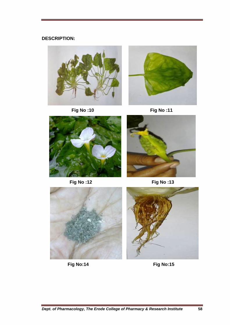
Dept. of Pharmacology, The Erode College of Pharmacy & Research Institute 58
DESCRIPTION:
Fig No :10 Fig No :11
Fig No :12 Fig No :13
Fig No:14 Fig No:15

Dept. of Pharmacology, The Erode College of Pharmacy & Research Institute 59
Habitat:
This species grows in streams, lakes, marshes, ponds, ditches,
canals14. Depth and turbidity of water affecting the penetration of light a clayey
and reducing substratum and little biotic disturbance.(Fig no : 10).
Leaves:
Leaves are extremely variable, with short or long petioles according to
the depth of the water. Blades of the submerged leaves are often narrow; the
floating ones, ovate or somewhat rounded, with a rounded or often heart
shaped base, thin and translucent 5 to 20 centimeters long. (Fig no : 11).
Flowers:
Flowers are white about 2cm in length Inflorescences 1-flowered;
spathes 3-10 winged. Flowers sepals 10-15, stamens 3-12; ovary 1, 3-9
carpellate. wrapped within spathes, cylindrical structures 24 cm long,
composed of green bracts that are ornamented with 3 or more ruffled wings.
Spathes born on long, angled stalks that become spiraled after flowering.
Sepals and shortlived petals of male flowers exert from the tip of the spathe
just above the water surface. Spathes containing female and/or bisexual
flowers are self fertile and remain submersed. Petals white, pink, blue or
purple, often tinged with yellow at the base. Flowering period autumn to
spring. (Fig no : 12).

Dept. of Pharmacology, The Erode College of Pharmacy & Research Institute 60
Fruit:
Fruit is oblong, 2.5 to 4 centimeters long, ovoid to cylindrical and
Fleshy encapsulated fruits contain as many as 2000 seeds Fruit with
numerous seeds(Fig no : 13).
Seeds:
Seeds densely covered with whitish, unicellular hairs. Seeds fusiform
1–2 mm long, 0.3–0.7 mm in diameter, with 2 faint slightly curved longitudinal
ridges. Testa light brown, dull to semi glossy, faintly wrinkled, with a tuft of
unicellular white hairs. Hilum inconspicuous. Embryo linear-spatulate
endosperm absent.( Fig no : 14).
Stems & Roots:
Stem small and cormlike, occasionally forked, with fibrous roots .The
roots in the sediment in water to 2 feet deep and has short. (Fig no : 15).
DISTRIBUTION133-134:
Ottelia alismoides (L.) pers. is Widely distributed in northeastern Africa,
South Korea, eastern and southeast Asia and the tropical regions of Australia,
Bangladesh. It is introduced in United states china (Anhui, Fujian,
Guangdong, Guangxi, Guizhou, Hainan, Hebei, Heilongjiang, Henan, Jiangsu,
Jiangi, Jilin, Liaoning, Sichuan, Yunnan, Zhejiang),Egypt, Hong Kong, India,
Srilanka. It has been reported from many countries across tropical and
subtropical Asia and Australia and covers a wide geographic range. It is found

Dept. of Pharmacology, The Erode College of Pharmacy & Research Institute 61
throughout India in tanks, ponds, streams and ditches. It was identified in
several places like the aquatic herb at the back waters of Madhuban Dam
near Dudhani, Silvassa region and Kunnathur tank Madurai. The plant was
identified in the location of Agaram village kudapakkam villianur Pondicherry.
PHYTOCHEMISTRY135-139:
Phytochemical present in this genus contain different natural
compounds like glycosides, Alkaloids, Flavonoids, Terpenoids, Tannins,
Phenolic compounds. Present knowledge about this endangered species of
medicinal plant is still limited with respect to its phytochemistry and biological
activity. Major chemical constituents of Ottelia alismoides Studies revealed
that isolated two diastereomeric 4-methylene-2-cyclohexenones, otteliones A
and B, ten new diarylheptanoids (2,3,4,5a-d,6,7&8) together with a
hydroxylated analog of otteliones A and B and 3a-hydroxyottelione.
ETHANOBOTANICAL AND MEDICINAL USES140:
The plants are used to improve the water quality in fish ponds by
capturing floting mud particles. The petioles and leaves are eaten as a
vegetable with excellent flavour, the leaves are used in Thailand for
seasoning rice. The fruit is also edible. The plant is used in the treatment of
haemorrhoids and applied as the poultice against fever. It is also grown as an
aquarium plant

Dept. of Pharmacology, The Erode College of Pharmacy & Research Institute 62
Table:1 Summarises the traditional use of different parts of the plant for
various ailments.
Plants part Uses/ailments
Leaves 1. Applied on poultice arm & leg in fever, Topical for haemorrhoids.
2. Decoction of ground leaves mixed with shoots of ipomoea aquatic, alium sativum, lasia spinosa, ocimum sanctum and Typha angustata for the treatment of pneumonia.
3. It is used as a rubefacient.
4. It is used as the treatment of stomach disorder.
5. It is used as spice for the rice and vegetables.
6. Leaves used to check bleeding.
Flowers
1. Flower paste 10-12 gram is taken orally early in morning to curing piles.
2. Local people eat lower part of the flower as raw or as vegetable.
Fruit 1. Arresting cough and inducing diuresis.
2. Fruit eaten as raw.
Whole plant 1. It is a promising drug raw material having anti-tuberculosis activity.
2. Plant prepared as paste and applied to abscesses of the breasts cancer, ulcers and burns.
3. It is also used for treatment of Asthma and applied externally for skin diseases.
4. Clearing away from heat.
5. Eliminating phlegm
6. It is used for the treatment of diabetes.

Dept. of Pharmacology, The Erode College of Pharmacy & Research Institute 63
Earlier work done on Ottelia alismoides (L.) Pers.,
1. Li H, Qu X, Zhao D et al., Preliminary study on the antitubercular
effect of Ottelia alismoides (L.) Pers. In this study a clinical trials, extract of
Ottelia alismoides cured two cases of bilateral tuberculosis of cervical lymph
glands within 3 months. Results suggest Ottelia alismoides to be a promising
medicinal herb with antitubercular effect.
2. Seif-Eldin, Ayyad N et al., Otteliones A and B: potently Cytotoxic
4-Methylene-2-cyclohexenones from Ottelia alismoides. In this study Isolated
Ottelione A the natural products showed remarkable in vitro cytotoxicity
against various cancer cell lines
3. TsaiYuan Chang, YunPeng Tu et al., Synthesis and
Antiproliferative Activities of Ottelione A Analogues. In this study Ottelione A,
isolated from the fresh water plant Ottelia alismoides, is among the most
potent natural product that possess in vitro antiproliferative activity, with an
IC50 in the pMnM range against 60 human cancer cell lines. Study
established the relationship of antimitotic ottelione against tubulin and various
cancer cell lines.

Dept. of Pharmacology, The Erode College of Pharmacy & Research Institute 64
4. SCOPE OF THE PRESENT STUDY
In the present situation, diabetes is possibly the world’s largest growing
metabolic disorders and as the knowledge on the heritogeneity of this disorder
is advanced, the need for more appropriate therapy increases. People with
diabetes can over time develop nerve damage throughout the body. About 60-
70 % of people with diabetes have neuropathy. A number of allopathic drugs
are used for the diabetic neuropathy effect like Aceclofenac, Aspirin,
Gabapentin, Paroxetine and Imipramine which have danger of drug
interaction, adverse effects etc. Traditional plant medicines are used
throughout the world for a range of diabetic neuropathy presentations. There
are many medicinal plants known to be used in the treatment of diabetic
neuropathy and a number of plants had been screened positively for their
Diabetic neuropathy effect. Most of these plants were found to belong to the
chemical group glycosides, alkaloids and Flavanoids and Tannins.
Ottelia alismoides(L.) PERS whole plant is having the Diabetic
neuropathy activity. As per the literature review, still no Diabetic neuropathy
activity has been reported on this plant. Hence, this study has been taken to
explore the Diabetic neuropathy activity of Ottelia alismoides. In
Streptozotocin induced diabetes in Wistar albino rats.

Dept. of Pharmacology, The Erode College of Pharmacy & Research Institute 65
5. AIM AND OBJECTIVES
Aim
The present investigation was aimed to screen the Diabetic neuropathy
activity of Ottelia alismoides (L.) PERS on Streptozotocin induced Diabetic
Wistar albino rats.
Objectives
The objective of the present study to:
a. Find out the phytochemical constituents present in the ethanolic
extract of the whole plant of Ottelia alismoides (L.) PERS
b. Acute toxicity studies
c. Evaluation of anti-diabetic activity
Blood glucose level
Body Weight
Organ Weight (Liver and Kidney)
d. Estimation of Antioxidant parameters
Superoxide dismutase level (SOD)
Catalase level (CAT)
Reduced glutathione (GSH)
Glutathione peroxidase (GPx)

Dept. of Pharmacology, The Erode College of Pharmacy & Research Institute 66
Other parameter:
Na+ ,K+, ATPase acivity
e. Estimation of Oxidative stress parameters
Lipid peroxidation (LPO)
f. Estimation of Behavioral assessment parameters
Hot plate (Thermal Hyperalgesia)
Cold plate (cold allodynia)
Tail immersion (Thermal hyperalgesia)
Tail clip (Mechanical hyperalgesia)
Tail Flick (Thermal hyperalgesia)
Formalin (Chemical hyperalgesia)
g. Histopathological studies of sciatic nerve
h. Statistical analysis

Dept. of Pharmacology, The Erode College of Pharmacy & Research Institute 67
6. PLAN OF WORK
1. Collection of plant.
2. Authentication of plant, shade drying of the Whole plant.
3. Extraction of plant materials with solvents.
4. Preliminary phytochemical screening of Ethanolic extract of Whole
plant of Ottelia alismoides (L.) PERS
5. Evaluation of acute toxicity studies to determine LD50 value
6. Evaluation of anti-diabetic activity.
7. Estimation of Nociceptive threshold parameters.
8. Histopathological study of sciatic nerve.
9. Statistical analysis

Dept. of Pharmacology, The Erode College of Pharmacy & Research Institute 68
7. MATERIALS AND METHODS
7.1 PLANT MATERIALS:
7.1.1 Collection and authentication, shade drying and granulation of
plant material
The whole plant Ottelia alismoides (L) PERS., were collected in the
month of February from Agaram Village Kudapakkam Villianur Pondicherry.
The plant material was taxonomically identified and authenticated by
Dr.N.Ayyapan Researcher, French Institute of Pondicherry, Pondicherry.
7.2 PREPARATION OF OTTELIA ALISMOIDES ETHANOLIC EXTRACT
The whole plant of Ottelia alismoides(L) PERS. were dried under
shade and then powdered with a mechanical grinder. The powder was passed
through sieve No:40 and stored in an airtight container for further use.
7.2.1 Method of Extraction
Soxhlet extraction:
Soxhlet extraction is the process of continuous extraction in which the
same solvent can be circulated through the extractor for several times. This
process involves in the extraction followed by evaporation of the solvent. The
vapours of the solvent are taken to a condenser and the condensed liquid is
returned to the drug for continuous extraction. Soxhlet apparatus designed
for such continuous extraction consists of a body of extractor attached with a
side tube and siphon tube. The lower of the extractor is attached to distillation

Dept. of Pharmacology, The Erode College of Pharmacy & Research Institute 69
flask and the mouth of extractor is fixed to a condenser by the standard joints.
The powdered crude drug is packed in the soxhlet apparatus directly or in a
thimble of filter paper or fine muslin cloth. The diameter of the thimble
corresponds to the internal diameter of the soxhlet extractor.
7.2.2 Chemicals and preparation of drug solutions:
Streptozotocin was obtained from Sisco research laboratories Pvt. Ltd,
Mumbai, India and Pregabalin was perchased from Swapnaroop drugs &
pharmaceuticals, Aurangabad, Maharastra, India. All otherchemicals and
reagents used were of analytical grade.
7.3 PRELIMINARY PHYTOCHEMICAL SCREENING
Qualitative Phytochemical Analysis141
The Ethanolic extract of Ottelia alismoides was analyzed for the
presence of various phytochemical constituents.
A.CARBOHYDRATE TESTS
1.Molisch’s test:
To the test solution, few drops of α-naphthol was added, then few
drops of concentrated sulphuric acid was added through the sides of test
tube, purple to violet colour ring appeared at the junction, indicates the
presence of carbohydrates.

Dept. of Pharmacology, The Erode College of Pharmacy & Research Institute 70
2.Fehling’s test:
To the test solution, equal quantity of Fehling’s A and B were added
and heated on water bath, brick red precipitate was formed, indicates the
presence of carbohydrates.
3.Benedict’s test:
To the test solution, 5ml of Benedict’s reagent was added and heated
on water bath, red precipitate was formed, indicates the presence of
carbohydrates.
4.Barfoed’s test:
To 1 ml of the test solution, add 1ml of Barfoed’s reagent was added
and heated on a water bath, red cupric oxide was formed, presence of
monosaccharide.
5.Test for pentoses:
To the test solution, equal volume of hydrochloric acid and
phloroglucinol was added and heated, no red colour was produced, indicates
the absence of pentoses.
6.Selivanoff’s test (test for ketones):
To the test solution, crystals of resorcinol and equal volume of
concentrated hydrochloric acid were added and heated on a water bath, rose
colour was produced, indicates the presence of ketones.

Dept. of Pharmacology, The Erode College of Pharmacy & Research Institute 71
B.ALKALOIDS
1.Dragendroff’s test:
To 1ml of the extract, Dragendroff’s reagent (potassium bismuth iodine
solution) was added, no reddish brown precipitate was formed, indicates the
absence of alkaloids.
2.Wagner’s test:
To 1ml of the extract, Wagner’s reagent (iodine potassium iodide
solution) was added, no reddish brown precipitate was formed, indicated the
absence of alkaloids.
3.Mayer’s test:
To 1 ml of the extract, Mayer’s reagent (potassium mercuric iodine
solution) was added, no cream colour precipitate was formed, indicates the
absence of alkaloids.
4.Hager’s test:
To 1 ml of the extract, Hager’s reagent (saturated aqueous solution of
picric acid) was added, no yellow coloured precipitate was formed, indicates
the absence of alkaloids.
5.Tannic acid test:
To the extract tannic acid solution was added, no buff colour precipitate
was produced, indicates the absence of alkaloids.
Dept. of Pharmacology, The Erode College of Pharmacy & Research Institute 72
C) GLYCOSIDE TEST
1.Legal’s test:
The extract in pyridine and sodium nitroprusside solution was added to
make it alkaline, formation of pink to red colour showed the presence of
glycosides.
2.Baljet’s test:
To 1 ml of the extract, 1 ml of sodium picrate solution was added and
the change of yellow to orange colour reveals the presence of glycosides.
3.Borntrager’s test:
Few ml of dilute sulphuric acid was added to 1 ml of the extract
solution. Boiled and filtered and then the filtrate was extracted with
chloroform. The chloroform layer was treated with 1 ml of ammonia. The
formation of red colour of the ammonical layer showed the presence of
anthraquinone glycosides.
4.Keller killani test:
1. 1gm of powdered drug was extracted with 10ml of 70% alcohol for 2
minutes, filtered, the filtrate was added to 10 ml of water and 0.5 ml of strong
solution of lead acetate and filter, and the filtrate was shaken with 5ml of
chloroform.

Dept. of Pharmacology, The Erode College of Pharmacy & Research Institute 73
2. The chloroform layer was separated in a porcelain dish and the solvent
was removed by gentle evaporation. The cooled residue was dissolved in 3 ml
of glacial acetic acid containing 2 drops of 5% ferric chloride solution.
The solution was carefully transferred to the surface of 2ml of
concentrated sulphuric acid. A reddish brown layer formed at the junction of
the two liquids and the upper layer slowly became bluish green, darkening
with standing.
D.TEST FOR SAPONINS
1. Foam test:
Small quantity of alcoholic extract was taken and 20ml of distilled water
was added and shaked in a graduated cylinder for 15 minutes length wise. No
Layer of foam, indicates the absence of saponins.
E. TEST FOR FLAVANOIDS
1. Shinoda’s test:
To the test solution, few magnesium turnings were added and
concentrated hydrochloric acid was added drop wise, pink scarlet, crimson
red or occasionally green to blue colour appeared after few minutes indicating
the presence of flavanoids.

Dept. of Pharmacology, The Erode College of Pharmacy & Research Institute 74
2. Alkaline reagent test:
To the test solution, few drops of sodium hydroxide solution was
added, intense yellow colour was formed which turned colourless on addition
of few drops of dilute acid indicating presence of flavanoids.
3. Zinc hydrochloride test:
To the test solution, a mixture of zinc dust and concentrated
hydrochloric acid were added. It gave red colour after few minutes showing
the presence of flavanoids.
F. TEST FOR TANNINS
1. Ferric chloride test:
To the test solution, ferric chloride solution was added, green colour
appeared showing the presence of condensed tannins.
2. Phenazone test:
To the test solution, 0.5 grams of sodium phosphate was added,
warmed and filtered. To the filtrate 2% phenazone solution was added, bulky
precipitate was formed which was often coloured, indicating the presence of
tannins.
3. Gelatin test:
To the test solution, 1% gelatine solution containing 10% sodium
chloride was added. Precipitate was formed, indicating the presence of
tannins.

Dept. of Pharmacology, The Erode College of Pharmacy & Research Institute 75
4. Test for Catechin:
Match stick was dipped in the test solution, it was dried and lastly
moistened with concentrated hydrochloric acid. Then the stick was warmed
near to flame. The colour of the wood changed to pink due to phloroglucinol.
(Phloroglucinol was formed when catechins were treated with acids),
indicating the presence of tannins.
7.4 ACUTE TOXICITY STUDY
Organisation for Economic co-operation and Development (OECD)
regulates guidelines for oral acute toxicity study. It is an international
organisation which works with the aim of reducing both the number of animals
and the level of pain associated with acute toxicity testing. To determine the
acute oral toxicity OECD frames the following guideline methods.
OECD 401 – Acute Oral Toxicity
OECD 420 – Acute Oral Toxicity: Fixed Dose procedure
OECD 423 –Acute Oral Toxicity: Acute Toxic Classic method
OECD 425 – Acute Oral Toxicity: Up and own Procedure
In the present study the acute oral toxicity of Ottelia alismoides was
carried out according to OECD 423 guideline (Acute Oral Toxicity: acute Toxic
Classic Method).

Dept. of Pharmacology, The Erode College of Pharmacy & Research Institute 76
ANIMALS:
Male Swiss albino mice weighing between 20 – 25 gm was used for the
study. The animals were obtained from animal house, Nandha College of
Pharmacy, Erode. The animals were placed at random and allocated to
treatment groups in polypropylene cages with paddy husk as bedding.
Animals were housed at a temperature of 24±2oC and relative humidity of 30
– 70 %. A 12:12 light: day cycle was followed. All animals were allowed to free
access to water and fed with standard commercial pelleted rat chaw. All the
experimental procedures and protocols used in this study were reviewed and
approved by the Institutional Animal Ethics Committee of Nandha College of
Pharmacy. (IAEC No: NCP/IAEC 2016-17-18) and were in accordance with
the guidelines of the CPCSEA.
Acute Toxicity Studies:
Acute toxicity studies were performed according to OECD-423
(Organization of Economic and Cooperation Development) guidelines. Male
Swiss mice selected by random sampling technique were employed in this
study. The animals were fasted for 4h with free access to water. The
Ethanolic extract of Ottelia alismoides (EEOA) was administered orally at a
dose of 5 mg/kg initially and mortality if any was observed for first 24 hrs and
after 72 hrs. If mortality was observed in two out of three animals, then the
dose administered was considered as toxic dose. However, if the mortality
was observed in only one animal out of three animals then the same dose
was repeated again to confirm the toxic effect. If no mortality was observed,

Dept. of Pharmacology, The Erode College of Pharmacy & Research Institute 77
then higher (50, 300, 2000 mg/kg) doses of the plant extracts were employed
for further toxicity studies.
OBSERVATION:
The following general behaviour was also observed during the acute toxicity
study (Ecobichon DJ, 1997)142.
1. Sedative
2. Hypnotics
3. Convulsion
4. Ptosis
5. Analgesia
6. Stupar reaction
7. Motor activity
8. Muscle relaxant
9. Pilo erection
10. Change in skin colour
11. Lacrimal secretion
12. Stool Consistency
Result:
There was no mortality with the extract of Ottelia alismoides after 24 &
72 hrs even at (2000mg/kg). The extract did not alter the general behaviour in
mice. No lethality or toxic reactions were found during and after the study
period.

Dept. of Pharmacology, The Erode College of Pharmacy & Research Institute 78
7.5 EXPERIMENTAL INDUCTION OF DIABETES
Sterptozotocin Induced Diabetes:
A freshly prepared solution of STZ (55 mg/kg in 0.01 M citrate buffer,
pH 4.5) was injected intraperitoneally to overnight-fasted rats. The rats
exhibited hyperglycaemia within 48 h of STZ administration. The rats having
fasting blood glucose (FBG) values of 250 mg/dl or above were considered for
the study. The study of test compound and standard drugs were dissolved in
distilled water and administered orally with the help of gastric oral tube.
7.5.1 EXPERIMENTAL DESIGN
The Diabetic Neuropathy activity was tested on a total of 30 rats (24
diabetic rats and 6 normal rats) and they were divided into five groups and
each group consists of 6 animals as follows,
Group I - Diabetic control received Streptozotocin injection (55mg/kg; b.wt;
i.p), freshly prepared in citrate buffer.
Group II- Streptozotocin injection (55mg/kg; b.wt; i.p) + insulin (5 IU/kg,
i.m).
Group III- STZ+insulin+ Standard drug Pregabalin (15mg/kg) dissolved in
0.5% CMC.
Group IV- STZ+insulin+ plant extract low dose (200mg/kg, b.wt; p.o)
suspended in 0.5% CMC.

Dept. of Pharmacology, The Erode College of Pharmacy & Research Institute 79
Group V- STZ+insulin+ plant extract high dose (400mg/kg, b.wt; p.o)
suspended in 0.5% CMC.
In the first four weeks of the study all the groups were left untreated.
From week 4 onwards the drug treatment started and continued up to the
week 8 after STZ injection. Body weight and Behavioural assessments like
thermal hyperalgesia (hot plate, tail flick & Tail immersion), Mechanical
hyperalgesia (Tail clip) thermal allodynia (cold plate) & (chemical
hyperalgesia) formalin were performed on week 0th, 4th, 6th & 8th of the study.
At the end of 8th week the blood was collected by retro-orbital sinus puncture,
rats were sacrificed by cervical dislocation organs collected and isolated
sciatic nerves were analyzed for following Biochemical and histopathological
study.
Blood Sample and Organs Collection:
The blood was collected by retro orbital under mild ether anaesthesia
from overnight fasted rats, into tubes containing potassium oxalate and
sodium fluoride as anticoagulant for estimation of fasting plasma glucose.
Plasma and serum were separated by centrifugation. After centrifugation at
2,000 rpm for 10 minutes, the clear supernatant was u sed for the analysis of
various biochemical parameters. After collection of blood, all the treated
animals were sacrificed the Sciatic nerve and liver tissues were isolated and
rinsed in ice- cold saline and kept in formalin solution (10%) for further
histopathological studies & oxidative stress parameters.

Dept. of Pharmacology, The Erode College of Pharmacy & Research Institute 80
Nerve collection and preparation of homogenate:
After 8 weeks of experiment, sciatic nerve was removed bilaterally from
the inguinal ligament to its trifurcation and incubated in triton x 100 for 20 min
to remove blood from sciatic nerve. Sciatic nerves were chopped into small
pieces and then homogenized with phosphate buffer solution (pH 7.0) by
using homogenizer. The homogenate was then centrifuged in a cold
centrifuge (Thermo Scientific) at 4ºC, 4000 x g for 10 min. The resulting
homogenate was passed through a cellulose filter to remove impurities and
was aliquoted for the measurement of SOD, GSH, CAT, GPx, LPO, Na+, K+,
ATPase activity.
7.6 EVALUATION OF PARAMETERS143
7.6.1 General parameters:
Body weight and Organ weight:
The body weight of rats was checked using a top loader weighing
balance. The body weight is expressed in grams. The vital organs such as
kidney and liver were collected. Blood was removed from the organs with the
filter paper and their weights were assessed with an electric weighing
balance. The organ weights were expressed in grams or milligrams.

Dept. of Pharmacology, The Erode College of Pharmacy & Research Institute 81
7.7 EVALUATION OF BIOCHEMICAL PARAMETERS:
7.7.1 Estimation of blood glucose level:
The Blood glucose levels were estimated by Hexokinase method144.
Glucose is phosphorylated by hexokinase (HK) in the presence of adenosine
triphosphate (ATP) and magnesium ions to produce glucose-6- phosphate
and adenosine diphosphate (ADP). Glucose-6-phosphate dehydrogenase
(G6P-DH) specifically oxidises glucose-6-phosphate to gluconate-6-
phosphate with the concurrent reduction of NAD+ to NADH. The increase in
absorbance at 340nm is proportional to the glucose concentration in the
sample.
Reagents
Reagent 1:
0.05 M Tris HCl buffer, pH 8.0 with 13.3 mM MgCl2
0.67 M Glucose in Tris-MgCl2 buffer
16.5 mM ATP in Tris-MgCl2 buffer
6.8 mM NAD in Tris-MgCl2 buffer
HK, Mg2+
Glucose + ATP Glucose-6-phosphate + ADP
Glucose-6-Phosphate + NAD+
Gluconate-6-P + NADH + H+

Dept. of Pharmacology, The Erode College of Pharmacy & Research Institute 82
Reagent 1A:
300 IU/ml Glucose-6-phosphate dehydrogenase in Tris-MgCl2 buffer
300 IU/ml Hexokinase in Tris-MgCl2 buffer
Procedure:
150 μl of reagent 1 was added with 30 μl of reagent 1A and to this 20 μl
of suitable diluents was added and the contents were mixed thoroughly. To
this mixture, 2 μl of serum sample was added. Then the contents were mixed
and incubated at 37°C for 10 seconds. After zeroing the instrument with blank
the absorbance of standard followed by the test sample was measured at 340
nm. The values were expressed as mg/dl.
7.8 EVALUATION OF NOCICEPTIVE PARAMETERS:
7.8.1 Tail Flick Method:
The tail flick test is a test of the pain response in animals, similar to
the hot plate test. It is used in basic pain research and to measure the
effectiveness of analgesics, by observing the reaction to heat. The
quantitative measurements of pain threshold in animal against thermal
radiation and for evaluation of analgesic activity. It was first described by D’
Amour and Smith in 1941145-146.
PROCEDURE:
The nociceptive response was evaluated regarding the latency to
withdrawal of the tail in response to noxious radiant heating. Animals are

Dept. of Pharmacology, The Erode College of Pharmacy & Research Institute 83
placed into individual restraining cages leaving the tail hanging out freely. The
animals are allowed to adapt to the cages for 30 min before testing. The
apparatus used is tail flick analgesiometer, the tip of tail of rat is placed on hot
metal wire and latency of withdrawal is calculated manually by stop watchR.
7.8.2 Tail Clip Method:
The method was described by Haffner147.
PROCEDURE:
Six screened Wister rats were assigned to each of the 5 groups. The
drug was administered 30 min prior testing. An artery clip was applied to
the root of the tail (approximately 1 cm from the body) to induce pain. The
animal quickly responds to this noxious stimuli by biting the clip or the tail near
the location of the clip. The time between stimulation onset and response was
measured by a stopwatch in 1/10 seconds increments. The length of time until
response indicates the period of greatest activity after dosing [Schleyerbach
2002] in different period of treatments was recorded. The values of
prolongation of latency time of experimental groups were compared with
control group for statistical analysis.
7.8.3 Hot Plate Method:
The method originally described by Woolfe and MacDonald148 (1944)
.The paws of the rats are very sensitive to heat at temperatures which are not
damaging the skin. The responses are jumping, withdrawal of the paws and

Dept. of Pharmacology, The Erode College of Pharmacy & Research Institute 84
licking of the paws. The time until these responses occur is prolonged after
administration of centrally acting analgesics.
PROCEDURE:
In this hot plate method animals from the each group were placed on
the hot plate (Eddys hot plate) which is commercially available consists of an
electrically heated surface. Temperature of this hot plate is maintained at 55 ̊
C- 56 ̊ C. This can be a copper plate or a heated glass surface. The
observation is done up to the time until paw licking or jumping was noted the
cut- off time was 10 sec. The reaction time was noted after the oral
administration of the drugs and test compounds.
7.8.4 Formalin Test:
The formalin test in rats has been proposed as a chronic pain model
which is sensitive to centrally active analgesic agents by Dubuisson and
Dennis149-150(1977).The formalin test uses a 10% formalin solution as a
chemical noxious stimulus. By injecting the formalin solution into the paw of a
rat, a model of persistent (chronic) pain caused by peripheral tissue injuries
and inflammation is created.
PROCEDURE
At the end of the study, all the groups were subjected to the formalin
test. Briefly, each rat was acclimatized to the observation box before the
formalin test. After an adaptation period of 15 min, the right hind foot paw was
injected with 50 mL of 2.5% formalin in the intraplantar region. Nociception
Dept. of Pharmacology, The Erode College of Pharmacy & Research Institute 85
was evaluated by quantifying paw licking time during the first 10 min (acute
phase) and at 20– 40 min (delayed phase) (Khalilzadeh et al., 2008; Luiz et
al., 2007)
7.8.5 Cold Plate Method:
A cold plate apparatus was designed to test the responses of
unrestrained rats to low temperature stimulation of the plantar aspect of the
paw. At plate temperatures of 10 degrees C and 5 degrees C, rats with either
chronic constriction injury (CCI) of the sciatic nerve or complete Freund's
adjuvant (CFA) induced inflammation of the hind paw displayed a stereotyped
behavior. Brisk lifts of the treated hind paw were recorded, while no evidence
of other nociceptive behaviors could be discerned151.
Procedure:
Cold-plate tests were conducted to evaluate the hyperalgesic
responses to cold stimuli. For this test, the hot/cold-plate test device (Ugo-
basile, 35100, Verase, Italy) was used. After setting the aluminium plate to 4
1C71 1C, the rats were placed in the Plexiglas compartment. The time that
elapsed between placing the animals in the device and the time point that
they quickly withdrew their paws was recorded (Rosellini et al., 2012). The
steps taken by the animals when walking or the slow paw-withdrawal behavior
related to locomotion were not considered (Ortega-Álvaro et al., 2012).
Measurements were repeated three times for each rat, and the average of
these three values was calculated (Rosellini et al., 2012). To avoid paw
damage, the experiment was not conducted for more than 30 s.

Dept. of Pharmacology, The Erode College of Pharmacy & Research Institute 86
7.8.6 Tail Immersion Method:
Hot and cold water Tail Immersion Test
This method was described by Sharma et al., 2006a152.
Procedure:
The procedure was based on the reaction time of the typical tail-
withdrawal reflex in rats induced by immersing the end of the tail in warm
water of 52.5±1ºC and cold water of 10±0.5ºC. The lower portion of the tail,
nearly 5 cm from tip, was marked and immersed in a beaker of freshly filled
warm water and cold water. The rats reacted by withdrawing the tail in a few
seconds. This reaction was measured two to three times, with an interval of at
least 15min, in order to obtain two consecutive values that differed no more
than 10%. After each determination, the tail was carefully dried. The cut-off
time for warm water tail immersion was 15 s and cold water tail immersion
30s.
7.9 EVALUATION OF ANTIOXIDANTS PARAMETERS:
7.9.1 Estimation Superoxide dismutase(SOD)
This enzyme catalyzes the dismutation of superoxide anion (O2-) to
hydrogen peroxide and molecular oxygen in the following manner153.
The enzyme activity was assayed by the method of Misra and Fridovich.
H2O + 2O2-. + 2H+ → 2H2O2 + O2
Dept. of Pharmacology, The Erode College of Pharmacy & Research Institute 87
Reagents:
0.1 M Carbonate-bicarbonate buffer; pH 10.2.
0.6 mM EDTA solution
1.8 mM Epinephrine (prepared in situ)
Absolute ethanol.
Chloroform
Procedure:
0.1 ml of tissue homogenate was added to the tubes containing 0.75 ml
ethanol and 0.15 ml chloroform (chilled in ice) and centrifuged. To 0.5 ml of
supernatant, added 0.5 ml of 0.6 mM EDTA solution and 1 ml of 0.1 M
carbonate-bicarbonate (pH 10.2) buffer. The reaction was initiated by the
addition of 0.5 ml of 1.8 mM epinephrine (freshly prepared) and the increase
in absorbance at 480 nm was measured. One unit of the SOD activity was the
amount of protein required to give 50% inhibition of epinephrine autoxidation.
7.9.2 Estimation of Catalase (CAT)
This enzyme catalyzes conversion of hydrogen peroxide into water
and molecular oxygen154.
The enzyme activity was assayed by the method of Sinha.
2H2O2 → 2H2O + O2

Dept. of Pharmacology, The Erode College of Pharmacy & Research Institute 88
Reagents:
Dichromate-acetic acid reagent: 5% potassium dichromate in water
was mixed with glacial acetic acid in the ratio of 1:3 (v/v).
0.01 M Phosphate buffer; pH 7.0.
0.2M Hydrogen peroxide
Procedure:
0.1 ml of the tissue homogenate was added to the reaction mixture
containing 1ml of 0.01 M phosphate buffer (pH 7.0) pre-warmed to 37°C, 0.4
ml of distilled water and the mixture was incubated at 37°C. The reaction was
initiated by the addition of 0.5 ml of 0.2 M hydrogen peroxide and the reaction
mixture was incubated at 37°C for one minute. The reaction was terminated
by the addition of 2 ml of dichromate-acetic acid reagent after 15, 30, 45, and
60 seconds. Standard hydrogen peroxide in the range of 4-20 μ moles were
taken and treated in the same manner. All the tubes were heated in a boiling
water bath for 10 minutes, cooled and the green colour that developed was
read at 590 nm against blank containing all components except the enzyme.
Catalase activity was expressed in terms of μ moles of H2O2
consumed/min/mg protein.
7.9.3 Estimation of Reduced glutathione (GSH):
The total reduced glutathione was determined according to the
method of Ellman155.

Dept. of Pharmacology, The Erode College of Pharmacy & Research Institute 89
The assay procedure is based on the reduction of Ellman´s reagent [5,
5΄- dithio dis (2- nitrobenzoic acid)] (DTNB) by SH groups of glutathione to
form 2-nitro-S-mercaptobenzoic acid per mole of glutathione. The product is
measured spectrophotometically at 412 nm.
Reagents
0.2 M Phosphate buffer; pH 8.0.
0.6 mM DTNB reagent.
5%TCA
Procedure
0.1 ml of tissue homogenate was precipitated with 5% TCA. The
contents were mixed well for complete precipitation of proteins and
centrifuged. To 0.1 ml of supernatant, 2 ml of 0.6 mM DTNB reagent and 0.2
M phosphate buffer (pH 8.0) were added to make up to a final volume of 4 ml.
The absorbance was read at 412 nm against a blank containing TCA instead
of sample. A series of standards treated in a similar way also run to determine
the glutathione content. The amount of glutathione was expressed as
nmoles/g tissue.

Dept. of Pharmacology, The Erode College of Pharmacy & Research Institute 90
7.9.4 Estimation of Glutathione peroxide (GPx):
Principle:
GPx activity was assessed by modified method of Flohe and Gunzler
(1984)156.
Reagents:
Sodium phosphate buffer 0.3 M, pH 7.0
Sodium azide 1Omhl
Reduced glutathione 4mM
Hydrogen peroxide 2.5 mM
TCA lPh
Phosphate solution 0.3 M disodium hydrogen phosphate.
DTNB 40 mg/100 ml of 1% sod~um cihate
EDTA 0.8 mM 9. Standard 20 mg of reduced glutathione in 100 rnl
distilled water. This solution contained 20 pg of glutathione/O. 1 ml.
Procedure:
GPx activity was assessed by modified method of Flohe and Gunzler
(1984). For the enzyme reaction, 0.2 ml of the post-mitochondrial supernatant
was placed into a tube and mixed with 0.4 ml reduced glutathione and the
mixture was put into an ice bath for 30 min. Then the mixture was centrifuged
for 10 min at 3000 rpm, 0.48 ml of the supernatant was placed into a cuvette,
and 2.2 ml of 0.32 M Na2HPO4 and 0.32 ml of 1.0 mmol/L DTNB were added
for color development. The absorbance at wavelength 412 nm was measured

Dept. of Pharmacology, The Erode College of Pharmacy & Research Institute 91
on spectrophotometer (LKB-Pharmacia, Mark II, Ireland) after 5 min. The
enzyme activity was calculated as nmol/mg protein.
7.10.EVALUATION OF OXIDATIVE STRESS PARAMETER
Estimation of Lipid Peroxidation (LPO):
Lipid peroxidation (LPO) was assayed by the method of Ohkawa et
al111, in which the malondialdehyde (MDA) released served as the index of
LPO. The extent of LPO in the hepatic tissue was assayed by measuring one
of the end products of this process, the thiobarbituric acid-reactive substances
(TBARS). As 99% TBARS is Malondialdehyde (MDA), thus this assay is
based on the reaction of 1 molecule of MDA with 2 molecules of TBARS at
low pH (2- 3) and at a temperature of 95°C for 60 min. The resultant pink
chromogen can be detected spectrophonometrically at 532nm157.
Reagents
Standard: 1, 1, 3, 3-tetra ethoxypropane (TEP).
8.1% Sodium dodecyl sulphate (SDS)
20%Acetic acid
0.8%Thiobarbituric acid (TBA)
15:1 v/v n-butanol: pyridine mixture

Dept. of Pharmacology, The Erode College of Pharmacy & Research Institute 92
Procedure
To 0.2 ml of tissue homogenate, 0.2ml of 8.1% SDS, 1.5ml of 20%
acetic acid (pH 3.5) and 1.5 ml of 0.8% TBA were added. The mixture was
made up to 4 ml with water and then heated in a water bath at 95.8ºC for 60
min using glass ball as a condenser. After cooling, 1ml of water and 5ml of n-
butanol: pyridine (15:1 v/v) mixture were added and shaken vigorously. After
centrifugation at 4000rpm for 10min, the organic layer was taken and its
absorbance was measured at 532nm. The level of lipid peroxides was
expressed as n moles of MDA formed/mg of protein.
7.11.OTHER PARAMETER:
Estimation of NA+ , K+ , ATPase activity158-159.
Principle:
Na+/K+-ATPase activity was measured according to the method of
Suhail and Rizvi 1987 The final assay mixture contained 0.5–1.2 mg
membrane protein/mL, 20 mmol/L KCl, 140 mmol/L NaCl, 3 mmol/L MgCl2,
30 mmol/L imidazole (pH 7.24), with or without 5 × 10⁻⁴ mol/L ouabain and
6 mmol/L ATP. Assay mixture was incubated for 30 min at 37 °C and the
reaction was stopped by the addition of 3.5 mL of a solution-A (0.5%
ammonium molybdate, 0.5 mol/L H2SO4, and 2% sodium dodecyl sulphate).
The amount of liberated phosphate (Pi) was estimated according to the
method of Fiske and Subbarow (1925).
Dept. of Pharmacology, The Erode College of Pharmacy & Research Institute 93
REAGENTS
0.5–1.2 mg membrane protein/mL,
20 mmol/L KCl, 140 mmol/L
NaCl, 3 mmol/L
MgCl2, 30 mmol/L
imidazole (pH 7.24), with or without 5 × 10⁻⁴ mol/L ouabain
f) nd 6 mmol/L ATP.
Assay mixture was incubated for 30 min at 37 °C and the reaction was
stopped by the addition of 3.5 mL of a solution-A (0.5% ammonium
molybdate, 0.5 mol/L H2SO4, and 2% sodium dodecyl sulphate). The amount
of liberated phosphate (Pi) was estimated and the values are recorded ( Fiske
and Subbarow(1925). Further by adding tissue extract to the above same
assay mixture and incubated for 30 min at 37 °C and the Na+/K+-ATPase
activity was measured in a spectrophotometer (Star 21 plus USA) and the
activity expressed as nmol pi released/mg protein per hour at 37 °C.
7.12 HISTOPATHOLOGICAL STUDIES
Hematoxylin, a basic bye is oxidized to hematein with a mordant, a
metallic ion suchas the salts of aluminium. The positively charged aluminium-
hematein complex combines with the negatively charged phosphate groups of
the nucleic acids (DNA and RNA) forming blue/purple colour, which is
characteristic of hematoxylin stains. Eosin is an acidic dye, which is
Dept. of Pharmacology, The Erode College of Pharmacy & Research Institute 94
considered to have a selective affinity for the basic parts of the cell, i.e., the
cytoplasm. Thus, the hematoxylin and eosin (H & E) stain is used to
demonstrate different structures of the tissue160.
The various steps involved in the preparation of pancreatic tissues for
histological studies are as follows:
Fixation:
In order to avoid tissue by the lysosomal enzymes and to preserve its
physical and chemical structure, a bit of tissue from each organ was cut and
fixed in bouin’s fluid immediately after removal from the animal body. The
tissues were fixed in bouin’s fluid for about 24 hours. The tissues were then
taken and washed in glass distilled water for a day to remove excess of picric
acid.
Dehydration:
The tissues were kept in the following solutions for an hour each; 30%,
50%, 70% and 100% alcohol. Inadequately dehydrated tissues cannot be
satisfactorily infiltered with paraffin. At the same time over dehydration results
in making the tissues brittle, which would be difficult for sectioning. So, careful
precautions were followed while performing the dehydration process
Clearing:
Dealcoholization or replacement of alcohol from the tissues with a
clearing agent is called as clearing. Xylene was used as the clearing agent for

Dept. of Pharmacology, The Erode College of Pharmacy & Research Institute 95
one or two hours, two or three times. Since, the clearing agent is miscible with
both dehydration and embedding agents, it permits paraffin to infilterate the
tissues. So, the clearing was carried out as the next step after dehydration to
permit tissue spaces to be filled with paraffin. The tissues were kept in the
clearing agent till they become transparent and impregnated with xylene.
Impregnation:
In this process the clearing agent xylene was placed by paraffin wax.
The tissues were taken out of xylene and were kept in molten paraffin
embedding bath, which consists of metal pots filled with molten wax
maintained at about 50oC. The tissues were given three changes in the
molten wax at half an hour intervals.
Embedding:
The paraffin wax used for embedding was fresh and heated upto the
optimum melting point at about 56–58oC. A clear glass plate was smeared
with glycerine. L-shaped mould was placed on it to from a rectangular cavity.
The molten paraffin wax was poured and air bubbles were removed by using
a hot needle. The tissue was placed in the paraffin and oriented with the
surface to be sectioned. Then the tissue was pressed gently towards the
glass plate to make settle uniformly with a metal pressing rod and allowed the
wax to settle and solidity room temperature. The paraffin block was kept in
cold water for cooling.
Dept. of Pharmacology, The Erode College of Pharmacy & Research Institute 96
Section Cutting:
Section cutting was done with a rotatory microtome. The excess of
paraffin around the tissue was removed by trimming, leaving ½ cm around the
tissue. Then the block was attached to the gently heated holder. Additional
support was given by some extra wax, which was applied along the sides of
the block. Before sectioning, all set screws holding the object holder and knife
were hand tightened to avoid vibration. To produce uniform sections, the
microtome knife was adjusted to the proper angle in the knife holder with only
the cutting edge coming in contact with the paraffin block. The tissue was cut
Sin the thickness range of about 7µm.
Flattening and Mounting of Sections:
The procedure was carried out in tissue flotation warm water bath. The
sections were spread on a warm water bath after they were detached from the
knife with the help of hair brush. Dust free clean slides were coated with egg
albumin over the whole surface. Required sections were spread on clean slide
and kept at room temperature
Staining of Tissue Sections:
The sections were stained as follows; deparaffinization with xylene two
time search for five minutes

Dept. of Pharmacology, The Erode College of Pharmacy & Research Institute 97
Dehydration through descending grades of ethyl alcohol
100% alcohol (absolute) - 2 minute
90% alcohol - 1minute
50% alcohol - 1 minute
Staining with Ehrlich’s Haemaoxylin was done for 15-20 minutes. Then
the sectioned tissues were thoroughly washed in tap water for 10 minutes.
Rinsed with distilled water and stained with Eosin. Dehydration again with
ascending grades of alcohol.
70% alcohol - 2 minute
90% alcohol - 2 minute
100%alcohol - 1 minute
Finally the tissues were cleared with xylene two times, each for about 3
minutes interval.
Mounting:
On the stained slide, DPX mountant was applied uniformly and micro
glass cover slides were spread. The slides were observed in Nikon
microscope and microphotographs were taken.

Dept. of Pharmacology, The Erode College of Pharmacy & Research Institute 98
7.13 STATISTICAL ANALYSIS
The data of all the results were represented as Mean ± S.E.M. on
statistically analysed by one-way ANOVA followed by Duncun’s Multiple range
test was used for statistical analysis p<0.05 was considered significant161.

Dept. of Pharmacology, The Erode College of Pharmacy & Research Institute 99
RESULTS
Appearance and percentage yield of EEMP
Table No: 2
Drug Ottelia alismoides (L.,) PERS
Solvent Ethanol
Colour Dark green
Consistency Semi solid
Percentage yield 19.5 % w/w

Dept. of Pharmacology, The Erode College of Pharmacy & Research Institute 100
PRELIMINARY PHYTOCHEMICAL SCREENING
Table No-3: Results of the Preliminary Phytochemical Constituents
present in Ethanolic extract of Ottelia alismoides(L) PERS.
Table no: 3 Preliminary phytochemical constituents present in EEPS
S. No Constituents Ottelia alismoides
Ethanolic extract
1. Alkaloids +
2. Carbohydrates _
3. Protein _
4. Terpinoids +
5. Phenols +
6. Tannins +
7. Flavanoids +
9. Glycosides +
10. Saponins _
+ve indicates the presence –ve indicates the absence
RESULTS:
The phytochemical constituents present in the Ethanolic extract of
Ottelia alismoides (L.) PERS were Phenols, Tannins, Flavanoids, Glycosides,
Alkaloids and Terpenoids.

Dept. of Pharmacology, The Erode College of Pharmacy & Research Institute 101
Estimation of Blood glucose level:
The Effect of the different doses of Ethanolic extract of Ottelia alismoides Pers
on Blood Glucose level.
Groups Blood glucose level(mg/dl)
0th week 8th week
Group I
STZ(55mg/kg)+Vehicle 330.15± 17.76 368.0± 59.05
Group II
STZ (55mg/kg)+
Insulin (5 IU/kg)
341.10 ±22.31 160.10± 20.22***
Group III
STZ+ insulin+ Pregabalin (15mg/kg)
319.15 ±13.18 140.01± 17.02** a
Group IV
STZ+ insuln+ EEOA (200mg/kg)
336.10 ±15.46 120.08 ±13.15ns
aa
Group V
STZ+ insulin+ EEOA (400mg/kg)
317.10 ±12.18 115.21 ±11.06ns
aa
Table no 4: Effect of EEOA on Blood Glucose level in experimentally induced Diabetic
rat model
The values were expressed as Mean ± S.E.M (n= 6 animals in each group)
*,**,***, indicates significance P < 0.05 , P < 0.01 & P <0.001 when compared to Insulin
Diabetic control.
a, aa indicates P < 0.05 & P < 0. 01 Significance between diabetic control group
NS indicates Non-significant
Data was analysed by one-way ANOVA followed by Duncun Multiple range test
(DMRT).

Dept. of Pharmacology, The Erode College of Pharmacy & Research Institute 102
Fig.No. 16 Effect of EEOA on Blood Glucose Level in
Experimentally induced Diabetic rat model
Estimation of Body weight & Organ weight:
The Effect of the different doses of Ethanolic extract of Ottelia alismoides Pers
on Blood Glucose level.
Groups Body weight Organ weight
Liver Kidney
Group I
STZ(55mg/kg)+Vehicle 204.16±6.93 4.55±0.45 1.93±0.05
Group II
STZ +Insulin (5 IU/kg) 249.06±5.31*** 3.09±0.34* 1.83±0.05*
Group III
STZ+ insulin+ Pregabalin (15mg/kg)
248.31±5.30* 2.84±0.31ns 1.62±0.07ns
Group IV
STZ+ insuln+ EEOA (200mg/kg)
240.56±5.29* 2.30±0.30ns 1.60±0.04ns
Group V
STZ+ insulin+ EEOA (400mg/kg)
245.10±5.30a 3.08±0.50ns 1.56±0.04ns
Table no 5: Effect of EEOA on Body weight & Organ weight in
Experimentally induced Diabetic rat model
0
50
100
150
200
250
300
350
400
450
0th week 8th week
Blo
od
Glu
co
se L
evel (m
g/d
l)
Group I STZ
Group II STZ (55mg/kg)+ Insulin (5 IU/kg)
Group III Pregabalin (15mg/kg)
Group IV EEOA (200mg/kg)
Group V EEOA (400mg/kg)

Dept. of Pharmacology, The Erode College of Pharmacy & Research Institute 103
The values were expressed as Mean ± S.E.M (n= 6 animals in each group)
*,**, ***, indicates significance P < 0.05 , P < 0.01 & P <0.001 when compared Insulin diabetic
control
a, aa indicates P < 0.05 & P < 0. 01 Significance between diabetic control group
NS indicates Non-significant
Data was analysed by one-way ANOVA followed by Duncun Multiple range test (DMRT).
RESULTS:
The Blood Glucose, Body weight and Organ Weight were measured
and showed in Table no-3 & 4.
The Diabetic control (Group 1) showed significant increase in Blood
Glucose, Kidney weight & liver and whereas the body weight were decreased
when compared insulin diabetic control (Group 2).
Standard (Group 3) and EEOA (200 mg/kg& 400 mg/kg) showed
statistically significant decrease in Blood Glucose and organ weight and
increase in body weight when compared to Diabetic control (Group1).

Dept. of Pharmacology, The Erode College of Pharmacy & Research Institute 104
ESTIMATION OF ANTIOXIDANT PARAMETERS
Effect of EEOA on Antioxidant level (SOD,CAT,GPx,GSH )
Groups
SOD
(units per mg protein )
CAT
(units per mg protein)
GPx
(units per mg p rotein)
GSH
(units per mgprotein )
Group I
STZ(55mg/kg)+Vehicle 1.627±1.06 1.08±0.09 1.13±0.05 1.26±0.54
Group II
STZ +Insulin (5 IU/kg) 2.155±1.03*** 2.08±0.09*** 3.08±0.31*** 2.86±0.54***
Group III
STZ+ insulin+ Pregabalin (15mg/kg)
2.654±1.84** 2.16±0.11** 2.65±0.63** 2.86±0.54**
Group IV
STZ+ insuln+ EEOA
(200mg/kg)
2.431±1.86*a 2.24±0.31ns a 2.98±0.74ns a 3.14±0.31ns a
Group V
STZ+ insulin+ EEOA (400mg/kg)
2.956±2.31 ns aa
2.49±0.26 ns aa
3.24±0.46 ns aa
3.89±0.48 ns aa
Table no 6: Effect of EEOA on Antioxidant level(SOD, CAT,GPx & CAT)
in Experimentally induced Diabetic rat model
The values were expressed as Mean ± S.E.M (n= 6 animals in each group)
*,**,***, indicates significance P < 0.05 , P < 0.01 & P <0.001 when compared to insulin
diabetic control
a, aa indicates P < 0.05 & P < 0. 01 Significance between diabetic control
NS indicates Non-significant
Data was analysed by one-way ANOVA followed by Duncun Multiple range test (DMRT).

Dept. of Pharmacology, The Erode College of Pharmacy & Research Institute 105
0
0.5
1
1.5
2
2.5
3
3.5
SOD
(u/m
g)
Group I STZ
Group II STZ (55mg/kg)+ Insulin (5 IU/kg)
Group III Pregabalin (15mg/kg)
Group IV EEOA (200mg/kg)
Group V EEOA (400mg/kg)
0
0.5
1
1.5
2
2.5
3
CAT
(u/m
g)
Group I STZ
Group II STZ (55mg/kg)+ Insulin (5 IU/kg)
Group III Pregabalin (15mg/kg)
Group IV EEOA (200mg/kg)
Group V EEOA (400mg/kg)

Dept. of Pharmacology, The Erode College of Pharmacy & Research Institute 106
Figure no 17: Effect of EEOA on Antioxidant level(SOD, CAT,GPx & GSH) in
Experimentally induced Diabetic rat model
0
0.5
1
1.5
2
2.5
3
3.5
4
GPx
(u/m
g)
Group I STZGroup II STZ (55mg/kg)+ Insulin (5 IU/kg)Group III Pregabalin (15mg/kg)Group IV EEOA (200mg/kg)Group V EEOA (400mg/kg)
0
0.5
1
1.5
2
2.5
3
3.5
4
4.5
GSH
(u/m
g)
Group I STZ
Group II STZ (55mg/kg)+ Insulin (5 IU/kg)
Group III Pregabalin (15mg/kg)
Group IV EEOA (200mg/kg)
Group V EEOA (400mg/kg)

Dept. of Pharmacology, The Erode College of Pharmacy & Research Institute 107
RESULTS:
The Superoxide dismutase (SOD), Catalase (CAT), Reduced
glutathione (GSH), Glutathione peroxides (GPx) levels were measured and
showed in Table no-6 and Fig. no-(17).
The Diabetic control (Group 1) showed significant decrease in
Superoxide dismutase (SOD), Catalase (CAT) and Reduced Glutathione
(GSH) level when compared to the insulin diabetic control (Group 2).
Standard (Group 3) and EEOA (200 & 400 mg/kg) showed statistically
significant increase in Glutathione peroxidase (GPx), Superoxide dismutase
(SOD), Catalase (CAT) and Reduced Glutathione (GSH) level when
compared to diabetic control (Group 1).

Dept. of Pharmacology, The Erode College of Pharmacy & Research Institute 108
ESTIMATION OF OXIDATIVE STRESS PARAMETER
Effect of EEOA on Malondialdehyde (MDA) level
GROUPS LPO(units per mg protein)
Group I STZ(55mg/kg)+vehicle 4.188±2.03
Group II
STZ +Insulin (5 IU/kg)
3.86±2.03***
Group III
STZ+ insulin + Pregabalin (15mg/kg)
3.256±1.84**
Group IV
STZ+ insulin+EEOA (200mg/kg)
2.416±1.13ns a
Group V
STZ+ insulin+ EEOA (400mg/kg)
2.136±1.39 ns aa
Table no 7: Effect of EEOA on Malondialdehyde (MDA) level
in Experimentally induced Diabetic rat model
The values were expressed as Mean ± S.E.M (n= 6 animals in each group)
*,**,***, indicates significance P < 0.05 , P < 0.01 & P <0.001 when compared diabetic control
group
a, aa indicates P < 0.05 & P < 0. 01 Significance between insulin diabetic treated group
NS indicates Non-significant
Data was analysed by one-way ANOVA followed by Duncun Multiple range test (DMRT).

Dept. of Pharmacology, The Erode College of Pharmacy & Research Institute 109
Figure no 18: Effect of EEOA on Malondialdehyde (MDA) level
in Experimentally induced Diabetic rat model
RESULTS:
The Liver Malondialdehyde (MDA) levels were measured and showed
in Table no-7 and Fig. no-(17). The Diabetic control (Group 1) showed
significant increase in Malondialdehyde (MDA) level when compared to the
normal control (Group 2).
Standard (Group 3) and EEOA (200&400 mg/kg) showed statistically
significant decrease in Malondialdehyde (MDA) level when compared to
Diabetic control (Group 1).
0
0.5
1
1.5
2
2.5
3
3.5
4
4.5
5
LPO
(u/m
g)
Group I STZ
Group II STZ (55mg/kg)+ Insulin (5 IU/kg)
Group III Pregabalin (15mg/kg)
Group IV EEOA (200mg/kg)
Group V EEOA (400mg/kg)

Dept. of Pharmacology, The Erode College of Pharmacy & Research Institute 110
ESTIMATION OF Na+ K+ ATP ase ACTIVITY:
Effect of EEOA on Na+ K+ ATP ase Activity
Group Na
+/K
+-ATPase activity
(nmol pi released/mg protein per hour)
Group I STZ(55mg/kg)+vehicle 2.634 ± 0.206
Group II STZ+
Insulin (5 IU/kg) 5.924±0.314***
Group III STZ+ insulin + Pregabalin (15 mg/kg)
3.929±0.321** ns
Group IV STZ+insulin+ EEOA (200mg/kg)
5.026±0.234ns a
Group V STZ+ insulin EEOA (400mg/
6.117±0.249ns aa
Table no 8: Effect of EEOA on Na+ K
+ ATP ase Activity in Experimentally induced
Diabetic rat model
The values were expressed as Mean ± S.E.M (n= 6 animals in each group)
*,**,***, indicates significance P < 0.05 , P < 0.01 & P <0.001 when diabetic control group
a, aa indicates P < 0.05 & P < 0. 01 Significance between Insulin diabetic treated group
NS indicates Non-significant
Data was analysed by one-way ANOVA followed by Duncun Multiple range test (DMRT)
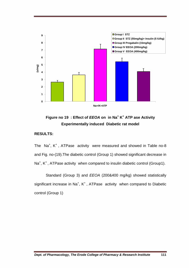
Dept. of Pharmacology, The Erode College of Pharmacy & Research Institute 111
Figure no 19 : Effect of EEOA on in Na+ K+ ATP ase Activity
Experimentally induced Diabetic rat model
RESULTS:
The Na+, K+ , ATPase activity were measured and showed in Table no-8
and Fig. no-(19).The diabetic control (Group 1) showed significant decrease in
Na+, K+ , ATPase activity when compared to insulin diabetic control (Group1).
Standard (Group 3) and EEOA (200&400 mg/kg) showed statistically
significant increase in Na+, K+ , ATPase activity when compared to Diabetic
control (Group 1)
0
1
2
3
4
5
6
7
8
9
Na+/K+ATP
(u/m
g)
Group I STZ
Group II STZ (55mg/kg)+ Insulin (5 IU/kg)
Group III Pregabalin (15mg/kg)
Group IV EEOA (200mg/kg)
Group V EEOA (400mg/kg)

Dept. of Pharmacology, The Erode College of Pharmacy & Research Institute 112
BEHAVIOURAL ASSESMENT
EFFECT OF EEOA ON HOT PLATE TEST:(Thermal Hyperalgesia)
Groups
Reaction Time in Sec
0 Week 4th
Week 6th
Week 8th
Week
Group I STZ
(55mg/kg)+vehicle 5.05 ± 0.84 4.46 ± 0.73 4.20 ± 0.27 3.91± 0.17
Group II STZ+
Insulin (5 IU/kg) 5.05 ± 0.31 *** 4.31 ± 0.67*** 4.11 ± 0.37*** 4.03 ± 0.54***
Group III STZ+ insulin + Pregabalin (15 mg/kg)
5.05 ± 0.56** ns
4.56 ± 0.16** 4.96 ± 0.56** 9.31 ± 0.16**
Group IV STZ+insulin+ EEOA (200mg/kg)
5.05 ± 0.31* a 4.60 ± 0.37* 4.70 ± 0.84ns a 9.54 ± 0.22ns aa
Group V STZ+ insulin EEOA (400mg/
5.05 ± 0.31ns a
5.20 ± 0.18ns a 8.81 ± 0.16ns aa 10.87 ± 0.12ns aa
Table no 9: Effect of EEOA on Hot plate test (Thermal hyperalgesia)
in Experimentally induced Diabetic rat model
The values were expressed as Mean ± S.E.M (n= 6 animals in each group)
*, **, ***, indicates significance P < 0.05 , P < 0.01 & P <0.001 when compared to insulin
diabetic control
a, aa indicates P < 0.05 & P < 0. 01 Significance between diabetic control
NS indicates Non-significant
Data was analysed by one-way ANOVA followed by Duncun Multiple range test (DMRT)

Dept. of Pharmacology, The Erode College of Pharmacy & Research Institute 113
Figure no 20 : Effect of EEOA on Hot plate test (Thermal Hyperalgesia) in
Experimentally induced Diabetic rat model
RESULTS:
The Paw Withdrawal latency were measured and showed in Table no-9
and Fig. no-(20). The diabetic control (Group 1) showed significant decrease
in Paw withdrawal latency when compared to the insulin diabetic control
(Group 2).
Standard (Group 3) and EEOA (200& 400 mg/kg) showed statistically
significant increase in Paw withdrawal latency when compared to Diabetic
control (Group 1).
0
2
4
6
8
10
12
14
0 week 4th week 6th week 8th week
Paw
wit
hd
raw
al (S
ec)
Group I STZ
Group II STZ (55mg/kg)+ Insulin (5 IU/kg)
Group III Pregabalin (15mg/kg)
Group IV EEOA (200mg/kg)
Group V EEOA (400mg/kg)

Dept. of Pharmacology, The Erode College of Pharmacy & Research Institute 114
EFFECT OF EEOA ON FORMALIN TEST: (Chemical hyperalgesia)
Groups FORMALIN
TEST(Acute phase)
FORMALIN TEST(Delayed
phase)
Group I STZ
(55mg/kg)+vehicle 32.93±1.08 30.11±1.86
Group II STZ+
Insulin (5 IU/kg) 58.54±2.16 60.88±1.96***
Group III STZ+ insulin + Pregabalin (15 mg/kg)
47.84±0.95 50.55±1.31aa
Group IV STZ+insulin+ EEOA (200mg/kg)
46.92±0.73 55.15±1.06aa
Group V STZ+ insulin EEOA (400mg/
53.65±1.18 62.56±1.18aa
Table no 10: Effect of EEOA on Formalin (chemical hyperalgesia) in Experimentally
induced Diabetic rat model
The values were expressed as Mean ± S.E.M (n= 6 animals in each group)
*,**, ***, indicates significance P < 0.05 , P < 0.01 & P <0.001 when compared to insulin
diabetic control
a, aa indicates P < 0.05 & P < 0. 01 Significance between diabetic control group
NS indicates Non-significant
Data was analysed by one-way ANOVA followed by Duncun Multiple range test (DMRT)

Dept. of Pharmacology, The Erode College of Pharmacy & Research Institute 115
Figure no 21 : Effect of EEOA on Formalin test (Chemical Hyperalgesia)
in Experimentally induced Diabetic rat model
RESULTS:
The response were measured and showed in Table no-10 and
Fig. no-(21). The Diabetic control (Group 1) showed significant decrease in
intensified nociceptive response and analgesic response when compared to
the insulin diabetic control (Group 2).
Standard (Group 3) and EEOA (200& 400 mg/kg) showed statistically
significant increase in intensified nociceptive response and analgesic when
compared to Diabetic control (Group 1).
0
10
20
30
40
50
60
70
80
Acute Phase Delayed Phase
Paw
lic
kin
g l
ate
ncy (
Sec)
Group I STZGroup II STZ (55mg/kg)+ Insulin (5 IU/kg)Group III Pregabalin (15mg/kg)Group IV EEOA (200mg/kg)Group V EEOA (400mg/kg)

Dept. of Pharmacology, The Erode College of Pharmacy & Research Institute 116
EFFECT OF EEOA ON TAIL FLICK TEST: (Thermal Hyperalgesia)
GROUPS
Reaction Time In Sec
0th
week 4th
week 6th
week 8th
week
Group I STZ
(55mg/kg)+vehicle 4.23 ± 016 4.21± 0.27 4.13± 0.15 4.09 ± 0.627
Group II STZ+
Insulin (5 IU/kg) 9.22 ± 0.57*** 4.36 ± 0.19*** 4.21 ± 0.15*** 4.45 ± 6.27***
Group III STZ+ insulin +
Pregabalin (15 mg/kg)
5.93 ± 0.27** ns
6.16 ± 0.31** ns
7.93 ± 0.96** ns
9.54 ± 0.73** ns
Group IV STZ+insulin+
EEOA (200mg/kg) 5.16 ± 0.16* 5.91 ± 0.22 ns 7.11 ± 0.54aa 9.6 ± 0.56aa
Group V STZ+ insulin
EEOA (400mg/ 5.39 ± 0.23 * 6.11 ± 0.56a 8.27 ± 0.22aa 9.93 ± 0.73aa
Table No11: Effect of EEOA on Tail Flick test (Thermal hyperalgesia)
in experimentally induced Diabetic rat model
The values were expressed as Mean ± S.E.M (n= 6 animals in each group)
*,**,***, indicates significance P < 0.05 , P < 0.01 & P <0.001 when insulin diabetic control
a, aa indicates P < 0.05 & P < 0. 01 Significance between diabetic control group
NS indicates Non-significant
Data was analysed by one-way ANOVA followed by Duncun Multiple range test (DMRT)
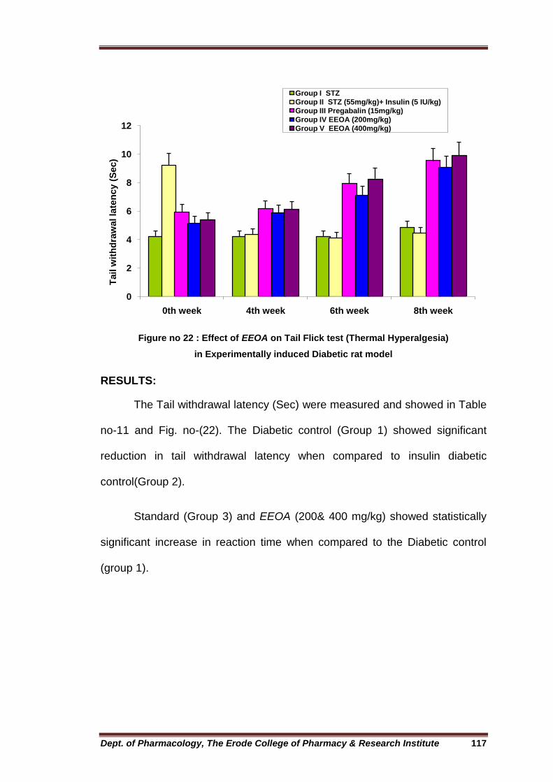
Dept. of Pharmacology, The Erode College of Pharmacy & Research Institute 117
Figure no 22 : Effect of EEOA on Tail Flick test (Thermal Hyperalgesia)
in Experimentally induced Diabetic rat model
RESULTS:
The Tail withdrawal latency (Sec) were measured and showed in Table
no-11 and Fig. no-(22). The Diabetic control (Group 1) showed significant
reduction in tail withdrawal latency when compared to insulin diabetic
control(Group 2).
Standard (Group 3) and EEOA (200& 400 mg/kg) showed statistically
significant increase in reaction time when compared to the Diabetic control
(group 1).
0
2
4
6
8
10
12
0th week 4th week 6th week 8th week
Tail w
ith
dra
wal la
ten
cy (
Sec)
Group I STZ
Group II STZ (55mg/kg)+ Insulin (5 IU/kg)
Group III Pregabalin (15mg/kg)
Group IV EEOA (200mg/kg)
Group V EEOA (400mg/kg)

Dept. of Pharmacology, The Erode College of Pharmacy & Research Institute 118
EFEECT OF TAIL IMMERSSION TEST: (Thermal hyperalgesia)
Effect of EEOA on Hot waterTail Immerssion test (Thermal hyperalgesia)
GROUPS Reaction Time in Sec
0th
week 4th
week 6th
week 8th
week
Group I STZ
(55mg/kg)+vehicle 10.31 ± 0.93 4.80 ± 0.24 4.98± 0.39 4.86 ± 0.29
Group II STZ+
Insulin (5 IU/kg) 5.56 ± 0.73*** 7.49± 0.27*** 6.13± 0.57*** 6.29 ± 0.11***
Group III STZ+
insulin +
Pregabalin (15
mg/kg)
5.96 ±0.16**ns 8.11 ± 0.56** ns 11.54 ± 0.67**
ns 3.08 ± 0.42** ns
Group IV
STZ+insulin+
EEOA (200mg/kg)
6.03 ± 0.18* 6.94 ± 0.013 a 7.93 ± 0.63 aa 8.93 ± 0.56aa
Group V STZ+
insulin EEOA
(400mg/
6.13 ± 0.31* 7.11 ± 0.015 aa 8.76 ± 0.23 aa 11.57 ± 0.27aa
Table no 12 : Effect of EEOA on Hot waterTail Immerssion test (Thermal hyperalgesia)
in Experimentally induced Diabetic rat model
The values were expressed as Mean ± S.E.M (n= 6 animals in each group)
*,**,***, indicates significance P < 0.05 , P < 0.01 & P <0.001 when compared insulin diabetic
control
a, aa indicates P < 0.05 & P < 0. 01 Significance between diabetic control
NS indicates Non-significant
Data was analysed by one-way ANOVA followed by Duncun Multiple range test (DMRT)

Dept. of Pharmacology, The Erode College of Pharmacy & Research Institute 119
Figure no 23 : Effect of EEOA on Hot water Tail Immerssion test (Thermal
Hyperalgesia) in Experimentally induced Diabetic rat model
RESULTS:
The Tail withdrawal latency (Sec) were measured and showed in Table
no-12 and Fig. no-(23). The Diabetic control (Group 1) showed significant
reduction in tail withdrawal latency when compared to the diabetic control
(Group 2).
Standard (Group 3) and EEOA (200& 400 mg/kg) showed statistically
significant increase in reaction time when compared to the Diabetic control
(group 1).
0
2
4
6
8
10
12
14
0th week 4th week 6th week 8th week
Tail w
ith
dra
wal la
ten
cy (
s)
Group I STZGroup II STZ (55mg/kg)+ Insulin (5 IU/kg)Group III Pregabalin (15mg/kg)Group IV EEOA (200mg/kg)Group V EEOA (400mg/kg)

Dept. of Pharmacology, The Erode College of Pharmacy & Research Institute 120
Effect of EEOA on Cold waterTail Immerssion test (Thermal allodynia)
GROUPS Reaction Time In Sec
0th
week 4th
week 6th
week 8th
week
Group I STZ
(55mg/kg)+vehicle 9.86 ± 0.67 5.36± 0.34 4.76 ± 0.13 1.13 ± 0.27
Group II STZ+
Insulin (5 IU/kg)
6.11 ± 0.27**
7.58 ± 0.16*** 6.53 ± 0.14*** 5.32 ±0.21***
Group III STZ+ insulin + Pregabalin (15
mg/kg)
6.34±0.18** ns
8.56 ± 0.27** ns
11.36 ±0.18**ns
13.24 ±0.18**ns
Group IV STZ+insulin+ EEOA
(200mg/kg) 6.46 ± 0.13* 7.67 ± 0.16a 8.54 ± 0.13aa 10.16 ± 0.93aa
Group V STZ+ insulin EEOA (400mg/
6.51 ± 0.15* 9.08 ± 0.53aa 10.56 ± 0.38aa 12.31 ± 0.84aa
Table no 13 : Effect of EEOA on Cold waterTail Immerssion test (Thermal allodynia) in
Experimentally induced Diabetic rat model
The values were expressed as Mean ± S.E.M (n= 6 animals in each group)
*,**,***, indicates significance P < 0.05 , P < 0.01 & P <0.001 when compared insulin diabetic
control
a, aa indicates P < 0.05 & P < 0. 01 Significance between diabetic control group
NS indicates Non-significant
Data was analysed by one-way ANOVA followed by Duncun Multiple range test (DMRT)

Dept. of Pharmacology, The Erode College of Pharmacy & Research Institute 121
Figure no 24 : Effect of EEOA on Cold water Tail Immersion test (Thermal allodynia)
in Experimentally induced Diabetic rat model
RESULTS:
The Tail withdrawal latency (Sec) were measured and showed in Table
no.13 and Fig. no-(24). The Diabetic control (Group 1) showed significant
reduction in tail withdrawal latency when compared to the insulin diabetic
control(Group 2).
Standard (Group 3) and EEOA (200& 400 mg/kg) showed statistically
significant increase in reaction time when compared to the Diabetic control
(group 1).
0
2
4
6
8
10
12
14
16
0th week 4th week 6th week 8th week
Tail w
ith
dra
wal la
ten
cy (
s)
Group I STZGroup II STZ (55mg/kg)+ Insulin (5 IU/kg)Group III Pregabalin (15mg/kg)Group IV EEOA (200mg/kg)Group V EEOA (400mg/kg)

Dept. of Pharmacology, The Erode College of Pharmacy & Research Institute 122
Effect of EEOA on Cold Plate test (cold allodynia)
Experimental Croups
Reaction Time in Sec
0 Week 4th
Week 6th
Week 8th
Week
Group I STZ
(55mg/kg)+vehicle 11.54 ± 0.27 4.96 ± 0.28 4.48 ± 0.27 4.13 ± 0.12
Group II STZ+
Insulin (5 IU/kg) 6.23 ± 0.28 *** 6.5 ± 0.92*** 4.46 ± 0.21*** 4.56 ± 0.38***
Group III STZ+ insulin +
Pregabalin (15 mg/kg)
6.78 ± 0.14** ns
6.98 ± 0.94** 8.56 ± 0.26** 9.78 ± 0.96**
Group IV STZ+insulin+
EEOA (200mg/kg) 6.82 ± 0.27* a 8.78 ± 0.24* 9.17 ± 0.34ns a 9.87 ± 0.34ns aa
Group V STZ+ insulin EEOA
(400mg/
6.35 ± 0.78ns a
8.31 ± 0.39ns a 9.26 ± 0.76ns
aa 10.13 ± 0.38ns
aa
Table no 14 : Effect of EEOA on Cold plate test (cold allodynia) in
Experimentally induced Diabetic rat model
The values were expressed as Mean ± S.E.M (n= 6 animals in each group)
*,**,***, indicates significance P < 0.05 , P < 0.01 & P <0.001 when compared insulin diabetic
control
a, aa indicates P < 0.05 & P < 0. 01 Significance between diabetic control.
NS indicates Non-significant
Data was analysed by one-way ANOVA followed by Duncun Multiple range test (DMRT)

Dept. of Pharmacology, The Erode College of Pharmacy & Research Institute 123
Figure no 25 : Effect of EEOA on Cold plate test (cold allodynia)
in Experimentally induced Diabetic rat model
RESULTS:
The paw escape latency (Sec) were measured and showed in Table
no.14 and Fig. no-(25). The Diabetic control (Group 1) showed significant
reduction in tail withdrawal latency when compared to the normal control
(Group 1).
Standard (Group 3) and EEOA (200& 400 mg/kg) showed statistically
significant increase in reaction time when compared to the Diabetic control
(Group 1).
0
2
4
6
8
10
12
14
0th week 4th week 6th week 8th week
Escap
e l
ate
ncy (
s)
Group I STZ
Group II STZ (55mg/kg)+ Insulin (5 IU/kg)
Group III Pregabalin (15mg/kg)
Group IV EEOA (200mg/kg)
Group V EEOA (400mg/kg)

Dept. of Pharmacology, The Erode College of Pharmacy & Research Institute 124
Effect of EEOA on Tail clip test (mechanical hypralgesia)
Experimental Groups
Reaction Time in Sec
0 Week 4th
Week 6th
Week 8th
Week
Group I STZ
(55mg/kg)+vehicle 6.93 ± 0.22 11.23 ± 0.56 11.87 ± 0.53 11.94 ± 0.73
Group II STZ+
Insulin (5 IU/kg)
11.67 ± 0.64 ***
10.26 ± 0.56*** 10.56 ± 0.53*** 10.62 ± 0.73***
Group III STZ+ insulin +
Pregabalin (15 mg/kg)
7.39 ± 0.13** ns
7.98 ± 0.61** 8.19 ± 0.36** 10.22 ± 0.86**
Group IV STZ+insulin+
EEOA (200mg/kg) 8.73 ± 0.87* a 8.92 ± 0.25* 9.16 ± 0.84ns a
10.58± 0.22ns aa
Group V STZ+ insulin EEOA
(400mg/
8.98 ± 0.54ns a
8.98 ± 0.11ns a 10.56 ± 0.58ns
aa 11.15± 0.18ns aa
Table no 15 : Effect of EEOA on Tail clip test (mechanical hypralgesia)
in Experimentally induced Diabetic rat model
The values were expressed as Mean ± S.E.M (n= 6 animals in each group)
*,**, ***, indicates significance P < 0.05 , P < 0.01 & P <0.001 when compared insulin diabetic
control
a, aa indicates P < 0.05 & P < 0. 01 Significance between diabetic control
NS indicates Non-significant
Data was analysed by one-way ANOVA followed by Duncun Multiple range test (DMRT)

Dept. of Pharmacology, The Erode College of Pharmacy & Research Institute 125
Figure no 26 : Effect of EEOA on Tail Clip test (Mechanical Hyperalgesia)
in Experimentally induced Diabetic rat model
RESULTS
The Tail withdrawal latency (Sec) were measured and showed in Table
no-15 and Fig. no-(26). The Diabetic control (Group 2) showed significant
reduction in tail withdrawal latency when compared to the diabetic
control(Group 1).
Standard (Group 3) and EEOA (200& 400 mg/kg) showed statistically
significant increase in reaction time when compared to the Diabetic control
(group 1).
0
2
4
6
8
10
12
14
0th week 4th week 6th week 8th week
Tail w
ith
dra
wal la
ten
cy
Group I STZ
Group II STZ (55mg/kg)+ Insulin (5 IU/kg)
Group III Pregabalin (15mg/kg)
Group IV EEOA (200mg/kg)
Group V EEOA (400mg/kg)

Dept. of Pharmacology, The Erode College of Pharmacy & Research Institute 126
Histopathology studies
GROUP 1: (Diabetic control): The nerve fascicle is surrounded by relatively
thick perineurium. The nerve axons are shrunken. Myelin sheath appears
fragmented and separated in some fibers. Edema is also noticed in between
the nerve fibers Other field shows marked thickening of less dense myelin
sheath in large myelinated fibers with loss of their axons.
GROUP II: (STZ+ insulin): sciatic nerve is surrounded by the dense
connective tissue epineurium. Each nerve fascicle is enclosed in the
specialized connective tissue perineurium. The axons appear clear with
variable diameters . The myelin is represented by dark ring surrounding the
axons.
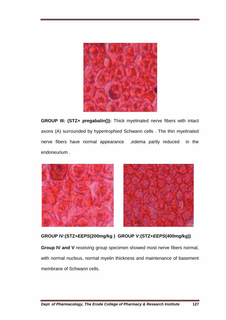
Dept. of Pharmacology, The Erode College of Pharmacy & Research Institute 127
GROUP III: (STZ+ pregabalin()): Thick myelinated nerve fibers with intact
axons (A) surrounded by hypertrophied Schwann cells . The thin myelinated
nerve fibers have normal appearance ,edema partly reduced in the
endoneurium .
GROUP IV:(STZ+EEPS(200mg/kg ) GROUP V:(STZ+EEPS(400mg/kg))
Group IV and V receiving group specimen showed most nerve fibers normal,
with normal nucleus, normal myelin thickness and maintenance of basement
membrane of Schwann cells.

Dept. of Pharmacology, The Erode College of Pharmacy & Research Institute 128
DISCUSSION
Hyperglycemia must be the originator in the pathogenesis of biabetic
complications. Diabetes is a heterogeneous set of disorders with a range of
pathologies and one of the most frequent complications of diabetes ids
peripheral neuropathy162. Diabetic neuropathy is characterised by progressive
chronic neuropathic pain that is tingling and burning in nature with
hyperesthesia(excessive physical sensitivity, especially of the skin) and
paresthesia(an abnormal sensation,typically tingling or pricking(pins and
needels), caused chiefly by pressure on or damage to peripheral nerves) with
deep aching and it is increased by touch 163. Neuropathic pain is defined as a
form of chronic pain that results from damage or abnormal function of central
or peripheral nervous system. The clinical presentation of neuropathic pain
induces hyperalgesia, allodynia and spontaneous pain164,165. Hyperglycemia
leads to the toxicity of neurons due to increased glucose oxidation , leading to
increased reactive oxygen species that may be controlled by the treatment
with antioxidant.166-168.
In this study, Ottelia alismoides(L) PERS., extract was given for the
treatment of neuropathic pain in STZ induced diabetic rats.
The preliminary phytochemical analysis of EEOA revealed the
presence of Glycosides, Alkaloids, Flavonoids, Tannins, Terpenoids, phenolic
compounds.The tannins, phenols and flavonoids present in the plant Ottelia
alismoides shows anti-diabetic action. The alkaloids and glycosides present in

Dept. of Pharmacology, The Erode College of Pharmacy & Research Institute 129
the herbal drugs are proved to be potent antioxidants as well as
nephroprotective agent169.
Acute toxicity studies revealed the non-toxic nature of the EEOA there
was no lethality or any toxic reactions found with high dose (2000 mg/kg body
weight) till the end of the study. According to the OECD 423 guidelines (Acute
Oral Toxicity: Acute Toxic Classic Method), an LD50 dose of 2000 mg/kg and
above was considered as unclassified so the EEOA was found to be safe.
In the present study STZ was used for induction of diabetes
neuropathy in rats. The development of neuropathy was observed at 0 week
after STZ induction, which was consistent with previous reports170. STZ
induced diabetic animal models are most accepted animal models of diabetic
neuropathy owing to their reproducibility and reversible diabetes171,172. STZ
action involved pancreatic DNA alkylation through GLUT2 transporter
mechanism. Which intern triggers multiple biochemical pathways such as
polyol pathway, hexosamine pathway, protein kinase C pathway(PKC),
advance glycation end (AGE) product and poly adipose ribose polymerase
(PARP) pathway all of these pathways contribute towards oxidative stress by
generating ROS in a mitochondria results in nerve damage and neuropathy173-
175. Rats injected with 55mg/kg showed significant increase in blood glucose
levels and decreased nociceptive thresholds. Similar thermal hyperalgesia,
mechanical and formalin evoked pain in STZ induced rats have been
demonstrated earlier176,177. Pregabalin a selective Ca 2.2 (α2-δ subunit)
channel anatagonist and it has has anti neuropathic pain, anticovulsant and
anxiolytic activities178. Treatment with EEOA extract along with insulin in

Dept. of Pharmacology, The Erode College of Pharmacy & Research Institute 130
diabetic rats significantly increased the nociceptive threshold, decreased
oxidative stress in a dose dependent manner.
Before recording the findings on the kidney and liver the effects of STZ
on the plasma glucose levels and body weights were noted. The observations
and results of the present study demonstrated that STZ was effective in
producing severe hyperglycaemia in experimental animals. The animals
Treated groups II,III & IV appeared ill-looking with loss of their body weights
because of injurious effects of STZ which caused alkylation of DNA and
produced hyperglycaemia and necrotic lesions179,180. Our in vivo results
showed that EEPS extract (200 and 400 mg/kg) treatment had an attenuating
effect on the serum glucose of the diabetic animals with significant
improvement in body weight it may be due to EEPS persisting insulin
secretogoge action like sulfonylureas.
Increase in the weight of kidney (hypertrophy) in STZ treated rats. The
mechanism of renal hypertrophy is unknown, evidence suggest that local
alterations in the production of one or more growth factors and/or their
receptors are crucial to this process181. An increase (hypertrophy) in the
weight of liver was compared with animals in treated groups. It could be
attributed to increased triglyceride accumulation leading to enlarged liver
which could be due to the increased influx of fatty acids into the liver induced
by hypoinsulinemia and the low capacity of excretion of lipoprotein secretion
from liver resulting from a deficiency of apolipoprotein B synthesis182. The
EEOA treated animals were found to have significant increased in organ

Dept. of Pharmacology, The Erode College of Pharmacy & Research Institute 131
weights when compared with that of animals with diabetes induced
neuropathy.
Oxidative stress is believed to be a biochemical trigger for sciatic nerve
dysfunction and reduced endoneurial blood flow in diabetic rats. Persistent
hyperglycemia disturbs the endogenous antioxidant defense mechanism and
prevents the free radicals scavenging activity183. In this study the activities of
various antioxidant enzymes SOD, CAT, GPx, LPO had been found to be
modulated in diabetic animals. The hyper activities of CAT, SOD could be a
response of overproduction of H2O2 and free radicals in the diabetic sciatic
nerve. The significant different in the activities of various biomarkers in
diabetic versus treated animals indicated the augmented oxidative stress and
tissue response during diabetes. Moreover nervous tissue is considered to be
rich in polyunsaturated fatty acids mainly arachidonic acid and
docosahexaenoic acids which are highly sensitive to LPO and depleted
antioxidants status. The diabetic animals exhibited the similar biochemical
imbalance in term of increased LPO levels and decreased GSH levels.
Hence, these levels revealed the magnitude of oxidative stress and extent of
structural and functional damage to the nervous system184.
Diabetes causes increases in the LPO products such as
malondialdehyde (MDA) or conjugated dienes in sciatic nerves. Scavenging
free radicals reduced the above effects of sciatic pain models185. In the
present study EEOA exhibited significant decrease in endogenous antioxidant
enzyme. This antioxidant activity of the Ottelia alismoides extract could also
have influenced its activity in diabetic neuropathic pain.

Dept. of Pharmacology, The Erode College of Pharmacy & Research Institute 132
Acute hyperglycemia induced by STZ elicits reduction of Na+, K+, ATPase
activity. Decrease in sciatic nerve Na+, K+, ATP ase activity could alter the
normal membrane axon repolarisation after the depolarization induced by an
action potential. EEOA treatment restored of Na+ K+ ATPase activity. It might
be possibly improved by inhibition of oxidative stress and also by amelioration
of vascular function186s.
Behavioral studies in diabetic rats often focus on the response to a
painful or non-painful sensory stimulus, there by measuring hyperalgesia and
allodynia respectively. The simplest of such tests measures the time to
withdrawal of a limb such as the tail or a paw from a noxious heat source, with
a faster withdrawal time being interpreted as hyperalgesia and a slower one
as hypoalgesia187. In the present study diabetic neuropathy using animal
models such as thermal hyperalgesia (tail flick, hot water tail immersion, hot
plate) mechanical hyperalgesia (tail clip method) cold allodynia (cold plate,
cold water tail immersion) and chemical hyperalgesia (formalin test)188.
These models are supposed to be behavioural biomarkers of diabetic
neuropathy. The hot plate and test involves two types of responses paw
licking and jumping. Both responses integrate at supraspinal structures with
the C and Aδ type I and II sensitive fibres participating in this model. The tail
withdrawal (tail flick, tail clip, tail immersion) respons after thermal stimuli is
elicited by the spinal motor reflex most probably via endogenous release of
substance P in the spinal cord189. EEOA extract showed antinociceptive effect
in the diabetic rats in both hot plate and tail immersion models, which indicate
that EEOA may act via central mechanism. Cold allodynia (cold plate) might

Dept. of Pharmacology, The Erode College of Pharmacy & Research Institute 133
be caused by disproportionate loss of Aδ fibers and sensitization of cold
receptors in peripheral neuropathic pain. Impaired blood flow also seems to
contribute to allodynia190. Vasodilator treatment has been demonstrated to
reduce allodynia in diabetic rats. EEOA may act on improved neuronal blood
flow by its direct vasorelaxant properties.
The nociception induced by formalin is associated with injured tissue. It
characterised by two phases acute & delayed. The acute phase(0-10min) is
short lived and initiates immediately after injection and is characterised by C-
fibre activation due to peripheral stimuli. The delayed phase(20-40min) is a
longer, persistent period caused by local tissue inflammation and also by
functional changes in the dorsal of the spinal cord. Therefore, this phase is
inhibited both both by opioids and analgesic agents. Substances that act
primarily as central analgesic inhibit both phases while peripherally acting
drugs inhibit only the delayed phase. In the present study the EEOA extract
exhibited analgesia only in the delayed phase which suggests that the extract
would possibly prevent the inflammatory markers induced pain perception.
The H&E stained lateral section of diabetic control sciatic nerve (A)
showed nerve fascicle is surrounded by relatively thick perineurium. The
nerve axons are shrunken. Myelin sheath appears fragmented and separated
in some fibers. Edema is also noticed in between the nerve fibers Other field
shows marked thickening of less dense myelin sheath in large myelinated
fibers with loss of their axons compared to (B). Treatment of Pregabalin and
Ottelia alismoides (400 mg/kg body weight) in diabetic rats was shown to
improve the thick myelinated nerve fibers with intact axons (A) surrounded by

Dept. of Pharmacology, The Erode College of Pharmacy & Research Institute 134
hypertrophied Schwann cells . The thin myelinated nerve fibers have normal
appearance ,edema partly reduced in the endoneurium in dose dependent
manner.
Therefore, Ottelia alismoides extract was proposed that in addition to
its antidiabetic, the antioxidant properties is the prominent features in
attenuation of diabetes induced neuropathy and its generating pain.

Dept. of Pharmacology, The Erode College of Pharmacy & Research Institute 135
10. SUMMARY AND CONCLUSION
The present study was to investigate the Neuroprotective effect of
EEOA in experimentally induced diabetic neuropathy in rats.
The phytochemical investigation reveals that the presence of tannins,
phenols & flavanoids are proved to be effective in diabetic treatment.
Diabetogenic effect of STZ is more reproducible and the metabolic
changes are much closer to those existing in human diabetes. Hence STZ is
considered the drug of choice for the induction of experimental diabetes.
The Blood glucose level significantly decreased with treatment of
EEOA proves that it has anti-diabetic activity.
The study shows marked inhibit in oxidative stress by enhancing
reduced activity of antioxidant enzyme SOD, CAT, GSH, GPx, Na+, K+,
ATPase & markedly elevate the level of LPO in liver compare to untreated
diabetic control animals, these findings further strengthen beneficial effect of
Ottelia alismoides as it showed production against STZ induced diabetic
neuropathic pain in rats.
After 4th week, diabetic control has shown signs of neuropathy while
treated group has showed significant improvement in their condition. From
this results, it is evident that EEOA has beneficial effect in protective against
STZ- induced diabetic neuropathy in rats.

Dept. of Pharmacology, The Erode College of Pharmacy & Research Institute 136
Histopathological assessment also showed that the damage caused by
STZ to the sciatic nerve is also markedly reduced by the administration of
Ottelia alismoides.
Finally these findings suggest that Ottelia alismoides treatment must be
beneficial to treat pain in diabetic animals.

Dept. of Pharmacology, The Erode College of Pharmacy & Research Institute 137
11. FUTURE PROSPECTIVES
Further study is required.,
1. To isolate and separate the active phytochemicals present in the
ethanolic extract of the Ottelia alismoides (L.) pers.
2. Formulation of the isolated lead molecule can be designed.
3. Clinical trial of the formulated molecule in healthy human volunteers
or diseased persons.

Dept. of Pharmacology, The Erode College of Pharmacy & Research Institute 138
12. BIBLIOGRAPHY
1. http://apps.who.int/iris/bitstream/10665/204871/1/9789241565257_eng.pdf
2. World Health Organization, Global Report on Diabetes. Geneva, 2016.
Accessed 30 August 2016.
3. "Simple treatment to curb diabetes". January 20, 2014.
4. Ramachandran A, Snehalatha C and Vijay Viswanathan. Burden of type 2
diabetes and its complications – The Indian scenario.Current Science.2002 ;
83(12):1471-1476.
5.http://study.com/academy//lesson/what-is-neuropathy-def-sym-
treatment,html
6.https://www.endocrineweb.com/diabetic-neuropathy/types-diabetic-
neuropathy.
7.http://www.mayoclinic.org/diseasesconditions/diabeticneuropathy/basics/tre
atment/con-20033336
8.https://en.wikipedia.org/wiki/Pain(Accessed 2ndApril 2015)
9. Smithira G, Ashma A, Rajamathanky H, Kavya V, Muhammed riyas K. A
review on ethanobotanical uses and pharmacology of plecospermum
spinosum . International journal of research in pharmacology and
pharmacotherapeutics.2017;6(1):45-49.
10.https://www.davita.com/kidney-disease/causes/diabetes/diabetes:-
definition,-causes-and-symptoms/e/4991(March 7 2017 )
11.Bansal v. Kalita j. Misra uk.Diabetic neuropathy. Postgraduate medical
journal.2006;82(964):95-100.
12.https://www.betterhealth.vic.gov.au/health/conditionsandtreatments/diabet
es-long-term-effects accessed on(28 6th 2017 )

Dept. of Pharmacology, The Erode College of Pharmacy & Research Institute 139
13.https://en.wikipedia.org/wiki/Macrovascular_disease (macrovascular)
14.http://www.mayoclinic.org/diseases-
conditions/diabetes/basics/complications/con-20033091 july 31 2014
15.http://www.nhs.uk/conditions/Cerebrovascular-
disease/pages/definition.aspx
16.http:www.medicalnewtoday.com/articles/184601.php
17.https://www.diapedia.org/acute-and-chronic-complications-of-
diabetes/7105379817/stroke Accessed on (march 10th 2015)
18.https://www.diabetesseifmanagement.com/diabetes-
resources/definition/peripheral-vascular-disease Accessed on(9.6.2017)
19.http://www.who.int/diabetes/action-online/bascies/en/Index3.html accessed
on (6.28.2017)
20.http://study.com/academy//lesson/what-is-neuropathy-def-sym-
treatment,html
21.https://www.endocrineweb.com/diabetic-neuropathy/types-diabetic-
neuropathy.
22. https://en.wikipedia.org/wiki/Peripheral_neuropathy
23.http://www.mayoclinic.org/diseases-conditions/peripheral-
neuropathy/symptoms-causes/dxc-20204947
24.Richard A C Hughes. "Clinical review: Peripheral neuropathy". British
Medical Journal. 324 (7335): 466–469. doi:10.1136/bmj.324.7335.466
25.Janet M. Torpy; Jennifer L. Kincaid; Richard M. Glass. "Patient page:
Peripheral neuropathy". Journal of the American Medical
Association. 303 (15):1556.

Dept. of Pharmacology, The Erode College of Pharmacy & Research Institute 140
26"Peripheral neuropathy fact sheet". National Institute of Neurological
Disorders and Stroke. 19 September 2012.
27. https://en.wikipedia.org/wiki/Proximal_neuropathy
28. https://en.wikipedia.org/wiki/Proximal_neuropathy
29.http://www.mayoclinic.org/diseasesconditions/autonomicneuropathy/basics
/definition/con-20029053
30.http://www.nytimes.com/health/guides/disease/autonomicneuropathy/overv
iew.html?mcubz=0(sep 11 2017)
31. http://emedicine.medscape.com/article/1173756-treatment
32. www.webmd.com/diabetes/tc/diabetic-focal-neuropathy-topic-overview
33. http://www.mcvitamines.com/cranial-neuropahty.htm.
34. http://emedicine.health.com/neuropathy/article_em.htm
35.Pourmand R. Diabetic neuropathy 1997;15:569-576.
36.Ibityoe, Rajbhandari neuropahy truncal pain. Q.J.Med.2021;15:1027-1031
37. Boulton AJM, Augur E, Ayyer DR. Diabetic thoracic polyradiculopathy
presending as an abdominal swelling. BMJ1984;289:798-799.
38. http://emedicine.medscape.com/article/249784-overview (Nov 11th 2015 )
39. https://en.wikipedia.org/wiki/Nerve_compression_syndrome
40.http://thebrain.mcgill.ca/flash/i/i_03/i_03_cl/i_03_cl_dou/i_03_cl_dou.html
(Accessed 2nd February 2017)
41. Ziegler D. Treatment of Diabetic Polyneuropathy. Update 2006. Ann N Y
Acad Sci 2006. 1084:250-266.

Dept. of Pharmacology, The Erode College of Pharmacy & Research Institute 141
42. Low PA, Nickander KK, Tritschler HJ. The roles of oxidative stress and
antioxidant treatment in experimental diabetic neuropathy. Diabetes
1997.46:38-42.
43. Packer L, Kraemer K, Rimbach G. Molecular aspects of lipoic acid in the
prevention of diabetes complications. Nutrition 2001;17:888-895.
44. Li N, Karin M. Is NF-kappaB the sensor of oxidative stress? FASEB
J.1999. 13:1137.
45. Shiba T, Inoguchi T, Sportsman JR, Health W, Bursell S, King GL.
Correlation of diacylglycerol and protein kinase C activity in rat retinal to
retinal circulation. Am J Physiol.1993;265:783-793.
46. Idris I, Donnelly R. Protein kinase Cbeta inhibition: a novel therapeutic
strategy for diabetic microangiopathy. Diabetes Vasc Dis.2006;3:172-178.
47. Nakamura Kato K, Hamada Y, Nakayama M, Chaya S, Nakashima E,
Naruse K et al. A protein kinase C-beta selective inhibitor ameliorates neural
dysfunction in streptozocin-induced diabetic rats. Diabetes.1999;48:2090-
2095.
48. Chattopadhyay M, Mata M, Fink D. Continuous delta opioid receptor
activation reduces neuronal voltage-gated sodium channel (Nav1.7) levels
through activation of protein kinase C in painful diabetic neuropathy. J
Neurosci.2008;28:6652-6658.
49. Cavaletti G, Miloso M, Nicolini G, Scuteri A, Tredici G. Emerging role of
mitogen-activated protein kinases in peripheral neuropathies. J Periph Nerv
Syst.2007; 12:175194.
50. Hall K, Liu KJ, Sima AA, Wiley JW. Impaired inhibitory G-protein function
contributes to increased calcium currents in rats with diabetic neuropathy. J
Neurophysiol.2001;86:760-770.
51. www.diabetes.niddk.nih.gov.

Dept. of Pharmacology, The Erode College of Pharmacy & Research Institute 142
52. Bhadada SK, Sahay RK, Jyotsna VP, Agarwal JK. Diabetic current
concept. Journal , Indian academy of clinical medicine.2001;2(4)305-318.
53.Dyck PJ. Invited review of limitation in predicting pathologic abnormality of
nerves from the EMG examination. Muscle nerve.1990;13(5):371-375.
54. Smieja M, Hunt DL, Edelman D, Etchells E, Cornuz J, Simel DL. Cinical
examination for the detection on protective sensation in the feet of diabetic
patients in cooperation group for ck=linical examination research, J Gen Intern
Med. 1999;14(7):418-424.
55. Maser RE, Nielson vk, Bass EB et al., Measuring diabetic neuropathy
assessment and comparison of clinical examination and quantitative sensory
testing.Diabetes care.1989;12(4)270-275.
56. Yang Z,Zhang Y, Chen R, Huang Y, Ji L,Sun F, Hong T, Zhan S.
Simpletests to screen for diabetic peripheral neuropathy (Protocol). Published
by John Wiley & Sons.17-37.
57. Mayfield JA, Sugarman JR. The use of the semmes Weinstein
monofilament and other threshold tests for preventing foot ulceration and
amputation in persons with dibetes. J.Fam Pract.2009;49(11):71-29.
58.Jimenez-Cohl, M.D.Carlos Grekin, M.D.Cristian Leyton M.D Claudio
VargasThermal Threshold: Research Study on Small Fiber Dysfunction in
Distal Diabetic Polyneuropathy Pedro.. Journal of Diabetes Science and
Technology Volume 6, Issue 1, January 2012.
59.Karagoz E, Tanridag T. The electrophysiology of diabetic neuropathy.
Internet journal of neurology.2004;5(1).
60. https://en.wikipedia.org/wiki/Pain
61. http://www.denalihealthcaremi.com/tag/classification-of-pain
62. Sherrington C. The Integrative Action of the Nervous System. Oxford:
Oxford University Press; 1906.

Dept. of Pharmacology, The Erode College of Pharmacy & Research Institute 143
63. Fein,A Nociceptors: the cells that sense
pain http://cell.uchc.edu/pdf/fein/nociceptors_fein_2012.pdf
64. Williams, S. J.; Purves, Dale (2001). Neuroscience. Sunderland, Mass:
Sinauer
65.Fields HL, Rowbotham M, Baron R (October 1998). "Postherpetic
neuralgia: irritable nociceptors and deafferentation". Neurobiol. Dis. 5 (4):
209–27
66. AitkenheadAR,Rowbotham DJ, Smith G. Textbook ofanaesthesia.4th ed.
Edinburgh: Churchill Livingstone; 2001.
67. nternational Association for the Study of pain. Retrieved 3 May 2015
68.https://www,nursingtimes.net/clinical-archive pain-management/
anatomyand physiology of pain /1860931/articles.
69. http://www.rnceus.com/ages/nociceptive.htm. (Accessed 20th February
2017)
70.https://www.78 steps health.us/body function/ascending-html
71.http://thebrain.mcgill.ca/flash/i/i_03/i_03_cl/i_03_cl_dou/i_03_cl_dou.html
(Accessed 2nd February 2017)
72.https://sites.google.com/site/geneticsofpain/descendingpainpathways.
(Accessed 2nd February 2017)
73.Tandan R, Lewis GA, Krusinski PB.topical capsaicin in painful diabetic
neuropathy.Diabetes care.1992;8-15
74. Max MB, Cunane M, Schafer SC et al., Amitryptiline releieves diabetic
pain in patients with normal or depressed mood. Neurology 1987:37-589.
75.Roseberg JM, Harrell C, Risitic H et al. The effect of gabapentine on
neuropathic pain.clin j pain.1997;13:251-255.

Dept. of Pharmacology, The Erode College of Pharmacy & Research Institute 144
76. Yasuda H, Terada M, Maeda K. Diabetic neuropathy and nerve
regeneration.Pubmed.2003;69(4):229-285.
77.htto://medical-dictionary.therefreebictionay.com/glucose control
78. Lynn Ang, Mamta, Jaiswal. Glucose control and diabetic neuropathy:
lesion from recent large clinical trails.2014;14(9):528.
79. Solders G, Tyden G. Improvement of nerve conduction in diabetic
neuropathy.Afollow up study 4 years later combined pancreatic and renal
transplantation. Diabetes.1992;41:946-951.
80. https://en.wikipedia.org/wiki/Aldose_reductase_inhibitor
81. Cameron NE, Cotter MA. Potential therapeutic approaches to the
treatment and prevention of diabetic neuropathy:Evidence from experimental
studies. Diabet Med.1993;10: 593-605.
82.http:// www. Adiabetesseifmanagement.comblog andioxidant-ala-
easespain from diabetic neuropathy.
83.https://www.healthcentral.com/article/acetyllcarnitinesupplement-a-
promising-teratment for-diabetic-peripheral-neuropathy.
84. Mendez JD, Ramos HG. Animal models in diabetes research. Archives of
medical research medicinal plants. Int Pharm Chem 2014;4:114-8.
1994;25(4):367-75.
85. Etuk EU, N. J. Animals models for studying diabetes mellitus. Agric Biol
2010;1:130-4.
86 . Iranloye BO, Arikawe AP, Rotimi G, Sogbade AO. Anti-diabetic and anti-
oxidant effects of Zingiber officinale on alloxaninduced and insulin-resistant
diabetic male rats. Nigeria.2011;26(1) 89-96.

Dept. of Pharmacology, The Erode College of Pharmacy & Research Institute 145
87. Brentjens R, Saltz L. pancreatic beta-cell damage.Islet cell tumors of the
pancreas: the medical oncologist's perspective of north
America.2001;81(3)527-542
88. 35 . Mythili MD, Vyas R, Akila G, Gunasekaran S. Effect of streptozotocin
on the ultrastructure of rat pancreatic islets. Microscopy research and
technique 2004;63(5):274-81.
89 . Patel R, Shervington A, Pariente JA, Martinez-Burgos MA, Salido GM,
Adeghate E, et al. Mechanism of exocrine pancreatic insufficiency in
streptozotocin-induced type 1 diabetes mellitus. Annals of the New York
Academy of Sciences.2006;1084:71-88.
90. Halim D, Khalifa K, Awadallah R, El-Hawary Z, El-Dessouky EA. Serum
mineral changes in dithizone-induced diabetes before and after insulin
treatment. Zeitschrift fur Ernahrungswissenschaft 1977;16(1):22-6.
91.Epand RM, Stafford AR, Tyers M, Nieboer E. Mechanism of action of
diabetogenic zinc-chelating agents. Model system studies. Molecular
pharmacology 1985;27(3):366-74.
92.Karasawa H, Takaishi K, Kumagae Y. Obesity-induced diabetes in mouse
strains treated with gold thioglucose: a novel animal model for studying β-cell
dysfunction. Obesity (Silver Spring, Md.) 2011;19(3):514-21.
93. Nagata M, Suzuki W, Iizuka S, Tabuchi M, Maruyama H, Takeda S, et al.
Type 2 diabetes mellitus in obese mouse model induced by monosodium
glutamate. Experimental animals / Japanese association for laboratory animal
science. 2006;55(2):109-115.
94. Arora Mahesh Kumar, Agarwal Anil, Baidya Dalim Kumar, Khanna Puneet
(2011). Pregabalin in acute and chronicpain. Journal of Anaesthesiology
Clinical Pharmacology. 2011;27 (3):307–14.
95. Martin D.J, McClelland D, Herd M.B, Sutton K.G, Hall M.D, Lee K,
Pinnock R.D, et al. Gabapentin-mediated inhibition of voltage-activated Ca2+

Dept. of Pharmacology, The Erode College of Pharmacy & Research Institute 146
channel currents in cultured sensory neurones is dependent on culture
conditions and channel sub-unit expression. Neuropharmacology. 2002;42:
353-66.
96. Faisal Mohd. The Pharmacological Evaluation of Epigallocatechin-3-
Gallate (EGCG) Against Diabetic Neuropathy in Wistar Rats.Int J sci Res Rev.
2012; 1(3): 75-87.
97. Stalin C, Gunasekaran V,Jayabalan G. Evaluation of Neuroprotective
Effect of Ficus benghalensis against Alloxan Induced Diabetic Neuropathy in
Rats. International Journal of Pharmacology, Phytochemistry and
Ethnomedicine. 2016;4:52-60.
98. Shakir D AlSharari, Salim S Al-Rejaie , Hatem M Abuohashish, Abdulaziz
M Aleisa, Mihir Y Parmar, Mohammed M Ahmed. Ameliorative Potential of
Morin in Streptozotocin-Induced Neuropathic Pain in Rats.Trop J Pharm Res.
2014;13(9):1429.
99.Mohammed Mubashir, Khan Dureshahwar, Hemant D. Une, Syed Shoaeb
Mohammad. Effects of Ethyl Acetate Fraction of Z. MauritianaLam. Leaves on
STZ Induced Diabetes and Diabetic Neuropathy In Mice.Int J Sci Res. 2014;
4(2): 109-116.
100. Amit Kumar Gangwar. Investigation of Neuroprotective Effect of
Rasagiline in Diabetic Neuropathy in Streptozotocin Induced Type 2 Diabetic
Rats.European Journal of Pharmaceutical and Medical Research.
2016;3(5):469-475 .
101. Parkar N, Addepalli V. Nobiletin on Diabetic Neuropathy in Experimental
rats. Austin J Pharmacol Ther. 2014;2 (5):1028.
102.Amal Jamil Fatani, Salim Salih AlRejaie, Hatem Mustafa Abuohashish,
Abdullah AlAssaf, MihirYogeshkumar Parmar, Mohammad Shamsul Ola, et
al. Neuroprotective effects of Gymnema sylvestre on streptozotocininduced

Dept. of Pharmacology, The Erode College of Pharmacy & Research Institute 147
diabetic neuropathy in rats. Experimental and Therapeutic
Medicine.2015;9(5): 1670–1678.
103. Ravikant, Abhay Kumar Verma, Priyanka Shrivastava. Effect of Coccinia
Indica Leaf Extract on Diabetic Neuropathy Pain In Rats. European Journal of
Pharmaceutical and Medical Research.2016;3(1): 415-420.
104. Sandeeep Kumar K, JameelaTahashildar, Karunakar Kota.
Neuroprotective effect of ethanolic root extract of Boerhaavia diffusa (Linn.)
against Streptozotocin induced Diabetic neuropathy in animal model. J.
Chem. Pharm. Res. 2016;8(3):831-840.
105. Pratibha D Nadig. Effect of Tinospora cordifoliaon experimental diabetic
neuropathy. Indian j pharmacol. 2012;44(5):580-583
106. Ranjithkumar R , Prathab Balaji S, Balaji B, Ramesh RV, Ramanathan
M.Standardized Aqueous Tribulus terristris (nerunjil) extract attenuates
hyperalgesia in experimentally induced diabetic neuropathic pain model: role
of oxidative stress and inflammatory mediators.Phytother Res.
2013;27(11):164657.
107.Ramdas B. PandhareI, B. Sangameswaran Popat B. MohiteI,Shantaram
G.Attenuating effect of seeds of Adenanthera pavonina aqueous extract in
neuropathic pain in streptozotocininduced diabetic rats: an evidence of
neuroprotective effects.Rev. bras. farmacogn. 2012;22(2).
108. Gowhar Ali, FazalSubhan, Muzaffar Abbas, JehanZeb, Muhammad
Shahid, Robert D. E, et al. streptozotocin-induced diabetic neuropathic pain
model for static or dynamic mechanical allodynia and vulvodynia: validation
using topical and systemic gabapentin. Naunyn-Schmiedeberg's Arch
Pharmacol. 2015; 388:1129–1140.
109.Nasser Zangiabadi, Hossein Mohtashami, Mohammad Shabani,Mandana
Jafari. Neuroprotective Effect of Cerebrolysin on Diabetic Neuropathy: A
Study on Male Rats.J Diabetes Metab. 2014;5(4):357.

Dept. of Pharmacology, The Erode College of Pharmacy & Research Institute 148
110. Jurairat Khongrum, Jintanaporn Wattanathorn, Supaporn Muchimapura,
Wipawee Thukhum-mee, Cholathip Thipkaew, Panakaporn Wannano.
Moringa oleifera Leaves Extract Attenuates Neuropathic Pain Induced by
Chronic Constriction Injury.Am.J. Applied Sci. 2012;9(8):1182-1187.
111.AyatKaeidi, Saeed Esmaeili-Mahani,VahidSheibani, Mehdi Abbasnejad,
Bahram Rasoulian, Zahra Hajializadeh, et al. Olive (Oleaeuropaea L.) leaf
extract attenuates early diabetic neuropathic pain through prevention of high
glucose-induced apoptosis: In vitro and in vivo studies. Journal of
Ethnopharmacology. 2011;136:188–196.
112.BhaskarNagilla, Pratap Reddy K. Neuroprotective and Antinociceptive
Effect of Curcumin in Diabetic Neuropathy in Rats.Int J Pharm PharmSci.
2014;6(5):131-138.
113.Premkumar N, Annamalai A.R, Thakur R S. Antinociceptive property of
EmbilicaofficinalisGaertn (Amla) in high fat diet-fed/low dose streptozotocin
induced diabetic neuropathy rats. Indian J Exp Biol. 2009;47:737-742.
114. Naveen kumar Gupta, Nithi Srivastva, Sanjeev puri. Neuroprotective
potential of azadirachta indica leaves in Diabetic rats. Asian J pharm clin
Res.2017;10(4):243-248.
115.Gomar Ali, Hosseini Abdolkarim, Mirazi Naser. Effect of hydroethanolic
extract of Rubus fruticosus on Neuropathic pain in wister diabetic rats.
Caspian journal of neurological science.2015;1(1):27-34.
116. Zhang yi, Yang Shao-long, Wang Ai-hong. Protective effect of ethanol
extracts of Hericium erinaceus on Alloxan-induced diabetic neuropathic pain
in rats.Hindawi publishing corporation.2015
117. Archana jorige, Akula Annapurna. Neuroprodective and antioxidant role
of pregabalin in sterptozotocine induced neurotoxicity. Indian journal of
pharmaceutical sciences and research.2016;7(11):4494-4500.
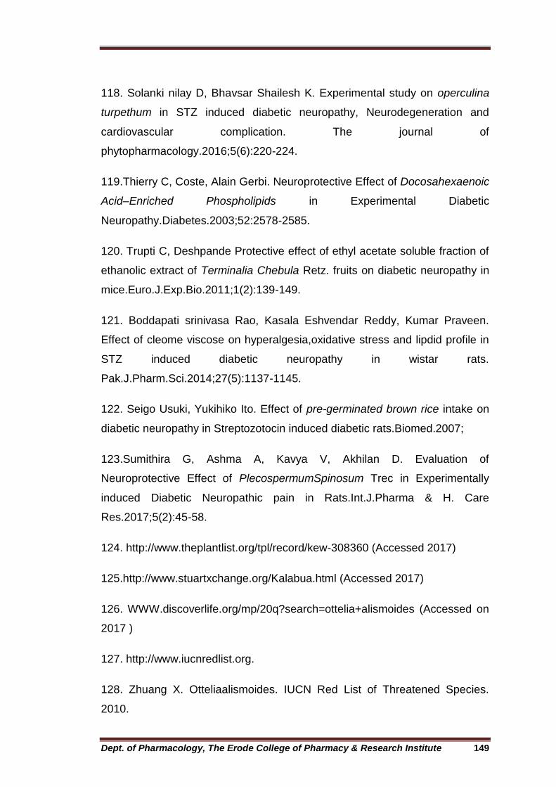
Dept. of Pharmacology, The Erode College of Pharmacy & Research Institute 149
118. Solanki nilay D, Bhavsar Shailesh K. Experimental study on operculina
turpethum in STZ induced diabetic neuropathy, Neurodegeneration and
cardiovascular complication. The journal of
phytopharmacology.2016;5(6):220-224.
119.Thierry C, Coste, Alain Gerbi. Neuroprotective Effect of Docosahexaenoic
Acid–Enriched Phospholipids in Experimental Diabetic
Neuropathy.Diabetes.2003;52:2578-2585.
120. Trupti C, Deshpande Protective effect of ethyl acetate soluble fraction of
ethanolic extract of Terminalia Chebula Retz. fruits on diabetic neuropathy in
mice.Euro.J.Exp.Bio.2011;1(2):139-149.
121. Boddapati srinivasa Rao, Kasala Eshvendar Reddy, Kumar Praveen.
Effect of cleome viscose on hyperalgesia,oxidative stress and lipdid profile in
STZ induced diabetic neuropathy in wistar rats.
Pak.J.Pharm.Sci.2014;27(5):1137-1145.
122. Seigo Usuki, Yukihiko Ito. Effect of pre-germinated brown rice intake on
diabetic neuropathy in Streptozotocin induced diabetic rats.Biomed.2007;
123.Sumithira G, Ashma A, Kavya V, Akhilan D. Evaluation of
Neuroprotective Effect of PlecospermumSpinosum Trec in Experimentally
induced Diabetic Neuropathic pain in Rats.Int.J.Pharma & H. Care
Res.2017;5(2):45-58.
124. http://www.theplantlist.org/tpl/record/kew-308360 (Accessed 2017)
125.http://www.stuartxchange.org/Kalabua.html (Accessed 2017)
126. WWW.discoverlife.org/mp/20q?search=ottelia+alismoides (Accessed on
2017 )
127. http://www.iucnredlist.org.
128. Zhuang X. Otteliaalismoides. IUCN Red List of Threatened Species.
2010.
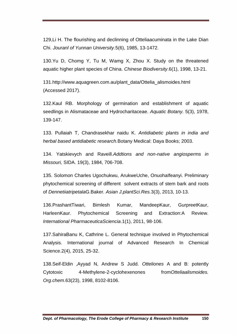
Dept. of Pharmacology, The Erode College of Pharmacy & Research Institute 150
129,Li H. The flourishing and declinning of Otteliaacuminata in the Lake Dian
Chi. Jouranl of Yunnan University.5(6), 1985, 131472.
130.Yu D, Chomg Y, Tu M, Wamg X, Zhou X. Study on the threatened
aquatic higher plant species of China. Chinese Biodiversity.6(1), 1998, 1321.
131.http://www.aquagreen.com.au/plant_data/Ottelia_alismoides.html
(Accessed 2017).
132.Kaul RB. Morphology of germination and establishment of aquatic
seedlings in Alismataceae and Hydrocharitaceae. Aquatic Botany. 5(3), 1978,
139147.
133. Pullaiah T, Chandrasekhar naidu K. Antidiabetic plants in india and
herbal based antidiabetic research.Botany Medical: Daya Books; 2003.
134. Yatskievych and Raveill.Addtions and non-native angiosperms in
Missouri, SIDA. 19(3), 1984, 706-708.
135. Solomon Charles Ugochukwu, ArukweUche, OnuohaIfeanyi. Preliminary
phytochemical screening of different solvent extracts of stem bark and roots
of DennetiiatripetalaG.Baker. Asian J.plantSci.Res.3(3), 2013, 10-13.
136.PrashantTiwari, Bimlesh Kumar, MandeepKaur, GurpreetKaur,
HarleenKaur. Phytochemical Screening and Extraction:A Review.
International PharmaceuticaSciencia.1(1), 2011, 98-106.
137.SahiraBanu K, Cathrine L. General technique involved in Phytochemical
Analysis. International journal of Advanced Research In Chemical
Science.2(4), 2015, 25-32.
138.Seif-Eldin ,Ayyad N, Andrew S Judd. Otteliones A and B: potently
Cytotoxic 4-Methylene-2-cyclohexenones fromOtteliaalismoides.
Org.chem.63(23), 1998, 8102-8106.
Dept. of Pharmacology, The Erode College of Pharmacy & Research Institute 151
139.Thomas R, Hoye, Seif-Eldin, Ayyad N, Hollie J, Beckord et al,. New
Diarylheptanoids and a HydroxylatedOttelionealismoides. Nat prod
commun.8(3), 2013, 351-358.
140.Sumithira G, Kavya V.,Ashma A, Kavinkumar MC. Areview of
Ethanobotanical and phytopharmacology of Otteli alismoides(L.) PERS. Int J
of Res in Pharmacology & Pharmacotherapeutics.2017;6(3):302-311.
141. Harboure. J.B. Phytochemical methods a guide to modern techniques of
plant analysis: London .2nd ed.chapman and hall.1984
142. Ecobichon D.J., The Basis of Toxicology Testing. New York, CRC
Press.1997
143. Seema Surendran ,Vijayalakshmi Krishna moorthy. Effect of Ethanolic
Extracts of Cyperus Rotundus on Biochemical Parameters of Diabetic
Cararact induced Wister Albino Rats. Int J Pharm Bio sci.2014;5(2):708-717
144. P.H. Agarkar, J.S. Kulkarni, V.L. Maheswari, R.A. Fursule. Practical
Biochemistry.Pune, India. NiraliPrakashan. 2008.
145. Kelli.A.Sullivan. Mouse Models of Diabetic Neuropathy. Neurobiology Of
Disease.2007;28(1):276–285.
146.Vogel H. Drug Discovery and Evaluation of Pharmacological Assays. 2nd
ed. Germany: Springer Publication. 2002
147. Schleyerbach R. (2002). Analgesic, anti-inflammatory, and anti-pyretic
activity. In Vogel HG editor. Drug discovery and evaluation. 2nd ed.
Germany: Springer;. p.696.
148. Schleyerbach R. Analgesic, anti-inflammatory, and anti-pyretic activity. In
Vogel HG editor. Drug discovery and evaluation of pharmacological assays.
2nd ed. Germany: Springer. 2002

Dept. of Pharmacology, The Erode College of Pharmacy & Research Institute 152
149. Khalilzadeh O, Anvari M, Khalilzadeh A, et al.. Involvement of
amlodipine, diazoxide, and glibenclamide in development of morphine
tolerance in mice. Int J Neurosci.2008;118:503–18.
150. Luiz AP, Moura JD, Meotti FC. (Antinociceptive action of ethanolic
extract obtained from roots of Humiriantheraampla Miers. J
Ethnopharmacol.2007;114:355–63.
151.Rosellini W, Casavant R, Engineer . Wireless peripheral nerve stimulation
increase pain thershold in twoneuropathic rat model.Exp.Neurol 235,621-626.
152. Sharma S, Kulkarni S.K., Agrewala J.N, Chopra K. Eur. J
Pharmacol.2008, 598, 32-36.
153. Misra HP, Fridovich I. The role of superoxide anion in the auto oxidation
of epinephrine and simple assay of superoxide dismutase.J.Biol. Chem. 1972;
247:3170-3184
154. Sinha AK, Colorimetric assay of catalase, Analytical Biochemistry.
1972;47(2): 389-394.
155. Bhesh Raj Sharma. Nelumbo nucifera leaf extract attenuated pancreatic
β-cell toxicity induced by interleukin-1β and interferon-ý, and increased insulin
secretion of pancreatic β-cell in streptozotocin-induced diabetic rats.
156. Rotruck. Glutathione peroxidase was assayed by the method of .1973
157. Okhawa H, Ohigni N, Yagi K. Assay of lipid peroxides in animal tissues
by thiobarbituric acid reaction. Anal Biochem. 1979;95:351-359.
158. Suhail M., Rizvi S.I. Red cell membrane (Na+ + K+)-ATPase in diabetes
mellitus. Biochem. Biophys. Res. Commun. 1987;146:179–186
159.Fiske C., Subbarow Y. The colourimetric determination of phosphorus. J.
Biol. Chem. 1925;66:375–400.
160. http://histologylab.ccnmtl.columbia.edu/histological_techniques/

Dept. of Pharmacology, The Erode College of Pharmacy & Research Institute 153
161.file:///C:/Users/USER/Desktop/Duncan's%20new%20multiple%20range%
20test%20%20Wikipedia.htm (Accessed 5th January 2017)
162. Saini AK, Kumar HAS, Sharma SS. Preventive and curarive effect of
ederavone on the nerve function and oxidative stress in experimental diabetic
neuropathy. Eur.J.Pharmacol. 2007;568.1-3:164-172.
163. Baynes JW .Role of oxidative stress in development of complications in
diabetes .Diabetes.1991;40(4):405-412.
164. Dickenson AH, Matthews EA, Suzuki R. Neurobiology of neuropathic
pain: mode of action of anticonvulsants. Europian journal of
pain.2002;1(6):51-60.
165. Abdi S, Lee DH, Chung JM. The anti-allodynic effects of
amitriptyline,gabaprntine and lidocaine in a rat model of neuropathic pain.
Anesh Analg.1998:87(6):1360-6.
166. Andrade P, Visser-Vandewalle V, Rosario JS. The thalidomide analgesic
effect is associated with differential TNF-a receptor expression in the dorsal
horn of the spinal cord as studied in a rat model of neuropathic pain. Brain
Res.2012;1450:24-32.
167. Pittenger GL, Malik RA, Burcus N, Boulton. Specific fibre deficits in
sensorimotor diabetic polyneuropathy corresponds to cytotoxicity against
neuroblastoma cells of sera from patients with diabetes. Diabetes
care.1999;22(5).
168. Greene DA, Stevens MJ, Obrosova I, Feldman. Glucose-induced
oxidative stress and programmed cell death in diabetic neuropathy. Eur. J.
Pharmacol.1999;30(375):217-223.
169. Atmani D, Chaher N, Berboucha M, Ayouni K, Ethanopharmacol
.2009;112(2):303-309

Dept. of Pharmacology, The Erode College of Pharmacy & Research Institute 154
170. Ramadas, Pandhare B, Sangameswaran B, Popat. RBFBJP.
2012,22(2),428-35.
171. Sima AA, Zhang WX, Tze WJ, Tai J. Diabetic neuropathy in STZ-induced
diabetic rats and effect of allogenic islet cell transplantation: Morphonometric
analysis. Diabetes.1988;37(8):1129-36.
172. Hussein JI, EI-Matty D, EI-Khayat ZA. Brain neurotransmitters in diabetic
rats treated with CO enzyme Q 10. Int .J.Pharm paharm Sci.2012;4:554-6.
173. Delaney CA, Dunger A, Di matteo M. Biochem pharmacol.1995;50.
174. Elsner M, Guldbakke B, Tiedge M. Diabetology.2000;43:1528-33.
175. Vincent AM, Russell JW, Low P. Endocrine Rev.2004;25:612-28.
176.Courteix C, Bardin M, Chantelauze J, LavarenneJ,Eschalier A. Study of
the sensitivity of the diabetes- induced pain model in rats to a range of
analgesics. Pain.1994;51:153- 160.
177.Calcutt NA, Jorge MC, Yaksh TL, Chaplan TL. Tactile allodynia and
formalin hyperalgesia in Streptozotocin- diabetic rats: effects of insulin, aldose
reductase inhibition and lidocaine. Pain.1996;68:293-299.
178. Stump P.Drugs today.2009;45:19-27.
179. Habibuddin M, Daghriri H, Humaira T. Antidiabetic effect of alcoholic
extract of Caralluma sinaica L. on sterptozotocin-induced diabetic rabbits.
J.Ethnopharmacol.2008;117(2):215-20
180. Piyachaturawat, P, poprasit. Gastric mucosal secretions and lesions by
different doses of STZ in rats. Toxicol.1991.55:21-29
181. Sharma K, Ziyadeh F,N. Hyperglycemia and diabetic kidney disease.
The case for transforming growth factor-beta as a key mediators.
Diabetes.1995;44(10):1139-46.

Dept. of Pharmacology, The Erode College of Pharmacy & Research Institute 155
182. Lee SI, Kim JS, Oh SH, Park KY. Antihyperglycemic effect of fomitopsis
pinicola extracts in sterptozotocin-induced diabetic rats.
J.Med.Food.2008;11(3):518-24.
183. Figueroa-Romero C, Sadidi M, Feldman EL. Mechanisms of disease: the
oxidative stress theory of diabetic neuropathy. Rev Endocr Metab
Disord.2008;9(4):301-14.
184. Gupta A, Gupta Y. Diabetic neuropathy. Part J Pak Med
Asssoc.2014;64:714-721.
185. Zeimmermann M. Ethical guidelines for investigation of experimental
pain in conscious animals.1983;16:109-110.
186. Faisal Mohd. The Pharmacological Evaluation of Epigallocatechin-3-
Gallate (EGCG) Against Diabetic Neuropathy in Wistar Rats.Int J sci Res Rev.
2012; 1(3): 75-87.
187. Michael B. Biochemistry and molecular cell biology of diabetic
complications. Nature. 2001;414:813-820.
188. Talbot S, Chahmi E, Dias JP, Key role Michael B. Biochemistry and
molecular for spinal dorsal horn micoglial kinin B1 receptor in early pain
diabetic neuropathy. Journal of neuroinflammation.2010;7(36):1-6
189. Yashpal et al., blocks the nociceptive responses to noxious thermal and
chemical stimuli in the rat. Neuroscience.1993;52:1039-1047.
190. Sharma et al.,curcumin attenuates thermal hypralgesia in a diabetic
mouse mode of neuropathic pain. Eur J pharmacol.2006;536:256-261.