Embed Size (px)
Citation preview
Evaluation of Glutathione Dependant Antioxidant Enzymes in Maternally Inherited Type 2 Diabetes
Mellitus. Utpal J. Dongre 1, V.G. Meshram 2*
1Utpal J. Dongre, Assistant Professor, Department of Biochemistry, Dr. Ambedkar College, Deeksha Bhoomi, Nagpur 440010, Maharashtra, India
2*Dr. Virendra G. Meshram, Professor, University Department of Biochemistry, RTM Nagpur University, Nagpur 440033, Maharashtra, India.
Abstract: Aim: This study has been undertaken to evaluate an activity of glutathione dependant antioxidant enzymes in patients with a history of maternal inherited type 2 diabetes mellitus. Method: The present study included three families among which two families are diabetic and one normal healthy family as control. The level of Glutathione, glutathione reductase, glutathione peroxidase and glutathione –s- transferase were estimated via various biochemical standard methods. Result: As compared to the normal healthy control samples the level of glutathione was found significantly decreased in both the diabetic families (family 1 p<0.01 and family 2 p<0.001). There was no significant difference in glutathione reductase activity in family 1 (p>0.05) and family 2 (p>0.05). This study shows an increase in an activity of glutathione peroxidase in family 1 (p<0.001), family 2 (p<0.001) and Glutathione –s-transferase in family 1 (p<0.01), family 2 (p<0.01). Conclusion: This study represents the levels of various glutathione based antioxidant enzymes, which can use as a marker for antioxidant levels in maternally inherited type 2 diabetes mellitus.
Keywords: Mitochondrial DNA, Glutathione, Glutathione Reductase, Glutathione Peroxidase, Diabetes Mellitus, Oxidative Stress etc.
Abbreviations: NA (Deoxyribo Nucleic Acid), GSH (Reduced Glutathione), GSSG (Oxidised Glutathione), GST (Glutathione –S- Transferase), GPX (Glutathione Peroxidase).
INTRODUCTION: Diabetes mellitus is widespread not only in India but also around the world. As per the American Diabetes Association (ADA) approximately 25.8 million people have been reported with diabetes, 18.8 million people are diagnosed with diabetes and 79 million people are in pre diabetic state [1,2]. In the year 2000 the widespread of the diabetes mellitus around the world was 171 millions, which may rise to 366 million in the year 2030 [3,4]. In current scenario, more than 40 million people in India are diabetic [5]. Mitochondria are involved in energy generation via oxidative phosphorylation. Oxidation of glucose generates various reducing equivalents like FADH2 and NADH, which exerts their electron to the electron transport chain and initiates an energy generation process [6,7]. But during the transport of electron through the electron transport chain it may get leaked. Leaked electron can generate free radicals like, peroxinitrite (ON00-), OH-, O, H2O2 etc., which are highly reactive in nature and can react with DNA, protein and other cell components. It may cause aberrant cellular communications [8]. Humans have generated a defence mechanism against these free radicals attack through various antioxidant enzymes, including catalase, superoxide dismutase, glutathione reductase, glutathione peroxidise, glutathione s transferase, etc. [9], among these enzymes glutathione based enzymes carrying potent scavenging activity for H2O2 [10].
Mitochondria are the main region where respiration takes place and inherited from mother to their offspring’s. Nuclear DNA in association with mitochondrial DNA codes for the polypeptide of an electron transport chain. An abnormal mitochondrial DNA may codes for an unusual polypeptide, which may result in lower ATP generation and thus reported as a causative agent for type 2 diabetes mellitus. Maternal inheritance of such defective mitochondria can generate more oxidative stress in families [11,12,13,14,15]. GSH contain a thiol group and prevalent in almost every tissues of mammals in an opulent amount to defend an oxidative stress and recorded as a potent biomarker for the redox imbalance within the cells [16]. Many studies corroborate the role of GSH in diabetes [17]. Reduced GSH level reported as one of the factors of DNA damage by oxidative stress in type 2 diabetes mellitus [18]. Oxidative stress is generated by an imbalance between free radicals and its scavenging systems. An aberrant activity of antioxidant enzymes may generate high oxidative stress in patients of type 2 diabetes mellitus, which may give rise to other serious complications [19].
MATERIAL AND METHOD: Sample collections and family history: Altogether eighteen samples from three families has been selected for this study, wherein family 1 and family 2 are diabetic
Utpal J. Dongre et al /J. Pharm. Sci. & Res. Vol. 7(3), 2015, 137-140
137
whereas family 3 is non diabetic (healthy control) between the age group of 18 to 70 years. Samples were collected after taking a signed consent form from the patients. All the samples and family history of patients were taken from “Diabetes Hospital” of Dr. Shailesh Pitale, located at Nagpur, Maharashtra, India. Inclusion Criteria: Families with a history of maternally inherited diabetes. Exclusion Criteria: Any kind of paternal history of type 2 diabetes mellitus, type 1 diabetes mellitus, Juvenile diabetes mellitus. Sample Preparation: 1 ml blood samples were collected in EDTA vacutainer tubes. Then it was centrifuged at 3000×g for 15 minutes for collection of plasma. The plasma samples were recentrifuged at 3000×g for same time to avoid the carryover of blood cells and were collected in new tubes. All collected plasma samples were stored at -20˚C until further analysis. Enzymatic Analysis: All standard methods were used to determine the concentration of various glutathione based antioxidant enzymes. Glutathione was estimated according to the method given by Beutler et. al. (1963) [20], an activity of Glutathione Reductase was assayed by the method of Racker et. al. (1955) [21], Glutathione Peroxidise was estimated by the method of Rotruck et. al. (1973) [22], Glutathione -S- Transferase was assayed as per Habig et. al. (1974) [23] and protein was estimated by Lowery method (1951) [24]. Statistical Analysis: Statistical analyses were done using Med Calc statistical software. All results were expressed in Mean ± SD. The two tailed probability student’s T test was used to differentiate between the two diabetic families assuming unequal variance. P< 0.05 was taken as a standard for significance difference.
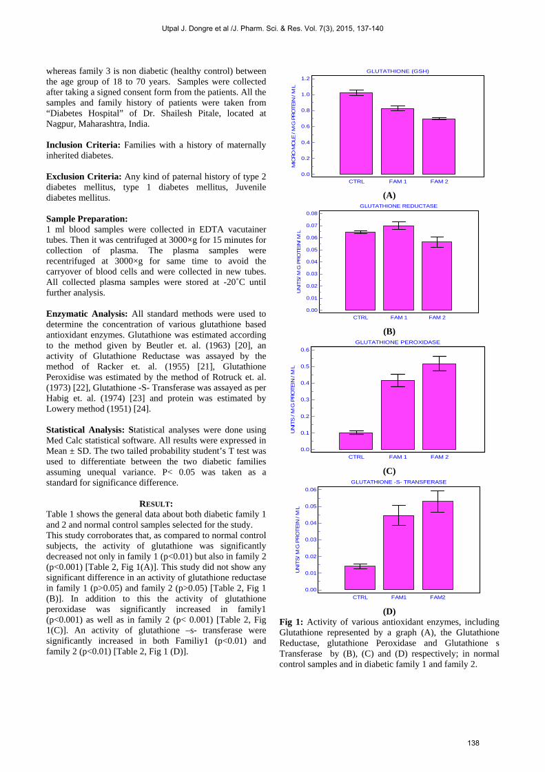
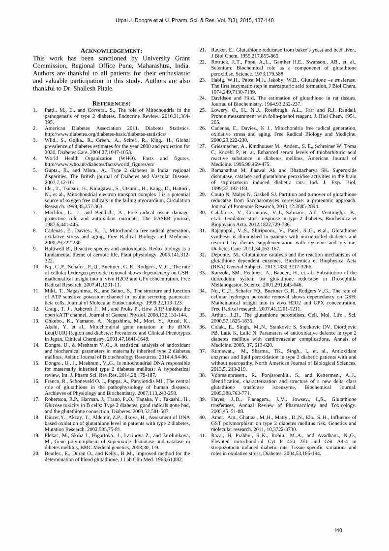
RESULT: Table 1 shows the general data about both diabetic family 1 and 2 and normal control samples selected for the study. This study corroborates that, as compared to normal control subjects, the activity of glutathione was significantly decreased not only in family 1 (p<0.01) but also in family 2 (p<0.001) [Table 2, Fig 1(A)]. This study did not show any significant difference in an activity of glutathione reductase in family 1 (p>0.05) and family 2 (p>0.05) [Table 2, Fig 1 (B)]. In addition to this the activity of glutathione peroxidase was significantly increased in family1 (p<0.001) as well as in family 2 (p< 0.001) [Table 2, Fig 1(C)]. An activity of glutathione –s- transferase were significantly increased in both Familiy1 (p<0.01) and family 2 (p<0.01) [Table 2, Fig 1 (D)].
GLUTATHIONE (GSH)
1.2
1.0
0.8
0.6
0.4
0.2
0.0
MIC
RO
MO
LE / M
G P
RO
TEIN
/ M
L
CTRL FAM 1 FAM 2
(A) GLUTATHIONE REDUCTASE
0.08
0.07
0.06
0.05
0.04
0.03
0.02
0.01
0.00
UN
ITS
/ M
G P
RO
TE
IN/ M
L
CTRL FAM 1 FAM 2
(B) GLUTATHIONE PEROXIDASE
0.6
0.5
0.4
0.3
0.2
0.1
0.0
UN
ITS / M
G P
RO
TEIN
/ M
L
CTRL FAM 1 FAM 2
(C) GLUTATHIONE -S- TRANSFERASE
0.06
0.05
0.04
0.03
0.02
0.01
0.00
UN
ITS
/ M
G P
RO
TE
IN / M
L
CTRL FAM1 FAM2
(D) Fig 1: Activity of various antioxidant enzymes, including Glutathione represented by a graph (A), the Glutathione Reductase, glutathione Peroxidase and Glutathione s Transferase by (B), (C) and (D) respectively; in normal control samples and in diabetic family 1 and family 2.
Utpal J. Dongre et al /J. Pharm. Sci. & Res. Vol. 7(3), 2015, 137-140
138
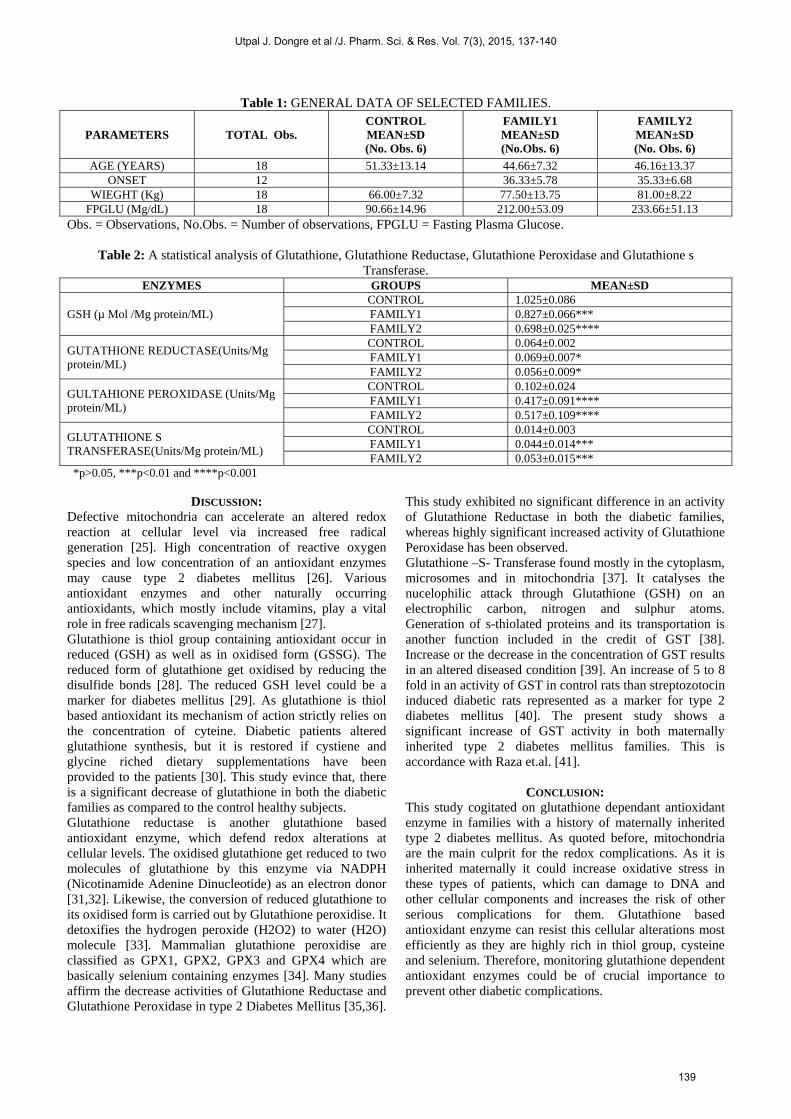
Table 1: GENERAL DATA OF SELECTED FAMILIES.
PARAMETERS TOTAL Obs. CONTROL MEAN±SD (No. Obs. 6)
FAMILY1 MEAN±SD (No.Obs. 6)
FAMILY2 MEAN±SD (No. Obs. 6)
AGE (YEARS) 18 51.33±13.14 44.66±7.32 46.16±13.37 ONSET 12 36.33±5.78 35.33±6.68
WIEGHT (Kg) 18 66.00±7.32 77.50±13.75 81.00±8.22 FPGLU (Mg/dL) 18 90.66±14.96 212.00±53.09 233.66±51.13
Obs. = Observations, No.Obs. = Number of observations, FPGLU = Fasting Plasma Glucose.
Table 2: A statistical analysis of Glutathione, Glutathione Reductase, Glutathione Peroxidase and Glutathione s Transferase.
ENZYMES GROUPS MEAN±SD
GSH (µ Mol /Mg protein/ML) CONTROL 1.025±0.086 FAMILY1 0.827±0.066*** FAMILY2 0.698±0.025****
GUTATHIONE REDUCTASE(Units/Mg protein/ML)
CONTROL 0.064±0.002 FAMILY1 0.069±0.007* FAMILY2 0.056±0.009*
GULTAHIONE PEROXIDASE (Units/Mg protein/ML)
CONTROL 0.102±0.024 FAMILY1 0.417±0.091**** FAMILY2 0.517±0.109****
GLUTATHIONE S TRANSFERASE(Units/Mg protein/ML)
CONTROL 0.014±0.003 FAMILY1 0.044±0.014*** FAMILY2 0.053±0.015***
*p>0.05, ***p<0.01 and ****p<0.001
DISCUSSION: Defective mitochondria can accelerate an altered redox reaction at cellular level via increased free radical generation [25]. High concentration of reactive oxygen species and low concentration of an antioxidant enzymes may cause type 2 diabetes mellitus [26]. Various antioxidant enzymes and other naturally occurring antioxidants, which mostly include vitamins, play a vital role in free radicals scavenging mechanism [27]. Glutathione is thiol group containing antioxidant occur in reduced (GSH) as well as in oxidised form (GSSG). The reduced form of glutathione get oxidised by reducing the disulfide bonds [28]. The reduced GSH level could be a marker for diabetes mellitus [29]. As glutathione is thiol based antioxidant its mechanism of action strictly relies on the concentration of cyteine. Diabetic patients altered glutathione synthesis, but it is restored if cystiene and glycine riched dietary supplementations have been provided to the patients [30]. This study evince that, there is a significant decrease of glutathione in both the diabetic families as compared to the control healthy subjects. Glutathione reductase is another glutathione based antioxidant enzyme, which defend redox alterations at cellular levels. The oxidised glutathione get reduced to two molecules of glutathione by this enzyme via NADPH (Nicotinamide Adenine Dinucleotide) as an electron donor [31,32]. Likewise, the conversion of reduced glutathione to its oxidised form is carried out by Glutathione peroxidise. It detoxifies the hydrogen peroxide (H2O2) to water (H2O) molecule [33]. Mammalian glutathione peroxidise are classified as GPX1, GPX2, GPX3 and GPX4 which are basically selenium containing enzymes [34]. Many studies affirm the decrease activities of Glutathione Reductase and Glutathione Peroxidase in type 2 Diabetes Mellitus [35,36].
This study exhibited no significant difference in an activity of Glutathione Reductase in both the diabetic families, whereas highly significant increased activity of Glutathione Peroxidase has been observed. Glutathione –S- Transferase found mostly in the cytoplasm, microsomes and in mitochondria [37]. It catalyses the nucelophilic attack through Glutathione (GSH) on an electrophilic carbon, nitrogen and sulphur atoms. Generation of s-thiolated proteins and its transportation is another function included in the credit of GST [38]. Increase or the decrease in the concentration of GST results in an altered diseased condition [39]. An increase of 5 to 8 fold in an activity of GST in control rats than streptozotocin induced diabetic rats represented as a marker for type 2 diabetes mellitus [40]. The present study shows a significant increase of GST activity in both maternally inherited type 2 diabetes mellitus families. This is accordance with Raza et.al. [41].
CONCLUSION: This study cogitated on glutathione dependant antioxidant enzyme in families with a history of maternally inherited type 2 diabetes mellitus. As quoted before, mitochondria are the main culprit for the redox complications. As it is inherited maternally it could increase oxidative stress in these types of patients, which can damage to DNA and other cellular components and increases the risk of other serious complications for them. Glutathione based antioxidant enzyme can resist this cellular alterations most efficiently as they are highly rich in thiol group, cysteine and selenium. Therefore, monitoring glutathione dependent antioxidant enzymes could be of crucial importance to prevent other diabetic complications.
Utpal J. Dongre et al /J. Pharm. Sci. & Res. Vol. 7(3), 2015, 137-140
139
ACKNOWLEDGEMENT: This work has been sanctioned by University Grant Commission, Regional Office Pune, Maharashtra, India. Authors are thankful to all patients for their enthusiastic and valuable participation in this study. Authors are also thankful to Dr. Shailesh Pitale.
REFERENCES: 1. Patti., M., E., and Corvera., S., The role of Mitochondria in the
pathogenesis of type 2 diabetes, Endocrine Review. 2010,31,364-395.
2. American Diabetes Association 2011. Diabetes Statistics. http://www.diabetes.org/diabetes-basic/diabetes-statistics/
3. Wild., S., Gojka., R., Green., A., Sciref., R., King., H., Global prevalence of diabetes estimates for the year 2000 and projection for 2030, Diabetes Care. 2004,27,1047-1053.
4. World Health Organization (WHO). Facts and figures. http://www.who.int/diabetes/facts/world_figures/en/
5. Gupta., R., and Misra., A., Type 2 diabetes in India: regional disparities, The British journal of Diabetes and Vascular Disease. 2007,7,12-16.
6. Ide., T., Tsutsui., H., Kinugawa., S., Utsumi., H., Kang., D., Hattori., N., et al., Mitochondrial electron transport complex I is a potential source of oxygen free radicals in the failing myocardium, Circulation Research. 1999,85,357-363.
7. Machlin., L., J., and Bendich., A., Free radical tissue damage: protective role and antioxidant nutrients, The FASEB journal, 1987,6,441-445.
8. Cadenas., E., Davies., K., J., Mitochondria free radical generation, oxidative stress and aging, Free Radical Biology and Medicine. 2000,29,222-230.
9. Halliwell B., Reactive species and antioxidants. Redox biology is a fundamental theme of aerobic life, Plant physiology. 2006,141,312-322.
10. Nq., C.,F., Schafer., F.,Q., Buettner., G.,R., Rodgers., V.,G., The rate of cellular hydrogen peroxide removal shows dependenecy on GSH: mathematical insight into in vivo H2O2 and GPx concentration, Free Radical Research. 2007,41,1201-11.
11. Miki., T., Nagashima., K., and Seino., S., The structure and function of ATP sensitive potassium channel in insulin secreting pancreatic beta cells, Journal of Molecular Endocrinology. 1999,22,113-123.
12. Craig., T. J., Ashcroft F., M., and Proks P., How ATP inhibits the open kATP channel, Journal of General Physiol. 2008,132,131-144.
13. Ohkubo., K., Yamano, A., Nagashima, M., Mori. Y., Anzai, K., Akehi, Y, et al., Mitochondrial gene mutation in the tRNA Leu(UUR) Region and diabetes: Prevalence and Clinical Phenotypes in Japan, Clinical Chemistry, 2001,47,1641-1648.
14. Dongre, U., & Meshram V.,G., A statistical analysis of antioxidant and biochemical parameters in maternally inherited type 2 diabetes mellitus, Asiatic Journal of Biotechnology Resources. 2014,4,94-96.
15. Dongre., U., J., Meshram., V.,G., Is mitochondrial DNA responsible for maternally inherited type 2 diabetes mellitus: A hypothetical review, Int. J. Pharm Sci. Rev.Res. 2014,28,179-187.
16. Franco, R., Schoneveld O. J., Pappa, A., Panyiotidis MI., The central role of glutathione in the pathophysiology of human diseases, Archieves of Physiology and Biochemistry. 2007,113,243-258.
17. Robertson, R.P., Harman. J., Trano, P.,O., Tanaka, Y., Takashi., H., Glucose toxicity in B cells: Type 2 diabetes, good radicals gone bad, and the glutathione connection, Diabetes. 2003,52,581-587.
18. Dincer,Y., Akcay, T., Aldemir, Z.P., Ilkova, H., Assesment of DNA based oxidation of glutathione level in patients with type 2 diabetes, Mutation Research. 2002,505,75-81.
19. Flekac, M., Skrha J., Higartova., J., Lacinova Z., and Jarolimkova, M., Gene polymorphism of superoxide dismutase and catalase in dibetes mellitus, BMC Medical genetics, 2008,30, 1-9.
20. Beutler., E., Duran O., and Kelly., B.,M., Improved method for the determination of blood glutathione, J Lab Clin Med. 1963,61,882.
21. Racker, E., Glutathione reducatse from baker’s yeast and beef liver., J Biol Chem. 1955,217,855-865.
22. Rotruck, J.,T., Pope, A.,L., Ganther H.E., Swanson., AB., et. al., Selenium: Biochemical role as a componenet of glutathione peroxidise, Science. 1973,179,588
23. Habig, W.H., Pabst M.J., Jakoby, W.B., Glutathione –s trnsferase. The first enzymatic step in mercapturic acid formation, J Biol Chem. 1974,249,7130-7139.
24. Davidson and Hird, The estimation of glutathione in rat tissues, Journal of Biochemistry. 1964,93,232-237.
25. Lowery, O., H., N.,J., Rosebrugh, A.L., Farr and R.J. Randall, Protein measurement with folin-phenol reagent, J. Biol Chem. 1951, 265.
26. Cadenas, E., Davies., K J., Mitochondria free radical generation, oxidative stress and aging, Free Radical Biology and Medicine. 2000,29,222-230.
27. Griesmacher, A., Kindhouser M., Andert., S. E., Schreiner W, Toma C, Knoebl P, et. al. Enhanced serum levels of thiobarbituric acid reactive substance in diabetes mellitus, American Journal of Medicine. 1995.98,469-475.
28. Ramanathan M, Jiaswal Ak and Bhattacharya SK. Superoxide dismutase, catalase and glutathione peroxidise activities in the brain of strptozotocin induced diabetic rats. Ind. J. Exp. Biol, 1999;37:182-183.
29. Couto N, Malys N, Gaskell SJ. Partition and turnover of glutathione reducatse from Saccharomyces cerevisiae: a proteomic approach. Journal of Proteome Research, 2013;12:2885-2894.
30. Calabrese., V., Cornelius., V.,L, Salinaro., AT., Ventimglia., B., et.al., Oxidative stress response in type 2 diabetes, Biochemica et Biophysica Acta. 2012,1822,729-736.
31. Rajagopal., V.,S., Shiripoom., V., Patel., S.,G., et.al., Glutathione synthesis is diminished in patients with uncontrolled diabetes and restored by dietary supplementation with cysteine and glycine, Diabetes Care. 2011,34,162-167.
32. Deponte., M., Glutathione catalysis and the reaction mechanisms of glutathione dependent enzymes, Biochemica et Biophysica Acta (BBA) General Subjects. 2013,1830,3217-3266.
33. Kanzok., SM., Fechner., A., Baucer., H., et. al., Substitution of the thioredoxin system for glutathione reducatse in Drosophilla Mellanogastor, Science. 2001,291,643-646.
34. Nq., C.,F., Schafer FQ., Buettner G.,R., Rodgers V.,G., The rate of cellular hydrogen peroxide removal shows depenedancy on GSH: Mathematical insight into in vivo H2O2 and GPX concentration, Free Radical research. 2007,41,1201-1211.
35. Arthur., J.,R., The glutathione peroxidises, Cell. Mol. Life . Sci. 2000,57,1825-1835.
36. Colak., E., Singh., M.,N., Stankovic S, Sreckovic DV, Diordjevic PB, Lalic K, Lalic N. Parameters of antioxidative defence in type 2 diabetes mellitus with cardiovascular complications, Annals of Medicine. 2005, 37, 613-620.
37. Kumawat., M., Sharma., TK., Singh., I., et. al., Antioxidant enzymes and lipid peroxidation in type 2 diabetic patients with and without neuropathy, North American Journal of Biological Sciences. 2013,5, 213-219.
38. Vdomsinprasert., R., Ponjaroenkit., S., and Ketterman., A.,J., Identification, characterization and structure of a new delta class glutathione trnsferase isoenzyme, Biochemical Journal. 2005,388,763-771.
39. Hayes, J.,D., Flanagem., J.,V., Jowsey., I.,R., Glutathione trnsferases, Annual Review of Pharmacology and Toxicology. 2005,45, 51-88.
40. Amer., Am., Ghattas., M.,H., Matty., D.,N., Ela., S.,H., Influence of GST polymorphism on type 2 diabetes mellitus risk, Genetics and molecular research. 2011, 10,3722-3730.
41. Raza., H, Prabhu., S.,K., Robin., M.,A., and Avadhani., N.,G., Elevated mitochondrial Cyt P 450 2E1 and GSt A4-4 in strepozotocin induced diabetic rats, Tissue specific variations and roles in oxidative stress, Diabetes. 2004,53,185-194.
Utpal J. Dongre et al /J. Pharm. Sci. & Res. Vol. 7(3), 2015, 137-140
140