Embed Size (px)
Citation preview

ORIGINAL CONTIBUTION Open Access
Evaluation of phytochemical compositionand antioxidative, hypoglycaemic andhypolipidaemic properties of methanolicextract of Hemidesmus indicus roots instreptozotocin-induced diabetic miceAnkita Joshi, Harsha Lad, Harsha Sharma and Deepak Bhatnagar*
Abstract
Background: Hemidesmus indicus is an important medicinal plant and extensively used in Ayurvedic and Unanisystem of medicine. The aim of the study was to evaluate the antioxidant, hypoglycaemic and hypolipidemic potentialof methanolic extract of roots of Hemidesmus indicus (HIE) in streptozotocin (STZ) induced diabetic mice.
Methods: HIE was analyzed by LC-MS to determine its phytochemical composition. The in-vitro antioxidant activity ofHIE was analyzed through inhibition of free radical scavenging activity (FRSA), total antioxidant power (TAP) andreducing power. Diabetes in mice was induced by a single dose of STZ followed by HIE treatment. The antioxidative,hypoglycaemic and hypolipidemic activity were studied ex-vivo in tissues of diabetic mice.
Results: Phytochemical composition of hemidesmus indicus roots (HIE) revealed the presence of phenols, flavanoids,terpenoids and about 40 different phytoconstituents by LC-MS analysis. Inhibition of lipid peroxidation (LPO) andmodulation in superoxide dismutase (SOD), catalase (CAT), glutathione-S- transferase (GST) activity and glutathione(GSH) content showed potent antioxidant activity of HIE in STZ induced diabetic mice, which was also substantiated byin-vitro antioxidant assays. The decrease in fasting blood glucose and serum lipid profile was also observed in miceadministered HIE.
Conclusion: It is proposed that HIE modulates the oxidant/antioxidant in favor of reducing oxidative stress, hypoglycemiaand improved the lipid profile in treated groups.
Keywords: Streptozotocin, Hypoglycaemic, Hypolipidaemic, Lipid peroxidation, Antioxidants, Hemidesmus indicus
BackgroundDiabetes mellitus is a complex chronic metabolic disordercharacterized by alterations in carbohydrate, protein andfat metabolism. The metabolic changes are caused by theinsufficiency of secretion or action of endogenous insulin[1]. Oxidative stress is suggested to be one of the mechan-ism underlying diabetes and diabetic complications. Itresults from an oxidant/antioxidant imbalance in favourof oxidants leading to damage of various intracellularcomponents such as proteins, lipids and nucleic acid as
well as extracellular matrix components such as proteo-glycans and collagens. Free radicals are formed by glucoseoxidation, non enzymatic glycation of proteins, oxidativedegradation of glycated proteins and increased lipid oxida-tion (LPO), which may promote oxidative stress and leadto the development of insulin resistance [2]. The harmfuleffects of oxidative stress are counteracted by the cellulardefense mechanism, which consists of enzymes, nonenzy-matic and metabolic antioxidants. To treat diabetes variousoral antihyperglycaemic agents have been developed overthe past years, which include sulphonylureas, biguanides,α-glucosidase inhibitors and thiazolidinediones [3].Hypoglycaemia, lactic acidosis and gastrointestinal
* Correspondence: [email protected] of Biochemistry, Devi Ahilya University, Khandwa Road, Indore, MP452017, India
© The Author(s). 2018 Open Access This article is distributed under the terms of the Creative Commons Attribution 4.0International License (http://creativecommons.org/licenses/by/4.0/), which permits unrestricted use, distribution, andreproduction in any medium, provided you give appropriate credit to the original author(s) and the source, provide a link tothe Creative Commons license, and indicate if changes were made.
Joshi et al. Clinical Phytoscience (2018) 4:7 DOI 10.1186/s40816-018-0064-0
intolerance are some of the adverse effect of these drugs.These side-effects of synthetic drugs along with drug-resistance have led to the resurgence of phytomedicine andsearch for novel type of antioxidant and antidiabetic frommedicinal plants.Hemidesmus indicus belongs to the family Periplocaceae
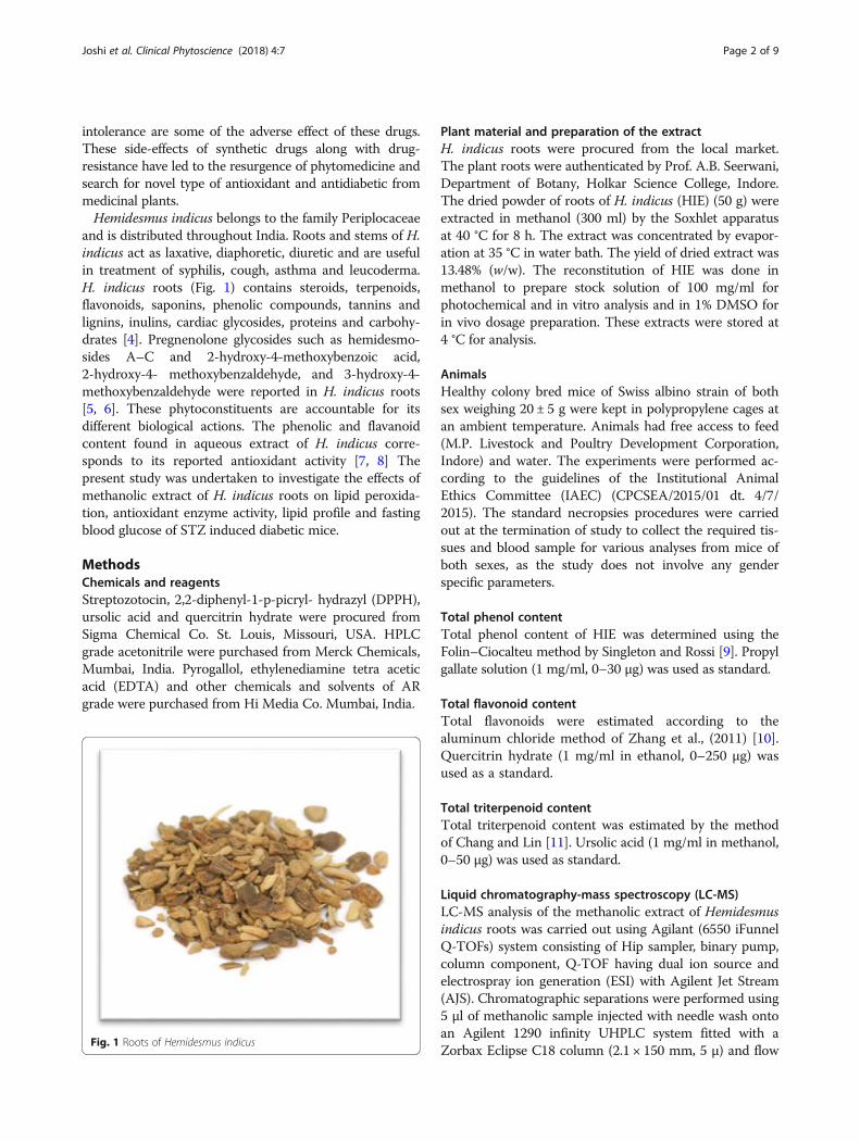
and is distributed throughout India. Roots and stems of H.indicus act as laxative, diaphoretic, diuretic and are usefulin treatment of syphilis, cough, asthma and leucoderma.H. indicus roots (Fig. 1) contains steroids, terpenoids,flavonoids, saponins, phenolic compounds, tannins andlignins, inulins, cardiac glycosides, proteins and carbohy-drates [4]. Pregnenolone glycosides such as hemidesmo-sides A–C and 2-hydroxy-4-methoxybenzoic acid,2-hydroxy-4- methoxybenzaldehyde, and 3-hydroxy-4-methoxybenzaldehyde were reported in H. indicus roots[5, 6]. These phytoconstituents are accountable for itsdifferent biological actions. The phenolic and flavanoidcontent found in aqueous extract of H. indicus corre-sponds to its reported antioxidant activity [7, 8] Thepresent study was undertaken to investigate the effects ofmethanolic extract of H. indicus roots on lipid peroxida-tion, antioxidant enzyme activity, lipid profile and fastingblood glucose of STZ induced diabetic mice.
MethodsChemicals and reagentsStreptozotocin, 2,2-diphenyl-1-p-picryl- hydrazyl (DPPH),ursolic acid and quercitrin hydrate were procured fromSigma Chemical Co. St. Louis, Missouri, USA. HPLCgrade acetonitrile were purchased from Merck Chemicals,Mumbai, India. Pyrogallol, ethylenediamine tetra aceticacid (EDTA) and other chemicals and solvents of ARgrade were purchased from Hi Media Co. Mumbai, India.
Plant material and preparation of the extractH. indicus roots were procured from the local market.The plant roots were authenticated by Prof. A.B. Seerwani,Department of Botany, Holkar Science College, Indore.The dried powder of roots of H. indicus (HIE) (50 g) wereextracted in methanol (300 ml) by the Soxhlet apparatusat 40 °C for 8 h. The extract was concentrated by evapor-ation at 35 °C in water bath. The yield of dried extract was13.48% (w/w). The reconstitution of HIE was done inmethanol to prepare stock solution of 100 mg/ml forphotochemical and in vitro analysis and in 1% DMSO forin vivo dosage preparation. These extracts were stored at4 °C for analysis.
AnimalsHealthy colony bred mice of Swiss albino strain of bothsex weighing 20 ± 5 g were kept in polypropylene cages atan ambient temperature. Animals had free access to feed(M.P. Livestock and Poultry Development Corporation,Indore) and water. The experiments were performed ac-cording to the guidelines of the Institutional AnimalEthics Committee (IAEC) (CPCSEA/2015/01 dt. 4/7/2015). The standard necropsies procedures were carriedout at the termination of study to collect the required tis-sues and blood sample for various analyses from mice ofboth sexes, as the study does not involve any genderspecific parameters.
Total phenol contentTotal phenol content of HIE was determined using theFolin–Ciocalteu method by Singleton and Rossi [9]. Propylgallate solution (1 mg/ml, 0–30 μg) was used as standard.
Total flavonoid contentTotal flavonoids were estimated according to thealuminum chloride method of Zhang et al., (2011) [10].Quercitrin hydrate (1 mg/ml in ethanol, 0–250 μg) wasused as a standard.
Total triterpenoid contentTotal triterpenoid content was estimated by the methodof Chang and Lin [11]. Ursolic acid (1 mg/ml in methanol,0–50 μg) was used as standard.
Liquid chromatography-mass spectroscopy (LC-MS)LC-MS analysis of the methanolic extract of Hemidesmusindicus roots was carried out using Agilant (6550 iFunnelQ-TOFs) system consisting of Hip sampler, binary pump,column component, Q-TOF having dual ion source andelectrospray ion generation (ESI) with Agilent Jet Stream(AJS). Chromatographic separations were performed using5 μl of methanolic sample injected with needle wash ontoan Agilent 1290 infinity UHPLC system fitted with aZorbax Eclipse C18 column (2.1 × 150 mm, 5 μ) and flow
Fig. 1 Roots of Hemidesmus indicus
Joshi et al. Clinical Phytoscience (2018) 4:7 Page 2 of 9

rate was 200 μl/min. The column was held at 95% SolventA (water) and 5% Solvent B (acetonitrile) for 2 min,followed by an 20 min step gradient from 5% B to 95% B,then 5 min with 5% A, 95% B. Then the elution wasachieved with a linear gradient from 5% A to 95% A for4 min. The following parameters were used throughoutthe MS experiment: for electro spray ionization with posi-tive ion polarity, the capillary voltage was set to 3500 V,the capillary temperature to 250 °C, the nebulizer pressureto 35 psi and the drying gas flow rate to 13 L/ min. Dataacquisition and mass spectrometric evaluation were car-ried out using software Agilent Mass Hunter Qualitativeanalysis B.06.
Free radical scavenging activity using 1, 1, 2,2-diphenyl-p-picryl hydrazylThe method is based on the reduction of an ethanolic solu-tion of DPPH by hydrogen donating groups of antioxidantsubstance [12]. The decrease in DPPH absorption at517 nm was measured.
Total antioxidant power using ferric reducing antioxidantpowerThe total antioxidant capacity (TAC) of HIE was deter-mined using the ferric reducing antioxidant power(FRAP) [13].
Reducing powerThe reducing power of the test samples was determinedaccording to the method of Oyaizu [14]. The reductiveability was measured by the reduction of FeCl3 inpresence of plant extracts. Ascorbic acid dissolved indistil water having concentration ranging from 0 to17.6 μg was used as positive control.
Induction of diabetesA single dose of freshly prepared STZ (180 mg/kg bodywt.) in cold 0.01 M citrate buffer (pH 4.5) was adminis-tered intraperitonially to overnight fasted mice to inducediabetes [15]. Streptozotocin causes β-cell toxicity viamechanism involving both free radical mediated damageand alkylation of DNA [16]. Mice were orally adminis-tered 10% (w/v) glucose for 24 h after STZ injection toovercome hypoglycaemic shock and after 72 h, fastingblood glucose (FBG) was measured. Mice having FBGlevels above 250 mg/dl were considered diabetic andwere selected for further experiments. The FBG of theanimals were estimated by glucometer (Akkiscan,Nempro Care, India) at every 3rd day, after the induc-tion of diabetes up to the end of treatment (day 12th).The variation in the body weight during the study periodwas recorded (provided in Additional file 1) and the foodand water intake was monitored in the animals.
Experimental designThe mice were divided into four groups of 6 animals each.The groups of animals were control mice without anytreatment (Group 1), STZ induced diabetic mice (Group2), mice treated with glibenclamide (10 mg/kg/day, orally)from 1stday of diabetes induction to 12th day (Group 3)and mice treated with HIE (35 mg/kg/day, orally) from 1stto 12th day after the induction of diabetes (Group 4). Theanimals were observed for the development of diabetes upto day 5 of STZ administration. Treatments in group 3and 4 were administered to diabetic mice for 12 days afterdevelopment of diabetes.
Collection and processing of biological samplesThe animals were sacrificed under mild ether anesthesia.Liver and kidney homogenate (10%) was prepared usingPotter-Elvehjem Homogenizer (Remi, Mumbai, India) inice cold phosphate buffer saline (PBS) (1: 9, v/v) followedby centrifugation at 16000 xg for 30 min at 4 °C. Bloodwas collected by cardiac puncture in citrated tubes.Erythrocytes lysate was prepared as described earlier[17]. The supernatant obtained after centrifugation oftissue homogenate and erythrocytes lysate were immedi-ately used to determine antioxidant enzymes and proteincontent.
Measurement of serum biochemical parametersBlood was collected from diabetic and treated groups andthe serum samples were analyzed using commerciallyavailable kits (Beacon Diagnostics, Navsari, India) forcholesterol, HDL, LDL, VLDL triglycerides by ELISA platereader (LISA Plus, Rapid Diagnostics, China) and spectro-photometer (Shimadzu, UV-1800, Japan). The quantitativeestimations were performed according to themanufacturer’s protocol.
Determination of malondialdehyde levelsMalondialdehyde (MDA) content in liver, kidney anderythrocytes was measured by HPLC method [18].
Determination of antioxidant statusThe activity of superoxide dismutase (SOD), catalase(CAT) and glutathione-S-transferase (GST) was measuredin tissue homogenates and erythrocytes lysate. The re-duced GSH content was measured in tissue homogenatesand blood [19–22].
Statistical analysisThe results obtained were analyzed by the SPSS softwarepackage version 20. The mean values obtained for thedifferent groups were compared by one-way ANOVA,followed by post hoc -Tukeys (HSD) test.
Joshi et al. Clinical Phytoscience (2018) 4:7 Page 3 of 9

ResultsDetermination of phytoconstituentsPhytochemical analysis of HIE was evaluated and theresults are represented in Table 1.
Qualitative mass spectral analysisHIE showed 73 unique mass signals, out of which puta-tive empirical formulas of 62 compounds were obtainedand identified by comparison with phytochemical data-base {developed by Sophisticated Analytical Instrument
Facility - Indian Institute of Technology, Bombay (SAIF-IITB) and Pubchem} and details of the compounds areprovided in Additional file 1. The MS spectrum of someof the compounds is represented in Fig. 2.
Total antioxidant power using ferric reducing antioxidantpowerThe results of total antioxidant power (TAP) usingFRAP are presented in Table 1.
Free radical scavenging activityHIE showed FRSA by donating the hydrogen atom orelectron to stable free radical DPPH. A linear relationshipwas observed in the DPPH radical scavenging activity andHIE concentrations (Fig. 3a). The FRSA of butylatedhydroxyl toluene (BHT, 0.18 mg/ml) was found to be55.51 ± 0.69%. The IC50 value for BHT and HIE were 0.06± 0.02 mg/ml and 0.10 ± 0.03 mg/ml respectively.
Reducing powerThe reduction of FeCl3 in the presence of methanolic ex-tract of HIE was monitored at 700 nm (Fig. 3 b). Thehigher absorbance of the reaction mixture indicated
Table 1 Estimation of phytoconstituents and total antioxidantpower of HIE
Constituents Content
Total phenolica 12.95 ± 0.77
Total triterpenoidb 79.42 ± 2.35
Total flavanoidc 57.68 ± 1.65
Total antioxidant powerd 77.36 ± 1.59
Values are Mean ± SE of four experimentsamg GAE/g of dry wt.bmg UAE /g of dry wt.cmg QHE/g of dry wt.dμM/g of dry wt.
Fig. 2 MFE MS Spectrum of some compounds of HIE detected by LC-MS
Joshi et al. Clinical Phytoscience (2018) 4:7 Page 4 of 9

greater reducing power. Ascorbic acid showed the absorb-ance of 0.64 ± 0.01 at the concentration of 176 μg/ml.The total phenolic content of HIE showed a strong and
positive correlation with reducing power (R2 = 0.997, p <0.05) and DPPH radical scavenging activity (R2 = 0.851)(Table 2). The total flavanoid content exhibited correlationof 0.997 (p < 0.05) with reducing power and 0.921 withDPPH radical scavenging activity. It is suggested that theradical scavenging activity and antioxidant activity of HIEis due to the presence of the phenolic and flavanoidcompounds.
Fasting blood glucose and lipid profileThe effect of HIE on fasting blood glucose is representedin Table 3. Glibenclamide and HIE (Group 3 and 4)produced significant decrease in blood glucose levelwhen compared to the diabetic control (Group 2) after6 days of treatment. Food intake and body weightshowed mild decrease after development of diabetes andnon-significant alterations in the body weight wasobserved in all groups throughout the study period(Additional file 2).The results showed that there was significant decrease
in serum cholesterol, triglycerides, LDL and VLDL inthe glibenclamide and HIE treated mice (Table 4; Group3 and 4) when compared to diabetic control (Group 2).
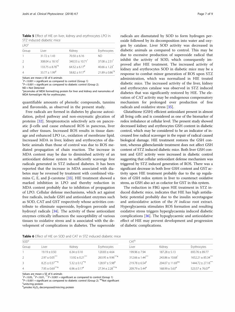
Determination of MDA levelsMDA content was found significantly increased in theliver, kidney and erythrocytes of the diabetic group(Table 5; Group 2) as compared to control (Group 1). Asignificant inhibition in the liver and kidney LPO wasobserved with glibenclamide (Group 3) and HIE (Group4) as compared to diabetic control (Group 2). The liverand kidney MDA content of HIE treated group of mice(Group 4) was comparable to that of control group(Group 1). Erythrocytes MDA content remained un-affected in glibenclamide and decreased in HIE treatedmice as compare to diabetic control.
Determination of antioxidant statusThe decrease in SOD activity was significant in the liver,whereas it was increased in kidney and erythrocytes indiabetic mice, as compared to control (Table 6; Group 1).Glibenclamide and HIE (Group 3 and 4) significantly ele-vated the SOD activity in the liver, while in kidney it wascomparable to control and in erythrocytes SOD activitywas remarkably reduced as compared to diabetic control.A significant increase in liver, kidney and erythrocyte
catalase activity was observed in diabetic animals (Table6; Group 2) when compared against the control (Group1). The treatment with glibenclamide (Group 3) signifi-cantly decreased the catalase activity in liver, while inerythrocytes and kidney the change was non-significantas compared to diabetic control (Group 2). However,with HIE (Group 4) treatment, liver, kidney and erythro-cytes catalase activity is normalized and significantlydecreased as compared to diabetic control (Group 2).GSH content increased in liver and decreased in kidney
and erythrocytes of diabetic animal (Group 2) as com-pared to control (Table 7; Group 1). Glibenclamide treatedanimal showed low GSH content in liver, while with HIEtreatment, the liver GSH content was near to normal. Inkidney, both the treatment resulted in recovery of GSHcontent to normal, whereas for erythrocytes there was noreduction in GSH content in glibenclamide treatment as
a
b
Fig. 3 Effect of different concentration of HIE on (a) DPPH radicalscavenging activity; (b) Reducing power. Values are Mean ± SE offour experiments
Table 2 Correlation coefficient between phytoconstituents andantioxidant properties of HIE
Antioxidant Properties FRSA RP
Phyto-Constituents
TPC 0.851 0.997*
TFC 0.921 0.997*
TTC 0.921 0.997*
FRSA Free radical scavenging activityRP Reducing powerTPC Total phenolic contentTFC Total flavanoid contentTTC Total triterpenoid content*Correlation coefficient is significant at P < 0.05
Joshi et al. Clinical Phytoscience (2018) 4:7 Page 5 of 9

compared to diabetic mice. Increase in GSH content oferythrocytes was observed in HIE as compared to diabeticgroup (Group 2).GST activity in tissues and erythrocytes was signifi-
cantly increased in diabetic animals (Table 7; Group 2)when compared against a control (Group 1), GST activ-ity was not improved by the treatment with glibencla-mide (Group 3) as compared to diabetic mice. HIE(Group 4) significantly reduced the liver and erythro-cytes GST activity when compared to diabetic control(Group 2) whereas it is restored to normal in kidney.
DiscussionROS produced in various tissues leads to tissue injury aswell as early events related to the development of dia-betes mellitus and its complications [23]. HIE may havebeneficial effects on type 1 and 2 diabetes, as the mech-anism of development of both the diabetes involveoxidative stress. However, the animal model used repre-sents only type 1 diabetes. Traditional plant remedieshave been used in the treatment of diabetes but only afew have been scientifically evaluated [24]. Methanolicextract of H. indicus roots contained high quantity offlavanoid and triterpenoid than phenolic content andcontributes to the observed antioxidant potential of theextract. The LC-MS analysis coupled with putative iden-tification of compounds indicated the presence ofphenolic and flavanoids such as DL-3,4-dihydroxyphenylglycol, catechin, amiloxate (cinnamic acid derivative);4-methyldaphnetin (coumarin), terpenoid such as punc-taporin B and podocarpatriene derivative. Various
alkaloids such as ecgonine, homatropine, β-erythroidine,butorphanol, securinine along with other phytoconstitu-ents such as phytosterols, lactones, prostaglandins,amino acids, lipids and fatty acids were also identified.Cholesterol lowering triparanol and nafronyl, which en-hances cellular oxidative capacity and nudifloramide anend product of NAD degradation shown to potentiallyinhibit PARP-1 were found in HIE. Natural compoundshaving anti-inflammatory activity such as safroglycol,etodolac derivative, anisodamine, β santonin, nabume-tone and securinine were identified by their molecularpeak (base peak) [25–28].The results of free radical scavenging activity showed
that HIE reduced DPPH free radical to non-radical DPPH-H by compounds having hydroxyl groups (catechin) orcompounds, which oxidise readily (sinomenine). The phe-nolics, flavanoids, diterpene and sesquiterpene compoundspresent in HIE can donate hydrogen to terminate the oddelectrons of the DPPH radical. The scavenging ability ofHIE can be attributed to these bioactive compounds. Thehigher FRAP value represents high total antioxidantpower of HIE. The phenolic compounds reduce Fe3+
to Fe2 + and interrupt free radical chain reaction, ei-ther by hydrogen atom or electron transfer processand form phenoxyl radical [29]. The reducing powerof HIE was positively correlated with the total phen-olic, flavanoid and triterpenoid content also evidentfrom mass spectral analysis. H. indicus is an edibleplant and contains significant amount of total pheno-lics and reported to have antioxidant activity [30]. Anearlier report by Rajan et al., [31] also showed theantioxidant activity of roots of H. indicus having
Table 3 Effects of HIE treatment on fasting blood glucose in streptozotocin induced diabetic mice
Group Day 1 Day 3 Day 6 Day 9 Day 12
1 91.75 ± 2.78 89.25 ± 2.72 96.25 ± 1.49 82.00 ± 2.27 92.00 ± 2.55
2 368.25 ± 12.29** 294.25 ± 9.29** 320.50 ± 16.06** 312.25 ± 17.4** 321.50 ± 22.77**
3 470.00 ± 22.39**# 276.75 ± 24** 167.75 ± 5.71*## 141.50 ± 6.96*## 112.25 ± 5.76##
4 541.75 ± 11.96**## 374.57 ± 23.39** 243.75 ± 13.29**# 178.25 ± 8.12**## 121.25 ± 5.22##
Values are mean ± SE of 6 animals*P < 0.01, **P < 0.001 = significant as compared to control (Group 1)#P < 0.01, ##P < 0.001 = significant as compared to diabetic control (Group 2)Values of FBG are in mg %
Table 4 Effects of HIE treatment on serum lipid profile in streptozotocin induced diabetic mice
Group Cholesterola Triglyceridesa HDLa LDLa VLDLa
1 125.03 ± 6.28 35.90 ± 7.68 101.82 ± 2.50 83.88 ± 3.76 7.18 ± 1.53
2 274.32 ± 15.82** 63.90 ± 2.05* 73.11 ± 3.17* 160.84 ± 5.26** 12.78 ± 0.41*
3 122.74 ± 7.09## 32.25 ± 6.24## 113.31 ± 5.59## 117.36 ± 7.59*## 6.45 ± 1.25##
4 122.77 ± 9.89## 32.99 ± 0.18## 122.74 ± 2.10## 117.84 ± 5.87*## 6.6 ± 0.04##
Values are mean ± SE of 6 animals*P < 0.01, **P < 0.001 = significant as compared to control (Group 1)#P < 0.01, ##P < 0.001 = significant as compared to diabetic control (Group 2)amg %
Joshi et al. Clinical Phytoscience (2018) 4:7 Page 6 of 9
quantifiable amounts of phenolic compounds, tanninsand flavonoids, as observed in the present study.Free radicals are formed in diabetes by glucose autoxi-
dation, polyol pathway and non-enzymatic glycation ofproteins [32]. Streptozotocin selectively acts on pancre-atic β-cells and cause enhanced ROS in pancreas, liverand other tissues. Increased ROS results in tissue dam-age and enhanced LPO i.e., oxidation of membrane lipid.Increased MDA in liver, kidney and erythrocytes of dia-betic animals than those of control was due to ROS me-diated propagation of chain reaction. The increase inMDA content may be due to diminished activity of anantioxidant defense system to sufficiently scavenge freeradicals generated in STZ induced diabetes. It has beenreported that the increase in MDA associated with dia-betes may be reversed by treatment with combined vita-mins C, E, and β-carotene [33]. HIE treatment showed amarked inhibition in LPO and thereby reduction inMDA content probably due to inhibition of propagationof LPO. Cellular defense mechanisms, which act againstfree radicals, include GSH and antioxidant enzymes suchas SOD, CAT and GST respectively whose activities con-tribute to eliminate superoxide, hydrogen peroxide andhydroxyl radicals [34]. The activity of these antioxidantenzymes critically influences the susceptibility of varioustissues to oxidative stress and is associated with the de-velopment of complications in diabetes. The superoxide
radicals are dismutated by SOD to form hydrogen per-oxide followed by its decomposition into water and oxy-gen by catalase. Liver SOD activity was decreased indiabetic animals as compared to control. This may bedue to excessive production of superoxide radical thatinhibit the activity of SOD, which consequently im-proved after HIE treatment. The increased activity ofkidney and erythrocytes SOD in diabetic mice may be aresponse to combat minor generation of ROS upon STZadministration, which was normalized in HIE treateddiabetic mice. The increased activity of the liver, kidneyand erythrocytes catalase was observed in STZ induceddiabetes that was significantly restored by HIE. The ele-vation of CAT activity may be endogenous compensatorymechanism for prolonged over production of freeradicals and oxidative stress [35].Glutathione (GSH) efficient antioxidant present in almost
all living cells and is considered as one of the biomarker ofredox imbalance at cellular level. The present study showeddecreased kidney and erythrocytes GSH content in diabeticcontrol, which may be considered to be an indicator of in-creased free radical scavenger in the repair of radical causedbiological damage. HIE treatment restores the GSH con-tent, whereas glibenclamide treatment does not affect GSHcontent of STZ induced diabetic mice. Both liver GSH con-tent and GST activity were increased in diabetic controlsuggesting that cellular antioxidant defense mechanism wastriggered by STZ induced generation of ROS. There was asignificant decrease in both liver GSH content and GST ac-tivity upon HIE treatment probably due to the up regula-tion of GSH redox system in liver to counteract oxidativestress, as GSH also act as cofactor for GST in this system.The reduction in FBG upon HIE treatment in STZ in-
duced diabetic mice, indicates that HIE has high antidia-betic potential probably due to the insulin secretagogueand antioxidative action of the H indicus root extract.Hyperglycaemia stimulates ROS formation and resultingoxidative stress triggers hyperglycaemia induced diabeticcomplications [36]. The hypoglycaemic and antioxidativeeffect of HIE may prevent development and progressionof diabetic complications.
Table 5 Effect of HIE on liver, kidney and erythrocytes LPO inSTZ induced diabetic mice
LPOa
Group Liver Kidney Erythrocytes
1 31.72 ± 1.43 70.50 ± 6.16 ND
2 308.04 ± 18.10* 340.53 ± 10.71* 37.08 ± 2.51*
3 133.75 ± 8.78*# 64.52 ± 6.17# 40.66 ± 1.23*
4 33.77 ± 1.99# 58.82 ± 9.17# 21.89 ± 0.86*#
Values are mean ± SE of 6 animals*P < 0.001 = significant as compared to control (Group 1)#P < 0.001 = significant as compared to diabetic control (Group 2).ND = Not Detectedapicomoles of MDA formed/mg protein for liver and kidney and nanomoles ofMDA formed/gm Hb for erythrocytes
Table 6 Effect of HIE on SOD and CAT in STZ induced diabetic mice
SODa CATb
Group Liver Kidney Erythrocytes Liver Kidney Erythrocytes
1 19.19 ± 0.50 6.34 ± 0.10 120.83 ± 4.64 199.96 ± 7.94 187.28 ± 5.13 693.76 ± 89.77
2 2.97 ± 0.05*** 13.92 ± 0.21*** 263.95 ± 9.96***# 312.66 ± 1.44*** 243.86 ± 10.68* 1652.21 ± 85.34***
3 8.25 ± 0.37***# 5.32 ± 0.12**# 128.97 ± 5.98# 219.78 ± 6.54# 204.07 ± 11.69NS 1444.72 ± 27.18***
4 7.95 ± 0.69***# 6.96 ± 0.17# 27.34 ± 2.28***# 209.79 ± 5.44# 168.99 ± 5.63# 523.57 ± 76.07#
Values are mean ± SE of 6 animals*P < 0.05, **P < 0.01, ***P < 0.001 = significant as compared to control (Group 1)#P < 0.001 = significant as compared to diabetic control (Group 2). NSNot significantaunits/mg proteinbμmoles H2O2 decomposed/min/mg protein
Joshi et al. Clinical Phytoscience (2018) 4:7 Page 7 of 9

Certain oxidative stress related defects in oxidative phos-phorylation machinery and mitochondrial β-oxidation leadto excess accumulation of intracellular triglyceride inmuscle and liver and subsequent insulin resistance [37].As compared to diabetic control, glibenclamide and HIEtreated mice, the total cholesterol, triglyceride, LDL andVLDL were significantly lowered with increased HDL indi-cating recovery of normal lipid metabolism in STZ in-duced diabetes. The results showed that HIE was found tobe effective against diabetic dyslipidaemia.
ConclusionThe methanolic extract of H. indicus roots containsvarious phytoconstituents having potent antioxidant,hypoglycaemic and hypolipidaemic activity. Oral admin-istration of HIE, lowers the FBG of STZ induced diabeticmice as well as it modulates the intracellular antioxidantdefense to overcome the oxidative damage and improvesthe serum lipid profile. It is suggested that the roots ofH. indicus may serve as an important hypoglycaemicand hypolipidaemic agent to protect the cells by mitigat-ing oxidative stress induced toxicity in STZ induceddiabetes.
Additional files
Additional file 1: Putative identification of compounds in HIE.(DOCX 17 kb)
Additional file 2: Effects of HIE treatment on body weight ofstreptozotocin induced diabetic mice. (DOCX 14 kb)
AbbreviationsAJS: Agilent Jet Stream; CAT: Catalase; DPPH: 2,2-diphenyl-1-p-picryl-hydrazyl; ESI: Electrospray ion generation; FBG: Fasting Blood Glucose;FRAP: Ferric Reducing Antioxidant Power; FRSA: Free Radical ScavengingActivity; GSH: Glutathione; GST: Glutathione-S- Transferase; HDL: High DensityLipoprotein; HIE: Methanolic extract of Hemidesmus indicus roots;LC-MS: Liquid Chromatography-Mass Spectroscopy; LDL: Low DensityLipoprotein; LPO: Lipid Peroxidation; MDA: Malondialdehyde;NAD: Nicotinamide-adenine dinucleotide; PARP-1: Poly ADP ribosepolymerase-1; PBS: Phosphate Buffer Saline; ROS: Reactive Oxygen Species;SOD: Superoxide Dismutase; STZ: Streptozotocin; TAP: Total AntioxidantPower; VLDL: Very Low Density Lipoprotein
AcknowledgementsWe acknowledge SAIF- IIT Bombay, India for providing LC-MS facility foranalysis of samples. AJ is thankful to University Grant Commission, New Delhifor providing Golden Jubilee Fellowship under UGC XIIth Plan grant.
FundingThe funding from Golden Jubilee Fellowship under University GrantCommission, New Delhi (UGC XIIth Plan) was provided to Ankita Joshi for thisresearch work.
Availability of data and materialsNot applicable
Authors’ contributionsAJ: designed and performed the experiments, participated in data analysisand manuscript preparation. HL, HS and DB: participated in design ofexperiments and helped in manuscript preparation. All authors read andapproved the final manuscript.
Ethics approvalThe experimental mice were treated following the ethical guidelines ofInstitutional Animal Ethics Committee (IAEC) constituted under supervision ofCPCSEA (Committee for the Purpose of Control and Supervision ofExperiments on Animals). The experimental procedures were approved bythe IAEC (CPCSEA/2015/01 dt. 4/7/2015) of Devi Ahilya University, India.
Consent for publicationNot applicable
Competing interestsThe authors declare that they have no competing interests.
Publisher’s NoteSpringer Nature remains neutral with regard to jurisdictional claims inpublished maps and institutional affiliations.
Received: 10 July 2017 Accepted: 24 January 2018
References1. Alberti KG, Zimmet PZ. New diagnostic criteria and classification of
diabetes-again. Diabet Med. 1998;15:535–6.2. Wolff SP, Jiang ZY, Hunt JV. Protein glycation and oxidative stress in
diabetes mellitus and ageing. Free Radic Biol Med. 1991;10:339–52.3. Scheen AJ, Lefèbvre PJ. Oral antidiabetic agents a guide to selection. Drugs.
1998;55:225–36.4. Lakshmi T, Rajendran R. Hemidesmus indicus commonly known as Indian
sarasaparilla- an update. Int J Pharm Bio Sci. 2013;4:397–404.5. Zhao Z, Matsunami K, Otsuka H, Negi N, Kumar A, Negi DS. A condensed
phenylpropanoid glucoside and pregnane saponins from the roots ofHemidesmus indicus. J Nat Med. 2013;67:137–42.
6. Fiori J, Leoni A, Fimognari C, Turrini E, Hrelia P, Mandrone M, Iannello C,Antognoni F, Poli F, Gotti R. Determination of phytomarkers in
Table 7 Effect of HIE on GSH and GST and in STZ induced diabetic mice
GSHa GSTb
Group Liver Kidney Erythrocytes Liver Kidney Erythrocytes
1 19.27 ± 0.62 25.33 ± 0.45 25.05 ± 0.91 0.33 ± 0.06 0.36 ± 0.07 10.22 ± 0.40
2 31.60 ± 1.98* 15.78 ± 0.39* 7.31 ± 1.11* 4.24 ± 0.42* 1.37 ± 0.13* 18.10 ± 1.45*
3 13.93 ± 1.78## 25.66 ± 1.61## 7.94 ± 0.21* 1.76 ± 0.23*## 1.11 ± 0.08 * 19.94 ± 1.61*
4 22.04 ± 0.67# 38.26 ± 0.61*## 13.82 ± 0.56*## 0.16 ± 0.02## 0.30 ± 0.05## 6.97 ± 0.30##
Values are mean ± SE of 6 animals*P < 0.001 = significant as compared to control (Group 1)#P < 0.01,##P < 0.001 = significant as compared to diabetic control (Group 2)ananomoles of DTNB conjugated/mg protein for liver and kidney and μmoles of DTNB conjugated/gm Hb for erythrocytesbμmoles of GSH conjugated/min/mg protein
Joshi et al. Clinical Phytoscience (2018) 4:7 Page 8 of 9

pharmaceutical preparations of Hemidesmus indicus roots by micellarelectrokinetic chromatography and high-performance liquidchromatography–mass spectrometry. Anal Lett. 2014;47:2629–42.
7. Ravikiran T, Shilpa S, Praveen Kumar N, Sowbhagya R, Anand S, AnupamaSK, Bhagyalakshmi D. Antioxidant activity of Hemidesmus indicus (L.) r.Br.Encapsulated poly (lactide-co-glycolide) (PLGA) nanoparticles. J Pharm BiolSci. 2016;11:9–17.
8. Kumar S, Pooja M, Harika K, Haswitha E, Nagabhushanamma G, Vidyavathi N.In-vitro antioxidant activities, total phenolics and flavonoid contents of wholeplant of Hemidesmus indicus (Linn.). Asian J Pharm Clin Res. 2013;6:249–51.
9. Singleton VL, Rossi JA. Colorimetry of total phenolics with phosphomolybdic-phosphotungstic acid reagents. Am J Enol Vitic. 1965;16:144–58.
10. Zhang L, Ravipati AS, Koyyalamudi SR, Jeong SC, Reddy N, Smith PT, MünchG, Wu MJ, Satyanarayanan M, Vysetti B. Antioxidant and anti-inflammatoryactivities of selected medicinal plants containing phenolic and flavonoidcompounds. J Agr Food Chem. 2011;59:12361–7.
11. Chang CL, Lin CS. Phytochemical composition, antioxidant activity, andneuroprotective effect of Terminalia chebula Retzius extracts. Evid BasedCompliment Alternat Med. 2012; https://doi.org/10.1155/2012/125247.
12. Mellors A, Tappel AL. The inhibition of mitochondrial peroxidation byubiquinone and ubiquinol. J Biol Chem. 1966;241:4353–6.
13. Benzie IF, Strain JJ. The ferric reducing ability of plasma as a measure ofantioxidant power: the FRAP assay. Anal Biochem. 1996;239:70–6.
14. Oyaizu M. Studies on product of browning reaction prepared fromglucosamine. Jpn J Nutr. 1986;44:307–15.
15. Arora S, Ojha SK, Vohora D. Characterisation of streptozotocin induceddiabetes mellitus in swiss albino mice. Glob J Pharmacol. 2009;3:81–4.
16. Lenzen S. The mechanisms of alloxan- and streptozotocin-induced diabetes.Diabetologia. 2008;51:216–26.
17. Kale M, Rathore N, John S, Bhatnagar D. Lipid peroxidative damage onpyrethroid exposure and alteration in antioxidant status in rat erythrocytes:a possible involvement of reactive oxygen species. Toxicol Lett. 1999;105:197–205.
18. Tukozkan N, Erdamar H, Seven I. Measurement of total malondialdehyde inplasma and tissue by high performance liquid chromatography andthiobarbituric acid assay. Firat Tip Dergisi. 2006;11:88–92.
19. Marklund S, Marklund G. Involvement of the superoxide anion radical inautoxidation of pyrogallol and convenient assay for superoxide dismutase.Eur J Biochem. 1974;47:469–74.
20. Aebi H. Catalase. In: Bergmeyer HU, editor. Methods in enzymatic assay. 3rded. New York: Academic press; 1983. p. 276–86.
21. Habig WH, Pabst MJ, Jakoby WB. Glutathione-S-transferase, the first enzymaticstep in mercapturic acid formation. J Biol Chem. 1974;249:7130–9.
22. Beutler E, Duran O, Kelly BM. Improved method for determination of bloodglutathione. J Lab Clin Med. 1963;61:882–8.
23. Maritim AC, Sanders RA, Watkins JB 3rd. Diabetes, oxidative stress, andantioxidants: a review. J Biochem Mol Toxicol. 2003;17:24–38.
24. Gupta RK, Kesari AN, Diwakarc S, Tyagia A, Tandona V, Chandra R, Watal G.In vivo evaluation of anti-oxidant and anti-lipidimic potential of Annonasquamosa aqueous extract in type 2 diabetic models. J Ethnopharmacol.2008;118:21–5.
25. Steinberg D, Avigan J, Feigelson EB. Effects of triparanol (mer-29) oncholesterol biosynthesis and on blood sterol levels in man. J Clin Invest.1961;40:884–93.
26. Martindale - the Extra Pharmacopoeia’ (30th ed), edited by J. E. F. Reynolds.Pp 1310. London: The Pharmaceutical Press. 1993. ISBN 0 85369300 5.
27. Shibata K, Mushiage M, Kondo T, Hayakawa T, Tsuge H. Effects of vitamin B6deficiency on the conversion ratio of tryptophan to niacin. Biosci BiotechnolBiochem. 1995;59:2060–3.
28. Galvez-Llompart M, Zanni R, Domenech RG. Modeling natural anti-inflammatory compounds by molecular topology. Int J Mol Sci. 2011;12:9481–503.
29. Steenken S, Neta P. One-electron redox potentials of phenols. Compoundsof biological interest. J Phys Chem. 1982;86:3661–7.
30. Jayawardena N, Watawana MI, Waisundara VY. Evaluation of the totalantioxidant capacity, polyphenol contents and starch hydrolase inhibitoryactivity of ten edible plants in an in vitro model of digestion. Plant FoodsHum Nutr. 2015;70:71–6.
31. Rajan S, Shalini R, Bharathi C, Aruna V, Thirunalasundari T, Brindha P. In vitroantioxidant screening of Hemidesmus indicus root from South India. Asian JPharm Biol Res. 2011;1:222–31.
32. Obrosova IG, Fathallah L, Greene DA. Early changes in lipid peroxidationand antioxidative defense in diabetic rat retina: effect of DL-α-lipoic acid.Eur J Pharmacol. 2000;398:139–46.
33. Mekinova D, Chorvathova V, Volkovova K, Staruchova M, Grancicova E,Klvanova J, Ondreic R. Effect of intake of exogenous vitamins C, E andβ-carotene on the antioxidative status in kidneys of rats withstreptozotocin-induced diabetes. Nahrung. 1995;39:257–61.
34. Soto C, Recoba R, Barron C, Alverez C, Favari L. Silymarin increasesantioxidant enzymes in alloxan-induced diabetes in rat pancreas. CompBiochem Physiol. 2003;136:205–12.
35. Aksoy N, Vural H, Sabuncu T, Aksoy S. Effect of melatonin on oxidative-antioxidative status of tissues in streptozotocin induced diabetic rats. CellBiochem Funct. 2003;21:121–5.
36. Valko M, Leibfritz D, Moncola J, Cronin MT, Mazura M, Telser J. Free radicalsand antioxidants in normal physiological functions and human disease. Int JBiochem Cell Biol. 2007;39:44–84.
37. Rosca MG, Mustata TG, Kinter MT, Ozdemir AM, Kern TS, Szweda LI,Brownlee M, Monnier VM, Weiss MF. Glycation of mitochondrial proteinsfrom diabetic rat kidney is associated with excess superoxide formation. AmJ Physiol-Renal Physiol. 2005;289:F420–30.
Joshi et al. Clinical Phytoscience (2018) 4:7 Page 9 of 9