Embed Size (px)
Citation preview

Evaluation of testicular cytotoxicity and genotoxicity of sofosbuvir and sofosbuvir - ribavirin in the adult
male albino rats
ORIGINAL ARTICLE Eur. J. Anat. 23 (6): 393-403(2019)
Wael B El-Kholy1, Manar A Faried
1, Rasha M. Salama
1, Mohammed M. El-
Fiky1, Islam El-Garawani
2
1Department of Anatomy and Embryology, Faculty of Medicine, Menoufia University, Egypt, 2Department of Zoology, Faculty of Science, Menoufia University, Egypt
SUMMARY Hepatitis C is a widely distributed problem all
over the world, especially Egypt. Chronically infect-ed people develop serious liver disease and now it is the most common cause for liver transplantation. Recently, a new regimen, sofosbuvir (sovaldi), alone or with combinations as sovaldi-ribavirin, was approved for treating this disease. There are limited studies that explore the effects of these drugs on the reproductive organs, and hence af-fection of male fertility while using these drugs. This study aims to throw more light on whether sovaldi or sovaldi-ribavirin causes testicular dam-aging effects in the adult male albino rats. We in-vestigated the effect of this regimen in a dose equivalent to that used in the human (41 mg/kg once daily orally for sovaldi and 41 mg/kg twice daily orally for ribavirin) for consecutive 5 and 10 days. There was highly significant decrease in tes-tosterone hormone level and marked degenerative changes in the seminiferous tubules and the testic-ular interstitium, with increase in collagen deposits in sovaldi treated rats, and in a more extensive manner in sovaldi-ribavirin treated rats. There was a significant increase of deoxyribonucleic acid (DNA) fragmentation in the treated groups after 10 days. However, there was a non-significant differ-
ence in DNA fragmentation in the treated groups after 5 days when compared with control. Immuno-histochemistry detection of caspase-3 showed sig-nificant increase in its expression in the treated groups after either 5 or 10 days. This denoted the specificity of caspase-3 immunohistochemistry technique in the detection of early apoptotic changes. It was concluded that sovaldi and soval-
di-ribavirin induced gonadotoxic effects through induction of DNA fragmentation via upregulation of caspase-3, and that the resulting damaging effects increased with longer duration of drug intake.
Key words: Hepatitis C – Sovaldi – Sovaldi-ribavirin – Testis – Apoptosis
INTRODUCTION
One of the most prevalent leading causes of mor-bidity and even mortality all over the world is hepa-titis C virus (HCV) with the highest prevalence in Egypt (Gomaa et al., 2017). Until now, there is no vaccine for HCV infection in spite of the existence of several routes of transmission (Reker and Islam, 2014).
Egypt, the single country with highest incidence
of HCV infection in the world, has embarked on a government-sponsored mass treatment program using several combinations of directly acting antivi-ral drugs (DAAs) (El-Fishawy et al., 2016).
393
Submitted: 29 August, 2018. Accepted: 5 April, 2019.
Corresponding author: Dr. Rasha M. Salama. Department of
Anatomy and Embryology, Faculty of Medicine, Menoufia Uni-
versity, Egypt.
E-mail adress: [email protected]
Effect of sofosbuvir and sofosbuvir-ribavirin on rat testis
394
Integration of pegylated interferon (PEGIFN) and ribavirin was prescribed by the standard care for chronic HCV infection between 2001 and 2011. In May 2011, two first-generation NS3/4A protease inhibitors, boceprevir, and telaprevir were ap-proved in combination with PEG-IFN and ribavirin. In December 2013, a second-generation NS3/4A protease inhibitor, simeprevir was approved for use with PEG-IFN and/or ribavirin. One of the most used combinations is sovaldi-ribavirin (Yau and Yoshida, 2014).
Until recently, HCV infections were treated with two antiviral drugs that had to be taken for almost a year and produced serious side effects, and these treatments were successful only in a small fraction of people. In 2013, a new drug, sovaldi, was approved for treating HCV and is of special interest. This drug cures more than 90% of pa-tients and is effective against several of the most common strains of HCV (Golanty and Edlin, 2015), with the chemical name L-Alanine, N-[[P(S),2′R]-2′-deoxy-2′-fluoro-2′-methyl-P-phenyl-5′-uridylyl]-, 1-methyl ethyl ester and a molecular formula of C22H29FN3O9P (Bhatia et al., 2014). NS5B is one of the non-structural proteins essential for viral ribonucleic acid (RNA) replication, and has been found to be a valuable target for DAAs (Lam et al., 2012). Sovaldi is a nucleotide analog that is a highly potent inhibitor of the NS5B polymerase in HCV. This drug has shown high efficacy in combi-nation with several other drugs with and without PEG- INF, against HCV (Bhatia et al., 2014).
Sovaldi is a promising therapy for chronic HCV infection, as it offers several advantages over the existing therapies, particularly in dealing with pa-tients with decompensated liver disease and pa-tients who cannot tolerate interferon-containing therapies. On account of its excellent performance in clinical trials, this drug has got food drug admin-istration (FDA) approval on 6 December, 2013, under the breakthrough therapy designation. This drug is effective against all HCV genotypes, has a better safety profile and low risk of development of resistance; however, careful clinical use and moni-toring are still essential to gather more data on this drug (Bhatia et al., 2014).
Ribavirin is a non-selective, antihepatitis, antiviral drug. It was synthesized in 1970. The broad spec-trum antiviral activity was reported in 1972 (Sidwell et al., 1972). In the early 1990s, ribavirin was stud-ied for the treatment of HCV infection. Ribavirin had no significant effect on HCV RNA levels when used as a single agent, despite observations of improvements in serum aminotransferase levels (Di Bisceglie et al., 1995) and liver histology (Hoofnagle et al., 2003). Prolonging the course of treatment did not add any benefit in terms of virolo-gy clearance (Hoofnagle et al., 1996). Therefore, ribavirin has been used for the treatment of chronic HCV infection only in combination (Te et al., 2007).
Following the success of sovaldi or sovaldi-
ribavirin regimen, further research is needed to clarify the optimal follow-up duration post-treatment and to evaluate the side effects of these drugs on different organs. Therefore, the testis was chosen in this study. Zhang et al. (2011) pos-tulated that reproductive system is very sensitive to toxic chemicals because of the high multiplica-tion rate of germ cells. On the other hand, the transmissible genetic damage from one generation to another takes place in this system only as re-ported by Au and Hsu (1980). Moreover, El-Atrebi et al. (2011) postulated that sexual dysfunction is a common side effect of Peg-IFN and ribavirin treat-ment, especially in middle-aged men in addition to advanced liver fibrosis that is an important co-factor in inducing sexual dysfunction during treat-ment.
The aim of this study was to evaluate the damag-ing effects of sovaldi and sovaldi- ribavirin at differ-ent durations on testicular tissue of adult male rats based on biochemical, histological, immunohisto-chemical and genetic assessments.
MATERIALS AND METHODS Animals
Sixty adult male albino rats weighing 150-180 g were maintained in the animal house of the Faculty of Medicine, Menoufia University, Egypt. The rats were caged in standardized room conditions and allowed unlimited access to chow and water. The experiment started after one week of caging. All procedures involving the use of the rats were ap-proved by the Animal Care and Use Committee, Faculty of Medicine, Menoufia University, Egypt.
Drugs
Sovaldi (Sofosbuvir, a product of Merck Compa-ny of pharmaceutical industries) was available in the form of tablets. Each tablet contained 400 mg of Sofosbuvir. They were crushed. The calculated dose was dissolved in distilled water.
Ribavirin (a product of Minapharm, Egypt) was available in the form of capsule. Each one con-tained 200 mg ribavirin. The contents of the cap-sule of the required dose were dissolved in distilled water.
Based on the clinical standard dose used for the human, animal equivalent dose (AED) calculation based on body surface area according to data adapted and modified from FDA draft guidelines was calculated. So, doses used in this study were calculated by using the following formula: - Rat Equivalent Dose (mg/kg) = Human does (mg/kg) × 6.2 (Nair and Jacop, 2016).
The clinical standard effective human dose for sovaldi is 400 mg once daily (Bhatia et al., 2014) and that for ribavirin is 800 mg/day divided into two equal doses (Jen et al., 2002) assuming the aver-age human body weight 60Kg. So, the doses used in this study, 41 mg/kg once daily for sovaldi and
R. M. Salam et al.
395
41 mg/kg twice daily for ribavirin, were approxi-mately equivalent to the doses used in human. Experimental design
The rats were divided into three groups (n=20 per group):
- Group I (control group): subdivided into two subgroups, (n= 10 per subgroup):-
Subgroup Ia: Ten rats received only standard diet and water for consecutive 5 days.
Subgroup Ib: Ten rats received only standard diet and water for consecutive 10 days.
- Group II (Sovaldi treated group): subdivided into two subgroups, (n= 10 per subgroup):-
Subgroup IIa:- Rats received sovaldi orally by gastric tube in a dose of 41 mg/kg dissolved in 2 ml distilled water once daily (equivalent to the clini-cal standard human dose 400 mg once daily) for 5 consecutive days.
Subgroup IIb:- Rats received sovaldi in the same previous dose and route of administration for 10 consecutive days.
- Group III (Sovaldi-Ribavirin treated group): sub-divided into two subgroups, (n= 10 per subgroup):-
Subgroup IIIa:- Rats received sovaldi in the same previous dose and route of administration plus rib-avirin orally by gastric tube in a dose of 41 mg/kg dissolved in 2 ml distilled water twice daily (approximately equivalent to the clinical standard human dose 400 mg twice/day) for 5 consecutive days.
Subgroup IIIb:- Rats received sovaldi and ribavi-rin in the same previous dose and route of admin-istration for 10 consecutive days.
At the end of the experiment, the rats were anes-thetized, intra-cardiac blood samples were collect-ed, and the testes were dissected for further pro-cessing and examinations.
Biochemical analysis
Serum was obtained by centrifugation of blood samples. The concentration of testosterone hor-mone in blood was measured. Quantitative data were expressed in the form of mean ± standard deviation (x̄ ± SD) and analyzed.
Molecular study
Fresh testicular tissues were subjected to molec-ular biological study as follows: Agarose gel tech-nique for detection of DNA fragmentation as a re-sult of apoptosis in testis.
Total genomic DNA extraction
Nucleic acid extraction was done according to the extraction method of Aljanabi and Martinez (1997), with some modifications introduced by El-Garawani and Hassab El-Nabi (2016), in which the direct staining of DNA sample was done. Apoptotic bands of DNA fragmentation appeared and locat-ed at 180 bp and its multiples 360, 540 and 720 bp against thirteen bands of 100 bp plus DNA ladder
(Thermo Scientific™ O'Gene Ruler™, USA). The intensity of released DNA fragments was meas-ured by image J software, as a mean of optical density values.
Histological, histochemical and immunohisto-chemical assessments
Testes were fixed in 10% neutral buffered formol solution for 24h and embedded in paraffin wax. For histological examination, 5-μm sections were de-paraffinized and rehydrated using a graded et-hanol (100%, 90%, and 70%) series and stained with hematoxylin and eosin (Hx&E) and Masson trichrome stain to show collagen fibers (Stevens and Wilson, 1996).
For caspase-3 immunohistochemistry, deparaffi-nized and hydrated testis sections were treated in 3% H2O2 for 5 min and rinsed with phosphate buf-fer saline (PBS) for 15 min. The sections were blo-cked with 1.5% normal goat serum in PBS and then incubated (45 min, room temperature) with rabbit polyclonal antihuman caspase-3 (0.5 μg/ml) in 1.5% normal goat serum in PBS. The sections then were incubated with biotin-conjugated goat antirabbit IgG (1:200, 1 h, room temperature), avi-din-biotin-peroxidase complex (Santa Cruz Bio-technology, Inc., rabbit peroxidase kit; 1 h) and DAB solution. Sections were counterstained with hematoxylin. Positive reaction was visualized as brown coloration. Negative controls were done using the same steps, except that phosphate buf-fered saline was applied instead of the primary antibodies (Jackson et al., 2008).
Morphometrical study
The percentage surface area of collagen depo-sits and percentage of caspase-3 immunoreactive cells were measured. Measurement was done using an image analyzer (Image J program). From each slide of experimental groups, 9 fields were randomly selected.
The total field and histochemical stained areas were calculated and the percentage of caspase-3 immunoreactivity was calculated as follows: % im-munoreactive stained cells = caspase-3 stained cell count/Total cell count × 100.
Statistical analysis
The data were collected, tabulated, and analyzed by SPSS (statistical package for social science) version 17.0 on IBM compatible computer (SPSS Inc., Chicago, IL, USA). The collected data were quantitative, which were described as mean ±SD and range; the data were compared using Mann Whitney U test. A P value of ≤ 0.05 was conside-red statistically significant. A P value of < 0.001 was considered statistically highly significant and P value > 0.05 was considered non-significant. This was done in the Public Health Department, Faculty of Medicine, Menoufia University, Egypt.
Effect of sofosbuvir and sofosbuvir-ribavirin on rat testis
396
RESULTS Endocrine disruptor effect of sovaldi and so-valdi-ribavirin in the rat testosterone hormone level
Analysis of testosterone hormone showed a non-significant difference (P˃0.05) between the control subgroups after 5 & 10 days, while there was a significant decrease (P≤0.05) in its level in sovaldi and sovaldi-ribavirin treated rats after 5 and 10 days when compared with the corresponding con-trol subgroups. Moreover, a significant decrease (P≤0.05) in testosterone hormone was observed in rats treated with sovaldi-ribavirin when compared with that treated with sovaldi only. In addition, the-re was a significant difference (P≤0.05) between the rats sacrificed after 5 days and that sacrificed after 10 days in the treated groups. (Table 1)
Total genomic DNA fragmentation and apopto-sis effect of sovaldi and sovaldi-ribavirin
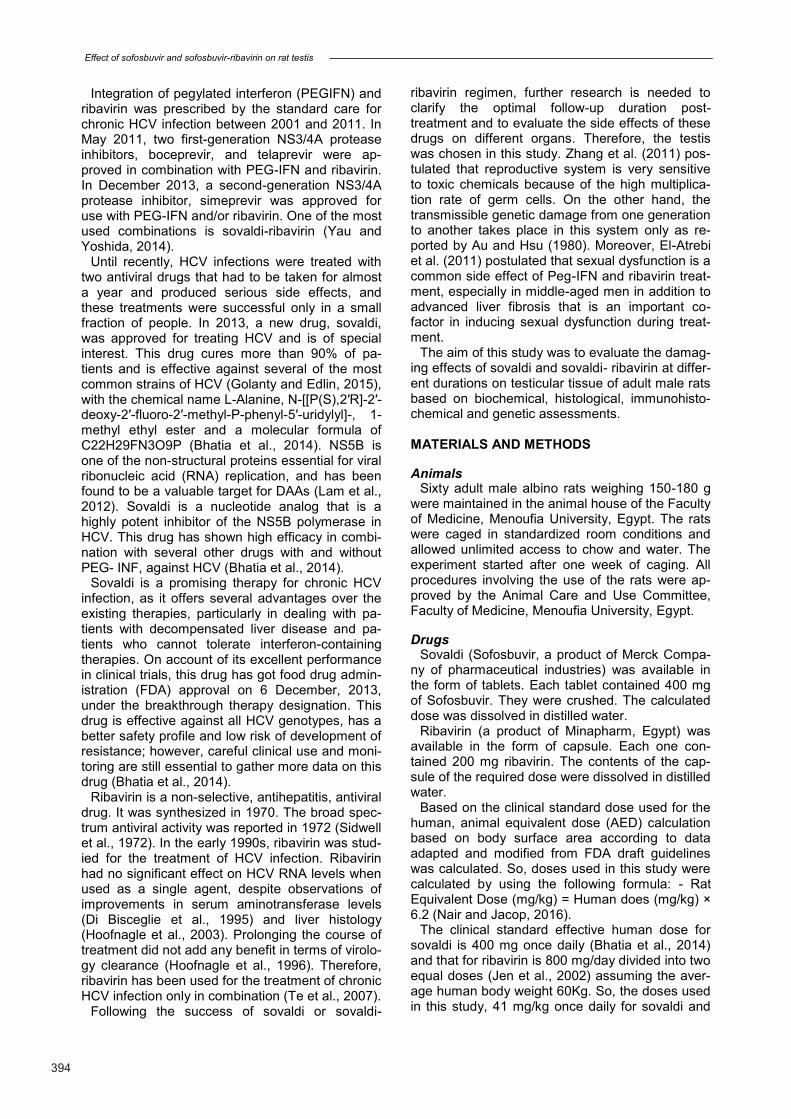
treated rats Optical density values of fragmented DNA ex-
tracted from testes of sovaldi treated rats and so-valdi-ribavirin treated rats after five days (10.9±2.29 and11±1.73) showed normal appearan-ce of intact DNA that was non-significant (P˃0.05) when compared with control (9.2±1.6). While the optical density values of fragmented DNA ex-tracted from testes of sovaldi-treated rats and so-valdi-ribavirin treated rats after ten days (101.3±8.08 and 106.7±13.32 respectively) showed significant increase (P≤0.05) of DNA frag-mentation appeared as smear pattern when com-pared with control (9.2±1.6) (Fig. 1 a,b).
Histopathological testicular changes of sovaldi and sovaldi-ribavirin-treated rats
- Hematoxylin and Eosin stain: The testes of the control group either sacrificed 5
or 10 days after the beginning of the experiment
Fig 1. (a): Digital photograph of DNA electrophoresis of rat testis tissue showing the effect of sovaldi and sovaldi-ribavirin treated subgroups. Where C: control; S: sovaldi; S+R:sovaldi-ribavirin. 5d: after five days; 10d: after ten days and M: DNA marker. (b): Total released DNA fragmentation in testis tissues of rats treated with sovaldi and sovaldi-ribavirin. C: control; S: sovaldi; S+R:sovaldi-ribavirin. 5d: after five days; 10d: after ten days. Data were presented as Mean± S.D., (n=6). Sovaldi and sovaldi-ribavirin treated rats after ten days showed significant increase of DNA frag-mentation (P≤0.05) and after five days showed non-significant (P˃0.05) with respect to control.
Subgroups The studied groups Test P value
Group I Group II Group III U test
Testosterone (ng/dl) in Subgroup (a) [n=5] Mean±SD Range
1.93±0.39 1.4 – 2.45
1.37±0.37 1.05 – 2.0
0.92±0.24 0.65 – 1.3
1.99 2.61 2.01
0.04711 0.00922 0.04533
Testosterone (ng/dl) in Subgroup (b) [n=5] Mean± SD Range
1.46±0.36 1 – 1.9
0.77±0.33 0.35 – 1.1
0.32±0.08 0.2 – 0.40
2.41 2.64 2.21
0.01611 0.00822 0.02733
U test 1.68 2.42 2.63
P value 0.0944 0.0244 0.00944
Table 1. Comparison between the studied groups regarding testosterone level
Subgroup (a): measurement after 5 days
Subgroup (b): measurement after 10 days
U test = Mann Whitney U test
1 = comparing group I with group II
2 = comparing group I with group III
3 = comparing group II with group III
4 = comparing subgroup a and subgroup b in each group
R. M. Salam et al.
397
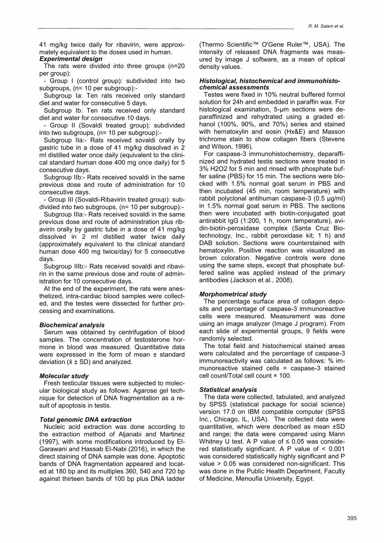
revealed normal testicular structure. The seminife-rous tubules appeared uniform lined by regularly arranged spermatogenic cells at different stages of maturation. They were separated by interstitial tissue. The interstitial cells of Leydig appeared rounded or polygonal in shape, with acidophilic cytoplasm and large rounded nuclei. The shown
spermatogenic cells were spermatogonia, primary spermatocytes, and spermatozoa filling the lumen of the tubules (Fig. 2).
Sovaldi treated group either sacrificed after 5 or 10 days showed distortion of the seminiferous tu-bules, especially in the rats sacrificed at the 10th day. The separation of spermatogenic cells from
Fig 2. Representative micrographs of a testicular section of a control rat showing seminiferous tubules lined by sper-matogonia (red arrow), primary spermatocytes (star) within the seminiferous tubules, and the tails of spermatozoa (S) are observed in the center of the seminiferous tubule. A few Leydig cells with an acidophilic cytoplasm (yellow arrow) are scattered between the seminiferous tubules (hematoxylin-eosin, 20x).
Subgroups The studied groups Test P value
Group I Group II Group III U test
%surface area of collagen deposition in Subgroup (a) Mean± SD Range
6.44±1.63 4.5 – 8.4
12.26±3.09 9.3 – 17
19.16±2.51 16.2 – 22.9
2.61 2.61 2.40
0.0091 0.0092 0.0163
%surface area of collagen deposition in Subgroup (b) Mean± SD Range
7.28±1.55 5.6 – 9
18.3±2.07 15.6 – 21.3
24.12±3.16 20.8 – 27.9
2.61 2.61 2.40
0.0091 0.0092 0.0163
U test 0.94 2.40 2.19
P value 0.354 0.0164 0.0284
Table 2. Comparison between the studied groups regarding % of surface area of collagen deposition.
Subgroup (a): measurement after 5 days
Subgroup (b): measurement after 10 days
U test = Mann Whitney U test
1 = comparing group I with group II
2 = comparing group I with group III
3 = comparing group II with group III
4 = comparing subgroup a and subgroup b in each group

Effect of sofosbuvir and sofosbuvir-ribavirin on rat testis
398
their underlying basement membrane was obser-ved in most of seminiferous tubules. Moreover, there were marked cytoplasmic vacuolations of the spermatogenic cells and atrophy of Leydig cells (Fig. 3).
The histological changes that were seen in the
testicular sections of the rats treated with both so-valdi and ribavirin were nearly similar to that occu-rred in sovaldi treated group, but in an extensive manner. There was a marked distortion of the tes-ticular seminiferous tubules with presence of con-gested dilated blood vessels in the testicular in-
Fig 3. Representative micrographs of a testicular section of a sovaldi treated rat showing distortion of some seminifer-ous tubules with disorganization of the spermatogenic cells (double black arrows), cytoplasmic vacuolation (green ar-row) of the spermatogenic cells, sloughing of the spermatogenic cells from the underlying basement membrane (red arrow) and atrophy of Leydig cells (asterisk). These changes are more obvious in the sovaldi treated group for 10 days (hematoxylin-eosin, 20x).
Subgroups Studied groups Test P value
Group I Group II Group III U test
% of spermatogenic cells in Subgroup (a) Mean± SD Range
10.4±2.30 8 – 13
60.60±4.83 55 – 66
72.60±6.80 64 – 80
2.91 2.61 2.19
0.0091 0.0092 0.0283
% of spermatogenic cells in Subgroup (b) Mean± SD Range
10.0±2.34 7 – 12
88.4±6.19 82 – 96
97.40±2.61 94 – 100
2.91 2.61 2.22
0.0091 0.0092 0.0263
U test 0.43 2.62 2.62
P value 0.674 0.0094 0.0094
Table 3. Comparison between the studied groups regarding % of surface area of collagen deposition.
Subgroup (a): measurement after 5 days
Subgroup (b): measurement after 10 days
U test = Mann Whitney U test
1 = comparing group I with group II
2 = comparing group I with group III
3 = comparing group II with group III
4 = comparing subgroup a and subgroup b in each group
R. M. Salam et al.
399
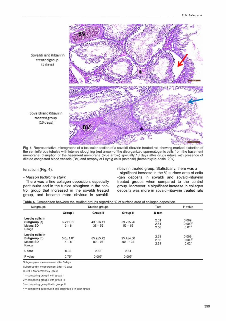
terstitium (Fig. 4).
- Masson trichrome stain: There was a fine collagen deposition, especially
peritubular and in the tunica albuginea in the con-trol group that increased in the sovaldi treated group, and became more obvious in sovaldi-
ribavirin treated group. Statistically, there was a significant increase in the % surface area of colla
-gen deposits in sovaldi and sovaldi-ribavirin treated groups when compared to the control group. Moreover, a significant increase in collagen deposits was more in sovaldi-ribavirin treated rats
Fig 4. Representative micrographs of a testicular section of a sovaldi-ribavirin treated rat showing marked distortion of the seminiferous tubules with intense sloughing (red arrow) of the disorganized spermatogenic cells from the basement membrane, disruption of the basement membrane (blue arrow) specially 10 days after drugs intake with presence of dilated congested blood vessels (BV) and atrophy of Leydig cells (asterisk) (hematoxylin-eosin, 20x).
Table 4. Comparison between the studied groups regarding % of surface area of collagen deposition.
Subgroup (a): measurement after 5 days
Subgroup (b): measurement after 10 days
U test = Mann Whitney U test
1 = comparing group I with group II
2 = comparing group I with group III
3 = comparing group II with group III
4 = comparing subgroup a and subgroup b in each group
Subgroups Studied groups Test P value
Group I Group II Group III U test
Leydig cells in Subgroup (a) Mean± SD Range
5.2±1.92 3 – 8
43.6±6.11 38 – 52
59.2±5.26 53 – 66
2.61 2.61 2.56
0.0091 0.0092 0.013
Leydig cells in Subgroup (b) Mean± SD Range
5.6± 1.81 4 – 8
85.2±5.72 80 – 93
95.4±4.50 90 – 102
2.63 2.62 2.31
0.0091 0.0092 0.023
U test 0.32 2.62 2.61
P value 0.754 0.0094 0.0094
Effect of sofosbuvir and sofosbuvir-ribavirin on rat testis
400
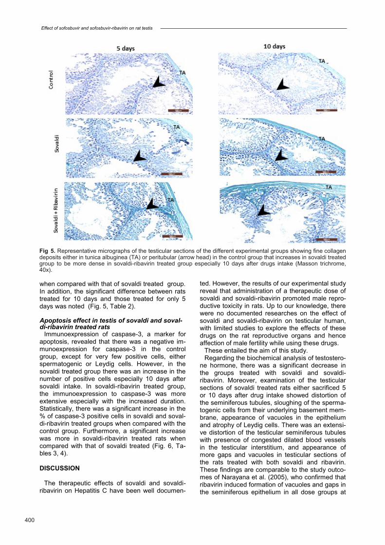
when compared with that of sovaldi treated group. In addition, the significant difference between rats treated for 10 days and those treated for only 5 days was noted (Fig. 5, Table 2).
Apoptosis effect in testis of sovaldi and soval-di-ribavirin treated rats
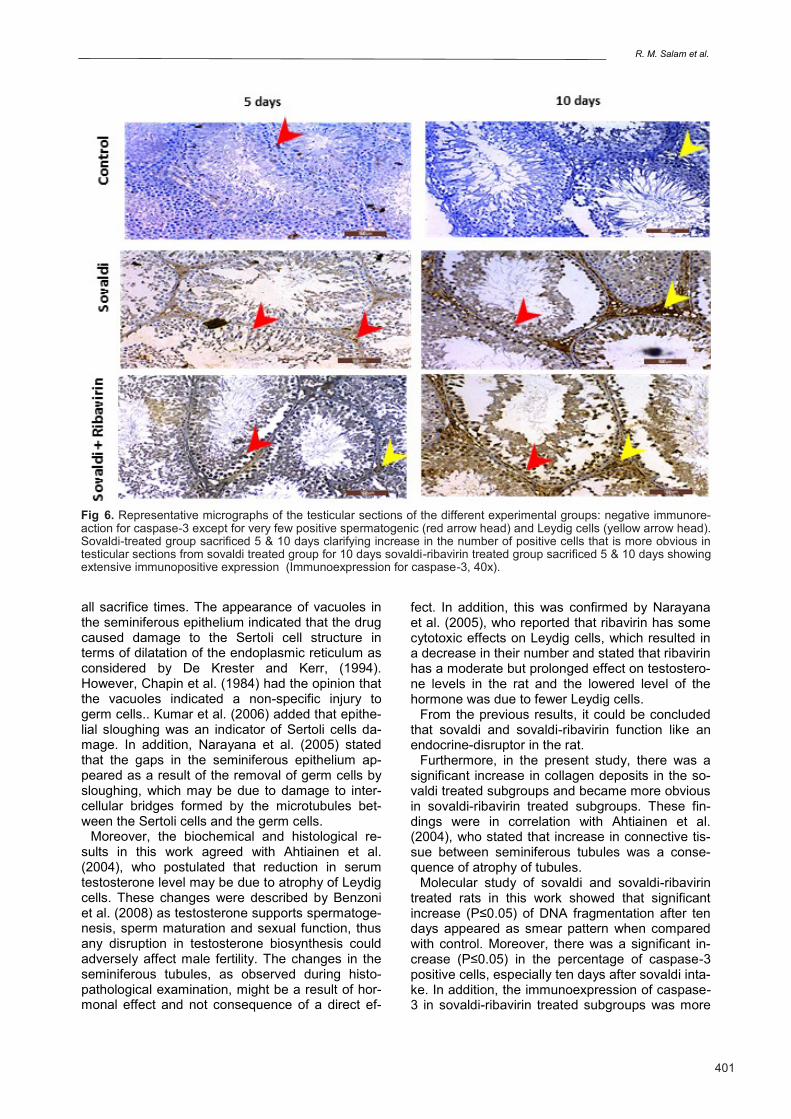
Immunoexpression of caspase-3, a marker for apoptosis, revealed that there was a negative im-munoexpression for caspase-3 in the control group, except for very few positive cells, either spermatogenic or Leydig cells. However, in the sovaldi treated group there was an increase in the number of positive cells especially 10 days after sovaldi intake. In sovaldi-ribavirin treated group, the immunoexpression to caspase-3 was more extensive especially with the increased duration. Statistically, there was a significant increase in the % of caspase-3 positive cells in sovaldi and soval-di-ribavirin treated groups when compared with the control group. Furthermore, a significant increase was more in sovaldi-ribavirin treated rats when compared with that of sovaldi treated (Fig. 6, Ta-bles 3, 4).
DISCUSSION
The therapeutic effects of sovaldi and sovaldi-ribavirin on Hepatitis C have been well documen-
ted. However, the results of our experimental study reveal that administration of a therapeutic dose of sovaldi and sovaldi-ribavirin promoted male repro-ductive toxicity in rats. Up to our knowledge, there were no documented researches on the effect of sovaldi and sovaldi-ribavirin on testicular human, with limited studies to explore the effects of these drugs on the rat reproductive organs and hence affection of male fertility while using these drugs.
These entailed the aim of this study. Regarding the biochemical analysis of testostero-
ne hormone, there was a significant decrease in the groups treated with sovaldi and sovaldi-ribavirin. Moreover, examination of the testicular sections of sovaldi treated rats either sacrificed 5 or 10 days after drug intake showed distortion of the seminiferous tubules, sloughing of the sperma-togenic cells from their underlying basement mem-brane, appearance of vacuoles in the epithelium and atrophy of Leydig cells. There was an extensi-ve distortion of the testicular seminiferous tubules with presence of congested dilated blood vessels in the testicular interstitium, and appearance of more gaps and vacuoles in testicular sections of the rats treated with both sovaldi and ribavirin. These findings are comparable to the study outco-mes of Narayana et al. (2005), who confirmed that ribavirin induced formation of vacuoles and gaps in the seminiferous epithelium in all dose groups at
Fig 5. Representative micrographs of the testicular sections of the different experimental groups showing fine collagen deposits either in tunica albuginea (TA) or peritubular (arrow head) in the control group that increases in sovaldi treated group to be more dense in sovaldi-ribavirin treated group especially 10 days after drugs intake (Masson trichrome, 40x).
R. M. Salam et al.
401
all sacrifice times. The appearance of vacuoles in the seminiferous epithelium indicated that the drug caused damage to the Sertoli cell structure in terms of dilatation of the endoplasmic reticulum as considered by De Krester and Kerr, (1994). However, Chapin et al. (1984) had the opinion that the vacuoles indicated a non-specific injury to germ cells.. Kumar et al. (2006) added that epithe-lial sloughing was an indicator of Sertoli cells da-mage. In addition, Narayana et al. (2005) stated that the gaps in the seminiferous epithelium ap-peared as a result of the removal of germ cells by sloughing, which may be due to damage to inter-cellular bridges formed by the microtubules bet-ween the Sertoli cells and the germ cells.
Moreover, the biochemical and histological re-sults in this work agreed with Ahtiainen et al. (2004), who postulated that reduction in serum testosterone level may be due to atrophy of Leydig cells. These changes were described by Benzoni et al. (2008) as testosterone supports spermatoge-nesis, sperm maturation and sexual function, thus any disruption in testosterone biosynthesis could adversely affect male fertility. The changes in the seminiferous tubules, as observed during histo-pathological examination, might be a result of hor-monal effect and not consequence of a direct ef-
fect. In addition, this was confirmed by Narayana et al. (2005), who reported that ribavirin has some cytotoxic effects on Leydig cells, which resulted in a decrease in their number and stated that ribavirin has a moderate but prolonged effect on testostero-ne levels in the rat and the lowered level of the hormone was due to fewer Leydig cells.
From the previous results, it could be concluded that sovaldi and sovaldi-ribavirin function like an endocrine-disruptor in the rat.
Furthermore, in the present study, there was a significant increase in collagen deposits in the so-valdi treated subgroups and became more obvious in sovaldi-ribavirin treated subgroups. These fin-dings were in correlation with Ahtiainen et al. (2004), who stated that increase in connective tis-sue between seminiferous tubules was a conse-quence of atrophy of tubules.
Molecular study of sovaldi and sovaldi-ribavirin treated rats in this work showed that significant increase (P≤0.05) of DNA fragmentation after ten days appeared as smear pattern when compared with control. Moreover, there was a significant in-crease (P≤0.05) in the percentage of caspase-3 positive cells, especially ten days after sovaldi inta-ke. In addition, the immunoexpression of caspase-3 in sovaldi-ribavirin treated subgroups was more
Fig 6. Representative micrographs of the testicular sections of the different experimental groups: negative immunore-action for caspase-3 except for very few positive spermatogenic (red arrow head) and Leydig cells (yellow arrow head). Sovaldi-treated group sacrificed 5 & 10 days clarifying increase in the number of positive cells that is more obvious in testicular sections from sovaldi treated group for 10 days sovaldi-ribavirin treated group sacrificed 5 & 10 days showing extensive immunopositive expression (Immunoexpression for caspase-3, 40x).
Effect of sofosbuvir and sofosbuvir-ribavirin on rat testis
402
extensive especially after ten days. These findings were in agreement with Lasheen et al. (2015), who hypothesized that the increase in testicular apopto-sis was the result of decline of gonadotropins, and subsequently testosterone hormonal level that in-fluenced the testicular cellular viability. Furthermo-re, El-Sharaky et al. (2010) reported that depletion of testosterone in the rats resulted in increased germ cell apoptosis. In addition, testosterone could affect Sertoli cells’ function and germinal cell dege-neration and dislocation could take place due to damage in function of Sertoli cells, and decreased testosterone level had been reported to enhance premature detachment of epithelial cells as men-tioned by Kumar et al. (2006) and Najafi et al. (2010).
Additionally, our work showed that there was in-crease in the number of caspase-3 positive cells after 5 days in sovaldi and sovaldi-ribavirin treated groups. This may be due to the central importance of caspase proteinases, particularly caspase-3, as the principal mediators of apoptosis thus conside-red as the primary activator of apoptotic DNA frag-mentation, as suggested by Wolf et al. (1999).
The detection of caspase-3 immunohistochemi-cally was considered a specific method for detec-tion of apoptosis as clarified by Arroyo et al. (2010); Cheng et al. (2011). Rainaldi et al. (2008) added that agarose gel electrophoresis, along with other methods, was successfully used to confirm apoptosis. Moreover, Duan et al. (2003) stated that detection of caspase-3 could be a more unique, direct and sensitive indicator of apoptosis than de-tection of secondary process as DNA fragmentati-on.
The findings of lowered testosterone hormone, testicular DNA fragmentation, caspase-3 activation and testicular damage, along with increased colla-gen deposits, were considered the most sensitive parameters for detection of testicular toxicity that could be of key importance for the clinical practice.
CONCLUSION
This study demonstrated the potential adverse effects of sovaldi and sovaldi-ribavirin on reproduc-tive system and function in male rats, and showed that sovaldi and sovaldi-ribavirin induced reproduc-tive disorders as revealed by reduction of serum testosterone level, toxic and degenerative effects on the histological architecture of testes, increase in collagen deposits, increased number of caspase-3 positive cells and DNA fragmentation. Also these reproductive disorders become more aggressive with increased duration of drugs intake.
Finally, sovaldi and sovaldi-ribavirin may induce reduction in the potential fertility of adult male rats due to their potential structural adverse effects on rats’ testes, so the physicians, while prescribing these drugs to the patients, must consider their possible gonadotoxic effects on his fertility. Further
studies are recommended to demonstrate whether these damaging effects will be reversible or not, and to use a protective agent to ameliorate the testicular structural changes induced by sovaldi and sovaldi-ribavirin.
REFERENCES AHTIAINEN M, TOPPARI J, POUTANEN M, HUHTA-
NIEMI I (2004) Indirect Sertoli cell-mediated ablation of germ cells in mice expressing the inhibin-α promoter/herpes simplex virus thymidine kinase transgene. Biol Reprod, 71: 1545-1550.
ALJANABI SM, MARTINEZ I (1997) Universal and rapid salt-extraction of high quality genomic DNA for PCR-based techniques. Nucleic Acids Res J, 25: 4692-4693.
ARROYO JA, LI C, SCHLABRITZ-LOUTSEVITCH N, MCDONALD T, NATHANIELSZ P, GALAN HL (2010) Increased placental XIAP and caspase 3 is associated with increased placental apoptosis in a baboon model of maternal nutrient reduction. Am J Obstet Gynecol, 203(4): 364 e13-18.
AU WW, HSU TC (1980) The genotoxic effects of adriamycin in somatic and germinal cells of mouse. Mutat Res, 79: 351-361.
BENZONI E, MINERVINI F, GIANNOCCARO A, FOR-NELLI F, VIGO D, VISCONTI A (2008) Influence of in vitro exposure to mycotoxin zearalenone and its deri-vatives on swine sperm quality. Reprod Toxicol, 25: 461-467.
BHATIA H, SINGH H, GREWAL N, NATT N (2014) So-fosbuvir: A novel treatment option for chronic hepatitis C infection. J Pharmacol Pharmacother, 5(4): 278-284.
CHAPIN RE, DUTTON SL, ROSS MD, SUMRELL BM, LAMB JC (1984) The effects of ethylene glycol mono-methyl ether on testicular histology in rats. J Androl, 5(5): 369-380.
CHENG XS, LI MS, DU J, JIANG QY, WANG L, YAN SY, YU DM, DENG JB (2011) Neuronal apoptosis in the developing cerebellum. Anat Histol Embryol, 40(1): 21-27.
DE KRESTER DM, KERR JB (1994) The cytology of the testis. In: Knobil NE, Neil JD (eds.) The physiology of reproduction. 2nd ed. Raven Press Ltd., New York, pp 1177-1280.
DI BISCEGLIE AM, CONJEEVARAM HS, FRIED MW (1995) Ribavirin as therapy for chronic hepatitis C. A randomized, double-blind, placebo-controlled trial. Ann Intern Med, 123: 897-903.
DUAN WR, GARNER DS, WILLIAMS SD, FUNCKES-SHIPPY CL, SPATH IS, BLOMME EA (2003) Compa-rison of immunohistochemistry for activated caspase-3 and cleaved cytokeratin 18 with the TUNEL method for quantification of apoptosis in histological sections of PC-3 subcutaneous xenografts. J Pathol, 119: 221-228.
EL-ATREBI KA, EL-ATREBI MA, EL-BASSYOUNI HT (2011) Sexual dysfunction in males with hepatitis c virus: relevance to histopathologic changes and pegin-terferon treatment. Saudi J Gastroenterol, 17(6): 406-

R. M. Salam et al.
403
410.
EL-FISHAWY H, SAADI G, HASSABALLA M, HUSSEIN M, DOSS W, RAGAB W, BARSOUM M (2016) Antivi-ral treatment prioritization in HCV-infected patients with extrahepatic manifestations – An Egyptian pers-pective. J Adv Res, 7(3): 391-402.
EL-GARAWANI IM, HASSAB EL-NABI SE (2016) In-creased sensitivity of apoptosis detection using direct and staining method and integration of acridine orange as an alternative safer fluorescent dye in agarose gel electrophoresis and micronucleus test. CJPAS, 10(2): 3865-3871.
EL-SHARAKY AS, NEWAIRY AA, ELGUINDY NM, ELWAFA AA (2010) Spermatotoxicity, biochemical changes and histological alteration induced by gossypol in testicular and hepatic tissues of male rats. Food Chem Toxicol, 48: 3354-3361.
GOMAA A, ALLAM N, ELSHARKWAY A, EL KASSAS M, WAKED I (2017) Hepatitis C infection in Egypt: prevalence, impact and management strategies. Hepat Med, 9: 17-25.
HOOFNAGLE JH, GHANY MG, KLEINER DE (2003) Maintenance therapy with ribavirin in patients with chronic hepatitis C who fail to respond to combination therapy with interferon alfa and ribavirin. Hepatology, 38: 66-74.
HOOFNAGLE JH, LAU D, CONJEEVARAM H (1996) Prolonged therapy of chronic hepatitis C with ribavirin. J Viral Hepat, 3: 247-252.
JACKSON P, BLYTHE D, BANCROFT JD, GAMPLE M (2008) Immunohistochemical techniques in theory and practice of histological technique (6th ed.). Elsevier, China, pp 423.
JEN J, LAUGHLIN M, CHUNG C, HEFT S, AFFRIME MB, GUPTA SK, GLUE P, HAJIAN G (2002) Ribavirin dosing in chronic hepatitis C: application of population pharmacokinetic-pharmacodynamic models. Clin Phar-macol Ther, 72(4): 349-361.
KUMAR SG, NARAYANA K, BAIRY KL, D'SOUZA UJ, SAMUEL VP, GOPALAKRISHNA K (2006) Dacarbazi-ne induces genotoxic and cytotoxic germ cell damage with concomitant decrease in testosterone and increa-se in lactate dehydrogenase concentration in the tes-tis. Mutat Res, 607: 240-252.
LAM AM, ESPIRITU C, BANSAL S, MICOLOCHICK STEUER HM, NIU C, ZENNOU V (2012) Genotype and subtype pro-ling of PSI-7977 as a nucleotide inhi-bitor of hepatitis C virus. Antimicrob Agents Chemot-her, 56: 3359-3368.
LASHEEN SS, REFAAT SH, EI-NEFIAWY NE, ABD-ELGAWAD RM, OTHMAN AI, OLAMA NK (2015) De-velopmental characteristics of rat testicular tissue and the impact of chronic noise stress exposure in the pre-natal and postnatal periods. Anat Physiol, S4.
NAIR AB, JACOB S (2016) A simple practice guide for dose conversion between animals and human. J Basic Clin Pharm, 7(2): 27-31.
NAJAFI G, RAZI M, HOSHYAR A, SHAHMOHAMAD-LOO S, FEYZI S (2010) The effect of chronic exposure with imidacloprid insecticide on fertility in mature male rats. Int J Fertil Steril, 4: 9-16.
NARAYANA K, D’SOUZA UJA, NARAYAN P, KUMAR G (2005) The antiviral drug ribavirin reversibly affects the reproductive parameters in male Wistar rat. Folia Morphol, 64(2): 65-71.
RAINALDI G, ROMANO R, INDOVINA P, FERRANTE A, MOTTA A, INDOVINA PL, SANTINI MT (2008) Me-tabolomics using 1H-NMR of apoptosis and necrosis in HL60 leukemia cells: differences between the two types of cell death and independence from the stimu-lus of apoptosis used. Radiat Res, 169(2): 170-180.
REKER C, ISLAM KM (2014) Risk factors associated with high prevalence rates of hepatitis C infection in Egypt. Int J Infect Dis, 25: 104-106.
SIDWELL RW, HUFFMAN JH, KHARE GP (1972) Broad-spectrum antiviral activity of Virazole: 1- beta-D-ribofuranosyl-1, 2, 4-triazole-3-carboxamide. Science, 177: 705-706.
GOLANTY E, EDLIN G (2015) Health & Wellness, 12th edit. Jones & Bartlett Publishers, USA, pp 272.
STEVENS A, WILSON IG (1996) The haematoxylin and eosin. In: Bancroft JD, Stevens A, Turner DR (eds.). Theory and practice of histological techniques. 4th edit. Churchill Livingstone, New York, pp 99-112.
TE H, RANDALL G, JENSEN D (2007) Mechanism of action of ribavirin in the treatment of chronic hepatitis C. Gastroenterol Hepatol, 3(3): 218-225.
WOLF BB, SCHULER M, ECHEVERRI F, GREEN DR (1999) Caspase-3 is the primary activator of apoptotic DNA fragmentation via DNA fragmentation factor-45/inhibitor of caspase- activated DNase inactivation. J Biol Chem, 22; 274(43): 30651-30656.
YAU AH, YOSHIDA EM (2014) Hepatitis C drugs: the end of the pegylated interferon era and the emergence of all-oral interferon-free antiviral regimens: a concise review. Can J Gastroenterol Hepatol, l28(8): 445-451.
ZHANG Q, YANG G, LI J, LI W, ZHANG B, ZHU W (2011) Melamine induces sperm DNA damage and abnormality, but not genetic toxicity. Regul Toxicol Pharmacol, 60: 144-150.


![Isolated Testicular Tuberculosis Mimicking Testicular ... involvement, but testicular involvement is an unusual clinical condition [3]. In this report, a case with isolated testicular](https://img.pdfslide.net/doc/110x75/5f3d57bf74280d66ef795ba2/isolated-testicular-tuberculosis-mimicking-testicular-involvement-but-testicular.jpg)