Embed Size (px)
Citation preview
Evaluation of the sagittal spine curvatures in the supine patients
1
MedDocs Publishers
Received: Jan 13, 2020Accepted: Feb 14, 2020Published Online: Feb 17, 2020Journal: Journal of Radiology and Medical ImagingPublisher: MedDocs Publishers LLCOnline edition: http://meddocsonline.org/Copyright: © Atalic B (2020). This Article is distributedunder the terms of Creative Commons Attribution 4.0 International License
*Corresponding Author(s): Bruno Atalic
Higher Research Associate Clinical Department of Radiology, Clinical Hospital Centre Rijeka, Kresimirova 42, 51000 Rijeka, Croatia Tel: 00-385-9967-34-736; Fax: 00-385-5165-8386, Email: [email protected]
Journal of Radiology and Medical ImagingOpen Access | Research Article
Cite this article: Atalic B, Barsic AR, Gudelj L. Evaluation of the sagittal spine curvatures in the supine patients. J Radiol Med Imaging. 2020: 3(1); 1023.
ISSN: 2637-885X
Bruno Atalic1*; Antonija Ruzic Barsic2; Lea Gudelj3
1Clinical Department of Radiology, Clinical Hospital Centre Rijeka, Rijeka, Croatia2Department of Radiology, Special Hospital for the Treatment, Rehabilitation and Prevention of Cardiovascular Diseases Thalas-sotherapia, Opatija, Croatia3Clinical Department of Psychiatry, Clinical Hospital Centre Rijeka, Rijeka, Croatia
Abstract
Objective: Providing suggestions for the evaluation of the sagittal spine curvatures in the supine patients because the sagittal spine curvatures are usually analyzed only in the erect patients.
Methods: 196 patients had their sagittal spine curva-tures analyzed in a retrospective study on both the standard X rays done in the erect position and on the native MR scans done in the supine position during the six months period, between the 1st January and the 30th June 2019, on the So-matom Avanto, Erlangen 1,5 T machine at the Department of Radiology of the Clinical Hospital Center Rijeka. Harrison’s posterior tangential method was used for the evaluation of the sagittal spine curvatures due to its lesser standard mis-take and a greater measurement precision than the Cobb’s method.
Results: In the cervical spines there were no significant changes in the patients with hyperlodoses and kyphoses, lesser changes were seen in the patients with hypolordoses and applanations, while the greatest changes were in the patients with normolordoses. The smallest changes overall were observed in the thoracic spines. Regarding the lum-bar spines there were again no significant changes in the patients with hyperlordoses and kyphoses, lesser changes were seen in the patients with hypolordoses and applana-tions, while the greatest changes were observed in the pa-tients with normolordoses.
Conclusion: In the supine patients the sagittal spine cu-rvatures could with a certainty be assessed in the cases of thoracic spines and lumbar normolordoses as well as in the cervical and lumbar hyperlordoses and pathological kypho-ses.
Keywords: X ray; Magnetic resonance imaging; Spine
Abbreviations: CT: Computed Tomography; MRI: Magnetic Resonance Imaging; P: Value of Statistical Significance
MedDocs Publishers
2Journal of Radiology and Medical Imaging
Introduction
Sagittal spine curvatures are usually evaluated in the erect patients while in the supine patients their evaluation is usually omitted due to the general understanding that they are changed and mostly reduced in the laying patient. In the context of the radiological imaging methods it means that only the standard X ray images obtained in the erect patient position are used for their evaluation, while the images obtained in the supine patient position [standard X rays, computed tomography/CT, magnetic resonance imaging/MRI] are unreasonably neglected in this segment of their imaging possibilities. When discussing sagittal spine curvatures, one should bear in mind that a normal spine in a healthy adult when looked at from the lateral perspective is S shaped. It is composed of cervical lordosis, thoracic kyphosis and lumbar lordosis which is of a great importance for its bio-mechanics. Cervical and lumbar lordoses are deemed as normal if their angles are between 40º and 60º and thoracic kyphosis if it is between 20º to 45º [1]. Lordoses designate ventral spine curvatures while kyphoses designate dorsal ones and in the above-mentioned combinations they are deemed as physiological [2]. They are deemed as pathological in the cases of the remodeling of the normal spine curvatures such as a cervical and a lumbar kyphosis with dorsal angles between 20 and 40º, while in the case of the thoracic kyphosis the greatest degree of a change is an applanation, because a thoracic pathological lordosis is almost never seen due to the rigidity of the mentioned segment of the spine [3].
Epstein et al have conducted a study on the compliance between the measurements of the acute traumatic deformities of the thoracolumbar segments of the spine on the standard X rays and the CT scans with the aim of evaluating the diagnostic value of the CT examinations in this respect. Coronal and sagittal reconstructions of the thoracolumbar segments of the spine in the patients which after their traumas had the whole body CTs conducted were compared with the standard X rays of the same patients in the supine position obtained with the transportable X ray machine. Four radiology consultants, two at a time, have used the Cobb’s method for the measurement of the pathological scolioses and kyphoses on the analyzed images. Measured data were statistically analyzed. Sagittal CT scans showed average mean difference of -1,13 degrees +/-SD 3,76 degrees, while the coronal ones showed average mean difference of 0,10 degrees +/-SD of 2,52 degrees in comparison with the standard X rays. Inter-observational difference between the four radiology consultants was 0,913 for the sagittal scans and 0,953 for the coronal ones which represented an excellent correlation. The authors have showed clinically important diagnostic value of the CT examinations in comparison with the standard X rays in the measurements of the traumatic deformities of the thoracolumbar segment of the spine [4].
Harimaya et al have conducted a retrospective evaluation of the changes of the lumbar lordoses in the patients with the spinal deformities which had undergone instrumental posterior spinal fusion with the Jackson’s cord. They have analyzed 44 standard X rays of the operated patients with the spinal deformities (43 women and 1 men). Lumbar lordoses were measured on the preoperative X rays of the erect and the supine patients, the intraoperative X rays of the supine patients and the postoperative X rays of the erect patients. Average angles of the lumbar lordoses in the preoperative erect and supine patients, the intraoperative supine patients and the postoperative erect patients were: -38,1; -46,0; -46,2 and -51,8 degrees respectively
(P<0,05). Two groups of patients were identified: ones with the increased lumbar lordosis during the intraoperative supine position (>5 degrees, n = 25) and the ones with the minimal changes of the lumbar lordoses during the intraoperative supine position (< or = 5 degrees, n = 19). They have concluded that the patients with the hypolordoses in the erect positions had greater and patients with hyperlordoses lesser changes in the supine position (P<0,05) [5].
Finsterer and Strobel have analyzed the causes of camptocormia or a bent spine syndrome which is characterized with the flexion of the thoracolumbar segment of the spine. Lumbar kyphosis which is evident during walking or standing is completely lost during laying. It is a neurological disease which is most often connected with Parkinson’s disease and to a lesser degree with dystonia, multi-system atrophy, Alzheimer’s disease, myopathy, motor neuron disease, myasthenia and chronic inflammatory demyelination polyneuropathy but it could occur as a side effect of some drugs, disc herniation, arthritis, spine trauma, para-neoplastic disorders and psychiatric diseases as well [6].
Finally, Tanahashi et al have analyzed changes of the degrees of the cervical spine curvatures in the neutral and the supine patient positions compared with the extension and the right rotation or the position for the corpectomy conducted with the aim of the decompression and the perforation of the vertebral artery canal. 50 patients with the cervical spine disorders were included. Cervical CT scans were obtained in the above described positions. At the same time a rotation at each dynamical segment of the spine was measured. In the positions for the corpectomy the rotation of the cervical spine of 37.2° ± 6.2° was measured between the occipital bone and the C7 level with the greatest rotation at the C1/C2 level and the significant rotation of the sub axial vertebrae [7].
Drawing from the above researches presented up to date, this paper tries to clarify the observed differences between the sagittal spine curvatures of patients in the erect and the supine positions with the aim of suggesting some possible guidelines for the assessment of the sagittal spine curvatures in the patients in which the radiological imaging methods have been conducted only in the supine position.
Objective
To establishment the guidelines for the evaluation of the sag-ittal spine curvatures in the supine patients, in which the evalu-ation is usually omitted due to the general understanding that they are changed, mostly reduced in supine position. Conse-quently, the sagittal spine curvatures are usually analyzed only on the standard X ray images obtained in the erect patients.
Patients and methods
Our retrospective research has included 196 patients regardless of their age and sex, which was in accordance with the above presented studies conducted by other researchers, which had undergone the native MRI examination of their cervical, thoracic or lumbosacral spine on the Siemens Somatom Avanto, Erlangen 1,5 T machine at the Department of Radiology of the Clinical Hospital Center Rijeka between the 1st January and the 30th June 2019. Analyzed patients comprised 25.93% of the overall number of the examined patients in the observed period of time which was 756. Inclusion criteria were the existence of the standard X rays of the examined part of the spine done in the erect position, that were performed not more than two
years prior to the current MRI examination, namely, they were done after the 1st January 2017, and finally their availability in the ISSA software of the hospital archive of the radiological examinations. Exclusion criteria were the nonexistence of the standard X rays of the examined part of the spine done in the erect position in the mentioned software of the hospital archive of the radiological examinations, the available standard X rays but performed before the 1st January 2017, therefore older than two years and finally if they were done in the supine position. Extra exclusion criterion was the existence of the implanted vertebro-synthesis material because it can create ferromagnetic artifacts during the examination. It also causes the changes in the natural alignment of the sagittal spine curvatures in the erect position of the patients and also complicates their adaptation in the supine position of the patients. The mentioned exclusion criterion was the predominant exclusion criteria due to the fact that major part of the MRI examinations of the spine in our Department are the control examinations after the spine surgeries. All the patients had their MRI examinations done according to the strict protocols, meaning with their necks fixed by the same fixator for the cervical spine examination and their legs slightly bowed over the same pillow for the lumbosacral spine examination.
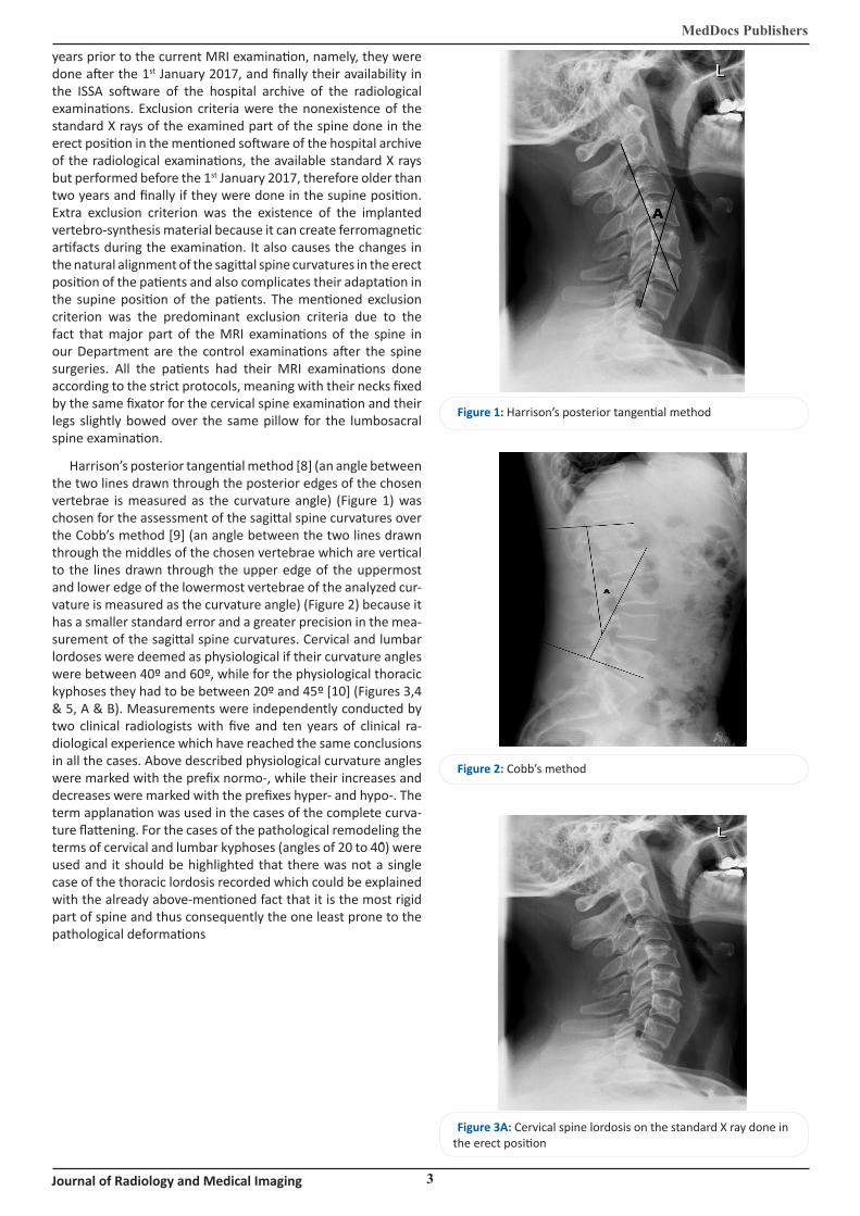
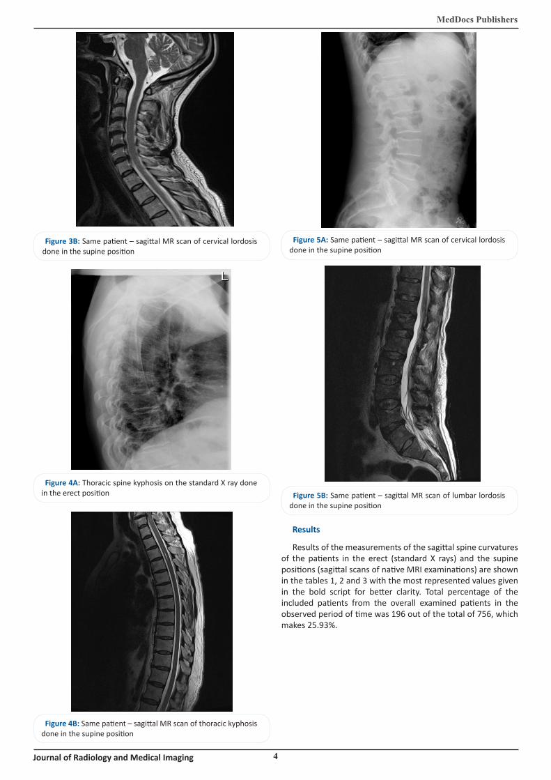
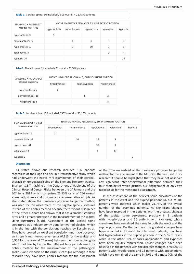
Harrison’s posterior tangential method [8] (an angle between the two lines drawn through the posterior edges of the chosen vertebrae is measured as the curvature angle) (Figure 1) was chosen for the assessment of the sagittal spine curvatures over the Cobb’s method [9] (an angle between the two lines drawn through the middles of the chosen vertebrae which are vertical to the lines drawn through the upper edge of the uppermost and lower edge of the lowermost vertebrae of the analyzed cur-vature is measured as the curvature angle) (Figure 2) because it has a smaller standard error and a greater precision in the mea-surement of the sagittal spine curvatures. Cervical and lumbar lordoses were deemed as physiological if their curvature angles were between 40º and 60º, while for the physiological thoracic kyphoses they had to be between 20º and 45º [10] (Figures 3,4 & 5, A & B). Measurements were independently conducted by two clinical radiologists with five and ten years of clinical ra-diological experience which have reached the same conclusions in all the cases. Above described physiological curvature angles were marked with the prefix normo-, while their increases and decreases were marked with the prefixes hyper- and hypo-. The term applanation was used in the cases of the complete curva-ture flattening. For the cases of the pathological remodeling the terms of cervical and lumbar kyphoses (angles of 20 to 40̊) were used and it should be highlighted that there was not a single case of the thoracic lordosis recorded which could be explained with the already above-mentioned fact that it is the most rigid part of spine and thus consequently the one least prone to the pathological deformations
MedDocs Publishers
3Journal of Radiology and Medical Imaging
Figure 1: Harrison’s posterior tangential method
Figure 2: Cobb’s method
Figure 3A: Cervical spine lordosis on the standard X ray done in the erect position
MedDocs Publishers
4Journal of Radiology and Medical Imaging
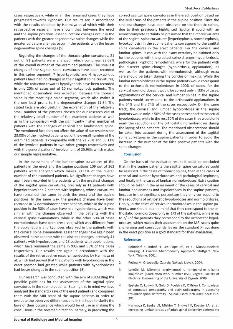
Figure 3B: Same patient – sagittal MR scan of cervical lordosis done in the supine position
Figure 4A: Thoracic spine kyphosis on the standard X ray done in the erect position
Figure 4B: Same patient – sagittal MR scan of thoracic kyphosis done in the supine position
Figure 5A: Same patient – sagittal MR scan of cervical lordosis done in the supine position
Figure 5B: Same patient – sagittal MR scan of lumbar lordosis done in the supine position
Results
Results of the measurements of the sagittal spine curvatures of the patients in the erect (standard X rays) and the supine positions (sagittal scans of native MRI examinations) are shown in the tables 1, 2 and 3 with the most represented values given in the bold script for better clarity. Total percentage of the included patients from the overall examined patients in the observed period of time was 196 out of the total of 756, which makes 25.93%.
MedDocs Publishers
5Journal of Radiology and Medical Imaging
Discussion
As stated above our research included 196 patients regardless of their age and sex in a retrospective study which had underwent the native MRI examination of their cervical, thoracic or lumbosacral spine on the Siemens Somatom Avanto, Erlangen 1,5 T machine at the Department of Radiology of the Clinical Hospital Center Rijeka between the 1st January and the 30th June 2019 which comprises 25,93% or ¼ of 756 overall examined patients and thus makes a representative sample. As also stated above the Harrison's posterior tangential method was used for the assessment of the sagittal spine curvatures instead of the Cobb's method because the previous researches of the other authors had shown that it has a smaller standard error and a greater precision in the measurement of the sagittal spine curvatures [8-10]. Assessment of the sagittal spine curvatures was independently done by two radiologists, which is in the line with the conclusions reached by Epstein et al. They have proved an excellent correlation and have observed an insignificant inter-observer error (0,913 for the sagittal and 0,953 for the coronal CT scans) between their four radiologists which had two by two in the different time periods used the Cobb's method for the measurement of the pathological scoliosis and kyphosis on the various scans [4]. Although in their research they have used Cobb's method for the assessment
Table 1: Cervical spine: 66 included / 303 overall = 21,78% patients
STANDARD X RAYS/ERECT PATIENT POSITION
NATIVE MAGNETIC RESONANCE / SUPINE PATIENT POSITION
hyperlordosis normolordosis hypolordosis aplanation kyphosis
hyperlordosis: 3 3
normolordosis: 15 1 8 3 3
hypolordosis: 19 2 10 2 5
aplanation: 13 9 4
kyphosis: 16 16
Table 2: Thoracic spine: 21 included / 91 overall = 23,08% patients
STANDARD X RAYS/ ERECT PATIENT POSITION
NATIVE MAGNETIC RESONANCE / SUPINE PATIENT POSITION
hyperkyphosis normokyphosis hypokyphosis
hyperkyphosis: 7 7
normokyphosis: 10 8 2
hypokyphosis: 4 4
Table 3: Lumbar spine: 109 included / 362 overall = 30,11% patients
STANDARD X RAYS / ERECT PATIENT POSITION
NATIVE MAGNETIC RESONANCE / SUPINE PATIENT POSITION
hyperlordosis normolordosis hypolordosis aplanation kyphosis
hyperlordosis: 11 11
normolordosis: 37 16 18 3
hypolordosis: 41 38 3
aplanation: 18 1 17
kyphosis: 2 2
of the CT scans instead of the Harrison's posterior tangential method for the assessment of the MR scans that we used in our research it should be highlighted that they have not observed any significant inter-observational difference between their four radiologists which justifies our engagement of only two radiologists for the mentioned assessment.
In the assessment of the cervical spine curvatures of the patients in the erect and the supine positions 66 out of 303 patients were analyzed which makes 21.78% of the overall number of the examined patients. No significant changes have been recorded in the patients with the greatest changes of the sagittal spine curvatures, precisely in 3 patients with hyperlordoses and 16 patients with kyphoses, whose curvatures have remained the same in both the erect and the supine positions. On the contrary, the greatest changes have been recorded in 15 normolordotic erect patients, that have had hypolordoses in the supine position in the 50% of cases, while in the other 50% of cases applanations and kyphoses have been equally represented. Lesser changes have been observed in the patients with the discreet changes, precisely 19 patients with hypolordoses and 13 patients with applanations, which have remained the same in 50% and almost 70% of the
cases respectively, while in all the remained cases they have progressed towards kyphoses. Our results are in accordance with the results obtained by Harimaya et al which with their retrospective research have shown that between the erect and the supine positions lesser curvature changes occur in the patients with the greater degenerative spine changes while the greater curvature changes occur in the patients with the lesser degenerative spine changes [5].
Regarding the changes of the thoracic spine curvatures, 21 out of 91 patients were analyzed, which comprises 23.08% of the overall number of the examined patients. The smallest changes of the sagittal spine curvatures have been recorded in this spine segment; 7 hyperkyphotic and 4 hypokyphotic patients have had no changes in their sagittal spine curvatures, while the reduction towards hypokyphosis have been recorded in only 20% of cases out of 10 normokyphotic patients. The mentioned observation was expected, because the thoracic spine is the most rigid spine segment, which also makes it the one least prone to the degenerative changes [1-3]. The stated facts are also useful in the explanation of the relatively small number of the patients chosen for our research out of the relatively small number of the examined patients as well as in the comparison with the significantly higher number of patients with the changes of cervical and lumbosacral spine. The mentioned fact does not affect the value of our results since 23.08% of the involved patients out of the overall number of the examined patients is comparable with the 21.78% and 30.11% of the involved patients in two other groups respectively and with the general patients’ involvement of 25.93% which makes our sample representative.
In the assessment of the lumbar spine curvatures of the patients in the erect and the supine positions 109 out of 362 patients were analyzed which makes 30.11% of the overall number of the examined patients. No significant changes have again been recorded in the patients with the greatest changes of the sagittal spine curvatures, precisely in 11 patients with hyperlordoses and 2 patients with kyphoses, whose curvatures have remained the same in both the erect and the supine positions. In the same way, the greatest changes have been recorded in 37 normolordotic erect patients, which in the supine position in the 50% of cases have had hypolordoses, which was similar with the changes observed in the patients with the cervical spine examinations, while in the other 50% of cases normolordoses have been preserved, which was different from the applanations and kyphoses observed in the patients with the cervical spine examination. Lesser changes have again been observed in the patients with the discreet changes, precisely 41 patients with hypolordoses and 18 patients with applanations, which have remained the same in 93% and 95% of the cases respectively. Our results are again in accordance with the results of the retrospective research conducted by Harimaya et al, which had proved that the patients with hypolordoses in the erect position had greater, while patients with hyperlordoses had lesser changes in the supine position [5].
Our research was conducted with the aim of suggesting the possible guidelines for the assessment of the sagittal spine curvatures in the supine patients. Bearing this in mind we have analyzed the standard X rays of the erect patients and compared them with the MRI scans of the supine patients in order to evaluate the observed differences and in the hope to clarify the laws of their occurrence which would be of use in making the conclusions in the reversed direction, namely, in predicting the
MedDocs Publishers
6Journal of Radiology and Medical Imaging
correct sagittal spine curvatures in the erect position based on the MRI scans of the patients in the supine position. Since the smallest changes have been observed on the thoracic spines, due to their previously highlighted rigidity, it could with an almost complete certainty be presumed that their three variants of the sagittal spine curvatures [hyperkyphosis, normokyphosis, hypokyphosis] in the supine patients correspond to the sagittal spine curvatures in the erect patients. For the cervical and lumbar spines, it can with the exact certainty be claimed only for the patients with the greatest spine changes [hyperlordosis, pathological kyphotic remodeling], while for the patients with the discreet spine changes [hypolordosis, applanation], as well as for the patients with normolordosis, although extra care should be taken during the conclusion making. While the lumbar normolordoses in the supine patients would correspond to the orthostatic normolordoses in 100% of cases, for the cervical normolordoses it would be correct only in 33% of cases. Applanations of the cervical and lumbar spines in the supine patients would correspond to the orthostatic applanations in the 64% and the 74% of the cases respectively. On the same track, the cervical and lumbar hypolordoses in the supine patients would only in 50% of the cases correspond to the actual hypolordoses, while in the rest 50% of the cases they would only mark the reductions of the orthostatic normolordoses during the laying of the patients. The mentioned observations should be taken into account during the assessment of the sagittal spine curvatures in the supine patients in order to avoid the increase in the number of the false positive patients with the spine changes.
Conclusion
On the basis of the evaluated results it could be concluded that in the supine patients the sagittal spine curvatures could be assessed in the cases of thoracic spines, then in the cases of cervical and lumbar hyperlordoses and pathological kyphoses, and finally in the cases of lumbar normolordoses. Extra caution should be taken in the assessment of the cases of cervical and lumbar applanations and hypolordoses in the supine patients, because in the significant percentage they only correspond to the reductions of orthostatic hypolordoses and normolordoses. Finally, in the cases of cervical normolordoses in the supine pa-tients, one should bear in mind that they correspond to the or-thostatic normolordoses only in 1/3 of the patients, while in up to 2/3 of the patients they correspond to the orthostatic hypol-ordoses, which makes their assessment on the MRI scans quite challenging and consequently leaves the standard X rays done in the erect position as a gold standard for their evaluation.
References
1. Bohndorf K, Imhof H, Lee Pope JrT, et al. Musculosceletal Imaging: A Concise Multimodality Approach. Stuttgart. New York: Thieme. 2001.
2. Pećina M. Ortopedija. Zagreb: Naklada Ljevak. 2004.
3. Lukežić M. Mjerenje zakrivljenosti u rendgenskim slikama kralješnice [Graduation work number 856]. Zagreb: Faculty of Electrical Engineering of the University of Zagreb. 2009.
4. Epstein O, Ludwig S, Gelb D, Poelstra K, O’Brien J. Comparison of computed tomography and plain radiography in assessing traumatic spinal deformity. J Spinal Disord Tech 2009; 22/3: 197-201.
5. Harimaya K, Lenke LG, Mishiro T, Bridwell K, Koester LA. et al. Increasing lumbar lordosis of adult spinal deformity patients via
MedDocs Publishers
7Journal of Radiology and Medical Imaging
intraoperative prone positioning. Spine 2009; 34/22: 2406-12.
6. Finsterer J, Strobl W. Causes of camptocormia. Disabil Rehabil 2003; 33/17-18: 1702-3.
7. Tanahashi H. Alterations in axial curvature of the cervical spine with a combination of rotation and extension in the conventional anterior cervical approach. Eur Spine J 2013; 22/12: 2850-6.
8. Harrison DE, Harrison DD, Cailliet R, Troyanovich SJ, Janik TJ, Holland B. Cobb method or Harrison posterior tangent method: which to choose for lateral cervical radiographic analysis. Spine 2000; 25/16: 2072-2078.
9. Xie J, Li T, Wang Y, Zhao Z, Zhang Y, et al. Change in Cobb angle of each segment of the major curve after posterior vertebral column resection [PVCR]: A preliminary discussion of correction mechanisms of PVCR. Eur Spine J. 2012; 21/4: 705-10.
10. Goh S, Price RI, Leedman PJ, Singer KP. A comparison of three methods for measuring thoracic kyphosis: implications for clinical studies. Perth: The University of Western Australia. 2000.