Embed Size (px)
Citation preview
i
EVALUATION OF THERMOSTABLE YEAST CYTOSINE DEAMINASE
AND HERPES SIMPLEX VIRUS THYMIDINE KINASE FUSIONS IN
DOUBLE SUICIDE GENE THERAPY FOR CANCER
By
MARILYN SANCHEZ-BONILLA
A thesis submitted in partial fulfillment of
the requirements for the degree of
MASTER OF SCIENCE IN PHARMACOLOGY AND TOXICOLOGY
WASHINGTON STATE UNIVERSITY
Division of Pharmacology and Toxicology
DECEMBER 2009
ii
To the Faculty of Washington State University:
The members of the Committee appointed to examine the thesis of MARILYN SANCHEZ-BONILLA find it satisfactory and recommend that it be accepted.
________________________________________ Margaret E. Black, Ph.D., Chair
________________________________________ Lisa M. Gloss, Ph.D.
________________________________________ Nancy S. Magnuson, Ph.D.
________________________________________ Arash Hatefi, Ph.D.
iii
EVALUATION OF THERMOSTABLE YEAST CYTOSINE DEAMINASE AND HERPES
SIMPLEX VIRUS THYMIDINE KINASE FUSIONS IN DOUBLE SUICIDE GENE
THERAPY FOR CANCER
Abstract
by Marilyn Sanchez-Bonilla, M.S. Washington State University
December 2009
Chair: Margaret E. Black
Suicide gene therapy is a promising alternative treatment for cancer that
specifically targets tumor cells for destruction by delivering a gene encoding a suicide
enzyme that is able to convert an anticancer prodrug to a cytotoxic product. Two widely
studied and clinically used enzyme/prodrug systems are cytosine deaminase (CD) with
5-fluorocytosine (5FC) and Herpes Simplex Virus thymidine kinase (HSV-TK) with
ganciclovir (GCV).
Both enzyme/prodrug systems have shown positive results in clinical studies but
still exhibit limitations such as low transduction efficiency due to current delivery
systems and low enzyme activity towards the prodrug. An approach to overcome the
latter limitation is to create a fusion of both CD and HSV-TK in what is known as double
suicide gene therapy (DSGT). DSGT takes advantage of two enzyme/prodrug systems
to create a synergistic cytotoxic effect with the lowest prodrug doses possible. DSGT
studies with bacterial CD (bCD) and TK fusion started in the late 1990s and showed
enhanced tumor growth inhibition and radiosensitization following 5FC and GCV
iv
treatment. 5FC effect on deoxynucleotide pools by allosteric regulation of the enzyme
ribonucleotide reductase was suggested as the cause of the synergistic effect observed
in bCD/TK. In contrast to bCD/TK studies, synergistic experiments are yet to be done
for yCD/TK fusions. Previous studies in our laboratory have created CD and TK
mutants (yCDdouble, yCDtriple and SR39, respectively), with either improved
thermostabilization or improved prodrug activity. These mutants were created using
computational design (yCD) or regio-specific random mutagenesis (TK). As CD mutants
and SR39 mutant showed improved activity and/or a greater tumor killing efficiency in
comparison to wild type enzymes, we sought to evaluate the synergistic effect of mutant
fusion enzymes. Even though yCD/SR39 fusions have been used with positive results
in clinical trials, we hypothesize that by using thermostabilized yCD in fusion with SR39
a greater killing effect could be obtained.
v
TABLE OF CONTENTS
Page
ABSTRACT .............................................................................................................iii
LIST OF TABLES ....................................................................................................vi
LIST OF FIGURES ..................................................................................................vii
CHAPTER
1. INTRODUCTION ..........................................................................................1
HSVTK/GCV ......................................................................................5
Improving Thymidine Kinase ..............................................................8
CD/5FC ..............................................................................................10
Improving Yeast Cytosine Deaminase ...............................................14
2. EVALUATION OF YEAST CYTOSINE DEAMINASE AND HSV-1
THYMIDINE KINASE FUSIONS IN DOUBLE SUICIDE
GENE THERAPY
Abstract ..............................................................................................17
Introduction ........................................................................................19
Materials and Methods ......................................................................23
Results and Discussion......................................................................26
Figure Legends ..................................................................................34
Tables and Figures ............................................................................36
3. SUMMARY AND FUTURE WORK ...............................................................42
4. REFERENCES .............................................................................................47
vi
LIST OF TABLES
Page
1.1 Differences between the E. coli (bCD) and S. cerevisiae (yCD) ...................14
2.1 List of oligonucleotides .................................................................................36
2.2 In vitro response of rat C6 glioma cells expressing individual enzymes .......36
wild type fusion or mutant fusion enzymes to 5FC or GCV
2.3 Statistically significance differences in IC50 values for 5FC treatment .........37
2.4 Statistically significance differences in IC50 values for GCV treatment ........37
2.5 Synergistic experiment with 5FC and GCV combination treatment ..............38
(percent cell killing)
vii
LIST OF FIGURES
Page
1.1 Schematic representation of suicide gene therapy .......................................2
1.2 Overview of regio-specific random mutagenesis ..........................................5
1.3 Activation of GCV by HSV-TK ......................................................................7
1.4 Cytosine and 5FC deamination by cytosine deaminase ...............................11
1.5 5FC metabolism ...........................................................................................12
2.1 CD/5FC and HSV-TK/GCV metabolic pathways ..........................................20
2.2 5FC sensitivity assays of rat C6 transfectants ..............................................39
2.3 GCV sensitivity assays of rat C6 transfectants .............................................40
2.4 Synergistic experiment with 5FC and GCV combination treatment ..............41
1
CHAPTER ONE
INTRODUCTION
According to the American Cancer Society, cancer is the second leading cause
of adult death in the United States after heart disease. It is estimated that in 2009 alone
1.5 million new cases will be diagnosed and approximately 512,340 million will die of
cancer which is roughly 1,400 deaths per day. Current cancer treatments include
surgery, chemotherapy and radiotherapy. Even though standard treatments are still
widely used, each approach has its problems and limitations. For example, although
surgery is most commonly used to remove solid tumors, there is a high probability of
incomplete tumor removal posing a high risk of relapse. While chemotherapy and
radiotherapy kill dividing cells including both normal and tumor cells they often cause
side effects such as nausea, vomiting, fatigue and loss of appetite. The possibility of
resistance to chemotherapeutic agents may also occur because of cellular changes that
could affect the amount of drug getting in or out of the cell (1). In addition to these side
effects, patients often develop secondary forms of cancer, such as leukemia, after
chemotherapeutic and radiation treatments have ended (2-6).
To overcome the limitations seen in standard treatments it is desirable to create
a therapy that will target tumor cells while sparing normal cells from damage. Suicide
gene therapy is an alternative approach to cancer treatment developed about 20 years
ago. The advantage of suicide gene therapy resides in its ability to specifically target
tumor cells for destruction by the delivery of a gene encoding a suicide enzyme. A
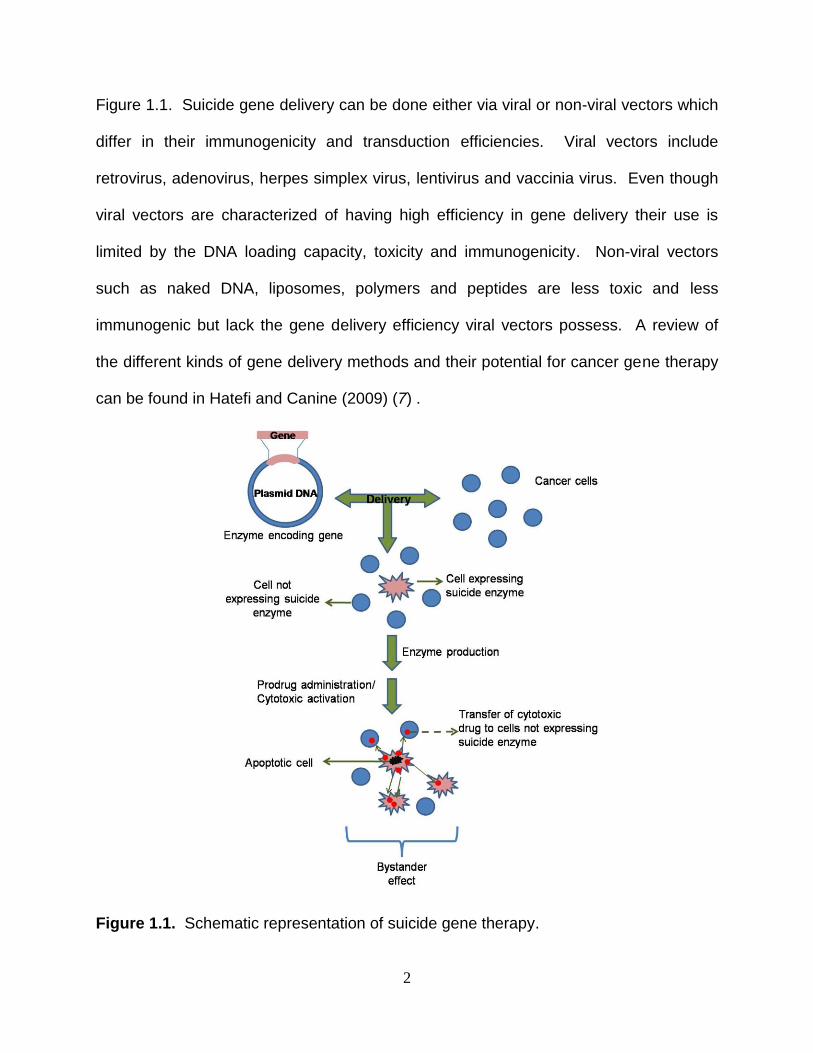
schematic representation of suicide gene therapy and how it works can be seen in
2
Figure 1.1. Suicide gene delivery can be done either via viral or non-viral vectors which
differ in their immunogenicity and transduction efficiencies. Viral vectors include
retrovirus, adenovirus, herpes simplex virus, lentivirus and vaccinia virus. Even though
viral vectors are characterized of having high efficiency in gene delivery their use is
limited by the DNA loading capacity, toxicity and immunogenicity. Non-viral vectors
such as naked DNA, liposomes, polymers and peptides are less toxic and less
immunogenic but lack the gene delivery efficiency viral vectors possess. A review of
the different kinds of gene delivery methods and their potential for cancer gene therapy
can be found in Hatefi and Canine (2009) (7) .
Figure 1.1. Schematic representation of suicide gene therapy.
3
Following successful gene delivery, the suicide enzyme is subsequently
produced and in turn converts an administered non-toxic prodrug into an active toxic
metabolite that induces apoptosis in transfected cells. This apoptotic effect may also be
transferred to neighboring non-transfected cells by a phenomenon known as the
bystander effect. The bystander effect by definition is the extension of killing effects
from transfected cells to neighboring non-transfected cells via diffusion (8, 9), gap
junctions (10-12) or apoptotic vesicles (13). Because of low cell transduction
efficiencies observed in current delivery systems, a strong bystander effect is crucial to
obtain total tumor regression. Although suicide gene therapy has its advantages and
potential for cancer treatment, another key limitation is the low enzyme activity
displayed toward the prodrug.
Ideally, a suicide enzyme suitable for suicide gene therapy protocols should
possess several important characteristics (14). Firstly, it should have high catalytic
activity towards a prodrug to be able to discriminate the enzyme‟s natural substrate
which could be present in normal cells decreasing treatment efficacy. Secondly, to
avoid damage to normal cells the suicide enzyme delivered to tumor cells must be
different from endogenous enzymes. Finally, the suicide enzyme should be expressed
in sufficient concentrations to be of clinical value. However, in reality, enzymes often
have greater activity towards their natural substrates and poor activity towards a
prodrug, thus requiring high doses of prodrug resulting in side effects. In addition, as
the enzyme in most cases is foreign to the host, an immune response could be
triggered which, depending on the situation, could be a positive event. Induction of an
immune response could eliminate remaining tumor cells but if induced too early could
4
destroy the vector carrying the suicide gene thus preventing gene delivery. To
overcome poor enzyme mediated prodrug activation we seek to improve suicide
enzymes in term of stability and activity towards their respective prodrugs.
Our laboratory focuses on optimizing enzymes through enzyme and pathway
engineering to obtain enzymes with higher sensitivity towards a prodrug for suicide
gene therapy (15-19). Several mutagenesis approaches may be used to achieve
enzyme improvement (15). One approach is site-directed mutagenesis where a single
amino acid is changed, however, this method has a low success rate and does not
usually provide variants with significantly improved activity. Another approach, random
mutagenesis, targets specific regions either in or out of the active site for amino acid
substitutions that might switch substrate specificity by favoring a prodrug over the
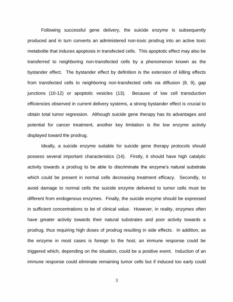
enzyme‟s natural substrate. Regio-specific random mutagenesis uses two or more
overlapping oligonucleotides, with at least one containing a randomized sequence for
the target region of interest within the gene(Figure 1.2) (20). The first and last
oligonucleotide used must contain a restriction site at their 5‟-end for cloning purposes.
Following annealing, extension and amplification, a randomized gene pool is obtained
and then cloned into a “dummy” vector. A “dummy” vector is created by introduction of
a stop codon that inactivates our gene of interest which is expressed in a bacterial
expression vector. Insertion of randomized fragments restores the enzyme open
reading frame. The randomized DNA pool is then transformed into a suitable host that
can be used in a genetic complementation system. Finally, improvement can also be
done using computer modeling programs to identify sequences of interest that might
alter the activity of an enzyme.
5
Figure 1.2. Overview of regio-specific random mutagenesis. N: randomized residues; R: restriction enzyme.
Herpes simplex virus thymidine kinase/Ganciclovir (HSV-TK/GCV)
Two widely studied and clinically used gene/prodrug systems are Herpes
Simplex Virus Thymidine Kinase (HSV-TK) with ganciclovir (GCV) and Cytosine
Deaminase (CD) with 5-fluorocytosine (5FC) (21, 22). Thymidine kinase (EC 2.7.1.21)
is an important enzyme in nucleotide metabolism and is responsible for the conversion
of thymidine to dTMP by the transfer of the γ-phosphate from ATP to thymidine. Unlike
human thymidine kinase, HSV-TK has broad substrate specificity and is able to
phosphorylate pyrimidines (thymidine and deoxycitidine), pyrimidine analogs
6
(azidothymidine and brivudin) and guanosine analogues (ganciclovir, acyclovir,
buciclovir, and penciclovir).
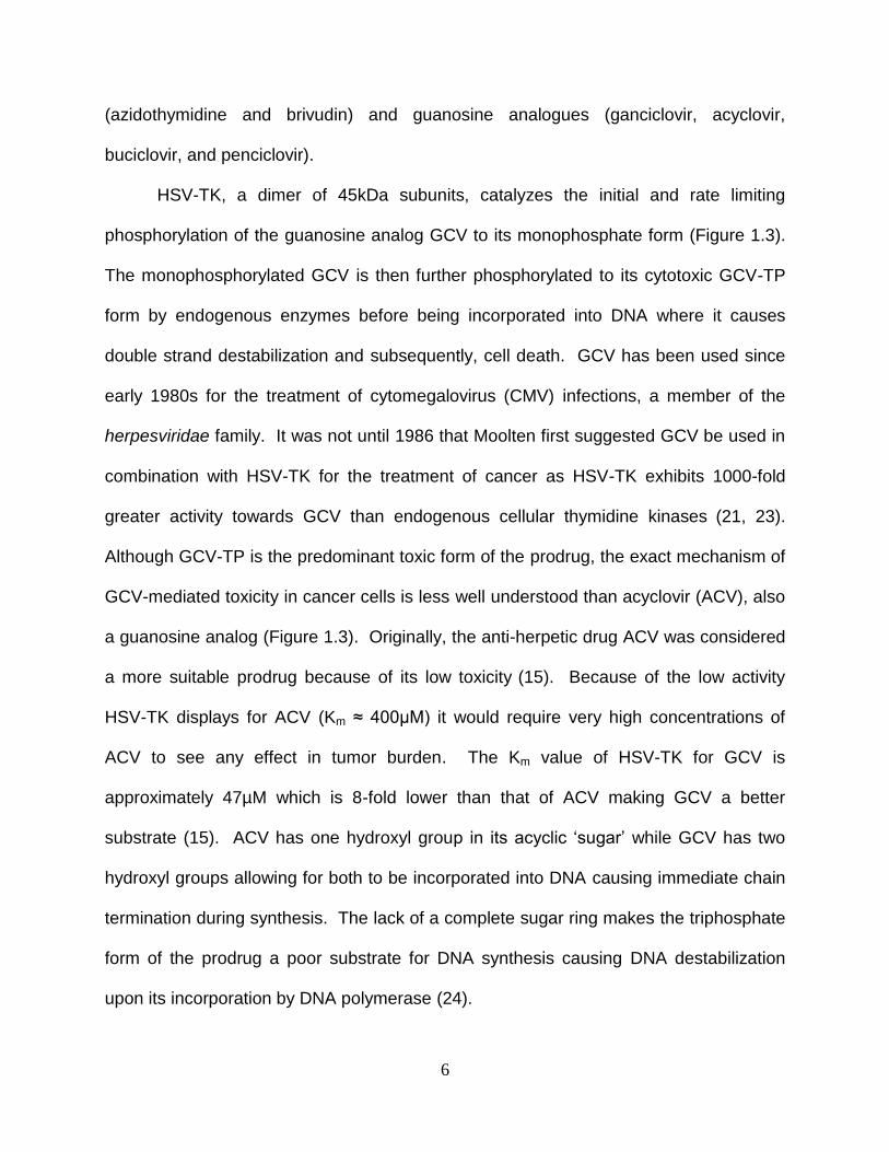
HSV-TK, a dimer of 45kDa subunits, catalyzes the initial and rate limiting
phosphorylation of the guanosine analog GCV to its monophosphate form (Figure 1.3).
The monophosphorylated GCV is then further phosphorylated to its cytotoxic GCV-TP
form by endogenous enzymes before being incorporated into DNA where it causes
double strand destabilization and subsequently, cell death. GCV has been used since
early 1980s for the treatment of cytomegalovirus (CMV) infections, a member of the
herpesviridae family. It was not until 1986 that Moolten first suggested GCV be used in
combination with HSV-TK for the treatment of cancer as HSV-TK exhibits 1000-fold
greater activity towards GCV than endogenous cellular thymidine kinases (21, 23).
Although GCV-TP is the predominant toxic form of the prodrug, the exact mechanism of
GCV-mediated toxicity in cancer cells is less well understood than acyclovir (ACV), also
a guanosine analog (Figure 1.3). Originally, the anti-herpetic drug ACV was considered
a more suitable prodrug because of its low toxicity (15). Because of the low activity
HSV-TK displays for ACV (Km ≈ 400µM) it would require very high concentrations of
ACV to see any effect in tumor burden. The Km value of HSV-TK for GCV is
approximately 47µM which is 8-fold lower than that of ACV making GCV a better
substrate (15). ACV has one hydroxyl group in its acyclic „sugar‟ while GCV has two
hydroxyl groups allowing for both to be incorporated into DNA causing immediate chain
termination during synthesis. The lack of a complete sugar ring makes the triphosphate
form of the prodrug a poor substrate for DNA synthesis causing DNA destabilization
upon its incorporation by DNA polymerase (24).
7
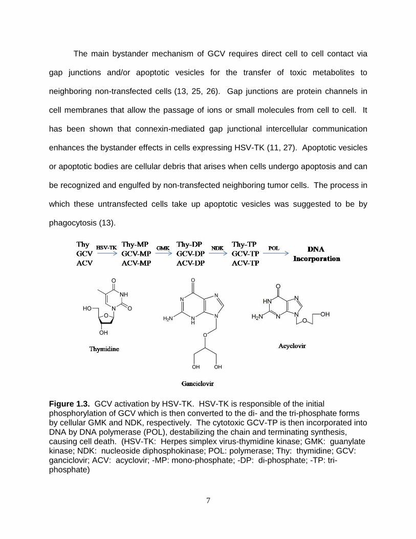
The main bystander mechanism of GCV requires direct cell to cell contact via
gap junctions and/or apoptotic vesicles for the transfer of toxic metabolites to
neighboring non-transfected cells (13, 25, 26). Gap junctions are protein channels in
cell membranes that allow the passage of ions or small molecules from cell to cell. It
has been shown that connexin-mediated gap junctional intercellular communication
enhances the bystander effects in cells expressing HSV-TK (11, 27). Apoptotic vesicles
or apoptotic bodies are cellular debris that arises when cells undergo apoptosis and can
be recognized and engulfed by non-transfected neighboring tumor cells. The process in
which these untransfected cells take up apoptotic vesicles was suggested to be by
phagocytosis (13).
Figure 1.3. GCV activation by HSV-TK. HSV-TK is responsible of the initial phosphorylation of GCV which is then converted to the di- and the tri-phosphate forms by cellular GMK and NDK, respectively. The cytotoxic GCV-TP is then incorporated into DNA by DNA polymerase (POL), destabilizing the chain and terminating synthesis, causing cell death. (HSV-TK: Herpes simplex virus-thymidine kinase; GMK: guanylate kinase; NDK: nucleoside diphosphokinase; POL: polymerase; Thy: thymidine; GCV: ganciclovir; ACV: acyclovir; -MP: mono-phosphate; -DP: di-phosphate; -TP: tri-phosphate)
8
Improving Thymidine Kinase
Although GCV is the current prodrug of choice for suicide gene therapy in
combination with HSV-TK, there are still limitations with the use of this prodrug. The Km
value of HSV-TK for GCV is approximately 100-fold higher than its Km value for
thymidine (Km = 47µM versus 0.5µM) (15). Because of the poor activity displayed by
HSV-TK towards GCV, prodrug doses administered to cancer patients are high and
may result in life-threatening side effects such as bone marrow depression or
myelosuppression (28). Myelosuppression is a condition that can range from mild to
life-threatening in which the bone marrow is unable to produce red blood cells, white
blood cells and platelets that can lead to anemia, an increased risk of infection, or
bleeding. Extensive work has been done to create improved HSV-TK variants with
increased activity towards GCV so that lower doses could be used to obtain effective
tumor ablation with reduced side effects (18, 29-31).
Before the structure of HSV-TK was known, it was suggested that amino acid
residues in proximity to two highly conserved sites known to play an important role in
substrate binding would be suitable targets for regio-specific random mutagenesis(32).
After the pool of random mutants was created, DNA was transformed into a suitable E.
coli strain (BL21(DE3)tdk-) and plated onto rich media plates to ascertain the number of
transformants. To identify variants with improved activity, a genetic complementation
system was used that aims to identify variants that are both TK active and GCV
sensitive and can be divided into two steps: positive and negative selection. As the E.
coli strain used was thymidine kinase (tdk) deficient, only transformed cells possessing
TK activity can complement the deficiency and therefore grow on selection plates
9
containing thymidine as the sole pyrimidine source. In the negative selection step, TK
active variants from positive selection plates are transferred to negative selection plates
containing thymidine and GCV. Variants that show improved activity are unable to form
colonies at lower concentrations of GCV than allow the wild type enzyme to grow.
In a previous study a DNA library of more than a million transformants was
obtained and processed through genetic complementation with only a handful showing
TK activity and a few with significant improvement in terms of GCV sensitivity in
comparison with wild type HSV-TK expressing cells (18, 29, 32). One TK mutant that
proved to be exceptional in terms of activity towards GCV is SR39. This mutant has five
amino acid substitutions (L159I, I160F, F161L, A168F, L169M) and exhibits high GCV
and ACV sensitivity in rat C6 glioma cells in vitro, with IC50 values of 0.017 µM and 0.11
µM respectively. These studies translated to a 294- and 182-fold reduction in IC50
compared to wild type HSV-TK for GCV and ACV, respectively. In vivo xenograft tumor
studies demonstrated that SR39 was better at inhibiting tumor growth requiring a 10-
fold lower GCV concentration (0.5 mg/kg) needed to see an effect in tumors expressing
wild type TK (5.0 mg/kg) (18). Low GCV doses needed to prevent tumor growth in
xenograft tumor models with SR39 demonstrated a significant decrease to current
doses used in most animal experiments (up to 300 mg/kg/day) (28). In another study
where immuno-deficient (SCID) mice were injected with metastatic prostate CL1 cells
expressing either wild type TK or SR39 and treated with 20 mg/kg GCV, SR39
demonstrated a 63% reduction in tumor growth in comparison with tumors expressing
wild type TK (33). In addition to rat C6 glioma cells, SR39 has also been shown to be
effective in increasing cell killing in various cell lines following treatment with GCV
10
including: human mesothelioma (I-45, REN, LRK, H513, H2052), human non-small cell
lung cancer (A549), cervical carcinoma (C33A) and mouse fibroblast tumors (EJ62)(34).
In bystander experiments, rat C6 glioma cells transfected with empty mammalian
expression vector (pUB) were mixed at different ratios with cells expressing SR39 and
treated with 80 µM GCV. Mutant SR39 displayed stronger bystander effect requiring
only 20% of cells expressing enzyme to obtain 50% cell killing as opposed to wild type
TK where no bystander effect was seen (35). The improvement of HSV-TK activity
towards GCV through the creation of SR39 has opened a number of possibilities for its
use, not only as an excellent suicide enzyme for suicide gene therapy protocols but also
as a reporter gene for positron emission tomography (PET) imaging (36-40). Positron
emission tomograpy is a non-invasive nuclear imaging technique that produces a three-
dimensional image and that can be used to monitor gene delivery, gene expression and
cancer therapy efficiency (40). SR39 has proven to be a powerful reporter protein as it
can phosphorylate a number of radiolabeled substrates used as reporter probes that
includes both pyrimidine (uracil-based probes) and purine (acycloguanosine-based
probes), while HSV-TK wild type has higher activity for pyrimidine-based probes (41).
Following initial phosphorylation by HSV-TK, radiolabeled compounds cannot diffuse
from cell to cell and thus allows an entrapment and accumulation of product within the
cells.
Cytosine Deaminase/5-Fluorocytosine (CD/5FC)
A second system that is widely used for suicide gene therapy is cytosine
deaminase (CD)/5-fluorocytosine (5FC). Cytosine deaminase (EC 3.5.4.1) is an
enzyme in the pyrimidine salvage pathway and is present in bacteria and fungi but not
11
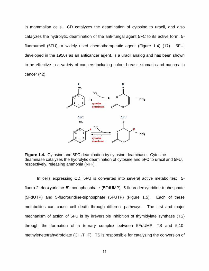
in mammalian cells. CD catalyzes the deamination of cytosine to uracil, and also
catalyzes the hydrolytic deamination of the anti-fungal agent 5FC to its active form, 5-
fluorouracil (5FU), a widely used chemotherapeutic agent (Figure 1.4) (17). 5FU,
developed in the 1950s as an anticancer agent, is a uracil analog and has been shown
to be effective in a variety of cancers including colon, breast, stomach and pancreatic
cancer (42).
Figure 1.4. Cytosine and 5FC deamination by cytosine deaminase. Cytosine deaminase catalyzes the hydrolytic deamination of cytosine and 5FC to uracil and 5FU, respectively, releasing ammonia (NH3). In cells expressing CD, 5FU is converted into several active metabolites: 5-
fluoro-2‟-deoxyuridine 5‟-monophosphate (5FdUMP), 5-fluorodeoxyuridine-triphosphate
(5FdUTP) and 5-fluorouridine-triphosphate (5FUTP) (Figure 1.5). Each of these
metabolites can cause cell death through different pathways. The first and major
mechanism of action of 5FU is by irreversible inhibition of thymidylate synthase (TS)
through the formation of a ternary complex between 5FdUMP, TS and 5,10-
methylenetetrahydrofolate (CH2THF). TS is responsible for catalyzing the conversion of
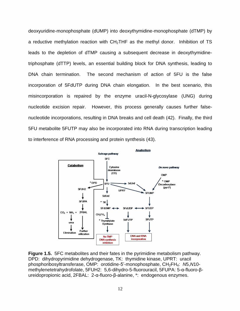
12
deoxyuridine-monophosphate (dUMP) into deoxythymidine-monophosphate (dTMP) by
a reductive methylation reaction with CH2THF as the methyl donor. Inhibition of TS
leads to the depletion of dTMP causing a subsequent decrease in deoxythymidine-
triphosphate (dTTP) levels, an essential building block for DNA synthesis, leading to
DNA chain termination. The second mechanism of action of 5FU is the false
incorporation of 5FdUTP during DNA chain elongation. In the best scenario, this
misincorporation is repaired by the enzyme uracil-N-glycosylase (UNG) during
nucleotide excision repair. However, this process generally causes further false-
nucleotide incorporations, resulting in DNA breaks and cell death (42). Finally, the third
5FU metabolite 5FUTP may also be incorporated into RNA during transcription leading
to interference of RNA processing and protein synthesis (43).
Figure 1.5. 5FC metabolites and their fates in the pyrimidine metabolism pathway. DPD: dihydropyrimidine dehydrogenase, TK: thymidine kinase, UPRT: uracil phosphoribosyltransferase, OMP: orotidine-5‟-monophosphate, CH2FH4: N5,N10-methylenetetrahydrofolate, 5FUH2: 5,6-dihydro-5-fluorouracil, 5FUPA: 5-α-fluoro-β-ureidopropionic acid, 2FBAL: 2-α-fluoro-β-alanine, *: endogenous enzymes.
13
An advantage of CD/5FC over HSV-TK/GCV is the small and uncharged nature
of 5FU making it capable to freely diffuse from cell to cell (44). As 5FC is transferred to
neighboring non-transfected cells by non-facilitated diffusion, only about 5% of tumor
cells expressing cytosine deaminase (CD) were necessary to obtain significant
antitumor effect, and direct cell contact was not required (8). The ability to diffuse may
be an important factor as cell to cell communication may be diminished in cancer cells
as a result of decreased gap junction formation (45, 46). 5FU has also been shown to
be a strong clinical radio-sensitizer effective in the treatment of solid tumors (47, 48).
Because 5FU asserts its cytotoxic effects in three different ways, it is more difficult if not
impossible for cells to repair DNA damage induced by 5FU metabolites which causes
cancer cells to display greater sensitivity to radiotherapy. There are two types of CD
enzymes currently used in suicide gene therapy: bacterial and yeast, isolated from
Escherichia coli and Saccharomyces cerevisiae, respectively. Although they both
deaminate cytosine and 5FC, these enzymes differ from each other in size, catalytic
metal, tertiary structure, thermostability and their efficiency in converting 5FC to 5FU
(Table 1.1) (16, 49-51). Yeast CD (yCD) has shown superiority in catalytic activity over
bacterial CD (bCD) with Km and Vmax values 22- and 4-fold lower for 5FC than bCD (49,
52). In comparison studies, yCD has improved radiosensitization, and the bystander
effect in cancer cells treated with 5FC is greater than bacterial CD (bCD), presumably
due to the difference in catalytic efficiencies (49, 52). Human colon cancer cells (HT29)
expressing yCD and irradiated with 1-2 Gy showed a significantly greater
radiosensitization over cells expressing bCD, requiring approximately 3-fold lower
concentration of 5FC to achieve similar response. Increased radiosensitization by 5FC
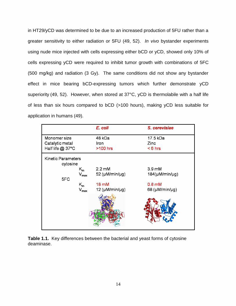
14
in HT29/yCD was determined to be due to an increased production of 5FU rather than a
greater sensitivity to either radiation or 5FU (49, 52). In vivo bystander experiments
using nude mice injected with cells expressing either bCD or yCD, showed only 10% of
cells expressing yCD were required to inhibit tumor growth with combinations of 5FC
(500 mg/kg) and radiation (3 Gy). The same conditions did not show any bystander
effect in mice bearing bCD-expressing tumors which further demonstrate yCD
superiority (49, 52). However, when stored at 37°C, yCD is thermolabile with a half life
of less than six hours compared to bCD (>100 hours), making yCD less suitable for
application in humans (49).
Table 1.1. Key differences between the bacterial and yeast forms of cytosine deaminase.
15
Improving Yeast Cytosine Deaminase
As mentioned above, although yCD has been shown to display more desirable
kinetics parameters than bCD, its thermolability limits its efficacy in suicide gene therapy
protocols. The half-life of yCD at 37°C is about six hours and its melting temperature
(Tm) is 52°C. We sought to optimize yCD by increasing thermostability and 5FC activity.
Computational design was used to identify possible amino acid substitutions outside the
active site of the enzyme that might thermostabilize yCD without affecting enzymatic
activity (53). Three amino acid substitutions (A23L, V108I and I140L) were determined
to each thermostabilize yCD by increasing the Tm by 2°C. When introducing two of
these substitutions into yCD (A23L and I140L), yCDdouble was created with a half-life of
21 hours and a Tm of 58°C. When adding the last substitution (V108I) to create yCDtriple,
a higher thermostabilization was obtained with an improvement in half-life from about
four hours to approximately 117 hours and an increase in Tm from 52°C to 62°C as
compared to the wild type enzyme. Of importance, the kinetic parameters the
thermostabilized yCDs displayed towards either cytosine or 5FC were unaltered. In
transfected rat C6 glioma cells both yCDdouble and yCDtriple showed a slight decrease in
IC50 values of 8 mM and 6 mM, respectively, in comparison with an IC50 value of >10
mM in wild type yCD (19). To address the goal of improving 5FC activity, regio-specific
random mutagenesis was used to target a sequence within the active site of yCD (19).
From these experiments several yCD variants were created and found to confer greater
sensitivity to 5FC in E. coli in comparison with wild type enzyme expressing cells. One
of these mutants, the substitution at residue 92 (yCDD92E) from aspartic acid to glutamic
acid is located at the homodimer interface of yCD and unexpectedly resulted in
16
increased thermostabilization with a Tm of 56°C, without alteration in 5FC activity. As
with thermostabilized enzymes, greater sensitivity to 5FC in yCDD92E could be explained
by the increase in enzyme half-life, which allows an extended conversion of 5FC to
5FU. In vitro cytotoxicity assays in transfected rat C6 glioma cells determined yCDD92E
sensitivity to 5FC to be similar to yCDdouble with an IC50 of approximately 8.5 mM. In an
in vivo xenograft tumor model yCDD92E displayed tumor growth inhibition similar to
yCDtriple. It was then hypothesized that by combining the D92E substitution with yCDtriple
a super sensitive variant would be obtained. However, although the combination of
yCDtriple with D92E resulted in a dramatic Tm increase of 16°C over wild type yCD, over
thermostabilization proved to be detrimental to 5FC sensitivity as the combined mutant
(yCDtriple-D92E) displayed an IC50 similar to yCD in rat C6 glioma cells. There are ongoing
experiments using regio-specific random mutagenesis to improve the prodrug activity of
the thermostabilized yCD that we anticipate will translate to both a lower 5FC
concentration needed to achieve effective cell killing and complete in vivo tumor
ablation.
17
CHAPTER TWO
Evaluation of Yeast Cytosine Deaminase and HSV-1 Thymidine Kinase Fusions in Double Suicide Gene Therapy
ABSTRACT
Suicide gene therapy is a promising cancer treatment that has the potential to
specifically target therapy to tumor cells while sparing normal cells from damage.
Despite its advantages, there are limitations in the system including poor delivery of the
gene to cancer cells and low enzyme activity towards the prodrug requiring
administration of high doses of prodrug. One approach to overcome poor prodrug
activation is to create a fusion of the cytosine deaminase (CD) and Herpes Simplex
Virus-thymidine kinase (HSV-TK) genes in what is known as double suicide gene
therapy (DSGT). The rationale behind DSGT is that the use of two enzyme/prodrug
systems will allow for a synergistic cytotoxic effect to occur that will ultimately destroy
the tumor with lower and safer prodrug doses. Studies have shown that enhanced
tumor growth inhibition and radiosensitization are observed when DSGT is utilized with
the bacterial CD/TK fusion due to a synergistic effect to combined 5FC and GCV
treatment.
Each of the enzymes used in this study, yeast CD (yCD) and HSV-TK have their
limitations. yCD is thermolabile with a melting temperature (Tm) of 52°C and a half-life
at 37°C of approximately six hours which precludes its use in human subjects. On the
other hand, HSV-TK has low activity towards GCV (Km 47µM) that requires high
concentrations of the prodrug to obtain efficacy. Previous studies in our laboratory have
18
created yCD and TK mutants (yCDdouble, yCDtriple and SR39, respectively), with either
improved thermostabilization or improved activity, by using computational design or
regio-specific random mutagenesis. As yCD mutants and SR39 showed improved
activity and/or a higher tumor killing efficiency in comparison to wild type enzymes, we
sought to evaluate the synergistic effect of mutant fusion enzymes. The results
obtained suggest that mutant yCDs in fusion with wild type TK have greater prodrug
sensitivity, as determined by IC50 values, to either 5FC or GCV than fusions with SR39.
In synergistic experiments, yCD/TK showed similar IC50 values to yCDdouble/TK or
yCDdouble/SR39 when treated with 1.5 mM 5FC prior to the addition of 3.0 µM GCV.
When comparing rat C6 glioma cells transfected with fusions with cells expressing
individual enzymes, mutant fusions showed significant improvement in 5FC sensitivity.
However, when treated with GCV and compared with SR39 an increased in IC50 value
was observed.
Further studies need to be done to elucidate whether there is a structural
conformational change in SR39 when it is expressed in a fusion with yCD that would
explain the apparent decrease in GCV sensitivity. The fusion of yCD/SR39 and
5FC/GCV treatment has shown promising results and is currently being used in Phase
III clinical trials for prostate cancer in combination with radiation. However, our results
suggest that fusions with TK could be a better choice over yCD/SR39 due to an
apparent higher synergistic effect that might translate to lower prodrug doses currently
used.
19
Introduction
Each wild type enzyme/prodrug system discussed in Chapter 1 has shown
positive results in clinical studies but still exhibits some limitations such as low enzyme
activity, low transduction efficiency due to current delivery systems and toxicity. An
approach being used to eliminate these limitations is through combinations of bCD or
yCD with HSV-TK as fusions in suicide gene protocols in what is known as double
suicide gene therapy (DSGT). DSGT takes advantage of two metabolic pathways that
use two entirely different enzyme/prodrug systems to obtain an effective cancer
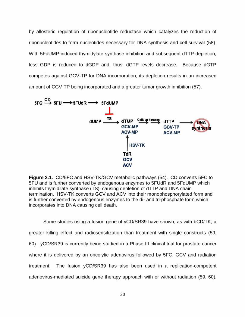
treatment at the lowest dose possible (Figure 2.1) (54). The rationale behind combining
drug treatment protocols includes preventing the emergence of drug resistant cells and
reducing toxicity to normal cells by decreasing the dose needed to achieve clinically
effective results. Several studies have used a bCD/TK fusion construct followed by
treatment with a combination of 5FC and GCV and showed a greater tumor inhibition
compared with individual treatments (54-57). The bCD/TK fusion was created in 1997
by Freytag and colleagues to test their hypothesis that a combination treatment of 5FC
and GCV will provide a synergistic cytotoxic effect (54). Their study showed that co-
administration of 5FC and GCV both radiosensitized tumor cells and resulted in a mild
synergistic cytotoxic effect. The synergistic cytotoxicity seen with bCD/TK after
treatment with 5FC and GCV was suggested to be caused by a shift in dGTP pools after
5FC treatment (57). The known 5FC mechanism of action includes the inhibition of
thymidylate synthase, by the metabolite 5FdUMP, which results in the subsequent
reduction of dTTP levels causing DNA synthesis termination (42). It is well known that
dTTP induces the reduction of guanosine diphosphate (GDP) to its deoxy form (dGDP)
20
by allosteric regulation of ribonucleotide reductase which catalyzes the reduction of
ribonucleotides to form nucleotides necessary for DNA synthesis and cell survival (58).
With 5FdUMP-induced thymidylate synthase inhibition and subsequent dTTP depletion,
less GDP is reduced to dGDP and, thus, dGTP levels decrease. Because dGTP
competes against GCV-TP for DNA incorporation, its depletion results in an increased
amount of CGV-TP being incorporated and a greater tumor growth inhibition (57).
Figure 2.1. CD/5FC and HSV-TK/GCV metabolic pathways (54). CD converts 5FC to 5FU and is further converted by endogenous enzymes to 5FUdR and 5FdUMP which inhibits thymidilate synthase (TS), causing depletion of dTTP and DNA chain termination. HSV-TK converts GCV and ACV into their monophosphorylated form and is further converted by endogenous enzymes to the di- and tri-phosphate form which incorporates into DNA causing cell death.
Some studies using a fusion gene of yCD/SR39 have shown, as with bCD/TK, a
greater killing effect and radiosensitization than treatment with single constructs (59,
60). yCD/SR39 is currently being studied in a Phase III clinical trial for prostate cancer
where it is delivered by an oncolytic adenovirus followed by 5FC, GCV and radiation
treatment. The fusion yCD/SR39 has also been used in a replication-competent
adenovirus-mediated suicide gene therapy approach with or without radiation (59, 60).
21
These studies used prostate and pancreatic tumor models and showed that yCD/SR39
fusion co-expressed with adenovirus death protein (APD) enhanced tumor cell killing
without adding toxicity. Expression of ADP makes the vector more cytolytic, allowing for
easier tumor cell infection and a facilitated spread to neighboring cells with the
possibility of increasing the bystander effect. Results obtained in both studies showed a
lack of toxicity and significant tumor control (59, 60). In 2003, a human sodium iodide
symporter (hNIS) gene was used in conjunction with yCD/SR39 adenovirus-mediated
suicide gene therapy to improve visualization of gene-therapy vectors (61). The hNIS
gene was used to transport anions including pertechnetate (99mTcO4-) which has
physical characteristics that could be exploited for nuclear imaging. From that study, a
valuable tool for gene therapy clinical trials was created by the utilization of three
reporter genes (hNIS, CD and HSV-1 TK), that resulted in a greater imaging resolution
allowing for noninvasive visualization of gene expression and vector delivery (61).
Unlike with the fusion of bCD/TK, basic synergistic experiments are yet to be
done with yCD/TK or mutant fusions. Because yCD has been shown to more efficiently
convert 5FC to 5FU than bCD, one might anticipate that the fusion of yCD/TK will
display a more powerful synergistic effect that that observed with bCD/TK. A greater
drug synergism with the combination treatment of 5FC and GCV in yCD/TK-expressing
cells might therefore translate to a more efficient cancer therapy using low drug doses
thereby decreasing side effects while attaining complete tumor ablation. In this study
we sought to perform cytotoxicity assays to determine synergistic effects when rat C6
glioma cells express thermostabilized yCD (yCDdouble and yCDtriple) and SR39 as fusion
genes. We hypothesized that, by using these mutants in double suicide gene therapy,
22
we will obtain a dramatic synergistic effect following combination treatment of 5FC and
GCV. However, results obtained were unexpected as fusions of yCD or
thermostabilized yCD with TK fusions appears to be more sensitive to either 5FC or
GCV than fusions with SR39. Our study suggests the use of thermostabilized yCD/TK
fusions, instead of yCD/SR39, in ongoing clinical trials might be the most appropriate
approach to obtain complete tumor ablation with safer prodrug doses. When comparing
our fusions with SR39 alone, a loss in GCV sensitivity was obtained that might be
caused by the enzyme impediment to bind GCV due to a partial blockade of the binding
site by yCD. Further biochemical and/or structural studies are needed to help us
explain what might be happening with fusions of yCD or thermostabilized yCD and
SR39 at a molecular level.
23
Materials and methods
Materials
Oligonucleotides used to introduce mutations and sequence pUB:yCD/TK fusions
were obtained from Integrated DNA Technologies (Coralville, IA). Introduction of
mutations was done using the QuikChange® II Site-Directed Mutagenesis kit from
Stratagene (La Jolla, CA). DNA purification was done using several kits: Wizard® PCR
prep kits from Promega (Madison, WI), HiSpeed® Plasmid Mini Kit from Qiagen
(Valencia, CA), and StrataPrep® EF Plasmid Midikit from Stratagene (La Jolla, CA).
AlamarBlue® was purchased from Serotec Limited (Oxford, UK). All cell culture
reagents were purchased from Gibco (Carlsbad, CA). All other reagents were
purchased from Sigma (St. Louis, MO) unless otherwise noted.
Bacterial strains
Escherichia coli strain NM522 [F+ lacIqΔ(lacZ)-M15proA+B+/supE thiΔ(lacproAB)
Δ(hsdMS-mcrB)5(rk-mk
-McrBC-)] and E. coli strain XL1-Blue [F‟::Tn10 proA+B+ lacIq
D(lacZ) M15/recA1 endA1 gyrA96 (Nalr) thi hsdR17 (rk-mk
+) supE44 relA1 lac] were
used as a recipient for certain cloning procedures. E. coli strain JM103 [endA1 glnV44
sbcBC rpsL thi-1 Δ(lac-proAB) F'(traD36 proAB+ lacIq lacZΔM15)] was used to obtain
plasmid DNA suitable for transfections.
Cell lines
Cell lines were maintained in a humidified incubator at 37 °C in 5% CO2. Rat C6
glioma cells (C6) were purchased from ATCC (Manasass, VA) and were grown in
Dulbecco‟s Modified Essential Medium (pH 7.2) containing 5% fetal bovine serum, 1 μM
sodium pyruvate, 10 mM HEPES, 100 μM nonessential amino acids, 100 U/mL
24
penicillin G, 10 μg/mL streptomycin sulfate, 292 μg/mL L-glutamine. Transfected cells
were cultured in media supplemented with the antibiotic blasticidin at a concentration of
4 μg/mL for selection of stable transfectants.
Introduction of mutations to yCD/TK
The QuikChange® kit was used to introduce amino acid substitutions into
pUB:yCD/TK to obtain combinations of wild type yCD, TK and mutant fusion constructs.
Refer to Table 2.1 for oligonucleotide information. Substitutions were confirmed by
sequencing DNA in the Washington State University Sequencing Core Laboratory using
oligo MB400 (5‟ TCAGTG TTAGACTAGTAAATTGTC3‟) to sequence the yCD portion
and MB468 (5‟ GCACCGTAT TGGC 3‟) for the TK portion.
In vitro cytotoxicity assays
In vitro studies were done using the mammalian expression vector pUB
(Invitrogen, CA) expressing wild type yCD, TK, fusion constructs and mutants
(pUB:yCDdouble, pUB:yCDtriple, pUB:SR39, pUB:yCD/TK, pUB:yCD/SR39,
pUB:yCDdouble/TK or pUB:yCDdouble/SR39, pUB:yCDtriple/TK or pUB:yCDtriple/SR39).
Transfection in rat C6 glioma cells were done by seeding 150,000 cells into 6-well
plates with overnight incubation (37°C, 5% CO2) followed by the addition of a
transfection mix in 1X Dulbecco‟s modified Eagle‟s medium (DMEM, pH 7.2) containing
3 µL of the transfection reagent FuGENE® 6 (Roche, Penzberg, Germany) per 1µg of
endonuclease-free DNA (ratio 3:1). Stable transfectants were created through multiple
passages in media containing the antibiotic blasticidin (1X DMEM, 5% fetal bovine
serum (FBS), 100 U/mL penicillin, 10 µg/mL streptomycin, 292 µg/mL glutamine, 10 mM
HEPES, 100 µM non-essential amino acids, 1 µM sodium pyruvate, 4 µg/mL blasticidin,
25
pH 7.2). To obtain IC50 values for each fusion construct, cytotoxicity assays were
performed where 70% confluent cells were treated with phosphate buffered saline
(PBS) (170 mM NaCl, 3 mM KCl, 10 mM Na2HPO4 and 1.7 mM KH2PO4), detached with
PBS containing 5 mM EDTA and trypsin and seeded into 96-well plates at 500
cells/well. Following overnight attachment, cells were treated with different
concentrations of 5FC (0-15 mM) or GCV (0-50 µM) for seven days, at which time the
redox indicator dye Alamar blue was added. Cell survival was determined using a multi-
detection microplate reader SynergyTM HT and microplate data collection and analysis
software Gen5 at a fluorescence wavelength of 530/590nm. Results were plotted as
percentage of cell survival versus drug concentration to obtain IC50 values for each
construct. At least two replicates were performed.
Synergistic experiments
After IC50 value determination, a combined treatment of 5FC and GCV was done
as in previous studies, where better results were shown when 5FC was administered
prior to GCV treatment as opposed to GCV first followed by 5FC (57). Briefly, for the
synergistic experiments, transfected cells were seeded at 250 cells/well and treated with
1.5 mM 5FC 24 hr prior to the addition of new media containing 3.0 µM GCV followed
by a six-day incubation period. To test if cell survival depended on when cells were
treated with GCV after 5FC addition, 5FC treatment was done at 0, 24, 48 and 72 hr.
Cell survival was determined as above.
26
Results and discussion
In vitro cytotoxicity assay experiments
As mentioned in Chapter 1, the use of wild type yeast cytosine deaminase (yCD)
and Herpes Simplex Virus-thymidine kinase (HSV-TK) is limited in suicide gene therapy
protocols due to low enzyme thermostability and/or poor enzyme activity, requiring high
doses of prodrug to achieve an effect in tumor cells. Various mutagenesis approaches
have been used to create super mutants as a means to overcome these limitations.
The determination of amino acid substitutions in yCD, that thermostabilized the enzyme,
allowed for an increased in half-life and melting temperature making yCD more
appropriate for clinical use in human subjects (53). Thermostabilization of yCD allowed
for an increased in half-life which then provides an extended conversion of 5-
fluorocytosine (5FC) into 5-fluorouracil (5FU), thereby resulting in a more effective cell
killing. Enzyme kinetic analysis reveals that increased 5FC sensitivity is not due to
alteration in kinetic parameters. In terms of HSV-TK, the improvement is due to a
greater activity towards ganciclovir (GCV). The HSV-TK mutant, SR39, displays more
than 2000-fold reduction in IC50 when compared to wild type TK and has proven to be
exceptional both in vitro and in vivo (18, 35, 62). Because of the significant
improvement in yCD we questioned whether the fusion of yCD/SR39 currently used in
clinical trials is the best candidate for double suicide gene therapy. As there are no
published synergistic data for yCD/TK fusions, we hypothesized that by using
thermostabilized yCD (yCDdouble and yCDtriple) in fusion with SR39 a stronger synergistic
effect would occur in comparison with yCD/TK or yCD/SR39. To test this hypothesis we
27
sought to first determine the IC50 values for 5FC and GCV in cells expressing fusion
enzymes to obtain optimal treatment conditions for synergistic experiments.
In vitro studies were done using the mammalian expression vector pUB
containing wild type yCD, TK, fusion constructs and mutants (pUB:yCDdouble,
pUB:yCDtriple, pUB:SR39, pUB:yCD/TK, pUB:yCD/SR39, pUB:yCDdouble/TK or
pUB:yCDdouble/SR39, pUB:yCDtriple/TK or pUB:yCDtriple/SR39). Stably transfected rat C6
glioma cells were seeded in 96-well plates and treated with either 5FC or GCV after
overnight incubation. Seven days later Alamar blue was added to each well and
fluorescence analysis was done to ascertain cell survival. Percent cell survival was
calculated by dividing average fluorescence of treated cells over that in untreated wells
for each drug concentration. The obtained values were then plotted against drug
concentration and IC50 values determined for each construct (Table 2.2). Statistical
significance of each fusion construct was evaluated using one way ANOVA and Holm‟s
multiple comparisons (Tables 2.3 and 2.4). Figures 2.2 and 2.3 show representative
survival curves of stably transfected cells treated with 5FC or GCV, respectively
expressing (A) yCD, (B) yCDdouble or (C) yCDtriple in fusion with either TK or SR39.
Values seen in Table 2.2 are the average IC50 of two separate experiments with
standard error of the mean. Cells expressing yCD/TK proved to be more sensitive to
5FC with an IC50 value of 1.85±0.15 mM compared with 5.55±0.35 mM with yCD/SR39
transfected cells. The same pattern was observed where stably transfected cells were
treated with different concentrations of GCV. Once again, yCD/TK showed
approximately 6-fold greater sensitivity to GCV than yCD/SR39 expressing cells with
IC50 values of 1.20±0.10 µM versus 10.15±1.65 µM. The greater sensitivity yCD/TK
28
displays for either 5FC or GCV individually suggests that the fusion of yCD/SR39
currently used in clinical trials is not the best candidate. Even though fusions of yCD/TK
showed greater sensitivity with either drug, as compared with yCD/SR39, the same was
not seen with thermostabilized yCD in fusions with TK or SR39. When cells expressing
fusion enzymes were treated with 5FC, yCDdouble/TK displayed a similar IC50 value to
that of yCDdouble/SR39 with an IC50 values of 0.95±0.55 mM and 2.15±1.35 mM,
respectively. In GCV cytotoxicity assays, the fusion of yCDdouble/TK also demonstrated
similar cell sensitivity with an IC50 of 0.75±0.25 µM as compared to yCDdouble/SR39
which displayed a GCV IC50 of 1.40±0.00 µM. Triple thermostabilized yCD (yCDtriple) in
fusion with TK or SR39 showed similar results as yCDdouble fusions. When C6 cells
were treated with 5FC yCDtriple/TK displayed an IC50 of 0.45±0.25 mM 5FC which
proved to be similar to cells expressing yCDtriple/SR39 (IC50 of 2.25±1.20 mM 5FC). The
same was true for C6 cells expressing yCDtriple/TK treated with GCV where an IC50-
value of 4.85±1.75 µM was similar to that obtained for yCDtriple/SR39-expressing cells
(2.35±0.45 µM),.
When comparing all fusion constructs tested, fusion of yCDtriple/TK conferred a
slightly greater 5FC sensitivity with an IC50 value of approximately 0.45±0.25 mM. As
TK does not contain 5FC activity, lower IC50 values observed for yCD in a fusion might
be due to yCD stabilization and/or an altered 5FC activity. Because pUB transfected
cells displayed an apparent toxicity to 5FC treatment similar to yCD expressing cells,
some concerns are raised. Other controls for this study include yCD, yCDdouble and
yCDtriple for which IC50 values have been previously determined to be over 10 mM, 8 mM
and 6 mM, respectively when expressed with the mammalian expression vector pcDNA
29
(19). However, in the results obtained the values were about 13.45±1.55 mM for yCD,
8.00±1.40 mM for yCDdouble and 11.05±1.95 mM for yCDtriple. Even though the
mammalian expression vectors used in each study have different promoters (ubiquitin C
(pUB) versus CMV (pcDNA)), the relative degree of 5FC sensitivity for each yCD wild
type and mutants should be the same.
As in cytotoxicity assays with 5FC, similar results where obtained when treating
cells with GCV in terms of fusions with TK showing similar sensitivity to fusions with
SR39, with the exception of yCD/TK which displayed about 6-fold lower IC50 for GCV as
compared with yCD/SR39. These results were unexpected as it was hypothesized that
fusions with SR39 will show greater sensitivity to GCV due to the significant
improvement in SR39 activity over TK (29, 32). When comparing each fusion with
individual enzymes, yCDtriple/TK showed the greatest sensitivity to 5FC only with an 11-
to 19-fold decrease in IC50 when compared with wild type or mutant yCD alone. In
experiments where cells expressing enzymes were treated with GCV only, yCDdouble/TK
displayed a more powerful cytotoxic effect with a 50-fold decrease in IC50 when
compared to wild type TK. However, none of our SR39 fusions improved cell killing
when compared with the GCV IC50 obtained for SR39 alone (0.45±0.05 µM). Results
observed in fusions expressing SR39 might be explained by the possibility that having
yCD in fusion with SR39 might be detrimental for the mutant activity towards GCV. This
effect might be due to destabilization or conformational changes in the structure of
mutant TK that alters its activity. Results presented in this study are preliminary and
need to be confirmed by additional experiments, including western blots to determine
protein expression of our fusion constructs and kinetic analysis.
30
Synergistic experiments
By definition drug synergism is the result of two drugs working in combination to
produce an effect greater than the sum of their individual effects (63-65). It has been
shown with bCD/TK, that there is mild synergism when cancer cells expressing the
fusion were treated with 5FC, GCV and radiation (54, 57). A shift in dGTP pools by 5FC
was suggested to be the cause for the synergistic cytotoxicity seen with the greatest
impact when GCV is added 24 hours after 5FC treatment, as opposed to simultaneously
(57). The 5FC mechanism of action includes inhibition of thymidylate synthase by the
metabolite 5FdUMP that results in the subsequent reduction of dTTP levels causing
DNA synthesis termination (42). It is well known dTTP induces the reduction of GDP to
form deoxyguanosine diphosphate (dGDP) by allosteric regulation of ribonucleotide
reductase which is the enzyme responsible for the conversion of ribonucleotides to
nucleotides necessary for cell survival (58). Following dTTP depletion, the conversion
of dGDP from GDP by ribonuclotide reductase is reduced and, thus, dGTP levels
decrease. As dGTP competes with GCV-TP for DNA incorporation, depletion of dGTP
pools results in increased amount of CGV-TP being incorporated into DNA causing
DNA synthesis inhibition and cell death (57).
Results obtained from in vitro cytotoxicity assays were used to study whether
there is synergism with the fusion constructs studied here and to determine which one is
the best in terms of IC50 value and cell killing efficiency. The treatment included either
5FC or GCV as controls or 5FC followed by incubation at different time points prior to
GCV treatment. After overnight incubation, cells were treated with either drug or media
containing no drug and incubated for seven days. A total of 40 replicates per treatment
31
were averaged and plotted as percent cell survival of treated cells over non-treated
cells. To find the optimal conditions for the combination treatment, cell killing efficiency
was tested using different times between treatment with 5FC and treatment with GCV
(0, 24, 48 and 72 hr) (data not shown). In accordance to similar experiments in bCD/TK
fusions, a greater cell killing was observed when using a 24-hour incubation period
between each drug treatment as opposed to treatment with 5FC and GCV
simultaneously (57). In their study, Boucher and colleagues also showed better results
when treating DU145 prostate carcinoma cells expressing bCD/TK with 5FC followed
with GCV, as opposed to treating first with GCV and then 5FC (57). Our results indicate
there was no difference in cell survival between a 5FC incubation period of 24 hr and 48
or 72 hours prior to GCV treatment (data not shown).
Following the determination of suitable conditions (time and drug dosing) for
treatment, experiments were repeated to determine which fusion construct was
exceptional in terms of a greater synergistic effect and an increased cell killing
efficiency. Pools of stable transfectants containing empty vector, wild type yCD, TK,
fusion constructs and mutants (pUB:yCDdouble, pUB:yCDtriple, pUB:SR39, pUB:yCD/TK,
pUB:yCD/SR39, pUB:yCDdouble/TK or pUB:yCDdouble/SR39, pUB:yCDtriple/TK or
pUB:yCDtriple/SR39) were seeded at 250 cells/well and treated with either 1.5 mM 5FC
(black columns), 3.0 µM GCV(white columns) or a combination of both (crimson
columns) (Figure 2.4). In combination treatments GCV was added 24 hr after 5FC
treatment. Each column (mean ± SEM; n=1; performed with 40 replicates) is expressed
as a percentage of the value for control wells with no treatment. Similar results in cell
survival were obtained for C6 cells expressing individual enzymes, with the exception of
32
TK and SR39 which appears to display some sensitivity to 5FC and GCV combination
treatment. As in cytotoxicity assays, cells expressing yCD/TK showed greater
sensitivity to combination treatment than yCD/SR39-expressing cells with a cell survival
of about 30% versus 70%, respectively. With the thermostabilized double yCD in fusion
with TK or SR39 the sensitivity to the 5FC and GCV combination treatment was similar,
with a slightly lower cell survival when cells express yCDdouble/TK. When C6 cells
express yCDtriple in fusion with TK or SR39, yCDtriple/TK displays a greater sensitivity to
combination treatment than yCDtriple/SR39. With the 5FC and GCV concentrations
tested, approximately 50% of cell killing was obtained when cells expressed yCDtriple/TK
fusion whereas only approximately 34% cell killing was observed in cells expressing
yCDtriple/SR39 (Table 2.5). For fusions enzymes our results indicate a similar cell killing
effect (about 67%) for cells expressing yCD/TK, yCDdouble/TK and yCDdouble/SR39.
These results were in accordance with what was obtained in cytotoxicity assays (Figure
2.2 and 2.3) where cells expressing yCD/TK, yCDdouble/TK and yCDdouble/SR39 showed
slightly greater sensitivity with individual treatments. We plan to repeat synergistic
experiments with lower concentrations of each drug, for example 0.5 mM 5FC and 1.5
µM GCV, to be able to discriminate between fusion constructs.
In drug synergy the therapeutic or toxic effect seen when two or more
compounds are used together is greater than the sum of each individual drug. In
accordance to Figure 2.4 and Table 2.5, it could be concluded that no synergy, but an
additive effect, was observed with all fusion constructs tested. The preliminary results
obtained, even though unexpected, gave us more insight of the effect present when
using combination of enzyme/prodrug systems. DSGT has great potential to improve
33
current suicide gene therapy outcomes with the possibility to reduce drug doses used in
clinical trials that could lead to a decreased or absence of side effects. The
development of tumors resistant to treatment can also be reduced or abolished by
combination of enzyme/prodrug/radiation therapy.
34
FIGURE LEGENDS
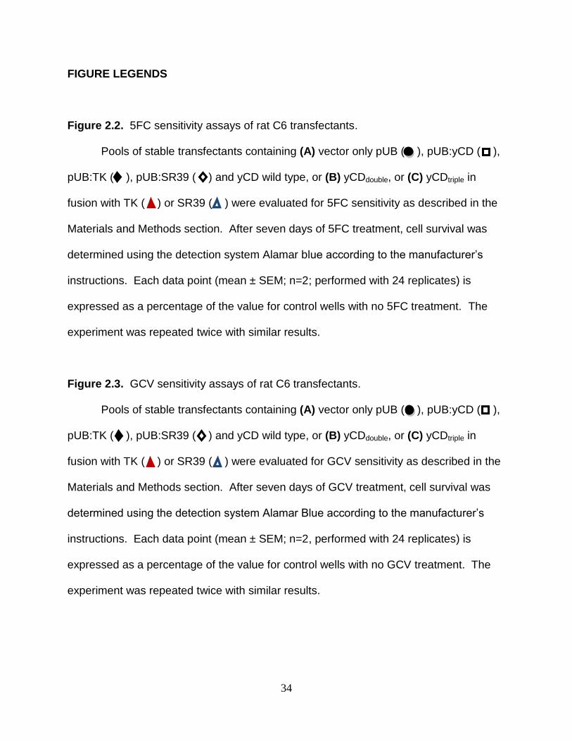
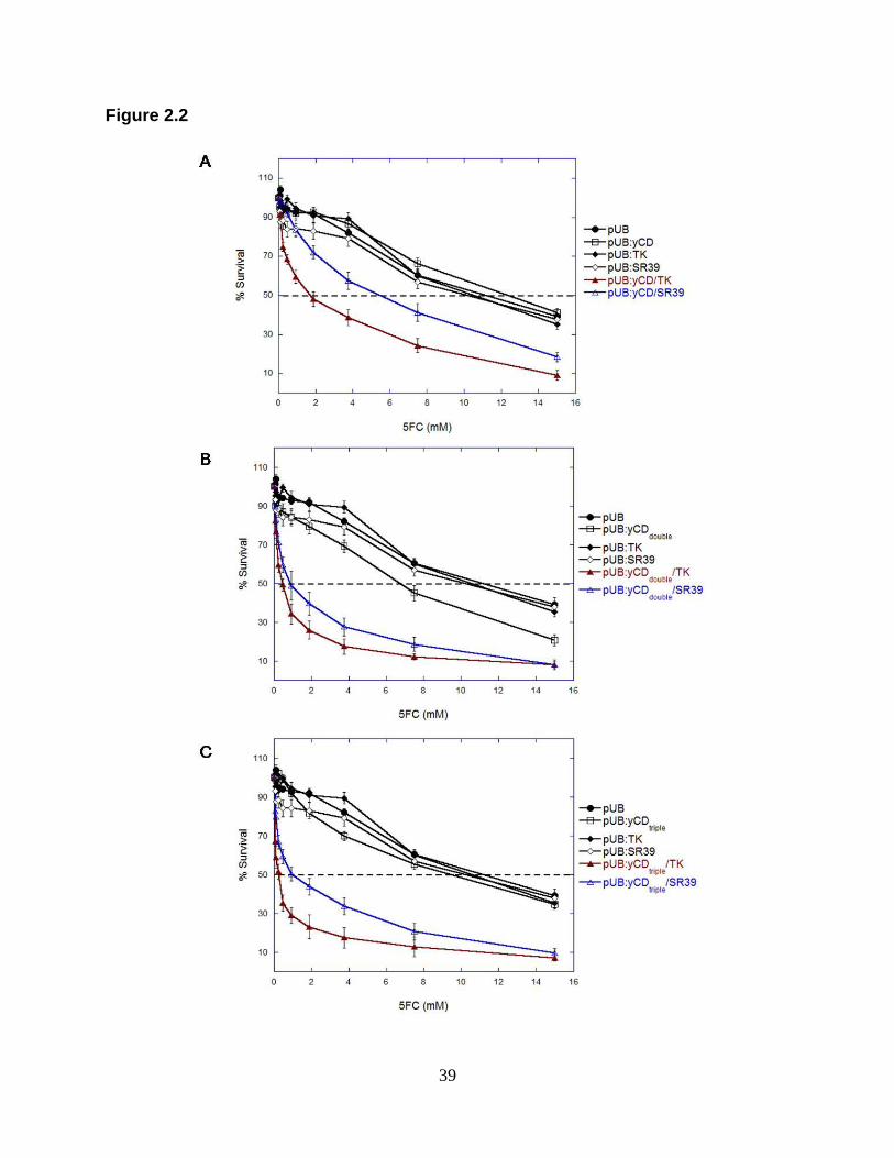
Figure 2.2. 5FC sensitivity assays of rat C6 transfectants.
Pools of stable transfectants containing (A) vector only pUB ( ), pUB:yCD ( ),
pUB:TK ( ), pUB:SR39 ( ) and yCD wild type, or (B) yCDdouble, or (C) yCDtriple in
fusion with TK ( ) or SR39 ( ) were evaluated for 5FC sensitivity as described in the
Materials and Methods section. After seven days of 5FC treatment, cell survival was
determined using the detection system Alamar blue according to the manufacturer‟s
instructions. Each data point (mean ± SEM; n=2; performed with 24 replicates) is
expressed as a percentage of the value for control wells with no 5FC treatment. The
experiment was repeated twice with similar results.
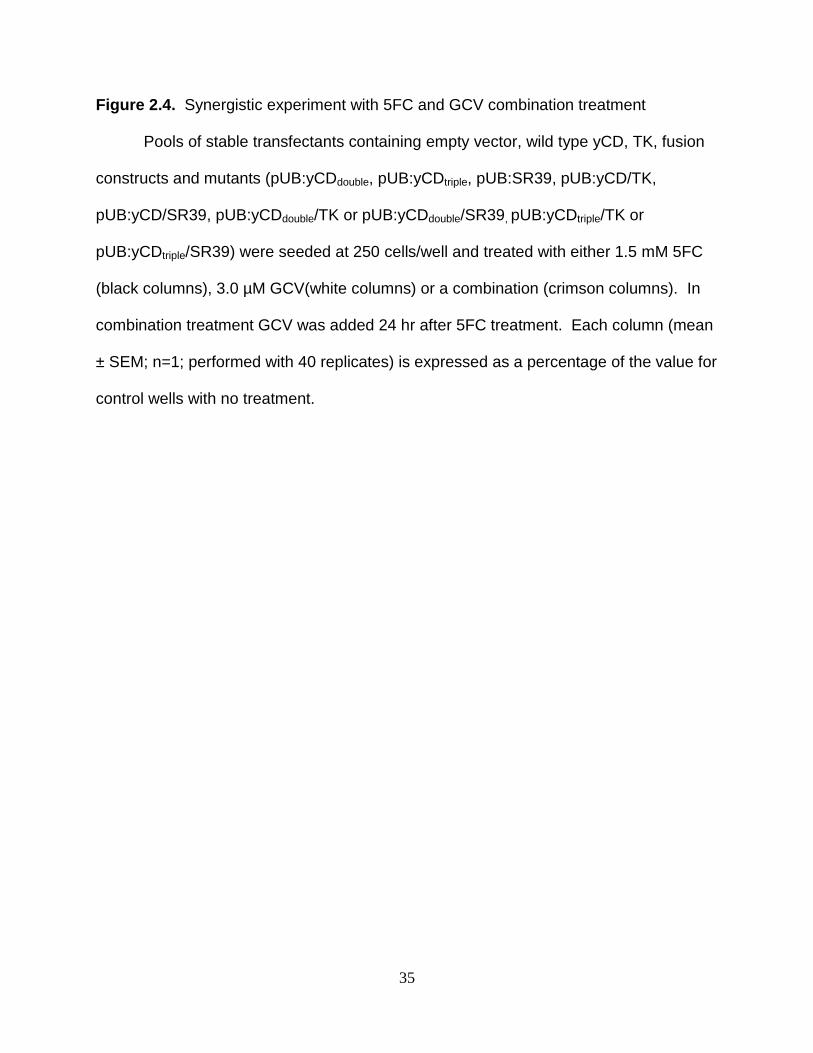
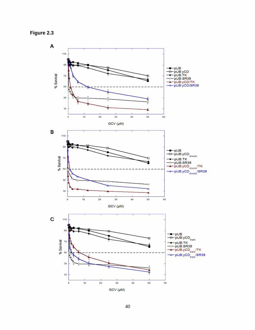
Figure 2.3. GCV sensitivity assays of rat C6 transfectants.
Pools of stable transfectants containing (A) vector only pUB ( ), pUB:yCD ( ),
pUB:TK ( ), pUB:SR39 ( ) and yCD wild type, or (B) yCDdouble, or (C) yCDtriple in
fusion with TK ( ) or SR39 ( ) were evaluated for GCV sensitivity as described in the
Materials and Methods section. After seven days of GCV treatment, cell survival was
determined using the detection system Alamar Blue according to the manufacturer‟s
instructions. Each data point (mean ± SEM; n=2, performed with 24 replicates) is
expressed as a percentage of the value for control wells with no GCV treatment. The
experiment was repeated twice with similar results.
35
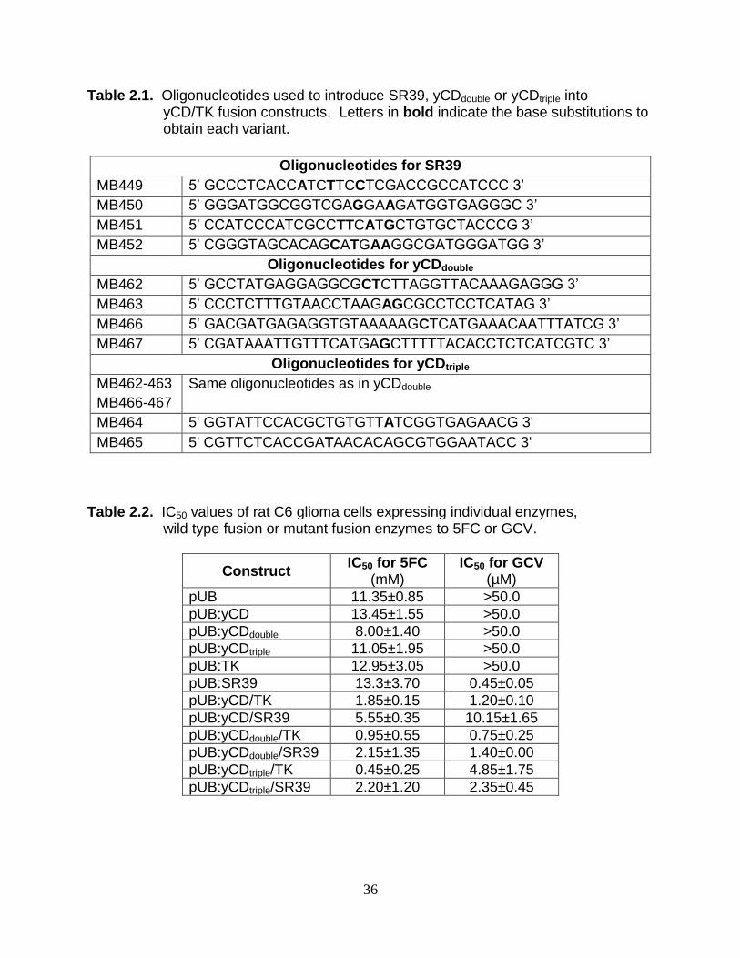
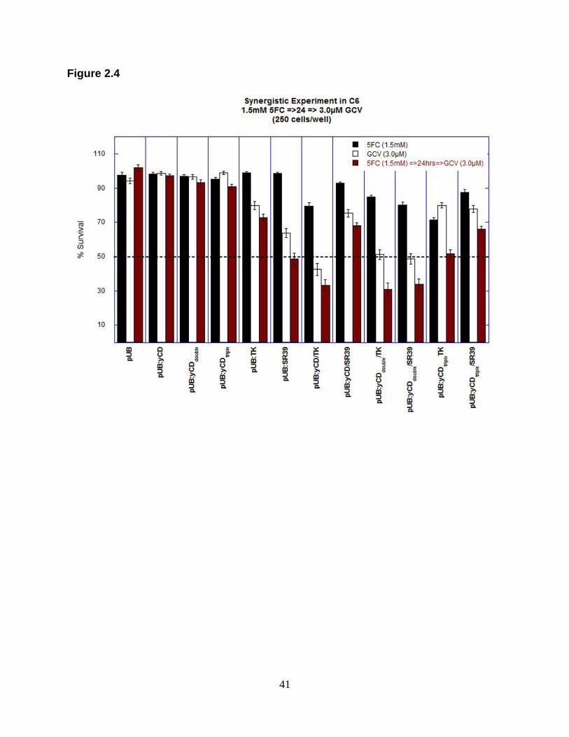
Figure 2.4. Synergistic experiment with 5FC and GCV combination treatment
Pools of stable transfectants containing empty vector, wild type yCD, TK, fusion
constructs and mutants (pUB:yCDdouble, pUB:yCDtriple, pUB:SR39, pUB:yCD/TK,
pUB:yCD/SR39, pUB:yCDdouble/TK or pUB:yCDdouble/SR39, pUB:yCDtriple/TK or
pUB:yCDtriple/SR39) were seeded at 250 cells/well and treated with either 1.5 mM 5FC
(black columns), 3.0 µM GCV(white columns) or a combination (crimson columns). In
combination treatment GCV was added 24 hr after 5FC treatment. Each column (mean
± SEM; n=1; performed with 40 replicates) is expressed as a percentage of the value for
control wells with no treatment.
36
Table 2.1. Oligonucleotides used to introduce SR39, yCDdouble or yCDtriple into yCD/TK fusion constructs. Letters in bold indicate the base substitutions to obtain each variant.
Oligonucleotides for SR39
MB449 5‟ GCCCTCACCATCTTCCTCGACCGCCATCCC 3‟
MB450 5‟ GGGATGGCGGTCGAGGAAGATGGTGAGGGC 3‟
MB451 5‟ CCATCCCATCGCCTTCATGCTGTGCTACCCG 3‟
MB452 5‟ CGGGTAGCACAGCATGAAGGCGATGGGATGG 3‟
Oligonucleotides for yCDdouble
MB462 5‟ GCCTATGAGGAGGCGCTCTTAGGTTACAAAGAGGG 3‟
MB463 5‟ CCCTCTTTGTAACCTAAGAGCGCCTCCTCATAG 3‟
MB466 5‟ GACGATGAGAGGTGTAAAAAGCTCATGAAACAATTTATCG 3‟
MB467 5‟ CGATAAATTGTTTCATGAGCTTTTTACACCTCTCATCGTC 3‟
Oligonucleotides for yCDtriple
MB462-463
MB466-467
Same oligonucleotides as in yCDdouble
MB464 5' GGTATTCCACGCTGTGTTATCGGTGAGAACG 3'
MB465 5' CGTTCTCACCGATAACACAGCGTGGAATACC 3'
Table 2.2. IC50 values of rat C6 glioma cells expressing individual enzymes, wild type fusion or mutant fusion enzymes to 5FC or GCV.
Construct IC50 for 5FC
(mM) IC50 for GCV
(µM)
pUB 11.35±0.85 >50.0
pUB:yCD 13.45±1.55 >50.0
pUB:yCDdouble 8.00±1.40 >50.0
pUB:yCDtriple 11.05±1.95 >50.0
pUB:TK 12.95±3.05 >50.0
pUB:SR39 13.3±3.70 0.45±0.05
pUB:yCD/TK 1.85±0.15 1.20±0.10
pUB:yCD/SR39 5.55±0.35 10.15±1.65
pUB:yCDdouble/TK 0.95±0.55 0.75±0.25
pUB:yCDdouble/SR39 2.15±1.35 1.40±0.00
pUB:yCDtriple/TK 0.45±0.25 4.85±1.75
pUB:yCDtriple/SR39 2.20±1.20 2.35±0.45
37
Table 2.3. Statistically significant differences in IC50 values for 5FC treatments.
Table 2.4. Statistically significant differences in IC50 values for GCV treatments.
38
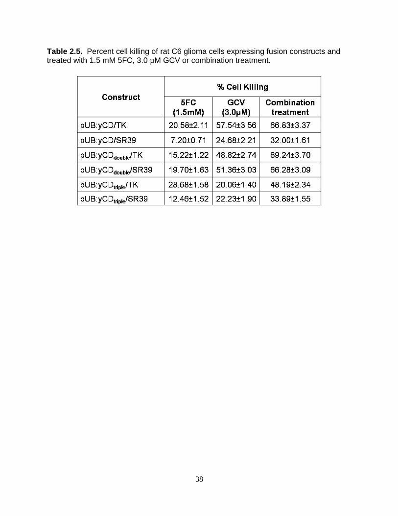
Table 2.5. Percent cell killing of rat C6 glioma cells expressing fusion constructs and treated with 1.5 mM 5FC, 3.0 µM GCV or combination treatment.
39
Figure 2.2
40
Figure 2.3
41
Figure 2.4
42
CHAPTER THREE
Summary and Future Directions
By definition drug synergism occurs when two or more drugs interact to
enhance or magnify the effects of each individual drug. Drug synergism has been
studied for decades in an attempt to find more effective drug treatments that will result
in less toxic effects and reduced drug resistance (63-65). In suicide gene therapy this
is particularly important because of the side effects that chemotherapeutic drugs pose
to the cancer patient, such as nausea, vomiting and/or bone marrow depression (3,
66, 67). To this end, a new approach to suicide gene therapy, double suicide gene
therapy (DSGT), was created by expressing fusion enzymes in cancer cells followed
by treatment with two different drugs. Synergistic effects were observed with bCD/TK
fusion in the late 1990s in which cells expressing the fusion and treated
simultaneously with 5FC and GCV displayed greater cell killing and radiosensitization
of tumor cells when compared to treatment with each individual prodrug (54, 57).
Unlike the bCD/TK system, synergistic experiments have yet to be done with
the yCD/TK system despite the advancement of yCD/SR39 to clinical trials. Fusion of
yCD/SR39 is currently being evaluated in Phase III clinical trials for prostate cancer in
combination with 5FC, GCV and radiation. In this current study we sought to test
thermostabilized yCD (yCDdouble and yCDtriple) and SR39 as fusion genes to test the
hypothesis that by using these mutants we will obtain a more potent synergistic effect
with 5FC and GCV treatment as compared to wild type fusions. A greater synergistic
effect for this system was anticipated because it has been shown yCD is superior to
bCD in converting 5FC to 5FU (49, 52). In addition, mutants of yCD and HSV-TK
43
have demonstrated to be superior to wild type enzymes in terms of thermostability or
activity towards the prodrug (19, 29, 32, 53).
To test our hypothesis, in vitro cytotoxic assays and synergistic experiments
were done in rat C6 glioma cells stably transfected with empty vector (pUB), individual
enzymes/mutants or fusions constructs and treated with 5FC or GCV individually, or
both drugs. Greater synergistic and cell killing effects were expected with fusions of
thermostabilized yCD and SR39 because of the significant improvements obtained
with each variant in terms of thermostabilization (yCDdouble and yCDtriple) and increased
prodrug activity (SR39) (18, 53). However, preliminary results obtained in this study
were unexpected as fusions of yCD or thermostabilized yCD with wild type TK appear
to be slightly more sensitive to combination treatment than fusions with SR39. As
SR39 has been shown to be superior to wild type TK in terms of providing a greater
killing effect in vitro and in vivo, we thought that perhaps the fusion of thermostabilized
yCD with SR39 would give a more powerful synergistic effect that could be translated
to more potent tumor ablation at low drug doses. When comparing with yCD wild type,
the increased sensitivity of fusion constructs treated with 5FC alone may be due to
yCD stabilization by having the enzyme in a fusion with TK. Another explanation
could be an increased in 5FC sensitivity when yCD is expressed in fusion with TK.
Previous studies have created a recombinant fusion protein (LinkCD) with yCD and a
linear polysaccharide, hyalorunan, and found that there was a slight increase in Tm of
4°C over wild type yCD (68). The Tm obtained with the yCD mutants, yCDdouble and
yCDtriple, were 6°C and 10°C higher, respectively, than that of wild type yCD (53).
Although the increase in Tm of LinkCD is lower than that obtained in thermostabilized
44
yCDdouble and yCDtriple, there was a significant increase in 5FC sensitivity as well as
increased survival rate of Balb/c mice bearing C26 murine adenocarcinoma (68).
In vitro cytotoxicity assays for GCV showed that cells expressing yCDdouble/TK
displayed similar sensitivity to GCV than fusions with SR39 (yCDdouble/SR39) The
same result was obtained for yCDtriple in fusion with TK or SR39 which also displayed
similar IC50 values. Even though fusion constructs tested showed approximately 25-
to 100-fold lower IC50 than wild type TK alone, results obtained are not as promising
as when cells express SR39 alone, which have a previously determined IC50 for GCV
of 0.02 µM (35). Results observed in yCD/SR39 fusions might be explained by the
possibility that having yCD in fusion with SR39 might be detrimental for GCV activity.
This effect might be due to destabilization or conformational changes in the structure
of mutant TK that alter its activity or its ability to bind GCV. As results indicate, a
decreased in GCV sensitivity in yCD, yCDdouble or yCDtriple in fusion with SR39
translated to higher IC50 values and greater cell survival when compared with SR39-
expressing cells.
To elucidate the reason behind the unexpected results obtained in this study,
experiments such as enzyme assays and structural studies are necessary. Enzyme
assays with thymidine and GCV will help to determine a decrease in activity in the
fusion constructs expressing SR39 when compared with SR39 by itself. Structural
studies, for example X-ray crystallography, might give us an insight of a possible
conformational change in fusions of yCD/SR39 that could further explain our results.
One of the mutations contained in SR39 is the substitution of phenylalanine at alanine
168 which was suggested to cause an opening of the active site that allows better
45
binding of larger substrates such as GCV (62). This raises the question of whether
yCD is somehow affecting the side chains displayed by A168F that allows an opening
of the active site which then decreases the amount of GCV bound to the enzyme. In
that case the opening in the active site of SR39 in fusion to yCD (wild type or mutant)
could be similar to that in wild type TK or smaller which could explain why fusions
display similar sensitivity to GCV. This might be easily solved by increasing the linker
between each enzyme.
In synergistic experiments, fusions of yCD/TK, yCDdouble/TK and yCDdouble/SR39
showed similar cytotoxicity to 5FC and GCV combination treatment tested with an
approximate cell survival of 30% as compared to individual treatment. Additional
experiments are necessary to discriminate between fusion constructs by further
lowering the concentrations used of each prodrug in combination treatment. To test
this we plan to repeat combination treatment experiments with lower concentrations of
each drug, for example 0.5 mM 5FC and 1.5 µM GCV, that will allow us to determine
the most exceptional fusion construct in terms of greater cell killing efficiency. The
next step after synergistic experiments is to study the bystander effect of cells
expressing fusion constructs to determine which fusion is the best in terms of kil ling
non-transfected cells with the least amount of transfected cells present.
In summary, preliminary results from this study opened more questions about
the efficacy of yCD thermostabilized enzymes in fusion with TK or SR39 in DSGT.
These questions need to be addressed in more extensive studies that could explain
our results before moving to an in vivo xenograft tumor model. Results suggest that
fusions with TK could be a better choice over yCD/SR39 for clinical trials due to an
46
apparent greater sensitivity to combination treatment that might translate to lower
prodrug doses currently used. Synergistic experiments using bCD or yCD in fusion
with other TK mutants might also give us another alternative to improve current DSGT
outcomes.
The superiority of yCD over bCD in terms of 5FC activity and radiosensitization,
the successful thermostabilization of yCD and the creation of super TK mutants, like
SR39, make us strongly believe that thermostabilized yCD/TK fusions would be much
more successful than bCD/TK fusions in double suicide gene therapy. This could
potentially lead to a more powerful cell killing effect thus requiring lower prodrug doses
and radiation that might translate to the absence of side effects to patients and a
complete tumor ablation. A comparison study between bCD/TK and thermostabilized
yCD/TK fusions could be useful to answers all these important questions. Because of
the advantages DSGT pose, we believe the determination of fusion enzyme/prodrugs
systems as the best candidate for cancer therapy would take us a step closer to curing
cancer.
FUNDING
This work was supported by National Institutes of Health grants [CA85939 (M.E.B.) and
CA97328 (Barry L. Stoddard and M.E.B.)].
47
CHAPTER FOUR
References
1. Longley, D. B., and Johnston, P. G. (2005) Molecular mechanisms of drug resistance, J Pathol 205, 275-292.
2. Relling, M. V., Rubnitz, J. E., Rivera, G. K., Boyett, J. M., Hancock, M. L., Felix, C. A., Kun, L. E., Walter, A. W., Evans, W. E., and Pui, C. H. (1999) High incidence of secondary brain tumours after radiotherapy and antimetabolites, Lancet 354, 34-39.
3. Kaldor, J. M., Day, N. E., Pettersson, F., Clarke, E. A., Pedersen, D., Mehnert, W., Bell, J., Host, H., Prior, P., Karjalainen, S., and et al. (1990) Leukemia following chemotherapy for ovarian cancer, N Engl J Med 322, 1-6.
4. Curtis, R. E., Boice, J. D., Jr., Stovall, M., Bernstein, L., Greenberg, R. S., Flannery, J. T., Schwartz, A. G., Weyer, P., Moloney, W. C., and Hoover, R. N. (1992) Risk of leukemia after chemotherapy and radiation treatment for breast cancer, N Engl J Med 326, 1745-1751.
5. Travis, L. B., Curtis, R. E., Glimelius, B., Holowaty, E. J., Van Leeuwen, F. E., Lynch, C. F., Hagenbeek, A., Stovall, M., Banks, P. M., Adami, J., and et al. (1995) Bladder and kidney cancer following cyclophosphamide therapy for non-Hodgkin's lymphoma, J Natl Cancer Inst 87, 524-530.
6. Moser, E. C., Noordijk, E. M., van Leeuwen, F. E., Baars, J. W., Thomas, J., Carde, P., Meerwaldt, J. H., van Glabbeke, M., and Kluin-Nelemans, H. C. (2006) Risk of second cancer after treatment of aggressive non-Hodgkin's lymphoma; an EORTC cohort study, Haematologica 91, 1481-1488.
7. Hatefi, A., and Canine, B. F. (2009) Perspectives in vector development for systemic cancer gene therapy., Gene Ther Mol Biol 13, 15-19.
8. Huber, B. E., Austin, E. A., Richards, C. A., Davis, S. T., and Good, S. S. (1994) Metabolism of 5-fluorocytosine to 5-fluorouracil in human colorectal tumor cells transduced with the cytosine deaminase gene: significant antitumor effects when only a small percentage of tumor cells express cytosine deaminase, Proc Natl Acad Sci U S A 91, 8302-8306.
9. Lawrence, T. S., Rehemtulla, A., Ng, E. Y., Wilson, M., Trosko, J. E., and Stetson, P. L. (1998) Preferential cytotoxicity of cells transduced with cytosine deaminase compared to bystander cells after treatment with 5-flucytosine, Cancer Res 58, 2588-2593.
48
10. Elshami, A. A., Saavedra, A., Zhang, H., Kucharczuk, J. C., Spray, D. C., Fishman, G. I., Amin, K. M., Kaiser, L. R., and Albelda, S. M. (1996) Gap junctions play a role in the 'bystander effect' of the herpes simplex virus thymidine kinase/ganciclovir system in vitro, Gene Ther 3, 85-92.
11. Mesnil, M., Piccoli, C., Tiraby, G., Willecke, K., and Yamasaki, H. (1996) Bystander killing of cancer cells by herpes simplex virus thymidine kinase gene is mediated by connexins, Proc Natl Acad Sci U S A 93, 1831-1835.
12. Mesnil, M., and Yamasaki, H. (2000) Bystander effect in herpes simplex virus-thymidine kinase/ganciclovir cancer gene therapy: role of gap-junctional intercellular communication, Cancer Res 60, 3989-3999.
13. Freeman, S. M., Abboud, C. N., Whartenby, K. A., Packman, C. H., Koeplin, D. S., Moolten, F. L., and Abraham, G. N. (1993) The "bystander effect": tumor regression when a fraction of the tumor mass is genetically modified, Cancer Res 53, 5274-5283.
14. Niculescu-Duvaz, I., and Springer, C. J. (2005) Introduction to the background, principles, and state of the art in suicide gene therapy, Mol Biotechnol 30, 71-88.
15. Black, M. E. (2001) Enzyme and pathway engineering for suicide gene therapy, Genet Eng (N Y) 23, 113-127.
16. Devanathan, S., Willmon, C., Mahan, S., and Black, M. (2002) Engineering enzymes for improved cancer gene therapy, pp 315-326, Res. Adv. in Cancer.
17. Mahan, S. D., Ireton, G. C., Knoeber, C., Stoddard, B. L., and Black, M. E. (2004) Random mutagenesis and selection of Escherichia coli cytosine deaminase for cancer gene therapy, Protein Eng Des Sel 17, 625-633.
18. Black, M. E., Kokoris, M. S., and Sabo, P. (2001) Herpes simplex virus-1 thymidine kinase mutants created by semi-random sequence mutagenesis improve prodrug-mediated tumor cell killing, Cancer Res 61, 3022-3026.
19. Stolworthy, T. S., Korkegian, A. M., Willmon, C. L., Ardiani, A., Cundiff, J., Stoddard, B. L., and Black, M. E. (2008) Yeast cytosine deaminase mutants with increased thermostability impart sensitivity to 5-fluorocytosine, J Mol Biol 377, 854-869.
20. Kurtz, J. E., and Black, M. E. (2004) Enhancement of suicide gene prodrug activation by random mutagenesis, Methods Mol Med 90, 331-344.
21. Moolten, F. L. (1986) Tumor chemosensitivity conferred by inserted herpes thymidine kinase genes: paradigm for a prospective cancer control strategy, Cancer Res 46, 5276-5281.
49
22. Mullen, C. A., Kilstrup, M., and Blaese, R. M. (1992) Transfer of the bacterial gene for cytosine deaminase to mammalian cells confers lethal sensitivity to 5-fluorocytosine: a negative selection system, Proc Natl Acad Sci U S A 89, 33-37.
23. Singhal, S., and Kaiser, L. R. (1998) Cancer chemotherapy using suicide genes, Surg Oncol Clin N Am 7, 505-536.
24. Fillat, C., Carrio, M., Cascante, A., and Sangro, B. (2003) Suicide gene therapy mediated by the Herpes Simplex virus thymidine kinase gene/Ganciclovir system: fifteen years of application, Curr Gene Ther 3, 13-26.
25. Bi, W. L., Parysek, L. M., Warnick, R., and Stambrook, P. J. (1993) In vitro evidence that metabolic cooperation is responsible for the bystander effect observed with HSV tk retroviral gene therapy, Hum Gene Ther 4, 725-731.
26. Fick, J., Barker, F. G., 2nd, Dazin, P., Westphale, E. M., Beyer, E. C., and Israel, M. A. (1995) The extent of heterocellular communication mediated by gap junctions is predictive of bystander tumor cytotoxicity in vitro, Proc Natl Acad Sci U S A 92, 11071-11075.
27. Dilber, M. S., Abedi, M. R., Christensson, B., Bjorkstrand, B., Kidder, G. M., Naus, C. C., Gahrton, G., and Smith, C. I. (1997) Gap junctions promote the bystander effect of herpes simplex virus thymidine kinase in vivo, Cancer Res 57, 1523-1528.
28. Greco, O., and Dachs, G. U. (2001) Gene directed enzyme/prodrug therapy of cancer: historical appraisal and future prospectives, J Cell Physiol 187, 22-36.
29. Black, M. E., Newcomb, T. G., Wilson, H. M., and Loeb, L. A. (1996) Creation of drug-specific herpes simplex virus type 1 thymidine kinase mutants for gene therapy, Proc Natl Acad Sci U S A 93, 3525-3529.
30. Mercer, K. E., Ahn, C. E., Coke, A., Compadre, C. M., and Drake, R. R. (2002) Mutation of herpesvirus thymidine kinase to generate ganciclovir-specific kinases for use in cancer gene therapies, Protein Eng 15, 903-911.
31. Degreve, B., Esnouf, R., De Clercq, E., and Balzarini, J. (2000) Selective abolishment of pyrimidine nucleoside kinase activity of herpes simplex virus type 1 thymidine kinase by mutation of alanine-167 to tyrosine, Mol Pharmacol 58, 1326-1332.
32. Black, M. E., and Loeb, L. A. (1993) Identification of important residues within the putative nucleoside binding site of HSV-1 thymidine kinase by random sequence selection: analysis of selected mutants in vitro, Biochemistry 32, 11618-11626.
33. Pantuck, A. J., Matherly, J., Zisman, A., Nguyen, D., Berger, F., Gambhir, S. S., Black, M. E., Belldegrun, A., and Wu, L. (2002) Optimizing prostate cancer
50
suicide gene therapy using herpes simplex virus thymidine kinase active site variants, Hum Gene Ther 13, 777-789.
34. Wiewrodt, R., Amin, K., Kiefer, M., Jovanovic, V. P., Kapoor, V., Force, S., Chang, M., Lanuti, M., Black, M. E., Kaiser, L. R., and Albelda, S. M. (2003) Adenovirus-mediated gene transfer of enhanced Herpes simplex virus thymidine kinase mutants improves prodrug-mediated tumor cell killing, Cancer Gene Ther 10, 353-364.
35. Ardiani, A., Sanchez-Bonilla, M., and Black, M. E. (2009) Fusion enzymes containing HSV-1 thymidine kinase mutants and guanylate kinase enhance prodrug sensitivity in vitro and in vivo, Cancer Gene Ther.
36. Gambhir, S. S., Bauer, E., Black, M. E., Liang, Q., Kokoris, M. S., Barrio, J. R., Iyer, M., Namavari, M., Phelps, M. E., and Herschman, H. R. (2000) A mutant herpes simplex virus type 1 thymidine kinase reporter gene shows improved sensitivity for imaging reporter gene expression with positron emission tomography, Proc Natl Acad Sci U S A 97, 2785-2790.
37. Pantuck, A. J., Berger, F., Zisman, A., Nguyen, D., Tso, C. L., Matherly, J., Gambhir, S. S., and Belldegrun, A. S. (2002) CL1-SR39: A noninvasive molecular imaging model of prostate cancer suicide gene therapy using positron emission tomography, J Urol 168, 1193-1198.
38. Yang, H., Berger, F., Tran, C., Gambhir, S. S., and Sawyers, C. L. (2003) MicroPET imaging of prostate cancer in LNCAP-SR39TK-GFP mouse xenografts, Prostate 55, 39-47.
39. Yaghoubi, S. S., Barrio, J. R., Namavari, M., Satyamurthy, N., Phelps, M. E., Herschman, H. R., and Gambhir, S. S. (2005) Imaging progress of herpes simplex virus type 1 thymidine kinase suicide gene therapy in living subjects with positron emission tomography, Cancer Gene Ther 12, 329-339.
40. Jacobs, A. H., Rueger, M. A., Winkeler, A., Li, H., Vollmar, S., Waerzeggers, Y., Rueckriem, B., Kummer, C., Dittmar, C., Klein, M., Heneka, M. T., Herrlinger, U., Fraefel, C., Graf, R., Wienhard, K., and Heiss, W. D. (2007) Imaging-guided gene therapy of experimental gliomas, Cancer Res 67, 1706-1715.
41. Likar, Y., Dobrenkov, K., Olszewska, M., Shenker, L., Cai, S., Hricak, H., and Ponomarev, V. (2009) PET imaging of HSV1-tk mutants with acquired specificity toward pyrimidine- and acycloguanosine-based radiotracers., Eur J Nucl Med Mol Imaging 36, 1273-1282.
42. Longley, D. B., Harkin, D. P., and Johnston, P. G. (2003) 5-fluorouracil: mechanisms of action and clinical strategies, Nat Rev Cancer 3, 330-338.
51
43. Ghoshal, K., and Jacob, S. T. (1994) Specific inhibition of pre-ribosomal RNA processing in extracts from the lymphosarcoma cells treated with 5-fluorouracil, Cancer Res 54, 632-636.
44. Domin, B. A., Mahony, W. B., and Zimmerman, T. P. (1993) Transport of 5-fluorouracil and uracil into human erythrocytes, Biochem Pharmacol 46, 503-510.
45. Mesnil, M., Crespin, S., Avanzo, J. L., and Zaidan-Dagli, M. L. (2005) Defective gap junctional intercellular communication in the carcinogenic process, Biochim Biophys Acta 1719, 125-145.
46. Wilgenbus, K. K., Kirkpatrick, C. J., Knuechel, R., Willecke, K., and Traub, O. (1992) Expression of Cx26, Cx32 and Cx43 gap junction proteins in normal and neoplastic human tissues, Int J Cancer 51, 522-529.
47. Byfield, J. E. (1989) 5-Fluorouracil radiation sensitization--a brief review, Invest New Drugs 7, 111-116.
48. Lawrence, T. S., Tepper, J. E., and Blackstock, A. W. (1997) Fluoropyrimidine-Radiation Interactions in Cells and Tumors, Semin Radiat Oncol 7, 260-266.
49. Kievit, E., Bershad, E., Ng, E., Sethna, P., Dev, I., Lawrence, T. S., and Rehemtulla, A. (1999) Superiority of yeast over bacterial cytosine deaminase for enzyme/prodrug gene therapy in colon cancer xenografts, Cancer Res 59, 1417-1421.
50. Ireton, G. C., Black, M. E., and Stoddard, B. L. (2003) The 1.14 A crystal structure of yeast cytosine deaminase: evolution of nucleotide salvage enzymes and implications for genetic chemotherapy, Structure 11, 961-972.
51. Ireton, G. C., McDermott, G., Black, M. E., and Stoddard, B. L. (2002) The structure of Escherichia coli cytosine deaminase, J Mol Biol 315, 687-697.
52. Kievit, E., Nyati, M. K., Ng, E., Stegman, L. D., Parsels, J., Ross, B. D., Rehemtulla, A., and Lawrence, T. S. (2000) Yeast cytosine deaminase improves radiosensitization and bystander effect by 5-fluorocytosine of human colorectal cancer xenografts, Cancer Res 60, 6649-6655.
53. Korkegian, A., Black, M. E., Baker, D., and Stoddard, B. L. (2005) Computational thermostabilization of an enzyme, Science 308, 857-860.
54. Rogulski, K. R., Kim, J. H., Kim, S. H., and Freytag, S. O. (1997) Glioma cells transduced with an Escherichia coli CD/HSV-1 TK fusion gene exhibit enhanced metabolic suicide and radiosensitivity, Hum Gene Ther 8, 73-85.
55. Aghi, M., Kramm, C. M., Chou, T. C., Breakefield, X. O., and Chiocca, E. A. (1998) Synergistic anticancer effects of ganciclovir/thymidine kinase and 5-
52
fluorocytosine/cytosine deaminase gene therapies, J Natl Cancer Inst 90, 370-380.
56. Uckert, W., Kammertons, T., Haack, K., Qin, Z., Gebert, J., Schendel, D. J., and Blankenstein, T. (1998) Double suicide gene (cytosine deaminase and herpes simplex virus thymidine kinase) but not single gene transfer allows reliable elimination of tumor cells in vivo, Hum Gene Ther 9, 855-865.
57. Boucher, P. D., Im, M. M., Freytag, S. O., and Shewach, D. S. (2006) A novel mechanism of synergistic cytotoxicity with 5-fluorocytosine and ganciclovir in double suicide gene therapy, Cancer Res 66, 3230-3237.
58. Moore, E. C., and Hurlbert, R. B. (1966) Regulation of mammalian deoxyribonucleotide biosynthesis by nucleotides as activators and inhibitors, J Biol Chem 241, 4802-4809.
59. Barton, K. N., Paielli, D., Zhang, Y., Koul, S., Brown, S. L., Lu, M., Seely, J., Kim, J. H., and Freytag, S. O. (2006) Second-generation replication-competent oncolytic adenovirus armed with improved suicide genes and ADP gene demonstrates greater efficacy without increased toxicity, Mol Ther 13, 347-356.
60. Freytag, S. O., Barton, K. N., Brown, S. L., Narra, V., Zhang, Y., Tyson, D., Nall, C., Lu, M., Ajlouni, M., Movsas, B., and Kim, J. H. (2007) Replication-competent adenovirus-mediated suicide gene therapy with radiation in a preclinical model of pancreatic cancer, Mol Ther 15, 1600-1606.
61. Barton, K. N., Tyson, D., Stricker, H., Lew, Y. S., Heisey, G., Koul, S., de la Zerda, A., Yin, F. F., Yan, H., Nagaraja, T. N., Randall, K. A., Jin, G. K., Fenstermacher, J. D., Jhiang, S., Ho Kim, J., Freytag, S. O., and Brown, S. L. (2003) GENIS: gene expression of sodium iodide symporter for noninvasive imaging of gene therapy vectors and quantification of gene expression in vivo, Mol Ther 8, 508-518.
62. Kokoris, M. S., and Black, M. E. (2002) Characterization of herpes simplex virus type 1 thymidine kinase mutants engineered for improved ganciclovir or acyclovir activity, Protein Sci 11, 2267-2272.
63. Berenbaum, M. C. (1989) What is synergy?, Pharmacol Rev 41, 93-141.
64. Chou, T. C., and Talalay, P. (1984) Quantitative analysis of dose-effect relationships: the combined effects of multiple drugs or enzyme inhibitors, Adv Enzyme Regul 22, 27-55.
65. Tallarida, R. J. (2001) Drug synergism: its detection and applications, J Pharmacol Exp Ther 298, 865-872.
53
66. Vermes, A., Guchelaar, H. J., and Dankert, J. (2000) Flucytosine: a review of its pharmacology, clinical indications, pharmacokinetics, toxicity and drug interactions, J Antimicrob Chemother 46, 171-179.
67. Bean, B. (1992) Antiviral therapy: current concepts and practices, Clin Microbiol Rev 5, 146-182.
68. Park, J. I., Cao, L., Platt, V. M., Huang, Z., Stull, R. A., Dy, E. E., Sperinde, J. J., Yokoyama, J. S., and Szoka, F. C. (2009) Antitumor therapy mediated by 5-fluorocytosine and a recombinant fusion protein containing TSG-6 hyaluronan binding domain and yeast cytosine deaminase, Mol Pharm 6, 801-812.