Embed Size (px)
Citation preview
Every month you have a whole new layer of skin.
Your nails grow .5mm per week.You will shed 40lbs of skin in a lifetime.The average amount of head hair is 120,000.Your eyelids have the thinnest skin.Hair grows 1cm per month.
True or False:
ProtectionCushions and insulates and is waterproofProtects from chemicals, heat, cold,
bacteriaScreens UV
Synthesizes vitamin D from UV raysRegulates body heatPrevents unnecessary water lossSensory reception (nerve endings)
Functions of skin
Key TermsEpi- “above”Derm- “skin”Hypo- “below”Kerat- “horn”Melano- “black/dark”Cyte- “cell”Strata- “cover”
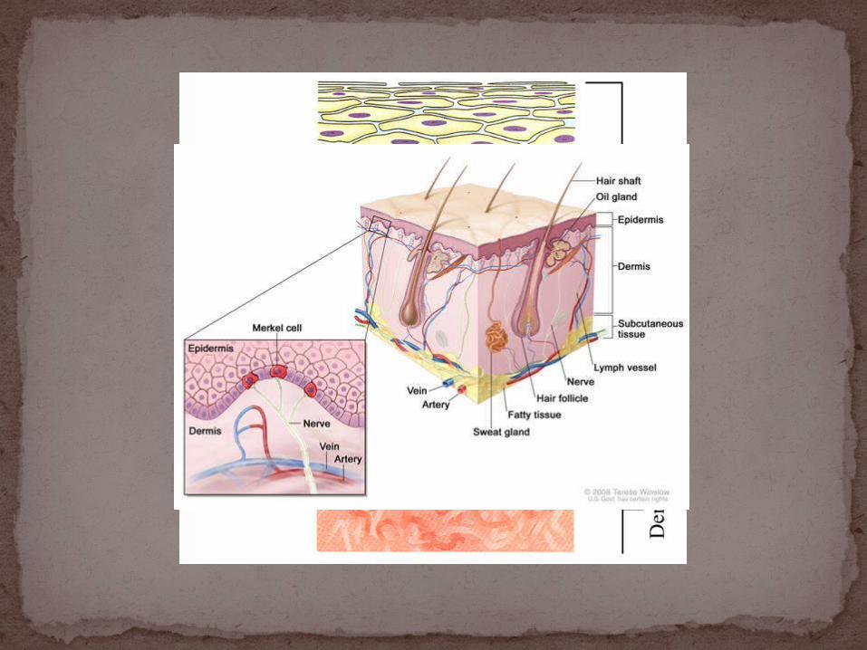
Integument is skinTwo distinct regions
Epidermis (keratinized stratified squamous)
Dermis (Dense Irregular CT w/ smooth muscle, nervous and blood tissue)
A fatty layer (hypodermis) lies deep to it- not a true layer of skin(areolar & adipose tissue)
The Integumentary System
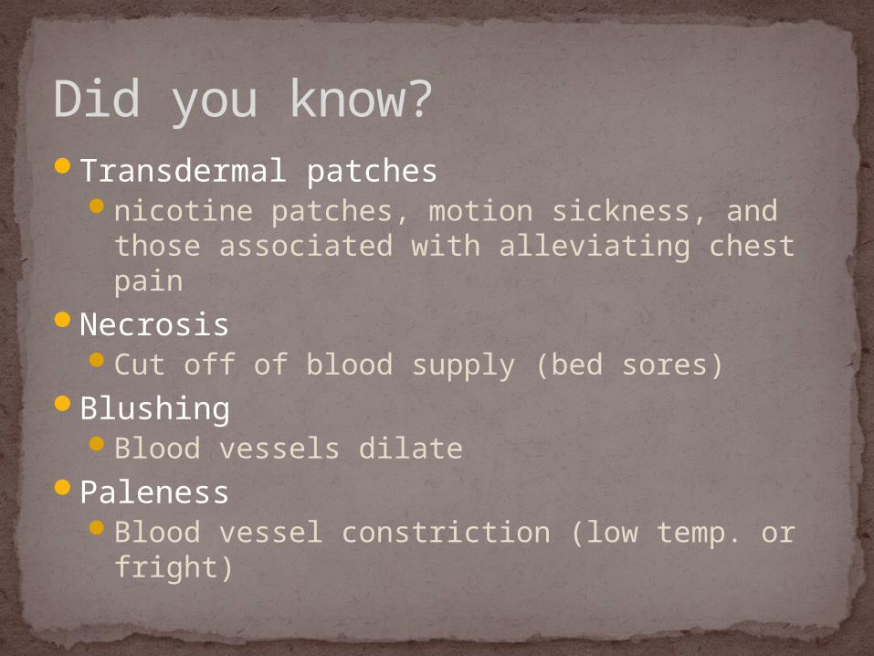
Transdermal patchesnicotine patches, motion sickness, and those
associated with alleviating chest painNecrosis
Cut off of blood supply (bed sores)Blushing
Blood vessels dilate Paleness
Blood vessel constriction (low temp. or fright)
Did you know?
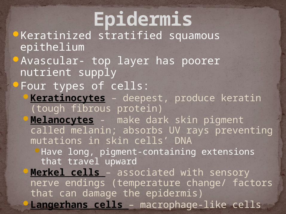
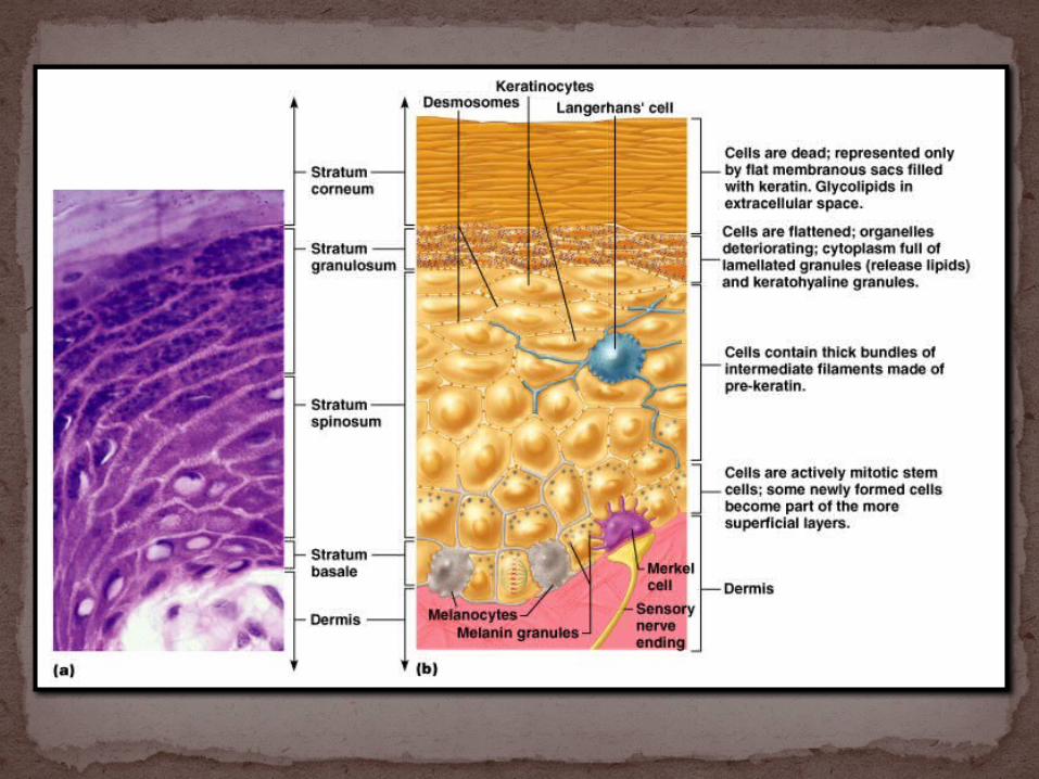
Keratinized stratified squamous epitheliumAvascular- top layer has poorer nutrient
supplyFour types of cells:
Keratinocytes – deepest, produce keratin (tough fibrous protein)
Melanocytes - make dark skin pigment called melanin; absorbs UV rays preventing mutations in skin cells’ DNAHave long, pigment-containing extensions that
travel upwardMerkel cells – associated with sensory nerve
endings (temperature change/ factors that can damage the epidermis)
Langerhans cells – macrophage-like cells
Epidermis
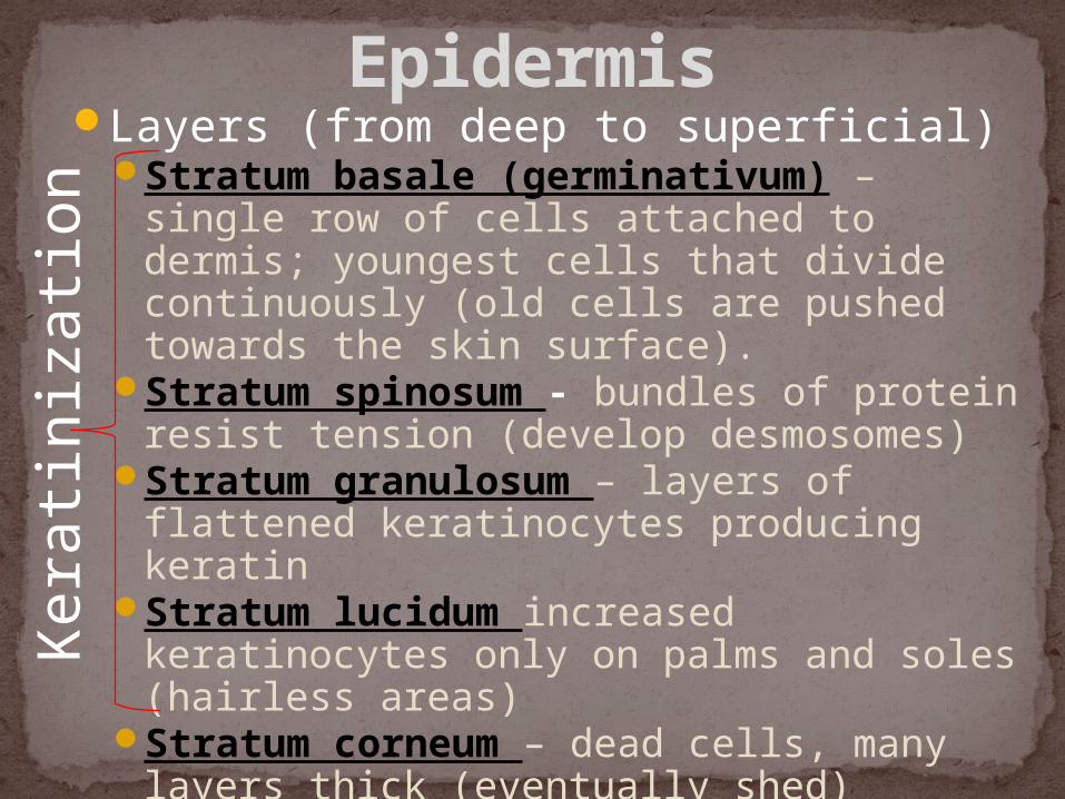
Layers (from deep to superficial)Stratum basale (germinativum) –
single row of cells attached to dermis; youngest cells that divide continuously (old cells are pushed towards the skin surface).
Stratum spinosum - bundles of protein resist tension (develop desmosomes)
Stratum granulosum – layers of flattened keratinocytes producing keratin
Stratum lucidum increased keratinocytes only on palms and soles (hairless areas)
Stratum corneum – dead cells, many layers thick (eventually shed)
EpidermisK
erat
iniz
atio
n

Create an acronym to remember them!
Create a Mnemonic
Bell WorkWhat are the five layers of the epidermis from deep to
superficial?

Lowest epidermal layer, near dermisGood nutrient supply (cells pushed away
from this layer die)Reproduces by mitosis (cells divide
continuously)Single row of cuboidal cells, columnar in
shapeWhere merkel cells are located (sensory
cells)Moves to upper epidermis in 27 days.
Stratum Basale

Stratum Basale
Living cells (keratinocytes, langerhans cells, melanocytes)
Cells are still dividing8-10 cells thickPolygonal in appearanceContain bundles of protein to resist
tension (support the skin)
Stratum Spinosum
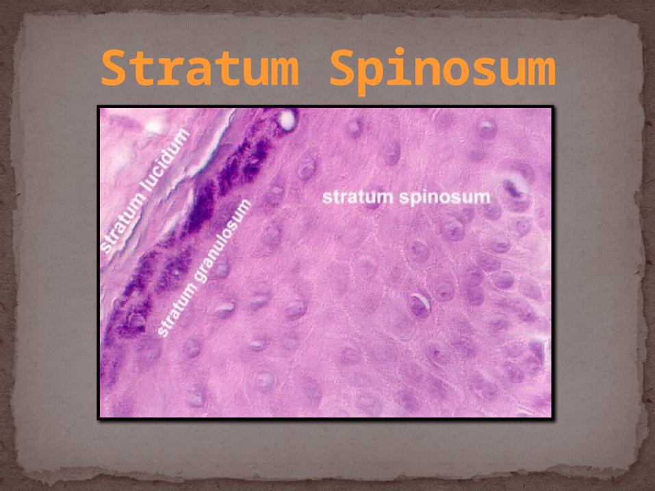
Stratum Spinosum
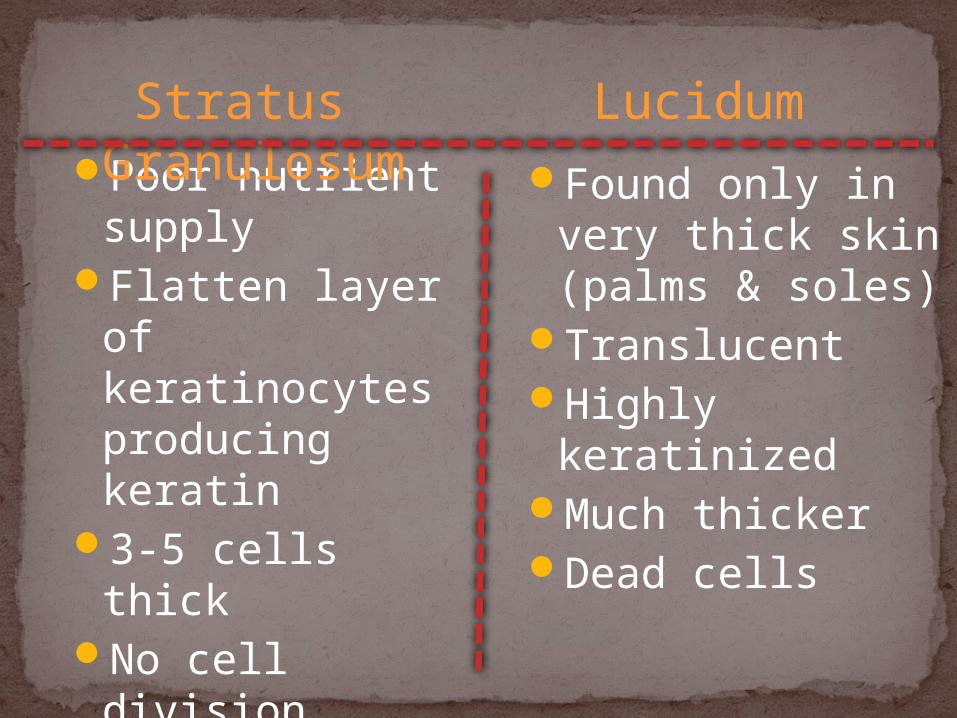
Poor nutrient supply
Flatten layer of keratinocytes producing keratin
3-5 cells thickNo cell division
Found only in very thick skin (palms & soles)
TranslucentHighly
keratinizedMuch thickerDead cells
Stratus Granulosum Lucidum
25-30 cells thick made of dead keratinocytes.
Cells are filled with keratin and hardened.
Sloughed off.Outer most layer of epidermis.
Stratum Corneum
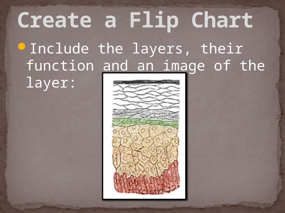
Include the layers, their function and an image of the layer:
Create a Flip Chart

Four basic types of tissue
Epithelium – epidermis just discussed
Connective tissue - dermisMuscle tissueNervous tissue
Remember…
Strong, flexible connective tissue: your “hide”Cells: fibroblasts, macrophages, mast cells,
WBCsFiber types: collagen, elastic, reticularRich supply of nerves and vesselsCritical role in temperature regulation (the
vessels)Two layers (see next slides)
Papillary – areolar connective tissue; includes dermal papillae (which connect to the epidermis)
Reticular – dense irregular connective tissue “reticulum” (network) of collagen and reticular fibers
Dermis
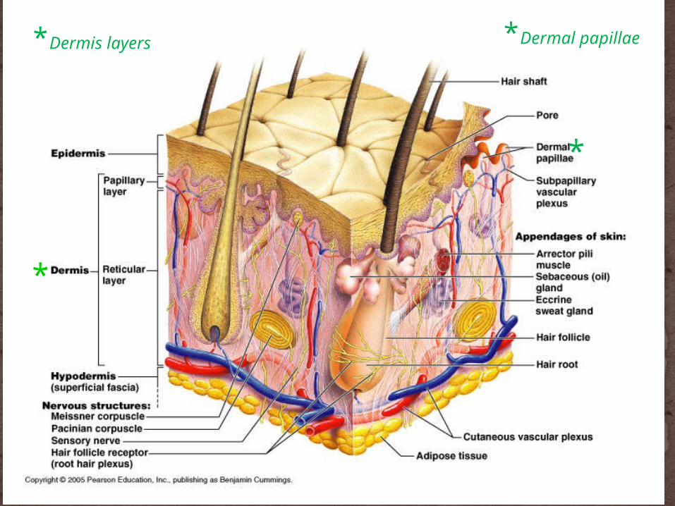
*Dermis layers
*
*
*Dermal papillae
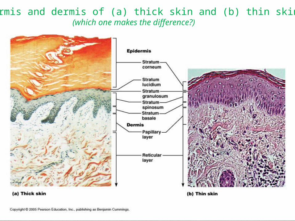
Epidermis and dermis of (a) thick skin and (b) thin skin(which one makes the difference?)
Prints:Keep skin from tearing and aid in
gripping objects & are “sweat films” because of sweat pores
Genetically determinedResult from dermal papillae
Finger like projects that connect the epidermis to the dermis
Elevate the overlying epidermis into ridges
Flexion creases:Deep dermis, from continual folding
Fingerprints, palmprints, footprints.
Names/meaning:“Hypodermis” (Gk) = below the skin“Subcutaneous” (Latin) = below the skin“Superficial fascia”= band or sheet of connective
tissueFatty tissue which stores fat and anchors skin
(areolar tissue and adipose cells)Different patterns of accumulation
(male/female)
Hypodermis

What layers of the epidermis go through mitosis?
What layer of the epidermis is the thickest?
What layer of the epidermis is only on the palms and soles?
Bell Work
Identify the three types of fingerprints.
What is the function of hair?
What is the lunula of the nail?
Bell Work
Derived from epidermis but extend into dermis
IncludeNailsHair and hair folliclesSebaceous (oil) glandsSweat (sudoiferous) glands
Skin appendages

Summarize the articleIllustrate
Create Class Questions
Play the Role

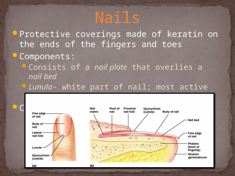
Protective coverings made of keratin on the ends of the fingers and toes
Components:Consists of a nail plate that overlies a nail bedLunula- white part of nail; most active growing
regionCorresponds to hooves and claws
Nails
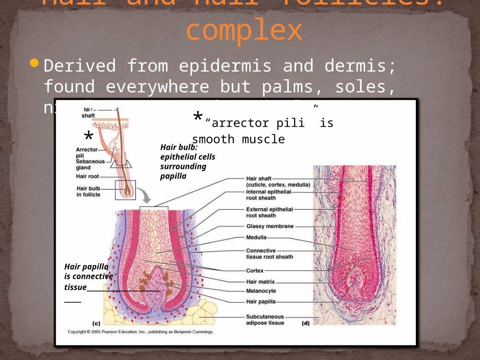
*“arrector pili” is smooth muscle*
Hair papilla is connective tissue________________
Hair bulb: epithelial cells surrounding papilla
Derived from epidermis and dermis; found everywhere but palms, soles, nipples, parts of genitalia
Hair and hair follicles: complex

Functions of hairWarmthSense light touch of the skinProtection – scalp
DevelopmentDevelops from epidermal cells at
the base of a tube-like depression- hair follicle
Components/PartsMade of hard keratinRoot imbedded in skinShaft projecting above skin surface
Three concentric layersMedulla (core)Cortex (surrounds medulla)Cuticle (single layers, overlapping
Hair growth: averages 2 mm/weekGrowth Phase: growing (90% of the
time)Resting phase then shed
Hair colorAmount of melanin for black or brown;
distinct form of melanin for redWhite: decreased melanin and air
bubbles in the medulla
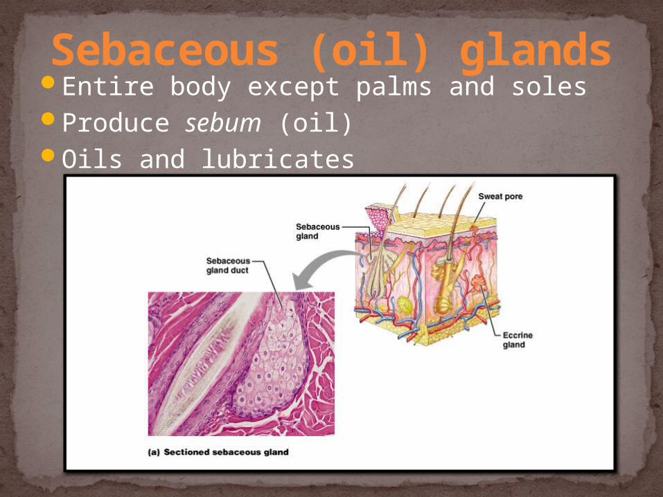
Entire body except palms and solesProduce sebum (oil)Oils and lubricates
Sebaceous (oil) glands

Sweat glandsEntire skin surface except nipples and part of
external genitaliaPrevent overheating500 mL to 12 L/day! (is mostly water) Produced in response to stress as well as heat
Eccrine or merocrineMost numerousTrue sweat: 99% water, some salts, traces of
wasteOpen through pores
ApocrineAxillary, anal and genital areas onlyDucts open into hair follicesThe organic molecules in it decompose with
time - odorModified sweat glands
Ceruminous – secrete earwaxMammary – secrete milk
Types of sweat glands

Identify each type of sweat gland:Eccrine Glands
Apocrine Glands
Ceruminous Glands
Mammary Glands
Bell Work
What is the difference between a first degree, second degree and
third degree burn (based on what you already know)?
Bell Work
Threat to lifeCatastrophic loss of body fluidsDehydration and fatal circulatory shockInfection
TypesBased on the depth of burnsTotal Surface Area (TBSA) affected by the burn
Burns!

Definition – a burn that involves only the epidermis Sign / Symptoms:
Skin is dry and erythematous (redness of the skin caused by dilation and congestion of the capillaries)
Pain to site The burned area blanches
(temporary whitening) with pressure
Edema (if present) will be minimal (accumulation of fluid under the skin)
First Degree/Superficial Burn

Definition – a burn in which the epidermis is burned through and the dermis is damaged Sign / Symptoms :
Deep, intense pain and swelling
Skin is moist Skin will be hyperemic
(pink)in color Blister formation Edema will be moderate
Second Degree/Partial Thickness
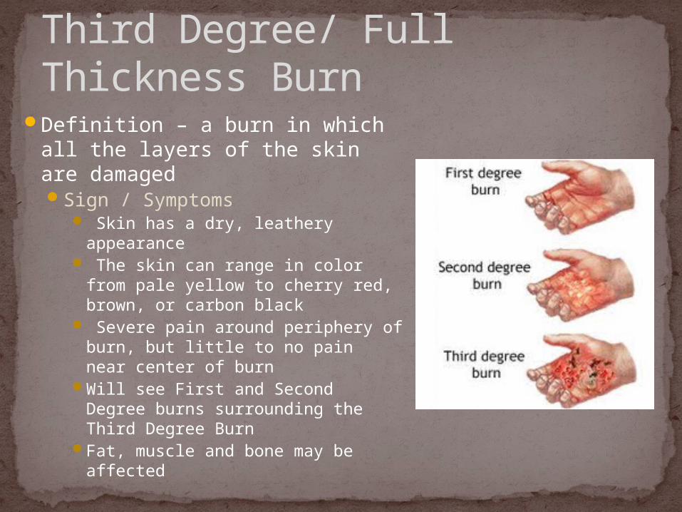
Definition – a burn in which all the layers of the skin are damaged Sign / Symptoms
Skin has a dry, leathery appearance
The skin can range in color from pale yellow to cherry red, brown, or carbon black
Severe pain around periphery of burn, but little to no pain near center of burn
Will see First and Second Degree burns surrounding the Third Degree Burn
Fat, muscle and bone may be affected
Third Degree/ Full Thickness Burn
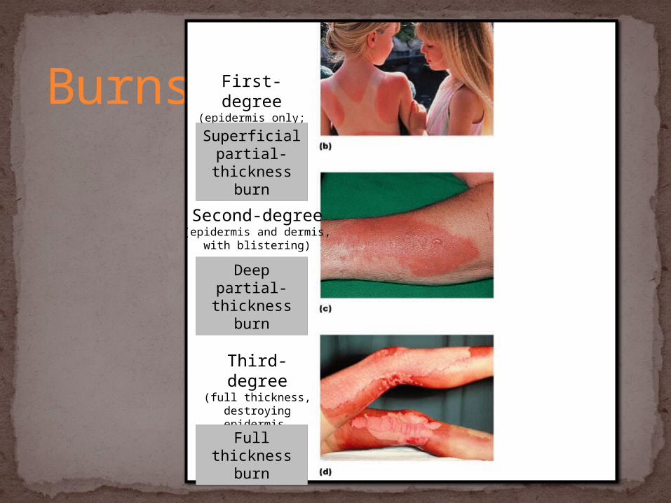
Burns First-degree(epidermis only;
redness)
Second-degree(epidermis and dermis,
with blistering)
Third-degree(full thickness,
destroying epidermis, dermis, often part of
hypodermis)
Superficial partial-
thickness burn
Deep partial- thickness burn
Full thickness burn
Burns can be categorized by the percentage of body surface damaged by the burn
Two Methods for Estimating the Total Body Surface Area (TBSA) affected by Burns: Rule of Nines (RON) Rule of Palm’s (ROP)
Total Body Surface Area (TBSA)
The Rule of Nines divides the TBSA into areas compromising 9% (multiples of 9%)The Rule of Nines is an estimate and is most
useful for adults and children over the age of 10.
The Rule of Nine’s is helpful for estimating the TBSA of large shaped burns.
Rule of Nines (RON)
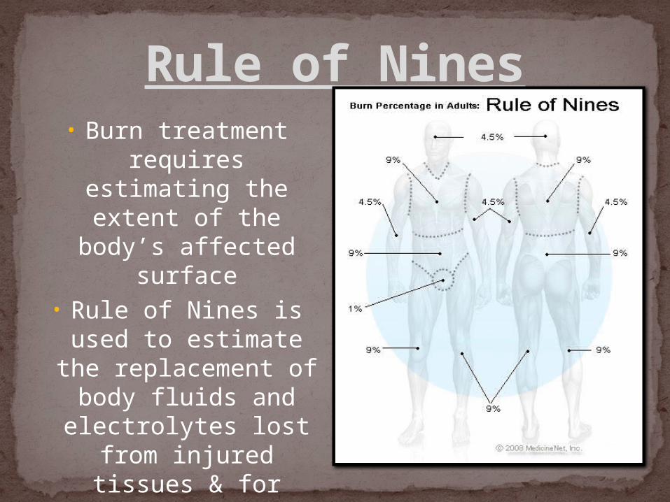
Rule of Nines• Burn treatment requires estimating
the extent of the body’s affected
surface• Rule of Nines is used
to estimate the replacement of body
fluids and electrolytes lost from injured
tissues & for covering the burned area with
skin.

The Rule of Palm’s assumes that the palm size of the patient represents approximately 1% of the TBSA.
TBSA is then estimated by approximating the number of “palms” it would take to completely cover the burn.
The rule of Palm’s is helpful for estimating the TBSA of small or irregularly formed burns.
Rule of Palm's
Inhalation Burns – burns in the upper and lower airways, caused by the inspiration of heat, toxic, smoke, or other gases.
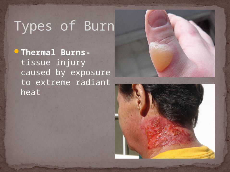
Thermal Burns- tissue injury caused by exposure to extreme radiant heat
Industrial/Chemical Burns- occur when the patient comes in direct contact with chemical agents
Electrical Burns- electrical current, including lightning, can cause severe damage to the body. The skin is burned where the energy enters the body and where it flows into the ground. Along the path of this flow, tissues are damaged due to heat.
Types of Burns
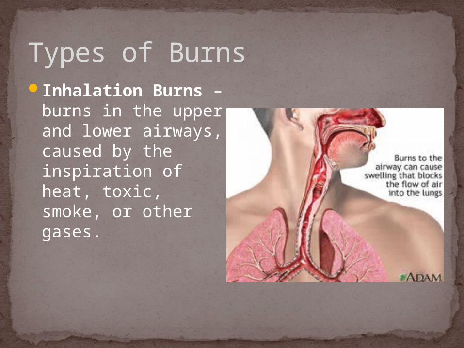
Types of BurnsInhalation Burns –
burns in the upper and lower airways, caused by the inspiration of heat, toxic, smoke, or other gases.
Types of Burns
Thermal Burns- tissue injury caused by exposure to extreme radiant heat

Types of Burns
Industrial/Chemical Burns- occur when the patient comes in direct contact with chemical agents

Types of BurnsElectrical Burns- electrical current,
including lightning, can cause severe damage to the body. The skin is burned where the energy enters the body and where it flows into the ground. Along the path of this flow, tissues are damaged due to heat.
Remove the patient from the environment where the burn occurred
Remove any substance which will continue to burn the patient
Fluid Replacement Therapy: Second- and Third- degree burns require
massive amounts of fluids to properly resuscitate a patient.
Dressings- initially, most burns can be irrigated with cool water then cover with a dry, sterile, bulky dressing
Treatment of Burns

What are the ABCDE’s of tumors on the skin?
Bell Work
Benign, e.g. warts, some moles Malignant (cancerous) – associated with UV
exposure (also skin aging)Basal Cell Carcinoma- cells of stratum basaleSquamous Cell Carcinoma - keratinocytesMelanoma – melanocytes: most dangerous;
recognition:A - AsymmetryB - Border irregularityC - ColorsD - Diameter larger than 6 mm
Tumors of the skin

Symmetrical, round or ovalBorder is sharp and well-definedColor is usually uniform tan, brown, or skin
colorUsually less than a quarter of an inch in
diameterDevelop throughout childhood and early
adulthoodNormal, benign moles usually look very
similar to each other
Healthy Moles
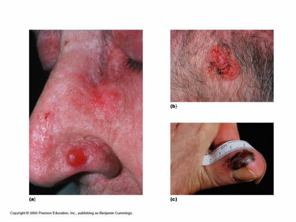
Basal cell carcinoma
Sqaumous cell carcinoma
Melanoma
Skin Cancer
The surrounding area of a wound becomes inflamedBlood vessels in the affected area become
dilated & more permeable (provide more nutrient & oxygen which aids healing)
If a wound is shallow, epithelial cells divide rapidly
If a wound is deep…blood clots form scabs to protect underlying
tissuesfibroblasts migrate to the injured areanew collagen fibers form & new blood vessels
extend beneath the scab
Healing of Wounds
Look at the depth of the open wound. Can you see yellow, fatty tissue? Is bone exposed? Is there a lot of flesh exposed?Is the wound more than 1/4 inch (6 mm) deep?
Look at the width of the wound.If the wound is too wide to be held together with
bandaging easily, then it will need stitching as this will pull the skin together so it can heal correctly.
Look at the location of the open wound. If the wound is in an area where you move a lot,
you need stitches
Stitches?
The epidermis thinsEpidermal cells grow larger/ more irregular in
shape but fewerPresence of “Liver Spots”- sites of oxidation of
fats in secretory cellsThe dermis is reduced
Production of elastin and collagen slows; loss of fat from the subcutaneous layer result in wrinkling
Fewer fibroblasts delay wound healingSebaceous glands secrete less oil which dries
the skinNumber of sweat glands decrease
The Beauty in Aging
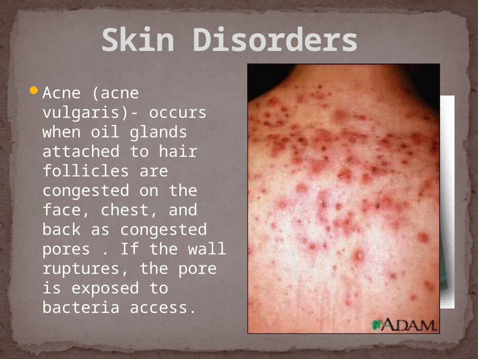
Skin Disorders Acne (acne vulgaris)-
occurs when oil glands attached to hair follicles are congested on the face, chest, and back as congested pores . If the wall ruptures, the pore is exposed to bacteria access.

Skin Disorders
Psoriasis- Red and inflamed and may become white scaly patches; commonly found on the elbows, knees, scalp and the lower back.Skin cells mature
about 5x fasterThese cells pile up on
the skin's surfaceCause is linked to
genes and triggers
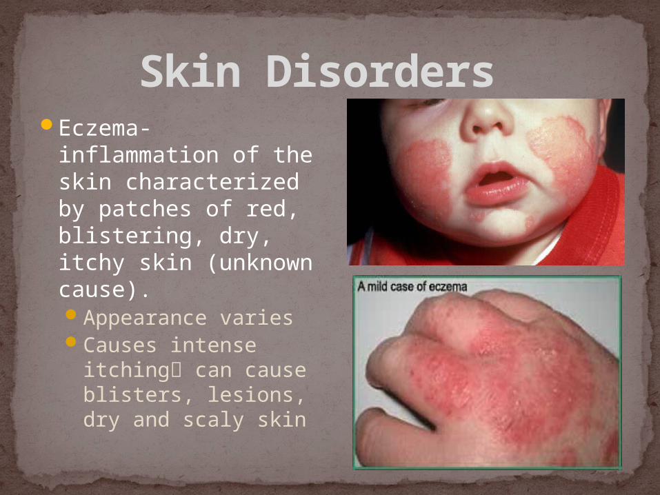
Skin Disorders Eczema-
inflammation of the skin characterized by patches of red, blistering, dry, itchy skin (unknown cause).Appearance variesCauses intense
itching can cause blisters, lesions, dry and scaly skin
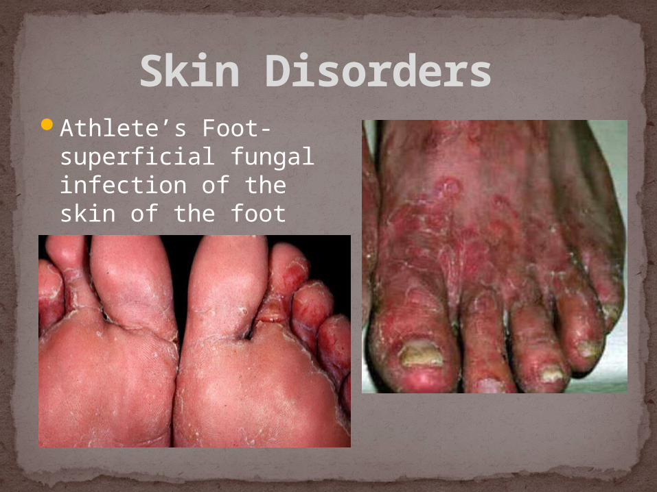
Skin Disorders Athlete’s Foot-
superficial fungal infection of the skin of the foot

Skin Disorders
Vitiligo- pigmentation disorder in which melanocytes in the skin are destroyed
resulting in white patches that appear on the skin in different parts of the body.
Skin Disorders
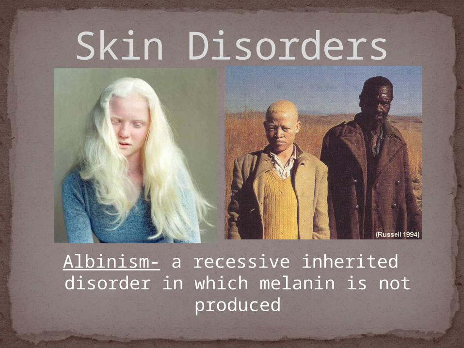
Albinism- a recessive inherited disorder in which melanin is not produced
What happens to the layers of skin as you
age?
Bell Work

Two GroupsQuestion given to each individual of each
groupIf the student gets the answer correct, the
team gets one point… the student gets to throw the ball into the trash canIf you choose to throw from across the room
and you make it, the team gets 3 pointsIf you choose to throw from 6 feet and make it
the team gets 1 point. The next team gets their chance.
Trash Ball


![BiStick 26.5ws 5mm Tiled[1]](https://img.pdfslide.net/doc/110x75/563db931550346aa9a9aed13/bistick-265ws-5mm-tiled1.jpg)


