Embed Size (px)
Citation preview
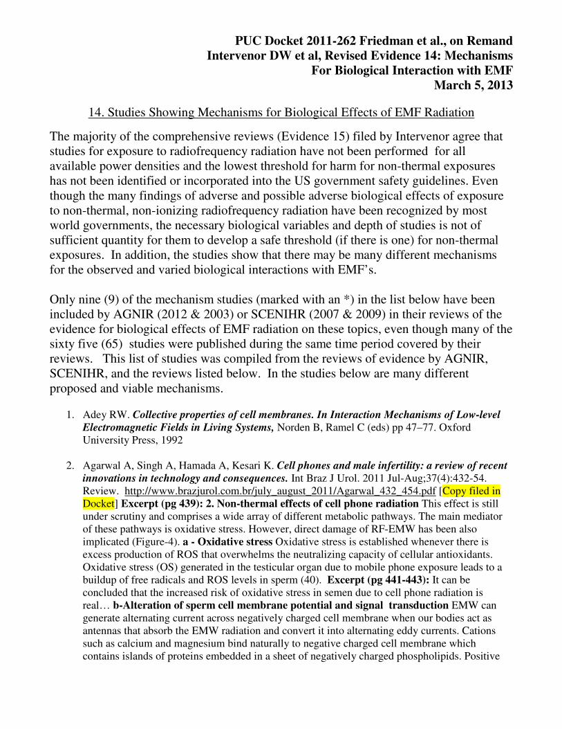
PUC Docket 2011-262 Friedman et al., on Remand
Intervenor DW et al, Revised Evidence 14: Mechanisms
For Biological Interaction with EMF
March 5, 2013
14. Studies Showing Mechanisms for Biological Effects of EMF Radiation
The majority of the comprehensive reviews (Evidence 15) filed by Intervenor agree that
studies for exposure to radiofrequency radiation have not been performed for all
available power densities and the lowest threshold for harm for non-thermal exposures
has not been identified or incorporated into the US government safety guidelines. Even
though the many findings of adverse and possible adverse biological effects of exposure
to non-thermal, non-ionizing radiofrequency radiation have been recognized by most
world governments, the necessary biological variables and depth of studies is not of
sufficient quantity for them to develop a safe threshold (if there is one) for non-thermal
exposures. In addition, the studies show that there may be many different mechanisms
for the observed and varied biological interactions with EMF’s.
Only nine (9) of the mechanism studies (marked with an *) in the list below have been
included by AGNIR (2012 & 2003) or SCENIHR (2007 & 2009) in their reviews of the
evidence for biological effects of EMF radiation on these topics, even though many of the
sixty five (65) studies were published during the same time period covered by their
reviews. This list of studies was compiled from the reviews of evidence by AGNIR,
SCENIHR, and the reviews listed below. In the studies below are many different
proposed and viable mechanisms.
1. Adey RW. Collective properties of cell membranes. In Interaction Mechanisms of Low-level
Electromagnetic Fields in Living Systems, Norden B, Ramel C (eds) pp 47–77. Oxford
University Press, 1992
2. Agarwal A, Singh A, Hamada A, Kesari K. Cell phones and male infertility: a review of recent
innovations in technology and consequences. Int Braz J Urol. 2011 Jul-Aug;37(4):432-54.
Review. http://www.brazjurol.com.br/july_august_2011/Agarwal_432_454.pdf [Copy filed in
Docket] Excerpt (pg 439): 2. Non-thermal effects of cell phone radiation This effect is still
under scrutiny and comprises a wide array of different metabolic pathways. The main mediator
of these pathways is oxidative stress. However, direct damage of RF-EMW has been also
implicated (Figure-4). a - Oxidative stress Oxidative stress is established whenever there is
excess production of ROS that overwhelms the neutralizing capacity of cellular antioxidants.
Oxidative stress (OS) generated in the testicular organ due to mobile phone exposure leads to a
buildup of free radicals and ROS levels in sperm (40). Excerpt (pg 441-443): It can be
concluded that the increased risk of oxidative stress in semen due to cell phone radiation is
real… b-Alteration of sperm cell membrane potential and signal transduction EMW can
generate alternating current across negatively charged cell membrane when our bodies act as
antennas that absorb the EMW radiation and convert it into alternating eddy currents. Cations
such as calcium and magnesium bind naturally to negative charged cell membrane which
contains islands of proteins embedded in a sheet of negatively charged phospholipids. Positive
Page 2 of 24
ions fit between the negatively charged phospholipid molecules and reduce their tendency to
repel one another contributing to cell membrane stability (62-64)… Short-term exposure to RF-
EMW may also lead to an increase in the activity of plasma membrane NADH oxidase enzyme,
which in turn increases ROS formation (10). Chronic exposure to EMW radiation, in association
with excess ROS exposure, leads to activation of heat shock proteins (hsp) as a protective
response (81). The job of these hsp is to combine with vital enzymes, forming a protective layer
around these enzymes. This in turn shields them from damage. However, this activation stops the
hsp from working properly and interferes with metabolism of the sperm (Figure-5).
3. Behari J, Paulraj R. Biomarkers of induced electromagnetic field and cancer. Indian J Exp Biol.
2007 Jan;45(1):77-85. Review. http://www.ncbi.nlm.nih.gov/pubmed/17249331 Abstract: The
present article delineates the epidemiological and experimental studies of electromagnetic field
which affects various tissues of human body. These affects lead to cell proliferation, which may
lead to cancer formation. Certain biomarkers have been identified which are one way or the
other responsible for tumor promotion or co-promotion. These are (i) melatonin, a hormone
secreted by pineal gland, (ii) Ca2+, which is essential in the regulation of the resting membrane
potential and in the sequence of events in synaptic excitation and neurotransmitter, release are
affected by electromagnetic field, (iii) ornithine decarboxylase (ODC), a rate-limiting enzyme in
the biosynthesis of polyamines, considered as a useful biological marker; over expression of
ODC can cause cell transformation and enhancement of tumor promotion. (iv) protein kinase is
an enzyme, which transfers phosphate groups from ATP to hydroxyl groups in the amino acid
chains of acceptor proteins, and (v) Na+-K+ ATPase, which transports sodium and potassium
ions across the membrane has a critical role in living cells. The various possible mechanisms
depending upon non equilibrium thermodynamics, co-operativism, stochastic and resonance are
discussed as possible models of signal transduction in cytosol, thereby controlling the
transcription phenomena. Finally a mechanism comprising the extremely low frequency and
radio frequency (RF)/microwave (MW) modulated field is compared.
4. Belyaev I. A Rationale for a Biologically-based Public Exposure Standard for
Electromagnetic Fields (ELF and RF)Section 15; Evidence for Disruption by Modulation
Role of Physical and Biological Variables in Bioeffects of Non-Thermal Microwaves for Reproducibility, Cancer Risk and Safety Standards. BioInitiative Working Group, BioInitiative
Report, 2012. Review [Copy filed in Docket] Excerpt (pg. 3): With increased absorption of
energy, so-called thermal effects of microwaves (MW) are usually observed that deal with MW-
induced heating. Specific absorption rate (SAR) or power density (PD) is a main determinate for
thermal MW effects. Several other physical parameters of exposure have been reported to be of
importance for so-called non-thermal (NT) biological effects, which are induced by MW at
intensities well below any measurable heating. Excerpt (pg. 3): To what degree SAR/PD can be
applied to the nowadays nonthermal MW chronic exposures is not known and the current state of
research demands reevaluation of the safety standards (Grigoriev, Nikitina et al. 2005). Excerpt
(pg. 9): Since 1980, there have been numerous reports of biological effects that show intensity
“windows,” that is, regions of intensity that cause changes surrounded by higher and lower
intensities that show no effects from exposure, see for review (Blackman 2009). These results
mean that lower intensity is not necessarily less bioactive, or less harmful.
Page 3 of 24
5. Belyaev I. Dependence of non-thermal biological effects of microwaves on physical and
biological variables: implications for reproducibility and safety. ICEMS Monograph, National
Institute for the Study and Control of Cancer and Environmental Diseases, Ramazzini Institute,
Eur. J. of Oncol.- Library, Vol. 5, 2010; Review [Copy Filed in Docket]
http://www.icems.eu/papers/ramazzini_library5_part1.pdf Abstract: biological responses,
including adverse health effects, to non-thermal (NT) microwaves (MW) have been described by
many research groups all over the world. The aim of this paper is to provide an overview of the
complex dependence of these effects on various physical and biological parameters, which must
be controlled in replication studies. Besides well-known dependencies on carrier frequency and
modulation, emerging data suggest dependencies of NT MW effects on polarization,
intermittence and coherence time of exposure, static magnetic field, electromagnetic stray fields,
genotype, gender, physiological and individual traits, cell density during exposure. Data also
indicate that duration of exposure may be as important as power density (PD) and specific absorption rate (SAR). Further evaluation of these dependencies are needed for understanding
the mechanisms by which NT MW affect biological systems, planning in vivo and
epidemiological studies, developing medical treatments, setting safety standards, and minimizing
the adverse effects of MW from mobile communication.
6. Blackman C. Cell phone radiation: Evidence from ELF and RF studies supporting more
inclusive risk identification and assessment. Pathophysiology 2009, 16:205-216. http://www.ncbi.nlm.nih.gov/pubmed/19264460 [Copy filed in Docket] Abstract: Many
national and international exposure standards for maximum radiation exposure from the use of
cell phone and other similar portable devices are ultimately based on the production of heat
particularly in regions of the head, that is, thermal effects (TE). The recent elevation in some
countries of the allowable exposure, that is, averaging the exposure that occurs in a 6min period
over 10g of tissue rather than over 1g allows for greater heating in small portions of the 10-g
volume compared to the exposure that would be allowed averaged over 1-g volume. There is
concern that 'hot' spots, that is, momentary higher intensities, could occur in portions of the 10-g
tissue piece, might have adverse consequences, particularly in brain tissue. There is another
concern about exposure to cell phone radiation that has been virtually ignored except for the
National Council of Radiation Protection and Measurements (NCRP) advice given in a
publication in 1986 [National Council for Radiation Protection and Measurements, Biological
Effects and Exposure Criteria for Radiofrequency Electromagnetic Fields, National Council for
Radiation Protection and Measurements, 1986, 400 pp.]. This NCRP review and guidance
explicitly acknowledge the existence of non-thermal effects (NTE), and included provisions for
reduced maximum-allowable limits should certain radiation characteristics occur during the
exposure. If we are to take most current national and international exposure standards as
completely protective of thermal injury for acute exposure only (6min time period) then the
recent evidence from epidemiological studies associating increases in brain and head cancers
with increased cell phone use per day and per year over 8-12 years, raises concerns about the
possible health consequences on NTE first acknowledged in the NCRP 1986 report [National
Council for Radiation Protection and Measurements, Biological Effects and Exposure Criteria
for Radiofrequency Electromagnetic Fields, National Council for Radiation Protection and
Measurements, 1986, 400 pp.]. This paper will review some of the salient evidence that
demonstrates the existence of NTE and the exposure complexities that must be considered and
understood to provide appropriate, more thorough evaluation and guidance for future studies and
Page 4 of 24
for assessment of potential health consequences. Unfortunately, this paper is necessary because
most national and international reviews of the research area since the 1986 report [National
Council for Radiation Protection and Measurements, Biological Effects and Exposure Criteria
for Radiofrequency Electromagnetic Fields, National Council for Radiation Protection and
Measurements, 1986, 400 pp.] have not included scientists with expertise in NTE, or given
appropriate attention to their requests to include NTE in the establishment of public-health-based
radiation exposure standards. Thus, those standards are limited because they are not
comprehensive.
7. Blank M, Goodman RM. Electromagnetic fields and health: DNA-based dosimetry.
Electromagn Biol Med. 2012 Dec;31(4):243-9. http://www.ncbi.nlm.nih.gov/pubmed/22676645
Abstract: We propose a biologically based measure of EMF radiation to replace the energy-
based "specific absorption rate" (SAR). A wide range of EMF frequencies has been linked to an
increased risk of cancer. The SAR value used to measure the EMF dose and set the safety
standard in the radiofrequency (RF) range fails as a standard for predicting cancer risk in the
ELF power frequency range. Because cancers are believed to arise from mutations in DNA,
changes in DNA induced by interaction with EMF could be a better measure of the biologically
effective dose in both frequency ranges. The changes can be measured by transcriptional
alterations and/or translational changes in specific proteins. Because ionizing radiation also
causes DNA damage, a biologically based standard related to stimulation of DNA could apply
over a much wider range of the electromagnetic spectrum. A safety standard for exposure to a
wide range of non ionizing frequencies can be based on the documented changes in DNA
biochemistry that arise from interactions with EMF.
8. Blank M, Goodman RM, A mechanism for stimulation of biosynthesisby electromagnetic
fields: charge transfer in DNA and base pair separation, J. Cell. Physiol. 214 (2008) 20–26.
http://www.ncbi.nlm.nih.gov/pubmed/17620313 Abstract: Electrons have been shown to move
in DNA, and a specific DNA sequence is associated with the response to EM fields. In addition,
there is evidence from biochemical reactions that EM fields can accelerate electron transfer.
Interaction with electrons could displace electrons in H-bonds that hold DNA together leading to
chain separation and initiating transcription. The effect of charging due to electron displacement
on the energetics of DNA aggregation shows that electron transfer would favor separation of
base pairs, and that DNA geometry is optimized for disaggregation under such conditions.
Electrons in the H-bonds of both DNA and the surrounding water molecules fluctuate at
frequencies that are much higher than the frequencies of the EM fields studied. The
characteristics of the fluctuations suggest that the applied EM fields are effectively DC pulses
and that interactions extend to microwave frequencies.
9. Blank M. Do electromagnetic fields interact with electrons in the Na,K-ATPase?
Bioelectromagnetics. 2005 Dec;26(8):677-83 http://www.ncbi.nlm.nih.gov/pubmed/16189824
Abstract: The effects of low frequency electric and magnetic fields on several biochemical
systems, including the Na,K-ATPase, indicate that electromagnetic (EM) fields interact with
electrons. The frequency optima for two enzymes in response to EM fields are very close to their
turnover numbers, suggesting that these interactions directly affect reaction rates. Nevertheless,
generally accepted ideas about Na,K-ATPase function and ion transport mechanisms do not
consider interactions with electrons. To resolve the clash of paradigms, we hypothesize
Page 5 of 24
interaction with transient electrons and protons that arise from flickering of H-bonds in the
hydrated protein. These transient charges in the enzyme could provide a trigger for the sequence
of conformation changes that are part of the ion transport mechanism. If the distributions of
transient electrons and protons in the membrane are affected by their concentration and the
membrane potential, as expected from electric double layer theory, this can account for the
different effects of low frequency electric and magnetic fields, as well as for the observation that
membrane hyperpolarization reverses the ATPase reaction to generate ATP.
10. Blank M, Goodman R. 2004. Initial Interactions in Electromagnetic Field-Induced
Biosynthesis.J Cellular Physiology 199: 359-363.
http://www.ncbi.nlm.nih.gov/pubmed/15095282 Abstract: Low frequency electromagnetic
(EM) fields induce gene expression, and recent insights into physical interactions of EM fields
with model systems suggest a mechanism that could initiate this process. The consistently low
thresholds at which EM fields stimulate biological processes indicate that they require little
energy. Since it has been shown that such weak fields accelerate electron transfer reactions, they
could stimulate transcription by interacting with electrons in DNA to destabilize the H-bonds
holding the two DNA strands together. Such a mechanism is consistent with the low electron
affinity of the bases in previously identified electromagnetic response elements (EMREs) needed
for EM field interaction with DNA. It is also in line with both endogenous and in vitro
stimulation of biosynthesis by electric fields. The frequency response of several EM sensitive
biological systems suggests that EM fields require repetition and are most effective at
frequencies that coincide with natural rhythms of the processes affected.
11. Blank M, Soo L. Electromagnetic acceleration of Belousov-Zhabotinski reaction.
Bioelectrochemistry 2003, 61: 93-97. http://www.ncbi.nlm.nih.gov/pubmed/14642914 Abstract:
Acceleration of the Belousov-Zhabotinski (BZ) reaction, in stirred homogeneous solutions, by
low frequency electromagnetic (EM) fields has provided new insights into EM interaction
mechanisms. The acceleration varies inversely with the basal reaction rate, indicating that the
applied magnetic field and the intrinsic chemical driving forces affect the same electron transfer
reaction. The amplitude and frequency dependence of the EM field interactions are also
consistent with interaction during electron transfer. A mechanism based on interaction with
moving electrons offers a way of explaining the ability of EM fields to stimulate gene expression,
in particular the stress response, since electrons have been shown to move in DNA.
12. Blank M, Goodman R. Electromagnetic initiation of transcription at specific DNA sites. J Cell
Biochem 2002, 81: 689-692. http://www.ncbi.nlm.nih.gov/pubmed/11329623 Abstract:
Initiation of transcription by electromagnetic (EM) fields offers an insight into mechanism. EM
field stimulated transcription appears to require specific DNA sequences, and these bases may be
sites where EM fields generate large repulsive forces between chains by accelerating electrons
that move within DNA. We can estimate the repulsion between chains by assuming that electron
affinity is a measure of electron density at each base, and inversely related to the velocity of
electrons (and the force). The repulsive force can be compared to the attraction between chains
due to H-bonds. From the difference between repulsion and attraction, we show that sites rich in
C and T, as in the specific sequences, would be more likely to come apart in EM fields. These
calculations suggest a plausible mechanism for initiation of transcription by EM fields, and
Page 6 of 24
provide a rationale for specific sequences to function as EM field response elements. Electron
flow could also be a factor in DNA chain melting due to Joule heating.
13. Blank M, Soo L. 2001a. Electromagnetic acceleration of electron transfer reactions. Journal of
Cellular Biochemistry 81: 278-283. http://www.ncbi.nlm.nih.gov/pubmed/11241667 Abstract:
The Moving Charge Interaction (MCI) model proposes that low frequency electromagnetic (EM)
fields affect biochemical reactions through interaction with moving electrons. Thus, EM field
activation of genes, and the synthesis of stress proteins, are initiated through EM field interaction
with moving electrons in DNA. This idea is supported by studies showing that EM fields
increase electron transfer rates in cytochrome oxidase. Also, in studies of the Na,K-ATPase
reaction, estimates of the speed of the charges accelerated by EM fields suggest that they too are
electrons. To demonstrate EM field effects on electron transfer in a simpler system, we have
studied the classic oscillating Belousov--Zhabotinski (BZ) reaction. Under conditions where the
BZ reaction oscillates at about 0.03 cycles/sec, a 60 Hz, 28 microT (280 mG) field accelerates
the overall reaction. As observed in earlier studies, an increase in temperature accelerates the
reaction and decreases the effect of EM fields on electron transfer. In all three reactions studied,
EM fields accelerate electron transfer, and appear to compete with the intrinsic chemical forces
driving the reactions. The MCI model provides a reasonable explanation of these observations.
14. Blank M, Goodman R. 2001. Electromagnetic initiation of transcription at specific DNA sites.
Journal of Cellular Biochemistry 81: 689-692. http://www.ncbi.nlm.nih.gov/pubmed/11329623
Abstract: Initiation of transcription by electromagnetic (EM) fields offers an insight into
mechanism. EM field stimulated transcription appears to require specific DNA sequences, and
these bases may be sites where EM fields generate large repulsive forces between chains by
accelerating electrons that move within DNA. We can estimate the repulsion between chains by
assuming that electron affinity is a measure of electron density at each base, and inversely related
to the velocity of electrons (and the force). The repulsive force can be compared to the attraction
between chains due to H-bonds. From the difference between repulsion and attraction, we show
that sites rich in C and T, as in the specific sequences, would be more likely to come apart in EM
fields. These calculations suggest a plausible mechanism for initiation of transcription by EM
fields, and provide a rationale for specific sequences to function as EM field response elements.
Electron flow could also be a factor in DNA chain melting due to Joule heating.
15. Blank M, Goodman R. Do electromagnetic fields interact directly with DNA?
Bioelectromagnetics. 1997;18(2):111-5. Abstract: The mechanisms whereby electromagnetic
(EM) fields stimulate changes in biosynthesis in cells are not known. It has has generally been
assumed that EM fields first interact with cell membranes, but this pathway may not be only one.
Interactions with membranes are well documented, but recent studies of EM signal transduction
in the membrane Na,K-ATPase are best explained by direct interaction of electric and magnetic
fields with mobile charges within the enzyme. Interaction with moving charges may be a
mechanism that is operative in other biopolymers. Recent studies on DNA have shown that large
electron flows are possible within the stacked base pairs of the double helix. Therefore, gene
activation by magnetic fields could be due to direct interaction with moving electrons within
DNA. Electric fields as well as magnetic fields stimulate transcription, and both fields could
interact with DNA directly. The mechanism of EM field-stimulated transcription may be related
Page 7 of 24
to the process in striated muscles, where endogenous electrical activity induces the synthesis of
new proteins.
16. Blank M. Biological effects of environmental electromagnetic fields: molecular mechanisms.
Biosystems. 1995, ;35(2-3):175-8. http://www.ncbi.nlm.nih.gov/pubmed/7488711 Abstract: In
recent years, epidemiological studies have shown an increased risk of leukemia in children living
near electric power distribution lines. Also, medical studies have shown that low frequency
electromagnetic (EM) fields accelerate the healing of bone fractures. The stimulation of
biosynthesis suggested by both epidemiological and clinical studies is observed in vitro, and
indicates that EM fields stimulate the biosynthetic 'stress response' in cells. Studies of EM field
effects on enzyme (Na,K-ATPase) function suggest that these effects should be considered in the
design of molecular electronic devices
17. *Brocklehurst B, McLauchlan KA. Free radical mechanism for the effects of environmental
electromagnetic fields on biological systems. Int J Radiat Biol. 1996 Jan;69(1):3-24.
http://www.ncbi.nlm.nih.gov/pubmed/8601753 Abstract: The radical pair mechanism is
discussed as a possible route whereby a magnetic field of environmental strength might affect a
biological system. It is well established as the origin of reproducible field effects in chemistry,
and these can be observed even at very low magnetic field strengths, including that of the
geomagnetic field. Here it is attempted to give a description which might assist experimentalists
working in biological laboratories to devize tests of its relevance to their work. The mechanism
is well understood and a specific theoretical approach is taken to explore and emphasize the
importance of the lifetime of the radical pair and the precise chemical natures of the radicals
which comprise it in affecting the size of the low-field effects. Further subsequent processes are
likely necessary to cause this primary effect to attain biological significance. Arguments are
provided to suggest that the encounters of freely diffusing pairs (F-pairs) of radicals are unlikely
to produce significant effects in biology.
18. *Chiabrera A, Bianco B, Moggia E, Kaufman JJ. Zeeman-Stark modeling of the RF EMF
interaction with ligand binding. Bioelectromagnetics. 2000 May;21(4):312-24. Abstract: The
influence of radiofrequency electromagnetic exposure on ligand binding to hydrophobic receptor
proteins is a plausible early event of the interaction mechanism. A comprehensive quantum
Zeeman-Stark model has been developed which takes into account the energy losses of the
ligand ion due to its collisions inside the receptor crevice, the attracting nonlinear endogenous
force due to the potential energy of the ion in the binding site, the out of equilibrium state of the
ligand-receptor system due to the basal cell metabolism, and the thermal noise. The biophysical
"output" is the change of the ligand binding probability that, in some instances, may be affected
by a suitable low intensity exogenous electromagnetic "input" exposure, e.g., if the depth of the
potential energy well of a putative receptor protein matches the energy of the radiofrequency
photon. These results point toward both the possibility of the electromagnetic control of
biochemical processes and the need for a new database of safety standards.
19. Chukova YP, Advances in Nonequilibrium Thermodynamics of the Systems under
Electromagnetic Radiation, Moscow: Academy of Sciences, 2001.
Page 8 of 24
20. Chukova YP. Bose Condensation and non thermal processes in living systems under
millimeter (MM) radiation. Electromagn Biol Med. 2009;28(1):41-5. Abstract: Positive and
negative experimental results on non thermal bioeffects of MM radiation are studied according to
the Bose Condensation model.
21. Cifra M, Fields JZ, Farhadi A. Electromagnetic cellular interactions. Prog Biophys Mol Biol.
2011 May;105(3):223-46. http://www.ncbi.nlm.nih.gov/pubmed/20674588 Review. Abstract:
Chemical and electrical interaction within and between cells is well established. Just the opposite
is true about cellular interactions via other physical fields. The most probable candidate for an
other form of cellular interaction is the electromagnetic field. We review theories and
experiments on how cells can generate and detect electromagnetic fields generally, and if the
cell-generated electromagnetic field can mediate cellular interactions. We do not limit here
ourselves to specialized electro-excitable cells. Rather we describe physical processes that are of
a more general nature and probably present in almost every type of living cell. The spectral range
included is broad; from kHz to the visible part of the electromagnetic spectrum. We show that
there is a rather large number of theories on how cells can generate and detect electromagnetic
fields and discuss experimental evidence on electromagnetic cellular interactions in the modern
scientific literature. Although small, it is continuously accumulating.
22. *de Pomerai D, Daniells C, David H, Allan J, Duce I, Mutwakil M, Thomas D, et al.
Nonthermal heat-shock response to microwaves. Nature 2000; 405:417-8.
http://www.ncbi.nlm.nih.gov/pubmed/10839528
23. Easterly CE. Cancer Link to Magnetic Field Exposure: A Hypothesis. American Journal of
Epidemiology 114, no. 2 (August 1981):169-74.
24. Frey AH. Differential biologic effects of pulsed and continuous electromagnetic fields and
mechanisms of effect. Ann N Y Acad Sci. 1974;238:273-9.
25. Frey AH, ed. On the Nature of Electromagnetic Field Interactions with Biological Systems.
Austin, TX:R.G. Landes Co., 1994.
26. Frey AH. Electromagnetic field interactions with biological systems. FASEB J. 1993 Feb
1;7(2):272-81. http://www.fasebj.org/content/7/2/272.full.pdf [Copy filed in Docket] Abstract:
This is a report on Symposia organized by the International Society for Bioelectricity and
presented at the 1992 FASEB Meeting. The presentations summarized here were intended to
provide a sampling of new and fruitful lines of research. The theme topics for the Symposia were
cancer, neural function, cell signaling, pineal gland function, and immune system interactions.
Living organisms are complex electrochemical systems that evolved over billions of years in a
world with a relatively simple weak magnetic field and with few electromagnetic energy emitters.
As is characteristic of living organisms, they interacted with and adapted to this environment of
electric and magnetic fields. In recent years there has been a massive introduction of equipment
that emits electromagnetic fields in an enormous range of new frequencies, modulations, and
intensities. As living organisms have only recently found themselves immersed in this new and
virtually ubiquitous environment, they have not had the opportunity to adapt to it. This gives us,
Page 9 of 24
as biologists, the opportunity to use these electromagnetic fields as probes to study the
functioning of living systems. This is a significant opportunity, as new approaches to studying
living systems so often provide the means to make great leaps in science. In recent years, a
diversity of biologists have carried out experiments using electromagnetic fields to study the
function of living cells and systems. This approach is now becoming quite fruitful and is yielding
data that are advancing our knowledge in diverse areas of biology.
27. Frey A, Review Biological Function as Influenced by Low Power Modulated EF Energy
Physiological Psychology Programs Office of Naval Research, 1970 (copy filed in Docket)
Abstract: In retent years, it has been recognized that low power density modalated rf energy can
affect the functioning of higher living orgnisms. In this paper the sparse data generated in the
western hemisphere on this subject is considered, the reasons for its sparseness noted, and the
hypotheses on mechanisms that may provide an explanation for the observed effects and other
possible effects are sketched. Possible conclusions with regard to hazards to personnel are then
considered. Excerpt (pg 28) : To sum then, we have complementary and overlapping
hypothesis, Wei's [1966, 1968, 1969] primarily on information transfer, Batteau's [1963]
primarily on information coding, and Augenstein's [primarily on Information storage ." Excerpt
(pg 31): As may be seen in the foregoing, if we recognize that our knowledge of how the
nervous system codes, transfers, and stores information is almost nil, one can list a number of
possible mechanisms through which rf energy could affect higher organisms. The problem in not
‘is there a possible mechanism" but rather which of numerous possible mechanisms' to use as a
guide for experimentation. It is only through experimentation that we will obtain the answers
about rf effects since calculations flounder on the assumptions that have to be made about how
nerves work. It should be noted that the foregoing is not an exhaustive list of possible
mechanisms. It should also be noted that more than one mechanismcan be involved in rf effects.
28. *Friedman J, Kraus S, Hauptman Y, Schiff Y, Seger R. Mechanism of short-term ERK
activation by electromagnetic fields at mobile phone frequencies. Biochem J. 2007;405:559–
568. http://www.ncbi.nlm.nih.gov/pmc/articles/PMC2267306/pdf/bj4050559.pdf [PubMed]
[Copy filed in Docket] Abstract: The exposure to non-thermal microwave electromagnetic
fields generated by mobile phones affects the expression of many proteins. This effect on
transcription and protein stability can be mediated by the MAPK (mitogen-activated protein
kinase) cascades, which serve as central signaling pathways and govern essentially all stimulated
cellular processes. Indeed, long-term exposure of cells to mobile phone irradiation results in the
activation of p38 as well as the ERK (extracellular-signal-regulated kinase) MAPKs. In the
present study, we have studied the immediate effect of irradiation on the MAPK cascades, and
found that ERKs, but not stress-related MAPKs, are rapidly activated in response to various
frequencies and intensities. Using signalling inhibitors, we delineated the mechanism that is
involved in this activation. We found that the first step is mediated in the plasma membrane by
NADH oxidase, which rapidly generates ROS (reactive oxygen species). These ROS then
directly stimulate MMPs (matrix metalloproteinases) and allow them to cleave and release Hb-
EGF [heparin-binding EGF (epidermal growth factor)]. This secreted factor activates the EGF
receptor, which in turn further activates the ERK cascade. Thus this study demonstrates for the
first time a detailed molecular mechanism by which electromagnetic irradiation from mobile
phones induces the activation of the ERK cascade and thereby induces transcription and other
cellular processes.
Page 10 of 24
29. Funk RH, Monsees TK. Effects of electromagnetic fields on cells: physiological and
therapeutical approaches and molecular mechanisms of interaction. Cells Tissues Organs.
2006;182(2):59-78. Review. http://www.ncbi.nlm.nih.gov/pubmed/16804297 Abstract: This
review concentrates on findings described in the recent literature on the response of cells and
tissues to electromagnetic fields (EMF). Models of the causal interaction between different forms
of EMF and ions or biomolecules of the cell will be presented together with our own results in
cell surface recognition. Naturally occurring electric fields are not only important for cell-surface
interactions but are also pivotal for the normal development of the organism and its physiological
functions. A further goal of this review is to bridge the gap between recent cell biological studies
(which, indeed, show new data of EMF actions) and aspects of EMF-based therapy, e.g., in
wounds and bone fractures.
30. Georgiou C. Oxidative stress-induced biological damage by low-level EMFs: mechanism of
free radical pair electron spinpolarizationand biochemical amplification Eu J Oncology, Libr.
Vol. V:63, an ICEMS Monograph, 2010 .
http://www.icems.eu/papers/ramazzini_library5_part1.pdf Excerpt (pg 63-64): In brief,
EMFexposure has been shown to cause high oxidative stress-induced biological damage,
manifested by a substantial increase of peroxidized lipids, oxidized proteins and
fragmented/nicked DNA. Substantial decrease has been also documented in the antioxidant
defense mechanisms, i.e., in the activity of crucial antioxidant enzymes and in the concentration
of endogenous antioxidants. Exogenous antioxidants and inhibitors of certain ROS/RNS-
producing enzymes reversed all these effects, which is another strong evidence for the causative
relation between oxidative stress and EMF exposure. EMF-induced oxidative stress has been
also shown in vitro by the increase of reactive oxygen/nitrogen species (ROS/RNS) indirectly
assessed by non-specific assays. New quantitative and specific in vivo ROS assays are proposed
for the conclusive verification of the oxidative stress mechanism, as well as specific quantitative
indicators of biological damage that can be used for the reassessment of the EMF exposure
limits. The present report offers a combined free radical air/oxidative stress mechanism in order
to explain how EMFs can cause disease in man. Moreover, it offers a scientifically solid
background mechanism for the experimental design of epidemiological studies, while it extends
its conclusions to the redefinition of safer EMF exposure limits for the public.
31. Georgiou C. Non thermal effects and mechanisms of interaction between EMF and living
matter; Oxidative stress-induced biological damage by low-level EMFs: mechanism of free radical pair electron spinpolarizationand biochemical amplification Eur. J. Oncol. Library, vol.
5, an ICEMS Monograph, 2010 . Review
http://www.icems.eu/papers/ramazzini_library5_part1.pdf [Copy Filed in Docket] Excerpt (pg
102-103): The overemphasized and monotonous argument of scientists supporting the idea of
nocasual connection between EMF exposure and disease in man is that there is no biochemical
mechanism by which such relationship can be established. Based on this argument, then, they
discount as experimentally and theoretically inadequate even epidemiological studies showing
such association. The EMF-induced oxidative stress mechanism uncovered in the present
treaty is based on the unification of sound physical, chemical and biochemical processes with fully supportive experimental evidence. Although it may not be the sole mechanism, the role of
oxidative stress in explaining the adverse EMF effects on man’s health may be central since free
Page 11 of 24
radicals are part of the physiology (both normal and abnormal) of organisms, and man. Thus, this
mechanism can be extended to all future research including epidemiological studies. For
example, in designing epidemiological studies based on this mechanism, parameters affecting the
antioxidant defense status of the participants should be accounted for. This mechanism predicts
that people with low or disease-compromised antioxidant defense due to various factors (e.g.
age, poor diet, iron overload, exposure to oxidative stress-inducing working/living conditions
and to various environmental pollutants, etc) are more vulnerable to the harmful effects of EMF
exposure. Until now, the evidence of oxidative stress formation under the influence of EMF’s is
only indirect because it has been based on the non-specific detection of ROS (free radical plus
non-free radical oxidants, see Table 1), on measuring oxidative stress induced biological effects
(e.g. lipid peroxidation, DNA and protein damage, perturbation of enzymic/non-enzymic
antioxidant defense), and on the reversal of all these effects by natural and artificial antioxidants
(such as melatonin, ROS spin traps etc). [See Diagram Model]
32. Goodman R, Blank M. Insights into electromagnetic interaction mechanisms. J Cell Physiol.
2002 Jul;192(1):16-22. http://www.ncbi.nlm.nih.gov/pubmed/12115732 Abstract: Low
frequency (< 300 Hz) electromagnetic (EM) fields induce biological changes that include effects
ranging from increased enzyme reaction rates to increased transcript levels for specific genes.
The induction of stress gene HSP70 expression by exposure to EM fields provides insight into
how EM fields interact with cells and tissues. Insights into the mechanism(s) are also provided
by examination of the interaction of EM fields with moving charges and their influence on
enzyme reaction rates in cell-free systems. Biological studies with in vitro model systems have
focused, in general, on the nature of the signal transduction pathways involved in response to
EM fields. It is likely, however, that EM fields also interact directly with electrons in DNA to
stimulate biosynthesis. Identification of an EM field-sensitive DNA sequence in the heat shock
70 (HSP70) promoter, points to the application of EM fields in two biomedical applications:
cytoprotection and gene therapy. EM field induction of the stress protein hsp70 may also provide
a useful biomarker for establishing a science-based safety standard for the design of cell phones
and their transmission towers
33. Grundler W, Kaiser F, Keilmann F, Walleczek J. Mechanisms of electromagnetic interaction
with cellular systems. Naturwissenschaften. 1992 Dec;79(12):551-9. Review.
http://www.ncbi.nlm.nih.gov/pubmed/1480219 Abstract: The question of how electromagnetic
fields--static or low to high frequency--interact with biological systems is of great interest. The
current discussion among biologists, chemists, and physicists emphasizes aspects of
experimental verification and of defining microscopic and macroscopic mechanisms. Both
aspects are reviewed here. We emphasize that in certain situations nonthermal interactions of
electromagnetic fields occur with cellular systems.
34. Grundler W, Jentzsch V, Keilmann F, Putterlik V. 1988. Resonant cellular effects of low
intensity microwaves. Biological Coherence and Response to External Stimuli H Frцlich Berlin,
Springer-Verlag:65-85.
35. Grundler W, Keilmann F. Resonant microwave effect on locally fixed yeast microcolonies. Z
Naturforsch C. 1989 Sep-Oct;44(9-10):863-6. http://www.ncbi.nlm.nih.gov/pubmed/2686673
Abstract: The microwave influence on the growth of yeast cells is studied in a novel
Page 12 of 24
experimental set-up designed to observe individual cells growing for several division cycles. The
results are in accordance with resonant microwave-induced growth stimulation as observed in
our earlier set-up where the turbidity of a stirred suspension of cells was used as the measure of
growth. The new experimental set-up is suited to decide on the proposed triplet mechanism of
resonant microwave biological effects.
36. Gaestel M. Biological monitoring of non-thermal effects of mobile phone radiation: recent
approaches and challenges. Biol Rev Camb Philos Soc. 2010 Aug;85(3):489-500.
http://www.ncbi.nlm.nih.gov/pubmed/20015314 Abstract: This review describes recent
developments in analysing the influence of radio-frequency electromagnetic fields (RF-EMFs )
on biological systems by monitoring the cellular stress response as well as overall gene
expression. Recent data on the initiation and modulation of the classical cellular stress response
by RF-EMFs, comprising expression of heat shock proteins and stimulation of stress-activated
protein kinases, are summarised and evaluated. Since isothermic RF-EMF exposure is assumed
rather than proven there are clear limitations in using the stress response to describe non-thermal
effects of RF-EMFs. In particular, further experiments are needed to characterise better the
threshold of the thermal heat shock response and the homogeneity of the cellular response in the
whole sample for each biological system used. Before then, it is proposed that the absence of the
classical stress response can define isothermal experimental conditions and qualifies other
biological effects of RF-EMFs detected under these conditions to be of non-thermal origin. To
minimise the probability that by making this assumption valuable insights into the nature of
biological effects of RF-EMFs could be lost, proteotoxic non-thermal RF-EMF effects should
also be monitored by measuring activities of labile intracellular enzymes and/or levels of their
metabolites before the threshold for the heat shock response is reached. In addition, non-thermal
induction of the stress response via promoter elements distinct from the heat shock element
(HSE) should be analysed using HSE-mutated heat shock promoter reporter constructs.
Screening for non-thermal RF-EMF effects in the absence of a classical stress response should be
performed by transcriptomics and proteomics. Recent approaches demonstrate that due to their
high-throughput characteristics, these methods inherently generate false positive results and
require statistical evaluation based on quantitative expression analysis from a sufficient number
of independent experiments with identical parameters. In future approaches, positive results must
be confirmed by independent quantitative methods and should also be evaluated in vivo to prove
possible non-thermal effects of RF-EMFs on living beings. If successful, this strategy should
contribute to identification of new underlying molecular mechanisms of interaction between RF-
EMFs and living beings distinct from absorption of thermal energy.
37. Golo VL. 2005. Three-wave interaction between interstrand modes of the DNA. Journal of
Experimental and Theoretical Physics 101:372-379.
38. Guiliani L, Soffritti M, ed., Non thermal effects and mechanisms of interaction between EMF
and living matter, ICEMS Monograph, National Institute for the Study and Control of Cancer
and Environmental Diseases, Ramazzini Institute, Eur. J. of Oncol.- Library, Vol. 5, 2010; [
Copy Filed in Docket] http://www.icems.eu/papers/ramazzini_library5_part1.pdf &
http://www.icems.eu/papers/ramazzini_library5_part2.pdf Excerpt: The EMF scientific
literature does now have several candidates for the biological explanation for non thermal
effects, such as the combined free radical pair/oxidative stress mechanism. (Giorgiou, C.D., p. 64
Page 13 of 24
and p. 103 for a diagramme illustrating this). Oxidative stress is implicated in cancer and
neurogenerative diseases such as Parkinsons and Alzheimers. There are also several other
possible biological explanations for low dose, non thermal effects of EMF, such as: chemical
kinetic effects, stochastic resonance, electrically induced phase transitions, cyclotron resonance,
resonant transport of ions, coherence effects, signal averaging rectification, parametric
resonance, ion interference, coherent excitations, alterations of metastable water states, effects of
torsion fields and combinations of the above.
39. Iskin VD. 1990. Biological effects of millimeter waves and correlation method of their
detection Kharkov, Osnova.
40. Iwasaka M, Miyakoshi J, Ueno S. Magnetic field effects on assembly pattern of smooth muscle
cells. In Vitro Cell Dev Biol Anim. 2003 Mar-Apr;39(3-4):120-3.
http://www.ncbi.nlm.nih.gov/pubmed/14505436 Abstract: Under a strong magnetic field, the
diamagnetic properties of biological cells modulate the behavior of the cells themselves. The
evidence that smooth muscle cells detect high-density magnetic flux and thus change their cell
orientation was shown as a visible pattern of cellular colonies. The speculated mechanism is a
diamagnetic torque force acting on cytoskeleton fibers, which are dynamically polymerizing-
depolymerizing during cell division and cell migration.
41. *Juutilainen J, Lang S, Rytömaa T. Possible cocarcinogenic effects of ELF electromagnetic
fields may require repeated long-term interaction with known carcinogenic factors. Bioelectromagnetics. 2000 Feb;21(2):122-8. http://www.ncbi.nlm.nih.gov/pubmed/10653623
Review. Abstract: Literature on cancer-related biological effects of extremely low frequency
(ELF) magnetic fields (MF) is discussed in the light of the current understanding of
carcinogenesis as a multistep process of accumulating mutations. Different animal models and
study designs have been used to address possible cocarcinogenic effects of MFs. Based on a
comparison of the results, we propose a hypothesis that MF exposure may potentiate the effects
of known carcinogens only when both exposures are chronic. We also discuss possible
mechanisms of MF effects on carcinogenesis and the adequacy of the classical two-step
initiation/promotion animal experiments for simulating human exposure to the complex mixture
of environmental carcinogens. We conclude that experiments designed according to the two-step
concept may not be sufficient for studying the possible role of MF in carcinogenesis. Possible
further animal studies are more likely to be productive if they include models that combine
chronic exposure to MFs with long-term exposures to known carcinogens.
42. Kovacic P, Somanathan R. Electromagnetic fields: mechanism, cell signaling, other
bioprocesses, toxicity, radicals, antioxidants and beneficial effects. J Recept Signal Transduct
Res. 2010 Aug;30(4):214-26. Review. http://www.ncbi.nlm.nih.gov/pubmed/20509751
Abstract: Electromagnetic fields (EMFs) played a role in the initiation of living systems, as well
as subsequent evolution. The more recent literature on electrochemistry is documented, as well
as magnetism. The large numbers of reports on interaction with living systems and the
consequences are presented. An important aspect is involvement with cell signaling and resultant
effects in which numerous signaling pathways participate. Much research has been devoted to
the influence of man-made EMFs, e.g., from cell phones and electrical lines, on human health.
The degree of seriousness is unresolved at present. The relationship of EMFs to reactive oxygen
Page 14 of 24
species (ROS) and oxidative stress (OS) is discussed. There is evidence that indicates a
relationship involving EMFs, ROS, and OS with toxic effects. Various articles deal with the
beneficial aspects of antioxidants (AOs) in countering the harmful influence from ROS-OS
associated with EMFs. EMFs are useful in medicine, as indicated by healing bone fractures.
Beneficial effects are recorded from electrical treatment of patients with Parkinson's disease,
depression, and cancer.
43. Kovacic P, Pozos RS. Cell signaling (mechanism and reproductive toxicity): redox chains,
radicals, electrons, relays, conduit, electrochemistry, and other medical implications. Birth
Defects Res C Embryo Today. 2006 Dec;78(4):333-44. Review.
http://www.ncbi.nlm.nih.gov/pubmed/17315245 Abstract: This article deals with a novel, simple,
integrated approach to cell signaling involving basic biochemical principles, and their
relationship to reproductive toxicity. Initially, an overview of the biological aspects is presented.
According to the hypothetical approach, cell signaling entails interaction of redox chains,
involving initiation, propagation, and termination. The messengers are mainly radicals and
electrons that are generated during electron transfer (ET) and hydrogen atom abstraction
reactions. Termination and initiation processes in the chain occur at relay sites occupied by redox
functionalities, including quinones, metal complexes, and imines, as well as redox amino acids.
Conduits for the messengers, comprising species with nonbonding electrons, are omnipresent.
Details are provided for the various electron transfer processes. In relation to the varying rates of
cell communication, rationale is based on electrons and size of radicals. Another fit is similarly
seen in inspection of endogenous precursors of reactive oxygen species (ROS); namely, proteins
bearing redox moieties, lipid oxidation products, and carbohydrate radicals. A hypothesis is
advanced in which electromagnetic fields associated with mobile radicals and electrons play a
role. Although radicals have previously been investigated as messengers, the area occupies a
minor part of the research, and it has not attracted broad consensus as an important component.
For the first time, an integrated framework is presented composed of radicals, electrons, relays,
conduits, and electrical fields. The approach is in keeping with the vast majority of experimental
observations. Cell signaling also plays an important role in reproductive toxicity. The main
classes that cause birth defects, including ROS, radiation, metal compounds, medicinals, abused
drugs, and miscellaneous substances, are known to participate in the signaling process. A
unifying basis exists, in that both signaling and reproductive toxicity are characterized by the
electron transfer-reactive oxygen species-oxidative stress (ET-ROS-OS) scheme. This article also
incorporates representative examples of the extensive investigations dealing with various
medical implications. There is considerable literature pointing to a role for cell communication in
a wide variety of illnesses.
44. Lawrence AF, Adey WR. Nonlinear wave mechanisms in interactions between excitable tissue
and electromagnetic fields. Neurol Res. 1982;4(1-2):115-53.
http://www.ncbi.nlm.nih.gov/pubmed/6127642 Abstract: It is now well established that intrinsic
electromagnetic fields play a key role in a broad range of tissue functions, including embryonic
morphogenesis, wound healing, and information transmission in the nervous system. These same
processes may be profoundly influenced by eletromagnetic fields induced by an external force.
Tissue exposure to extremely low frequency (ELF) and ELF-modulated microwave fields at
levels below those inducing significant thermal effects has revealed highly nonlinear
mechanisms as a basis for observed effects. Interactions of phonons and excitons along linear
Page 15 of 24
molecules may produce nonlinear molecular vibrations in the form of soliton waves. Solitons
exist in a minimal energy state and are extremely long-lived in comparison to linear oscillations.
Solitons may convey energy released by chemical reactions from one site to another in enzymes
of other long-chain proteins. These nonlinear waves may also couple reaction-diffusion
processes in the intracellular and extracellular domains. A model is proposed for interaction
between excitable tissue and electromagnetic fields, based on nonlinear waves in the cell
membrane, with ionic interactions as an essential step. Calcium fluxes in the extracellular space
of the central system are modeled by a nonlinear reaction-diffusion system. Membrane molecular
solitons may exist in long-chain molecules (Davydov type) and play a significant role in charge
transfer; or they may exist as nonlinear waves conveying energy along gel-lipid domains from
one protein site to another (Sine-Gordon soliton). Soliton movements occur at subsonic
velocities.
45. *Leszczynski D, Joenvaara s, Reivinen J, Kuokka R. Non-thermal activation of the
hsp27/p38MAPK stress pathway by mobile phone radiation in human endothelial cells: molecular mechanism for cancer- and blood-brain barrier-related effects. Differentiation;
research in biological diversity. 70:120-129 (2002).
http://www.ncbi.nlm.nih.gov/pubmed/12076339 Abstract: We have examined whether non-
thermal exposures of cultures of the human endothelial cell line EA.hy926 to 900 MHz GSM
mobile phone microwave radiation could activate stress response. Results obtained demonstrate
that 1-hour non-thermal exposure of EA.hy926 cells changes the phosphorylation status of
numerous, yet largely unidentified, proteins. One of the affected proteins was identified as heat
shock protein-27 (hsp27). Mobile phone exposure caused a transient increase in phosphorylation
of hsp27, an effect which was prevented by SB203580, a specific inhibitor of p38 mitogen-
activated protein kinase (p38MAPK). Also, mobile phone exposure caused transient changes in
the protein expression levels of hsp27 and p38MAPK. All these changes were non-thermal
effects because, as determined using temperature probes, irradiation did not alter the
temperature of cell cultures, which remained throughout the irradiation period at 37 +/- 0.3
degrees C. Changes in the overall pattern of protein phosphorylation suggest that mobile phone
radiation activates a variety of cellular signal transduction pathways, among them the
hsp27/p38MAPK stress response pathway. Based on the known functions of hsp27, we put
forward the hypothesis that mobile phone radiation-induced activation of hsp27 may (i) facilitate
the development of brain cancer by inhibiting the cytochrome c/caspase-3 apoptotic pathway and
(ii) cause an increase in blood-brain barrier permeability through stabilization of endothelial cell
stress fibers. We postulate that these events, when occurring repeatedly over a long period of
time, might become a health hazard because of the possible accumulation of brain tissue damage.
Furthermore, our hypothesis suggests that other brain damaging factors may co-participate in
mobile phone radiation-induced effects.
46. Leszczynski D, Nylund R, Joenväärä S, Reivinen J: Applicability of discovery science approach
to determine biological effects of mobile phone radiation. Proteomics. 2004; 4: 426-31.
http://www.ncbi.nlm.nih.gov/pubmed/14760712Abstract: We argue that the use of high-
throughput screening techniques, although expensive and laborious, is justified and necessary in
studies that examine biological effects of mobile phone radiation. The "case of hsp27 protein"
presented here suggests that even proteins with only modestly altered (by exposure to mobile
phone radiation) expression and activity might have an impact on cell physiology. However, this
Page 16 of 24
short communication does not attempt to present the full scientific evidence that is far too large
to be presented in a single article and that is being prepared for publication in three separate
research articles. Examples of the experimental evidence presented here were designed to show
the flow of experimental process demonstrating that the use of high-throughput screening
techniques might help in rapid identification of the responding proteins. This, in turn, can help in
speeding up of the process of determining whether these changes might affect human health.
47. Lagroye I, Percherancier Y, Juutilainen J, De Gannes FP, Veyret B. ELF magnetic fields:
animal studies, mechanisms of action. Prog Biophys Mol Biol. 2011 Dec;107(3):369-73.
http://www.ncbi.nlm.nih.gov/pubmed/21914452 Abstract: Animal studies can contribute to
addressing the issue of possible greater health risk for children exposed to 50-60 Hz extremely
low frequency (ELF) magnetic fields (MFs), mostly in terms of teratological effects and cancer.
Teratology has been extensively studied in animals exposed to ELF MFs but experiments have
not established adverse developmental effects. Childhood leukaemia has been the only cancer
consistently reported in epidemiological studies as associated with exposure to ELF MFs. This
association has been the basis for the classification as "possibly carcinogenic to humans" by the
International Agency for Research on Cancer in 2002. Animal experiments have provided only
limited support for these epidemiological findings. However, none but one study used an animal
model for acute lymphoblastic leukaemia (ALL), the main form of childhood leukaemia, and
exposures to ELF MFs were not carried out over the whole pregnancy period, when the first hit
of ALL is assumed to occur. Moreover, there are no generally accepted biophysical mechanisms
that could explain carcinogenic effects of low-level MFs. The radical pair mechanism and related
cryptochromes (CRY) molecules have recently been identified in birds and other non-
mammalian species, as a sensor of the geomagnetic field, involved in navigation. The hypothesis
has to be tested in mammalian models. CRY, which is part of the molecular circadian clock
machinery, is a ubiquitous protein likely to be involved in cancer cell growth and DNA repair. In
summary, we now have some clues to test for a better characterization of the interaction between
ALL and ELF MFs exposure.
48. Luben RA. Effects of low-energy electromagnetic fields (pulsed and DC) on membrane signal
transduction processes in biological systems. Health Phys. 1991 Jul;61(1):15-28.
http://www.ncbi.nlm.nih.gov/pubmed/14603534 Abstract: The vertebrate organism possesses a
number of internal processes for signaling and communication between cell types. Hormones
and neurotransmitters move from one cell type to another and carry chemical "messages" that
modulate the metabolic responses of tissues to the environment. Interaction with these signaling
systems is a potential mechanism by which very low-energy electromagnetic fields might produce
metabolic responses in the body. Hormone and neurotransmitter receptors are specialized protein
molecules that use a variety of biochemical activities to pass chemical signals from the outside of
a cell across the plasma membrane to the interior of the cell. Since many low-energy
electromagnetic fields have too little energy to directly traverse the membrane, it is possible that
they may modify the existing signal transduction processes in cell membranes, thus producing
both transduction and biochemical amplification of the effects of the field itself. As an example
of the kinds of processes that may be involved in these interactions, one metabolic process in
which the physiological effects of low-energy electromagnetic fields is well established is the
healing of bone fractures. The process of regulation of bone turnover and healing is reviewed in
Page 17 of 24
the context of clinical applications of electromagnetic energy to the healing process, especially
for persistent nonunion fractures. A hypothetical molecular mechanism is presented that might
account for the observed effects of electromagnetic fields on bone cell metabolism in terms of
the fields' interference with signal transduction events involved in the hormonal regulation of
osteoblast function and differentiation.
49. *Mashevich M, Folkman D, Kesar A, Barbul A, Korenstein R, Jerby E, Avivi L. 2003. Exposure
of human peripheral blood lymphocytes to electromagnetic fields associated with cellular phones leads to chromosomal instability. Bioelectromagnetics 24: 82-90.
http://www.ncbi.nlm.nih.gov/pubmed/12524674 Abstract: Whether exposure to radiation
emitted from cellular phones poses a health hazard is at the focus of current debate. We have
examined whether in vitro exposure of human peripheral blood lymphocytes (PBL) to
continuous 830 MHz electromagnetic fields causes losses and gains of chromosomes
(aneuploidy), a major "somatic mutation" leading to genomic instability and thereby to cancer.
PBL were irradiated at different average absorption rates (SAR) in the range of 1.6-8.8 W/kg for
72 hr in an exposure system based on a parallel plate resonator at temperatures ranging from
34.5-37.5 degrees C. The averaged SAR and its distribution in the exposed tissue culture flask
were determined by combining measurements and numerical analysis based on a finite element
simulation code. A linear increase in chromosome 17 aneuploidy was observed as a function of
the SAR value, demonstrating that this radiation has a genotoxic effect. The SAR dependent
aneuploidy was accompanied by an abnormal mode of replication of the chromosome 17 region
engaged in segregation (repetitive DNA arrays associated with the centromere), suggesting that
epigenetic alterations are involved in the SAR dependent genetic toxicity. Control experiments
(i.e., without any RF radiation) carried out in the temperature range of 34.5-38.5 degrees C
showed that elevated temperature is not associated with either the genetic or epigenetic
alterations observed following RF radiation-the increased levels of aneuploidy and the
modification in replication of the centromeric DNA arrays. These findings indicate that the
genotoxic effect of the electromagnetic radiation is elicited via a non-thermal pathway.
Moreover, the fact that aneuploidy is a phenomenon known to increase the risk for cancer,
should be taken into consideration in future evaluation of exposure guidelines.
50. *Marinelli F, La Sala D, Cicciotti G, Cattini L, Trimarchi C, Putti S, Zamparelli A, Giuliani L,
Tomassetti G, Cinti C Exposure to 900 MHz electromagnetic field induces an unbalance
between pro-apoptotic and pro-survival signals in T-lymphoblastoid leukemia CCRF-CEM cells. J Cell Physiol. 2004 Feb;198(2):324-32. Erratum in: J Cell Physiol. 2004 Mar;198(3):479-
80. http://www.ncbi.nlm.nih.gov/pubmed/14603534 Abstract: It has been recently established
that low-frequency electromagnetic field (EMFs) exposure induces biological changes and could
be associated with increased incidence of cancer, while the issue remains unresolved as to
whether high-frequency EMFs can have hazardous effect on health. Epidemiological studies on
association between childhood cancers, particularly leukemia and brain cancer, and exposure to
low- and high-frequency EMF suggested an etiological role of EMFs in inducing adverse health
effects. To investigate whether exposure to high-frequency EMFs could affect in vitro cell
survival, we cultured acute T-lymphoblastoid leukemia cells (CCRF-CEM) in the presence of
unmodulated 900 MHz EMF, generated by a transverse electromagnetic (TEM) cell, at various
exposure times. We evaluated the effects of high-frequency EMF on cell growth rate and
apoptosis induction, by cell viability (MTT) test, FACS analysis and DNA ladder, and we
investigated pro-apoptotic and pro-survival signaling pathways possibly involved as a function
Page 18 of 24
of exposure time by Western blot analysis. At short exposure times (2-12 h), unmodulated 900
MHz EMF induced DNA breaks and early activation of both p53-dependent and -independent
apoptotic pathways while longer continuous exposure (24-48 h) determined silencing of pro-
apoptotic signals and activation of genes involved in both intracellular (Bcl-2) and extracellular
(Ras and Akt1) pro-survival signaling. Overall our results indicate that exposure to 900 MHz
continuous wave, after inducing an early self-defense response triggered by DNA damage, could
confer to the survivor CCRF-CEM cells a further advantage to survive and proliferate.
[mechanisms by which EMFs influence the life cycle of cells by inducing abnormal apoptotic and
survival signals]
51. Marjanović AM, Pavičić I, Trošić I. Biological indicators in response to
radiofrequency/microwave exposure. Arh Hig Rada Toksikol. 2012 Sep 25;63(3):407-16.
http://www.ncbi.nlm.nih.gov/pubmed/23152390 Abstract: Over the years, due to rapid
technological progress, radiation from man-made sources exceeded that of natural origin. There
is a general concern regarding a growing number of appliances that use radiofrequency/
microwave (RF/MW) radiation with particular emphasis on mobile communication systems.
Since nonthermal biological effects and mechanisms of RF/MW radiation are still uncertain,
laboratory studies on animal models, tissues, cells, and cell free system are of extraordinary
importance in bioelectromagnetic research. We believe that such investigations play a supporting
role in public risk assessment. Cellular systems with the potential for a clear response to RF/MW
exposures should be used in those studies. It is known that organism is a complex
electrochemical system where processes of oxidation and reduction regularly occur. One of the
plausible mechanisms is connected with generation of reactive oxygen species (ROS).
Depending on concentration, ROS can have both beneficial and deleterious effects. Positive
effects are connected with cell signalling, defence against infectious agents, and proliferative cell
ability. On the other hand, excessive production, which overloads antioxidant defense
mechanism, leads to cellular damage with serious potential for disease development. ROS
concentration increase within the cell caused by RF/MW radiation seems to be a biologically
relevant hypothesis to give clear insight into the RF/MW action at non-thermal level of
radiation. In order to better understand the exact mechanism of action and its consequences,
further research is needed in the field. We would like to present current knowledge on possible
biological mechanisms of RF/MW actions.
52. Matronchik AI, Alipov ED, Beliaev IY. A model of phase modulation of high frequency
nucleoid oscillations in reactions of E coli cells to weak static and low-frequency magnetic fields. Biofizika 1996. 41:642-649. http://www.ncbi.nlm.nih.gov/pubmed/8924464 Abstract:
The method of anomalous viscosity time dependencies was used to investigated the influence of
extremely low frequency alternating and static magnetic field on the genome conformational
state of E. coli K12 AB1157 cells. The physical model was developed on the base of obtained
results to describe effects of weak static and alternating magnetic fields on the living cells. In this
model the high frequency of oscillations of a charged nucleoid are considered with a classical
approach. The wave-like dependence of maximum effect on magnetic induction within the range
from 0 to 110 microT was established.
Page 19 of 24
53. Matronchik AI, Beliaev IY. 2005. Model of slow nonuniform rotation of the charged DNA
domain for effects of microwaves, static and alternating magnetic fields on conformation of nucleoid in living cells. Fröhlich Centenary International Symposium Coherence and
Electromagnetic Fields in Biological Systems .CEFBIOS-2005. Journal of Pokorny Prague,
Czech Republic, Institute of Radio Engineering and Electronics, Academy of Sciences of the
Czech Republic:63-64.
54. Matronchik AI, Beliaev IY. Mechanism for combined action of microwaves and static
magnetic field: slow non uniform rotation of charged nucleoid. Electromagnetic Biology &
Medicine 2008, 27:340-354. http://www.ncbi.nlm.nih.gov/pubmed/19037783 Abstract: Recent
data show that microwaves (MW) and extremely low-frequency (ELF) magnetic fields at low
intensities affect conformation of nucleoids in bacterial E. coli cells and human lymphocytes.
Experimental data suggest that magnitude of the effects of both MW and ELF depend on
frequency and static magnetic field. We have previously proposed the physical model for the
effects of combined ELF/static magnetic fields on the charged DNA-domain/nucleoid. In this
article, we present the model of slow non uniform rotation of the charged DNA-domain/nucleoid
for the combined effects of MW and static magnetic field. The solution of this model suggests
that the combined action of MW and static magnetic field results in slow non uniform rotation of
nucleoid with angular speed that depends on Larmor frequency. The model predicts that non
thermal effects of MW are dependent on carrier frequency and also static magnetic field in the
area of exposure.
55. Panagopoulos DJ, Messini N, Karabarbounis A, Philippetis AL, Margaritis LH. A mechanism
for action of oscillating electric fields on cells. Biochem Biophys Res Commun. 2000 Jun
16;272(3):634-40. http://www.ncbi.nlm.nih.gov/pubmed/10860806 Abstract: The biological
effects of electromagnetic fields have seriously concerned the scientific community and the
public as well in the past decades as more and more evidence has accumulated about the
hazardous consequences of so-called "electromagnetic pollution." This theoretical model is based
on the simple hypothesis that an oscillating external electric field will exert an oscillating force
to each of the free ions that exist on both sides of all plasma membranes and that can move
across the membranes through transmembrane proteins. This external oscillating force will cause
a forced vibration of each free ion. When the amplitude of the ions' forced vibration transcends
some critical value, the oscillating ions can give a false signal for opening or closing channels
that are voltage gated (or even mechanically gated), in this way disordering the electrochemical
balance of the plasma membrane and consequently the whole cell function.
56. Panagopoulos DJ, Karabarbounis A, Margaritis LH. Mechanism for action of electromagnetic
fields on cells. Biochem Biophys Res Commun. 2002 Oct 18;298(1):95-102. Abstract: A
biophysical model for the action of oscillating electric fields on cells, presented by us before
[Biochem. Biophys. Res. Commun. 272(3) (2000) 634-640], is extended now to include
oscillating magnetic fields as well, extended to include the most active biological conditions, and
also to explain why pulsed electromagnetic fields can be more active biologically than
continuous ones. According to the present theory, the low frequency fields are the most bioactive
ones. The basic mechanism is the forced-vibration of all the free ions on the surface of a cell's
plasma membrane, caused by an external oscillating field. We have shown that this coherent
vibration of electric charge is able to irregularly gate electrosensitive channels on the plasma
Page 20 of 24
membrane and thus cause disruption of the cell's electrochemical balance and function
[Biochem. Biophys. Res. Commun. 272(3) (2000) 634-640]. It seems that this simple idea can be
easily extended now and looks very likely to be able to give a realistic basis for the explanation
of a wide range of electromagnetic field bioeffects.
57. *Okano H. Effects of static magnetic fields in biology: role of free radicals. Front Biosci. 2008
May 1;13:6106-25. http://www.ncbi.nlm.nih.gov/pubmed/18508647 Abstract: Biological
systems can respond to a wide range of static magnetic fields (SMF). Some of these responses
seem to be mediated partly through free radical reactions. For example, in magnetic sense and
navigation using the geomagnetic field, one of the most promising mechanisms for explaining
magnetic compass is "a radical pair mechanism". Biological free radicals are most commonly
oxygen or nitrogen based with an unpaired electron, leading to the terms "reactive oxygen
species (ROS)" or "reactive nitrogen species (RNS)". When applying SMF to medical treatment,
coupling SMF exposure with possible chemotherapy of cancers is a novel fascinating area that
SMF could enhance agent-induced ROS production against tumors. In addition, one of the potent
mechanisms of SMF effects on hemodynamics and blood pressure has sometimes been linked to
nitric oxide pathway. However, health and environmental concerns have been raised because the
SMF effects on oxidative stress leading to genetic mutation and apoptosis/necrosis have been
found. It seems to take place from free radical generation.
58. Perov S, Balzano Q, Kuster N, Merger of Two Different Dosimetry Rationales Progress In
Electromagnetics Research Symposium Proceedings, Moscow, Russia, August 18{21, 2009 157
[Copy filed in Docket]
59. Pilla A, Fitzsimmons R, Muehsam D, Wu J, Rohde C, Casper D. Electromagnetic fields as first
messenger in biological signaling: Application to calmodulin-dependent signaling in tissue repair. Biochim Biophys Acta. 2011 Dec;1810(12):1236-45
http://www.ncbi.nlm.nih.gov/pubmed/22005645 Abstract: BACKGROUND: The transduction
mechanism for non-thermal electromagnetic field (EMF) bioeffects has not been fully elucidated.
This study proposes that an EMF can act as a first messenger in the calmodulin-dependent
signaling pathways that orchestrate the release of cytokines and growth factors in normal cellular
responses to physical and/or chemical insults. METHODS: Given knowledge of Ca(2+) binding
kinetics to calmodulin (CaM), an EMF signal having pulse duration or carrier period shorter than
bound Ca(2+) lifetime may be configured to accelerate binding, and be detectable above thermal
noise. New EMF signals were configured to modulate calmodulin-dependent signaling and
assessed for efficacy in cellular studies. RESULTS: Configured EMF signals modulated CaM-
dependent enzyme kinetics, produced several-fold increases in key second messengers to include
nitric oxide and cyclic guanosine monophosphate in chondrocyte and endothelial cultures and
cyclic adenosine monophosphate in neuronal cultures. Calmodulin antagonists and downstream
blockers annihilated these effects, providing strong support for the proposed mechanism.
CONCLUSIONS: Knowledge of the kinetics of Ca(2+) binding to CaM, or for any ion binding
specific to any signaling cascade, allows the use of an electrochemical model by which the
ability of any EMF signal to modulate CaM-dependent signaling can be assessed a priori or a
posteriori. Results are consistent with the proposed mechanism, and strongly support the
Ca/CaM/NO pathway as a primary EMF transduction pathway.GENERAL
SIGNIFICANCE:The predictions of the proposed model open a host of significant possibilities
Page 21 of 24
for configuration of non-thermal EMF signals for clinical and wellness applications that can
reach far beyond fracture repair and wound healing.
60. Saxena A, Jacobson J, Yamanashi W, Scherlag B, Lamberth J, Saxena B. A hypothetical
mathematical construct explaining the mechanism of biological amplification in an experimental model utilizing picoTesla (PT) electromagnetic fields. Med Hypotheses. 2003
Jun;60(6):821-39. http://www.ncbi.nlm.nih.gov/pubmed/12699707 Abstract: We seek to
answer the conundrum: What is the fundamental mechanism by which very weak, low frequency
Electromagnetic fields influence biosystems? In considering the hydrophobicity of
intramembranous protein (IMP) H-bonds which cross the phospholipid bilayer of plasma
membranes, and the necessity for photonic recycling in cell surface interactions after dissipation
of energetic states, present models lack structure and thermodynamic properties to maintain
(DeltaE) sufficient energy sources necessary for amplifications by factors of 10(12). Even
though one accepts that the ligand-receptor association alters the conformation of extracellular,
extruding portions of IMP's at the cell surface, and that this change can be transmitted to the
cytoplasm by the transmembranous helical segments by nonlinear vibrations of proteins with
generation of soliton waves, one is still unable to account for repair and balanced function.
Indeed, responses of critical molecules to certain magnetic field signals may include enhanced
vibrational amplitudes, increased quanta of thermal energies and order inducing interactions. We
may accept that microtrabecular reticulum-receptor is associated with actin filaments and ATP
molecules which contribute to the activation of the cyclase enzyme system through
piezoelectricity. Magnetic fields will pass through the membrane which sharply attenuates the
electric field component of an EM field, due to its high impedance. Furthermore, EM oscillations
are converted to mechanical vibrations; i.e., photon-phonon transduction, to induce molecular
vibrations of frequencies specifically responsible for bioamplifications of weak triggers at the
membrane surface, as well as GAP junctions. The hydrogen bonds of considerable importance
are those in proteins (10(12)Hz) and DNA (10(11)Hz) and may be viewed as centers of EM
radiation emission in the range from the mm microwaves to the far IR. However, classical
electrodynamical theory does not yield a model for biomolecular resonant responses which are
integrated over time and account for the connection between the phonon field and photons.
Jacobson Resonance does supply an initial physical mechanism, as equivalencies in energy to
that of Zeeman Resonance (i.e., zero-order magnetic resonance) and cyclotron resonance may be
derived from the DeBroglie wave particle equation. For the first time, we view the introduction
of Relativity Theory to biology in the expression, mc(2)=BvLq, where m is the mass of a particle
in the 'box' or 'string' (molecule in a biosystem), c is the velocity of electromagnetic field in
space, independent of its inertial frame of reference, B is the magnetic flux density,v is the
velocity of the carrier or 'string' (a one or two dimensional 'box') in which the particle exists, L is
its dimension (length) and q represents a unit charge q=1C, by defining electromotive force as
energy per unit charge. Equivalencies suggest that qvBL is one of the fundamental expressions of
energy of a charged wave-particle in magnetic fields, just as Zeeman and cyclotron resonance
energy expressions, gbetaB and qhB/2pim, and is applicable to all charged particles (molecules
in biological systems). There may exist spontaneous, independent and incessant interactions of
magnetic vector B and particles in biosystems which exert Lorentz forces. Lorentz forces may be
transmitted from EM field to gravitational field as a gravity wave which return to the phonon
field as microgravitational fluctuations to therein produce quantum vibrational states that
increase quanta of thermal energies integrated over time. This may account for the differential of
Page 22 of 24
10(12) between photonic energy of ELF waves and the Boltzman energy kT. Recent data from in
vivo controlled studies are included as empirical support for the various hypotheses presented.
61. Scaiano JC, Cozens FL, McLean J. Model for the rationalization of magnetic field effects in
vivo. Application of the radical-pair mechanism to biological systems. Photochem Photobiol.
1994 Jun;59(6):585-9. http://www.ncbi.nlm.nih.gov/pubmed/8066117 Abstract: A model for
magnetic field effects in biological systems is proposed. This model employs the basic concepts
of the radical pair mechanism, and predicts that magnetic fields will increase the average
radical concentration, lengthen their lifetime and enhance the probability of radical reactions
with cellular components. The relevance of these effects in relation to cancer initiation,
promotion and progression is discussed.
62. Trošić I, Pavičić I, Marjanović AM, Bušljeta I. Non-thermal biomarkers of exposure to
radiofrequency/microwave radiation. Arh Hig Rada Toksikol. 2012;63 Suppl 1:67-73.
http://www.ncbi.nlm.nih.gov/pubmed/22548854 Review. Abstract: This article gives a review
or several hypotheses on the biological effects of non-thermal radiofrequency/microwave
(RF/MW) radiation and discusses our own findings from animal and in vitro studies performed
over the last decade. We have found that RF/MW radiation disturbs cell proliferation and leads
to cell differentiation in the bone marrow, which is reflected in the peripheral blood of rats.
Repeated RF/MW radiation can also temporarily disrupt melatonin turnover. The observed
changes seem to be a sign of adaptation to stress caused by irradiation rather than of malfunction.
The article looks further into the basic mechanisms of RF/MW biological action, including cell
growth parameters, colony-forming ability, viability, and the polar and apolar protein
cytoskeleton structures. The observed reversible cell changes significantly obstructed cell
growth. In contrast to the apolar intermediate proteins, the intracellular polar microtubule and
actin fibres were damaged by radiation in a time-dependent manner. These significantly altered
parameters can be considered as the biomarkers of exposure. Future research should combine
dosimetry, experimental studies, and epidemiological data.
63. Vander Vorst A. RF/Microwave Interaction with Biological Tissues IEEE, 2006; John Wiley &
Sons Review Excerpt (pg 105-106): Among these investigations, the studies showing increased
BBB permeability in experimental animals are about equal in number with those that do not
report BBB disruption at high as well as at low SARs. Some of the apparent discrepancies
undoubtedly stemmed from the complexity of the BBB and from differences in microwave
exposure conditions, such as frequency power level and SAR distribution, and from differences
in the use of a variety of assays and procedures to detect changes in BBB permeability…More
recent reports, however, using the leakage of serum albumin, suggest that microwave exposure
can alter BBB permeability at SARs that are well below the maximal permissive level for cellular
telephone, for instance 1.6Wkg-1, including extremely low levels (0.016Wkg-1). A plausible
question is, then, under repeated exposures of the human brain to microwaves from cellular
mobile telephones, could albumin and other toxic molecules leak into and accumulate around
and in the brain cells? [41]. Excerpt (pg 108): It has been observed that rats acutely exposed
(45 min) to pulsed 2.45GHz (2-ms pulses, 500pps, power density 1mWcm-2, average whole-
body SAR 0.6Wkg-1) showed retarded learning while performing in the radial-arm maze to
obtain food rewards [49]. Excerpt (pg 125): The SAR is proportional to absorption losses, and
there is a temperature elevation when the SAR is positive; if there is absorption, there is a
Page 23 of 24
temperature elevation. Hence, from a phenomenological point of view, it should be emphasized
that EM theory does not possess the mathematical tool for imposing a constant temperature. As a
consequence, it cannot investigate the possibility of nonthermal effects: When using EM theory,
only thermal effects can be evaluated. In other words, using exclusively EM tools offers no
chance to display nonthermal effects. If EM energy is not an adequate tool for investigating
nonthermal RF and microwave effects, then what is the good tool?
64. Vanderstraeten J, Vander Vorst A. Theoretical evaluation of dielectric absorption of microwave
energy at the scale of nucleic acids. Bioelectromagnetics. 2004 Jul;25(5):380-9.
http://www.ncbi.nlm.nih.gov/pubmed/15197763 Abstract: A theoretical model is proposed for
the evaluation of dielectric properties of the cell nucleus between 0.3 and 3 GHz, as a function of
its nucleic acids (NA) concentration (CNA). It is based on literature data on dielectric properties
of DNA solutions and nucleoplasm. In skeletal muscle cells, the specific absorption rate (SAR)
ratio between nucleoplasm and cytoplasm is found to be larger than one for CNA above 30
mg/ml. A nearly linear relationship is found between CNA and this nucleocytoplasmic SAR
ratio. Considering the nanoscale of the layer of condensed counterions and bound water
molecules at the NA-solution interface, the power absorption per unit volume is evaluated at this
precise location. It is found to be between one and two orders of magnitude above that in muscle
tissue as a whole. Under realistic microwave (MW) exposure conditions, however, these SAR
inhomogeneities do not generate any significant thermal gradient at the scale considered here.
Nevertheless, the question arises of a possible biological relevance of nonnegligible and
preferential heat production at the location of the cell nucleus and of the NA molecules.
65. Velizarov S, Raskmark P, Kwee S. The effects of radiofrequency fields on cell proliferation are
non-thermal. Bioelectrochem Bioenerg. 1999 Feb;48(1):177-80.
http://www.ncbi.nlm.nih.gov/pubmed/10228585 Abstract: The number of reports on the effects
induced by radiofrequency (RF) electromagnetic fields and microwave (MW) radiation in
various cellular systems is still increasing. Until now no satisfactory mechanism has been
proposed to explain the biological effects of these fields. One of the current theories is that heat
generation by RF/MW is the cause, in spite of the fact that a great number of studies under
isothermal conditions have reported significant cellular changes after exposure to RF/MW.
Therefore, this study was undertaken to investigate which effect MW radiation from these fields
in combination with a significant change of temperature could have on cell proliferation. The
experiments were performed on the same cell line, and with the same exposure system as in a
previous work [S. Kwee, P. Raskmark, Changes in cell proliferation due to environmental non-
ionizing radiation: 2. Microwave radiation, Bioelectrochem. Bioenerg., 44 (1998), pp. 251-255].
The field was generated by signal simulation of the Global System for Mobile communications
(GSM) of 960 MHz. Cell cultures, growing in microtiter plates, were exposed in a specially
constructed chamber, a Transverse Electromagnetic (TEM) cell. The Specific Absorption Rate
(SAR) value for each cell well was calculated for this exposure system. However, in this study
the cells were exposed to the field at a higher or lower temperature than the temperature in the
field-free incubator i.e., the temperature in the TEM cell was either 39 or 35 +/- 0.1 degrees C.
The corresponding sham experiments were performed under exactly the same experimental
conditions. The results showed that there was a significant change in cell proliferation in the
exposed cells in comparison to the non-exposed (control) cells at both temperatures. On the other
hand, no significant change in proliferation rate was found in the sham-exposed cells at both
Page 24 of 24
temperatures. This shows that biological effects due to RF/MW cannot be attributed only to a
change of temperature. Since the RF/MW induced changes were of the same order of magnitude
at both temperatures and also comparable to our previous results under isothermal conditions at
37 degrees C, cellular stress caused by electromagnetic fields could initiate the changes in cell
cycle reaction rates. It is widely accepted that certain classes of heat-shock proteins are involved
in these stress reactions.