Embed Size (px)
Citation preview

Evidence-Based Care Guideline 17 Cytomegalovirus Prevention following Solid Organ Transplantation
Copyright © 2019 Cincinnati Children's Hospital Medical Center; all rights reserved. Page 1 of 40
March 1, 2019
INTRODUCTION / BACKGROUND
Cytomegalovirus (CMV) is a significant cause of morbidity and mortality after solid organ transplantation. CMV presents along a continuum of symptoms ranging from asymptomatic replication of virus to a viral syndrome with malaise and fever to end-organ disease including hepatitis, colitis, pneumonitis and retinitis. CMV in adults has also been associated with opportunistic infections including fungal infections. In pediatrics, the indirect effects of CMV are less clear but concerns include potential associations with late graft failure (kidney) (Li 2007 [4b]; (risk factors), early mortality (lung, small bowel) (Danziger-Isakov 2009 [4a]; (incidence), Florescu 2012 [4b]; (risk factors) and coronary artery vasculopathy (heart) (Mahle 2009 [3a]; (incidence), Potena 2006 [3b]; (treatment), Simmonds 2008 [4a]; (risk factors). Therefore, prevention of CMV disease aims to decrease post-transplant morbidity and mortality.
Challenges in Prevention of CMV • Monitoring: o Variability exists between laboratory assays (Pang 2009 [5a]; (Diagnostics)). Until all assays are normalized to
international WHO standards, consistency in laboratory and assay use is desirable. o Treatment thresholds for preemptive therapy are unknown (Ghisetti 2004 [2a]; (prognosis)). o Optimal specimen type (whole blood vs. plasma) to predict CMV is unknown (Lisboa 2011 [4b]; (diagnosis)).
• Prevention strategies: o Adherence to medications and monitoring are critical to the success of all prevention strategies. o Prophylaxis, preemptive, and sequential strategies have risks and benefits to be weighed by each individual transplant
team.
Guideline Objective • To prevent CMV disease in at-risk solid organ transplant recipients through risk stratification and targeted and cost-
effective prevention strategies.
Epidemiology Although definitions vary within the literature, recent data report continued CMV infection and disease in pediatric solid organ transplant (SOT), despite the use of prevention strategies (Table 1).
Table 1: Epidemiology by Organ Type
Organ CMV Infection CMV Disease References
Kidney 8 to 38% 8 to 12% Martin-Pena 2009 [2a]; (incidence), Ginevri 1998 [3b]; (incidence), Kranz 2008 [4a]; (incidence), Robinson 2002 [4a]; (incidence), Bock 1997 [4a]; (incidence)
Liver 15 to 30% 12 to 22% Krampe 2010 [3b]; (prevention), Turmelle 2009 [3b]; (prevention), Bedel 2012 [4a]; (incidence), Kullberg-Lindh 2003 [4b]; (risk factors)
Heart 38% 8 to 18% Mahle 2009 [3a]; (incidence), Simmonds 2008 [4a]; (risk factors)
Lung 30% 22 to 38% Danziger-Isakov 2009 [4a]; (incidence), Danziger-Isakov 2003 [4a]; (incidence), Metras 1999 [4b]; (incidence)
Small Bowel 13% 8 to 24% Mazariegos 2008 [4b]; (prevention), Florescu 2012 [4b]; (risk factors), Bueno 1997 [4b]; (risk factors)
Risk Factors CMV serostatus of the donor and recipient at the time of transplant is the major risk factor associated with subsequent CMV infection. The highest risk occurs in a seronegative recipient who receives an organ from a seropositive donor. However, even CMV D-/R- pediatric SOT are at risk from nosocomial or community acquisition of CMV (Danziger-Isakov 2009 [4a]; (incidence)). Risk is further stratified by D/R serostatus and organ type in Table 2.
Additional risks include:
• Use of unfiltered blood products that are not leukocyte-depleted (Ho 1994 [5b])
• Increased immunosuppression, directly or indirectly leading to activation of latently infected cells (Hokeberg 1995 [2b]; (incidence), Kirklin 1994 [4a]; (incidence), Best 1995 [4b]; (risk factors), Patel 1996 [5a]; Ho 1994 [5b]; Tolkoff-Rubin 1994 [5b]; Stratta 1993 [5b]). This therapy may be: o antithymocyte immunoglobulins (ATG, ALG) for either induction therapy or rejection treatment, or o anti-rejection therapy in the past 14 days (Best 1995 [4b]; (risk factors), which includes high doses of corticosteroids
(Stratta 1993 [5b]).
• Environmental exposures, including child care settings (Centers for Disease Control and Prevention 2000 [5a]). Definitions for terms marked with * and Abbreviations may be found in an Abbreviations and Definitions section.

Evidence-Based Care Guideline 17 Cytomegalovirus Prevention following Solid Organ Transplantation
Copyright © 2019 Cincinnati Children's Hospital Medical Center; all rights reserved. Page 2 of 40
TARGET POPULATION FOR THE RECOMMENDATION
Inclusion Criteria These recommendations are intended for use in patients with SOT, ages birth to adult.
Exclusion Criteria These recommendations are NOT intended for use in the following:
• Patients with CMV disease
• Patients with non-solid organ transplants
TARGET USERS FOR THE RECOMMENDATIONS
Target users include, but are not limited to, clinicians caring for inpatients and outpatients; patient care staff, including nurse practitioners and nurses; patients and families; pharmacists; primary care providers; residents; and transplant teams.
EVIDENCE-BASED CARE RECOMMENDATIONS Click on the {Evidence Discussion and Dimensions for Recommendation #} hyperlinks for the Discussion/Synthesis of the Evidence and
the Table of Dimensions for Judging Recommendation Strength related to individual care recommendation statements.
Assessment
Laboratory Assessment / Monitoring
Care Recommendation Statement 1 It is recommended that the following standardized elements be employed for prophylaxis and monitoring of CMV infection in SOT recipients:
• that whole blood CMV DNA PCR be used for monitoring (Lisboa 2011 [4b]; (diagnosis)), and
• that monitoring occur at specified intervals (see Table 2) (Citations included in table 2; Kotton 2018 [5a]; Local Consensus 2018 [5]). {Evidence Discussion & Dimensions for Recommendation 1}
Care Recommendation Statement 2 It is recommended, to assure consistent results, that the same laboratory facility and assay be used for serial samples (Rychert 2014 [2a]; Pang 2009 [5a]; (Diagnostics), Local Consensus 2018 [5]).
Note 1: The laboratory facility at Cincinnati Children’s Hospital Medical Center (Cincinnati Children’s) will be used for Cincinnati Children’s and local patients. Note 2: For non-local patients, whole blood CMV DNA PCR samples can be mailed to Cincinnati Children’s laboratory to assure consistent results.
• If outside laboratory is unable to mail samples to Cincinnati Children’s, serial CMV DNA PCR samples should be monitored at the same outside laboratory to assure consistent results.
Note 3: Inconsistent test results may be the result of testing being performed:
• at different laboratory facilities,
• using different assays, or
• on a different specimen type (e.g. whole blood vs. plasma) (Lisboa 2011 [4b]; (diagnosis)).
Contributing factors may be:
• patient use of different laboratory facility due to geographic need or insurance
• the designated laboratory facility transitions to use a different assay
• unreliable implementation processes (see Implementation section). {Evidence Discussion & Dimensions for Recommendation 2}
Recommendation Strength Weak
Recommendation Strength Moderate

Evidence-Based Care Guideline 17 Cytomegalovirus Prevention following Solid Organ Transplantation
Copyright © 2019 Cincinnati Children's Hospital Medical Center; all rights reserved. Page 3 of 40
Table 2: Prophylaxis and Monitoring Recommendations for CMV Prevention
Organ Serostatus* Risk Level Recommended Prophylaxis and Monitoring Citations
Kidney
D-/R- Low†
Prophylaxis: 3 months of oral acyclovir§
Monitoring: for clinical symptoms (see Recommendation #3 for list)
Varela-Fascinetto 2017 [2b]; (prevention), Melgosa Hijosa 2004 [3b]; (prognosis), Ginevri 1998 [3b]; (incidence), Hocker 2016 [4a]; (prevention), Jongsma 2013 [4a]; (prognosis), Lapidus-Krol 2010 [4a]; (prevention), Camacho-Gonzalez 2011 [4a]; (risk), Bock 1997 [4a]; (incidence), Local Consensus 2018 [5]
R+ or D+/R- Intermediate
to High
Prophylaxis: 3 months of VGCV‡
Monitoring: for clinical symptoms (see Recommendation #3 for list)
Liver
D-/R- Low†
Prophylaxis: GCV IV once daily until able to take acyclovir orally§ to complete 120 days of antiviral therapy post-transplant.
Serial monitoring: every 3 months x 12 months post-transplant
Krampe 2010 [3b]; (prevention), Bedel 2012 [4a]; (incidence), Saitoh 2011 [4a]; (prevention), Lapidus-Krol 2010 [4a]; (prevention), Madan 2009 [4a]; (prevention), Local Consensus 2018 [5] R+ or D+/R-
Intermediate to High
Prophylaxis: GCV IV once daily until able to take VGCV orally until 120 days post-transplant ‡ (VGCV not FDA approved in liver)
Serial monitoring: once monthly x 12 months
Heart
D-/R- Low†
Prophylaxis: none
Serial monitoring: every 2 weeks × 1 month, then once monthly months 2-12, after 12 months with biopsies or clinically indicated
Mahle 2009 [3a]; (incidence), Snydman 2010 [4a]; (prevention), Lin 2012 [4b]; (incidence), Local Consensus 2018 [5] R+ or D+/R-
Intermediate to High
Prophylaxis: GCV IV until able to take VGCV orally; twice daily x 2 weeks then once daily to complete 6 months post-transplant‡
CMVIG 150 mg/kg within 72 hours of transplant and 100 mg/kg at 4 and 8 weeks post-transplant
Serial monitoring: every 2 weeks × 1 month, then once monthly months 2-6, one week after stopping valganciclovir then monthly 7-12 months, after 12 months with biopsies or clinically indicated
Lung
D-/R- Low† Prophylaxis: 3 months of oral acyclovir§
Serial monitoring: once monthly x 12 months Palmer 2010 [2a]; (treatment), Danziger-Isakov 2009 [4a]; (incidence), Ranganathan 2009 [4a]; (prevention), Local Consensus 2018 [5]
R+ or D+/R- High
Prophylaxis: GCV IV once daily until able to take VGCV orally to complete 12 months post-transplant‡
Serial monitoring: once monthly x 12 months
Small Bowel**
D-/R- Low†
Prophylaxis: GCV IV once daily for 8 weeks
Serial monitoring: at Cincinnati Children’s laboratory once monthly x 12 months Mazariegos 2008 [4b];
(prevention), Florescu 2012 [4b]; (risk factors), Bueno 1997 [4b]; (risk factors), Local Consensus 2018 [5] R+ or D+/R- High
Prophylaxis: GCV IV once daily for 8 weeks and then transition to oral VGCV if on full feeds to complete 6 months total prophylaxis.
Serial monitoring: at Cincinnati Children’s laboratory every 2 weeks × 3 months and then once monthly to 12 months
Note: There are no randomized studies indicating that CMV immunoglobulin is any more effective than GCV or VGCV alone for intermediate and higher-risk recipients. These regimens
represent local consensus and do not imply an exclusive course of action.
* Refer to Table 3 serostatus recommendation for infants less than 12 months of age. † Risk of CMV infection in D-/R- is approximately 5% to 7% within 12 months of transplantation (Danziger-Isakov 2009 [4a]; (incidence), Danziger-Isakov 2003 [4a]; (incidence)). ‡ T-cell depleting induction is associated with increased risk of CMV DNAemia and disease; consider prolonged prophylaxis or more intensive monitoring (Camacho-Gonzalez 2011 [4a];
(risk)). § Acyclovir is given for risk of Herpes Simplex Virus reactivation in D-/R- liver, lung, and kidney recipients (Wilck 2013 [5a]; (treatment)).
** Use caution with VGCV in patients with small bowel transplants due to concerns for malabsorption (Florescu 2012 [4b]; (risk factors).
Abbreviations: CINCINNATI CHILDREN’S = Cincinnati Children’s Hospital Medical Center; CMV = cytomegalovirus; D- = donor CMV negative serologic status before transplant; D+ = donor CMV positive serologic status before transplant; FDA = Federal Drug Administration; GCV = ganciclovir; IV = intravenous; R- = recipient CMV negative serologic status before transplant; R+ = recipient CMV positive serologic status before transplant; VGCV= valganciclovir

Evidence-Based Care Guideline 17 Cytomegalovirus Prevention following Solid Organ Transplantation
Copyright © 2019 Cincinnati Children's Hospital Medical Center; all rights reserved. Page 4 of 40
Clinical Assessment
Care Recommendation Statement 3 It is recommended that patients with any of the following clinical conditions be evaluated for CMV by examination, whole blood PCR and end-organ histopathology, if indicated by clinical suspicion and pre-test risk (Kotton 2018 [5a]; Local Consensus 2018 [5]).
• fever • thrombocytopenia • gastroenteropathy • muscle pain • anemia • pneumonitis • leukopenia • hepatitis • retinitis
{Evidence Discussion & Dimensions for Recommendation 3}
Management Recommendations
General Recommendations for CMV disease prevention in solid organ transplant recipients are based on the organ transplanted and previously defined risk levels (Table 2).
Primary Strategy
Care Recommendation Statement 4 It is recommended that targeted prophylaxis be the primary strategy for prevention of CMV disease (Hocker 2016 [4a]; Madan 2009 [4a]; Lin 2012 [4b]; Kotton 2018 [5a]; Local Consensus 2018 [5]). See definition. {Evidence Discussion and Dimensions for Recommendation 4}
Risk Stratification
Care Recommendation Statement 5 It is recommended that targeted prophylaxis be risk stratified based on donor/recipient CMV serostatus (Table 2) (Martin-Pena 2009 [2a]; Mahle 2009 [3a]; Danziger-Isakov 2009 [4a]; Kranz 2008 [4a]; Kotton 2018 [5a]; Local Consensus 2018 [5]). {Evidence Discussion and Dimensions for Recommendation 5}
Care Recommendation Statement 6 It is recommended to assign infants < 12 months of age to the high risk category unless D-/R-, as serology in infants <12 months of age may be confounded by maternal antibody (Table 3) (Kotton 2018 [5a]; Local Consensus 2018 [5]). {Evidence Discussion and Dimensions for Recommendation 6}
Table 3: Assignment of Donor/Recipient Serostatus in Infants < 12 months of age
Donor Recipient Highest Risk Assignment
+ + or − D+/R−*
− + D−/R+
− − D−/R−
*If recipient confirmed positive by CMV culture or NAT (nucleic acid amplification testing), assign D+/R+.
Medications
Care Recommendation Statement 7 It is recommended to use age- and BSA-based antiviral dosing to optimize therapy (Table 4) ( Bradley 2016 [2a]; Asberg 2014 [2a]; Varela-Fascinetto 2017 [2b]; (prevention), Pescovitz 2010 [3a]; (treatment), Vaudry 2009 [3a]; (treatment), Villeneuve 2013 [3b]; (treatment), Launay 2012 [3b]; (treatment), Local Consensus 2018 [5]). {Evidence Discussion and Dimensions for Recommendation 7}
Care Recommendation Statement 8 It is recommended that valganciclovir be dosed around a meal for best absorption (Local Consensus [5a]). {Evidence Discussion and Dimensions for Recommendation 8}
Care Recommendation Statement 9 Consider re-initiation of prophylaxis for a minimum of 3 months for patients who undergo treatment of acute rejection with antilymphocyte antibodies who are serologically at risk (D+ or R+) (Local Consensus 2018 [5]). {Evidence Discussion and Dimensions for Recommendation 9}
Recommendation Strength Moderate
Recommendation Strength Moderate
Recommendation Strength Moderate
Recommendation Strength Moderate
Recommendation Strength Strong
Recommendation Strength Consensus
Statement Strength Consensus
Evidence-Based Care Guideline 17 Cytomegalovirus Prevention following Solid Organ Transplantation
Copyright © 2019 Cincinnati Children's Hospital Medical Center; all rights reserved. Page 5 of 40
Table 4: Valganciclovir and Ganciclovir
A. Valganciclovir and Ganciclovir Dosing by Age
Age Valganciclovir (oral) Ganciclovir (IV)
< 3 years 7 × BSA × GFR* daily
Monitor for signs of toxicity† All ages:
5 mg/kg IV every 24 hours‡ 3 to 18 years 7 × BSA × GFR* daily Up to 900 mg daily‡
> 18 years 900 mg daily‡
* See GFR calculations below † Toxicity includes neutropenia, thrombocytopenia and renal dysfunction ‡ Requires dose adjustments with renal dysfunction, see below
B. *GFR Calculations: Patient Equation Comment
Less than 18 years
Bedside Schwartz equation:
• 0.413 × height (cm) / SCr (mg/dL)
◦ For less than 12 months: calculate to a maximum GFR of 100 mL/min/1.73m2
◦ Ages 1-18 years: calculate to a maximum GFR of 120 mL/min/1.73m2
• This equation has not been validated below age 2 years. It was developed in children with chronic kidney disease but is reasonable to use in this population.
• For patients less than 12 months old, there is no validated equation to estimate GFR. For VCV dosing, the Schwartz equation has been used but likely overestimates clearance. By 1 year of age normal GFR is in the range of 100 mL/min/1.73 m2 therefore recommend maxing at this for dose calculations. Consultation with nephrology may be appropriate to help assess GFR.
• This equation will overestimate GFR in children with markedly decreased muscle mass (see cystatin C-based alternative below).
> 18 years with Renal Dysfunction
Modification of Diet in Renal Disease (MDRD)
• 175 × SCr −1.154 × age −0.203 × 1.212 (if patient is black) × 0.742 (if female)
◦ Maximum GFR reported as > 60 mL/min/1.73m2
• See renal dose adjustments below for this population. ‡
Alternatives
Cystatin C-based, using the Larsson equation:
• 77.239 × CysC in mg/L −1.2623
◦ For less than 12 months: calculate to a maximum GFR of 100 mL/min/1.73m2
◦ Ages 1-18 years: calculate to a maximum GFR of 120 mL/min/1.73m2
• This is a muscle mass-independent alternative for GFR estimation for children older than 1 year of age (though this equation has not been validated below age 2 years).
• With this method there is a risk of under dosing valganciclovir and ganciclovir in patients exposed to high dose steroids and calcineurin inhibitors; an elevated cystatin C in these patients will result in falsely low calculated GFR (Muto 2010 [4a]; (prognosis), Risch 2001 [4b]; (prognosis)).
CKiD 2012 formula:
• 39.8 x [ht (cm) / SCr (mg/dL)]0.456 x [1.8 / CysC (mg/L)]0.418 x [30 / BUN (mg/dL)]0.079 x [1.076]male x [1.00]female x [ht / 1.4]0.179
◦ calculate to a maximum GFR of 120 mL/min/1.73m2
• This is a serum creatinine and cystatin C based alternative for GFR estimation for children between 1 and 16 years of age (Schwartz 2012 [2a]).
• This method has been shown to better predict measured GFR in kidney transplant patients when GFR is < 90 ml/min/1.73 m2 (de Souza 2015 [2a]).
• While this equation is more cumbersome to calculate it may be the most accurate assessment of GFR for pediatric transplant patients.
Consultation • Consultation with nephrology may be appropriate if there is
uncertainty about the utility of creatinine- or cystatin C-based GFR calculations, or discrepancies between methods.
Nuclear Medicine
• calculated GFR in mL/min/1.73m2
• A measured GFR (nuclear medicine) remains the gold standard for the precise assessment of kidney function, but it is somewhat complicated, costly, and it involves radiation.
Abbreviations: BSA = body surface area, cm = centimeters, CysC = cystatin C; dL = deciliter, GFR = glomerular filtration rate, IV = intravenous, kg = kilogram, L = liter, mg = milligrams, mL/min = milliliters per minute, m2 = meters squared, SCr = serum creatinine

Evidence-Based Care Guideline 17 Cytomegalovirus Prevention following Solid Organ Transplantation
Copyright © 2019 Cincinnati Children's Hospital Medical Center; all rights reserved. Page 6 of 40
C. ‡ Renal Dose Adjustments Valganciclovir
(>18 years or who meet maximum daily dosing based on weight)
Ganciclovir
GFR 50 to 69 mL/min 2.5 mg/kg/dose every 24 hours
GFR 40 to 59 mL/min 450 mg once daily GFR 25 to 49 mL/min 1.25 mg/kg/dose every 24 hours
GFR 25 to 39 mL/min 450 mg every 2 days GFR 10 to 24 mL/min 0.625 mg/kg/dose every 24 hours
GFR 10 to 24 mL/min 450 mg twice weekly GFR <10 mL/min 0.625 mg/kg/dose 3 times/week following hemodialysis
ABBREVIATIONS AND DEFINITIONS
Abbreviations
For CMV IgG Serologic Status before Transplant
• D–: donor CMV negative
• D+: donor CMV positive
• R–: recipient CMV negative
• R+: recipient CMV positive
Definitions (Adapted from Kotton 2018 [5a]; Humar 2006 [5a])
CMV Infection and Disease:
• CMV infection: evidence of CMV replication by CMV DNA polymerase chain reaction (PCR) in the absence of symptoms
• CMV disease: evidence of CMV infection with attributable symptoms; CMV disease can be further categorized as either: o CMV syndrome with fever, malaise, leukopenia, and/or thrombocytopenia o CMV disease with evidence of tissue invasive disease (hepatitis, colitis, pneumonitis, etc.)
CMV Prevention Strategies:
• Prophylaxis: antiviral medication for a specified period of time (usually 3 to 12 months). Prophylaxis can be universal (given to all recipients) or targeted (given based on risk profile to selected groups of recipients).
• Preemptive therapy: serial monitoring for CMV replication with initiation of therapy at a pre-determined threshold viral load prior to the onset of symptoms
• Surveillance after prophylaxis (SAP): universal prophylaxis followed by serial monitoring and preemptive therapy as above
IMPLEMENTATION
Applicability & Feasibility Issues
Implementation Issues for CMV Monitoring Related to External Laboratory Facility Use Attempts to implement Care Recommendation Statement 2 may encounter difficulties, when use of external laboratory facilities cannot be avoided. Under such circumstances, a reliable process to document the following relevant details will enable appropriate interpretation of assay results.
Specifics to be documented for each specimen: 1. Laboratory facility 2. Specimen type (whole blood or plasma) 3. Unit of measure for results (copies/mL, IU/mL, etc.) 4. Assay used (if available)
In addition, implementation of this interpretation requires reliable access to these details within the context of clinic flow. Components of the process to implement the guideline include staff education regarding changes to the guideline, updating organ-specific protocols to reflect changes, and revision of organ-specific order sets to ensure successful implementation.
Relevant Cincinnati Children’s Tools The following health topics were updated in this revision of the guideline:
• Cytomegalovirus (CMV) in the Immunocompromised Patient
• Medications to Prevent Infections Following Kidney Transplant

Evidence-Based Care Guideline 17 Cytomegalovirus Prevention following Solid Organ Transplantation
Copyright © 2019 Cincinnati Children's Hospital Medical Center; all rights reserved. Page 7 of 40
Outcome Measures
Outcome measures will be assessed after implementation of the revised EBC Guideline for the prevention of CMV in solid organ transplant recipients. Incidence of CMV disease events in the at-risk population will be monitored to assess for unintended increases in event rates.
Process Measures
To address the decreased serial monitoring in the early post-transplant period, evaluation of both number of CMV viral load surveillance tests within the first year post-transplant performed with the balancing measure of CMV disease and infection events can be collected. This will identify if decreasing frequency of testing is associated with an increased risk of the undesired outcome, CMV disease. It will additionally address adherence to the new monitoring guideline.
DISCUSSION / SYNTHESIS OF THE EVIDENCE AND TABLES OF DIMENSIONS FOR JUDGING
RECOMMENDATIONS STRENGTH BY CARE RECOMMENDATION STATEMENT
Given the dimensions below for each recommendation and that more answers to the left of the scales indicate support for a stronger recommendation, the recommendation statements reflect the strength of each recommendation as judged by the development group. (Note that for negative recommendations, the left/right logic may be reversed for one or more dimensions.)
Care Recommendation Statement 1 It is recommended that the following standardized elements be employed for prophylaxis and monitoring of CMV infection in SOT recipients:
• that whole blood CMV DNA PCR be used for monitoring (Lisboa 2011 [4b]; (diagnosis)), and
• that monitoring occur at specified intervals (see Table 2) (Palmer 2010 [2a]; (treatment), Varela-Fascinetto 2017 [2b]; (prevention), Mahle 2009 [3a]; (incidence), Krampe 2010 [3b]; (prevention), Hocker 2016 [4a]; (prevention), Danziger-Isakov 2009 [4a]; (incidence), Melgosa Hijosa 2004 [3b]; (prognosis), Ginevri 1998 [3b]; (incidence), Jongsma 2013 [4a]; (prognosis), Bedel 2012 [4a]; (incidence), Camacho-Gonzalez 2011 [4a]; (risk), Saitoh 2011 [4a]; (prevention), Lapidus-Krol 2010 [4a]; (prevention), Snydman 2010 [4a]; (prevention), Madan 2009 [4a]; (prevention), Ranganathan 2009 [4a]; (prevention), Bock 1997 [4a]; (incidence), Florescu 2012 [4b]; (risk factors), Lin 2012 [4b]; (incidence), Mazariegos 2008 [4b]; (prevention), Bueno 1997 [4b]; (risk factors), Kotton 2018 [5a]; Local Consensus 2018 [5]).
Clinical Question
Among patients with SOT, does monitoring with whole blood samples, compared to plasma, at specific intervals improve or reduce CMV disease incidence?
Dimensions of Judging the Recommendation Strength for CMV Disease Incidence
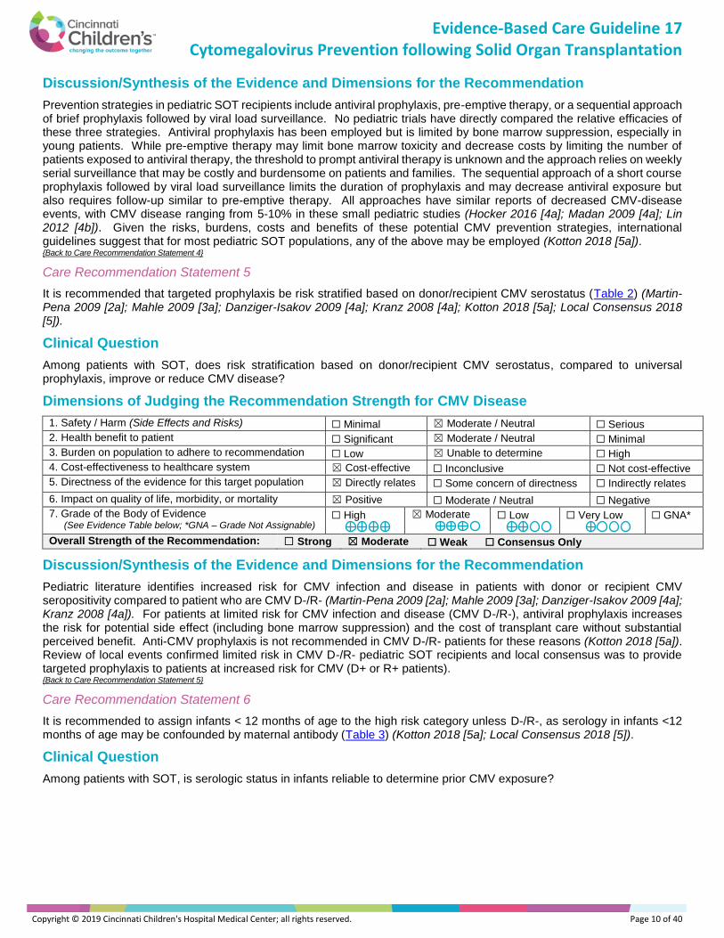
1. Safety / Harm (Side Effects and Risks) ☒ Minimal ☐ Moderate / Neutral ☐ Serious
2. Health benefit to patient ☐ Significant ☒ Moderate / Neutral ☐ Minimal
3. Burden on population to adhere to recommendation ☒ Low ☐ Unable to determine ☐ High
4. Cost-effectiveness to healthcare system ☒ Cost-effective ☐ Inconclusive ☐ Not cost-effective
5. Directness of the evidence for this target population ☐ Directly relates ☐ Some concern of directness ☒ Indirectly relates
6. Impact on quality of life, morbidity, or mortality ☐ Positive ☒ Moderate / Neutral ☐ Negative
7. Grade of the Body of Evidence (See Evidence Table below; *GNA – Grade Not Assignable)
☐ High
☒ Moderate
☐ Low
☐ Very Low
☐ GNA*
Overall Strength of the Recommendation: ☐ Strong ☐ Moderate ☒ Weak ☐ Consensus Only
Discussion/Synthesis of the Evidence and Dimensions for the Recommendation
The determination of which sample to use for CMV viral load testing, whole blood or plasma, was reviewed based on the currently available evidence. Direct comparisons between whole blood and plasma samples are limited in the literature. At least one study, indicates that neither whole blood nor plasma is superior in detecting the clearance of virus or in the prediction of relapse of CMV infection (Lisboa 2011 [4b]; (diagnosis)). Therefore, consensus was to continue using whole blood for viral load measurement to maintain consistency of measurement with the implementation of the new guideline as it does not impact the burden to the population, cost or potential benefits.
Monitoring schema were developed based on a review of internal CMV infection and disease incidence, including the timing of events in the post-transplant period over the past 5 years (Palmer 2010 [2a]; (treatment), Varela-Fascinetto 2017 [2b]; (prevention), Mahle 2009 [3a]; (incidence), Krampe 2010 [3b]; (prevention), Melgosa Hijosa 2004 [3b]; (prognosis), Ginevri 1998 [3b]; (incidence), Hocker 2016 [4a]; (prevention), Jongsma 2013 [4a]; (prognosis), Bedel 2012 [4a]; (incidence),

Evidence-Based Care Guideline 17 Cytomegalovirus Prevention following Solid Organ Transplantation
Copyright © 2019 Cincinnati Children's Hospital Medical Center; all rights reserved. Page 8 of 40
Camacho-Gonzalez 2011 [4a]; (risk), Saitoh 2011 [4a]; (prevention), Lapidus-Krol 2010 [4a]; (prevention), Snydman 2010 [4a]; (prevention), Danziger-Isakov 2009 [4a]; (incidence), Madan 2009 [4a]; (prevention), Ranganathan 2009 [4a]; (prevention), Bock 1997 [4a]; (incidence), Florescu 2012 [4b]; (risk factors), Lin 2012 [4b]; (incidence), Mazariegos 2008 [4b]; (prevention), Bueno 1997 [4b]; (risk factors). Balancing cost of increased numbers of test with the risk of delayed diagnosis of CMV infection or disease, internal data supported decreased viral load monitoring during the early post-transplant period, while the patients were taking CMV prophylaxis. No monitoring during prophylaxis is supported by the Transplantation Society CMV Guideline (Kotton 2018 [5a]); however, episodes of CMV infection and disease occurred in our local population. Therefore, a consensus decision to decrease but not eliminate monitoring during this period was made.
In the absence of relevant, published evidence for this care recommendation statement, developers reviewed previous statements and local data generated since its implementation. Events in the population were reviewed to determine what, if any, modifications to the care recommendation statement would be necessary. Consensus was pursued through open discussion with all committee members. Following presentation of the data and evidence results (or lack thereof), questions were answered and objections or concerns were addressed from all team members. All members agreed to the final recommendation statement with complete consensus. {Back to Care Recommendation Statement 1}
Care Recommendation Statement 2
It is recommended, to assure consistent results, that the same laboratory facility and assay be used for serial samples (Rychert 2014 [2a]; Pang 2009 [5a]; (Diagnostics), Local Consensus 2018 [5]).
Note 1: The laboratory facility at Cincinnati Children’s will be used for Cincinnati Children’s and local patients. Note 2: For non-local patients, whole blood CMV DNA PCR samples can be mailed to Cincinnati Children’s laboratory to assure consistent results.
• If outside laboratory is unable to mail samples to Cincinnati Children’s, serial CMV DNA PCR samples should be monitored at the same outside laboratory to assure consistent results.
Note 3: Inconsistent test results may be the result of testing being performed:
• at different laboratory facilities,
• using different assays, or
• on a different specimen type (e.g. whole blood vs. plasma) (Lisboa 2011 [4b]; (diagnosis)).
Contributing factors may be:
• patient use of different laboratory facility due to geographic need or insurance
• the designated laboratory facility transitions to use a different assay
• unreliable implementation processes (see Implementation section).
Clinical Question
Among patients with SOT, does using the same laboratory for serial testing, compared to using different laboratories, improve consistency of results?
Dimensions of Judging the Recommendation Strength for Consistent Lab Results
1. Safety / Harm (Side Effects and Risks) ☐ Minimal ☒ Moderate / Neutral ☐ Serious
2. Health benefit to patient ☐ Significant ☒ Moderate / Neutral ☐ Minimal
3. Burden on population to adhere to recommendation ☐ Low ☐ Unable to determine ☒ High
4. Cost-effectiveness to healthcare system ☐ Cost-effective ☒ Inconclusive ☐ Not cost-effective
5. Directness of the evidence for this target population ☒ Directly relates ☐ Some concern of directness ☐ Indirectly relates
6. Impact on quality of life, morbidity, or mortality ☒ Positive ☐ Moderate / Neutral ☐ Negative
7. Grade of the Body of Evidence (See Evidence Table below; *GNA – Grade Not Assignable)
☐ High
☒ Moderate
☐ Low
☐ Very Low
☐ GNA*
Overall Strength of the Recommendation: ☐ Strong ☒ Moderate ☐ Weak ☐ Consensus Only
Discussion/Synthesis of the Evidence and Dimensions for the Recommendation
Inter-laboratory variability of quantitative CMV viral loads is well reported in the literature even with the introduction of international unit calibration (Pang 2009 [5a]; Rychert 2014 [2a]). However, intra-laboratory results present with decreased variability. Therefore, balancing the potential inconvenience of arranging for sample processing and assays in the same lab with the issues related to decreased reliability of assay interpretation when samples are resulted serially from multiple labs, consensus decision to recommend assay performance predominantly at Cincinnati Children’s was made. Alternative options were developed to address the potential barriers to Cincinnati Children’s performing these tests. {Back to Care Recommendation Statement 2}

Evidence-Based Care Guideline 17 Cytomegalovirus Prevention following Solid Organ Transplantation
Copyright © 2019 Cincinnati Children's Hospital Medical Center; all rights reserved. Page 9 of 40
Care Recommendation Statement 3
It is recommended that patients with any of the following clinical conditions be evaluated for CMV by examination, whole blood PCR and end-organ histopathology, if indicated (Kotton 2018 [5a]; Local Consensus 2018 [5]).
• fever • thrombocytopenia • gastroenteropathy • muscle pain • anemia • pneumonitis • leukopenia • hepatitis • retinitis
Clinical Question
Among patients with SOT, who should be evaluated for CMV and by what methods to improve diagnosis of CMV disease?
Dimensions of Judging the Recommendation Strength for Diagnosis of CMV Disease
1. Safety / Harm (Side Effects and Risks) ☒ Minimal ☐ Moderate / Neutral ☐ Serious
2. Health benefit to patient ☒ Significant ☐ Moderate / Neutral ☐ Minimal
3. Burden on population to adhere to recommendation ☒ Low ☐ Unable to determine ☐ High
4. Cost-effectiveness to healthcare system ☐ Cost-effective ☒ Inconclusive ☐ Not cost-effective
5. Directness of the evidence for this target population ☒ Directly relates ☐ Some concern of directness ☐ Indirectly relates
6. Impact on quality of life, morbidity, or mortality ☒ Positive ☐ Moderate / Neutral ☐ Negative
7. Grade of the Body of Evidence (See Evidence Table below; *GNA – Grade Not Assignable)
☐ High
☐ Moderate
☐ Low
☒ Very Low
☐ GNA*
Overall Strength of the Recommendation: ☐ Strong ☒ Moderate ☐ Weak ☐ Consensus Only
Discussion/Synthesis of the Evidence and Dimensions for the Recommendation
CMV is a significant infectious complication in pediatric solid organ transplant recipients with up to 38% in some populations experiencing infection and/or disease (Martin-Pena 2009 [2a]; (incidence), Mahle 2009 [3a]; (incidence), Krampe 2010 [3b]; (prevention), Turmelle 2009 [3b]; (prevention), Ginevri 1998 [3b]; (incidence), Bedel 2012 [4a]; (incidence), Danziger-Isakov 2009 [4a]; (incidence), Kranz 2008 [4a]; (incidence), Simmonds 2008 [4a]; (risk factors), Danziger-Isakov 2003 [4a]; (incidence), Robinson 2002 [4a]; (incidence), Bock 1997 [4a]; (incidence), Florescu 2012 [4b]; (risk factors), Mazariegos 2008 [4b]; (prevention), Kullberg-Lindh 2003 [4b]; (risk factors), Metras 1999 [4b]; (incidence), Bueno 1997 [4b]; (risk factors). Non-specific signs and symptoms may portend infection secondary to CMV or other infectious post-transplant complications that would require differential therapy based on determination of underlying etiology. Therefore, known signs and symptoms of potential CMV infection and/or disease in pediatric SOT recipients require evaluation to prompt appropriate treatment to avoid CMV-related morbidity and mortality (Kotton 2018 [5a]; Local Consensus [5]). {Back to Care Recommendation Statement 3}
Care Recommendation Statement 4
It is recommended that targeted prophylaxis be the primary strategy for prevention of CMV disease (Hocker 2016 [4a]; Madan 2009 [4a]; Lin 2012 [4b]; Kotton 2018 [5a]; Local Consensus 2018 [5]). See definition.
Clinical Question
Among patients with SOT, does targeted prophylaxis, compared to universal prophylaxis or pre-emptive therapy, improve or reduce CMV disease?
Dimensions of Judging the Recommendation Strength for CMV Disease
1. Safety / Harm (Side Effects and Risks) ☐ Minimal ☒ Moderate / Neutral ☐ Serious
2. Health benefit to patient ☒ Significant ☐ Moderate / Neutral ☐ Minimal
3. Burden on population to adhere to recommendation ☐ Low ☐ Unable to determine ☒ High
4. Cost-effectiveness to healthcare system ☐ Cost-effective ☒ Inconclusive ☐ Not cost-effective
5. Directness of the evidence for this target population ☐ Directly relates ☒ Some concern of directness ☐ Indirectly relates
6. Impact on quality of life, morbidity, or mortality ☒ Positive ☐ Moderate / Neutral ☐ Negative
7. Grade of the Body of Evidence (See Evidence Table below; *GNA – Grade Not Assignable)
☐ High
☐ Moderate
☐ Low
☒ Very Low
☐ GNA*
Overall Strength of the Recommendation: ☐ Strong ☒ Moderate ☐ Weak ☐ Consensus Only
Evidence-Based Care Guideline 17 Cytomegalovirus Prevention following Solid Organ Transplantation
Copyright © 2019 Cincinnati Children's Hospital Medical Center; all rights reserved. Page 10 of 40
Discussion/Synthesis of the Evidence and Dimensions for the Recommendation
Prevention strategies in pediatric SOT recipients include antiviral prophylaxis, pre-emptive therapy, or a sequential approach of brief prophylaxis followed by viral load surveillance. No pediatric trials have directly compared the relative efficacies of these three strategies. Antiviral prophylaxis has been employed but is limited by bone marrow suppression, especially in young patients. While pre-emptive therapy may limit bone marrow toxicity and decrease costs by limiting the number of patients exposed to antiviral therapy, the threshold to prompt antiviral therapy is unknown and the approach relies on weekly serial surveillance that may be costly and burdensome on patients and families. The sequential approach of a short course prophylaxis followed by viral load surveillance limits the duration of prophylaxis and may decrease antiviral exposure but also requires follow-up similar to pre-emptive therapy. All approaches have similar reports of decreased CMV-disease events, with CMV disease ranging from 5-10% in these small pediatric studies (Hocker 2016 [4a]; Madan 2009 [4a]; Lin 2012 [4b]). Given the risks, burdens, costs and benefits of these potential CMV prevention strategies, international guidelines suggest that for most pediatric SOT populations, any of the above may be employed (Kotton 2018 [5a]). {Back to Care Recommendation Statement 4}
Care Recommendation Statement 5
It is recommended that targeted prophylaxis be risk stratified based on donor/recipient CMV serostatus (Table 2) (Martin-Pena 2009 [2a]; Mahle 2009 [3a]; Danziger-Isakov 2009 [4a]; Kranz 2008 [4a]; Kotton 2018 [5a]; Local Consensus 2018 [5]).
Clinical Question
Among patients with SOT, does risk stratification based on donor/recipient CMV serostatus, compared to universal prophylaxis, improve or reduce CMV disease?
Dimensions of Judging the Recommendation Strength for CMV Disease
1. Safety / Harm (Side Effects and Risks) ☐ Minimal ☒ Moderate / Neutral ☐ Serious
2. Health benefit to patient ☐ Significant ☒ Moderate / Neutral ☐ Minimal
3. Burden on population to adhere to recommendation ☐ Low ☒ Unable to determine ☐ High
4. Cost-effectiveness to healthcare system ☒ Cost-effective ☐ Inconclusive ☐ Not cost-effective
5. Directness of the evidence for this target population ☒ Directly relates ☐ Some concern of directness ☐ Indirectly relates
6. Impact on quality of life, morbidity, or mortality ☒ Positive ☐ Moderate / Neutral ☐ Negative
7. Grade of the Body of Evidence (See Evidence Table below; *GNA – Grade Not Assignable)
☐ High
☒ Moderate
☐ Low
☐ Very Low
☐ GNA*
Overall Strength of the Recommendation: ☐ Strong ☒ Moderate ☐ Weak ☐ Consensus Only
Discussion/Synthesis of the Evidence and Dimensions for the Recommendation
Pediatric literature identifies increased risk for CMV infection and disease in patients with donor or recipient CMV seropositivity compared to patient who are CMV D-/R- (Martin-Pena 2009 [2a]; Mahle 2009 [3a]; Danziger-Isakov 2009 [4a]; Kranz 2008 [4a]). For patients at limited risk for CMV infection and disease (CMV D-/R-), antiviral prophylaxis increases the risk for potential side effect (including bone marrow suppression) and the cost of transplant care without substantial perceived benefit. Anti-CMV prophylaxis is not recommended in CMV D-/R- patients for these reasons (Kotton 2018 [5a]). Review of local events confirmed limited risk in CMV D-/R- pediatric SOT recipients and local consensus was to provide targeted prophylaxis to patients at increased risk for CMV (D+ or R+ patients). {Back to Care Recommendation Statement 5}
Care Recommendation Statement 6
It is recommended to assign infants < 12 months of age to the high risk category unless D-/R-, as serology in infants <12 months of age may be confounded by maternal antibody (Table 3) (Kotton 2018 [5a]; Local Consensus 2018 [5]).
Clinical Question
Among patients with SOT, is serologic status in infants reliable to determine prior CMV exposure?

Evidence-Based Care Guideline 17 Cytomegalovirus Prevention following Solid Organ Transplantation
Copyright © 2019 Cincinnati Children's Hospital Medical Center; all rights reserved. Page 11 of 40
Dimensions of Judging the Recommendation Strength for Reliable Serologic Status
1. Safety / Harm (Side Effects and Risks) ☐ Minimal ☒ Moderate / Neutral ☐ Serious
2. Health benefit to patient ☐ Significant ☒ Moderate / Neutral ☐ Minimal
3. Burden on population to adhere to recommendation ☒ Low ☐ Unable to determine ☐ High
4. Cost-effectiveness to healthcare system ☐ Cost-effective ☒ Inconclusive ☐ Not cost-effective
5. Directness of the evidence for this target population ☐ Directly relates ☒ Some concern of directness ☐ Indirectly relates
6. Impact on quality of life, morbidity, or mortality ☐ Positive ☒ Moderate / Neutral ☐ Negative
7. Grade of the Body of Evidence (See Evidence Table below; *GNA – Grade Not Assignable)
☐ High
☐ Moderate
☐ Low
☒ Very Low
☐ GNA*
Overall Strength of the Recommendation: ☐ Strong ☒ Moderate ☐ Weak ☐ Consensus Only
Discussion/Synthesis of the Evidence and Dimensions for the Recommendation
Interpretation of donor and recipient serostatus for infants less than 12 months of age is confounded by the potential presence of trans placentally-acquired maternal CMV antibodies (Kotton 2018 [5a]). Attempts to definitively categorize infants as CMV seropositive by demonstrating CMV shedding is confounded by the fact that CMV shedding in saliva or urine among infected infants is intermittent. Risk stratification, as discussed in Care Recommendation 5, drives prevention strategy and accurate categorization with appropriate prophylaxis can decrease risk for CMV infection and/or disease. Therefore, international and local consensus both promote placing infants in the highest risk category unless CMV status, including prior infection, can be confirmed (Kotton 2018 [5a]). {Back to Care Recommendation Statement 6}
Care Recommendation Statement 7
It is recommended to use age- and BSA-based antiviral dosing to optimize therapy (Table 4) (Bradley 2016 [2a]; Asberg 2014 [2a]; Varela-Fascinetto 2017 [2b]; (prevention), Pescovitz 2010 [3a]; (treatment), Vaudry 2009 [3a]; (treatment), Villeneuve 2013 [3b]; (treatment), Launay 2012 [3b]; (treatment), Local Consensus 2018 [5]).
Clinical Question
Among patients with SOT, does age-based and BSA-based antiviral dosing, compared to weight-based dosing, improve or reduce CMV disease?
Dimensions of Judging the Recommendation Strength for CMV Disease
1. Safety / Harm (Side Effects and Risks) ☐ Minimal ☒ Moderate / Neutral ☐ Serious
2. Health benefit to patient ☐ Significant ☒ Moderate / Neutral ☐ Minimal
3. Burden on population to adhere to recommendation ☒ Low ☐ Unable to determine ☐ High
4. Cost-effectiveness to healthcare system ☐ Cost-effective ☒ Inconclusive ☐ Not cost-effective
5. Directness of the evidence for this target population ☒ Directly relates ☐ Some concern of directness ☐ Indirectly relates
6. Impact on quality of life, morbidity, or mortality ☒ Positive ☐ Moderate / Neutral ☐ Negative
7. Grade of the Body of Evidence (See Evidence Table below; *GNA – Grade Not Assignable)
☐ High
☒ Moderate
☐ Low
☐ Very Low
☐ GNA*
Overall Strength of the Recommendation: ☒ Strong ☐ Moderate ☐ Weak ☐ Consensus Only
Discussion/Synthesis of the Evidence and Dimensions for the Recommendation
Pharmacokinetics (PK) studies in older children (Pescovitz 2010 [3a]; Vaudry 2009 [3a]) support the currently recommended dosing schedule provided in the package insert. In addition, emerging data since the last iteration of these guidelines suggest that PK in younger SOT populations, including infants down to 4 months of age should follow the same BSA-based dosing recommendations. Current models support BSA-based dosing to reach targeted ganciclovir AUC as opposed to weight-based dosing previously recommended in pediatric SOT recipients under 3 years of age (Bradley 2016 [2a]; Asberg 2014 [2a]; Varela-Fascinetto 2017 [2b]; Villeneuve 2013 [3b]; Launay 2012 [3b]). {Back to Care Recommendation Statement 7}
Care Recommendation Statement 8
It is recommended that valganciclovir be dosed around a meal for best absorption (Local Consensus 2018 [5]).
Clinical Question
Among patients with SOT, does ingestion of valganciclovir with food, compared to fasting, improve or reduce valganciclovir bioavailability?

Evidence-Based Care Guideline 17 Cytomegalovirus Prevention following Solid Organ Transplantation
Copyright © 2019 Cincinnati Children's Hospital Medical Center; all rights reserved. Page 12 of 40
Dimensions of Judging the Recommendation Strength for Valganciclovir Bioavailability
1. Safety / Harm (Side Effects and Risks) ☒ Minimal ☐ Moderate / Neutral ☐ Serious
2. Health benefit to patient ☒ Significant ☐ Moderate / Neutral ☐ Minimal
3. Burden on population to adhere to recommendation ☒ Low ☐ Unable to determine ☐ High
4. Cost-effectiveness to healthcare system ☒ Cost-effective ☐ Inconclusive ☐ Not cost-effective
5. Directness of the evidence for this target population ☐ Directly relates ☒ Some concern of directness ☐ Indirectly relates
6. Impact on quality of life, morbidity, or mortality ☐ Positive ☒ Moderate / Neutral ☐ Negative
7. Grade of the Body of Evidence (See Evidence Table below; *GNA – Grade Not Assignable)
☐ High
☐ Moderate
☐ Low
☐ Very Low
☒ GNA*
Overall Strength of the Recommendation: ☐ Strong ☐ Moderate ☐ Weak ☒ Consensus Only
Discussion/Synthesis of the Evidence and Dimensions for the Recommendation
Valganciclovir PK studies provided in the package insert for the product provide information about drug bioavailability with and without food from healthy volunteers, HIV-positive patients and solid organ transplant recipients. Recommendations from this data show improved bioavailability of valganciclovir when taken with food (package insert; https://www.gene.com/download/pdf/valcyte_prescribing.pdf) (Local Consensus [5a]). {Back to Care Recommendation Statement 8}
Care Recommendation Statement 9
Consider re-initiation of prophylaxis for a minimum of 3 months for patients who undergo treatment of acute rejection with antilymphocyte antibodies who are serologically at risk (D+ or R+) (Local Consensus 2018 [5]).
Clinical Question
Among patients with SOT, does valganciclovir prophylaxis, compared to clinical monitoring, improve or reduce CMV disease after treatment of acute rejection with antilymphocyte antibodies?
Dimensions of Judging the Recommendation Strength for CMV Disease
1. Safety / Harm (Side Effects and Risks) ☐ Minimal ☒ Moderate / Neutral ☐ Serious
2. Health benefit to patient ☐ Significant ☒ Moderate / Neutral ☐ Minimal
3. Burden on population to adhere to recommendation ☐ Low ☒ Unable to determine ☐ High
4. Cost-effectiveness to healthcare system ☐ Cost-effective ☒ Inconclusive ☐ Not cost-effective
5. Directness of the evidence for this target population ☐ Directly relates ☐ Some concern of directness ☒ Indirectly relates
6. Impact on quality of life, morbidity, or mortality ☐ Positive ☒ Moderate / Neutral ☐ Negative
7. Grade of the Body of Evidence (See Evidence Table below; *GNA – Grade Not Assignable)
☐ High
☐ Moderate
☐ Low
☐ Very Low
☒ GNA*
Overall Strength of the Recommendation: ☐ Strong ☐ Moderate ☐ Weak ☒ Consensus Only
Discussion/Synthesis of the Evidence and Dimensions for the Recommendation
In children at risk for CMV infection and/or disease who receive significantly intensified immunosuppression (e.g. antilymphocyte therapy, intravenous steroids), international consensus guidelines recommend either prophylaxis with valganciclovir/ganciclovir or an intensified DNAemia surveillance program with preemptive treatment. Further, no data exist to suggest specific duration in these circumstances. Due to the risk for significant CMV events, local consensus determined prophylaxis only after antilymphocyte therapy for a 3-month period, consistent with timing of immune reconstitution after antilymphocyte therapy, as the preferred method for CMV prevention in this circumstance weighing cost of prophylaxis, side effect of antiviral therapy, cost and convenience of monitoring schedules as factors in the decision. {Back to Care Recommendation Statement 9}

Evidence-Based Care Guideline 17 Cytomegalovirus Prevention following Solid Organ Transplantation
Copyright © 2019 Cincinnati Children's Hospital Medical Center; all rights reserved. Page 13 of 40
CLINICAL QUESTIONS, CRITERIA FOR INCLUSION, AND SEARCH STRATEGIES
& RESULTS
Clinical Questions
Among patients with SOT aged birth to young adult,
1. Does monitoring with whole blood samples, compared to plasma, at specific intervals, improve or reduce CMV disease incidence?
2. Does using the same laboratory for serial testing, compared to using different laboratories, improve consistency of results?
3. Who should be evaluated for CMV and by what methods to improve diagnosis of CMV disease?
4. Does targeted prophylaxis, compared to universal prophylaxis or pre-emptive therapy, improve or reduce CMV disease?
5. Does risk stratification based on donor/recipient CMV serostatus, compared to universal prophylaxis, improve or reduce CMV disease?
6. Is serologic status in infants reliable to determine prior CMV exposure?
7. Does age-based and BSA-based antiviral dosing, compared to weight-based dosing, improve or reduce CMV disease?
8. Does ingestion of valganciclovir with food, compared to fasting, improve or reduce valganciclovir bioavailability?
9. Does valganciclovir prophylaxis, compared to clinical monitoring, improve or reduce CMV disease after treatment of acute rejection with antilymphocyte antibodies?
Criteria for considering studies for this review
Types of Studies Study designs were not restricted for inclusion in the systematic review
Types of Participants Patients following SOT, Ages birth to young adult
Types of Interventions Monitoring – whole blood, Diagnostic evaluation / Testing, Prevention of CMV infection, Targeted prophylaxis, antiviral dosing based on age or BSA, Valganciclovir ingestion with food or prophylaxis
Types of Comparisons Monitoring – plasma/clinical, Prevention of CMV infection, Prophylaxis – universal or preemptive therapy, weight-based dosing, Valganciclovir ingestion while fasting
Types of Outcomes Improvement or reduction of CMV disease Consistent and reliable laboratory results, Valganciclovir bioavailability
Exclusion Criteria Patients with CMV disease or with non-solid organ transplants
Search Strategy
Search Methods
To select evidence for critical appraisal by the group for this guideline, the databases below were searched using search terms, limits, filters, and date parameters to generate an unrefined, “combined evidence” database. This search strategy focused on answering the clinical questions addressed in this document and employing a combination of Boolean searching on human-indexed thesaurus terms (e.g., MeSH) as well as “natural language” searching on words in the title, abstract, and indexing terms.
Search Databases Search Terms Limits, Filters, &
Search Date Parameters Date of Most
Recent Search
☒ MedLine
via PubMed or Ovid
☐ CINAHL
☒ Cochrane Database
for Systematic Reviews
☐ PsycInfo
☒ Other: Embase
• CMV or Cytomegalovirus
• SOT or “Solid Organ Transplant”
• Specific pharmacokinetics or medications – Ganciclovir, valganciclovir, acyclovir, cytomegalovirus hyperimmune globulin
Publication Dates or Search Dates:
• August 2013 to January 2018
1 / 2018
☒ English Language
☒ Pediatric Evidence Only:
• Pediatric
☐ Other Limits or Filters
Search Results
The citations were reduced by eliminating duplicates and non-English articles. The resulting abstracts and full text articles were reviewed to eliminate low quality and irrelevant citations or articles. During the course of the guideline development, additional articles were identified from subsequent refining searches for evidence, clinical questions added to the guideline and subjected to the search process, and hand searching of reference lists. The initial search for evidence identified 300 articles. 55 articles met the inclusion criteria above.

Evidence-Based Care Guideline 17 Cytomegalovirus Prevention following Solid Organ Transplantation
Copyright © 2019 Cincinnati Children's Hospital Medical Center; all rights reserved. Page 14 of 40
TEAM MEMBERS & CONFLICTS OF INTEREST
Group / Team Members
Multidisciplinary Team
Team Leader/Author/Chair: Lara Danziger-Isakov, MD, MPH, Infectious Diseases, Cincinnati Children’s Hospital Medical Center
Team Members/Co-Authors: Scott Pangonis, MD, Infectious Disease, Cincinnati Children’s Hospital Medical Center *John Bucuvalas, MD, Liver Transplant, Cincinnati Children’s Hospital Medical Center Alex Miethke, MD, Liver Transplant, Cincinnati Children’s Hospital Medical Center Anna Peters, MD, Liver Transplant, Cincinnati Children’s Hospital Medical Center Clifford Chin, MD, Cardiology/Transplant, Cincinnati Children’s Hospital Medical Center * Samuel Kocoshis, MD Intestinal Transplant, Cincinnati Children’s Hospital Medical Center Francisco Flores, MD, Nephrology/Transplant, Cincinnati Children’s Hospital Medical Center * Marc Schechter, MD, Pulmonary/Transplant, Cincinnati Children’s Hospital Medical Center
Patient Services * Trina Hemmelgarn, PharmD, Pharmacy, Cincinnati Children’s Hospital Medical Center * Danielle Lazear, PharmD, Pharmacy, Cincinnati Children’s Hospital Medical Center BreAnn Taylor, PharmD, Pharmacy, Cincinnati Children’s Hospital Medical Center
*Member of previous CMV Prevention guideline development Team
Other Evidence-Based Care Recommendation Development Support
Content Reviewers: Grant Paulsen, MD, Pediatric Infectious Diseases, Cincinnati Children’s Hospital Medical Center
Support/Consultant & Evidence Methodologist: Danette Stanko-Lopp, MA, MPH, Cincinnati Children’s Hospital Medical Center
Conflicts of Interest were declared for each team member and:
☒ No financial or intellectual conflicts of interest were found.
☒ No external funding was received for development of this recommendation.
☒ The following conflicts of interest were disclosed:
Conflict of interest declarations information is maintained in Cincinnati Children’s ePAS (electronic Protocol Administration System).
FUTURE RESEARCH AGENDA
1. Among children with SOT, what is the efficacy of prevention strategies, and what are the important differences between prophylaxis, preemptive therapy, and sequential/hybrid strategies?
2. Among children with SOT, what economic and safety concerns are important to consider when anticipating use of antiviral medications?
3. Among children with SOT, what is the optimal schedule for antiviral dosing and therapeutic drug monitoring?
4. Among children with SOT, what novel options are effective for the prevention and treatment of CMV infection and disease?
5. Among children with SOT, what indirect effects are associated with CMV infection?
6. Among children with SOT, what are the clinically relevant viral load thresholds to guide risk stratification, preemptive therapy, and therapeutic assessments?
7. Among children with SOT, which assays for the assessment of T cell immunity to CMV are able to predict the development of CMV disease, thereby allowing better risk stratification of patients and more targeted prevention strategies?
LEGEND EVIDENCE EVALUATION SYSTEM (LET EVIDENCE GUIDE EVERY NEW DECISION)
Full tables of the LEGEND evidence evaluation system are available in separate documents:
• Table of Evidence Levels of Individual Studies by Domain, Study Design, & Quality (abbreviated table below)
• Grading a Body of Evidence to Answer a Clinical Question
• Judging the Strength of a Recommendation (Evidence Discussion and Dimensions for Recommendations section)
Evidence-Based Care Guideline 17 Cytomegalovirus Prevention following Solid Organ Transplantation
Copyright © 2019 Cincinnati Children's Hospital Medical Center; all rights reserved. Page 15 of 40
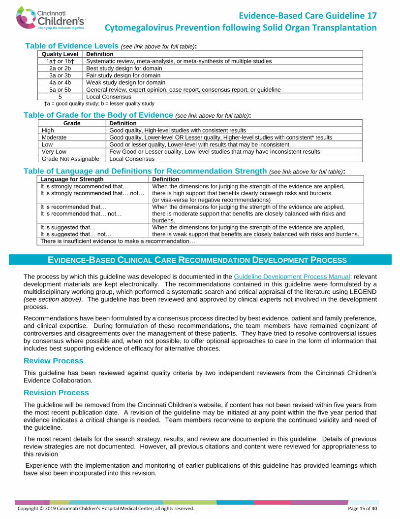
Table of Evidence Levels (see link above for full table):
†a = good quality study; b = lesser quality study
Table of Grade for the Body of Evidence (see link above for full table):
Table of Language and Definitions for Recommendation Strength (see link above for full table): Language for Strength Definition It is strongly recommended that… It is strongly recommended that… not…
When the dimensions for judging the strength of the evidence are applied, there is high support that benefits clearly outweigh risks and burdens. (or visa-versa for negative recommendations)
It is recommended that… It is recommended that… not…
When the dimensions for judging the strength of the evidence are applied, there is moderate support that benefits are closely balanced with risks and burdens.
It is suggested that… It is suggested that… not…
When the dimensions for judging the strength of the evidence are applied, there is weak support that benefits are closely balanced with risks and burdens.
There is insufficient evidence to make a recommendation…
EVIDENCE-BASED CLINICAL CARE RECOMMENDATION DEVELOPMENT PROCESS
The process by which this guideline was developed is documented in the Guideline Development Process Manual; relevant development materials are kept electronically. The recommendations contained in this guideline were formulated by a multidisciplinary working group, which performed a systematic search and critical appraisal of the literature using LEGEND (see section above). The guideline has been reviewed and approved by clinical experts not involved in the development process.
Recommendations have been formulated by a consensus process directed by best evidence, patient and family preference, and clinical expertise. During formulation of these recommendations, the team members have remained cognizant of controversies and disagreements over the management of these patients. They have tried to resolve controversial issues by consensus where possible and, when not possible, to offer optional approaches to care in the form of information that includes best supporting evidence of efficacy for alternative choices.
Review Process
This guideline has been reviewed against quality criteria by two independent reviewers from the Cincinnati Children’s Evidence Collaboration.
Revision Process
The guideline will be removed from the Cincinnati Children’s website, if content has not been revised within five years from the most recent publication date. A revision of the guideline may be initiated at any point within the five year period that evidence indicates a critical change is needed. Team members reconvene to explore the continued validity and need of the guideline.
The most recent details for the search strategy, results, and review are documented in this guideline. Details of previous review strategies are not documented. However, all previous citations and content were reviewed for appropriateness to this revision
Experience with the implementation and monitoring of earlier publications of this guideline has provided learnings which have also been incorporated into this revision.
Quality Level Definition
1a† or 1b† Systematic review, meta-analysis, or meta-synthesis of multiple studies
2a or 2b Best study design for domain
3a or 3b Fair study design for domain
4a or 4b Weak study design for domain
5a or 5b General review, expert opinion, case report, consensus report, or guideline
5 Local Consensus
Grade Definition
High Good quality, High-level studies with consistent results
Moderate Good quality, Lower-level OR Lesser quality, Higher-level studies with consistent* results
Low Good or lesser quality, Lower-level with results that may be inconsistent
Very Low Few Good or Lesser quality, Low-level studies that may have inconsistent results
Grade Not Assignable Local Consensus

Evidence-Based Care Guideline 17 Cytomegalovirus Prevention following Solid Organ Transplantation
Copyright © 2019 Cincinnati Children's Hospital Medical Center; all rights reserved. Page 16 of 40
Review History
Date Event Outcome
March 1, 2019 5-Year Review Guideline revised and published
September 30, 2013 5-Year Review Guideline revised and published
July 6, 2007 5-Year Review Guideline revised and published
June 7, 2001 Original Publication New guideline developed and published
Permission to Use the Guideline
This Evidence-Based Care Guideline (EBCG) and any related implementation tools (if applicable, e.g., screening tools, algorithms, etc.) are available online and may be distributed by any organization for the global purpose of improving child health outcomes.
Website address: http://www.cincinnatichildrens.org/service/j/anderson-center/evidence-based-care/recommendations/default/
Examples of approved uses of the EBCG include the following: • copies may be provided to anyone involved in the organization’s (outside of Cincinnati Children’s) process for
developing and implementing evidence-based care guidelines; • hyperlinks to the Cincinnati Children’s website may be placed on the organization’s website; • the EBCG may be adopted or adapted for use within the organization, provided that Cincinnati Children’s receives
appropriate attribution on all written or electronic documents; and • copies may be provided to patients and the clinicians who manage their care.
Notification to Cincinnati Children’s ([email protected]) is appreciated for all uses of any EBCG or its companion documents which are adopted, adapted, implemented, or hyperlinked.
Please cite as
Danziger-Isakov, L, Pangonis, S, Bucuvalas, J, Miethke, A, Peters, A, Chin, C, Kocoshis, S, Flores, F, Schechter, M, Witte, D, Hemmelgarn, T, Lazear, D, Taylor, B: CMV Guideline Development Team (2018). Cincinnati Children's Hospital Medical Center: Evidence-based clinical care guideline for Cytomegalovirus Prevention following Solid Organ Transplantation. http://www.cincinnatichildrens.org/service/j/anderson-center/evidence-based-care/recommendations/default/, Guideline 17, pages 1–19, March 1, 2019
For more information
About this guideline, its companion documents, or the Cincinnati Children’s Evidence-Based Care Recommendation Development process, contact Lara Danziger-Isakov, MD, MPH in Infectious Diseases at (513) 636-9101 or [email protected] or the Cincinnati Children’s Evidence Collaboration at [email protected].
Note/Disclaimer
This guideline addresses only key points of care for the target population; it may not be a comprehensive practice guideline. These care recommendations result from review of literature and practices current at the time of their formulations. This guideline does not preclude using care modalities proven efficacious in studies published subsequent to the current revision of this document. This document is not intended to impose standards of care preventing selective variances from the recommendations to meet the specific and unique requirements of individual patients. Adherence to this guideline is voluntary. The clinician in light of the individual circumstances presented by the patient must make the ultimate judgment regarding any specific care recommendation.

Evidence-Based Care Guideline 17 Cytomegalovirus Prevention following Solid Organ Transplantation
Copyright © 2019 Cincinnati Children's Hospital Medical Center; all rights reserved. Page 17 of 40
REFERENCES Evidence Level in [ ], Table of Evidence Levels in LEGEND section above
Note: When using the electronic version of this document, the hyperlink to the PubMed abstract may be located at the end of citations.
1. Asberg, A., Bjerre, A., Neely, M.: New algorithm for valganciclovir dosing in pediatric solid organ transplant recipients. Pediatr Transplant, 18(1): 103-11, 2014, [2a].
2. Bedel, A. N.; Hemmelgarn, T. S.; and Kohli, R.: Retrospective review of the incidence of cytomegalovirus infection and disease after liver transplantation in pediatric patients: comparison of prophylactic oral ganciclovir and oral valganciclovir. Liver Transpl, 18(3): 347-54, 2012, [4a] (incidence) http://www.ncbi.nlm.nih.gov/pubmed/22139888.
3. Best, N. G.; Trull, A. K.; Tan, K. K.; Spiegelhalter, D. J.; Wreghitt, T. G.; and Wallwork, J.: Blood cyclosporine concentrations and cytomegalovirus infection following heart transplantation. Transplantation, 60(7): 689-94., 1995, [4b] (risk factors) http://www.ncbi.nlm.nih.gov/pubmed/7570978.
4. Bock, G. H.; Sullivan, E. K.; Miller, D.; Gimon, D.; Alexander, S.; Ellis, E.; and Elshihabi, I.: Cytomegalovirus infections following renal transplantation--effects on antiviral prophylaxis: a report of the North American Pediatric Renal Transplant Cooperative Study. Pediatr Nephrol, 11(6): 665-71, 1997, [4a] (incidence) http://www.ncbi.nlm.nih.gov/pubmed/9438638.
5. Bradley, D., Moreira, S., Subramoney, V., Chin, C., Ives, J., Wang, K.; Valcyte NP22523 Study Team: Pharmacokinetics and Safety of Valganciclovir in Pediatric Heart Transplant Recipients 4 Months of Age and Younger. Pediatr Infect Dis J, 35(12): 1324-1328, 2016 [2a].
6. Bueno, J. et al.: Cytomegalovirus infection after intestinal transplantation in children. Clin Infect Dis, 25(5): 1078-83, 1997, [4b] (risk factors) http://www.ncbi.nlm.nih.gov/pubmed/9402361.
7. Camacho-Gonzalez, A. F.; Gutman, J.; Hymes, L. C.; Leong, T.; and Hilinski, J. A.: 24 weeks of valganciclovir prophylaxis in children after renal transplantation: a 4-year experience. Transplantation, 91(2): 245-50, 2011, [4a] (risk)] http://www.ncbi.nlm.nih.gov/pubmed/21076375.
8. Centers for Disease Control and Prevention: MMWR Guidelines for preventing opportunistic infections among hematopoietic stem cell transplant recipients. Recommendations and Reports 49(RR-10): 1-125, CE1-7, 2000, [5a] http://www.ncbi.nlm.nih.gov/pubmed/11718124.
9. Danziger-Isakov, L. A.; DelaMorena, M.; Hayashi, R. J.; Sweet, S.; Mendeloff, E.; Schootman, M.; Huddleston, C. B.; and DeBaun, M. R.: Cytomegalovirus viremia associated with death or retransplantation in pediatric lung-transplant recipients. Transplantation, 75(9): 1538-43, 2003, [4a] (incidence) http://www.ncbi.nlm.nih.gov/pubmed/12792511.
10. Danziger-Isakov, L. A. et al.: The risk, prevention, and outcome of cytomegalovirus after pediatric lung transplantation. Transplantation, 87(10): 1541-8, 2009, [4a] (incidence) http://www.ncbi.nlm.nih.gov/pubmed/19461492.
11. de Souza, V.; Cochat, P.; Rabilloud, M.; Selistre, L.; Wagner, M.; Hadj-Aissa, A.; Dolomanova, O.; Ranchin, B.; Iwaz,J.; and Dubourg, L.: Accuracy of Different Equations in Estimating GFR in Pediatric Kidney Transplant Recipients. Clin J Am Soc Nephr, 10: 463–470, 2015, [2a].
12. Florescu, D. F.; Langnas, A. N.; Grant, W.; Mercer, D. F.; Botha, J.; Qiu, F.; Shafer, L.; and Kalil, A. C.: Incidence, risk factors, and outcomes associated with cytomegalovirus disease in small bowel transplant recipients. Pediatr Transplant, 16(3): 294-301, 2012, [4b] (risk factors) http://www.ncbi.nlm.nih.gov/pubmed/22212495.
13. Ghisetti, V.; Barbui, A.; Franchello, A.; Varetto, S.; Pittaluga, F.; Bobbio, M.; Salizzoni, M.; and Marchiaro, G.: Quantitation of cytomegalovirus DNA by the polymerase chain reaction as a predictor of disease in solid organ transplantation. J Med Virol, 73(2): 223-9, 2004, [2a] (prognosis) http://www.ncbi.nlm.nih.gov/pubmed/15122796.
14. Ginevri, F. et al.: Acyclovir plus CMV immunoglobulin prophylaxis and early therapy with ganciclovir are effective and safe in CMV high-risk renal transplant pediatric recipients. Transpl Int, 11(1): S130-4, 1998, [3b] (incidence) http://www.ncbi.nlm.nih.gov/pubmed/9664962.
15. Ho, M.: Advances in understanding cytomegalovirus infection after transplantation. Transplant Proc, 26(5 Suppl 1): 7-11., 1994, [5b] http://www.ncbi.nlm.nih.gov/pubmed/7940978.
16. Hocker, B. et al.: Cytomegalovirus Infection in Pediatric Renal Transplantation and the Impact of Chemoprophylaxis With (Val-)Ganciclovir. Transplantation, 100(4): 862-70, 2016, [4a] (prevention) http://www.ncbi.nlm.nih.gov/pubmed/26736017.
17. Hokeberg, I.; Eriksson, B. M.; Zweygberg-Wirgart, B.; Tufvesson, G.; Olding-Stenkvist, E.; and Grillner, L.: Diagnostic markers and risk factors of cytomegalovirus infection and disease in renal allograft recipients. Scand J Infect Dis, 27(5): 435-40, 1995, [2b] (incidence) http://www.ncbi.nlm.nih.gov/pubmed/8588130.
18. Humar, A., and Michaels, M.: American Society of Transplantation recommendations for screening, monitoring and reporting of infectious complications in immunosuppression trials in recipients of organ transplantation. Am J Transplant, 6(2): 262-74, 2006, [5a] http://www.ncbi.nlm.nih.gov/pubmed/16426310.
19. Jongsma, H.; Bouts, A. H.; Cornelissen, E. A.; Beersma, M. F.; and Cransberg, K.: Cytomegalovirus prophylaxis in pediatric kidney transplantation: The Dutch experience. Pediatr Transplant, 17(6): 510-7, 2013, [4a] (prognosis) http://www.ncbi.nlm.nih.gov/pubmed/23890076.

Evidence-Based Care Guideline 17 Cytomegalovirus Prevention following Solid Organ Transplantation
Copyright © 2019 Cincinnati Children's Hospital Medical Center; all rights reserved. Page 18 of 40
20. Kirklin, J. K.; Naftel, D. C.; Levine, T. B.; Bourge, R. C.; Pelletier, G. B.; O'Donnell, J.; Miller, L. W.; and Pritzker, M. R.: Cytomegalovirus after heart transplantation. Risk factors for infection and death: a multiinstitutional study. The Cardiac Transplant Research Database Group. J Heart Lung Transplant, 13(3): 394-404., 1994, [4a] (incidence) http://www.ncbi.nlm.nih.gov/pubmed/8061014.
21. Kotton CN, Kumar D, Caliendo AM, Huprikar S, Chou S, Danziger-Isakov L, Humar A; Transplantation Society International CMV Consensus Group*: The Third International Consensus Guidelines on the Management of Cytomegalovirus in Solid Organ Transplantation. Transplantation. 2018 Mar 29 [5a].
22. Krampe, K.; Briem-Richter, A.; Fischer, L.; Nashan, B.; and Ganschow, R.: The value of immunoprophylaxis for cytomegalovirus infection with intravenous immunoglobulin in pediatric liver transplant recipients receiving a low-dose immunosupressive regimen. Pediatr Transplant, 14(1): 67-71, 2010, [3b] (prevention) http://www.ncbi.nlm.nih.gov/pubmed/19175517.
23. Kranz, B.; Vester, U.; Wingen, A. M.; Nadalin, S.; Paul, A.; Broelsch, C. E.; and Hoyer, P. F.: Acute rejection episodes in pediatric renal transplant recipients with cytomegalovirus infection. Pediatr Transplant, 12(4): 474-8, 2008, [4a] (incidence) http://www.ncbi.nlm.nih.gov/pubmed/18466436.
24. Kullberg-Lindh, C.; Ascher, H.; Krantz, M.; and Lindh, M.: Quantitative analysis of CMV DNA in children the first year after liver transplantation. Pediatr Transplant, 7(4): 296-301, 2003, [4b] (risk factors) http://www.ncbi.nlm.nih.gov/pubmed/12890008.
25. Lapidus-Krol, E.; Shapiro, R.; Amir, J.; Davidovits, M.; Steinberg, R.; Mor, E.; and Avitzur, Y.: The efficacy and safety of valganciclovir vs. oral ganciclovir in the prevention of symptomatic CMV infection in children after solid organ transplantation. Pediatr Transplant, 14(6): 753-60, 2010, [4a] (prevention) http://www.ncbi.nlm.nih.gov/pubmed/20477976.
26. Launay, E. et al.: Pharmacokinetic profile of valganciclovir in pediatric transplant recipients. Pediatr Infect Dis J, 31(4): 405-7, 2012, [3b] (treatment) http://www.ncbi.nlm.nih.gov/pubmed/22198827.
27. Li, L.; Chaudhuri, A.; Weintraub, L. A.; Hsieh, F.; Shah, S.; Alexander, S.; Salvatierra, O., Jr.; and Sarwal, M. M.: Subclinical cytomegalovirus and Epstein-Barr virus viremia are associated with adverse outcomes in pediatric renal transplantation. Pediatr Transplant, 11(2): 187-95, 2007, [4b] (risk factors) http://www.ncbi.nlm.nih.gov/pubmed/17300499.
28. Lin, A.; Worley, S.; Brubaker, J.; Boyle, G.; Nasman, C.; Sabella, C.; and Danziger-Isakov, L.: Assessment of cytomegalovirus hybrid preventative strategy in pediatric heart transplant patients. J Ped Infect Dis, 1(4): 278-283, 2012, [4b] (incidence).
29. Lisboa, L. F. et al.: The clinical utility of whole blood versus plasma cytomegalovirus viral load assays for monitoring therapeutic response. Transplantation, 91(2): 231-6, 2011, [4b] (diagnosis) http://www.ncbi.nlm.nih.gov/pubmed/21048530.
30. Local Consensus: During guideline development timeframe. 2017-2018, [5]] . 31. Madan, R. P. et al.: A hybrid strategy for the prevention of cytomegalovirus-related complications in pediatric liver
transplantation recipients. Transplantation, 87(9): 1318-24, 2009, [4a] (prevention) http://www.ncbi.nlm.nih.gov/pubmed/19424031.
32. Mahle, W. T.; Fourshee, M. T.; Naftel, D. M.; Alejos, J. C.; Caldwell, R. L.; Uzark, K.; Berg, A.; and Kanter, K. R.: Does cytomegalovirus serology impact outcome after pediatric heart transplantation? J Heart Lung Transplant, 28(12): 1299-305, 2009, [3a] (incidence) http://www.ncbi.nlm.nih.gov/pubmed/19783178.
33. Martin-Pena, A.; Cordero, E.; Fijo, J.; Sanchez-Moreno, A.; Martin-Govantes, J.; Torrubia, F.; and Cisneros, J.: Prospective study of infectious complications in a cohort of pediatric renal transplant recipients. Pediatr Transplant, 13(4): 457-63, 2009, [2a] (incidence) http://www.ncbi.nlm.nih.gov/pubmed/18673356.
34. Mazariegos, G. V. et al.: Pediatric intestinal retransplantation: techniques, management, and outcomes. Transplantation, 86(12): 1777-82, 2008, [4b] (prevention) http://www.ncbi.nlm.nih.gov/pubmed/19104421.
35. Melgosa Hijosa, M.; Garcia Meseguer, C.; Pena Garcia, P.; Alonso Melgar, A.; Espinosa Roman, L.; Pena Carrion, A.; and Navarro Torres, M.: Preemptive treatment with oral ganciclovir for pediatric renal transplantation. Clin Nephrol, 61(4): 246-52, 2004, [3b] (prognosis) http://www.ncbi.nlm.nih.gov/pubmed/15125030.
36. Metras, D.; Viard, L.; Kreitmann, B.; Riberi, A.; Pannetier-Mille, A.; Garbi, O.; Marti, J. Y.; and Geigle, P.: Lung infections in pediatric lung transplantation: experience in 49 cases. Eur J Cardiothorac Surg, 15(4): 490-4, 1999, [4b] (incidence) http://www.ncbi.nlm.nih.gov/pubmed/10371127.
37. Muto, H.; Ohashi, K.; Ando, M.; Akiyama, H.; and Sakamaki, H.: Cystatin C level as a marker of renal function in allogeneic hematopoietic stem cell transplantation. Int J Hematol, 91(3): 471-7, 2010, [4a] (prognosis) http://www.ncbi.nlm.nih.gov/pubmed/20195929.
38. Palmer, S. M. et al.: Extended valganciclovir prophylaxis to prevent cytomegalovirus after lung transplantation: a randomized, controlled trial. Ann Intern Med, 152(12): 761-9, 2010, [2a] (treatment) http://www.ncbi.nlm.nih.gov/pubmed/20547904.
39. Pang, X. L.; Fox, J. D.; Fenton, J. M.; Miller, G. G.; Caliendo, A. M.; and Preiksaitis, J. K.: Interlaboratory comparison of cytomegalovirus viral load assays. Am J Transplant, 9(2): 258-68, 2009, [5a] (Diagnostics) http://www.ncbi.nlm.nih.gov/pubmed/19178413.

Evidence-Based Care Guideline 17 Cytomegalovirus Prevention following Solid Organ Transplantation
Copyright © 2019 Cincinnati Children's Hospital Medical Center; all rights reserved. Page 19 of 40
40. Patel, R.; Snydman, D. R.; Rubin, R. H.; Ho, M.; Pescovitz, M.; Martin, M.; and Paya, C. V.: Cytomegalovirus prophylaxis in solid organ transplant recipients. Transplantation, 61(9): 1279-89., 1996, [5a] http://www.ncbi.nlm.nih.gov/pubmed/8629285.
41. Pescovitz, M. D. et al.: Pharmacokinetics of oral valganciclovir solution and intravenous ganciclovir in pediatric renal and liver transplant recipients. Transpl Infect Dis, 12(3): 195-203, 2010, [3a] (treatment) http://www.ncbi.nlm.nih.gov/pubmed/20002356.
42. Potena, L. et al.: Acute rejection and cardiac allograft vascular disease is reduced by suppression of subclinical cytomegalovirus infection. Transplantation, 82(3): 398-405, 2006, [3b] (treatment) http://www.ncbi.nlm.nih.gov/pubmed/16906040.
43. Ranganathan, K. et al.: Cytomegalovirus immunoglobulin decreases the risk of cytomegalovirus infection but not disease after pediatric lung transplantation. J Heart Lung Transplant, 28(10): 1050-6, 2009, [4a] (prevention) http://www.ncbi.nlm.nih.gov/pubmed/19782286.
44. Risch, L.; Herklotz, R.; Blumberg, A.; and Huber, A. R.: Effects of glucocorticoid immunosuppression on serum cystatin C concentrations in renal transplant patients. Clin Chem, 47(11): 2055-9, 2001, [4b] (prognosis) http://www.ncbi.nlm.nih.gov/pubmed/11673383.
45. Robinson, L. G.; Hilinski, J.; Graham, F.; Hymes, L.; Beck-Sague, C. M.; Hsia, J.; and Nesheim, S. R.: Predictors of cytomegalovirus disease among pediatric transplant recipients within one year of renal transplantation. Pediatr Transplant, 6(2): 111-8, 2002, [4a] (incidence) http://www.ncbi.nlm.nih.gov/pubmed/12000465.
46. Rychert J, Danziger-Isakov L, Yen-Lieberman B, Storch G, Buller R, Sweet SC, Mehta AK, Cheeseman JA, Heeger P, Rosenberg ES, Fishman JA. Multicenter comparison of laboratory performance in cytomegalovirus and Epstein-Barr virus viral load testing using international standards. Clin Transplant, 28(12): 1416-23, 2014 Dec, [2a].
47. Saitoh, A. et al.: A universal preemptive therapy for cytomegalovirus infections in children after live-donor liver transplantation. Transplantation, 92(8): 930-5, 2011, [4a] (prevention) http://www.ncbi.nlm.nih.gov/pubmed/21941226.
48. Schwartz, G. J.; Schneider, M. F.; Maier, P. S.; Moxey-Mims, M.; Dharnidharka, V. R.; Warady, B. A.; Furth, S. L.; and Munoz, A.: Improved equations estimating GFR in children with chronic kidney disease using an immunonephelometric determination of cystatin C. Kidney International, 82: 445-453, 2012, [2a].
49. Simmonds, J. et al.: Endothelial dysfunction and cytomegalovirus replication in pediatric heart transplantation. Circulation, 117(20): 2657-61, 2008, [4a] (risk factors) http://www.ncbi.nlm.nih.gov/pubmed/18474812.
50. Snydman, D. R.; Kistler, K. D.; Ulsh, P.; and Morris, J.: Cytomegalovirus prevention and long-term recipient and graft survival in pediatric heart transplant recipients. Transplantation, 90(12): 1432-8, 2010, [4a] (prevention) http://www.ncbi.nlm.nih.gov/pubmed/21076378.
51. Stratta, R. J.: Clinical patterns and treatment of cytomegalovirus infection after solid-organ transplantation. Transplant Proc, 25(5 Suppl 4): 15-21., 1993, [5b] http://www.ncbi.nlm.nih.gov/pubmed/8212302.
52. Tolkoff-Rubin, N. E., and Rubin, R. H.: The interaction of immunosuppression with infection in the organ transplant recipient. Transplant Proc, 26(5 Suppl 1): 16-9., 1994, [5b] http://www.ncbi.nlm.nih.gov/pubmed/7940970.
53. Turmelle, Y. P.; Nadler, M. L.; Anderson, C. D.; Doyle, M. B.; Lowell, J. A.; and Shepherd, R. W.: Towards minimizing immunosuppression in pediatric liver transplant recipients. Pediatr Transplant, 13(5): 553-9, 2009, [3b] (prevention) http://www.ncbi.nlm.nih.gov/pubmed/19067920.
54. Varela-Fascinetto, G.; Benchimol, C.; Reyes-Acevedo, R.; Genevray, M.; Bradley, D.; Ives, J.; and Silva, H. T., Jr.: Tolerability of up to 200 days of prophylaxis with valganciclovir oral solution and/or film-coated tablets in pediatric kidney transplant recipients at risk of cytomegalovirus disease. Pediatr Transplant, 21(1), 2017, [2b] (prevention) http://www.ncbi.nlm.nih.gov/pubmed/27753183.
55. Vaudry, W.; Ettenger, R.; Jara, P.; Varela-Fascinetto, G.; Bouw, M. R.; Ives, J.; and Walker, R.: Valganciclovir dosing according to body surface area and renal function in pediatric solid organ transplant recipients. Am J Transplant, 9(3): 636-43, 2009, [3a] (treatment) http://www.ncbi.nlm.nih.gov/pubmed/19260840.
56. Villeneuve, D.; Brothers, A.; Harvey, E.; Kemna, M.; Law, Y.; Nemeth, T.; and Gantt, S.: Valganciclovir dosing using area under the curve calculations in pediatric solid organ transplant recipients. Pediatr Transplant, 17(1): 80-5, 2013, [3b] (treatment) http://www.ncbi.nlm.nih.gov/pubmed/23240598.
57. Wilck, M. B., and Zuckerman, R. A.: Herpes simplex virus in solid organ transplantation. Am J Transplant, 13 Suppl 4: 121-7, 2013, [5a] (treatment) http://www.ncbi.nlm.nih.gov/pubmed/23465005.

Copyright © 2019 Cincinnati Children's Hospital Medical Center; all rights reserved. Page 20 of 40
Evidence-Based Care Guideline 17 Cytomegalovirus Prevention following Solid Organ Transplantation
EBDM Evidence Summary Table
Cytomegalovirus (CMV) Prevention following Solid Organ Transplantation (SOT) By Evidence Level and Author Alphabetically
CONTROLLED CLINICAL TRIALS (CCT) OR RANDOMIZED, CONTROLLED TRIALS (RCT) – [2A]
Study Citation Study Type N
Sample Size Population
(Setting, Patients) Intervention / Comparison Groups Outcomes
Evidence Level
Significant Results and Conclusions Including estimates with associated precision (e.g., Odds Ratios or NNT with Confidence Intervals) as well as Limitations / Risk of Bias, Gaps, Applicability, Consistency, or other Notes
Asberg 2014
CCT 45 25 renal and 18 liver transplant recipients between 0.5 to 16 years of age (median age – 9 years)
All patients received both v-GCV (powder for oral solution) and intravenous GCV. Doses based on adult dose recommendations adapted to children by BSA scaling; 520 mg/m2 of v-GCV and 260 mg/m2 for intravenous GCV (administered as a one h infusion), both adjusted for estimated renal function by the Schwartz formula. Patients received 4 doses: intravenous (IV) GCV on days 1 & 2 and v-GCV on days 3 & 4.
• Plasma GCV concentrations
• Serum creatinine
• Model
2a
• Lambda model: Population bias=0.175; Imprecision=0.050; R2-values = Population and individual predicted = 0.78 vs observed plots 0.98
• Allometric scaling to weight superior to height scaling [AIC lower by 25.6] and BSA scaling [AIC lower by 52.1]
• Pescovitz algorithm (Dose = 7*BSA*CLcreat) overdoses almost all young children and underdoses most of the older pediatric patients.
• SCH dose strategy has better target achievement as compared with the Pescovitz algorithm for the youngest children, but is still inferior to this algorithm.
“Based on the final non-parametric model, the following algorithm is suggested for CMV prophylactic dosing of v-GCV in pediatric patients:”
• v-GCV dose [mg] = body weight [kg] * (0.07 * GFR [mL/min] +k) where k = 5 for GFR ≤ 30 mL/min, k = 10 for GFR > 30 mL/min and weight > 30 kg and k = 15 for GFR > 30 mL/min and weight ≤ 30 kg.
Bradley 2016 CCT 17 Heart transplant recipients 4 months of age and younger, hemodynamically stable, adequate hematologic and renal functions
Patients received 2 doses of VGCV on consecutive days using the pediatric dosing algorithm
• Plasma concentrations of ganciclovir (GCV)
2a
• The VGCV pediatric dosing algorithm uses body surface area and renal function to provide adequate systemic GCV exposures and risk/benefit in the young patients similar to older pediatric patients and may be used across all pediatric age groups including those less than 4 months of age.
• Mean AUC0–24h=68.1 μg*h/mL; Median AUC0–24h=64.6 μg*h/mL, Coefficient of variation (CV)=29%; Bioavailability=64%; CL=1.25 L/h; Vcent=2.13 L, Vperiph=2.09 L; Cmax=10.5 μg/mL
• Adverse events = 19 AEs in 8 patients (47%) = 1 anemia from study medication, 1 serious dehydration, 1 serious postoperative wound infection, 4 less serious anemia, 2 vomiting
Ghisetti 2004 CCT 47 Solid organ recipients (35 liver, 12 heart) undergoing transplantation and developing CMV infection in the first 6 months after surgery
All patients received conventional triple immunosuppressive regimen (azathioprine, cyclosporine, and prednisone). Organ rejection episodes were treated with steroid bolus (steroid-resistant rejection received OKT3 or tacrolimus). No CMV prophylaxis administered. Blood samples were withdrawn biweekly for the first 3 months, every 15 days until the 6th month of follow-up, and tested for CMV DNA with the COBAS AMPLICOR system.
• CMV DNA
• Viral kinetics 2a
• No significant difference was observed between symptomatic and asymptomatic infected patients concerning age and sex.
• There was no significant difference between patients who eventually developed CMV disease and those who did not for CMV DNA detection at day 23 after transplantation (median value – mean+SD: 25+18; range 10–93 days; median viral load at the first PCR positive sample in patients with CMV disease: 3 log10 copies/106 PBLs, mean+SD: 3+0.6 logs; median viral load in patients with asymptomatic infection: 2.7 logs, mean+SD: 2.7+0.4 logs, P=0.32).
• There was no significant difference from week 4 to the peak, with DNA increases < 1 log10 copies/106 PBLs observed in all patients. Any DNA increase >1 log from first detection to week 4 was associated significantly with the clinical progression of CMV infection (OR = 1.74, 95%CI 1.22–2.48).
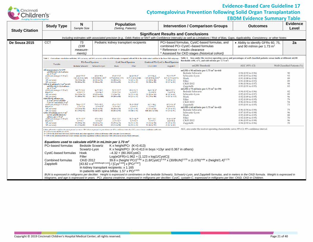
Copyright © 2019 Cincinnati Children's Hospital Medical Center; all rights reserved. Page 21 of 40
Evidence-Based Care Guideline 17 Cytomegalovirus Prevention following Solid Organ Transplantation
EBDM Evidence Summary Table
Study Citation Study Type N
Sample Size Population
(Setting, Patients) Intervention / Comparison Groups Outcomes
Evidence Level
Significant Results and Conclusions Including estimates with associated precision (e.g., Odds Ratios or NNT with Confidence Intervals) as well as Limitations / Risk of Bias, Gaps, Applicability, Consistency, or other Notes
De Souza 2015 CCT 73 (199
measure-ments)
Pediatric kidney transplant recipients PCr-based formulas, CystC-based formulas, and combined PCr-CystC–based formulas * Reference = insulin clearance * Assessed for CKD stages (historical cohort)
• Ability to identify GFRs 60, 75, and 90 ml/min per 1.73 m2
2a
Equations used to calculate eGFR in mL/min per 1.73 m2 PCr-based formulas Bedside Scwartz K x height/PCr (K=0.413) Scwartz-Lyon K x height/PCr (K=0.413 in boys >13yr and 0.367 in others) CystC-based formulas Hoek –4.32 + (80.35/CystC) Filler Log(eGFR)=1.962 + [1.123 x log(1/CystC)] Combined formulas CKiD 2012 39.8 x (height/ PCr)0.456 x (1.8/CystC)0.418 x (30/BUN)0.079 x (1.076)male x (height/1.4)0.179 Zappitelli [43.82 x e0.0033height (cm)] / [Cys0.635] x [PCr0.547] In kidney transplant recipients: x 1.165 In patients with spina bifida: 1.57 x PCr0.925 BUN is expressed in milligrams per deciliter. Height is expressed in centimeters in the bedside Schwartz, Schwartz-Lyon, and Zappitelli formulas, and in meters in the CKiD formula. Weight is expressed in
kilograms, and age is expressed in years. PCr, plasma creatinine, expressed in milligrams per deciliter; CystC, cystatin C, expressed in milligrams per liter; CKiD, CKD in Children.

Copyright © 2019 Cincinnati Children's Hospital Medical Center; all rights reserved. Page 22 of 40
Evidence-Based Care Guideline 17 Cytomegalovirus Prevention following Solid Organ Transplantation
EBDM Evidence Summary Table
Study Citation Study Type N
Sample Size Population
(Setting, Patients) Intervention / Comparison Groups Outcomes
Evidence Level
Significant Results and Conclusions Including estimates with associated precision (e.g., Odds Ratios or NNT with Confidence Intervals) as well as Limitations / Risk of Bias, Gaps, Applicability, Consistency, or other Notes
Martin-Pena 2009 CCT 36 patients (of 208 at center)
Pediatric renal transplantation patients with one or more infections (58.3%) followed for 2 years after transplantation until graft loss or until the end of the study
Triple drug immunosuppression (steroids, tacrolimus, mycophenolate mophetil) and induction therapy (daclizumab) as well as a single dose of cefotaxime as surgical site infection (SSI) prophylaxis. During the first three months, patients were given: * TMP–SMX & nystatin daily to prophylax against Pneumocystis jiroveci and fungal infections
* hyperimmune CMV immunoglobulins * Acyclovir for high-risk CMV seronegative patients who received a CMV seropositive organ
CMV monitoring based on weekly pp65 antigenemia assay from 2nd week to 3rd month post-transplantation
• Independent predictors of infection
2a
• Incidence: 1.5 episodes per patient in the first year of transplantation (19.4%)
• Incidence of active CMV infection in CMV seronegative recipient/seropositive donor = 31.2%
• Incidence in other patients = 10%
• Infections: 33 bacterial (73.3%), 11 viral (24.4%), and 1 protozoal
• UTI and CMV infections were the most common syndromes – (CMV 15.5%, n=7 and UTI 48.3%, n=28)
• Active CMV infection = 9 episodes in 7 patients (3 episodes of asymptomatic viremia (33.3%), 2 episodes of viral syndrome (22.2%), 4 episodes of invasive disease
• All CMV infections occurred in the first six months after transplantation.
• During the first four months after transplantation, 60.3% of infections occurred, with viral infections diagnosed mostly between second and sixth months after transplantation
Palmer 2010 RCT 136 Adult lung transplant recipients from 11 US lung transplant centers who completed 3 months of open-label valganciclovir prophylaxis
Patients randomly assigned to 9 additional months of oral valganciclovir (extended-course: n=70 – 46 completed study) or placebo (short-course: n=66 – 45 completed) The study ended 1 month after completion of randomized study medication (or 13 months after transplant).
• Freedom from CMV disease (syndrome or tissue-invasive) on an intention-to-treat basis 300 days after randomization
• CMV disease severity
• CMV infection
• Acute rejection
• Opportunistic infections
• Ganciclovir resistance
• Safety
2a
• Significantly less CMV disease, CMV infections, and disease severity were found in the extended-course group compared to the short-course group:
• CMV disease: 32% of short-course group (95% CI 20.1%-44.1%) versus 4% of extended-course group (95% CI 0.0%-8.5%; P<0.001), which remained after adjustment for CMV mismatch status (positive or negative CMV serologic status of donor or recipient) – Hazard Ratio (HR)=0.09 (95% CI 0.021-0.39; P<0.001) and HR=0.11 (0.047-0.27; P<0.001), respectively.
• CMV infection: 64% short-course versus 10% extended-course (P<0.001)
• Disease severity: 110,000 copies/mL short-course versus 3200 copies/mL extended-course, P<0.009
• Each component of the primary composite also showed a significant reduction with extended prophylaxis for short-course versus extended-course therapy:
• CMV syndrome (19% short vs. 4% extended; P<0.004)
• Invasive CMV disease (21% short vs. 2% extended; P<0.001)
• No significant differences between groups were reported in the incidence of non-CMV opportunistic infections or acute rejection after randomization.
• During the 6 months after study completion, a low incidence of CMV disease was observed in both groups.
• Extended-course patients (versus short-course patients) had significantly reduced platelet counts (<180x109 cells/L = 217 (165–251) vs 275 (200–331)) and lower median platelet counts at study conclusion.
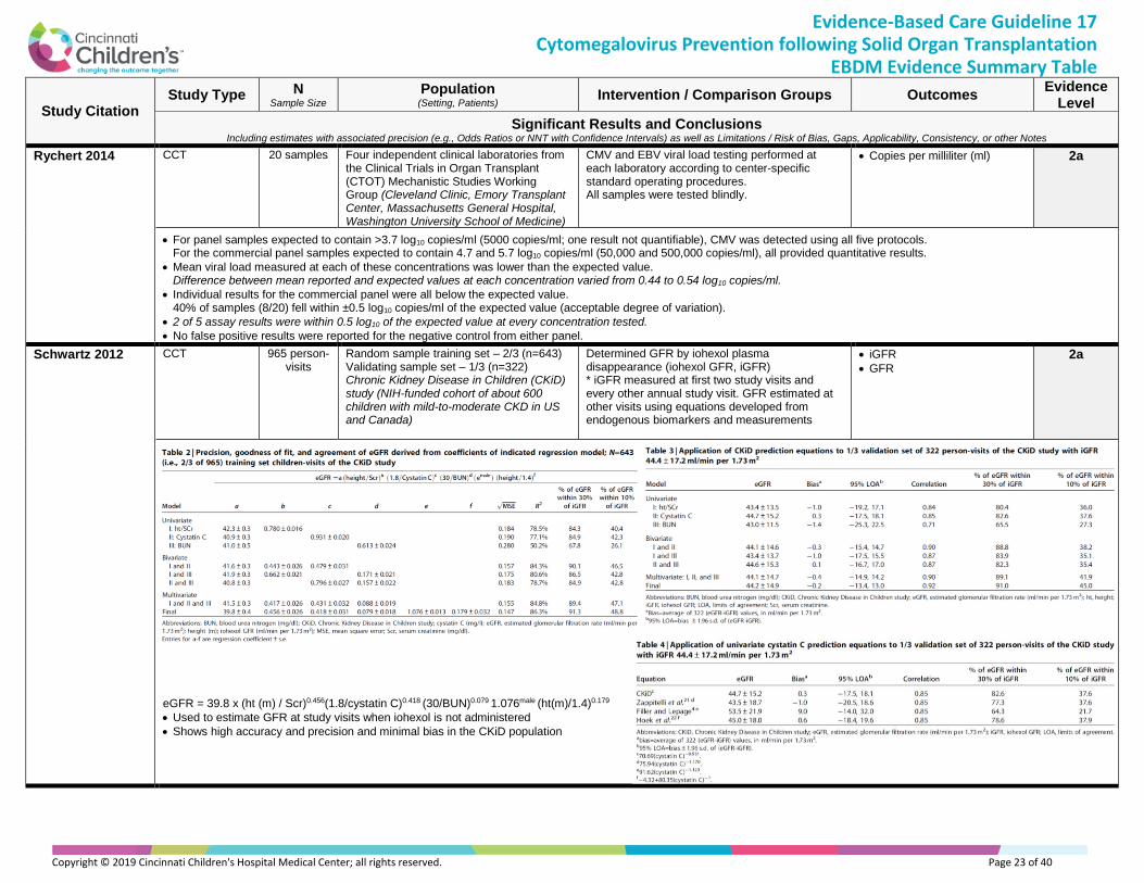
Copyright © 2019 Cincinnati Children's Hospital Medical Center; all rights reserved. Page 23 of 40
Evidence-Based Care Guideline 17 Cytomegalovirus Prevention following Solid Organ Transplantation
EBDM Evidence Summary Table
Study Citation Study Type N
Sample Size Population
(Setting, Patients) Intervention / Comparison Groups Outcomes
Evidence Level
Significant Results and Conclusions Including estimates with associated precision (e.g., Odds Ratios or NNT with Confidence Intervals) as well as Limitations / Risk of Bias, Gaps, Applicability, Consistency, or other Notes
Rychert 2014 CCT 20 samples Four independent clinical laboratories from the Clinical Trials in Organ Transplant (CTOT) Mechanistic Studies Working Group (Cleveland Clinic, Emory Transplant Center, Massachusetts General Hospital, Washington University School of Medicine)
CMV and EBV viral load testing performed at each laboratory according to center-specific standard operating procedures. All samples were tested blindly.
• Copies per milliliter (ml) 2a
• For panel samples expected to contain >3.7 log10 copies/ml (5000 copies/ml; one result not quantifiable), CMV was detected using all five protocols. For the commercial panel samples expected to contain 4.7 and 5.7 log10 copies/ml (50,000 and 500,000 copies/ml), all provided quantitative results.
• Mean viral load measured at each of these concentrations was lower than the expected value. Difference between mean reported and expected values at each concentration varied from 0.44 to 0.54 log10 copies/ml.
• Individual results for the commercial panel were all below the expected value. 40% of samples (8/20) fell within ±0.5 log10 copies/ml of the expected value (acceptable degree of variation).
• 2 of 5 assay results were within 0.5 log10 of the expected value at every concentration tested.
• No false positive results were reported for the negative control from either panel.
Schwartz 2012 CCT 965 person-visits
Random sample training set – 2/3 (n=643) Validating sample set – 1/3 (n=322) Chronic Kidney Disease in Children (CKiD) study (NIH-funded cohort of about 600 children with mild-to-moderate CKD in US and Canada)
Determined GFR by iohexol plasma disappearance (iohexol GFR, iGFR) * iGFR measured at first two study visits and every other annual study visit. GFR estimated at other visits using equations developed from endogenous biomarkers and measurements
• iGFR
• GFR
2a
eGFR = 39.8 x (ht (m) / Scr)0.456(1.8/cystatin C)0.418 (30/BUN)0.079 1.076male (ht(m)/1.4)0.179
• Used to estimate GFR at study visits when iohexol is not administered
• Shows high accuracy and precision and minimal bias in the CKiD population
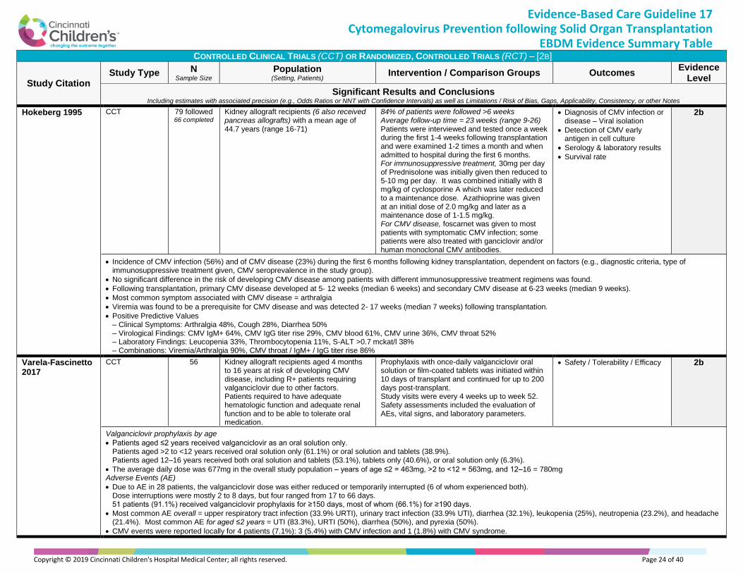
Copyright © 2019 Cincinnati Children's Hospital Medical Center; all rights reserved. Page 24 of 40
Evidence-Based Care Guideline 17 Cytomegalovirus Prevention following Solid Organ Transplantation
EBDM Evidence Summary Table
CONTROLLED CLINICAL TRIALS (CCT) OR RANDOMIZED, CONTROLLED TRIALS (RCT) – [2B]
Study Citation Study Type N
Sample Size Population
(Setting, Patients) Intervention / Comparison Groups Outcomes
Evidence Level
Significant Results and Conclusions Including estimates with associated precision (e.g., Odds Ratios or NNT with Confidence Intervals) as well as Limitations / Risk of Bias, Gaps, Applicability, Consistency, or other Notes
Hokeberg 1995 CCT 79 followed 66 completed
Kidney allograft recipients (6 also received pancreas allografts) with a mean age of 44.7 years (range 16-71)
84% of patients were followed >6 weeks Average follow-up time = 23 weeks (range 9-26) Patients were interviewed and tested once a week during the first 1-4 weeks following transplantation and were examined 1-2 times a month and when admitted to hospital during the first 6 months. For immunosuppressive treatment, 30mg per day of Prednisolone was initially given then reduced to 5-10 mg per day. It was combined initially with 8 mg/kg of cyclosporine A which was later reduced to a maintenance dose. Azathioprine was given at an initial dose of 2.0 mg/kg and later as a maintenance dose of 1-1.5 mg/kg. For CMV disease, foscarnet was given to most patients with symptomatic CMV infection; some patients were also treated with ganciclovir and/or human monoclonal CMV antibodies.
• Diagnosis of CMV infection or disease – Viral isolation
• Detection of CMV early antigen in cell culture
• Serology & laboratory results
• Survival rate
2b
• Incidence of CMV infection (56%) and of CMV disease (23%) during the first 6 months following kidney transplantation, dependent on factors (e.g., diagnostic criteria, type of immunosuppressive treatment given, CMV seroprevalence in the study group).
• No significant difference in the risk of developing CMV disease among patients with different immunosuppressive treatment regimens was found.
• Following transplantation, primary CMV disease developed at 5- 12 weeks (median 6 weeks) and secondary CMV disease at 6-23 weeks (median 9 weeks).
• Most common symptom associated with CMV disease = arthralgia
• Viremia was found to be a prerequisite for CMV disease and was detected 2- 17 weeks (median 7 weeks) following transplantation.
• Positive Predictive Values – Clinical Symptoms: Arthralgia 48%, Cough 28%, Diarrhea 50% – Virological Findings: CMV IgM+ 64%, CMV IgG titer rise 29%, CMV blood 61%, CMV urine 36%, CMV throat 52% – Laboratory Findings: Leucopenia 33%, Thrombocytopenia 11%, S-ALT >0.7 mckat/l 38% – Combinations: Viremia/Arthralgia 90%, CMV throat / IgM+ / IgG titer rise 86%
Varela-Fascinetto 2017
CCT 56 Kidney allograft recipients aged 4 months to 16 years at risk of developing CMV disease, including R+ patients requiring valganciclovir due to other factors. Patients required to have adequate hematologic function and adequate renal function and to be able to tolerate oral medication.
Prophylaxis with once-daily valganciclovir oral solution or film-coated tablets was initiated within 10 days of transplant and continued for up to 200 days post-transplant. Study visits were every 4 weeks up to week 52. Safety assessments included the evaluation of AEs, vital signs, and laboratory parameters.
• Safety / Tolerability / Efficacy 2b
Valganciclovir prophylaxis by age
• Patients aged ≤2 years received valganciclovir as an oral solution only. Patients aged >2 to <12 years received oral solution only (61.1%) or oral solution and tablets (38.9%). Patients aged 12–16 years received both oral solution and tablets (53.1%), tablets only (40.6%), or oral solution only (6.3%).
• The average daily dose was 677mg in the overall study population – years of age ≤2 = 463mg, >2 to <12 = 563mg, and 12–16 = 780mg Adverse Events (AE)
• Due to AE in 28 patients, the valganciclovir dose was either reduced or temporarily interrupted (6 of whom experienced both). Dose interruptions were mostly 2 to 8 days, but four ranged from 17 to 66 days. 51 patients (91.1%) received valganciclovir prophylaxis for ≥150 days, most of whom (66.1%) for ≥190 days.
• Most common AE overall = upper respiratory tract infection (33.9% URTI), urinary tract infection (33.9% UTI), diarrhea (32.1%), leukopenia (25%), neutropenia (23.2%), and headache (21.4%). Most common AE for aged ≤2 years = UTI (83.3%), URTI (50%), diarrhea (50%), and pyrexia (50%).
• CMV events were reported locally for 4 patients (7.1%): 3 (5.4%) with CMV infection and 1 (1.8%) with CMV syndrome.
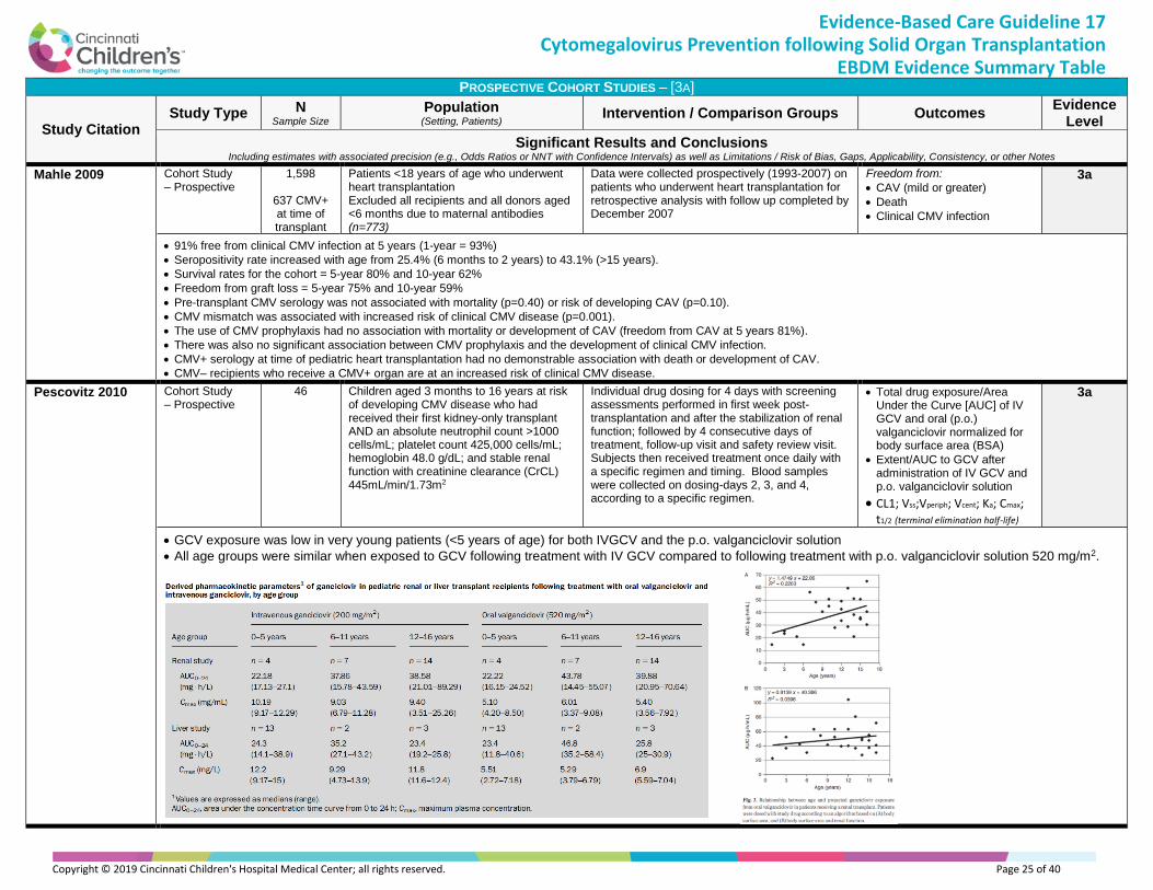
Copyright © 2019 Cincinnati Children's Hospital Medical Center; all rights reserved. Page 25 of 40
Evidence-Based Care Guideline 17 Cytomegalovirus Prevention following Solid Organ Transplantation
EBDM Evidence Summary Table
PROSPECTIVE COHORT STUDIES – [3A]
Study Citation Study Type N
Sample Size Population
(Setting, Patients) Intervention / Comparison Groups Outcomes
Evidence Level
Significant Results and Conclusions Including estimates with associated precision (e.g., Odds Ratios or NNT with Confidence Intervals) as well as Limitations / Risk of Bias, Gaps, Applicability, Consistency, or other Notes
Mahle 2009 Cohort Study – Prospective
1,598
637 CMV+ at time of transplant
Patients <18 years of age who underwent heart transplantation Excluded all recipients and all donors aged <6 months due to maternal antibodies (n=773)
Data were collected prospectively (1993-2007) on patients who underwent heart transplantation for retrospective analysis with follow up completed by December 2007
Freedom from:
• CAV (mild or greater)
• Death
• Clinical CMV infection
3a
• 91% free from clinical CMV infection at 5 years (1-year = 93%)
• Seropositivity rate increased with age from 25.4% (6 months to 2 years) to 43.1% (>15 years).
• Survival rates for the cohort = 5-year 80% and 10-year 62%
• Freedom from graft loss = 5-year 75% and 10-year 59%
• Pre-transplant CMV serology was not associated with mortality (p=0.40) or risk of developing CAV (p=0.10).
• CMV mismatch was associated with increased risk of clinical CMV disease (p=0.001).
• The use of CMV prophylaxis had no association with mortality or development of CAV (freedom from CAV at 5 years 81%).
• There was also no significant association between CMV prophylaxis and the development of clinical CMV infection.
• CMV+ serology at time of pediatric heart transplantation had no demonstrable association with death or development of CAV.
• CMV– recipients who receive a CMV+ organ are at an increased risk of clinical CMV disease.
Pescovitz 2010 Cohort Study – Prospective
46 Children aged 3 months to 16 years at risk of developing CMV disease who had received their first kidney-only transplant AND an absolute neutrophil count >1000 cells/mL; platelet count 425,000 cells/mL; hemoglobin 48.0 g/dL; and stable renal function with creatinine clearance (CrCL) 445mL/min/1.73m2
Individual drug dosing for 4 days with screening assessments performed in first week post-transplantation and after the stabilization of renal function; followed by 4 consecutive days of treatment, follow-up visit and safety review visit. Subjects then received treatment once daily with a specific regimen and timing. Blood samples were collected on dosing-days 2, 3, and 4, according to a specific regimen.
• Total drug exposure/Area Under the Curve [AUC] of IV GCV and oral (p.o.) valganciclovir normalized for body surface area (BSA)
• Extent/AUC to GCV after administration of IV GCV and p.o. valganciclovir solution
• CL1; Vss;Vperiph; Vcent; Ka; Cmax; t1/2 (terminal elimination half-life)
3a
• GCV exposure was low in very young patients (<5 years of age) for both IVGCV and the p.o. valganciclovir solution
• All age groups were similar when exposed to GCV following treatment with IV GCV compared to following treatment with p.o. valganciclovir solution 520 mg/m2.

Copyright © 2019 Cincinnati Children's Hospital Medical Center; all rights reserved. Page 26 of 40
Evidence-Based Care Guideline 17 Cytomegalovirus Prevention following Solid Organ Transplantation
EBDM Evidence Summary Table
Study Citation Study Type N
Sample Size Population
(Setting, Patients) Intervention / Comparison Groups Outcomes
Evidence Level
Significant Results and Conclusions Including estimates with associated precision (e.g., Odds Ratios or NNT with Confidence Intervals) as well as Limitations / Risk of Bias, Gaps, Applicability, Consistency, or other Notes
Vaudry 2009 Controlled Clinical Trial (phase II/III,
multicenter, open-
label, single dose
level, non-
comparative
study
investigating
safety, tolerability
&
pharmacokinetics
of valganciclovir
oral solution and
tablets given as
prophylaxis to
pediatric de novo
SOT recipients)
63 patients 7 countries
All SOT recipients aged between 3 months and 16 years who were at risk of developing CMV disease (i.e. donor and/or recipient were seropositive for CMV) * Patients were to have adequate hematological function (absolute neutrophil count >1300 cells/lL; platelet count >40 000 cells/lL; hemoglobin levels >8.0 g/dL) and adequate renal function (an estimated CrCL calculated by the Schwartz formula, CrCLS > 35 mL/min/1.73 m2) Exclusion criteria: previous allergic or other adverse reactions to aciclovir, valaciclovir or ganciclovir; severe, uncontrolled diarrhea; liver enzyme elevations >5 times the upper limit of normal (except for heart or liver transplant recipients); pregnancy or lactation.
Patients received once-daily prophylaxis with oral valganciclovir from 1–2 days post-transplantation up to day 100 post-transplantation. Valganciclovir powder for oral solution was provided as 12 g of powder (containing 5 g valganciclovir) for reconstitution with 91 mL of purified water to a final volume of 100 mL (valganciclovir concentration, 50 mg/mL). Patients were followed for safety and assessment of CMV disease until 26 weeks posttransplant. Safety assessments took place at each visit (days 1 and 7, weeks 2, 6 and 10, day 100, and weeks 16, 20 and 26 posttransplant) and included monitoring of adverse events (AEs), including opportunistic infections, laboratory safety tests (hematology, urinalysis and blood chemistry) and assessment of vital signs.
• Diagnosis of CMV disease
• Treatment failure
• Biopsy-proven acute rejection
• Graft survival
3a
• Seven patients had CMV viremia or antigenemia during the study, of which most cases (n = 5) occurred after the cessation of valganciclovir and were asymptomatic (n = 6).
• Incidence of biopsy-proven rejection higher in ≤2 years age group (29.4% vs. 9.5% in the >2 to<12 years group and 8.0% in the 12–16 years group).

Copyright © 2019 Cincinnati Children's Hospital Medical Center; all rights reserved. Page 27 of 40
Evidence-Based Care Guideline 17 Cytomegalovirus Prevention following Solid Organ Transplantation
EBDM Evidence Summary Table
PROSPECTIVE COHORT STUDIES – [3B]
Study Citation Study Type N
Sample Size Population
(Setting, Patients) Intervention / Comparison Groups Outcomes
Evidence Level
Significant Results and Conclusions Including estimates with associated precision (e.g., Odds Ratios or NNT with Confidence Intervals) as well as Limitations / Risk of Bias, Gaps, Applicability, Consistency, or other Notes
Ginevri 1998 Cohort Study – Prospective
79 recipients
30/79 CMV
patients
Renal transplant pediatric recipients were followed up for at least 12 months – 45 males/34 females; mean age at time of kidney transplantation of 14.1 & 4.9 years (range 2.5-20).
Patients were subdivided into four groups according to the CMV infection antibody status of donors/recipients. High-risk recipient group – CMV R– & CMV D+ = 33 patients
• CMV infection & syndrome by serostatus
3b
• Median time to onset of CMV infection = 48+4.1 days (range 14-105)
• No statistically significant difference between seronegative (45.4+5.8) and seropositive (58.2+8.2 days) recipients.
• 6 patients required 2-3 separate courses of ganciclovir therapy for relapsing CMV infections.
• Median time to recurrence of CMV infection = 14.1+4.3 days (range 4-33)
• None of these patients manifested CMV syndrome or disease when CMV infection relapsed or they experienced the emergence of CMV strains resistant to ganciclovir.
Krampe 2010 Cohort Study – Prospective
28 patients 71
transplants
Consecutive children at risk for CMV infection in the first six months following liver transplant (donor CMV+, recipient CMV-)
Prophylactic IVIG with prospective monitoring to perform preemptive ganciclovir therapy * clinical, laboratory, and microbiological course
• Immunosuppression
• Acute graft rejection
• CMV status
3b
• Patient survival 100% at six-month followup
• Patient graft survival 92.9%
• Incidence of acute graft rejection 28.6%
Launay 2012 Cohort Study – Prospective
20 PK profiles
10 children
Median age = 5.2 years Range = 8 months to 13.1 years
Transplant recipients (solid organ and HSC transplants) Aged 6 months to 18 years
Preemptive therapy with IV GCV given to prevent CMV disease and transplant patients tested weekly for CMV viremia * CMV viral load assessed * IV GCV started when CMV viral load + (viral load >300 copies/mL) and continued until viral load undetectable * When positive, IV GCV (10 mg/kg/d divided into 2 doses) started and continued until viral loads undetectable then IV GCV switched to VGCV * VGCV administered until weekly test for viremia measured a second undetectable viral load
• Viral load measured / Pharmacokinetic Profiles
3b
Medians for IV GCV and for VGCV
• Dosage = 9.8 mg/kg/d 19.1 mg/kg/d (theoretical dosage = 36.1 mg/kg/d
• Delay between treatment start and PK study = 13 days 6 days
• Cmin = 0.33 mcg/mL 0.27 mcg/mL
• AUC0–24 = 22.9 mcg*h/mL 34.6 mcg*h/mL AUC0–24 normalized 20 mg/kg/d = 37.6 mcg*h/mL
Authors concluded that, although not statistically significant, the “GCV PK profile obtained after oral VGCV and normalized for a dose of 20 mg/kg/d divided into 2 doses seems equal to or greater than that observed after an effective IV GCV 10 mg/kg/d dosage divided into 2 doses… The use of oral VGCV at a dose normalized to 20 mg/kg/d provides an exposure to GCV at least equal to or greater than the exposure on 10 mg/kg/d of IV GCV in most cases.”

Copyright © 2019 Cincinnati Children's Hospital Medical Center; all rights reserved. Page 28 of 40
Evidence-Based Care Guideline 17 Cytomegalovirus Prevention following Solid Organ Transplantation
EBDM Evidence Summary Table
Study Citation Study Type N
Sample Size Population
(Setting, Patients) Intervention / Comparison Groups Outcomes
Evidence Level
Significant Results and Conclusions Including estimates with associated precision (e.g., Odds Ratios or NNT with Confidence Intervals) as well as Limitations / Risk of Bias, Gaps, Applicability, Consistency, or other Notes
Melgosa Hijosa 2004
Cohort Study – Prospective
42 Children in the first year after renal transplantation (28 boys, 14 girls) Mean age at transplant 11.17 ± 5.86 years (median 11.01 and range 1.78–20.6 years)
Children received IV ganciclovir prophylaxis for CMV in the immediate post-transplant period. Quantitative antigenemia (pp68) determinations and blood, urine and throat cultures were done on a scheduled basis to detect CMV.
• Signs of CMV infection
• Relapse 3b
• The primary renal disease was uropathy in 12 (28.6%), renal dysplasia in 10 (23.8%), glomerulopathy in 6 (14.3%), nephronophthisis in 5 (11.9%), reflux nephropathy in 5 (12.9%), cystinosis in 2 (4.8%) and other causes in the last 2 (4.8%).
• Average cold time was 13±8.29 hours
• Of those who developed an infection at time of transplantation, 59% of recipients were CMV IgG(+)(R+) and 41% were negative (R–); 54% received a kidney CMV seropositive (D+), 9% seronegative (D–) and serological situation was unknown in 36%. Of 5 patients with clinical symptoms, 2 were D(+)/R(+); 3 were D(+)/R(–).
• In 47.6% of patients, no sign of CMV was detected at any time during the first-year follow-up. CMV infection was detected in 22 (52.4%) at a mean of 44.31±27.38 days post-treatment, with earliest infection detected at 14 days (range: 14–142 days).
• No significant differences were reported between groups with infection and without for age at time of transplant, sex or time of cold ischemia, nor with the number of days with IV ganciclovir treatment, nor with mean GFR post-treatment for a year.
• Scheduled and patient-specific measurements of pp68 antigenemia and establishment of preemptive treatment may be as effective as continuous oral prophylaxis for prevention.
Potena 2006 Cohort Study – Prospective
66 patients Consecutive patients undergoing first heart transplantation
Aggressive CMV prophylaxis compared with standard prophylaxis, both based on pretransplant donor (D) and recipient (R) CMV serology: R–/D+ received aggressive prophylaxis and R+ received standard prophylaxis All patients completed one year of follow-up.
• CMV infection
• Acute rejection
• Cardiac allograft vascular (CAV) disease
3b
• Estimated incidence = 87+4% at month 12 Difference in Rejection Score
• Month 1 – when all the patients received CMV prophylaxis, there was no difference between two groups (aggressive vs standard)
• Months 2-6 – patients treated with the aggressive prophylaxis remained significantly lower (P=0.03)
• Months 7-9 – aggressively treated patients peaked, reaching those who received only one month of anti-CMV prophylaxis Aggressively treated patients (compared to standard prophylaxis) had
• a lower incidence of CMV infection (73+10% vs. 94+4%; P=0.038)
• an independent reduced relative risk (RR) for acute rejection graded>3A (RR [95% CI] = 0.55 [0.26–0.96]; P=0.03)
• a slower progression of CAV (coronary artery lumen volume change= -21+13% vs. -10+14%; P=0.05) and vessel shrinkage (vessel volume change= -15+11% vs. -3+18%; P=0.03)
Villeneuve 2013 Cohort Study – Prospective
23 patients
28 AUC results
All pediatric SOT recipients six months to three yrs. old who were treated with oral valganciclovir suspension and underwent any AUC measurement – Single-center observational study
Pharmacokinetic study comparing valganciclovir dosing regimens and the potential benefits of individualized dose adjustments in children following organ transplantation – Dose determined by individual provider preference, generally 14–16 mg/kg
• Patient AUC
• Valganciclovir doses
• Ganciclovir levels
3b
• Weight-based valganciclovir doses (14–16 mg/kg once-daily for prophylaxis or twice-daily for treatment) resulted in therapeutic ganciclovir levels in 50% of patient cases, sub-therapeutic levels in 38.5%, and supra-therapeutic levels in the remaining 11.5%.
• Current manufacturer-recommended pediatric dose resulted in therapeutic AUCs in 15.4%, subtherapeutic levels in 3.8%, and supra-therapeutic levels in 80.8%.
• Current manufacturer-recommended dosing based on BSA and CrCl was estimated to result in therapeutic AUCs in fewer patients than the simple weight-based formula used in our institution (4 vs. 13; p = 0.017).
• An AUC calculation using only 2h & 5h measurements was strongly correlated with the AUC using all four-time measurements (R2 = 0.846; p < 0.001).
• 7 of 28 valganciclovir AUCs measured were being treated for active CMV infection – 4 were receiving CMV prophylaxis prior to CMV viremia development Only 1 had a therapeutic AUC at the time
• There were no cases of CMV disease while patients were on valganciclovir, all episodes of viremia resolved with valganciclovir treatment, and there was no evidence of antiviral resistance.
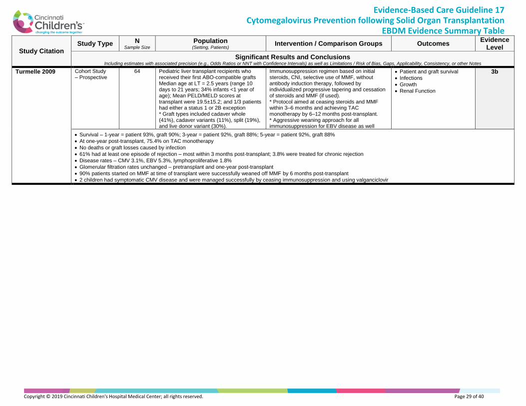
Copyright © 2019 Cincinnati Children's Hospital Medical Center; all rights reserved. Page 29 of 40
Evidence-Based Care Guideline 17 Cytomegalovirus Prevention following Solid Organ Transplantation
EBDM Evidence Summary Table
Study Citation Study Type N
Sample Size Population
(Setting, Patients) Intervention / Comparison Groups Outcomes
Evidence Level
Significant Results and Conclusions Including estimates with associated precision (e.g., Odds Ratios or NNT with Confidence Intervals) as well as Limitations / Risk of Bias, Gaps, Applicability, Consistency, or other Notes
Turmelle 2009 Cohort Study – Prospective
64 Pediatric liver transplant recipients who received their first ABO-compatible grafts Median age at LT = 2.5 years (range 10 days to 21 years; 34% infants <1 year of age); Mean PELD/MELD scores at transplant were 19.5±15.2; and 1/3 patients had either a status 1 or 2B exception * Graft types included cadaver whole (41%), cadaver variants (11%), split (19%), and live donor variant (30%).
Immunosuppression regimen based on initial steroids, CNI, selective use of MMF, without antibody induction therapy, followed by individualized progressive tapering and cessation of steroids and MMF (if used). * Protocol aimed at ceasing steroids and MMF within 3–6 months and achieving TAC monotherapy by 6–12 months post-transplant. * Aggressive weaning approach for all immunosuppression for EBV disease as well
• Patient and graft survival
• Infections
• Growth
• Renal Function
3b
• Survival – 1-year = patient 93%, graft 90%; 3-year = patient 92%, graft 88%; 5-year = patient 92%, graft 88%
• At one-year post-transplant, 75.4% on TAC monotherapy
• No deaths or graft losses caused by infection
• 61% had at least one episode of rejection – most within 3 months post-transplant; 3.8% were treated for chronic rejection
• Disease rates – CMV 3.1%, EBV 5.3%, lymphoproliferative 1.8%
• Glomerular filtration rates unchanged – pretransplant and one-year post-transplant
• 90% patients started on MMF at time of transplant were successfully weaned off MMF by 6 months post-transplant
• 2 children had symptomatic CMV disease and were managed successfully by ceasing immunosuppression and using valganciclovir

Copyright © 2019 Cincinnati Children's Hospital Medical Center; all rights reserved. Page 30 of 40
Evidence-Based Care Guideline 17 Cytomegalovirus Prevention following Solid Organ Transplantation
EBDM Evidence Summary Table
RETROSPECTIVE COHORT AND OTHER LOWER LEVEL STUDIES – [4A]
Study Citation Study Type N
Sample Size Population
(Setting, Patients) Intervention / Comparison Groups Outcomes
Evidence Level
Significant Results and Conclusions Including estimates with associated precision (e.g., Odds Ratios or NNT with Confidence Intervals) as well as Limitations / Risk of Bias, Gaps, Applicability, Consistency, or other Notes
Bedel 2012 Retrospective Cohort Study
56 Pediatric liver transplant recipients prescribed either oral ganciclovir (n=37) or valganciclovir (n=19)
Patients followed until 200 days post-transplant or death
Incidence of:
• Early onset and Late Onset CMV infection & CMV disease
• Patient specific factors for CMV acquisition
Rate of adverse drug effects Discontinuation
4a
• Incidence of CMV infection and CMV disease were not statistically different when comparing oral valganciclovir and ganciclovir.
• Early onset CMV disease – 0% valganciclovir and 5.4% ganciclovir (p=0.54)
• No statistically significant differences in secondary outcomes
• Trend for increased incidence of late onset CMV disease – valganciclovir (22.2%) vs. ganciclovir (8.1%; p=0.23)
• No reported differences in adverse events
Bock 1997 Case-Control Study
142 Registry records in the North American Pediatric Renal Transplant Cooperative Study (NAPRTCS) registry records – Patients hospitalized for viral infection within 12 months of renal transplantation (including CMV infections) and transplanted prior to age 20 years
Patient Data included age and calendar year transplanted, patient gender/race, kidney donor source, use/type of anti-T cell induction therapy, cyclosporine dose day 30 post-transplant, allograft data, and survival during the first 36 post-transplant months CMV-Related Data included diagnostic method(s) for CMV, time post-transplant of hospitalization for CMV infection, CMV immune status of donor and recipient at time of transplant, viral prophylactic agent(s) administered, existence of concomitant transplant rejection episode and affected specific organ systems Control Patients matched by transplant year, not hospitalized with CMV, and selected randomly from the total group of transplanted patients
• Risk factors for CMV disease
• Clinical manifestations of CMV disease
4a
Hospitalization for CMV Disease
• Mean time to hospitalization for CMV disease = 51 days (90% of patients within the first 5 months post-transplant)
• Most significant risk factor for hospitalization = CMV+ kidney donor Odds Ratio (OR) = 5.2 [95% Confidence Interval (CI) 2.8±10.3, P<0.0001] (irrespective of recipient age or CMV immune status)
• Risk reduction for CMV hospitalization – Antiviral agents (acyclovir, ganciclovir) or pooled IgG, prophylaxis with enriched anti-CMV IgG – OR = 0.31, P = 0.03 Other Risk
• Risk reduction of major organ involvement during CMV infection – The prophylactic use of antiviral agents – OR = 0.34, P<0.005
• Any form of prophylaxis was better than none for patients with CMV and 3-year graft survival (88% vs. 52%, P<0.001)
• CMV risk not significantly greater in CMV± recipients of CMV+ donor kidneys compared with CMV+ recipients of CMV+ donor kidneys – OR = 0.90 (0.42±1.9), P = 0.85
• No significantly increased risk for CMV associated with recipient’s CMV+ status, age, race, nor allograft type
• For those receiving CMV+ donor kidneys, viral prophylaxis with enriched anti-CMV IgG had a decreased risk of hospitalization for CMV disease versus no prophylaxis OR = 0.31, 95%CI 0.24±0.99, P = 0.03
• No effect for pooled IgG products – OR = 0.54, 95%CI 0.19±1.52, P = 0.3
• For recipients of donor CMV+ kidneys hospitalized with CMV, prophylaxis with acyclovir/ganciclovir was associated with a significantly decreased risk of major organ involvement OR = 0.34, 95%CI 0.18±0.85, P<0.005 but not for either form of prophylactic IgG product – OR = 0.69, P = NS
• Combined therapy with an IgG product and antiviral agent had a significantly decreased risk of major organ disease – OR = 0.25, 95%CI 0.09±0.78, P<0.02

Copyright © 2019 Cincinnati Children's Hospital Medical Center; all rights reserved. Page 31 of 40
Evidence-Based Care Guideline 17 Cytomegalovirus Prevention following Solid Organ Transplantation
EBDM Evidence Summary Table
Study Citation Study Type N
Sample Size Population
(Setting, Patients) Intervention / Comparison Groups Outcomes
Evidence Level
Significant Results and Conclusions Including estimates with associated precision (e.g., Odds Ratios or NNT with Confidence Intervals) as well as Limitations / Risk of Bias, Gaps, Applicability, Consistency, or other Notes
Camacho-Gonzalez 2011
Cohort Study – Retrospective
111 Pediatric renal transplant patients (60% males, 46% African Americans, median age at transplant 14.5 years (range 1.4–20.4 years))
Patients received 24 weeks valganciclovir prophylaxis - 15 mg/kg/day, max 900 mg/day
• Incidence of CMV disease
• Toxicity of valganciclovir 4a
• 69% donors and 44% f recipients were seropositive pretransplant
• Median duration of valganciclovir use = 5.9 months (range 0.5–24 months)
• CMV viremia 27% and CMV disease 4.5%
• All patients with disease presented after prophylaxis ended and all were D+/R−.
• Thymoglobulin use (P=0.04) and positive donor CMV status (P=0.02) were associated with a higher risk of CMV viremia.
• 24% had hematologic toxicity directly associated with valganciclovir.
Danziger-Isakov 2009
Cohort Study – Retrospective,Multi-center
577 Primary lung or heart-lung transplant recipients from 14 pediatric lung transplant centers in the United States, Canada, Austria, Germany and the United Kingdom from the International Pediatric Lung Transplant Collaborative (IPTLC)
* Pretransplant evaluation – “standard protocol” Induction immunosuppressive therapy – varied from no induction therapy to receipt of lympholytic agents or IL-2 receptor antagonists * After transplantation – triple-drug immunosuppression with a calcineurin inhibitor (CNI), prednisone, and either azathioprine or mycophenolate mofetil. * Immunosuppressive therapy – gradually reduced as time from transplant increased * CMV Prophylaxis and routine transbronchial biopsies to assess for rejection – not standard across, and changed over time within, centers
• Acute Rejection
• BOS – bronchiolitis obliterans
4a
• For patients D-/R-, CMV episodes were only associated with receipt of induction therapy (HR=5.2; 95% CI 1.5, 18.8).
• Development of a CMV episode was associated with donor CMV seropositivity regardless of: D+/R+ HR=2.1; 95% CI 1.3-3.5 D+/R– HR=1.9; 95% CI 1.2-3.0 Receipt of a living donor organ – HR=2.5; 95% CI 1.4-4.3 Transplant in the earliest transplant era in this study – HR=2.3; 95% CI 1.5-3.8 A2 rejection prior to CMV episode – HR=1.4; 95% CI 1.00-1.9
• Duration of prophylaxis was not associated with a decrease in CMV episodes for recipients with history of pre-transplant CMV exposure (R+). CMV D–/R– patients were less likely to have prophylaxis administered (P<0.001). CMV D/R status was not otherwise associated with prophylaxis duration.
• For CMV mismatched subjects D+/R–, discontinued prophylaxis at the time of CMV episode doubled the risk of CMV (HR=1.9, 95% CI 1.02-3.7).
• Each month of prophylaxis received was associated with a 30% increase in risk of CMV infection or disease (HR=1.3, 95% CI 1.00-1.6).
• Extending prophylaxis was not associated with CMV episodes.
• For CMV patients D+/R+, discontinued prophylaxis significantly increased the risk of CMV episodes (HR=3.5; 95% CI 1.4-8.4).

Copyright © 2019 Cincinnati Children's Hospital Medical Center; all rights reserved. Page 32 of 40
Evidence-Based Care Guideline 17 Cytomegalovirus Prevention following Solid Organ Transplantation
EBDM Evidence Summary Table
Study Citation Study Type N
Sample Size Population
(Setting, Patients) Intervention / Comparison Groups Outcomes
Evidence Level
Significant Results and Conclusions Including estimates with associated precision (e.g., Odds Ratios or NNT with Confidence Intervals) as well as Limitations / Risk of Bias, Gaps, Applicability, Consistency, or other Notes
Danziger-Isakov 2003
Case-Cohort Study – Retrospective
194 Pediatric primary lung-transplant recipients Patients at high risk for CMV infection received ganciclovir prophylaxis for 42 days post-transplantation. * Retrospective chart review was conducted on the medical records of all primary lung transplants – Data from time of transplant to 1 year or until either death or re-transplantation (if occurred prior to end of the 1-year observational period)
• Time to first episode of CMV viremia
• Risk factors / Adverse events
• Retransplantation or death within 1 year after transplantation
4a
CMV Viremia
• First episode of CMV viremia was associated with retransplantation or death between days 90 and 365 (RR=4.1, 95% CI 1.1–14.5) and was not associated with BOS (RR=1.3, 95% CI 0.5–3.3; BOS – bronchiolitis obliterans).
• 36.7% CMV viremia in patients who received organs from CMV-seropositive donors versus 9.9% in recipients of CMV-seronegative organs.
• Recipients of seropositive donor organs were more than five times as likely to develop CMV viremia than recipients of seronegative donor organs (OR 5.3, 95% CI 2.4–11.8, P<0.001)
• Among CMV-negative allograft recipients, no statistical difference was found in CMV viremia rate = CMV+ recipients 16.7% versus CMV- recipients 6.6% (P=0.129)
• Time from transplant to onset of viremia was significantly earlier when donor CMV status was seropositive versus seronegative (log rank test, P<0.001).
• Date quartile of transplantation did not affect incidence of CMV viremia (range 18.3%–27.1% positive for CMV viremia).
• Patients with CMV viremia were more likely to experience 2+ episodes of acute rejection (OR=4.0, 95% CI 1.6–10.1, P<0.002).
• CMV serostatus for all patients without and with viremia D–/R–: without 38.3% v. with 6.6% D–/R+: without 16.8% v. with 16.7% D+/R–: without 24.2% v. with 34.5% D+/R+: without 17.4% v. with 39.5% Unknown: 3.3%
• Death before 90 days post-transplantation was not positively associated with an episode of CMV viremia OR=0.11, 95%CI 0.01–0.82, P<0.01
• Retransplantation or death between 90 and 365 days was associated with a first episode of CMV viremia OR=4.1, 95%CI 1.1–14.5, P<0.02
• BOS was not associated with CMV viremia OR=1.3, 95%CI 0.5–3.3, P<0.65
Hocker 2016 Cohort Study – Retrospective
242 eligible 157 with
data for the first 3 years
after transplant
All kidney allograft recipients 21 years or younger at the time of transplantation on a calcineurin inhibitor (CNI)–based immunosuppressive regimen (Cooperative European Paediatric Renal Transplant Initiative (CERTAIN) Registry)
Patients with a complete and validated data set for at least 1 year after transplantation, whether or not they had received antiviral chemoprophylaxis with VGCV or GCV and in whom CMV replication was accurately determined by CMV nucleic acid testing (NAT) and/or the pp65 antigenemia assay at the above-mentioned points in time
• Anemia
• Thrombocytopenia
• Time to CMV Replication
4a
• No significant difference between rate of CMV syndromes in the chemoprophylaxis and preemptive therapy groups (6.1% v 2.8%, P = 0.355).
• Incidence of CMV-related tissue-invasive disease was similar (P = 0.646) in the both groups.
• In years 2-3 after transplantation, rate of CMV replication was low – 4.5%
• The frequency of CMV replication was related to the CMV serostatus of donors and/or recipients: D+/R− (34.9%) – 25.0% in the prophylaxis cohort vs 66.7% in the preemptive therapy cohort; P < 0.01 D+/R+ (12.1%) – 5.9% prophylaxis vs 18.8% preemptive therapy; P = 0.109 D−/ (R− or R+) – 5.9% prophylaxis vs 4.2% preemptive therapy cohort; P = 0.751 D−/R− (3.4%)
• Patients with a high (D+/R−) or intermediate CMVrisk (D+/R+), who had received VGCV or GCV prophylaxis, experienced a significantly lower eGFR loss at 3 years after transplantation than patients receiving preemptive therapy.
• D+/R− and D+/R+ patients receiving chemoprophylaxis (20.7%) had a lower CMV replication rate within the first year by transplantation than D+/R− and D+/R+ patients receiving preemptive therapy (38.3%) (P < 0.05).
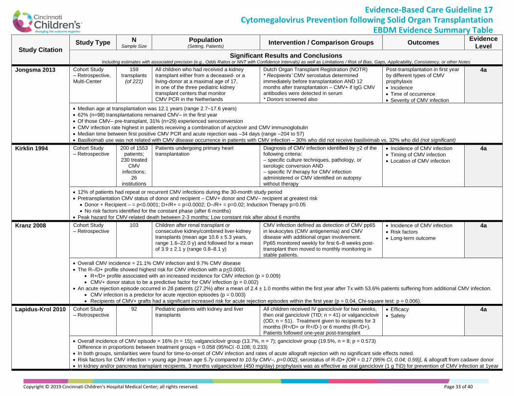
Copyright © 2019 Cincinnati Children's Hospital Medical Center; all rights reserved. Page 33 of 40
Evidence-Based Care Guideline 17 Cytomegalovirus Prevention following Solid Organ Transplantation
EBDM Evidence Summary Table
Study Citation Study Type N
Sample Size Population
(Setting, Patients) Intervention / Comparison Groups Outcomes
Evidence Level
Significant Results and Conclusions Including estimates with associated precision (e.g., Odds Ratios or NNT with Confidence Intervals) as well as Limitations / Risk of Bias, Gaps, Applicability, Consistency, or other Notes
Jongsma 2013 Cohort Study – Retrospective, Multi-Center
159 transplants
(of 221)
All children who had received a kidney transplant either from a deceased- or a living-donor at a maximal age of 17, in one of the three pediatric kidney transplant centers that monitor CMV PCR in the Netherlands
Dutch Organ Transplant Registration (NOTR) * Recipients’ CMV serostatus determined immediately before transplantation AND 12 months after transplantation – CMV+ if IgG CMV antibodies were detected in serum * Donors screened also
Post-transplantation in first year by different types of CMV prophylaxis
• Incidence
• Time of occurrence
• Severity of CMV infection
4a
• Median age at transplantation was 12.1 years (range 2.7–17.6 years)
• 62% (n=98) transplantations remained CMV– in the first year
• Of those CMV– pre-transplant, 31% (n=29) experienced seroconversion
• CMV infection rate highest in patients receiving a combination of acyclovir and CMV immunoglobulin
• Median time between first positive CMV PCR and acute rejection was –34 days (range –204 to 57)
• Basiliximab use was not related with CMV disease occurrence in patients with CMV infection – 30% who did not receive basiliximab vs. 32% who did (not significant)
Kirklin 1994 Cohort Study – Retrospective
200 of 1553 patients;
230 treated CMV
infections; 26
institutions
Patients undergoing primary heart transplantation
Diagnosis of CMV infection identified by >2 of the following criteria: – specific culture techniques, pathology, or serologic conversion AND – specific IV therapy for CMV infection administered or CMV identified on autopsy without therapy
• Incidence of CMV infection
• Timing of CMV infection
• Location of CMV infection
4a
• 12% of patients had repeat or recurrent CMV infections during the 30-month study period
• Pretransplantation CMV status of donor and recipient – CMV+ donor and CMV– recipient at greatest risk
• Donor + Recipient – = p<0.0001; D+/R+ = p=0.0002; D–/R+ = p=0.02; Induction Therapy p=0.05
• No risk factors identified for the constant phase (after 6 months)
• Peak hazard for CMV-related death between 2-3 months; Low constant risk after about 6 months
Kranz 2008 Cohort Study – Retrospective
103 Children after renal transplant or consecutive kidney/combined liver-kidney transplants (mean age 10.6 ± 5.3 years, range 1.6–22.0 y) and followed for a mean of 3.9 ± 2.1 y (range 0.8–8.1 y)
CMV infection defined as detection of CMV pp65 in leukocytes (CMV antigenemia) and CMV disease with additional organ involvement. Pp65 monitored weekly for first 6–8 weeks post-transplant then moved to monthly monitoring in stable patients.
• Incidence of CMV infection
• Risk factors
• Long-term outcome
4a
• Overall CMV incidence = 21.1% CMV infection and 9.7% CMV disease
• The R–/D+ profile showed highest risk for CMV infection with a p<0.0001.
• R+/D+ profile associated with an increased incidence for CMV infection (p = 0.009)
• CMV+ donor status to be a predictive factor for CMV infection (p = 0.002)
• An acute rejection episode occurred in 28 patients (27.2%) after a mean of 2.4 ± 1.0 months within the first year after Tx with 53.6% patients suffering from additional CMV infection.
• CMV infection is a predictor for acute rejection episodes (p = 0.003)
• Recipients of CMV+ grafts had a significant increased risk for acute rejection episodes within the first year (p = 0.04, Chi-square test: p = 0.006).
Lapidus-Krol 2010 Cohort Study – Retrospective
92 Pediatric patients with kidney and liver transplants
All children received IV ganciclovir for two weeks, then oral ganciclovir (TID; n = 41) or valganciclovir (OD; n = 51). Treatment given to recipients for 3 months (R+/D+ or R+/D-) or 6 months (R-/D+). Patients followed one-year post-transplant
• Efficacy
• Safety
4a
• Overall incidence of CMV episode = 16% (n = 15); valganciclovir group (13.7%, n = 7); ganciclovir group (19.5%, n = 8; p = 0.573) Difference in proportions between treatment groups = 0.058 (95%CI -0.108; 0.233)
• In both groups, similarities were found for time-to-onset of CMV infection and rates of acute allograft rejection with no significant side effects noted.
• Risk factors for CMV infection = young age [mean age 5.7y compared to 10.5y CMV–, p=0.002], serostatus of R-/D+ [OR = 0.17 (95% CI, 0.04; 0.59)], & allograft from cadaver donor
• In kidney and/or pancreas transplant recipients, 3 months valganciclovir (450 mg/day) prophylaxis was as effective as oral ganciclovir (1 g TID) for prevention of CMV infection at 1year

Copyright © 2019 Cincinnati Children's Hospital Medical Center; all rights reserved. Page 34 of 40
Evidence-Based Care Guideline 17 Cytomegalovirus Prevention following Solid Organ Transplantation
EBDM Evidence Summary Table
Study Citation Study Type N
Sample Size Population
(Setting, Patients) Intervention / Comparison Groups Outcomes
Evidence Level
Significant Results and Conclusions Including estimates with associated precision (e.g., Odds Ratios or NNT with Confidence Intervals) as well as Limitations / Risk of Bias, Gaps, Applicability, Consistency, or other Notes
Madan 2009 Retrospective Descriptive Study
122 Pediatric liver transplant recipients Followed for a median of 2.3 years post-transplantation
Patients received a minimum of 14 days of postoperative ganciclovir, followed by monthly CMV PCR monitoring – Both CMV donor and recipient serostatus had to be documented.
• CMV Risk Stratification
• CMV Disease
• Acute rejection
4a
• CMV PCR screening was performed biweekly for the first 3 postoperative months, monthly for the remainder of the first postoperative year, and every 2 to 3 months thereafter.
• 119 patients received postoperative IV ganciclovir, with a mean +SD duration of therapy of 12.9+5.6 days
• High risk for CMV = 43 CMV– recipients receiving CMV+ grafts & Routine risk = 79 subjects
• CMV was detected by PCR in the absence of symptoms in 34.4% of subjects – more likely in high risk (58.1%) than in routine risk (21.8%) recipients (P<0.0001)
• A total of 38.5% of subjects were spared antiviral medications beyond their initial postoperative prophylaxis.
• CMV disease developed in 12 patients [8 D+/R-, 2 D+/R+, 1 D-/R+, 1 D-/R-] = overall incidence 9.8%
• Overall acute rejection rate at any time = 41.8%; All episodes of acute rejection after CMV occurred at least 3 months after the diagnosis of CMV.
Muto 2010 Cohort Study – Retrospective
75 Patients who underwent a single HSCT (hematopoietic stem cell transplant) with at least one opportunity to monitor serum cystatin C levels during the same period
Chart review of allogeneic HSCT recipients with 1+ serum cystatin C level measured March 2006 to October 2008 – Renal dysfunction in the acute phase or acute on chronic phase was judged according to AKIN classification.
• Cystatin C –pre- and post- transplant serum levels
• CKD staging / deterioration
• AKI
4a
• Cystatin C levels were significantly higher post-transplantation than pre-transplantation.
• Advanced disease status may be less likely to interfere with serum cystatin C level. Variables associated with the risk of cystatin C elevation
• Calcineurin inhibitor use (OR = 7.26, 95%CI 1.096–48.053, P=0.04; multiple logistic regression analysis)
• Previous AKI event (Hazard Ratio 31.8, 95% CI 4.037–250.7, P<0.001)
• Sepsis (OR 0.77, P=0.048 univariate) Variables associated with the risk of worsening CKD
• Cystatin C level C0.90 mg/L before transplantation (OR = 4.88, P=0.041 univariate)
• Previous AKI event (OR = 61.3, P<0.001 univariate) Correlations
• Inverse – cystatin C and eGFR (r = -0.682, P<0.001)
• 1/cystatin C and eGFR (r = 0.815, P<0.001)
• Cystatin C was elevated in the creatine blind area (GFR 40–70 mL/min)
Ranganathan 2009
Cohort Study – Retrospective
599 totals * 329 with 3+ weeks IV ganciclovir * 62 (19%) CMVIG
Pediatric lung transplant recipients 14 sites in North America and Europe, <21 years of age, primary lung or heart-lung transplant, survived 2+ weeks after surgery, and data available from transplantation date to one-year post-transplant, death, or re-transplantation
Each institution had a diagnostic methodology to identify CMV episodes, but included demonstration of antigenemia, positive viral culture or positive polymerase chain reaction (PCR) – Added cytomegalovirus immunoglobulin (CMVIG) prophylaxis to at least three weeks of IV ganciclovir in pediatric lung transplant recipients
• Association of time to CMV and risk factors (CMVIG use)
• BOS/BO – Bronchiolitis obliterans syndrome or bronchiolitis obliterans
• PTLD – Post-transplant lymphoproliferative disease
4a
• CMVIG administered more frequently with CMV D+/R− (p<0.05) – CMV episodes (infection/disease) common after pediatric lung transplant = Incidence in the first year 36% – Dosing intervals=1 day–1 month; Most common=every 2 weeks; Median duration=84 days (range 1–192); Median dose administered = 150 mg/kg (mean 133 mg/kg)
• Duration of antiviral prophylaxis other than CMVIG was longer for patients administered CMVIG compared to those patients taking prophylactic ganciclovir alone (p = 0.025).
• Time to first episode of CMV disease with CMVIG occurred at a median of 122 days (mean 137 days) and without CMVIG a median of 96 days (mean 118 days) (p>0.05). CMV Disease – Risk Factors
• Donor CMV seropositivity – HR=3.8 (95% CI 2.0–7.5) [D+R+ HR=4.9 (1.4, 16.9) p=0.012; D+R− HR=6.0 (1.8, 19.8) p=0.003; D−R+ HR=2.0 (0.51, 8.0) p=0.32]
• Earlier era of transplant – HR=5.5 (1.9, 15.5) p=0.001 * Transplant type (Single Lung Transplant versus all others) – HR=5.3 (1.8, 15.3) p=0.002 CMV Infection
• Subjects who did not receive CMVIG as part of their prophylaxis were three times more likely to develop CMV infection (HR 3.4; 95% CI 1.2, 9.5) independent of CMV serostatus
• Donor CMV seropositivity – HR=3.8 (95% CI 2.0–7.5) [D+R+ HR=12.3 (2.9, 52.0) p<0.001; D+R− HR=9.2 (2.1, 39.2) p=0.003; D−R+ HR=8.6 (1.8, 39.8) p=0.006]
• Not receiving CMVIG – HR=3.4 (1.2–9.5) p=0.022 *Transplant in the earliest era (1992–1994) – HR=2.1 (1.1–3.8) p=0.022

Copyright © 2019 Cincinnati Children's Hospital Medical Center; all rights reserved. Page 35 of 40
Evidence-Based Care Guideline 17 Cytomegalovirus Prevention following Solid Organ Transplantation
EBDM Evidence Summary Table
Study Citation Study Type N
Sample Size Population
(Setting, Patients) Intervention / Comparison Groups Outcomes
Evidence Level
Significant Results and Conclusions Including estimates with associated precision (e.g., Odds Ratios or NNT with Confidence Intervals) as well as Limitations / Risk of Bias, Gaps, Applicability, Consistency, or other Notes
Robinson 2002 Cohort Study – Retrospective
72 patients 73 transplants
All renal transplant recipients from Egleston Children’s Hospital, Atlanta, Georgia, from January 27, 1993 to December 3, 1998
Patient medical records who had received renal transplants were identified using ICD-9 codes for this procedure and reviewed. Abstracted, standardized data including clinical and laboratory data for 1 year post-transplant
• Presumed or proven CMV disease
• CMV infection
4a
• Incidence of CMV Disease = 12.3% (9/73; 95% CI 5.8–22.1%)
• Median time to onset of CMV disease = 52 days post-transplant
• Median age with CMV disease = 13.6 years (range 5.6–18.4 years, SD 54.0) – not significantly different from those who did not develop CMV disease.
• CMV– Recipient (R-) serostatus associated (but not significantly) with CMV disease (univariate RR 4.91, p<0.139)
• CMV+ Donor (D+) serostatus strongly associated with the CMV disease development (univariate RR 8.52, p<0.01) and stronger when pediatric (<18y)
• CMV disease varied significantly by dose of CsA – Median CsA dose significantly lower for recipients who developed CMV disease (7.69 versus 12.85; p<0.003).
• Independently associated with a significantly increased risk of CMV disease: CsA dose <8.0 mg/kg, age, recipient serostatus and other CMV-associated variables, with a FET p-value <0.2, donor CMV+ serostatus, and transplant in October and November
• Anatomic site of CMV disease did not differ by month of transplant.
Saitoh 2011 Cohort Study – Retrospective
113 Children after live-donor liver transplant at the largest pediatric LT center in Japan (median age: 16 months)
Universal preemptive therapy for CMV infection – CMV-pp65 antigenemia monitored weekly for all patients & Ganciclovir therapy initiated when CMV-pp65 antigenemia was positive Monitored for at least six months
• Event-free survival 6 months after LT
• Incidence of HCMV infection
• Incidence of HCMV disease
• Death from any cause
4a
• Overall success rate of LT = 91.7%
• CMV-pp65 antigenemia became positive in 37 (33%) recipients – D+/R–: 62%, D+/R+: 36%, D–/R+: 11%, D–/R–: 8% Among the D+ recipients: 38% /R+ (11 of 29) & 64% /R– (28 of 44) avoided the use of ganciclovir.
• Median time to become positive for CMV-pp65 antigenemia = 33 days postoperatively (interquartile range [IQR]=17.5 days, range=8-115 days).
• CMV-pp65 antigenemia positivity observed in 63% in the D+/R–patients and 38% D+/R+ patients CMV serostatus (donors & recipients) significantly affected the proportion of recipients who remained negative for CMV-pp65 antigenemia for 6 months after LT
• Human CMV disease was documented in six (5%) recipients, and they were successfully treated with ganciclovir without any sequelae.
Simmonds 2008 Case Control Study
50 Pediatric heart transplant recipients 8 to 17 years of age (27 male) Returning for their annual review and free of angiographic evidence of cardiac allograft vasculopathy
Patients were separated into 2 groups according to CMV status: those without evidence of CMV replication after transplantation (n=38; 19 male) and patients with evidence of viremia after transplantation (n=12; 8 male)
• Brachial artery flow-mediated dilation (FMD)
4a
• Brachial artery flow-mediated dilation (FMD) was significantly reduced in patients with evidence of CMV replication after transplantation (Mean 6.64+ SE 1.12%) compared with those without evidence of replication (9.48+0.56%; P<0.02) and remained significant when adjusted for age, time since transplantation, and medication.
• The difference in FMD was not due to differences in smooth muscle function, baseline arterial diameter (2.96+0.079 versus 3.23+0.12 mm; P=0.094) and flow (reactive hyperemia, 489+41% versus 437+128%; P=0.618).
• Donor CMV status, recipient pretransplantation status, and traditional CMV risk stratification were not predictive of FMD.
• Maximum CMV PCR detected or duration of PCR positivity were not correlated to FMD.
Snydman 2010 Cohort Study – Retrospective
3697 All pediatric recipients of primary, single-organ heart transplants <18 years of age
Pediatric heart recipients who received (1) CMV prophylaxis with CMVIG (with or without antivirals) [n=455] (2) antivirals without CMVIG [n=1358], and (3) no prophylaxis [n=1884]
• Recipient death and graft loss at 7 years post-transplantation
• Associations between CMV prophylaxis/CMVIG & acute rejection or clinical outcomes
4a
• CMVIG (with or without antivirals) and antivirals without CMVIG were both associated with significantly (P0.05) lower rates of graft loss and death versus no prophylaxis.
• After adjustment, CMVIG was associated with a significantly decreased adjusted risk for graft loss and a borderline (P0.09) decreased adjusted mortality risk; antiviral prophylaxis was associated with decreased adjusted risk for graft loss and mortality.
• In the CMV-positive donor/CMV-negative recipient cohort, CMVIG (with or without antivirals) was associated with decreased adjusted risk for graft loss and death.

Copyright © 2019 Cincinnati Children's Hospital Medical Center; all rights reserved. Page 36 of 40
Evidence-Based Care Guideline 17 Cytomegalovirus Prevention following Solid Organ Transplantation
EBDM Evidence Summary Table
RETROSPECTIVE COHORT AND OTHER LOWER LEVEL STUDIES – [4B]
Study Citation Study Type N
Sample Size Population
(Setting, Patients) Intervention / Comparison Groups Outcomes
Evidence Level
Significant Results and Conclusions Including estimates with associated precision (e.g., Odds Ratios or NNT with Confidence Intervals) as well as Limitations / Risk of Bias, Gaps, Applicability, Consistency, or other Notes
Best 1995 Cohort Study – Retrospective
95 Consecutive heart transplant recipients (N=111) studied for 4 months with 95 surviving 1 month
Patients maintained on triple therapy – CsA/Sandimmune, aziothioprine, oral prednisone Rejection episodes treated with short course high dose IV methylprednisone then daily oral steroid dose increased and gradually reduced back to maintenance dose
• CMV infection
• CMV disease
• Risk factors
4b
• 56% experienced a CMV infection from 5 weeks to 4 months after transplantation
• 70% of those were CMV antibody+ before transplantation
• Donor and recipient CMV antibody status are significantly different in CMV infection rate Risk factors for CMV infection in months 1 & 2
• CsAbc in previous week per 100 mcg L-1 increase = RR 1.25, 95%CI 1.02-1.53
• CsAbc >550 mcg L-1 in previous week = RR 4.43, 95%CI 1.21-16.16
• Rejection treatment in previous 14 days = RR 9.04, 95%CI 2.57-31.64 Risk factors with constant effect for months 1-4
• CMV Recipient+ = RR 1.09, 95%CI 0.55-2.16
• CMV Donor+ = RR 2.46, 95%CI 1.25-4.89
• Primary diagnosis Cardiomyopathy = RR 0.37, 95%CI 0.19-0.75
Bueno 1997 Cohort Study – Retrospective
41 children (16 CMV
disease in 10 children)
Children who received either isolated small bowel (SB), liver-small bowel (L-SB), or multivisceral transplants
All children received a combination of tacrolimus and steroids as well as prostaglandin E1 until IV tacrolimus (in all but 8)
• Incidence and outcome of CMV
• Survival – Patient, Graft, & CMV-disease-free
4b
• Resolution of CMV disease = 93.3% of episodes (no deaths)
• CMV in D+/R− children = more extensive and persistent disease
• Survival rates (patient and graft) similar in D/R subgroups and between children with and without CMV disease
• History of rejection not a risk factor (RR=1.19; 95% CI 0.30–4.73) Increased Incidence of CMV Disease
• Cumulative dose of steroid boluses (RR=1.59; 95% CI 1.14–2.21)
• History of steroid recycles (RR=2.72; 95% CI, 1.21–6.13)
Florescu 2012 Cohort Study – Retrospective
98 Pediatric patients (<19 years of age) who underwent isolated SBT or LSBT
Induction therapy consisted of basiliximab; Antithymocyte globulin used in patients with renal failure, prior transplantation or evidence of sensitization; Maintenance immunosuppressive regimen consisted of steroids (for the first year after transplantation) and tacrolimus
• Incidence of CMV disease
• Timing of CMV disease
• Impact on patient outcome
4b
• Median follow-up time for the cohort = 1654 days (IQR: 1.14–2.14 days)
• CMV infection = 18 patients; CMV disease = 7 patients; CMV viremia = 11 patients Risk factors for CMV disease
• CMV D+/R- mismatch (OR 2.5; 95% CI 0.52–12.07; p = 0.25)
• VGC prophylaxis (OR 2.0; 95% CI 0.38–10.63; p = 0.41)
• Median total HLA-DR mismatches (OR 0.34; 95% CI 0.10–1.17; p = 0.09) Survival analysis - Risk of death
• Patients with CMV disease – OR = 11.1 (95% CI 1.3–95.9; p = 0.03 compared to patients without CMV disease – OR = 4)

Copyright © 2019 Cincinnati Children's Hospital Medical Center; all rights reserved. Page 37 of 40
Evidence-Based Care Guideline 17 Cytomegalovirus Prevention following Solid Organ Transplantation
EBDM Evidence Summary Table
Study Citation Study Type N
Sample Size Population
(Setting, Patients) Intervention / Comparison Groups Outcomes
Evidence Level
Significant Results and Conclusions Including estimates with associated precision (e.g., Odds Ratios or NNT with Confidence Intervals) as well as Limitations / Risk of Bias, Gaps, Applicability, Consistency, or other Notes
Kullberg-Lindh 2003
Retrospective Descriptive Study
18 Children who had a liver transplantation <18 years of age
CMV DNA levels in serum were analyzed by quantitative PCR and CMV antibodies by in-house assays, ELISA for IgG, and immunofluorescence for IgM. Immunosuppressive protocol: Methylprednisolone/prednisolone, azathioprine and CsA or tacrolimus started at transplantation. Methylprednisolone given IV perioperatively as a bolus dose and continued with prednisolone.
• CMV Infection 4b
• 4 children with symptomatic CMV infection were all <2 years of age (mean age 7.5 months) – 3 had received grafts from seropositive donors.
• 16 episodes of acute rejection in 11 children during the first 9 months after transplantation, histologically verified in all cases.
• Rejection treated with a 4-day tapered dose of steroids, sometimes repeated because of poor response.
• CMV DNA detectable by CA Monitor in all 4 patients with symptomatic infection at levels from 970–26,400 copies/mL (CMV IgM detected in all 4 patients)
• 1 patient with asymptomatic infection (415 copies/mL) & None with latent infection
• In 3 patients with asymptomatic infection (5 occasions), nested PCR in serum was positive when CA Monitor was negative
• Risk of acquiring CMV disease after SOT highest in D+R– transplant patients
Li 2007 Case-Control Study
102 Pediatric renal transplant recipients at Stanford University (1995-2003) – Median age 13.7 yrs. (range 0.83–22.25): 11 steroid-free and 12 steroid-based patients <5 yrs. old 6 patients in both cohorts >18 yrs. old
51 pediatric and young adult renal transplant recipients with steroid-free immunosuppression AND a matched cohort of 51 steroid-based renal transplant recipients – Mean time of follow-up: 55.3 ± 10.4 months for steroid-free cohort 79.8 ± 21.1 months for the steroid-based cohort
• incidence – CMV disease
• Incidence – subclinical viremia 4b
Incidence – CMV disease = 0.98% and Subclinical viremia = CMV 12.7% and CMV+EBV 6.9% Risk factors for subclinical viremia
• Age <5 years (odds ratio = 5.6, p = 0.01 for viremia development)
• Lack of prophylaxis (p = 0.01), (82% vs. 51%; p ¼ 0.07)
• Steroid usage (odds ratio = 12.8, p = 0.0001 for viremia development) and Associations with subclinical viremia
• Increased risk of acute rejection (odds ratio = 2.07; p = 0.025)
• Lower 3-year graft function (p = 0.03)
• Hypertension (p = 0.04)
• Graft loss (p = 0.03)
Subclinical asymptomatic CMV and EBV viremia is a risk factor for graft injury and loss.
Lin 2012 Cohort Study – Retrospective
25 patients
26 transplants
Pediatric heart transplant recipients who received a hybrid strategy of 2–4 weeks IV ganciclovir followed by serial whole blood CMV monitoring
CMV D+/R− patients received 5 mg/kg ganciclovir IV every 12 hours for 2 weeks after transplant followed by 5 mg/kg ganciclovir IV once daily for 2 additional weeks. R+ patients received 5 mg/kg ganciclovir IV every 12 hours for 2 weeks
• Acute rejection and antibody-mediated rejection (AMR)
• CAV & IVUS grading
• Stenosis from angiography
4b
• 54% (n=14) were CMV donor (D)+ /recipient (R)−; 31% D+/R+; 15% D−/R+
• Median prophylaxis duration was 25 days (range, 7–70 days) & 38% subjects developed CMV infection
• Median time to first CMV DNAemia = 2.3 months (range, 9 days to 24.8 months) & to viral load clearance = 29 days (range, 4–233 days)
• 25 D−/R− patients were transplanted and received no prophylaxis with 8% developing CMV infection
• 23% died of complications unrelated to CMV

Copyright © 2019 Cincinnati Children's Hospital Medical Center; all rights reserved. Page 38 of 40
Evidence-Based Care Guideline 17 Cytomegalovirus Prevention following Solid Organ Transplantation
EBDM Evidence Summary Table
Study Citation Study Type N
Sample Size Population
(Setting, Patients) Intervention / Comparison Groups Outcomes
Evidence Level
Significant Results and Conclusions Including estimates with associated precision (e.g., Odds Ratios or NNT with Confidence Intervals) as well as Limitations / Risk of Bias, Gaps, Applicability, Consistency, or other Notes
Lisboa 2011 Cohort Study – Retrospective
219 of 259 Solid organ transplant recipients with symptomatic CMV disease (clinical and virological evidence) and day 0 plasma viral loads more than or equal to 600 copies/mL AND enrolled in a trial to treat CMV disease for 21 days of regular viral load monitoring (VICTOR Study – dataset)
Treatment doses of IV ganciclovir (5 mg/kg IV twice daily) or oral valganciclovir (900 mg orally twice daily) were given for 21 days, followed by valganciclovir maintenance dose (900 mg orally once daily) up to day 49, having doses been adjusted for renal function.
• Virologic recurrence
• Clinical recurrence 4b
• Virus was still detectable by day 21 in 154 of 219 (70.3%) patients with the whole blood versus 105 of 219 (52.1%; P<0.001) patients with the plasma assay.
• The positive predictive value of persistent plasma viremia at day 21 for virologic recurrence was 41.9% vs. 36.3% for the whole blood assay.
• In the subset of patients with a negative plasma but positive whole blood at day 21 (n=49), the incidence of virologic recurrence was similar to that of all patients with a negative plasma assay (23.1% vs. 23.6%).
• Good correlation between plasma and whole blood viral loads (Spearman’s r2=0.79, P<0.001; Fig. 1). Absolute value for whole blood viral loads were mostly about 1-log higher compared to plasma viral loads. Early median half-life of whole blood viral load (1.7 days) was shorter than the paired plasma ones (4.72 days, P<0.001).
• 17.2% – incidence of CMV disease recurrence in patients with positive plasma viremia at day 21 8.2% – incidence of disease recurrence in patients with a negative plasma viremia at day 21 (P=0.08; PPV 17.2%; NPV 91.8%)
• 15.1% – incidence of CMV disease recurrence at day 21 in positive whole blood patients 6.3% – incidence of CMV disease recurrence at day 21 in patients who had negative whole blood at day 21 (P=0.12; PPV 15.1%, NPV 93.8%)
Mazariegos 2008 Cohort Study – Retrospective
14 All pediatric intestinal re-transplant (Re-ITx) recipients (14 of 172 transplant recipients)
Records of all pediatric intestinal retransplant recipients analyzed for the incidence, indications, techniques, management, complications encountered, and outcomes.
• Incidence
• Outcomes for retransplant with minimal 1-year follow-up
4b
• Mean time of initial graft survival = 34.2 months
• Re-ITx was with isolated bowel 2, liver-bowel 4, and multivisceral 9 (4 kidney)
• 71.4% patients (10) alive with functioning grafts at a mean current follow-up time of 55.9 months
• All surviving patients weaned-off total parenteral nutrition at a median time of 32 days and 90% are off intravenous fluids
Metras 1999 Cohort Study – Prospective
42 patients 49
transplants
Pediatric patients (6–16 years, mean 12y)
• Transplantations: 10 En bloc double-lung;
31 Bilateral sequential-lung; 1 Single-lung;
7 Heart-lung; 7 retransplantations in 6
patients; 8 patients on mechanical
ventilation, 3 post tracheostomy
Included protocols for immunosuppression, antibiotics, antifungal agents, pneumocystis, toxoplasmosis, CMV, and post-transplant routine surveillance
• Survival
• Infection episodes 4b
• Among the 13 deaths in the 1st year, 10 were directly related to infection, 60% due to CMV.
• Survival – 3 months 85%, 1 year 65.7%, 3 years 47.5%, 5 years 28.5%
• 3-year survival was significantly different between patients receiving CMV– negative organs (40%) and CMV+ organs (17%)
• The incidence of CMV pneumonitis was evaluated in relation with the various immunosuppressive protocols and CMV prophylaxis. It appears that there is no significant correlation between these factors.
• D–/R– 20% D–/R+ 33% D+/R+ 60% D+/R– 75%
Risch 2001 Nested Case-Control Study
60 Renal transplant patients seen for routine follow-up – clinically stable & prospectively monitored during a 1-year period
20 patients on therapy for immunosuppression with low-dose glucocorticoids MATCHED with 20 patients receiving cyclosporin A alone AND 20 patients receiving cyclosporin A with azathioprine
• Influence of glucorticoid immunosuppression on cystatin C concentrations in serum
4b
• Renal transplant patients receiving glucocorticoid medication have higher cystatin C than two comparable groups with glucocorticoid-free immunosuppression.
• Patients receiving long-term, low-dose glucocorticoid therapy had higher cystatin C concentrations compared to control patients.
• IV high-dose methylprednisolone yielded significant differences in cystatin C values at different time points (before administration, after three doses, and 8 days after discontinuation; P<0.001). After 3 daily doses of 500 mg, cystatin C concentrations increased from 2.13 mg/L (IQR, 1.72–2.80) to 2.69 mg/L (IQR, 2.34 –3.5; P<0.05). Eight days after discontinuation, cystatin C concentrations significantly decreased to 1.96 mg/L (IQR, 1.63–2.4; P<0.05).

Copyright © 2019 Cincinnati Children's Hospital Medical Center; all rights reserved. Page 39 of 40
Evidence-Based Care Guideline 17 Cytomegalovirus Prevention following Solid Organ Transplantation
EBDM Evidence Summary Table
GUIDELINES, EXPERT CONSENSUS, OTHER REVIEW ARTICLES – [5A] OR [5B]
Study Citation Study Type N
Sample Size Population
(Setting, Patients) Intervention / Comparison Groups Outcomes
Evidence Level
Significant Results and Conclusions Including estimates with associated precision (e.g., Odds Ratios or NNT with Confidence Intervals) as well as Limitations / Risk of Bias, Gaps, Applicability, Consistency, or other Notes
Humar 2006 Guideline
N/A Recipients of Organ Transplantation American Society of Transplantation Recommendations for Screening, Monitoring and Reporting of Infectious Complications in Immunosuppression Trials
5a
• CMV active infection: Replicative infection can be diagnosed by growing the virus in vitro, finding evidence of viral infection by intra-cytoplasmic or intra-nuclear inclusions or by antibody-based staining techniques for CMV in histopathologic sections or finding evidence of replication using nucleic acid-based assays or antigenemia studies.
• CMV disease: Defined by evidence of CMV infection with attributable symptoms. Can be subclassified into CMV viral syndrome or tissue invasive disease.
Kotton 2018 Guideline N/A Recipients of Solid Organ Transplantation The Transplantation Society International CMV Consensus Group, Infectious Diseases Section
5a
• CMV infection: evidence of CMV replication regardless of symptoms (differs from latent CMV); “defined as virus isolation or detection of viral proteins (antigens) or nucleic acid in any body fluid or tissue specimen”
• CMV disease: evidence of CMV infection with attributable symptoms. CMV disease can be further categorized as a viral syndrome (ie, fever, malaise, leukopenia, and/or thrombocytopenia), or as tissue invasive (“end organ”) disease
• Universal prophylaxis: entails the administration of antiviral medication to all patients or “at-risk” patients within 10 days after transplant and continuing for a finite period (3-6 months) RECOMMENDATIONS - PEDIATRIC:
• In general, the principles that guide the use of prophylaxis in adults are similar in children as defined by the organ transplanted and CMV donor and recipient serostatus.
• Performing donor and recipient CMV IgG serology pretransplantation for risk stratification (strong, high).
• NO IgM testing (strong, low).
• Repeat serologic testing at the time of transplant if pretransplantation serology is negative (strong, low).
• Risk assessment in this age group should assume the highest risk level for purposes of CMV prevention, given the challenge of characterizing donor and recipient serostatus in those less than 12 months of age, due to the possible presence of maternal antibodies (strong, moderate).
• Prophylaxis, preemptive therapy and surveillance after prophylaxis strategies (strong, moderate) dependent on organ type and risk stratification.
• Use of the valganciclovir-dosing algorithm that adjusts for body surface area and renal function using the updated Schwartz formula381 provides ganciclovir exposures similar to those established as safe and effective in adults and is recommended in infants and children for prophylaxis (strong, moderate).
• Recent data strongly supports BSA-based dosing algorithm over the prior suggestion of 16 mg/kg dosing for young infants (strong, moderate).
• Recipients undergo frequent monitoring for CMV DNAemia for at least this time period (strong, moderate).
• Use oral valganciclovir for the treatment of asymptomatic DNAemia (strong, low).
• Initial treatment of severe CMV disease in children with IV ganciclovir at a dose of 5 mg/kg every 12 hours with appropriate adjustments for renal function (strong, moderate). Some experts consider switching to oral therapy towards the end of their treatment courses (strong, low). Treatment decisions regarding delivery method should be individualized based on age, adherence, and other modifying factors (weak, very low).
• In the management of CMV infection and disease, immunosuppression should be reduced where feasible (strong, low).
• CMV Ig therapy is not routinely recommended for CMV disease (weak, low).
• Prophylaxis with (val) ganciclovir or preemptive therapy in children at risk for CMV who receive significantly intensified immunosuppression for rejection, primary disease recurrence, or other complicating condition (strong, low) – no data to suggest a specific duration of prophylaxis in these circumstances
Pang 2009 Laboratory Study 37 laboratories
Laboratories – 22 in the USA, 13 in Canada 2 in Europe – Utilizing QNAT for CMV VL determination in peripheral blood – Direct contact through the American Society of Transplantation and the Canadian Society of Transplantation
Panel samples coded and shipped on dry ice by overnight courier. Recipient laboratories asked to report the arrival and condition of the panel samples by e-mail and to return the results within 6 weeks. Questionnaire responses obtained technical and methodological information detailing the procedures employed. To ensure confidentiality, all laboratories were requested to send their results and information to the central laboratory for analysis.
• Laboratory results 5a
• Decreased variability was significantly associated with the use of commercially available reagents/procedures and intralaboratory variability.
• Interlaboratory variation - Actual (range, 2.0–4.0 log10 copies/mL) and Self-reported lower limits of detection (range, 1.0–4.0 log10 copies/mL)
• Individual sample variation observed in reported results ranged from 2.0 log10 (minimum) to 4.3 log10 (maximum) and was greatest at low VLs.

Copyright © 2019 Cincinnati Children's Hospital Medical Center; all rights reserved. Page 40 of 40
Evidence-Based Care Guideline 17 Cytomegalovirus Prevention following Solid Organ Transplantation
EBDM Evidence Summary Table
Study Citation Study Type N
Sample Size Population
(Setting, Patients) Intervention / Comparison Groups Outcomes
Evidence Level
Significant Results and Conclusions Including estimates with associated precision (e.g., Odds Ratios or NNT with Confidence Intervals) as well as Limitations / Risk of Bias, Gaps, Applicability, Consistency, or other Notes
Patel 1996 Review Article N/A Overview summary of currently available data addressing the prophylaxis of CMV, key points for the design of rational prophylaxis regimens utilizing current antiviral agents, and future alternative strategies as new agents and approaches become available
5a
• Ideal CMV prophylactic regimen: “(1) effective in an oral formulation if frequent administration is required or in an intravenous formulation that can be given at infrequent intervals (i.e., weekly); (2) safe, thus requiring minimal laboratory evaluations and having a wide therapeutic range to avoid monitoring of levels; (3) having minimal interactions with conventional “transplantation” medications; (4) pan virustatic-cidal to cover not only CMV but other herpes family viruses (herpes simplex virus, varicella-zoster virus, Epstein-Barr virus, human herpesvirus 6), with a low chance of inducing antiviral resistance; (5) administered only to patients at risk for symptomatic CMV infection; and (6) cost effective.”
Wilck 2013 Review Article N/A Herpes simplex virus types 1 and 2 (HSV-1, HSV-2) • N/A 5a
• Prevention for pediatric patients: ACV 30–80 mg/kg p.o. in 3 divided doses VACV 15–30 mg/kg/p.o. tid [Grade III evidence] For recurrent infection: Lower doses for recurrent labialis, higher doses for recurrent genital or ocular disease.
• For patients receiving CMV antiviral prophylaxis (typically continued for ≥100 days), additional HSV prevention is not necessary.
CDC 2000 (outdated)
Guideline N/A Recommendations Regarding CMV for HSCT adults, adolescents, and pediatric patients
Recommendations of CDC, the Infectious Disease Society of America, and the American Society of Blood and Marrow Transplantation (and American Academy of Pediatrics)
5b
Preventive regimens for pediatric hematopoietic stem cell transplant (HSCT) recipients
Ho 1994 Review Article N/A N/A N/A N/A 5b
Susceptibility to CMV infection and disease varies by degree of immunosuppression, antecedent immunity to CMV, virus sources, and host responses.
• Processes that modulate activation of two general types – Immune processes that restrict lytic virus development; Cell developmental processes that activate infections
• Viral Factors determining CMV morbidity – Source and quantity of the virus infecting the host; Virulence; Dissemination
• Host Factors relevant to susceptibility to CMV morbidity – Factors regulating virus activation, regulating viral replication and spread, and determining inflammation
• Susceptibility to CMV Morbidity – Viral Factors – Primary infection, Reactivation infection, and Types of virus-infected cells (infected cells – latently, abortively, and lytically)
Stratta 1993 Review Article N/A N/A N/A N/A 5b
• 3 potential sources of CMV infection – donor organs, cellular blood products (packed cells and platelets), and reactivation of endogenous virus
• 3 characteristics which play important roles in determining clinical manifestations – latency, strong propensity for cell association/lability, potential for inducing malignant transformation
• Risk factors for CMV disease – donor CMV seropositivity, use of antilymphocyte therapy, and retransplantation for acute rejection
• Clinical management of CMV disease – early CMV infection detection followed by initiation of specific antiviral therapy; variable reduction in immunosuppression; optimizing nutritional and metabolic support; prophylaxis and/or treatment of superinfection; selective use of IV immunoglobulin; surveillance viral cultures and titers to monitor the therapy response
Tolkoff-Rubin 1994
Review Article N/A N/A N/A N/A 5b
• Infection risk = patient’s net state of immunosuppression + epidemiologic exposures encountered by the patient
• Net state of immunosuppression = nature of immunosuppressive therapy administered + infection presence with viral agents like CMV
• Benefit = link preventive antimicrobial strategies to immunosuppressive programs – optimal CMV prevention = a standard prophylactic regimen + preemptive therapy (to treat rejection)
CI – Confidence Interval; HR – Hazard Ratio; OR – Odds Ratio; RR – Risk Ratio or Relative Risk; SD – Standard Deviation