Embed Size (px)
Citation preview
HIGHLIGHTED ARTICLE| INVESTIGATION
Evidence for Amino Acid Snorkeling from aHigh-Resolution, In Vivo Analysis of Fis1 Tail-Anchor
Insertion at the Mitochondrial Outer MembraneAbdurrahman Keskin,1 Emel Akdogan, and Cory D. Dunn2
Department of Molecular Biology and Genetics, Koç University, Sarıyer, 34450 İstanbul, Turkey
ORCID ID: 0000-0003-2393-5944 (C.D.D.)
ABSTRACT Proteins localized to mitochondria by a carboxyl-terminal tail anchor (TA) play roles in apoptosis, mitochondrialdynamics, and mitochondrial protein import. To reveal characteristics of TAs that may be important for mitochondrial targeting, wefocused our attention upon the TA of the Saccharomyces cerevisiae Fis1 protein. Specifically, we generated a library of Fis1p TAvariants fused to the Gal4 transcription factor, then, using next-generation sequencing, revealed which Fis1p TA mutationsinhibited membrane insertion and allowed Gal4p activity in the nucleus. Prompted by our global analysis, we subsequentlyanalyzed the ability of individual Fis1p TA mutants to localize to mitochondria. Our findings suggest that the membrane-associateddomain of the Fis1p TA may be bipartite in nature, and we encountered evidence that the positively charged patch at the carboxylterminus of Fis1p is required for both membrane insertion and organelle specificity. Furthermore, lengthening or shortening of theFis1p TA by up to three amino acids did not inhibit mitochondrial targeting, arguing against a model in which TA length directsinsertion of TAs to distinct organelles. Most importantly, positively charged residues were more acceptable at several positionswithin the membrane-associated domain of the Fis1p TA than negatively charged residues. These findings, emerging from the firsthigh-resolution analysis of an organelle targeting sequence by deep mutational scanning, provide strong, in vivo evidence thatlysine and arginine can “snorkel,” or become stably incorporated within a lipid bilayer by placing terminal charges of their sidechains at the membrane interface.
KEYWORDS mitochondrial protein targeting; mitochondrial division; membrane insertion; amino acid snorkeling; deep mutational scanning
PROTEINS inserted within the mitochondrial outer mem-brane (OM) by a carboxyl-terminal tail anchor (TA)
are important for programmed cell death, mitochondrial pro-tein import, and the control of mitochondrial shape andnumber (Wattenberg and Lithgow 2001). While ER-directedtail-anchored proteins can take advantage of a conserved setof soluble proteins and membrane-bound receptors (Denicet al. 2013; Johnson et al. 2013; Aviram et al. 2016), themachinery targeting many TAs to mitochondria is yet to be
discovered (Lee et al. 2014; Neupert 2015), and genetic andbiochemical evidence suggest that spontaneous insertion of TAsat mitochondria may occur without the need for a translocationmachinery (Setoguchi et al. 2006; Kemper et al. 2008; Krumpeet al. 2012). Furthermore, some tail-anchored proteins can bedual localized to mitochondria and other organelles (Borgeseand Fasana 2011), but how membrane specificity is controlledis unclear. TA targeting seems to depend, in general, uponincompletely defined structural characteristics of the TArather than a defined consensus sequence (Beilharz 2003;Rapaport 2003; Borgese et al. 2007).
Genetic selectionschemesusing theorganismSaccharomycescerevisiae have been of high value in understanding howproteins reach their proper destination within eukaryoticcells. During such studies, a protein required for survivalunder selective conditions can be mislocalized, and therebymade inactive, by a targeting sequence utilized by the trans-port process being studied. Next, mutations that allow
Copyright © 2017 by the Genetics Society of Americadoi: 10.1534/genetics.116.196428Manuscript received September 30, 2016; accepted for publication December 12,2016; published Early Online December 18, 2016.Available freely online through the author-supported open access option.Supplemental material is available online at www.genetics.org/lookup/suppl/doi:10.1534/genetics.116.196428/-/DC1.1Present address: Department of Biological Sciences, Columbia University, NewYork, NY 10027.
2Corresponding author: Koç Üniversitesi, Fen Fakultesi, Rumelifeneri Yolu,34450 Sarıyer, İstanbul, Turkey. E-mail: [email protected]
Genetics, Vol. 205, 691–705 February 2017 691

return of this mistargeted protein to a region of the cellat which it can perform its function are recovered underselective conditions. Trans factors related to protein target-ing are identified by standard genetic approaches. Alterna-tively, cis mutations in the targeting sequence are revealed,typically by Sanger sequencing of individual fusion con-struct clones. Most prominently, this genetic approachto studying protein targeting and transport has been impor-tant in understanding protein transit to and through theendomembrane system (Deshaies and Schekman 1987;Robinson et al. 1988; Stirling et al. 1992). This approachhas also been applied to the study of protein import andexport at the mitochondrial inner membrane (Jensen et al.1992; Maarse et al. 1992; He and Fox 1999).
Even with the availability of these powerful geneticstrategies, a fine-grained analysis of any single eukaryoticprotein targeting signal has been lacking. However, with theadvent of next-generation sequencing, more comprehensivestudies of protein targeting sequences are possible. In thisstudy, we successfully coupled genetic selection to next-generation sequence analysis to define characteristics im-portant for localization of the tail-anchored Fis1 protein tothe mitochondrial OM.
Materials and Methods
Yeast strains and plasmids
Details of strains used in this study are provided in Supple-mental Material, Table S2. Plasmid acquisition details andassociated references, as well as details of plasmid construc-tion, are found in Table S3. Oligonucleotides used in thisstudy are listed in Table S4.
Culture conditions
Synthetic complete (SC) medium contains 0.67% yeast ni-trogen base without amino acids, 2% dextrose, 0.1% casa-mino acids, 50 mg/ml adenine hemisulfate, and either25 mg/ml uracil (SC 2Trp) or 100 mg/ml tryptophan(SC 2Ura). Supplemented minimal medium (SMM) con-tains 0.67% yeast nitrogen base without amino acids, 2%dextrose, 20 mg/ml adenine hemisulfate, 20 mg/ml uracil,20 mg/ml methionine, and 30 mg/ml lysine. SMM also con-tains, depending on selection needs, 20 mg/ml histidine,100 mg/ml leucine, and/or 20 mg/ml tryptophan, as indi-cated. SLac medium lacking histidine contains 0.67% yeastnitrogen base without amino acids, 1.2% NaOH, a volume oflactic acid sufficient to subsequently bring the pH to 5.5,20 mg/ml adenine hemisulfate, 20 mg/ml uracil, 20 mg/mlmethionine, 30 mg/ml lysine, 100 mg/ml leucine, and20 mg/ml tryptophan. Solid media also contain 1.7% bacte-riological agar. For serial dilution assays, strains in logarith-mic proliferation phase were diluted to an OD600 of 0.1, and4 ml of this dilution and three serial fivefold dilutions werespotted to solid medium. Experiments have been carried outat 30� unless otherwise noted.
Fis1p TA mutant library construction
Recombination-based cloning (Oldenburg et al. 1997) wasused to generate constructs expressing Gal4–superfolderGFP (sfGFP)–Fis1p under control of the FIS1 promoter andmutated at one of 27 positions within the Fis1p TA. Two DNAsegments generated by PCRwere fused in this recombinationreaction. The 59 portion was amplified by PCR from templateplasmid b100 using primer 698 and the appropriate primer(rvsposX) listed in Table S4. The 39 section was generatedfrom template b100 using primers 517 and the relevantprimer (fwdposX) listed in the same table. PCR productswere recombined into NotI-linearized pKS1 by cotransforma-tion of vector and PCR products into strain MaV203. Thesublibrary for each Fis1p TA position was generated individ-ually by selection of Trp+ clones in liquid medium, with aportion of each transformation reaction plated to solidSC 2Trp medium to confirm recombination and transforma-tion efficiency. To generate the total pool prior to selection forGal4p-mediated transcription, an equal quantity of cells, asdetermined by OD600 measurement, was taken from over-night cultures of each sublibrary and combined within thesame liquid culture.
Deep mutational scanning of the Fis1p TA library
The pool of constructs containing Fis1p TA mutations wascultured for four generations in SC 2Trp medium, SC 2Uramedium, or SMM2Trp2His medium containing 0, 5, 10, or20 mM 3-aminotriazole (3-AT). Plasmids present under eachculture condition were then recovered from 10OD600 units ofcells. To harvest each plasmid library, cells were pelleted at4000 3 g for 3 min, then washed with 5 ml 0.9 M D-sorbitoland resuspended in 1 ml of 0.9 M D-sorbitol. One “stick-full”of zymolyase 20T (Amsbio, Abingdon, UK), was added, andcells were incubated at 37� for 45 min. Cells were againcollected at 40003 g for 3min and processed using a plasmidpurification kit (GeneJET Plasmid Miniprep Kit, Thermo Sci-entific, Waltham, MA) according to the manufacturer’s in-structions. Primers 882 and 883 were used to amplify thegenomic region encoding the Fis1p TA from each plasmidpool. Using the provided PCR products, next-generation,paired-end sequencing was performed by Microsynth(Balgach, Switzerland) on a MiSeq Nano (2x150v2). FASTQoutput from paired ends, stripped of adaptor sequences,was combined into a single segment using version 2.8 ofthe PANDAseq assembler (Masella et al. 2012). The TRIMfunction (trimmer Galaxy tool version 0.0.1) was performedusing the resources of the Galaxy Project (Goecks et al. 2010)to remove sequences not directly encoding the defined Fis1pTA and stop codon. Further processing in Microsoft Excel(Redmond, WA) subsequently allowed conversion of DNAsequence to amino acid sequence and removal of those TAswith more than one amino acid alteration from further anal-ysis. Enrichment values reflect, at a given amino acid posi-tion, the ratio of the fraction of amino acid counts followingselection to the fraction of amino acid counts in the starting
692 A. Keskin, E. Akdogan, and C. D. Dunn
library. Enrichment values are not derived through compari-sons across different amino acid positions. Counts for thenative amino acid at each position were set as the total num-ber of TA counts for which all amino acids were wild-type(WT) within a given selected pool. When calculating enrich-ment values, TA amino acid replacements for which therewere zero reads in the SC 2Trp sample had their valuechanged to one to allow possible detection of enrichmentunder selective conditions by preventing division by zero.Heat maps were generated using the Matrix2png utility(Pavlidis and Noble 2003).
Microscopy
For epifluorescencemicroscopy, cells in the logarithmic phaseof culture proliferation were examined using an Eclipse 80imicroscope with a 3100 Plan Fluor objective and linked toa DS-Qi1Mc camera (Nikon, Tokyo). Cells were cultured inSMM medium appropriate for plasmid selection. Exposuretimes were automatically determined, and images were cap-tured using NIS-Elements version AR 3.2. mCherry fusionsare driven by the ADH1 promoter and universally containFis1p amino acids 119–128 linking mCherry to the Fis1pTA, the region of Fis1p that is necessary and sufficient formitochondrial insertion (Mozdy et al. 2000; Beilharz 2003;Kemper et al. 2008; Förtsch et al. 2011) or to alternative TAs.All images of mCherry expressionwere brightness adjusted inAdobe Photoshop CS5 (Adobe, San Jose, CA) to an equiva-lent extent, except when the mCherry-BAX(TA) signal wasassessed. For presentation of data associated with that spe-cific fusion protein, the “autolevels” adjustment was used. Topromote Fis1p-dependent mitochondrial fragmentation, so-dium azide was added at a concentration of 500 mM for60 min before fluorescence microscopy (Fekkes et al. 2000;Klecker et al. 2015). Scoring of mitochondrial morphologywas performed blind to genotype.
Genetic assessment of Fis1p functionality
Cells lose mitochondrial DNA (mtDNA) and the ability toproliferate on nonfermentable medium when mitochondrialfusion is blocked, unless mitochondrial division is also abro-gated (Sesaki and Jensen 1999; Fekkes et al. 2000; Mozdyet al. 2000; Tieu and Nunnari 2000). Strain CDD688 (Mutluet al. 2014) harbors chromosomal deletions in FZO1 and FIS1and a CHX-counterselectable plasmid expressing FZO1. Uponremoval of plasmid-expressed FZO1, cells will maintainmtDNA and respire unless functional FIS1 is also present toallow mitochondrial division. To assess the functionality ofFis1p variants containing TA mutations, strain CDD688 wastransformed with plasmids expressing WT FIS1 or variantsmutated within the Fis1p TA. Transformants were culturedovernight in SMM2His medium lacking CHX to permit cellsto lose the FZO1-encoding plasmid. Serial dilutions werethen spotted to SLac 2His + 3 mg/mL CHX (“lactate/nofusion”) and incubated for 5 days to test for maintenance ofmtDNA following counterselection for FZO1, with cell pro-liferation indicating a lack of Fis1p function. As a control for
cell proliferation under conditions not selective for mtDNAmaintenance, an equal volume of culture was also spotted toSMM 2Trp 2His medium (“glucose/fusion”) and incubatedfor 2 days.
Data availability
FASTQoutput resulting fromamplicon sequencingperformedto quantify Gal4–sfGFP–Fis1p library variants under differingmedia conditions can be found within the Dryad data repos-itory (http://dx.doi.org/10.5061/dryad.j14r5).
Results
Localization to mitochondria via the Fis1p tail anchorprevents Gal4-mediated transcriptional activation
The TA of Fis1p is necessary (Mozdy et al. 2000; Beilharz2003) and sufficient (Kemper et al. 2008; Förtsch et al.2011) for insertion of this polypeptide into the mitochondrialOM. No cellular machinery involved in Fis1p insertion hasbeen identified (Kemper et al. 2008; Krumpe et al. 2012).Fis1p has been suggested to reach a final topology in theOM in which the amino-terminal bulk of the protein facesthe cytosol, a very short and positively charged carboxyl ter-minus protrudes into the mitochondrial intermembranespace, and the two are connected by a membrane-anchoringdomain (MAD) passing through the OM (Mozdy et al. 2000).In developing our selection for TA mutations that diminishFis1p targeting, we reasoned that fusion of a transcriptionfactor to Fis1p would lead to insertion within the mitochon-drial OM and a lack of nuclear function (Figure 1A). Muta-tions within the TA of Fis1p that prevent effective membraneinsertion would, however, presumably allow the linked tran-scription factor to enter the nucleus, promote expression ofits targets, and allow survival under specific selective condi-tions, provided that the fusion protein is not degraded, ag-gregated, or misdirected to another cellular location. Towardthis goal, we generated a construct containing the Gal4ptranscription factor at the amino terminal end of the poly-peptide and full-length Fis1p at the carboxyl terminal end ofthe protein, since S. cerevisiae strains allowing titratable se-lection based upon nuclear entry and subsequent binding toGal4p-responsive DNA elements are readily available. sfGFP(Pédelacq et al. 2005) was placed between the Gal4 and Fis1moieties and was visible at mitochondria upon overexpres-sion of this fusion protein (Figure 1B). While Fis1p has beenreported to be homogenously distributed on themitochondrialsurface (Mozdy et al. 2000), puncta containing Gal4–sfGFP–Fis1p are observed, perhaps due to the formation of hetero-meric complexes with nuclear import components attemptingto transport Gal4–sfGFP–Fis1p to the nucleus.
To assess failed Gal4–sfGFP–Fis1p targeting tomitochon-dria, we specifically took advantage of the Gal4p-drivenHIS3 and URA3 auxotrophic markers in MaV203, a straincommonly used for yeast two-hybrid assays (Vidal et al.1996a). Similar to cells containing an empty vector, Gal4p
Comprehensive Analysis of the Fis1 TA 693
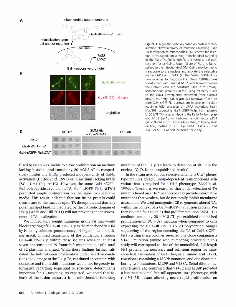
fused to Fis1p was unable to allow proliferation on mediumlacking histidine and containing 20 mM 3-AT to competi-tively inhibit any His3p produced independently of Gal4pactivation (Durfee et al. 1993) or in medium lacking uracil(SC 2Ura) (Figure 1C). However, the same Gal4–sfGFP–Fis1 polypeptide devoid of its TA [Gal4–sfGFP–Fis1p(DTA)]permitted ample proliferation on the same two selectivemedia. This result indicated that our fusion protein couldtranslocate to the nucleus upon TA disruption and that anypotential lipid binding mediated by the cytosolic domain ofFis1p (Wells and Hill 2011) will not prevent genetic assess-ment of TA localization.
We immediately sought mutations in the TA that wouldblock targeting of Gal4–sfGFP–Fis1p to themitochondrial OMby isolating colonies spontaneously arising on medium lack-ing uracil. Limited sequencing of the constructs encodingGal4–sfGFP–Fis1p within these isolates revealed at leastseven nonsense and 19 frameshift mutations out of a totalof 32 plasmids analyzed. While these findings further vali-dated the link between proliferation under selective condi-tions and damage to the Fis1p TA, continued encounters withnonsense and frameshift mutations would not be greatly in-formative regarding sequential or structural determinantsimportant for TA targeting. As expected, we noted that re-lease of the fusion construct from mitochondria following
mutation of the Fis1p TA leads to detection of sfGFP in thenucleus (C. D. Dunn, unpublished results).
In the strain used for our selective scheme, a Ura+ pheno-type requires greater Gal4p-dependent transcriptional acti-vation than is required for a His+ phenotype (Vidal et al.1996b). Therefore, we reasoned that initial selection of TAmutants based on a His+ phenotype may provide informativemutations that weaken, but do not totally inhibit membraneassociation. We used mutagenic PCR to generate altered TAswithin the context of a Gal4–sfGFP–Fis1 fusion protein. Wethen isolated four colonies that proliferated upon SMM2Hismedium containing 20 mM 3-AT, yet exhibited diminishedproliferation on SC 2Ura medium when compared to cellsexpressing the Gal4–sfGFP–Fis1(DTA) polypeptide. Sangersequencing of the region encoding the TA of Gal4–sfGFP–Fis1p within these colonies revealed one clone containing aV145E mutation (amino acid numbering provided in thisstudy will correspond to that of the unmodified, full-lengthFis1 protein; the necessary and sufficient region for mito-chondrial association of Fis1p begins at amino acid L129),two clones containing a L139P mutation, and one clone har-boring two mutations: L129P and V138A. Serial dilution as-says (Figure 2A) confirmed that V145E and L139P provideda less than maximal, but still apparent Ura+ phenotype, withthe V145E mutant allowing more rapid proliferation on
Figure 1 A genetic selection based on protein misloc-alization allows recovery of mutations blocking Fis1pTA localization to mitochondria. (A) Scheme for selec-tion of mutations preventing mitochondrial targetingof the Fis1p TA. Full-length Fis1p is fused to the tran-scription factor Gal4p. Upon failure of Fis1p to be lo-calized to the mitochondrial OM, Gal4p may be free totranslocate to the nucleus and activate the selectablemarkers HIS3 and URA3. (B) The Gal4–sfGFP–Fis1 fu-sion localizes to mitochondria. Strain CDD898 wastransformed with plasmid b102, which overexpressesthe Gal4–sfGFP–Fis1p construct used in this study.Mitochondria were visualized using mCherry fusedto the Cox4 presequence expressed from plasmidpHS12–mCherry. Bar, 5 mm. (C) Removal of the TAfrom Gal4–sfGFP–Fis1p allows proliferation on mediumrequiring HIS3 activation or URA3 activation. StrainMaV203 expressing Gal4–sfGFP–Fis1p from plasmidb100 (WT TA), a variant lacking the Fis1p TA from plas-mid b101 (DTA), or harboring empty vector pKS1was cultured in SC 2Trp medium, then, following serialdilution, spotted to SC 2Trp, SMM 2His + 20 mM3-AT, or SC 2Ura and incubated for 2 days.
694 A. Keskin, E. Akdogan, and C. D. Dunn
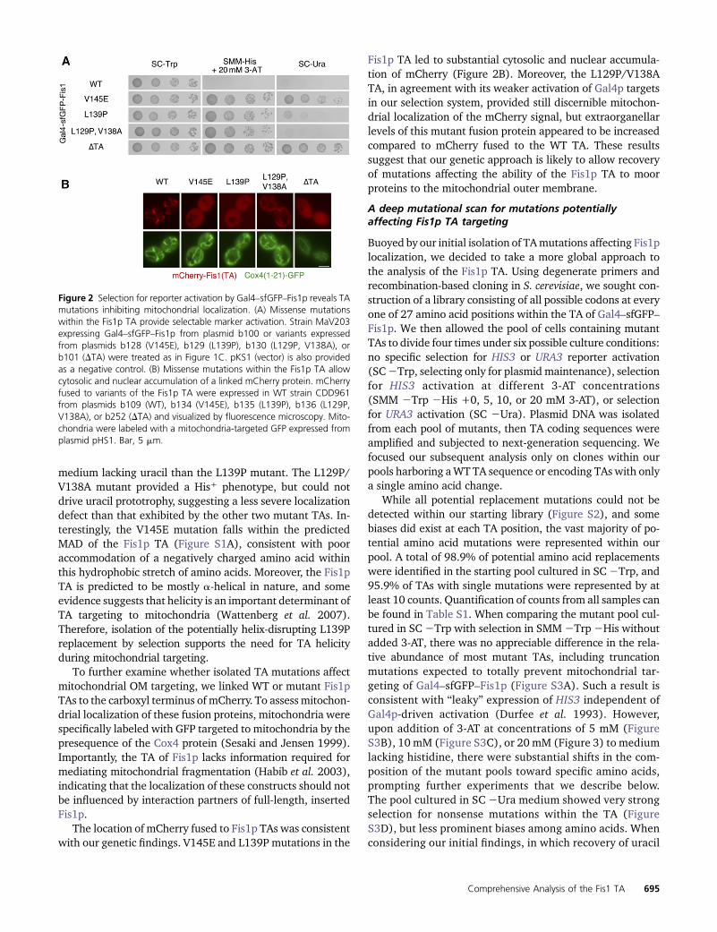
medium lacking uracil than the L139P mutant. The L129P/V138A mutant provided a His+ phenotype, but could notdrive uracil prototrophy, suggesting a less severe localizationdefect than that exhibited by the other two mutant TAs. In-terestingly, the V145E mutation falls within the predictedMAD of the Fis1p TA (Figure S1A), consistent with pooraccommodation of a negatively charged amino acid withinthis hydrophobic stretch of amino acids. Moreover, the Fis1pTA is predicted to be mostly a-helical in nature, and someevidence suggests that helicity is an important determinant ofTA targeting to mitochondria (Wattenberg et al. 2007).Therefore, isolation of the potentially helix-disrupting L139Preplacement by selection supports the need for TA helicityduring mitochondrial targeting.
To further examine whether isolated TA mutations affectmitochondrial OM targeting, we linked WT or mutant Fis1pTAs to the carboxyl terminus of mCherry. To assess mitochon-drial localization of these fusion proteins, mitochondria werespecifically labeled with GFP targeted to mitochondria by thepresequence of the Cox4 protein (Sesaki and Jensen 1999).Importantly, the TA of Fis1p lacks information required formediating mitochondrial fragmentation (Habib et al. 2003),indicating that the localization of these constructs should notbe influenced by interaction partners of full-length, insertedFis1p.
The location of mCherry fused to Fis1p TAs was consistentwith our genetic findings. V145E and L139P mutations in the
Fis1p TA led to substantial cytosolic and nuclear accumula-tion of mCherry (Figure 2B). Moreover, the L129P/V138ATA, in agreement with its weaker activation of Gal4p targetsin our selection system, provided still discernible mitochon-drial localization of the mCherry signal, but extraorganellarlevels of this mutant fusion protein appeared to be increasedcompared to mCherry fused to the WT TA. These resultssuggest that our genetic approach is likely to allow recoveryof mutations affecting the ability of the Fis1p TA to moorproteins to the mitochondrial outer membrane.
A deep mutational scan for mutations potentiallyaffecting Fis1p TA targeting
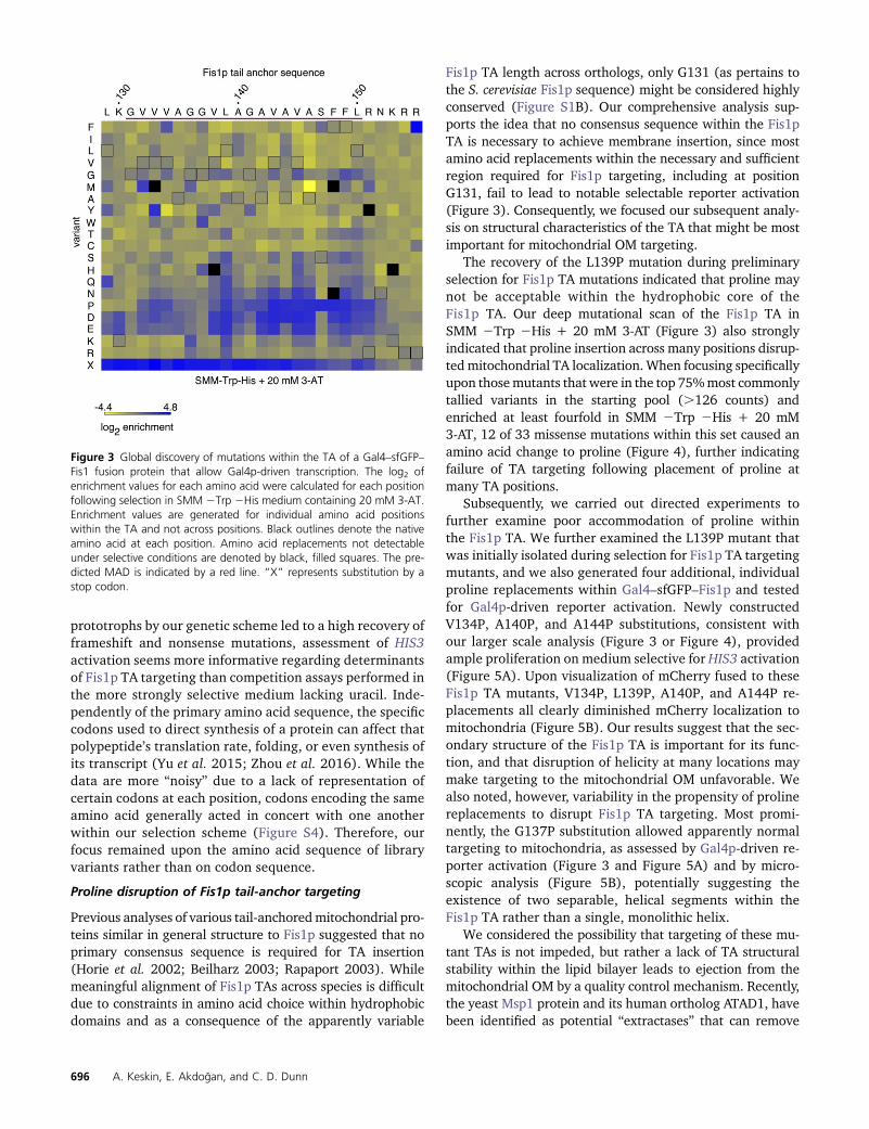
Buoyed by our initial isolation of TAmutations affecting Fis1plocalization, we decided to take a more global approach tothe analysis of the Fis1p TA. Using degenerate primers andrecombination-based cloning in S. cerevisiae, we sought con-struction of a library consisting of all possible codons at everyone of 27 amino acid positions within the TA of Gal4–sfGFP–Fis1p. We then allowed the pool of cells containing mutantTAs to divide four times under six possible culture conditions:no specific selection for HIS3 or URA3 reporter activation(SC2Trp, selecting only for plasmid maintenance), selectionfor HIS3 activation at different 3-AT concentrations(SMM 2Trp 2His +0, 5, 10, or 20 mM 3-AT), or selectionfor URA3 activation (SC 2Ura). Plasmid DNA was isolatedfrom each pool of mutants, then TA coding sequences wereamplified and subjected to next-generation sequencing. Wefocused our subsequent analysis only on clones within ourpools harboring aWT TA sequence or encoding TAs with onlya single amino acid change.
While all potential replacement mutations could not bedetected within our starting library (Figure S2), and somebiases did exist at each TA position, the vast majority of po-tential amino acid mutations were represented within ourpool. A total of 98.9% of potential amino acid replacementswere identified in the starting pool cultured in SC2Trp, and95.9% of TAs with single mutations were represented by atleast 10 counts. Quantification of counts from all samples canbe found in Table S1. When comparing the mutant pool cul-tured in SC2Trp with selection in SMM2Trp2His withoutadded 3-AT, there was no appreciable difference in the rela-tive abundance of most mutant TAs, including truncationmutations expected to totally prevent mitochondrial tar-geting of Gal4–sfGFP–Fis1p (Figure S3A). Such a result isconsistent with “leaky” expression of HIS3 independent ofGal4p-driven activation (Durfee et al. 1993). However,upon addition of 3-AT at concentrations of 5 mM (FigureS3B), 10 mM (Figure S3C), or 20 mM (Figure 3) to mediumlacking histidine, there were substantial shifts in the com-position of the mutant pools toward specific amino acids,prompting further experiments that we describe below.The pool cultured in SC 2Ura medium showed very strongselection for nonsense mutations within the TA (FigureS3D), but less prominent biases among amino acids. Whenconsidering our initial findings, in which recovery of uracil
Figure 2 Selection for reporter activation by Gal4–sfGFP–Fis1p reveals TAmutations inhibiting mitochondrial localization. (A) Missense mutationswithin the Fis1p TA provide selectable marker activation. Strain MaV203expressing Gal4–sfGFP–Fis1p from plasmid b100 or variants expressedfrom plasmids b128 (V145E), b129 (L139P), b130 (L129P, V138A), orb101 (DTA) were treated as in Figure 1C. pKS1 (vector) is also providedas a negative control. (B) Missense mutations within the Fis1p TA allowcytosolic and nuclear accumulation of a linked mCherry protein. mCherryfused to variants of the Fis1p TA were expressed in WT strain CDD961from plasmids b109 (WT), b134 (V145E), b135 (L139P), b136 (L129P,V138A), or b252 (DTA) and visualized by fluorescence microscopy. Mito-chondria were labeled with a mitochondria-targeted GFP expressed fromplasmid pHS1. Bar, 5 mm.
Comprehensive Analysis of the Fis1 TA 695
prototrophs by our genetic scheme led to a high recovery offrameshift and nonsense mutations, assessment of HIS3activation seems more informative regarding determinantsof Fis1p TA targeting than competition assays performed inthe more strongly selective medium lacking uracil. Inde-pendently of the primary amino acid sequence, the specificcodons used to direct synthesis of a protein can affect thatpolypeptide’s translation rate, folding, or even synthesis ofits transcript (Yu et al. 2015; Zhou et al. 2016). While thedata are more “noisy” due to a lack of representation ofcertain codons at each position, codons encoding the sameamino acid generally acted in concert with one anotherwithin our selection scheme (Figure S4). Therefore, ourfocus remained upon the amino acid sequence of libraryvariants rather than on codon sequence.
Proline disruption of Fis1p tail-anchor targeting
Previous analyses of various tail-anchoredmitochondrial pro-teins similar in general structure to Fis1p suggested that noprimary consensus sequence is required for TA insertion(Horie et al. 2002; Beilharz 2003; Rapaport 2003). Whilemeaningful alignment of Fis1p TAs across species is difficultdue to constraints in amino acid choice within hydrophobicdomains and as a consequence of the apparently variable
Fis1p TA length across orthologs, only G131 (as pertains tothe S. cerevisiae Fis1p sequence) might be considered highlyconserved (Figure S1B). Our comprehensive analysis sup-ports the idea that no consensus sequence within the Fis1pTA is necessary to achieve membrane insertion, since mostamino acid replacements within the necessary and sufficientregion required for Fis1p targeting, including at positionG131, fail to lead to notable selectable reporter activation(Figure 3). Consequently, we focused our subsequent analy-sis on structural characteristics of the TA that might be mostimportant for mitochondrial OM targeting.
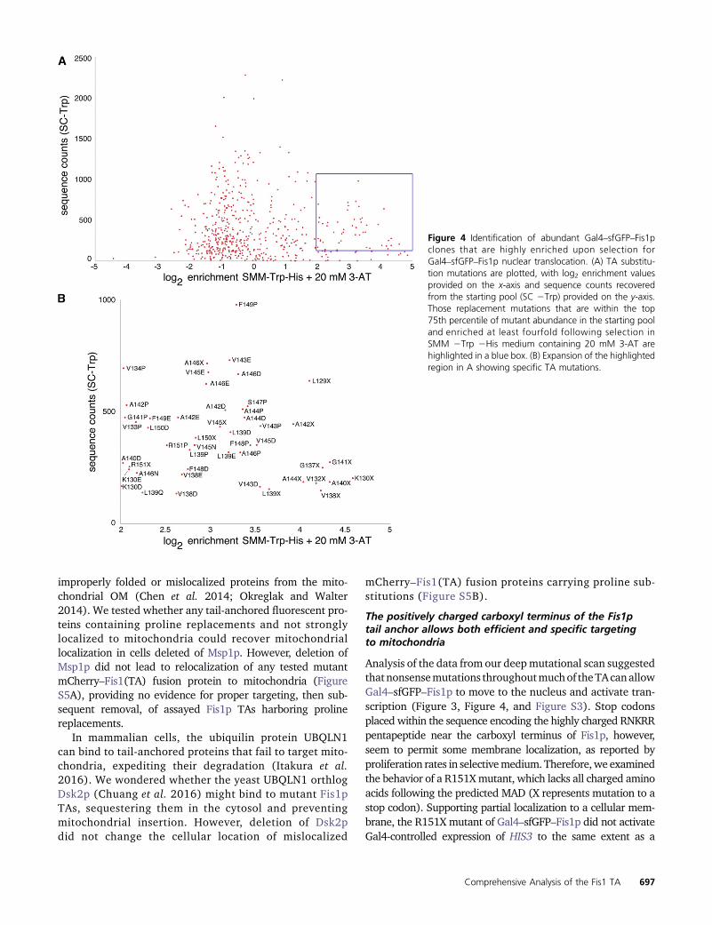
The recovery of the L139P mutation during preliminaryselection for Fis1p TA mutations indicated that proline maynot be acceptable within the hydrophobic core of theFis1p TA. Our deep mutational scan of the Fis1p TA inSMM 2Trp 2His + 20 mM 3-AT (Figure 3) also stronglyindicated that proline insertion across many positions disrup-tedmitochondrial TA localization. When focusing specificallyupon thosemutants that were in the top 75%most commonlytallied variants in the starting pool (.126 counts) andenriched at least fourfold in SMM 2Trp 2His + 20 mM3-AT, 12 of 33 missense mutations within this set caused anamino acid change to proline (Figure 4), further indicatingfailure of TA targeting following placement of proline atmany TA positions.
Subsequently, we carried out directed experiments tofurther examine poor accommodation of proline withinthe Fis1p TA. We further examined the L139P mutant thatwas initially isolated during selection for Fis1p TA targetingmutants, and we also generated four additional, individualproline replacements within Gal4–sfGFP–Fis1p and testedfor Gal4p-driven reporter activation. Newly constructedV134P, A140P, and A144P substitutions, consistent withour larger scale analysis (Figure 3 or Figure 4), providedample proliferation on medium selective for HIS3 activation(Figure 5A). Upon visualization of mCherry fused to theseFis1p TA mutants, V134P, L139P, A140P, and A144P re-placements all clearly diminished mCherry localization tomitochondria (Figure 5B). Our results suggest that the sec-ondary structure of the Fis1p TA is important for its func-tion, and that disruption of helicity at many locations maymake targeting to the mitochondrial OM unfavorable. Wealso noted, however, variability in the propensity of prolinereplacements to disrupt Fis1p TA targeting. Most promi-nently, the G137P substitution allowed apparently normaltargeting to mitochondria, as assessed by Gal4p-driven re-porter activation (Figure 3 and Figure 5A) and by micro-scopic analysis (Figure 5B), potentially suggesting theexistence of two separable, helical segments within theFis1p TA rather than a single, monolithic helix.
We considered the possibility that targeting of these mu-tant TAs is not impeded, but rather a lack of TA structuralstability within the lipid bilayer leads to ejection from themitochondrial OM by a quality control mechanism. Recently,the yeast Msp1 protein and its human ortholog ATAD1, havebeen identified as potential “extractases” that can remove
Figure 3 Global discovery of mutations within the TA of a Gal4–sfGFP–Fis1 fusion protein that allow Gal4p-driven transcription. The log2 ofenrichment values for each amino acid were calculated for each positionfollowing selection in SMM 2Trp 2His medium containing 20 mM 3-AT.Enrichment values are generated for individual amino acid positionswithin the TA and not across positions. Black outlines denote the nativeamino acid at each position. Amino acid replacements not detectableunder selective conditions are denoted by black, filled squares. The pre-dicted MAD is indicated by a red line. “X” represents substitution by astop codon.
696 A. Keskin, E. Akdogan, and C. D. Dunn
improperly folded or mislocalized proteins from the mito-chondrial OM (Chen et al. 2014; Okreglak and Walter2014). We tested whether any tail-anchored fluorescent pro-teins containing proline replacements and not stronglylocalized to mitochondria could recover mitochondriallocalization in cells deleted of Msp1p. However, deletion ofMsp1p did not lead to relocalization of any tested mutantmCherry–Fis1(TA) fusion protein to mitochondria (FigureS5A), providing no evidence for proper targeting, then sub-sequent removal, of assayed Fis1p TAs harboring prolinereplacements.
In mammalian cells, the ubiquilin protein UBQLN1can bind to tail-anchored proteins that fail to target mito-chondria, expediting their degradation (Itakura et al.2016). We wondered whether the yeast UBQLN1 orthlogDsk2p (Chuang et al. 2016) might bind to mutant Fis1pTAs, sequestering them in the cytosol and preventingmitochondrial insertion. However, deletion of Dsk2pdid not change the cellular location of mislocalized
mCherry–Fis1(TA) fusion proteins carrying proline sub-stitutions (Figure S5B).
The positively charged carboxyl terminus of the Fis1ptail anchor allows both efficient and specific targetingto mitochondria
Analysis of the data from our deepmutational scan suggestedthatnonsensemutations throughoutmuchof theTAcanallowGal4–sfGFP–Fis1p to move to the nucleus and activate tran-scription (Figure 3, Figure 4, and Figure S3). Stop codonsplaced within the sequence encoding the highly charged RNKRRpentapeptide near the carboxyl terminus of Fis1p, however,seem to permit some membrane localization, as reported byproliferation rates in selectivemedium. Therefore,we examinedthe behavior of a R151Xmutant, which lacks all charged aminoacids following the predicted MAD (X represents mutation to astop codon). Supporting partial localization to a cellular mem-brane, the R151X mutant of Gal4–sfGFP–Fis1p did not activateGal4-controlled expression of HIS3 to the same extent as a
Figure 4 Identification of abundant Gal4–sfGFP–Fis1pclones that are highly enriched upon selection forGal4–sfGFP–Fis1p nuclear translocation. (A) TA substitu-tion mutations are plotted, with log2 enrichment valuesprovided on the x-axis and sequence counts recoveredfrom the starting pool (SC 2Trp) provided on the y-axis.Those replacement mutations that are within the top75th percentile of mutant abundance in the starting pooland enriched at least fourfold following selection inSMM 2Trp 2His medium containing 20 mM 3-AT arehighlighted in a blue box. (B) Expansion of the highlightedregion in A showing specific TA mutations.
Comprehensive Analysis of the Fis1 TA 697
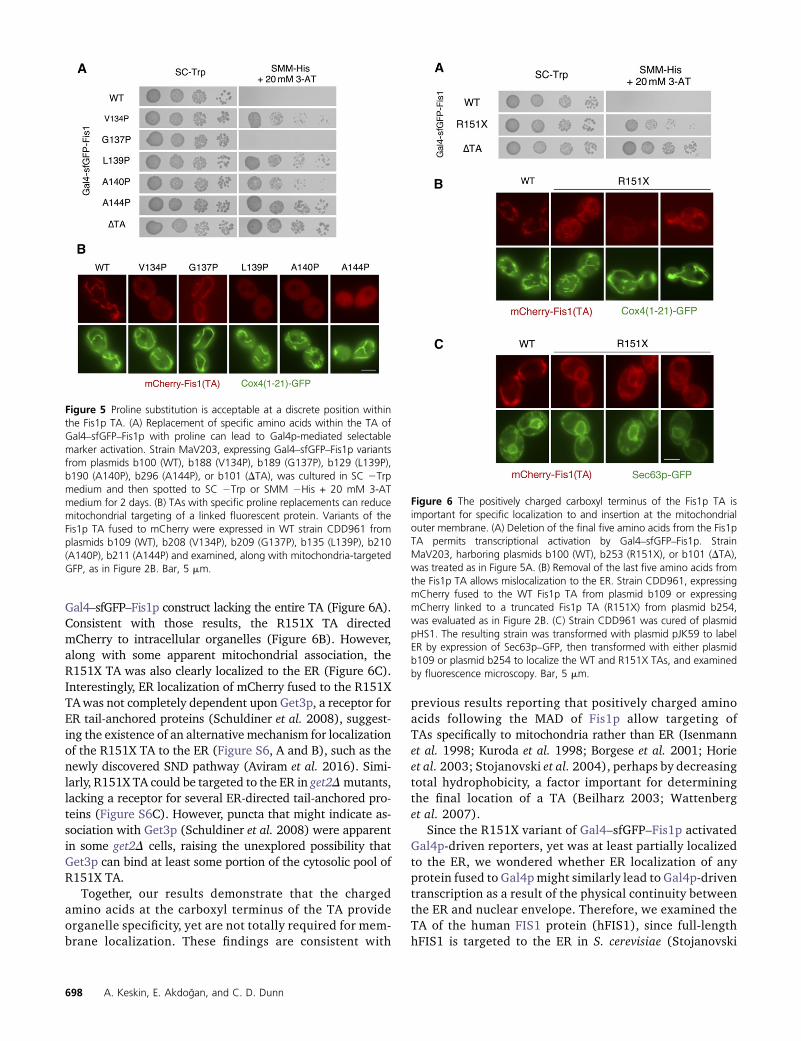
Gal4–sfGFP–Fis1p construct lacking the entire TA (Figure 6A).Consistent with those results, the R151X TA directedmCherry to intracellular organelles (Figure 6B). However,along with some apparent mitochondrial association, theR151X TA was also clearly localized to the ER (Figure 6C).Interestingly, ER localization of mCherry fused to the R151XTAwas not completely dependent upon Get3p, a receptor forER tail-anchored proteins (Schuldiner et al. 2008), suggest-ing the existence of an alternative mechanism for localizationof the R151X TA to the ER (Figure S6, A and B), such as thenewly discovered SND pathway (Aviram et al. 2016). Simi-larly, R151X TA could be targeted to the ER in get2Dmutants,lacking a receptor for several ER-directed tail-anchored pro-teins (Figure S6C). However, puncta that might indicate as-sociation with Get3p (Schuldiner et al. 2008) were apparentin some get2D cells, raising the unexplored possibility thatGet3p can bind at least some portion of the cytosolic pool ofR151X TA.
Together, our results demonstrate that the chargedamino acids at the carboxyl terminus of the TA provideorganelle specificity, yet are not totally required for mem-brane localization. These findings are consistent with
previous results reporting that positively charged aminoacids following the MAD of Fis1p allow targeting ofTAs specifically to mitochondria rather than ER (Isenmannet al. 1998; Kuroda et al. 1998; Borgese et al. 2001; Horieet al. 2003; Stojanovski et al. 2004), perhaps by decreasingtotal hydrophobicity, a factor important for determiningthe final location of a TA (Beilharz 2003; Wattenberget al. 2007).
Since the R151X variant of Gal4–sfGFP–Fis1p activatedGal4p-driven reporters, yet was at least partially localizedto the ER, we wondered whether ER localization of anyprotein fused to Gal4p might similarly lead to Gal4p-driventranscription as a result of the physical continuity betweenthe ER and nuclear envelope. Therefore, we examined theTA of the human FIS1 protein (hFIS1), since full-lengthhFIS1 is targeted to the ER in S. cerevisiae (Stojanovski
Figure 5 Proline substitution is acceptable at a discrete position withinthe Fis1p TA. (A) Replacement of specific amino acids within the TA ofGal4–sfGFP–Fis1p with proline can lead to Gal4p-mediated selectablemarker activation. Strain MaV203, expressing Gal4–sfGFP–Fis1p variantsfrom plasmids b100 (WT), b188 (V134P), b189 (G137P), b129 (L139P),b190 (A140P), b296 (A144P), or b101 (DTA), was cultured in SC 2Trpmedium and then spotted to SC 2Trp or SMM 2His + 20 mM 3-ATmedium for 2 days. (B) TAs with specific proline replacements can reducemitochondrial targeting of a linked fluorescent protein. Variants of theFis1p TA fused to mCherry were expressed in WT strain CDD961 fromplasmids b109 (WT), b208 (V134P), b209 (G137P), b135 (L139P), b210(A140P), b211 (A144P) and examined, along with mitochondria-targetedGFP, as in Figure 2B. Bar, 5 mm.
Figure 6 The positively charged carboxyl terminus of the Fis1p TA isimportant for specific localization to and insertion at the mitochondrialouter membrane. (A) Deletion of the final five amino acids from the Fis1pTA permits transcriptional activation by Gal4–sfGFP–Fis1p. StrainMaV203, harboring plasmids b100 (WT), b253 (R151X), or b101 (DTA),was treated as in Figure 5A. (B) Removal of the last five amino acids fromthe Fis1p TA allows mislocalization to the ER. Strain CDD961, expressingmCherry fused to the WT Fis1p TA from plasmid b109 or expressingmCherry linked to a truncated Fis1p TA (R151X) from plasmid b254,was evaluated as in Figure 2B. (C) Strain CDD961 was cured of plasmidpHS1. The resulting strain was transformed with plasmid pJK59 to labelER by expression of Sec63p–GFP, then transformed with either plasmidb109 or plasmid b254 to localize the WT and R151X TAs, and examinedby fluorescence microscopy. Bar, 5 mm.
698 A. Keskin, E. Akdogan, and C. D. Dunn
et al. 2004). Indeed, we found that mCherry fused to thehFIS1 TA was poorly localized to mitochondria (FigureS7A) and abundant at the ER (Figure S7B). However, afusion protein consisting of Gal4p fused to the TA of hFIS1did not provide HIS3 activation (Figure S7C), indicatingthat the hFIS1 TA is quantitatively membrane targeted inS. cerevisiae and that activation of Gal4p-dependent re-porters upon removal of the positively charged carboxylterminus from Gal4–sfGFP–Fis1p is unlikely to be a conse-quence of ER localization.
Due to the mislocalization of the hFIS1 TA, we then in-vestigated thepossibility that othermitochondrial TAproteinsfromhumanwould be targeted improperly in S. cerevisiae.Wefused mCherry to the TA of human BAX, a region that issufficient for insertion at the mammalian mitochondrial OM(Schinzel et al. 2004). While mCherry signal was diminishedin comparison with other mCherry fusion proteins examinedin this study and expressed under the same promoter,mCherry fused to the BAX TA was properly targeted to mito-chondria (Figure S7D). Gal4p fused to the BAX TA did notactivate selectable reporters (C. D. Dunn, unpublished re-sults), further indicating effective mitochondrial targetingmediated by the BAX TA.
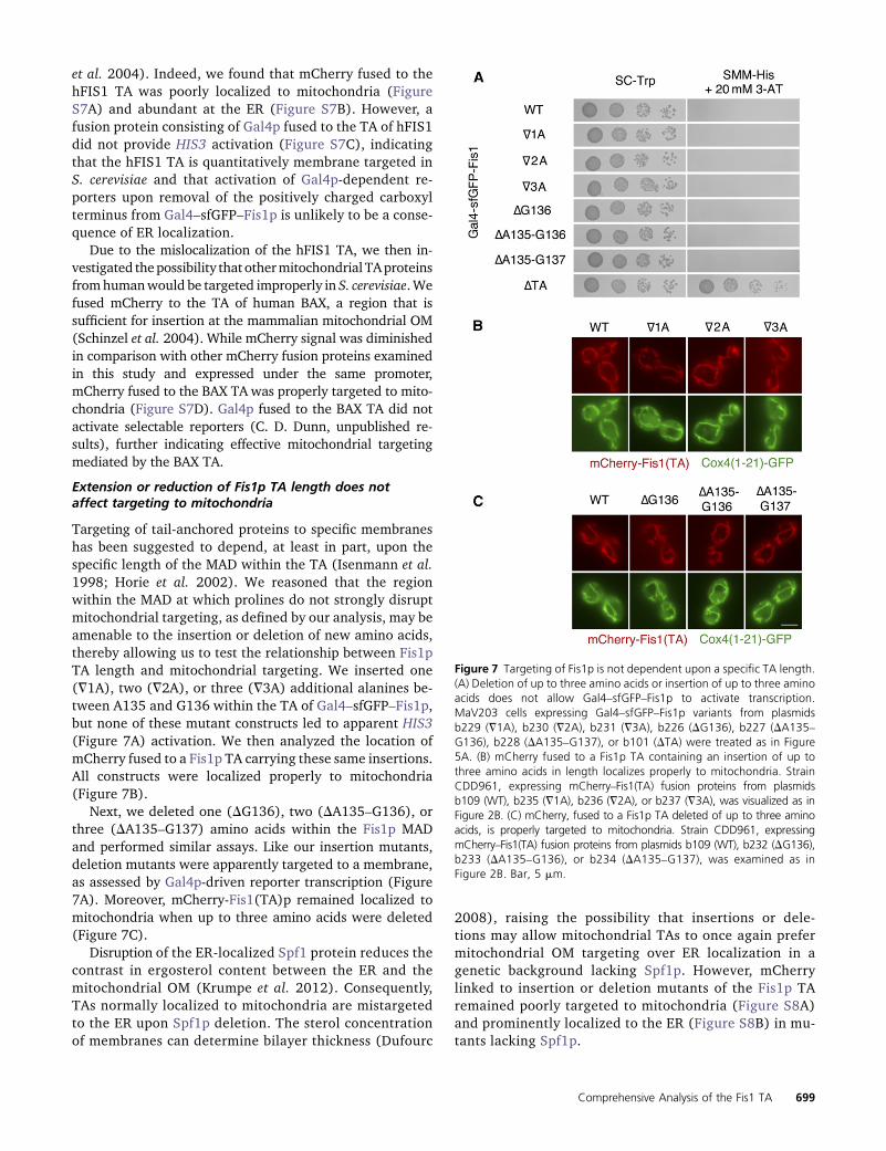
Extension or reduction of Fis1p TA length does notaffect targeting to mitochondria
Targeting of tail-anchored proteins to specific membraneshas been suggested to depend, at least in part, upon thespecific length of the MAD within the TA (Isenmann et al.1998; Horie et al. 2002). We reasoned that the regionwithin the MAD at which prolines do not strongly disruptmitochondrial targeting, as defined by our analysis, may beamenable to the insertion or deletion of new amino acids,thereby allowing us to test the relationship between Fis1pTA length and mitochondrial targeting. We inserted one(=1A), two (=2A), or three (=3A) additional alanines be-tween A135 and G136 within the TA of Gal4–sfGFP–Fis1p,but none of these mutant constructs led to apparent HIS3(Figure 7A) activation. We then analyzed the location ofmCherry fused to a Fis1p TA carrying these same insertions.All constructs were localized properly to mitochondria(Figure 7B).
Next, we deleted one (DG136), two (DA135–G136), orthree (DA135–G137) amino acids within the Fis1p MADand performed similar assays. Like our insertion mutants,deletion mutants were apparently targeted to a membrane,as assessed by Gal4p-driven reporter transcription (Figure7A). Moreover, mCherry-Fis1(TA)p remained localized tomitochondria when up to three amino acids were deleted(Figure 7C).
Disruption of the ER-localized Spf1 protein reduces thecontrast in ergosterol content between the ER and themitochondrial OM (Krumpe et al. 2012). Consequently,TAs normally localized to mitochondria are mistargetedto the ER upon Spf1p deletion. The sterol concentrationof membranes can determine bilayer thickness (Dufourc
2008), raising the possibility that insertions or dele-tions may allow mitochondrial TAs to once again prefermitochondrial OM targeting over ER localization in agenetic background lacking Spf1p. However, mCherrylinked to insertion or deletion mutants of the Fis1p TAremained poorly targeted to mitochondria (Figure S8A)and prominently localized to the ER (Figure S8B) in mu-tants lacking Spf1p.
Figure 7 Targeting of Fis1p is not dependent upon a specific TA length.(A) Deletion of up to three amino acids or insertion of up to three aminoacids does not allow Gal4–sfGFP–Fis1p to activate transcription.MaV203 cells expressing Gal4–sfGFP–Fis1p variants from plasmidsb229 (=1A), b230 (=2A), b231 (=3A), b226 (DG136), b227 (DA135–G136), b228 (DA135–G137), or b101 (DTA) were treated as in Figure5A. (B) mCherry fused to a Fis1p TA containing an insertion of up tothree amino acids in length localizes properly to mitochondria. StrainCDD961, expressing mCherry–Fis1(TA) fusion proteins from plasmidsb109 (WT), b235 (=1A), b236 (=2A), or b237 (=3A), was visualized as inFigure 2B. (C) mCherry, fused to a Fis1p TA deleted of up to three aminoacids, is properly targeted to mitochondria. Strain CDD961, expressingmCherry–Fis1(TA) fusion proteins from plasmids b109 (WT), b232 (DG136),b233 (DA135–G136), or b234 (DA135–G137), was examined as inFigure 2B. Bar, 5 mm.
Comprehensive Analysis of the Fis1 TA 699
Together, our results demonstrate that the Fis1p TA isproperly localized to mitochondria even when its length issubstantially altered.
Positively charged amino acids are more acceptablethan negatively charged amino acids within thepredicted transmembrane domain of the Fis1ptail anchor
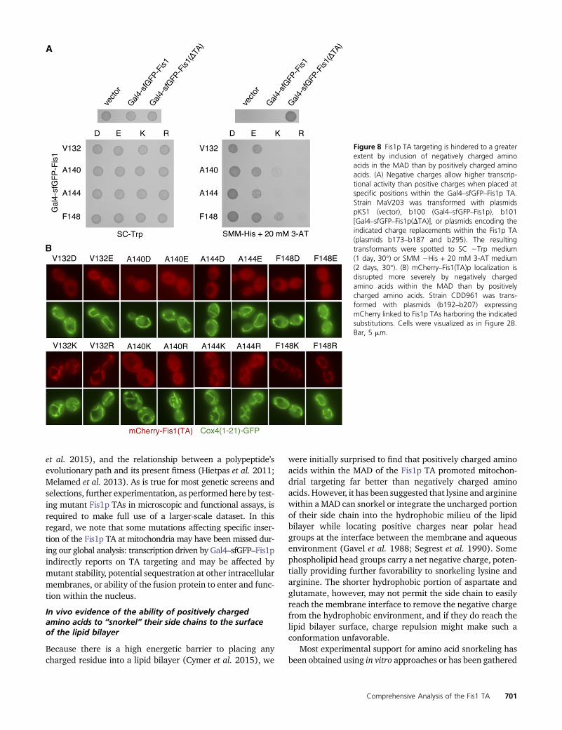
Our deep mutational scan of the Fis1p TA demonstrated thatGal4–sfGFP–Fis1pwas generally able to activate gene expres-sion when aspartate or glutamate was placedwithin theMAD(Figure 3). In fact, upon examination of those amino acidreplacements found within the top three quartiles of countsin the initial library and also enriched at least fourfold uponculture in SMM2Trp2His + 20mM3-AT, 18 of 33missensemutations were aspartate or glutamate substitutions (Figure4). We were surprised to find that placement of positivelycharged arginine or lysine residues appeared to be muchmore acceptable within the MAD of Fis1p than aspartate orglutamate; none of the amino acid substitutions within thishigh-count, high-enrichment set were by lysine or arginine.
To further pursue the possibility that positively chargedamino acids can be accommodated within the Fis1pMAD, wemutated four amino acids within the hydrophobic stretch ofthe Fis1p TA to aspartate, glutamate, lysine, or arginine. Spe-cifically, we generated amino acid replacements at positionsV132, A140, A144, or F148, then retested these mutants un-der selection for Gal4–sfGFP–Fis1p transcriptional activity.The results from our global analysis were verified, with as-partate and glutamate mutations providing stronger reporteractivation than lysine and arginine mutations (Figure 8A).Only the A144D mutation provided sufficient Gal4p activityfor proliferation on medium lacking uracil (Figure S9A) after2 days of incubation, suggesting a very severe TA localizationdefect caused by this substitution. We noted that these mu-tant Gal4–sfGFP–Fis1p constructs exhibit altered behavior atdifferent temperatures. For example, lysine and arginine sub-stitutions at positions A140, A144, or F148 led to reducedproliferation at 37� under conditions selective for HIS3 ac-tivation (Figure S9B) when compared to the same TA sub-stitutions assessed at 18� (Figure S9C) or 30� (extendedincubation, Figure S9D). This outcome is consistent withthe idea that altered phospholipid dynamics at differenttemperatures may lead to consequent changes in TA inser-tion efficiency (de Mendoza and Cronan 1983).
We then tested the ability of these Fis1p TAs containingcharge substitutions to promotemitochondrial localization ofmCherry. At positions V132 and F148, locations within theMAD nearer to the putative water–lipid bilayer interface,mutation to positively charged amino acids allowed abun-dant localization to mitochondria (Figure 8B). In contrast,mutation to negatively charged amino acids clearly hinderedmitochondrial targeting. We noted that F148D and F148Ereplacements hampered mitochondrial localization more se-verely than V132D and V132E replacements, consistent withphenotypic testing of Gal4–sfGFP–Fis1 fusion proteins. At
position A144, lying more deeply within the predictedMAD, all charge mutations inhibited mCherry targeting, yetA144K and A144R substitutions allowed some mCherry lo-calization at mitochondria, while A144D and A144E mu-tants appeared undetectable at mitochondria. Finally, nomitochondrial signal was apparent for any of the chargemutants tested at position A140 of the Fis1p TA. However,A140K and A140R mutants differed from A140D and A140Emutants by localizing to other membranes within the cell,including the plasma membrane, rather than providing adiffuse cytosolic signal. Msp1p removal did not permitrelocalization of tail-anchored fluorescent proteins to mi-tochondria (Figure S5C), supporting the idea that chargereplacements within the Fis1p TA lead to a failure of asso-ciation with the OM rather than enhanced removal frommitochondria. Removal of UBQLN1 ortholog Dsk2p alsohad no discernible effect on mCherry–Fis1(TA)p mutantlocalization (Figure S5D).
Negligible Fis1p activity at mitochondria is apparently suf-ficient to promote mitochondrial fission (Habib et al. 2003;Krumpe et al. 2012), suggesting that even minimal localiza-tion and related functionality at the OM would be detectableby functional assays. Interestingly, Fis1p variants harboringcharged residues, positive or negative, at positions V132,A144, or V148, with the exception of Fis1p carrying theA144D mutation, provided at least some Fis1p function, asindicated by microscopic (Figure S10, A and B) and genetic(Figure S10C) assays. Such a result is consistent with thefinding that only the A144D charge mutant provides suffi-cient URA3 activation for rapid proliferation on medium lack-ing uracil (Figure S9A).
Together, our results demonstrate that positively chargedamino acids within the MAD can better promote Fis1p locali-zation than negatively charged amino acids, but that even neg-atively charged amino acids can be accommodated within theMAD and lead to a detectable level of mitochondrial targeting.
Discussion
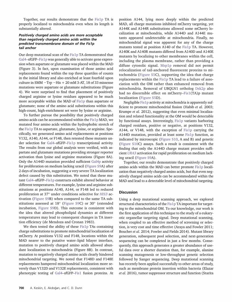
Using a deep mutational scanning approach, we exploredstructural characteristics of the Fis1p TA important for target-ing to the mitochondrial OM. To our knowledge, our work isthe first application of this technique to the study of a eukary-otic organellar targeting signal. Deep mutational scanning,when coupled to an effective method of screening or selec-tion, is very cost and time effective (Araya and Fowler 2011;Boucher et al. 2014; Fowler and Fields 2014). Mutant librarygeneration, subsequent pool selection, and next-generationsequencing can be completed in just a few months. Conse-quently, this approach generates a greater abundance of use-ful data over a shorter duration than, for example, alaninescanning mutagenesis or low-throughput genetic selectionfollowed by Sanger sequencing. Deep mutational scanninghas recently been applied successfully to other areas of study,such as membrane protein insertion within bacteria (Elazaret al. 2016), tumor suppressor structure and function (Starita
700 A. Keskin, E. Akdogan, and C. D. Dunn
et al. 2015), and the relationship between a polypeptide’sevolutionary path and its present fitness (Hietpas et al. 2011;Melamed et al. 2013). As is true for most genetic screens andselections, further experimentation, as performed here by test-ing mutant Fis1p TAs in microscopic and functional assays, isrequired to make full use of a larger-scale dataset. In thisregard, we note that some mutations affecting specific inser-tion of the Fis1p TA at mitochondria may have been missed dur-ing our global analysis: transcription driven by Gal4–sfGFP–Fis1pindirectly reports on TA targeting and may be affected bymutant stability, potential sequestration at other intracellularmembranes, or ability of the fusion protein to enter and func-tion within the nucleus.
In vivo evidence of the ability of positively chargedamino acids to “snorkel” their side chains to the surfaceof the lipid bilayer
Because there is a high energetic barrier to placing anycharged residue into a lipid bilayer (Cymer et al. 2015), we
were initially surprised to find that positively charged aminoacids within the MAD of the Fis1p TA promoted mitochon-drial targeting far better than negatively charged aminoacids. However, it has been suggested that lysine and argininewithin a MAD can snorkel or integrate the uncharged portionof their side chain into the hydrophobic milieu of the lipidbilayer while locating positive charges near polar headgroups at the interface between the membrane and aqueousenvironment (Gavel et al. 1988; Segrest et al. 1990). Somephospholipid head groups carry a net negative charge, poten-tially providing further favorability to snorkeling lysine andarginine. The shorter hydrophobic portion of aspartate andglutamate, however, may not permit the side chain to easilyreach the membrane interface to remove the negative chargefrom the hydrophobic environment, and if they do reach thelipid bilayer surface, charge repulsion might make such aconformation unfavorable.
Most experimental support for amino acid snorkeling hasbeen obtained using in vitro approaches or has been gathered
Figure 8 Fis1p TA targeting is hindered to a greaterextent by inclusion of negatively charged aminoacids in the MAD than by positively charged aminoacids. (A) Negative charges allow higher transcrip-tional activity than positive charges when placed atspecific positions within the Gal4–sfGFP–Fis1p TA.Strain MaV203 was transformed with plasmidspKS1 (vector), b100 (Gal4–sfGFP–Fis1p), b101[Gal4–sfGFP–Fis1p(DTA)], or plasmids encoding theindicated charge replacements within the Fis1p TA(plasmids b173–b187 and b295). The resultingtransformants were spotted to SC 2Trp medium(1 day, 30�) or SMM 2His + 20 mM 3-AT medium(2 days, 30�). (B) mCherry–Fis1(TA)p localization isdisrupted more severely by negatively chargedamino acids within the MAD than by positivelycharged amino acids. Strain CDD961 was trans-formed with plasmids (b192–b207) expressingmCherry linked to Fis1p TAs harboring the indicatedsubstitutions. Cells were visualized as in Figure 2B.Bar, 5 mm.
Comprehensive Analysis of the Fis1 TA 701
during structural studies (Monné et al. 1998; Long et al.2005; Kim et al. 2012; Öjemalm et al. 2016), and littlein vivo evidence has been reported in support of this phenom-enon. Our comprehensive study of the Fis1p TA, a regiondedicated only to the process of membrane integration(Habib et al. 2003; Kemper et al. 2008), strongly supportsthe ability of lysine or arginine to be accommodated by snor-keling at numerous positions within the Fis1p MAD. We notethat snorkeling, if operative for positive charges within theFis1p TA, may not be permitted within the context of all mi-tochondrial TAs: replacement of S184 within the BAX TA bylysine does not seem to permit mitochondrial localization ofthis protein (Nechushtan et al. 1999).We also note that whilethe Fis1p TA is typically modeled as bitopic, or reachingthrough the mitochondrial outer membrane from the cytosolto the intermembrane space, the possibility that the Fis1p TAlies side-long in the outer membrane has not been ruled out.However, snorkeling is thought to be possible for a MADfound in either the monotopic or the bitopic configuration(Strandberg and Killian 2003).
Further comprehensive mutational scans of MADs mayfurther substantiate the concept of snorkeling. Interestingly,since those Fis1p TAs mutated to contain positively chargedamino acids within the MAD were often not targeted to theOM with full efficiency, one might imagine a scenario inwhich dual localization of a protein with a single MAD tocytosol and to intracellular membranes can be easily evolvedby the appearance of positively charged amino acids at posi-tions previously lacking such charges. Moreover, some pre-diction methods for MADs might be considered overlyconservative, especially for prediction programs emphasizingamino acid charge. Therefore, the accumulating evidence ofamino acid snorkeling should prompt the development ofimproved algorithms that explicitly consider positivelycharged amino acids to be more acceptable at certain posi-tions of a potential MAD.
The membrane-associated domain of the Fis1p tailanchor may consist of two separable segments
Computational analyses suggest that the Fis1p TA is mostlya-helical in nature (Buchan et al. 2013; Drozdetskiy et al.2015). We found that proper localization of the Fis1p TAlikely requires its predicted a-helicity within the MAD, sinceproline, which is known to break or kink helices (Senes et al.2004), profoundly disrupts targeting when substituted atmany positions throughout this hydrophobic region. How-ever, we found that replacement by proline is more accept-able at a specific location, G137, than proline mutationsfound at many other locations within this region, potentiallyindicating that the Fis1p MAD is bipartite in nature. Furthersupporting a bipartite structure of the Fis1pMAD, insertion ofnew amino acids between A135 and G136 did not apparentlyaffect mitochondrial TA targeting. Moreover, mutations to-ward the carboxyl-terminal end of the Fis1p MAD appear toaffect mitochondrial targeting more drastically, as reportedby deep mutational scanning, than mutations toward the
amino terminus of the transmembrane segment. Previousanalysis of the rat OMP25 TA also support a bipartite struc-ture of theMAD, with higher sensitivity to mutation nearer tothe carboxyl-terminal end of this hydrophobic stretch (Horieet al. 2002). In addition, prolines are found within the MADof the mammalian OMb and OMP25 TAs. These results sug-gest that those prolines might demarcate the boundary be-tween distinct structural regions of this type of targetingsequence. On the other hand, prolines within a single helicalsegment may simply be more easily housed within an a-helixwhen buried deep in the lipid bilayer (Li et al. 1996; Seneset al. 2004) and may not reflect two separable MAD seg-ments. If this is the case, prolines found within mitochondrialTAs might indicate the portion of the TA found at the mid-point of the OM.
Glycine is not preferred within a-helices (Chou andFasman 1974; O’Neil and DeGrado 1990) as a consequenceof its conformational flexibility. However, our deep muta-tional scan does not indicate reduced membrane targetingwhen most amino acids within the Fis1p TA are individuallymutated to glycine. This might be surprising in light of thepronounced effects provided by several proline substitutionmutations throughout this domain. Yet, glycinemay not be asdisruptive for a-helices found within a lipid bilayer environ-ment when compared with a-helices of soluble proteins,due to better intrahelical hydrogen bonding within the hy-drophobic environment of the membrane (Dong et al. 2012).Indeed, four glycines already exist within the S. cerevisiaeFis1p TA, and the TAs of Fis1p orthologs are also litteredwith glycines (Stojanovski et al. 2004 and Figure S1B), fur-ther indicating that glycines are less disruptive of the Fis1pTA than prolines. Interestingly, GXXXG motifs, and othersimilarly spaced small amino acids like alanine and serine,can promote helix packing within lipid bilayers (Russ andEngelman 2000; Gimpelev et al. 2004; Senes et al. 2004).However, our findings indicate that the sole Fis1p GXXXGmotif and a nearby AXXXA motif do not play a significantrole in targeting of the Fis1p TA to membrane. Furthermore,a GXXXXG motif may mediate multimerization of the mito-chondrial OM protein Bcl-XL (Ospina et al. 2011), and sucha motif can be found within the S. cerevisiae Fis1p and sev-eral of its orthologs. However, the GXXXXGmotif also seemsto have little to no role in insertion, at least as determined bythe ability of Gal4–sfGFP–Fis1p to activate gene expression.
Does a machinery dedicated to mitochondrialtail-anchor insertion exist?
So far, no cellular machinery dedicated to the insertionof mitochondrial tail-anchored proteins has been revealed.Evidence supporting the existence of such a machinery in-cludes saturable import of mitochondrial TA-containing pro-teins in mammalian cells (Setoguchi et al. 2006), potentiallyindicating a finite number of binding sites for mitochondrialTAs. Furthermore, the TOM complex has been reported toassist in insertion of full-length BAX into mitochondria (Ottet al. 2007; Cartron et al. 2008; Colin et al. 2009). Consistent
702 A. Keskin, E. Akdogan, and C. D. Dunn

with the need for a mitochondrial TA translocation machin-ery, the hFIS1 TA localizes specifically to mitochondria inhuman cells (Suzuki et al. 2003), but cannot effectivelylocalize mCherry to S. cerevisiae mitochondria, possiblysuggesting evolutionary divergence and a structural mis-match between the hFIS1 TA and any putative yeast TAtranslocation apparatus. However, we note that not allhuman mitochondrial TAs fail to be imported at the properorganelle in yeast, since our genetic andmicroscopic resultsindicate that the human BAX TA can be targeted to yeastmitochondria.
Other, perhaps more abundant evidence supports theidea that mitochondrial tail-anchored proteins similar instructure to Fis1p do not require a translocation machineryand can spontaneously insert into the OM. First, the MADof the TA is protected from chemical modification uponexposure to lipid vesicles devoid of protein (Kemperet al. 2008), suggesting that the Fis1p TA can insert intolipid bilayers without assistance. Moreover, blockadeor destruction of the general insertion pore for mitochon-drial proteins, its associated receptors, or the SAM andMIM protein biogenesis machineries of the mitochondrialOM did not prevent Fis1p insertion at yeast mitochondria(Stojanovski et al. 2007; Kemper et al. 2008; Sinzel et al.2016). Tail-anchored proteins also appear to have the abil-ity to spontaneously and rapidly insert into mammalianmitochondria without the need for the TOM complexor soluble cytosolic chaperones (Setoguchi et al. 2006),although cytosolic chaperones potentially play a role inmaintaining solubility of tail-anchored proteins whilethey are en route to mitochondria (Itakura et al. 2016).Further supporting the absence of a translocation machin-ery dedicated to TA insertion, a large-scale screen for pro-teins required for proper localization of mitochondrialtail-anchored proteins uncovered no putative transloconcomponents (Krumpe et al. 2012). We note that Fis1p isalso localized to peroxisomes (Kuravi et al. 2006), sug-gesting that any machinery that allows Fis1p TA insertionwould potentially be shared by both mitochondria andperoxisomes. However, no dual-localized translocationmachinery has yet been identified.
While a fraction of Fis1p can be targeted to the peroxi-some, we found that detection of mutant Fis1p TAs in thecytosol and nucleus by microscopic and genetic assays is notdriven by a specific failure of peroxisomal insertion. mCherryfused to the WT Fis1p TA is not evident in the cytosol ornucleus upon severe disruption of peroxisomal biogenesis(Hettema et al. 2000) by deletion of the peroxisomal mem-brane protein import components Pex3p (Figure S11A) orPex19p (C. D. Dunn, unpublished results). Moreover, re-porter activation by Gal4–sfGFP–Fis1p containing an unmu-tated TA is not increased when PEX3 is removed (FigureS11B), and the relative level of Gal4p-driven reporter activa-tion among proline substitution mutants (Figure S11C) orcharge substitution mutants (Figure S11D) did not differ be-tween PEX3 and pex3D cells.
If a TA insertionmachinery does exist at themitochondrialOM, loss-of-function mutations affecting this machinerywould presumably be recovered by the application of ourgenetic selection scheme. Moreover, we expect that refinedanalysis of other organelle-targeting signals and membraneinsertion sequenceswill beaccomplishedbyapplying thedeepmutational scanning approach outlined in this study.
Acknowledgments
We thank Gülaysxe İnce Dunn, Bengisu Seferoglu, GüleycanLutfullahoglu Bal, Funda Kar, and Sara Nafisi for commentson this manuscript. This work was supported by a EuropeanMolecular Biology Organization Installation Grant (2138)to C.D.D., a European Research Council Starting Grant(637649-RevMito) to C.D.D., and by Koç University. Theauthors have no known conflict of interest affecting the out-come or interpretation of this study.
Literature Cited
Araya, C. L., and D. M. Fowler, 2011 Deep mutational scanning:assessing protein function on a massive scale. Trends Biotech-nol. 29: 435–442.
Aviram, N., E. A. Costa, E. C. Arakel, S. G. Chuartzman, C. H. Janet al., 2016 The SND proteins constitute an alternative target-ing route to the endoplasmic reticulum. Nature 540: 134–138.
Beilharz T., B. Egan, P. A. Silver, K. Hofmann, T. Lithgow,2003 Bipartite signals mediate subcellular targeting of tail-anchored membrane proteins in Saccharomyces cerevisiae.J. Biol. Chem. 278: 8219–8223.
Borgese, N., and E. Fasana, 2011 Targeting pathways of C-tail-anchored proteins. Biochim. Biophys. Acta 1808: 937–946.
Borgese, N., I. Gazzoni, M. Barberi, S. Colombo, and E. Pedrazzini,2001 Targeting of a tail-anchored protein to endoplasmic re-ticulum and mitochondrial outer membrane by independent butcompeting pathways. Mol. Biol. Cell 12: 2482–2496.
Borgese, N., S. Brambillasca, and S. Colombo, 2007 How tailsguide tail-anchored proteins to their destinations. Curr. Opin.Cell Biol. 19: 368–375.
Boucher, J. I., P. Cote, J. Flynn, L. Jiang, A. Laban et al.,2014 Viewing protein fitness landscapes through a next-genlens. Genetics 198: 461–471.
Buchan, D. W. A., F. Minneci, T. C. O. Nugent, K. Bryson, and D. T.Jones, 2013 Scalable web services for the PSIPRED proteinanalysis workbench. Nucleic Acids Res. 41: W349–W357.
Cartron, P.-F., G. Bellot, L. Oliver, X. Grandier-Vazeille, S. Manonet al., 2008 Bax inserts into the mitochondrial outer mem-brane by different mechanisms. FEBS Lett. 582: 3045–3051.
Chen, Y.-C., G. K. E. Umanah, N. Dephoure, S. A. Andrabi, S. P.Gygi et al., 2014 Msp1/ATAD1 maintains mitochondrial func-tion by facilitating the degradation of mislocalized tail-anchoredproteins. EMBO J. 33: 1548–1564.
Chou, P. Y., and G. D. Fasman, 1974 Conformational parametersfor amino acids in helical, beta-sheet, and random coil regionscalculated from proteins. Biochemistry 13: 211–222.
Chuang, K.-H., F. Liang, R. Higgins, and Y. Wang, 2016 Ubiquilin/Dsk2promotes inclusion body formation and vacuole (lysosome)-mediateddisposal of mutated huntingtin. Mol. Biol. Cell 27: 2025–2036.
Colin, J., J. Garibal, B. Mignotte, and I. Guenal, 2009 The mito-chondrial TOM complex modulates Bax-induced apoptosis inDrosophila. Biochem. Biophys. Res. Commun. 379: 939–943.
Comprehensive Analysis of the Fis1 TA 703
Cymer, F., G von Heijne, and S. H. White, 2015 Mechanisms ofintegral membrane protein insertion and folding. J. Mol. Biol.427: 999–1022.
de Mendoza, D., and J. E. Cronan, Jr.., 1983 Thermal regulationof membrane lipid fluidity in bacteria. Trends Biochem. Sci. 8:49–52.
Denic, V., V. Dötsch, and I. Sinning, 2013 Endoplasmic reticulumtargeting and insertion of tail-anchored membrane proteins bythe GET pathway. Cold Spring Harb. Perspect. Biol. 5: a013334.
Deshaies, R. J., and R. Schekman, 1987 A yeast mutant defectiveat an early stage in import of secretory protein precursors intothe endoplasmic reticulum. J. Cell Biol. 105: 633–645.
Dong, H., M. Sharma, H.-X. Zhou, and T. A. Cross, 2012 Glycines:role in a-helical membrane protein structures and a potentialindicator of native conformation. Biochemistry 51: 4779–4789.
Drozdetskiy, A., C. Cole, J. Procter, and G. J. Barton, 2015 JPred4:a protein secondary structure prediction server. Nucleic AcidsRes. 43: W389–W394.
Dufourc, E. J., 2008 Sterols and membrane dynamics. J. Chem.Biol. 1: 63–77.
Durfee, T., K. Becherer, P. L. Chen, S. H. Yeh, Y. Yang et al.,1993 The retinoblastoma protein associates with the proteinphosphatase type 1 catalytic subunit. Genes Dev. 7: 555–569.
Elazar, A., J. Weinstein, I. Biran, Y. Fridman, E. Bibi et al.,2016 Mutational scanning reveals the determinants of proteininsertion and association energetics in the plasma membrane.eLife 5: 1302.
Fekkes, P., K. A. Shepard, and M. P. Yaffe, 2000 Gag3p, an outermembrane protein required for fission of mitochondrial tubules.J. Cell Biol. 151: 333–340.
Fowler, D. M., and S. Fields, 2014 Deep mutational scanning: anew style of protein science. Nat. Methods 11: 801–807.
Förtsch, J., E. Hummel, M. Krist, and B. Westermann, 2011 Themyosin-related motor protein Myo2 is an essential mediator ofbud-directed mitochondrial movement in yeast. J. Cell Biol.194: 473–488.
Gavel, Y., L. Nilsson, and G. Heijne von, 1988 Mitochondrial tar-geting sequences. Why “non-amphiphilic” peptides may still beamphiphilic. FEBS Lett. 235: 173–177.
Gimpelev, M., L. R. Forrest, D. Murray, and B. Honig, 2004 Helicalpacking patterns in membrane and soluble proteins. Biophys. J.87: 4075–4086.
Goecks, J., A. Nekrutenko, and J. Taylor Galaxy Team,2010 Galaxy: a comprehensive approach for supporting acces-sible, reproducible, and transparent computational research inthe life sciences. Genome Biol. 11: R86.
Habib, S. J., A. Vasiljev, W. Neupert, and D. Rapaport,2003 Multiple functions of tail-anchor domains of mitochon-drial outer membrane proteins. FEBS Lett. 555: 511–515.
He, S., and T. D. Fox, 1999 Mutations affecting a yeast mitochon-drial inner membrane protein, Pnt1p, block export of a mito-chondrially synthesized fusion protein from the matrix. Mol.Cell. Biol. 19: 6598–6607.
Hettema, E. H., W. Girzalsky, M. Van den Berg, R. Erdmann, and B.Distel, 2000 Saccharomyces cerevisiae Pex3p and Pex19p arerequired for proper localization and stability of peroxisomalmembrane proteins. EMBO J. 19: 223–233.
Hietpas, R. T., J. D. Jensen, and D. N. A. Bolon,2011 Experimental illumination of a fitness landscape. Proc.Natl. Acad. Sci. USA 108: 7896–7901.
Horie, C., H. Suzuki, M. Sakaguchi, and K. Mihara,2002 Characterization of signal that directs C-tail-anchoredproteins to mammalian mitochondrial outer membrane. Mol.Biol. Cell 13: 1615–1625.
Horie, C., H. Suzuki, M. Sakaguchi, and K. Mihara,2003 Targeting and assembly of mitochondrial tail-anchoredprotein Tom5 to the TOM complex depend on a signal distinct
from that of tail-anchored proteins dispersed in the membrane.J. Biol. Chem. 278: 41462–41471.
Isenmann, S., Y. Khew-Goodall, J. Gamble, M. Vadas, and B. W.Wattenberg, 1998 A splice-isoform of vesicle-associated mem-brane protein-1 (VAMP-1) contains a mitochondrial targetingsignal. Mol. Biol. Cell 9: 1649–1660.
Itakura, E., E. Zavodszky, S. Shao, M. L. Wohlever, R. J. Keenanet al., 2016 Ubiquilins chaperone and triage mitochondrialmembrane proteins for degradation. Mol. Cell 63: 1–14.
Jensen, R. E., S. Schmidt, and R. J. Mark, 1992 Mutations in a19-amino-acid hydrophobic region of the yeast cytochrome c1presequence prevent sorting to the mitochondrial intermem-brane space. Mol. Cell. Biol. 12: 4677–4686.
Johnson, N., K. Powis, and S. High, 2013 Post-translational trans-location into the endoplasmic reticulum. Biochim Biophys Acta.1833: 2403–2409.
Kemper, C., S. J. Habib, G. Engl, P. Heckmeyer, K. S. Dimmer et al.,2008 Integration of tail-anchored proteins into the mitochon-drial outer membrane does not require any known import com-ponents. J. Cell Sci. 121: 1990–1998.
Kim, C., T. Schmidt, E.-G. Cho, F. Ye, T. S. Ulmer et al., 2012 Basicamino-acid side chains regulate transmembrane integrin signal-ling. Nature 481: 209–213.
Klecker, T., M. Wemmer, M. Haag, A. Weig, S. Böckler et al.,2015 Interaction of MDM33 with mitochondrial inner mem-brane homeostasis pathways in yeast. Sci. Rep. 5: 18344.
Krogh, A., B. Larsson, G. von Heijne, and E. L. Sonnhammer,2001 Predicting transmembrane protein topology with a hiddenMarkov model: application to complete genomes11 J. Mol. Biol305: 567–580.
Krumpe, K., I. Frumkin, Y. Herzig, N. Rimon, C. Özbalci et al.,2012 Ergosterol content specifies targeting of tail-anchoredproteins to mitochondrial outer membranes. Mol. Biol. Cell23: 3927–3935.
Kuravi, K., S. Nagotu, A. M. Krikken, K. Sjollema, M. Deckers et al.,2006 Dynamin-related proteins Vps1p and Dnm1p controlperoxisome abundance in Saccharomyces cerevisiae. J. CellSci. 119: 3994–4001.
Kuroda, R., T. Ikenoue, M. Honsho, S. Tsujimoto, J. Y. Mitomaet al., 1998 Charged amino acids at the carboxyl-terminal por-tions determine the intracellular locations of two isoforms ofcytochrome b5. J. Biol. Chem. 273: 31097–31102.
Lee, J., D. H. Kim, and I. Hwang, 2014 Specific targeting of pro-teins to outer envelope membranes of endosymbiotic organelles,chloroplasts, and mitochondria. Front. Plant Sci. 5: 173.
Li, S. C., N. K. Goto, K. A. Williams, and C. M. Deber, 1996 Alpha-helical, but not beta-sheet, propensity of proline is determinedby peptide environment. Proc. Natl. Acad. Sci. USA 93: 6676–6681.
Long, S. B., E. B. Campbell, and R. MacKinnon, 2005 Voltagesensor of Kv1.2: structural basis of electromechanical coupling.Science 309: 903–908.
Maarse, A. C., J. Blom, L. A. Grivell, and M. Meijer, 1992 MPI1, anessential gene encoding a mitochondrial membrane protein, ispossibly involved in protein import into yeast mitochondria.EMBO J. 11: 3619–3628.
Masella, A. P., A. K. Bartram, J. M. Truszkowski, D. G. Brown, andJ. D. Neufeld, 2012 PANDAseq: paired-end assembler for illu-mina sequences. BMC Bioinformatics 13: 31.
Melamed, D., D. L. Young, C. E. Gamble, C. R. Miller, and S.Fields, 2013 Deep mutational scanning of an RRM domainof the Saccharomyces cerevisiae poly(A)-binding protein. RNA19: 1537–1551.
Monné, M., I. Nilsson, M. Johansson, N. Elmhed, and G. von Heijne,1998 Positively and negatively charged residues have differenteffects on the position in the membrane of a model transmem-brane helix. J. Mol. Biol. 284: 1177–1183.
704 A. Keskin, E. Akdogan, and C. D. Dunn
Mozdy, A. D., J. M. McCaffery, and J. M. Shaw, 2000 Dnm1pGTPase-mediated mitochondrial fission is a multi-step processrequiring the novel integral membrane component Fis1p. J. CellBiol. 151: 367–380.
Mutlu, N., G. Garipler, E. Akdogan, and C. D. Dunn,2014 Activation of the pleiotropic drug resistance pathwaycan promote mitochondrial DNA retention by fusion-defectivemitochondria in Saccharomyces cerevisiae. G3 4: 1247–1258.
Nechushtan, A., C. L. Smith, Y. T. Hsu, and R. J. Youle,1999 Conformation of the Bax C-terminus regulates subcellu-lar location and cell death. EMBO J. 18: 2330–2341.
Neupert, W., 2015 A perspective on transport of proteins intomitochondria: a myriad of open questions. J. Mol. Biol. 427:1135–1158.
Öjemalm, K., T. Higuchi, P. Lara, E. Lindahl, H. Suga, and G. vonHeijne, 2016 Energetics of side-chain snorkeling in transmem-brane helices probed by nonproteinogenic amino acids. Proc.Natl. Acad. Sci. USA 113: 10559–10564.
Okreglak, V., and P. Walter, 2014 The conserved AAA-ATPaseMsp1 confers organelle specificity to tail-anchored proteins.Proc. Natl. Acad. Sci. USA 111: 8019–8024.
Oldenburg, K. R., K. T. Vo, S. Michaelis, and C. Paddon,1997 Recombination-mediated PCR-directed plasmid con-struction in vivo in yeast. Nucleic Acids Res. 25: 451–452.
O’Neil, K. T., and W. F. DeGrado, 1990 A thermodynamic scale forthe helix-forming tendencies of the commonly occurring aminoacids. Science 250: 646–651.
Ospina, A., A. Lagunas-Martínez, J. Pardo, and J. A. Carrodeguas,2011 Protein oligomerization mediated by the transmembranecarboxyl terminal domain of Bcl-XL. FEBS Lett. 585: 2935–2942.
Ott, M., E. Norberg, K. M. Walter, P. Schreiner, C. Kemperet al., 2007 The mitochondrial TOM complex is requiredfor tBid/Bax-induced cytochrome c release. J. Biol. Chem.282: 27633–27639.
Pavlidis, P., and W. S. Noble, 2003 Matrix2png: a utility for visu-alizing matrix data. Bioinformatics 19: 295–296.
Pédelacq, J.-D., S. Cabantous, T. Tran, T. C. Terwilliger, and G. S.Waldo, 2005 Engineering and characterization of a super-folder green fluorescent protein. Nat. Biotechnol. 24: 79–88.
Rapaport, D., 2003 Finding the right organelle. Targeting signals inmitochondrial outer-membrane proteins. EMBO Rep. 4: 948–952.
Robinson, J. S., D. J. Klionsky, L. M. Banta, and S. D. Emr,1988 Protein sorting in Saccharomyces cerevisiae: isolation ofmutants defective in the delivery and processing of multiplevacuolar hydrolases. Mol. Cell. Biol. 8: 4936–4948.
Russ, W. P., and D. M. Engelman, 2000 The GxxxG motif: aframework for transmembrane helix-helix association. J. Mol.Biol. 296: 911–919.
Schinzel, A., T. Kaufmann, M. Schuler, J. Martinalbo, D. Grubbet al., 2004 Conformational control of Bax localization andapoptotic activity by Pro168. J. Cell Biol. 164: 1021–1032.
Schuldiner, M., J. Metz, V. Schmid, V. Denic, M. Rakwalska et al.,2008 The GET complex mediates insertion of tail-anchoredproteins into the ER membrane. Cell 134: 634–645.
Segrest, J. P., H. De Loof, J. G. Dohlman, C. G. Brouillette, andG. M. Anantharamaiah, 1990 Amphipathic helix motif: classesand properties. Proteins 8: 103–117.
Senes, A., D. E. Engel, and W. F. DeGrado, 2004 Folding of helicalmembrane proteins: the role of polar, GxxxG-like and prolinemotifs. Curr. Opin. Struct. Biol. 14: 465–479.
Sesaki, H., and R. E. Jensen, 1999 Division versus fusion: Dnm1pand Fzo1p antagonistically regulate mitochondrial shape. J. CellBiol. 147: 699–706.
Setoguchi, K., H. Otera, and K. Mihara, 2006 Cytosolic factor- andTOM-independent import of C-tail-anchored mitochondrialouter membrane proteins. EMBO J. 25: 5635–5647.
Sinzel, M., T. Tan, P. Wendling, H. Kalbacher, C. Özbalci et al.,2016 Mcp3 is a novel mitochondrial outer membrane proteinthat follows a unique IMP-dependent biogenesis pathway.EMBO Rep. 17: 965–981.
Starita, L. M., D. L. Young, M. Islam, J. O. Kitzman, J. Gullingsrudet al., 2015 Massively parallel functional analysis of BRCA1RING domain variants. Genetics 200: 413–422.
Stirling, C. J., J. Rothblatt, M. Hosobuchi, R. Deshaies, and R.Schekman, 1992 Protein translocation mutants defective inthe insertion of integral membrane proteins into the endoplas-mic reticulum. Mol. Biol. Cell 3: 129–142.
Stojanovski, D., O. S. Koutsopoulos, K. Okamoto, and M. T. Ryan,2004 Levels of human Fis1 at the mitochondrial outer mem-brane regulate mitochondrial morphology. J. Cell Sci. 117:1201–1210.
Stojanovski, D., B. Guiard, V. Kozjak-Pavlovic, N. Pfanner, and C.Meisinger, 2007 Alternative function for the mitochondrialSAM complex in biogenesis of alpha-helical TOM proteins.J. Cell Biol. 179: 881–893.
Strandberg, E., and J. A. Killian, 2003 Snorkeling of lysine sidechains in transmembrane helices: How easy can it get? FEBSLett. 544: 69–73.
Suzuki, M., S. Y. Jeong, M. Karbowski, R. J. Youle, and N. Tjandra,2003 The solution structure of human mitochondria fissionprotein Fis1 reveals a novel TPR-like helix bundle. J. Mol. Biol.334: 445–458.
Tieu, Q., and J. Nunnari, 2000 Mdv1p is a WD repeat protein thatinteracts with the dynamin-related GTPase, Dnm1p, to triggermitochondrial division. J. Cell Biol. 151: 353–366.
Vidal, M., R. K. Brachmann, A. Fattaey, E. Harlow, and J. D. Boeke,1996a Reverse two-hybrid and one-hybrid systems to detectdissociation of protein-protein and DNA-protein interactions.Proc. Natl. Acad. Sci. USA 93: 10315–10320.
Vidal, M., P. Braun, E. Chen, J. D. Boeke, and E. Harlow,1996b Genetic characterization of a mammalian protein-protein interaction domain by using a yeast reverse two-hybridsystem. Proc. Natl. Acad. Sci. USA 93: 10321–10326.
Wattenberg, B., and T. Lithgow, 2001 Targeting of C-terminal(tail)-anchored proteins: understanding how cytoplasmic activ-ities are anchored to intracellular membranes. Traffic 2: 66–71.
Wattenberg, B. W., D. Clark, and S. Brock, 2007 An artificialmitochondrial tail signal/anchor sequence confirms a require-ment for moderate hydrophobicity for targeting. Biosci. Rep.27: 385–401.
Wells, R. C., and R. B. Hill, 2011 The cytosolic domain of Fis1binds and reversibly clusters lipid vesicles. PLoS One 6: e21384.
Yu, C.-H., Y. Dang, Z. Zhou, C. Wu, F. Zhao et al., 2015 Codonusage influences the local rate of translation elongation to reg-ulate co-translational protein folding. Mol. Cell 59: 744–754.
Zhou, Z., Y. Dang, M. Zhou, L. Li, C.-H. Yu et al., 2016 Codonusage is an important determinant of gene expression levelslargely through its effects on transcription. Proc. Natl. Acad.Sci. USA 113: E6117–E6125.
Communicating editor: O. Cohen-Fix
Comprehensive Analysis of the Fis1 TA 705