Embed Size (px)
Citation preview
DISEASES OF AQUATIC ORGANISMSDis Aquat Org
Vol. 96: 89–96, 2011doi: 10.3354/dao02376
Published September 9
INTRODUCTION
Marine mammals possess exquisite biochemical,anatomical, physiological, and behavioural adapta-tions to life in the oceans, including mechanisms toprevent in vivo gas bubble formation during repetitivediving (Ridgway 1972, Elsner 1999). Elevated arterialblood nitrogen tensions have been documented infree-diving pinnipeds (Falke et al. 1985), and elevatedmuscle nitrogen tensions have been measured intrained dolphins following a series of dives (Ridgway &Howard 1979). To date there have been no reported
clinical cases of gas bubble formation in living marinemammals, although lesions in dead cetaceans associ-ated with gas bubbles indicate bubbles have formed invivo (Fernandez et al. 2005, Jepson et al. 2005). Houserand colleagues have recently re-visited the question ofnitrogen saturation in the bottlenose dolphin Tursiopstruncatus and concluded that nitrogen supersaturationis likely. However, they were unable to demonstratevascular bubble formation in a repetitively divingtrained animal (Houser et al. 2010).
In spite of the evidence for tolerance of elevated gastensions, gas bubbles are found and sometimes associ-
© Inter-Research 2011 · www.int-res.com*Email: [email protected]
Evidence of injury caused by gas bubbles in a livemarine mammal: barotrauma in a California sea
lion Zalophus californianus
W. Van Bonn1,*, E. Montie2, S. Dennison1, N. Pussini1, P. Cook3, D. Greig1, J. Barakos4, K. Colegrove5, F. Gulland1
1Veterinary Science Department, The Marine Mammal Center, Sausalito, California 94965, USA2University of South Carolina Beaufort, Bluffton, South Carolina 29909, USA
3Psychology Department, University of California at Santa Cruz, Santa Cruz, California 95064, USA4California Pacific Medical Center, University of California at San Francisco, San Francisco, California 94143, USA
5Zoological Pathology Program, College of Veterinary Medicine, University of Illinois at Urbana-Champaign, Maywood,Illinois 60153, USA
ABSTRACT: A yearling male California sea lion Zalophus californianus with hypermetric ataxia andbilateral negative menace reflexes was brought to The Marine Mammal Center, Sausalito, California,USA, in late 2009 for medical assessment and treatment. The clinical signs were due to multiple gasbubbles within the cerebellum. These lesions were intraparenchymal, multifocal to coalescing, spher-ical to ovoid, and varied from 0.5 to 2.4 cm diameter. The gas composed 21.3% of the total cerebellumvolume. Three rib fractures were also noted during diagnostic evaluation and were presumed to beassociated with the gas bubbles in the brain. The progression of clinical signs and lesion appearancewere monitored with magnetic resonance imaging, cognitive function testing and computed tomo -graphy. Gas filled voids in the cerebellum were filled with fluid on follow up images. Clinical signsresolved and the sea lion was released with a satellite tag attached. Post release the animal travelledapproximately 75 km north and 80 km south of the release site and the tag recorded dives of over150 m depth. The animal re-stranded 25 d following release and died of a subacute bronchopneumo-nia and pleuritis. This is the first instance of clinical injury due to gas bubble formation described ina living pinniped and the first sea lion with quantifiable cerebellar damage to take part in spatiallearning and memory testing.
KEY WORDS: Gas bubbles · Barotrauma · California sea lion · Zalophus californianus
Resale or republication not permitted without written consent of the publisher
Dis Aquat Org 96: 89–96, 201190
ated with injury in marine mammals. There are reportsof gas bubbles discovered in various tissues during thenecropsies of stranded marine mammals, some ofwhich stranded in temporal and spatial associationwith mid-frequency active sonar, as well as in animalsby-caught in nets (Jepson et al. 2003, 2005, Fernandezet al. 2005, Cox et al. 2006, Moore et al. 2009). Tissuereactions, including disseminated haemorrhages inlipid-rich tissues (Fernandez et al. 2005) and fibrousencapsulation of gas cavities (Jepson et al. 2005), indi-cate gas bubbles can occur in live animals and persistfor long enough to generate cellular re sponses. In con-trast to bubble formation as a result of supersaturationconditions, gas escaping from a normally gas-filledstructure during change in ambient pressure withresultant physical damage is best classified as baro-trauma. The true prevalence of injuries due to super-saturation and gas bubble formation or barotrauma inmarine mammals is unknown (Gulland & Hall 2007),and like gas bubble formation following supersatura-tion, naturally occurring barotrauma has never beenreported in a living marine mammal.
Here we present the first clinical evidence of gasbubble injury in a living marine mammal. Gas bubbleswere detected in the brain of a yearling sea lion thatwas brought to our rehabilitation hospital facility.Changes in the appearance of the lesions, the clinicalsigns and laboratory assessment of the animal werefollowed for over 100 d. By combining magnetic reso-nance imaging studies with cognitive testing, thisstudy also provides some insight into brain function inthese unique animals.
MATERIALS AND METHODS
In September 2009, a 26.5 kg body weight, yearlingmale California sea lion Zalophus californianus wasbrought to The Marine Mammal Center (TMMC) forclinical care following rescue off a public dock where itwas described as ‘head weaving and lethargic’. Onadmission, clinical signs of hypermetric ataxia andbilateral negative menace reflexes were observed,suggesting a localized cerebellar lesion. During theentire stay at TMMC the animal was housed in a pur-pose-built rehabilitation enclosure with other con-specifics of the same age class and fed thawed, previ-ously fresh-frozen herring at approximately 10% ofbody weight daily.
An initial health assessment was conducted upon ar-rival following standard TMMC protocol. Briefly, the an-imal was manually restrained, a standard body lengthmeasurement obtained, gender confirmed and the ani-mal was thoroughly visually inspected, auscultated andpalpated. Whole blood was collected by veinipuncture
of the caudal gluteal vein using K+EDTA and serumseparator Vacutainer® tubes (BD) and submitted to ourlaboratory for automated cell counts (Vet ABC Hematol-ogy Analyzer, Scil Animal Care Company), manualwhite cell differential determination and a serum chem-istry panel (ACE, Alfa Wassermann).
Survey radiographs of the head and thorax were ob-tained under general anaesthesia 2 d after initial assess-ment. Anaesthesia was induced and maintained withisoflurane (Webster Veterinary) in oxygen delivered bymask using a semi-closed circle configuration. Respira-tion was spontaneous throughout. Orthogonal radi-ographic projections of the head and thorax were ob-tained using a fixed diagnostic x-ray unit and standardmedical grade screen-film combinations. A cere-brospinal fluid (CSF) sample was collected while theanimal was anaesthetized via dural puncture at the cis-terna magna and submitted to an outside reference laboratory (IDEXX Laboratories) for fluid analysis. Theanimal was transported to a veterinary magnetic reso-nance imaging (MRI) facility (Animal Scan) 2 d aftersurvey radigraphs were obtained. Anaesthesia was in-duced with sevoflurane (Webster Veterinary) in oxygenby mask. The animal was intubated with a cuffed endo-tracheal tube and maintained on isoflurane in oxygenusing a semi-closed circle configuration. Respirationwas mechanically controlled throughout the imagingperiod delivering a tidal volume of 250 ml at a rate of10 breaths min–1 and peak inspiratory pressure of 18 cmH2O.
MRI scanning of the brain was completed with a 1.5-T Siemens Magnetom Symphony scanner (Siemens)equipped with a CP Extremity Coil. Following the localizer scan, 2-dimensional proton density (PD) andT2-weighted (T2W) images in the transverse planewere acquired using a turbo spin-echo (TSE) sequencewith the following parameters: time to repetition (TR) =3650 ms; time to echo (TE) = 14/98 ms for PD and T2Wrespectively; slice thickness = 2.5 mm; field of view(FOV) = 150 × 150 mm; voxel size = 0.3 × 0.3 × 2.5 mm.Anatomical structures were identified using the MRI-based, brain atlas of the California sea lion (Montie etal. 2009). Initial evaluation of MR images was com-pleted at the MRI unit. Post- pro cessing, segmentation(i.e. assigning pixels to particular structures), 3D recon-structions, and volume analysis were performed usingthe software program AMIRA 4.1.1 (Mercury ComputerSystems). Segmenting brain structures, creating 3-di-mensional reconstructions, and volume analysis usedmethods de scribed in the MRI-based brain atlas of alive California sea lion (Montie et al. 2009).
Recovery was uneventful and the animal returned toTMMC. Follow up radiographs and blood sampleswere obtained on Day 25 of hospitalization using meth-ods described previously.
Van Bonn et al.: Barotrauma in a live California sea lion 91
The animal participated in cognitive testing duringthe rehabilitation period. Two tests were conducted, amotor-learning assay involving alternation in a T-maze, and a long-term spatial memory assay involvingregularly baited locations. The T-maze has been usedin motor learning and spatial memory tests involvingmice with cerebellum damage. The long-term spatialtask described here is similar in structure to the Morriswater maze, also frequently used in spatial learningand memory tasks involving mice with cerebellumdamage (Petrosini et al. 1996).
The T-maze in this study consisted of a ~3 m chute,open on one end and terminating at the other end inhinged doors, one on the right and one on the left. Dur-ing experimental sessions, the animal was free to exitthe chute from either door. The doors opened only out-ward, so having exited, the animal had to return to theentrance of the chute to re-enter the maze. Duringexperimental sessions the animal was allowed to con-tinuously navigate the T-maze. Each trip through themaze and selection of a door constituted a trial. Oneach trial, a fish reward was provided immediately fol-lowing selection of the correct door: no reward wasprovided if the animal selected the incorrect door. Theanimal was rewarded for selecting either door on thefirst trial of an experimental session, but on followingtrials within the session, the correct door was the oppo-site of that selected on the trial immediately preceding.Thus, maximum reinforcement could be obtained bynavigating the maze in an alternating pattern, left,right, left, right, etc. A variable number of trials wereconducted on each day: no fewer than 20 and no morethan 100. The animal continued participating in T-maze sessions until meeting a criterion of 85% or morecorrect trials on 2 consecutive sessions of 20 trials each.Successful alternation during continuous navigation ofa T-maze is believed to be a measure of procedural ormotor learning (Wood et al. 2000).
In the long-term spatial memory task, 4 potentialfood locations — opaque buckets — were made avail-able once every 24 h. The same location was baitedwith fish on each presentation. For each daily presenta-tion the buckets were made available until the animalhad eaten all of the fish from the baited bucket and vis-ited each of the other buckets at least once, or until theanimal had eaten the fish and at least 5 min had passedsince the buckets were presented. A ‘visit’ was definedas the animal’s nose being within 6 in (ca. 15 cm) of abucket rim while head orientation was within 15 de-grees of the bucket’s midline. The buckets were pre-sented on 12 separate occasions. This task was con-structed to be similar to the Morris water maze task(Morris 1984), in which a mouse or rat must find a hid-den, submerged platform and is then tested on theirability to re-locate the platform following delay. Initial
learning of such tasks can be supported by proceduraland motor memory, associative memory (e.g. the plat-form is next to the bolt), and allocentric spatial memory.
Following the 21 d period of cognitive testing, wholebody computed tomography (CT) imaging was per-formed. The animal was transported to a 64-row multi-slice scanner (Lightspeed VCT ®, GE Healthcare).Anaesthesia was induced with isoflurane by mask. Theanimal was intubated with a cuffed endotracheal tubeand maintained on isoflurane in oxygen delivered viasemi-closed circle configuration. Re spiration was spon-taneous throughout. Proprietary bone and detailreconstruction algorithms (medium and high fre-quency reconstruction algorithms) with 1.25 mm slicethickness and pitch of 1.4 (single slice scanner defini-tion) and gantry rotation time of 0.8 s were applied toacquire images of the entire body without contrastadministration. Contrast administration into the sub-clavian vein for a post contrast series was attemptedbut was unsuccessful. Recovery from anaesthesia wasuneventful and the animal again returned to TMMCenclosures.
A second MRI was obtained 73 d after initial presen-tation using the scanner and image acquisition proto-col described previously. Immediately following imag-ing and while still under general anaesthesia, theanimal was fitted with a satellite tag (SPLASH, WildlifeComputers) glued to the hair of the head with Loctite™
422 (Henkel). The tag was programmed to transmitbinned dive data in 6 h intervals: 09:00–15:00, 15:00–21:00, 21:00–03:00, and 03:00–09:00 h. Dive depth wassampled every 10 s and recorded in depth bins: 2–10,10–20, 20–30, 30–40, 40–50, 50–60, 60–70, 70–80, 80–100, 100–120, 120–140, 140–160, and >160 m. Diveduration was recorded in time bins: 0, 1, 2, 3, 4, 5, 6, 7,8, 9, 10, 15, 20, 25, and >25 s. Time at depth wasrecorded in the same depth bins as dive depth. The tagmeasured depth to the resolution of 0.5 m and divesless than 2 m were ignored.
On Day 74 whole blood was collected as describedpreviously and complete blood counts, white cell dif-ferential determination and serum chemistries re -peated. The animal was released from ChimneyRock, Point Reyes National Seashore (37.9902° N,122.9639° W) the following day.
The sea lion was found stranded again 25 d followingrelease (99 d from initial presentation) inside San Fran-cisco Bay (36.805° N, 121.792° W). It was returned toTMMC and another health assessment conductedincluding complete blood count (CBC), white cell dif-ferential and serum chemistries. The animal was freefrom clinical signs of neurologic impairment and atewell. It was found dead in its enclosure 3 d later. Thebody was held overnight at refrigeration temperaturesand a third MRI study of the head obtained the next
Dis Aquat Org 96: 89–96, 2011
day using the same scanner and image acquisition pro-tocols described previously. Immediately following theMRI a complete necropsy examination was performedin accordance with standard TMMC protocols. Repre-sentative tissue sections from all organs were fixed in10% neutral buffered formalin and submitted to a vet-erinary pathologist (Zoological Pathology Program) forexamination and interpretation. Tissues were pro-cessed routinely, embedded in paraffin, and 5 µm sec-tions were stained with haematoxilin and eosin (H&E).Select sections of brain were stained with antibodies tothe endothelial marker Factor VIII (rabbit polycolonal,Dako). Sections of lung were stained with Brown andHopps.
Swabs (Transporter™ HealthLink) of exudates recov-ered from the thoracic cavity for microbiological isola-tion and identification at tempts were submitted to theCalifornia Animal Health and Food Safety Laboratory
System (California De partment of Food & Agriculture,Animal Health Branch).
RESULTS AND DISCUSSION
Results of the initial CBC, serum chemistry paneland the CSF analyses were clinically unremarkable.For brevity, specifics of parameters and values forthese and the repeated cell counts and chemistryanalyses (also clinically unremarkable) are notshown.
The survey radiographs of the head revealed gasopacities throughout the caudal fossa. Radiographs ofthe thorax revealed non-displaced fractures of the leftthird, fourth and fifth ribs. The rib fractures showedslight rounding of fracture margins without evidence ofcallus formation suggestive of fractures between 1 and
92
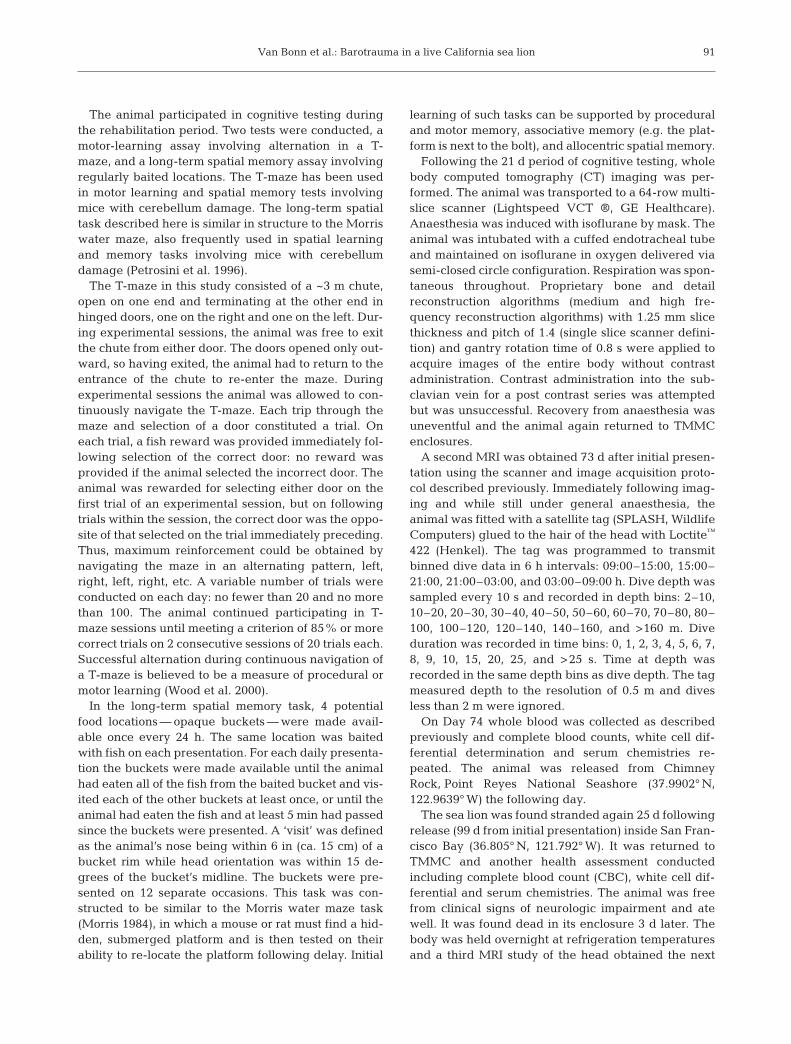
Fig. 1. Zalophus californianus. Initial magnetic resonance imaging (MRI). (A,B) T2-weighted images of the brain showing hydro-cephalus and signal voids consistent with gas bubbles. These images were acquired in the transverse plane. The central andventral horns of the lateral ventricle are enlarged (yellow arrows). Gas bubbles are visible in the cerebellum but not cerebral hemi-spheres. (C,D) 3-dimensional reconstructions of hydrocephalus and gas bubbles. In (C), the brain is viewed rostral to caudal,where grey and white matter is translucent, ventricles are blue, left air is white, and right air is dark green/brown. In (D), the brainis viewed caudal to rostral grey matter is translucent and ventricles are removed. The gas bubbles in the right cerebellum arelarger in volume than the bubbles in the left cerebellum (15.25 compared to 6.87 cm3), causing a mass-effect, deforming and dis-placing the vermis (orange arrow in B) and right caudal cerebellar peduncle (green arrow in B), and shifting the mesencephalicaqueduct and fourth ventricle leftward (white arrow in C). The gas composed 21.3% of the cerebellum. Scale bars = 8 cm
Van Bonn et al.: Barotrauma in a live California sea lion
2 wk chronicity. There was no evidence of pneumoniaor pneumothorax.
The first MRI of the brain revealed signal voids onT2W and PD images consistent with gas that wererestricted to the cerebellum (Fig. 1). These lesionswere intraparenchymal, multifocal to coalescing, sphe -rical to ovoid, and varied from 0.5 to 2.4 cm diameter.The gas composed 21.3% of the total cerebellum volume. The volume of gas in the right cerebellum(15.2 cm3) was larger than the volume in the left cere-bellum (6.9 cm3). This difference exerted a mass effect,which deflected the cerebellar vermis leftward andcaused an outflow obstruction, resulting in mild hydro-cephalus as compared to the normal sea lion brain(Montie et al. 2009). The presence and distribution ofthe signal void lesions correlated with the gas opacitiesobserved in the radiographs within the caudal fossa,and a diagnosis of pneumocerebellum was made.
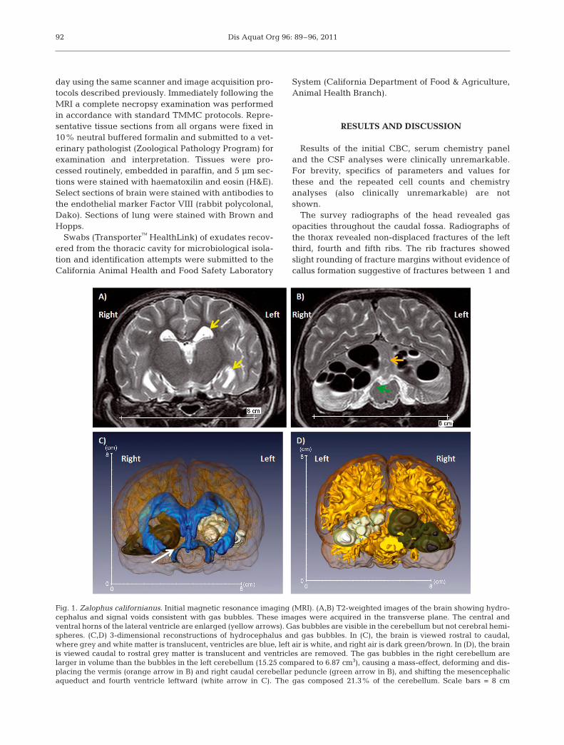
The CT performed 2 mo after the initial MRIrevealed round to ovoid structures within the cerebel-lum, in similar locations and of similar size to the gaslesions identified using MRI. However, these lesionshad Hounsfield units similar to CSF, indicating that thegas had resolved and was replaced by fluid. The cal-varium, nasal cavity, and tympanic bullae were all nor-mal ruling out head trauma as the cause of the gas-leakage into the caudal fossa. Bony proliferationconsistent with callus formation was identified in theproximal portion of the third, fourth and fifth left ribs,indicating that the rib fractures were healing and heal-ing was consistent with the speculated time of trauma.There was no evidence of pneumonia or pneumotho-rax. The second MRI performed 2 wk after the CTdemonstrated an alteration in the size and signal inten-sity of the cerebellar lesions (Fig. 2). The signal inten-sity from all lesions had changed from signal voids to
93
Fig. 2. Zalophus californianus. Follow up magnetic resonance imaging (MRI) showing fluid replacement of previously gas-filledvesicles. (A) Hydrocephalus is still present (yellow arrows), although it is less pronounced in the central and ventral horns of thelateral ventricles compared to the initial MRI. (B) Gas is now replaced with fluid. Hydrocephalus is more prominent in the lateralrecess of the fourth ventricle as compared to the initial MRI (yellow arrow). (C,D) 3-dimensional reconstructions of hydro-cephalus and fluid. In (C), the brain is viewed rostral to caudal, where grey and white matter are translucent, ventricles are blue,left fluid is green, and right fluid is pink. In (D), the brain is viewed caudal to rostral; grey mattter is translucent and ventricles areremoved. The fluid in the right cerebellum is larger in volume than the fluid in the left cerebellum (7.83 compared to 3.43 cm3).However, the mass-effect is not as predominant, and the vermis (orange arrow in B) and right caudal cerebellar peduncle (green
arrow in B) are less deformed and displaced. The fluid composed 11.6% of the cerebellum. Scale bars in (A) and (B) = 8 cm
Dis Aquat Org 96: 89–96, 2011
intensities similar to CSF on both T2W and PDsequences, consistent with CT findings of fluid-filledintraparenchymal cavitations. These findings indi-cated a progression of the cerebellar lesions to cysticencephalomalacia. The fluid in the right cerebellumremained larger in volume than the fluid in the leftcerebellum (7.8 compared to 3.4 cm3); fluid in totalcomposed 11.6% of the cerebellum. The vermis andfourth ventricle were no longer displaced from mid-line, reflecting a reduction in mass effect secondary toreduction in lesion size. However, the hydrocephaluswas more prominent in the lateral recess of the fourthventricle, as compared to the initial MRI.
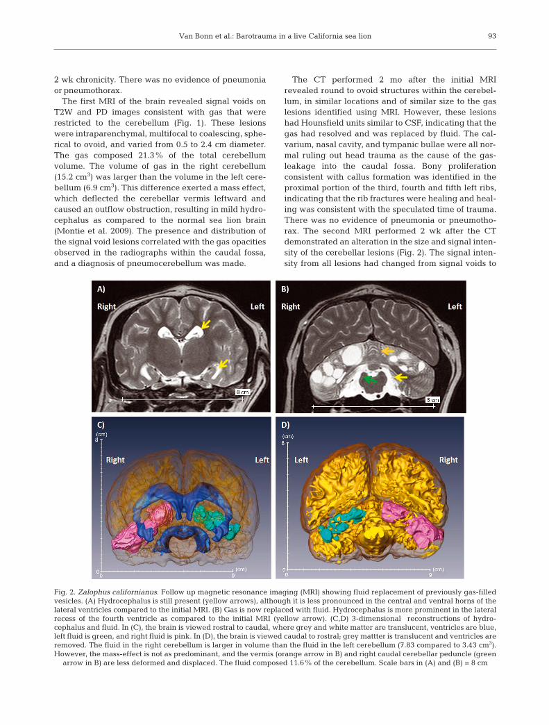
The animal was severely impaired on acquisition ofalternation in the T-maze, requiring 696 trials to reachcriterion, defined as 85% or more correct trials on 2consecutive 20-trial sessions. This finding was morethan 2.32 standard deviations (139.4) above the meantrials to criterion (372.0) of 10 other California sea lionsundergoing the same testing (Fig. 3). The next highestnumber of trials for any subject to reach criterion in thistask was 511. Five of these 10 sea lions had healthybrains, while 5 had brain damage particular to themedial temporal lobe, suggesting that the impairmentof the California sea lion in this study cannot be dis-missed as a mere byproduct of general brain damage.
The animal was able to learn the location of thebaited bucket in the once daily long-term spatial mem-ory test. The animal went directly to the baited locationwithout first visiting any of the non-baited buckets onthe fifth and all 7 remaining days of testing. With 4 pos-sible locations, the probability of correctly selecting thebaited bucket on 7 consecutive days is astronomicallysmall (<0.0001). The other 10 animals also took part inthe long-term spatial memory test. None of them reli-ably visited the correct bucket first prior to the thirdpresentation.
Impairment in the T-maze suggests impaired proce-dural or motor learning, while the ability to rememberthe correct location of the baited bucket in the long-term memory test indicates some sparing of allocentricor associative memory. This suggests that the sea lioncerebellum is essential in guiding search behaviourand related learning, but not necessary for long-termlocation-based recall. These findings directly mirrorthose gleaned from extensive testing with rodents (Pet-rosini et al. 1996, Molinari & Leggio 2007).
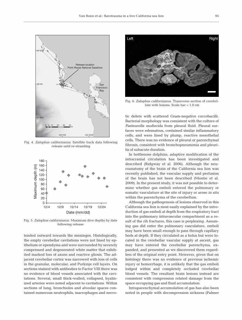
Following release the animal travelled approxi-mately 75 km north and 80 km south of the releaselocation before coming ashore in San Francisco Bay(Fig. 4). A total of 8200 dives were recorded and 90%of the dives were 40 m or less. Maximum dive depthincreased over time from 40 m to approximately 120 mwith a maximum recorded depth of 156 m (Fig. 5). Theanimal lost a third of its body weight in 25 d, which isour best indication that the sea lion was not foragingefficiently. The maximum dive depths imply that thiswas not a physiological constraint, but this young ani-mal may not have been an experienced feeder or maynot have been behaviourally competent during thistime.
Necropsy and histopathology confirmed that the sealion died from subacute bronchopneumonia and pleu-ritis due to Pasteurella multocida infection. The statusof nutrition score was 1/5, indicating emaciation.Grossly, there was mild fecal staining around the per-ineum, and the subcutaneous adipose was atrophicand mildly jaundiced. The thoracic cavity containedapproximately 100 ml of pale yellow viscous exudate.Cultures of this exudate grew large numbers of P. mul-tocida. The lungs were diffusely congested and tra-cheobronchial lymph nodes enlarged. A bony callus atthe mid-shaft of the left third, forth, and fifth ribs corre-sponded to the fractures noted on ante mortem radi-ographs.
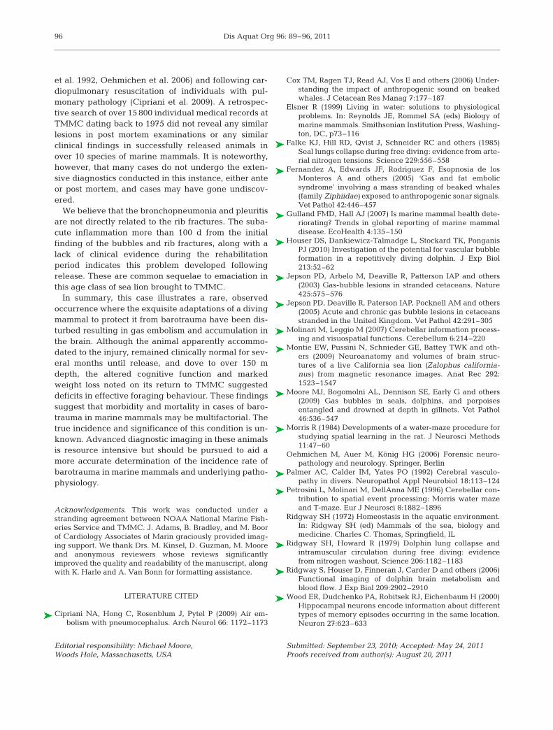
Multifocal cavitations, up to 1.5 × 1.0 cm were notedwithin the cerebellum. These cavitations contained asmall amount of colorless fluid and were larger andmore numerous on the right side of the cerebellum(Fig. 6). The cavitations were localized to white matterat the base of cerebellar folia and in some areas ex-
94
0
100
200
300
400
500
600
700
800
Tria
ls t
o cr
iterio
n
Fig. 3. Zalophus californianus. Number of trials to criterion,defined as 85% or more correct trials on 2 consecutive 20-trialsessions in the T-maze. Mean ±SD are represented. Each dotrepresents a sea lion. The California sea lion in this study is
represented by an ×
Van Bonn et al.: Barotrauma in a live California sea lion
tended outward towards the meninges. Histologically,the empty cerebellar cavitations were not lined by ep-ithelium or ependyma and were surrounded by severelycompressed and degenerated white matter that exhib-ited marked loss of axons and reactive gliosis. The ad -jacent cerebellar cortex was narrowed with loss of cellsin the granular, molecular, and Purkinje cell layers. Onsections stained with antibodies to Factor VIII there wasno evidence of blood vessels associated with the cavi -tations. Several, small thick-walled, collapsed, hyalin-ized arteries were noted adjacent to cavitations. Withinsections of lung, bronchioles and alveolar spaces con-tained numerous neutrophils, macrophages and necro -
tic debris with scattered Gram-negative cocco bacilli.Bacterial morphology was consistent with the culture ofPasteurella multocida from pleural fluid. Pleural sur-faces were edematous, contained similar inflammatorycells, and were lined by plump, reactive mesothelialcells. There was no evidence of pleural or parenchymalfibrosis, consistent with bronchopneumonia and pleuri-tis of subacute duration.
In bottlenose dolphins, adaptive modification of theintracranial circulation has been investigated anddescribed (Ridgway et al. 2006). Although the neu-roanatomy of the brain of the California sea lion wasrecently published, the vascular supply and perfusionof the brain has not been described (Montie et al.2009). In the present study, it was not possible to deter-mine whether gas emboli entered the pulmonary orsomatic vasculature at the site of injury or arose in situwithin the parenchyma of the cerebellum.
Although the pathogenesis of lesions observed in thisCalifornia sea lion is most easily explained by the intro-duction of gas emboli at depth from the respiratory tractinto the pulmonary intravascular compartment as a re-sult of the rib fractures, this case is perplexing. Assum-ing gas did enter the pulmonary vasculature, embolimay have been small enough to pass through capillarybeds at depth. If they circulated as a bolus but were lo-cated in the cerebellar vascular supply at ascent, gasmay have entered the cerebellar parenchyma, ex-panded, and presented as we discovered them regard-less of the original entry point. However, given that onhistology there was no evidence of previous ischemicinjury or hemorrhage, it is unlikely that the gas embolilodged within and completely occluded cerebellarblood vessels. The resultant brain lesions instead areconsistent with compression related damage from thespace occupying gas and fluid accumulation.
Intraparenchymal accumulation of gas has also beennoted in people with decompression sickness (Palmer
95
122°30'W123°0'W
38°30'N
38°0'N
37°30'N
0 10 20 30 405km
SanFrancisco
Bay
Release locationPoint Reyes National Seashore
Fig. 4. Zalophus californianus. Satellite track data followingrelease until re-stranding
Fig. 6. Zalophus californianus. Transverse section of cerebel-lum with lesions. Scale bar = 1.0 cm
020406080
100120140160180
12/4 12/9 12/14
Date (mm/dd)12/19 12/24
Max
imum
dep
th (m
)
Fig. 5. Zalophus californianus. Maximum dive depths by datefollowing release
Dis Aquat Org 96: 89–96, 2011
et al. 1992, Oehmichen et al. 2006) and following car-diopulmonary resuscitation of individuals with pul-monary pathology (Cipriani et al. 2009). A retrospec-tive search of over 15 800 individual medical records atTMMC dating back to 1975 did not reveal any similarlesions in post mortem examinations or any similarclinical findings in successfully released animals inover 10 species of marine mammals. It is noteworthy,however, that many cases do not undergo the exten-sive diagnostics conducted in this instance, either anteor post mortem, and cases may have gone undiscov-ered.
We believe that the bronchopneumonia and pleuritisare not directly related to the rib fractures. The suba-cute inflammation more than 100 d from the initialfinding of the bubbles and rib fractures, along with alack of clinical evidence during the rehabilitationperiod indicates this problem developed followingrelease. These are common sequelae to emaciation inthis age class of sea lion brought to TMMC.
In summary, this case illustrates a rare, observedoccurrence where the exquisite adaptations of a divingmammal to protect it from barotrauma have been dis-turbed resulting in gas embolism and accumulation inthe brain. Although the animal apparently accommo-dated to the injury, remained clinically normal for sev-eral months until release, and dove to over 150 mdepth, the altered cognitive function and markedweight loss noted on its return to TMMC suggesteddeficits in effective foraging behaviour. These findingssuggest that morbidity and mortality in cases of baro-trauma in marine mammals may be multifactorial. Thetrue incidence and significance of this condition is un -known. Advanced diagnostic imaging in these animalsis resource intensive but should be pursued to aid amore accurate determination of the incidence rate ofbarotrauma in marine mammals and underlying patho-physiology.
Acknowledgements. This work was conducted under astranding agreement between NOAA National Marine Fish-eries Service and TMMC. J. Adams, B. Bradley, and M. Boorof Cardiology Associates of Marin graciously provided imag-ing support. We thank Drs. M. Kinsel, D. Guzman, M. Mooreand anonymous reviewers whose reviews significantlyimproved the quality and readability of the manuscript, alongwith K. Harle and A. Van Bonn for formatting assistance.
LITERATURE CITED
Cipriani NA, Hong C, Rosenblum J, Pytel P (2009) Air em-bolism with pneumocephalus. Arch Neurol 66: 1172–1173
Cox TM, Ragen TJ, Read AJ, Vos E and others (2006) Under-standing the impact of anthropogenic sound on beakedwhales. J Cetacean Res Manag 7:177–187
Elsner R (1999) Living in water: solutions to physiologicalproblems. In: Reynolds JE, Rommel SA (eds) Biology ofmarine mammals. Smithsonian Institution Press, Washing-ton, DC, p73–116
Falke KJ, Hill RD, Qvist J, Schneider RC and others (1985)Seal lungs collapse during free diving: evidence from arte-rial nitrogen tensions. Science 229:556–558
Fernandez A, Edwards JF, Rodriguez F, Esopnosia de losMonteros A and others (2005) ‘Gas and fat embolic syndrome’ involving a mass stranding of beaked whales(family Ziphiidae) exposed to anthropogenic sonar signals.Vet Pathol 42:446–457
Gulland FMD, Hall AJ (2007) Is marine mammal health dete-riorating? Trends in global reporting of marine mammaldisease. EcoHealth 4:135–150
Houser DS, Dankiewicz-Talmadge L, Stockard TK, PonganisPJ (2010) Investigation of the potential for vascular bubbleformation in a repetitively diving dolphin. J Exp Biol213:52–62
Jepson PD, Arbelo M, Deaville R, Patterson IAP and others(2003) Gas-bubble lesions in stranded cetaceans. Nature425:575–576
Jepson PD, Deaville R, Paterson IAP, Pocknell AM and others(2005) Acute and chronic gas bubble lesions in cetaceansstranded in the United Kingdom. Vet Pathol 42:291–305
Molinari M, Leggio M (2007) Cerebellar information process-ing and visuospatial functions. Cerebellum 6:214–220
Montie EW, Pussini N, Schnieder GE, Battey TWK and oth-ers (2009) Neuroanatomy and volumes of brain struc-tures of a live California sea lion (Zalophus california -nus) from magnetic resonance images. Anat Rec 292:1523–1547
Moore MJ, Bogomolni AL, Dennison SE, Early G and others(2009) Gas bubbles in seals, dolphins, and porpoisesentangled and drowned at depth in gillnets. Vet Pathol46:536–547
Morris R (1984) Developments of a water-maze procedure forstudying spatial learning in the rat. J Neurosci Methods11:47–60
Oehmichen M, Auer M, König HG (2006) Forensic neuro -pathology and neurology. Springer, Berlin
Palmer AC, Calder IM, Yates PO (1992) Cerebral vasculo -pathy in divers. Neuropathol Appl Neurobiol 18:113–124
Petrosini L, Molinari M, DellAnna ME (1996) Cerebellar con-tribution to spatial event processing: Morris water mazeand T-maze. Eur J Neurosci 8:1882–1896
Ridgway SH (1972) Homeostasis in the aquatic environment.In: Ridgway SH (ed) Mammals of the sea, biology andmedicine. Charles C. Thomas, Springfield, IL
Ridgway SH, Howard R (1979) Dolphin lung collapse andintramuscular circulation during free diving: evidencefrom nitrogen washout. Science 206:1182–1183
Ridgway S, Houser D, Finneran J, Carder D and others (2006)Functional imaging of dolphin brain metabolism andblood flow. J Exp Biol 209:2902–2910
Wood ER, Dudchenko PA, Robitsek RJ, Eichenbaum H (2000)Hippocampal neurons encode information about differenttypes of memory episodes occurring in the same location.Neuron 27:623–633
96
Editorial responsibility: Michael Moore,Woods Hole, Massachusetts, USA
Submitted: September 23, 2010; Accepted: May 24, 2011Proofs received from author(s): August 20, 2011